role for the outer membrane ferric siderophore receptor envelope
TRANSCRIPT

The EMBO Journal vol.13 no.12 pp.2805-2813, 1994
Role for the outer membrane ferric siderophore receptorPupB in signal transduction across the bacterial cellenvelope
Margot Koster1, Wim van Klompenburg,Wilbert Bitter, John Leong andPeter Weisbeek
University of Utrecht, Departnent of Molecular Cell Biology,Padualaan 8, 3584 CH Utrecht, The Netherlands'Corresponding author
Communicated by B.de Kruijff
The outer membrane protein PupB of Pseudomonasputida WCS358 facilitates transport of iron complexedto the siderophores pseudobactin BN8 and pseudobactinBN7 into the cell. Its synthesis is induced by the presenceof these specific siderophores under iron limitation. Thesignal transduction pathway regulating siderophore-dependent expression of pupB was shown to consist oftwo regulatory proteins, PupI and PupR, and the PupBreceptor itself. Mutational analysis of the regulatory genessuggested that PupI acts as a positive regulator ofpupBtranscription, whereas PupR modifies PupI activitydependent on the presence of pseudobactin BN8. PupIand PupR do not share homology with the classicalbacterial two-component systems but display significantsimilarity to the Fecl and FecR proteins of Escherichiacoli involved in regulation of ferric dicitrate transport.The function of the PupB receptor in pupB regulationwas studied by the use of chimeric receptor proteinscomposed of PupB and the ferric pseudobactin 358receptor PupA. This experiment revealed that PupB isinvolved in the initiation of the signal transductionpathway, implying a so far unique role for an outermembrane protein in signal transduction.Key words: outer membrane receptorlPseudomonas putidalsiderophores/signal transduction
IntroductionIron is essential for the growth of most microorganisms, butits biological availability in the environment is oftenrestricted. Bacteria respond to iron deprivation by producingsmall water-soluble iron binding compounds, called sidero-phores, which deliver iron to the cell via specific high-affinitytransport systems (Neilands, 1981, 1982). Plant-growthpromoting Pseudomonas putida WCS358 synthesizes underiron limitation the fluorescent siderophore pseudobactin 358,which is structurally related to the siderophores producedby other fluorescent pseudomonads (Geels and Schippers,1983; van der Hofstad et al., 1986). This group ofsiderophores, called pseudobactins or pyoverdines, arecomposed of a fluorescent chromophore attached to a peptidemoiety, which differs in length and composition betweensiderophores produced by different Pseudomonas strains.P.putida WCS358 has the capacity to exploit a large number
of pseudobactins of heterologous origin for iron acquisition(Bakker et al., 1990).The ability of a fluorescent Pseudomonas strain to utilize
a given pseudobactin is related to the presence of an outermembrane receptor which is specific for that ferricpseudobactin complex and facilitates its uptake into the cell.In addition, less specific proteins are involved in transportof ferric pseudobactins across the inner membrane and therelease of iron (Marugg et al., 1989; Koster et al., 1993).In P.putida WCS358, two ferric pseudobactin receptors,PupA and PupB, have been characterized. The PupAreceptor is involved in iron transport via the native sidero-phore pseudobactin 358 (Bitter et al., 1991), whereas PupBfunctions in transport of two heterologous siderophores,namely pseudobactins BN8 and BN7 (Koster et al., 1993).The two receptor proteins share considerable sequencehomology and are both functionally dependent on the innermembrane proteins TonB, ExbB and ExbD (Bitter et al.,1993), which provide energy required for the transportprocess (for a review, see Postle, 1990). Mutants of strainWCS358 deficient in PupA or PupB production retain theability to utilize pseudobactin 358 or pseudobactin BN8,respectively, albeit with reduced efficiency (Bitter et al.,1991; Koster et al., 1993). This implies the presence ofadditional receptors for these ferric siderophores in thisstrain.
Synthesis of PupA and pseudobactin 358 is regulated bythe concentration of available iron (Marugg et al., 1988).PupB expression is also iron-controlled but has in additionan absolute requirement for the presence of one of its cognatepseudobactins (Koster et al., 1993). A similar type ofregulation has been described for the enterobactin- andferrioxiamine B receptor of Pseudomonas aeruginosa(Cornelis et al., 1987; Poole et al., 1990) and for the ferricdicitrate transport system ofEscherichia coli (Hussein et al.,1981; Pressler et al., 1988). In addition to pseudobactinsBN8 and BN7, other heterologous pseudobactins also inducethe synthesis of specific outer membrane proteins in P.putidaWCS358 which suggests the presence of multiple induciblereceptors for ferric pseudobactins in this strain. The largevariety of ferric siderophore uptake systems reflects theimportance of iron competition in the natural habitat of thisbacterium.
Siderophore-dependent regulation implies the presence ofa signalling system capable of monitoring a specificsiderophore and converting this signal into a cellularresponse. Induction of synthesis of the E. coli ferric dicitratetransport system is mediated by two proteins, FecI and FecR,which do not show homology to other known bacterialregulatory proteins (van Hove et al., 1990). Their genes arelocated immediately upstream of thefecA gene encoding theferric dicitrate outer membrane receptor. It has beenproposed that Fecd functions as a transcriptional activatorof the fec genes and FecR as a sensor, repressing Fecd
© Oxford University Press 2805

M.Koster et al.
activity in the absence of ferric dicitrate (van Hove et al.,1990). Expression of the ferric enterobactin receptor PfeAin P. aeruginosa is under control of two regulatory proteins,PfeR and PfeS (Dean and Poole, 1993), which displayhomology to the histidine kinase sensors and responseregulators of a large family of bacterial two-componentregulatory systems (Albright et al., 1989). Thusinterestingly, two evolutionarily distinct signal transductionsystems are employed for siderophore-dependent regulationof thefec genes in E.coli and the pfeA gene in P.aeruginosa.The aim of the present study is to investigate the regulation
of PupB expression in P.putida WCS358. Two genes areidentified upstream of thepupB gene, pupI andpupR, whichare involved in the siderophore-dependent regulation of PupBexpression. The predicted translation products exhibit highsimilarity to the FecI and FecR proteins of E. coli providingevidence for a conservation of this regulatory system inGram-negative bacteria. Furthermore, it is demonstrated thatthe stimulus to which this two-component system respondsis not the ferric siderophore complex directly but a signaltransduced by the PupB receptor upon transport of itssubstrate. This is to our knowledge the first example of anouter membrane protein that acts as a component of a signaltransduction cascade.
ResultsIdentification and characterization of pupl and pupRPreviously, a transcriptional unit upstream of thepupB genewas shown to be essential for PupB expression (Koster et al.,1993). In order to identify and characterize gene(s)controlling PupB synthesis, the nucleotide sequence of a 1.7kb region immediately upstream ofpupB was determined.A physical map encompassing this region is depicted inFigure 1. Two open reading frames (ORFs), designated pupIandpupR, were found in the same transcriptional orientationas the pupB gene (Figure 2). The codon usage of the twoORFs is characteristic of Pseudomonas species (Viebrockand Zumft, 1988; Wong and Abdelal, 1990). The stop codonof the pupI ORF overlaps with the start codon of pupR(Figure 2) suggesting thatpupI andpupR are cotranscribedin one operon. The putative translation initiation sites ofpupIand pupR located at position 113 and 631 are not precededby obvious Shine-Dalgarno sequences.ThepupIORF potentially encodes a protein of 173 amino
acid residues (mol. wt, 19 474) and the putative PupR proteinis 324 amino acid residues (mol. wt, 35 846). To identifypotential transmembrane helices, hydrophobicity analysiswas performed on the deduced PupI and PupR proteins (vonHeijne, 1992). No putative transmembrane domains werepredicted for PupI which is suggestive of a cytoplasmiclocation for this protein. PupR contains two potentialtransmembrane segments between residues 85-105 and238-258 (Figure 3) and may therefore represent an integralinner membrane protein. The amino acid sequences of PupIand PupR were compared with known proteins present inthe SwissProt sequence database. The only significantsequence similarity found was with the Fecd and FecRproteins (Figure 3), a two-component system regulatingferric dicitrate transport in E. coli (van Hove et al., 1990).The PupI protein shows an overall identity of 42.8% withthe FecI protein, and a helix-tum-helix DNA bindingmotif (Dodd and Egan, 1990) is present in both proteins at
E c E E '-
(L to CO k0 co &I I I
pup/ pupR
IIEI
pupS
1 kb
Fig. 1. Physical map of the DNA region containing pupI, pupR andpupB. Indicated are the ORFs and their direction of transcription. Theflag represents the position of the TnS insertion abolishing pupBexpression in strain KV51. Bg, Bgll; E, EcoRI; Pv, Pvull; P, PsM;S, Sall; Sm, SnaI; St, StuI.
1 GACCAGTTGCCGTCGCGGCATTTGTCGCGCGCGCGTGTTGCAGGGCCGCCAGGCATGTAT
_w. pupl61 ATATGATAAGCATTATCGTTTGCAGTAATGACTCACCTGGCGGCCGCACACCATGCTCCC
M L P
121
181
241
301
361
421
481
541
601
661
721
781
841
TTCCTCTGATCCCCTTTTGTGCGATGTCGCGCTGCTCTACCGCCAGCAGCACAGCTGGTTS S D P L L C D V A L L Y R Q Q H S W L
GACGCGCTGGCTCAGGCAGCGCCTGAATTGCTCGCAAAGCGCAGCGGACCTGGCCCAAGAT R W L R Q R L N C S Q S A A D L A Q D
CACCTTCATCCGGCTGTTGAACAAGGAGCAGGTGCCGCAACTGCATGCGCCGCGTACCTTT F I R L L N K E Q V P Q L H A P R T F
TCTGGCCAAGGTGGCGCAAAGCGTGTTGTGTAACCACTACCGGCGGCAAAAGCTCGAACGL A X V A Q S V L C N H Y R R Q K L E R
CGCCTACCTTGAGGCGCTGGCGATGCTGCCTGAGCCGGTGGTGCCAAGCCTGGAGACCCAA Y L E A L A M L P E P V V P S L E T Q
A I LL E T L I A L D A A L D G L E R P
V R E A F L L S Q V D G L G H T E I A Q
ACGGCTGGCCGTGTCGGTCACCACGGTCAAGCGCTACATCATCAAGGCAGGTGCACTGTGR L A V S V T T V K R Y I I K A G A L C
,o pupfCATCATGCTCGACCACAGCCTGGACCTGCCATGAACGGCCAGGGCGCAACGTCGATCCCG
I M L D H S L D L PM N G O G A T S I P
GGCGAGGTGGCCGAGCAGGCCATGCACTGGCACCTCGAACTGCAAGAGCCGGCCGTCAGTG E V A E Q A M H W H L E L Q E P A V S
GCCGCCACCCTGGCCGCCTGCATGAGCTGGCGCCAGGCGCACCCGCTACATGAACATGCCA A T L A A C M S W R Q A H P L H Z H A
W Q R T Q V F a Q R L R E M R S P G Q R
CCCTTGGCCCATGCGGCGCTGCGGCCGCAGCAGTCGCGGCGCACTGCGCTCAAGCAATTGP L A H A A L R P Q Q S R R T A L 0Q L
901 TCGCTGCTGATGGCCGCAGGCGCAGGGGCrTGGTArTTG.AAGGACGCCGC(CTGGTACAAS L L M A A G A G A W Y L K D A A L V Q
961
1021
1081
1141
1201
1261
1321
1381
1441
1501
1561
GACTGGCGCGCTGATTACCACAGCCGCATCGGCGAGCAGCGGCGCCTGACCCTGGCCGATD W R A D Y H S R I G E Q R R L T L A D
GGCACCCAGGTACAGCTGAACACCGACAGCGCCCTCAATGTGGCATTCGACCAGCAGGCAG T Q V Q L N T D S A L N V A F D Q Q A
CGCCGCCTGAGGCTCGTGCGCGGCGAGATGCTGATCACCCGCCCCGCCCTCGCCGATAGCR R L R L V R G E M L I T R P A L A D S
CGCCCACTGTGGGTAGACACCGAGCACGGCCGCCTTGAGTCGACGCTGGCGCAGTTCAATR P L W V D T E H G R L E S T L A Q F N
GTGCGGCTGCACGGCCAACACACCCAGGCCACCGTGTATCAGGGCAGCGTGGCGCTGCAAV R L H G Q H T Q A T V Y Q G S V A L Q
CCAGCCCTGCATGCCTACCCGCCCATCCTGCTGGGCGCCGGTGAACAGGCCAGTTTCAACP A L H A Y P PF L L G A G 0Q A S F N
CAGCAGGGCTTGCTGGCGCGGCAGGCCGTGGCCGCTGTCGCCCCCGCCTGGAGCCAAGGCQ Q G L L A R 0 A V A A V A P A H S Q G
ATGCTGGTTGCTCAAGGCCAGCCCCTGGCTGCGTTTATCGAAGACCTGGCCCGCTATCGCM L V A Q G Q P L A A F I E D L A R Y R
CGCGGACACCTGGCCTGCGACCCGGCCTTGGCCGGCCTGCGTGTATCCGGCACGTTCCCGR G H L A C D P A L A G L R V S G T F P
CTGGAAAACACCGACAAGATCATTGCTGCCGTGGCAGAAACCTTGCAGTTGGAGGTGCAGL E N T D K I I A A V A E T L Q L E V Q
H---- - ---- - _- -T W L K F
-R A
L L
H F T R Y tI V T L K P R M A
1621 GCAGGGGTGGTCCGATTTCGCGCCTCACGAGACTTAACCCTCAGCACTTCAAATCAATGG
,1 pupa1681 GGGAACACTTGTGAATCACACCGCACGC
M N H T A R
Fig. 2. Nucleotide sequence of the DNA fragment carrying pupj andpupR. The deduced amino acid sequences are presented in one-lettersymbols. The putative Fur repressor binding site upstream of pupI isunderlined.
2806

Signal transduction across the cell envelope
an equivalent position (Figure 3). The PupR and FecRproteins share 36.6% identity with the highest homology intheir C-termini (Figure 3). Only the most N-terminal putativetransmembrane domain of PupR is conserved in the FecRprotein.To study the regulation ofpup! and pupR expression, a
transcriptional pupI-lacZ fusion construct (pMWl) wasmade. Strain WCS358 harbouring this construct displayediron-repressed 3-galactosidase activity. The activity was
- 10-fold higher under iron-depleted than under iron-sufficient conditions and was not increased by the presenceof pseudobactin BN8 (Table I). A sequence was identifiedin the promoter region showing homology to the bindingsite for the Fur protein of E. coli (de Lorenzo et al., 1987)(Figure 2). Fur acts as a repressor of transcription in thepresence of sufficient iron, and has been identified in manydifferent bacteria including P.putida WCS358 (V.Venturi,personal communication).
Functional analysis of PupI and PupRThe previously described transposon insertions abrogatingPupB synthesis, including the chromosomal TnS insertion
PUPI
FECI
PUP I
FECI
PUPI
FECI
PUPI
FECI
ML--PSSDPLLCDVALLYRQQHSWLTRWLRQRLNCSQSAA
MSDRATTTASLT-FESLYGTHHGWLKSWLTRKLQSAFDAD
DLAQDTFIRLLNKEQVPQLHAPRTFLAKVAQSVLCNHYRR11111 1 11 11 11
DIAQDTFLRVMVSETLSTIRDPRSFLCTIAKRVMVDLFRR
QKLERAYLEALAMLPEPVVPSLETQAILLETLIALDAALD11 1111111 1 11 1 1111 11 11
NALEKAYLEM1LALMPEGGAPSPEERESQLETLQLLDSMLD
GLERPVREAFLLSQVDGLGHTEIAORLAVSVTTVKRYIIK
GLNGKTREAFLLSQLDGLTYSEIAHKLGVSISSVKKYVAK
38
39
78
79
118
119
158
159
in mutant KV51 (Koster et al., 1993), were all located inthe pupI ORF (Figure 1). Mutant KV5 1, lacking the pupIand presumably also the pupR gene product, was impairedin the synthesis of detectable amounts of the PupB receptor(Figure 4). A transcriptional pupB-lacZ fusion (pMW2)was used to study the transcriptional regulation of thepupBgene. (3-galactosidase activity in strain WCS358(pMW2) wasincreased 3-fold in low iron conditions and - 12-fold inresponse to the presence of pseudobactin BN8 relative tothe activity in high iron conditions (Table II). Thepseudobactin-dependent induction was completely abolishedin thepup::TnS mutant, consistent with a role of this locusin controlling PupB expression at the transcriptional level.The induction could be restored by the introduction ofplasmid pMCl carrying pupI and pupR. The level ofpromoter activity in the complemented mutant was increaseddiscernibly relative to the wild-type strain which could bedue to the presence of multiple copies of pupI and pupR.Complementation with plasmid pMC2 carrying only pupIresulted in pupB expression irrespective of the presence ofpseudobactin BN8 (Table 11). This suggested a role for PupRin negatively regulating pupB expression in the absence ofthe siderophore.To assess the function exerted by the PupR protein, a
chromosomal pupR mutant of strain WCS358 wasconstructed. For this purpose, the internal SmaI fragmentof the pupR gene on pEW4, a pEMBL18 derivative, wasreplaced with the Q interposon containing a streptomycinresistance gene resulting in plasmid pEW5. Since thisplasmid cannot replicate in Pseudomonas, introduction ofpEW5 into strain WCS358 could only result in streptomycinresistant colonies by homologous recombination of the
PUPI AGALCIMLDHSLDLP 173
FECI AVEHCLLFRLEYGL 173
PUPR
FECR
PUPR
FECR
PUPR
FECR
PUPR
FECR
PUPR
FECR
PUPR
FECR
PUPR
FECR
PUPR
FECR
MNGQGATSIPGEVAEQAMHWHLELQEPAVSAATLAACMSW11 I 11 11MNPLLTDS-RRQALRSASHWYAVLSGERVSPQQEARWQQW
RQAHPLHEHAWQRTQVFAQRLREMRSPGQRPLAHAALR-PIII 11 11 11
YEQDQDNQWAWQ--QV--ENLRNQLGGVPGDVASRALHDT
11 1 1 11 11 1 1111RLTRRHVMKGLLLLLGAG-GGWOLWOSETGEGLRADYRTA
IGEQRRLTLADGTQVQLNTDSALNVAFDQQARRLRLVRGE
KGTVSRQQLEDGSLLTLNTQSAADVRFDAHQRTVRLWYGE
MLITRPALADSRPLWVDTEHGRLESTLA-QFNVRLHGQHT
IAITTAKDALQRPFRVLTRQGQL-TALGTEFTVRQQDNFT
QATVYQGSVALQPALHAYPPILLGAGEQASFNQQGLLARQlD HlI E
l111QLDVQQHAVEVLLASAPAQKRIVNAGESLQFSASEFGAVK
II I II I II
PLDDESTSWTKDILSFSDKPLGEVIATLTRYRNGVLRCDP
ALAGLRVSGTFPLENTDKIIAAVAETLQLEVQHFTRYWVT1111 111111 III I 11 1iii
AVAGLRLSGTFPLKNTDAILNVIAQTLPVKIQSITRYWIN
40
39
79
75
119
114
159
154
198
193
238
233
278
273
318
313
Table I. Expression of a pupI-lacZ fusion in WCS358, KV51 andBWV29 grown under different conditions
,B-Galactosidase activity (U)
Strain Genomic mutation +Fe -Fe ps.BN8
WCS358 - 64 700 616KV51 pupl::TnS 12 501 468BWV29 pupR::Q 43 515 479
All strains harbour plasmid pMWl carrying the transcriptionalpupI-lacZ fusion. Cells were grown in iron-deficient RSM medium(-Fe), or in RSM medium supplemented with either 100 aM FeCl3(+Fe), or 40 yM pseudobactin BN8 (ps.BN8).
WCS358 KV51 BWV29
kDa 1 2 3 1 2 3 1 2 3
fRFIts,-{1 - i
68
PUPR LKPRMAX 325
FECR ISPL 317
Fig. 3. Alignment of the deduced protein products of pupI and pupRwith the FecI and FecR proteins of E.coli, respectively. Vertical lines
indicate identical residues. The conserved helix-tum-helix motif
found in PupI and FecI and the putative transmembrane domains in
PupR and FecR are underlined.
Fig. 4. Expression of the PupB protein in wild-type strain WCS358,pupI::TnS mutant KV51, and the pupR::O mutant BWV29. Cellenvelope fractions of cells grown in KB medium supplemented with100 AM FeCl3 (lane 1), iron-limiting KB medium (lane 2) and iron-limiting KB medium supplemented with 40 AM pseudobactin BN8(lane 3) were analysed by SDS-PAGE. The positions of themolecular weight standard proteins (left) and the PupB protein (arrow)are indicated.
2807
PupB

M.Koster et al.
Table H. Regulation of pupB-lacZ expression by PupI and PupR
(3-Galactosidase activity (U)
Strains Genomic mutation Plasmid +Fe -Fe ps.BN8
WCS358 - 51 168 636KV51 pupl::TnS - 40 112 156KV51 pupl::TnS (pMClpupIIpupR+) 40 136 1912KV51 pupl::Tn5 (pMC2pupI+) 91 635 780BWV29 pupR::Q - 56 244 274BWV29 pupR::Q (pMC1pupI+pupR+) 36 147 2112BWV29 pupR: :Q (pMC2pupI+) 83 982 1080WCS358 - (pMC2pupI+) 126 623 2914WCS358 - (pMClpupI+pupR+) 75 140 2393
All strains harbour plasmid pMW2 carrying a transcriptional pupB-lacZ fusion. Cells are grown as described in Table I.
inactivated gene into the chromosome (see Materials andmethods). The resulting mutant BWV29 exhibitedpseudobactin-independent synthesis of the PupB receptor(Figure 4). ,B-galactosidase activity resulting from expressionof the pupB-lacZ construct in strain BWV29(pMW2) wasalso constitutive, although at a significantly lower level thanin the parental strain (Table II). Introduction of plasmidpMC2 carrying pupI into this mutant resulted in an increasein pseudobactin-independent expression, whereas normalregulation was re-established by plasmid pMC 1 containingboth regulatory genes, pupI and pupR (Table II). Theseresults supported a function of PupR in repressing PupBsynthesis in the absence of pseudobactin BN8 and showedin addition that an intact pupR gene is required for optimalpupB promoter activity under induction conditions.
Introduction ofplasmid pMC2 carrying pupI into the wild-type strain WCS358 resulted in very high levels of pupBpromoter activity even in the absence of pseudobactin BN8,whereas transcription remained normally regulated whenpupI and pupR were both overexpressed (Table II). Theseresults were consistent with the idea that PupI acts as apositive regulator of pupB transcription, whereas PupRprevents transcriptional activation by PupI in the absenceof pseudobactin BN8. The observation that the amount ofchromosomally encoded PupR was not sufficient to represscompletely the activity of the plasmid-encoded PupI underiron limitation was suggestive of repression of PupI by PupRthrough the formation of a stoichiometric complex ratherthan by enzymatic interaction.
Since many regulatory proteins control the transcriptionof their own genes, the possibility of autoregulation ofpupIand pupR was examined by measuring ,B-galactosidaseactivity in the genomic mutants BWV29 and KV5 1harbouring the pupI-lacZ fusion pMWl. No differences inpromoter activity were found between the mutants lackingthe regulatory proteins and the parental strain (Table I).Therefore, PupI and PupR synthesis appeared not to beautoregulated.
Function of the PupB receptor in signal transductionPseudobactin-dependent activation of thepupB promoter wasabolished in the chromosomal pupB mutant KV53 (TableIl). Apparently, the PupB protein is required for tran-scriptional activation of its structural gene. Since the transportactivity of the receptor is dependent on the TonB energycoupling system, it was determined whether the TonB protein
receptor
PupAB
PupBA
plasmid
pMM30
pMM40
I§ 1L
47 82 673
45 86 690
Fig. 5. Schematic representation of the chimeric proteins. The openbar represents PupA, the dotted bar represents PupB, and the signalsequence is indicated by the black bar. The length of the differentdomains is shown underneath each bar in number of amino acidresidues.
is also necessary for induction of pupB expression.Therefore, the pupB-lacZ fusion pMW2 was introducedinto the tonB mutant TE156 (Bitter et al., 1993). Since strainTE156 is unable to grow under iron limitation, activity ofthe pupB promoter in this mutant was studied in the presenceof iron. Under these conditions, the mutant secretessiderophore in high amounts and iron-regulated promoters,which are normally repressed, are transcribed (Bitter et al.,1993). No induction in ,B-galactosidase activity in responseto the presence of pseudobactin BN8 could be observed inTE156(pMW2) (Table HI). Thus, PupB and the TonBprotein are both essential for siderophore-dependentexpression of thepupB gene. It is possible that, for induction,the ferric pseudobactin complex has to be transported viathe PupB receptor into the periplasm, in order to mediatesignal ransduction. However, the chromosomalpupB mutantKV53 has 50% residual uptake of ferric pseudobactin BN8(Koster et al., 1993), suggesting that the internalized ferricpseudobactin is not providing the signal for induction. Thisfact led to the hypothesis that the PupB receptor in concertwith the TonB system has a specific role in signal trans-duction besides its transport function.A topology model has been proposed for the folding of
the PupB receptor in the outer membrane (W.Bitter et al.,in preparation). According to this model, the most extensiveperiplasmic domain of PupB is formed by the N-terminal70 amino acid residues which are thus probably involvedin a putative regulatory function of the receptor. To assessthe function of this region, a chimeric receptor wasconstructed in which the first 86 amino acid residues ofmature PupB were replaced with the corresponding part ofthe PupA protein, the receptor for ferric pseudobactin 358(Figure 5). Replacement of this region does not involve anycell-surface exposed domains and would therefore not alter
2808

Signal transduction across the cell envelope
Table m. Involvement of the PupB receptor in regulation of pupB-1acZ expression
,B-Galactosidase activity (U)
Strain Genomic mutation Plasmid +Fe -Fe psBN8 ps358
WCS358 - 51 168 636 NDTE156 tonB::TnS 113 ND 93a NDKV53 pupB::TnS 45 85 117 NDKV53 pupB::TnS (pMM1pupB+) 78 135 493 NDKV53 pupB::TnS (pMM30pupAB+) 84 110 150 NDKV53 pupB::Tn5 (pMM40pupBA+) 96 760 656 NDWCS3 58 - (pMM40pupBA+) 58 600 ND NDJM205 sid::TnS (pMM40pupBA+) 55 185 ND 661KV51 pupl::TnS (pMM40pupBA+) 59 142 ND ND
All strains harbour plasmid pMW2 carrying the pupB-lacZ fusion. Cells were grown as described in Table I.ND, not deternined.aFor this strain additional iron is added to permit growth of the strain.
the ferric siderophore binding capacity of the protein. Asecond hybrid receptor was constructed consisting of thesignal sequence and the first 86 amino acid residues of PupBfused to the C-terminal 690 amino acid residues of the PupAreceptor (Figure 5). The fusion sites in the hybrid receptorsare located within the first postulated transmembrane domainwhich is highly conserved between PupA and PupB. Thehybrid genes were made by using newly introducedrestriction sites created by PCR-mediated mutagenesis (seeMaterials and methods). Introduction of the restriction sitesresulted in a change of the serine residues at positions 131and 133 in the PupA and the PupB receptor, respectively,into alanine. The hybrid pupAB and pupBA genes werecloned in the broad host range vector pML130 behind thelac promoter resulting in the plasmids pMM30 and pMM40,respectively (Figure 5).The chimeric receptors were first tested for their ferric
pseudobactin transport capacity. Since strain WCS358 hasmultiple outer membrane receptors for the same ferricpseudobactin complex, it was not possible to study thetransport activity of the chimeric receptors in the pupA andpupB mutants of this strain. Therefore, the constructs wereintroduced in Pseudomonas sp. A124 which does not possessouter membrane receptors for utilization of pseudobactinBN8 or pseudobactin 358 (Koster et al., 1993). PlasmidpMM30, carrying the pupAB gene, provided strain A124with the ability to utilize specifically pseudobactin BN8 asdetermined by a bioassay based upon reversal of EDDA-induced iron starvation. A124(pMM30) could grow asefficiently with ferric pseudobactin BN8 as iron source asstrain A124 harbouring pMMl carrying the intactpupB gene(data not shown). Thus, replacement of the N-terminaldomain had not altered the transport ability and specificityof the PupB receptor. Similarly, it was established that thehybrid PupBA receptor still took up ferric pseudobactin 358.The chimeric receptors were subsequently tested for
restoration ofpupB-lacZ induction in strain KV53, thepupBmutant of strain WCS358. Strain KV53 harbouring pMM1carrying an intactpupB gene exhibited induction of thepupBpromoter comparable to that of the wild-type strain, whereasin KV53(pMM30) expressing the PupAB hybrid receptor,no promoter activity was observed (Table III). Since thechimeric receptor still transports iron via pseudobactin BN8,this result confirmed a distinct function of PupB in regulation
of gene expression. Interestingly, introduction of thepupBAgene in strain KV53 resulted in high ,B-galactosidase activityunder iron-depleted conditions independent of the presenceof pseudobactin BN8 (Table E). A possible explanation forthis result could be that the PupBA receptor mediatedactivation of the pupB promoter in response to its cognatesiderophore, pseudobactin 358, which is produced under ironlimitation by strain KV53. To investigate this possibility,pMM40 with the hybrid pupBA gene was introduced intothe wild-type strain WCS358 and a mutant strain JM205,defective in siderophore biosynthesis (Marugg et al., 1985).The presence of the hybrid receptor led to pupB promoteractivity under iron limitation in the wild-type strain, whereasno pupB expression was observed in the biosynthesis mutantunless exogenous siderophore 358 was added to the medium(Table IE). This result provided evidence for pseudobactin358-dependent pupB induction mediated by the chimericPupBA receptor. No pupB promoter activity was observedin the pupI: :Tn5 mutant KV51(pMM40) which showed thatthe PupI and PupR proteins are required for this induction.Thus, the chimeric receptor PupBA could induce pupBexpression via the PupI and PupR proteins in response tothe PupA-related siderophore, pseudobactin 358, instead ofto pseudobactin BN8. These results demonstrated that thestimulus to which the PupI/PupR system responds is not theferric siderophore complex itself, but a signal transducedby the receptor in response to the presence of its substrate.Furthermore, the domain of PupB involved in signaltransduction is located in the N-terminal 86 amino acidresidues of the receptor.
DiscussionThe ability of P.putida WCS358 to transport iron complexedto a large variety of siderophores seems to be associated withthe presence of a large number of outer membrane receptorswhich are specifically up-regulated when the correspondingsiderophore is encountered in the environment (Koster et al.,1993). The expression of the inducible PupB receptor wasstudied in order to obtain insight in the underlying regulatorynetwork. Two genes, pupI and pupR, were identified whichare involved in controlling pseudobactin BN8-dependentexpression of pupB. The genes, located immediatelyupstream of the pupB locus, are apparently organized in an
2809
M.Koster at a!.
operon. Their expression is strongly increased upon irondeprivation as deduced from experiments with apupl-lacZtranscriptional fusion. The presence of a putative Fur boxupstream of the pupI gene suggests that the Fur protein ofP.putida WCS358 is involved in this regulation. Recently,a positive regulatory element, named PfrA, has beenidentified which is required for expression of siderophorebiosynthetic genes under low iron conditions in strainWCS358 (Venturi et al., 1993). This element seems to bespecifically involved in iron-responsive regulation ofsiderophore biosynthesis since activity of the pupI promoterwas not dependent on the PfrA protein (unpublished results).The deduced PupI and PupR protein products are predicted
to be located in the cytoplasm and inner membrane,respectively. The two proteins display significant homologyto the FecI and FecR proteins of E. coli (van Hove et al.,1990). No similarity was found with other bacterialregulatory proteins, thus these two systems may comprisea new class of prokaryotic signal transduction systems.Mutational analysis showed that the PupI protein acts as apositive regulator ofpupB transcription. ApupL: :TnS mutantno longer exhibited induction of the pupB promoter, andcomplementation of this mutant by the pupI gene alone wassufficient to restore pupB expression, although in apseudobactin BN8-independent manner. The fact that ahelix-turn-helix motif is predicted in the C-terminal partof the PupI protein is suggestive of a role for PupI infacilitating gene expression by interaction with the pupBpromoter region. Such a role is supported by the finding thatits E. coli counterpart FecI binds specifically to the fecAoperator region (V.Braun, personal communication).
Overexpression ofpupl resulted in activation of thepupBpromoter even in the absence of pseudobactin BN8, whereassimultaneous overexpression ofpupR restored pseudobactin-responsive regulation. Apparently, PupR inhibits the activityof the PupI protein in the absence of the siderophore.Consistent with this role, a mutant deficient in PupRproduction exhibited PupB synthesis independent ofpseudobactin BN8. It should be noted that thepupB promoteractivity was markedly reduced in this mutant relative to thatin the parental strain under induction conditions. Thisreduction may be due to an effect of the pupR mutation onthe stability of the pupI mRNA or PupI protein.Alternatively, it is possible that the PupR protein alsofunctions as a positive regulator of pupB expression bystimulating PupI activity when pseudobactin BN8 is present.Exactly how the PupR protein modulates the activity of PupIis not known. The lack of homology between the PupI/PupRsystem and other regulatory systems indicates a differentmechanism of signal transduction than phosphorylation,which is the most commonly found signalling strategy inbacteria (for a review, see Parkinson, 1993).
In addition to the PupI/PupR system, the PupB receptorwas also shown to play a crucial role in signal transduction.Replacement of the N-terminal domain of PupB by thecorresponding domain of PupA, the receptor for ferricpseudobactin 358, did not interfere with the transportfunction of the PupB receptor but did affect its ability toactivate pupB expression in response to the presence ofpseudobactin BN8. This result reveals a specific functionof the receptor in regulation of gene expression.Furthermore, a chimeric PupBA protein consisting of theN-terminal 86 amino acid residues of the PupB receptor and
A
B
0
OM
Pp
CM
OM
pp
CM
activationpupB promoter
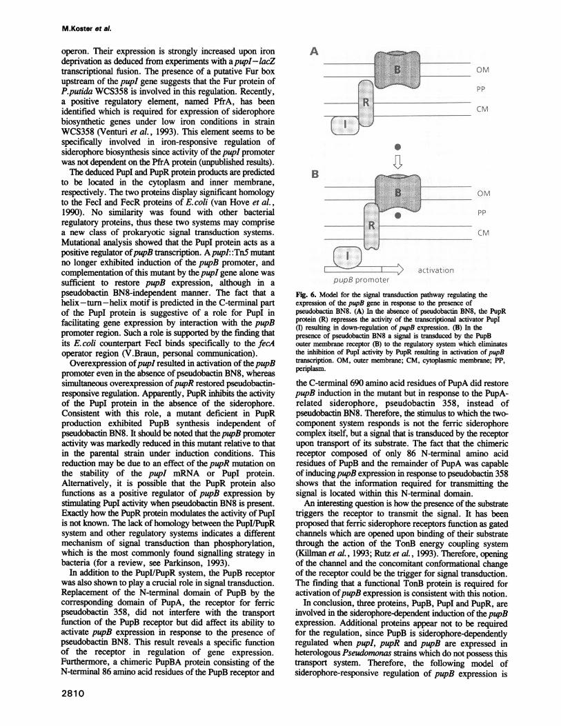
Fig. 6. Model for the signal transduction pathway regulating theexpression of the pupB gene in response to the presence ofpseudobactin BN8. (A) In the absence of pseudobactin BN8, the PupRprotein (R) represses the activity of the transcriptional activator PupI(I) resulting in down-regulation of pupB expression. (B) In thepresence of pseudobactin BN8 a signal is transduced by the PupBouter membrane receptor (B) to the regulatory system which eliminatesthe inhibition of PupI activity by PupR resulting in activation of pupBtranscription. OM, outer membrane; CM, cytoplasmic membrane; PP,periplasm.
the C-terminal 690 amino acid residues of PupA did restorepupB induction in the mutant but in response to the PupA-related siderophore, pseudobactin 358, instead ofpseudobactin BN8. Therefore, the stimulus to which the two-component system responds is not the ferric siderophorecomplex itself, but a signal that is transduced by the receptorupon transport of its substrate. The fact that the chimericreceptor composed of only 86 N-terminal amino acidresidues of PupB and the remainder of PupA was capableof inducingpupB expression in response to pseudobactin 358shows that the information required for transmitting thesignal is located within this N-terminal domain.An interesting question is how the presence of the substrate
triggers the receptor to transmit the signal. It has beenproposed that ferric siderophore receptors function as gatedchannels which are opened upon binding of their substratethrough the action of the TonB energy coupling system(Kiliman et al., 1993; Rutz et al., 1993). Therefore, openingof the channel and the concomitant conformational changeof the receptor could be the trigger for signal transduction.The finding that a functional TonB protein is required foractivation ofpupB expression is consistent with this notion.
In conclusion, three proteins, PupB, PupI and PupR, areinvolved in the siderophore-dependent induction of thepupBexpression. Additional proteins appear not to be requiredfor the regulation, since PupB is siderophore-dependentlyregulated when pupI, pupR and pupB are expressed inheterologous Pseudomonas strains which do not possess thistransport system. Therefore, the following model ofsiderophore-responsive regulation of pupB expression is
2810
k..,.O-.j

Signal transduction across the cell envelope
Table IV. Bacterial strains, siderophores and plasmids
Strain/siderophore/plasmid Relevant characteristicsa Source/Reference
StrainsP.putida WCS358 wild-type, NXR Geels and Schippers (1983)KV51 WCS358pupI: :TnS, KmR, NxR Koster et al. (1993)KV53 WCS358pupB: :TnS, KmR, NxR Koster et al. (1993)BWV29 WCS358pupR::Q, SmR, NXR This workTE156 WCS358tonB::TnS, KmR, NxR Bitter et al. (1993)JM205 WCS358sid::TnS, KmR, NxR Marugg et al. (1985)Pseudomonas sp. A124 wild-type, RifR Suslow and Schroth (1982)Pseudomonas sp. BN8 wild-type, NxR Bitter et al. (1991)E.coli PC2495 recA, hsdS, lacZY, thi, F' Phabagen collectionSiderophoresPseudobactin BN8 produced by Pseudomonas sp. BN8Pseudobactin 358 produced by P.putida WCS358Plasmids pEMBL18, ApR, ColEl replicon Dente et al. (1983)pEMBLl9 ApR, ColEl replicon Dente et al. (1983)PRK2013 KMR, Tra+, Mob+, ColEl replicon Figurski and Helinski (1979)pMP220 TcR, 'lacZ Spaink et al. (1987)pML123 GmR, pNm Labes et al. (1990)pML130 GmR, placZ Labes et al. (1990)pJRI TcR, pRK767 carrying pupI, pupR and pupB Koster et al. (1993)pJRM43 SmR, pJRD253 carrying pupB Koster et al. (1993)pUWi ApR, pUC18 carrying pupA W.Bitter, H.Zomer, P.Weisbeek
and J.Tommassen (inpreparation)
pMWl TcR, pMP220 carrying pupl-lacZ fusion This workpMW2 TcR, pMP220 carrying pupB-lacZ fusion This workpMCl GmR, pML123 carrying pup!, pupR This workpMC2 GmR, pML123 carrying pup! This workpMM1 GmR, pML130 carrying pupB This workpMM30 GmR, pML130 carrying pupAB This workpMM40 GmR, pML130 carrying pupBA This workpEW4 ApR, pEMBL18 carrying partial pupR This workpEW5 ApR, pEMBL18 carrying partial pupR::Q This workpHP45Q ApR, SmR Prentki and Krisch (1984)
aAbbreviations for drug resistance: Nx, nalidixic acid; Ap, ampicillin; Gm, gentamicin; Km, kanamycin; Rif, rifampicin; Sm, streptomycin; Tc,tetracycline.
proposed (Figure 6). Under low iron conditions theregulatory proteins PupI and PupR and small amounts ofthe PupB receptor are synthesized. In this situation, PupRprevents transcriptional activation of the pupB promoter byinhibiting the activity of the PupI protein. When ferricpseudobactin BN8 is present in the environment it will betransported across the outer membrane by the PupB receptorin a TonB-dependent manner. During this transport a signal,probably the conformational change of the receptor, istransduced to the regulatory system. It is attractive tospeculate that the PupR protein is the receiver of this signal.After transmission of the signal the PupR protein will nolonger repress PupI activity which in turn activates pupBgene transcription. Exactly where the PupI and PupRproteins are located in the cell and how the signal istransduced between the different components remains to bedetermined. According to this model, two functions can beassigned to the PupB receptor, i.e. ferric pseudobactintransport and initiation of the signal transduction pathwaythat leads to regulation of its own synthesis. This isreminiscent of the situation found for some periplasmicbinding protein-dependent transport systems like thephosphate-specific Pst system, which have in addition to their
transport function, a role in signal transduction (Cox et al.,1988). The PupB receptor is the first example of an outermembrane protein displaying such a role. FecA, the receptorfor ferric dicitrate of E. coli was also found to beindispensable for induction of the fec genes which has ledto the assumption that for induction ferric citrate has to betransported into the periplasm to interact with the regulatoryproteins (Zimmermann et al., 1984). Alternatively, the FecAreceptor may, in analogy with the PupB system, act as acomponent of the signal transduction pathway. However,the FecA receptor does not possess an extended periplasmicN-terminal domain like the PupA and the PupB receptor(Pressler et al., 1988). Therefore, it cannot be excluded thatthe two systems respond to different signals despite theconservation in primary structure of the regulatory proteins.
In strain WCS358 at least three other outer membraneproteins and probably many more are expressed in responseto a specific siderophore (Koster et al., 1993). Hence, as
many different regulatory systems have to be present tocontrol the synthesis of these proteins. Responding to thetransport activity of the receptor instead of to the ferricsiderophore in the periplasm could be an efficient means toavoid cross-talk between the different systems.
2811

M.Koster et al.
Materials and methodsBacterial strains and culture conditionsThe bacterial strains used in this work are listed in Table IV. Pseudomonasstrains were grown at 30°C in King's medium B (KB) (King et al., 1954)or in RSM medium (Buyer et al., 1989) supplemented when required with100 itM FeCl3 or 40 /M pseudobactin. E.coli was cultured at 37°C in LBmedium (Miller, 1972). For Pseudomonas, antibiotics were used at thefollowing concentrations: nalidixic acid, 25 ag/ml; rifampicin, 40 jig/mi;tetracycline, 40 isg/ral; kanamycin, 50 Ag/ml; gentamicin, 50 Ag/ml;streptomycin, 50 pg/ml and piperacillin, 75 Ag/ml. For E.coli, the antibioticsused were tetracycline, 10 pg/ml; kanamycin, 50 itg/ml; gentamicin, 25ag/ml; streptomycin, 25 isg/ml; and ampicillin, 50 ug/ml.
Plasmids and recombinant DNA techniquesPlasmids used in this study are listed in Table IV. For the construction ofpMWl carrying the pupl-lacZ transcriptional fusion, the 0.5 kbStuI-PvuII(l) fragment of plasmid pJRl (Figure 1) was cloned intopEMBL18, excised with EcoRI and PstI and ligated in pMP220 in the properorientation to direct lacZ transcription. Plasmid pMW2, carrying thepupB-lacZ fusion, was constructed by cloning the 1.2 kb Sall(l)-SalI(2)fragment of pJRl (Figure 1) in pEMBL18, followed by ligation into pMP220in the proper orientation using the EcoRI and PstI restriction sites. PlasmidpMC2, carrying the pupI gene, was constructed by cloning the 1.0 kbStuI-SmaI(2) fragment of pJR1 (Figure 1) in pEMBL18, followed byligation in vector pML123. To obtain construct pMC1, with the pupI andpupR genes, the 3.5 kb EcoRI-PstI fragment of pJRl (Figure 1) was ligatedinto pEMBL19, excised with BamHI and ClaI and ligated in pML123. Theplasmids pMCl and pMC2 were constructed in such a way that the directionof transcription of the genes was in the opposite orientation with respectto the neomycin promoter. Plasmid pEW4, which was used for theconstruction of the pupR chromosomal mutant, was made by ligation ofthe 1.5 kb Stul-Sal(l) fiament ofpJRl (Figure 1) into pEMBL18. PlasmidpMMl, carrying the pupB gene, was constructed by cloning the 4 kbSmaI(3)-SmaI(4) fragment of plasmid pJR1 (Figure 1) into pML130.
Plasmids were isolated by using the rapid procedure described by Bimboim(1983). Digestions with restriction enzymes, agarose gel electrophoresis,purification of DNA fragments and ligation with T4 DNA ligase were
performed as described by Maniatis et al. (1982). Plasmids were introducedinto E. coli by transformation using the calcium chloride procedure (Cohenet al., 1972), and into Pseudomonas by triparental mating (Marugg et al.,1988).
Determination of nucleotide sequenceDNA segments of the 1.7 kb region of pJRl upstream of the pupB gene(Figure 1) were obtained by digestion with different restriction endonucleasesand ligated into the corresponding sites of pEMBL18 and pEMBL19. Theconstructs were encapsidated as single-stranded DNA after superinfectionwith phage M13-IRl. Nucleotide sequences were determined by the dideoxychain termination method (Sanger et al., 1977) using [a-35S]dATP forlabelling and 7-deaza-dGTP (Boehringer Mannheim, Germany) instead ofdGTP to avoid compression in the sequencing gels. The DNA fragmentswere separated with a Bio-Rad electrophoresis system.
Cell envelope preparations and SDS-PAGEFractions containing outer membranes were isolated by centrifugation ofultrasonically disrupted cells (15 min at 10 000 g) followed by extractionwith 3% sarkosyl (de Weger et al., 1986). SDS-PAGE was performedon 8% acrylamide gels as described by Laemmli (1970).
Enzyme assayTo determine fl-galactosidase enzyme activity cells were grown in RSMmedium with the required supplements until late log phase. Enzyme activityof 200 p1 cells was determined by using o-nitrophenyl-j3-galactoside (ONPG)as a substrate as described by Miller (1972). The data are representativeof three independent experiments.
Construction of a genomic mutant by gene replacementFor insertional inactivation ofpupR, plasmid pEW4 was used, a derivativeofpEMBL18 carrying a part of thepupR gene. The 175 bp SmaI fragmentlocated inpupR was replaced by the Q interposon containing a streptomycinresistance gene (Prentki and Krisch, 1984), using plasmid pHP45Q as thesource of the interposon. The resulting construct, pEW5, was introducedinto strain WCS358 by electroporation (Hattermann and Stacey, 1990). SincepEWS cannot replicate in Pseudomonas, streptomycin resistance can onlybe established by homologous recombination of the inactivated pupR gene
into the chromosome. The streptomycin-resistant colonies were tested forpiperacillin sensitivity to confirm the loss of plasmid sequences.
Construction of hybrid receptor genesThe following oligonucleotides were used to introduce a unique restrictionsite at equivalent positions in the pupA and pupB sequence: 1, 5'-GGC-CAAATCGAGCTAGCAGCGACCA-3'; 2, 5'-TGGTCGCTGCTAGCT-CGATTGGCC-3'; 3, 5'-GGCGCCCTGGAGCTAGCCGCGGTGT-3'; 4,5'-ACACCGCGGCTAGCTCCAGGGCGCC-3'. The oligonucleotideswere complementary to either one of the strands ofpupA (1 and 2) or thepupB gene (3 and 4) and contained mismatches (underlined) to introducea unique restriction site for NheI. DNA fragments of the pupB gene wereamplified by PCR using plasmid pJRM43 as a template, the primers 3 and4 containing the AMeI site and primers complementary to sequences upstreamand downstream of the pupB gene. In the same way fragments of thepupAgene were obtained using plasmid pUWl as a template. The hybrid geneswere constructed by cloning the fragments using the NheI site in differentcombinations in plasmid pML130 behind the lac promoter (Figure 5).
Siderophore utilizationPseudobactins were harvested from cultures grown at 30°C for 48 h in RSMmedium as described previously (Yang and Leong, 1984). Pseudobactinutilization of Pseudomonas strains was determined by reversal of ironstarvation induced by ethylenediamine di(o-hydroxyphenylacetic acid)(EDDA). Bacterial suspensions were added to KB agar with 50 /sg/ml EDDAat a concentration of 1000 c.f.u./ml. Filter paper discs were placed on agarcontaining 4 ul of the different siderophore solutions (100 jtM), and after24 h of incubation at 30°C, the plates were examined for bacterial growth.
Computer analysesPutative membrane spanning domains were identified using the TOPREDprogram developed by von Heijne (1992). For comparison of the aminoacid sequences of PupI and PupR with proteins present in the SwissProtsequence database, the FASTA program was used (Pearson and Lipman,1988). Sequences were analysed using programs included in the programpackage PC/GENE (IntelliGenetics, Inc.).
AcknowledgementsWe thank G.von Heijne for the prediction of potential transmembranedomains and J.Tommassen for helpful discussions and critical reading ofthe manuscript. These investigations were supported by a grant from theEuropean Economic Community (Eclair program).
ReferencesAlbright,L.M., Huala,E. and Ausubel,F.M. (1989) Annu. Rev. Genet., 23,
311-336.Bakker,P.A.H.M., van Peer,R. and Schippers,B. (1990) In Homby,D. (ed.),
Biological Control of Soil-borne Plant Pathogens. CAB International,Wallingford, pp. 131-142.
Birnboim,H.C. (1983) Methods Enzymol., 100, 243-255.Bitter,W., Marugg,J.D., de Weger,L.A., Tommassen,J. and Weisbeek,P.J.
(1991) Mol. Microbiol., 5, 647-655.Bitter,W., Tommassen,J. and Weisbeek,P.J. (1993) Mol. Microbiol., 7,
117-130.Buyer,J.S., Sikora,L.J. and Chaney,R.L. (1989) Biol. Fertil. Soils, 8,98- 101.
Cohen,S.N., Chang,A.C.Y. and Hsu,C.L. (1972) Proc. Natl Acad. Sci.US4, 69, 2110-2114.
Cornelis,P., Moguilevsky,N., Jacques,J.F. and Masson,P.L. (1987) InDoring,G., Holder,I.A. and Botzenhart,K. (eds), Basic Research andClinical Aspects of Pseudomonas aeruginosa. S.Karger, Basel, pp.290-306.
Cox,G.B., Webb,D., Godovac-Zinmermann,J. and Rosenberg,H. (1988)J. Bacteriol., 170, 2283-2286.
Dean,C.R. and Poole,K. (1993) Mol. Microbiol., 8, 1095-1103.de Lorenzo,V., Wee,S. Herrero,M. and Neilands,J.B. (1987) J. Bacteriol.,
169, 2624-2630.Dente,L., Cesareni,G. and Cortese,R. (1983) Nucleic Acids Res., 11,
1645-1655.de Weger,L.A., van Boxtel,R., van der Burg,B., Gruters,R., Geels,F.P.,
Schippers,B. and Lugtenberg,B. (1986) J. Bacteriol., 165, 585-594.Dodd,I.B. and Egan,J.B. (1990) Nucleic Acids Res., 18, 5019.Figurski,D.H. and Helinski,D.R. (1979) Proc. Natl Acad. Sci. USA, 76,
1648-1652.
2812

Signal transduction across the cell envelope
Geels,F.P. and Schippers,B. (1983) Phytopatol. Z., 108, 207-221.Hattermann,D.R. and Stacey,G. (1990) Appl. Environ. Microbiol., 56,
833-836.Hussein,S., Hantke,K. and Braun,V. (1981) Eur. J. Biochem., 117,431-437.
Killman,H., Benz,R. and Braun,V. (1993) EMBO J., 12, 3007-3016.King,E.O., Ward,M.K. and Raney,D.E. (1954) J. Lab. Clin. Med., 44,
301 -307.Koster,M., van de Vossenberg,J., Leong,J. and Weisbeek,P.J. (1993) Mol.
Microbiol., 8, 591-601.Labes,M., Piihler,A. and Simon,R. (1990) Gene, 89, 37-46.Laemmli,U.K. (1970) Nature, 227, 680-685.Maniatis,T., Fritsch,E.F. and Sambrook,J. (1982) Molecular Cloning: A
Laboratory Manual. Cold Spring Harbor Laboratory Press, Cold SpringHarbor, NY.
Marugg,J.D., van Spanje,M., Hoekstra,W.P.M., Schippers,B. andWeisbeek,P.J. (1985) J. Bacteriol., 164, 563-570.
Marugg,J.D., Nielander,H.B., Horrevoets,A.J.G., van Megen,I., vanGenderen,I. and Weisbeek,P.J. (1988) J. Bacteriol., 170, 1812- 1819.
Marugg,J.D., De Weger,L.A., Nielander,H.B., Oorthuizen,M.,Recourt,K., Lugtenberg,B.J.J., van der Hofstad,G.A.J.M. andWeisbeek,P.J. (1989) J. Bacteriol., 171, 2819-2826.
Miller,J.H. (1972) Experiments in Molecular Biology. Cold Spring HarborLaboratory Press, Cold Spring Harbor, NY.
Neilands,J.B. (1981) Annu. Rev. Biochem., 50, 715-731.Neilands,J.B. (1982) Annu. Rev. Microbiol., 36, 285-309.Parkinson,J.S. (1993) Cell, 73, 857-871.Pearson,W.R. and Lipman,D.J. (1988) Proc. Natl Acad. Sci. USA, 85,2444-2448.
Poole,K., Young,L. and Neshat,S. (1990) J. Bacteriol., 172, 6991-6996.Postle,K. (1990) Mol. Microbiol., 4, 2019-2025.Prentki,P. and Krisch,H.M. (1984) Gene, 29, 303-313.Pressler,U., Staudenmaier,H., Zimmermann,L. and Braun,V. (1988)
J. Bacteriol., 170, 2716-2724.Rutz,J.M., Liu,J., Lyons,J., Goranson,J., Armstrong,S.K., McIntosh,M.A.,
Feix,J.B. and Klebba,P.E (1993) Science, 258, 471-475.Sanger,F., Nicklen,S. and Coulson,A.R. (1977) Proc. NatlAcad. Sci. USA,
74, 5463-5467.Spaink,H.P., Okker,R.J.H., Wijffelman,C.A., Pees,E. and Lugten-
berg,B.J.J. (1987) Plant. Mol. Biol., 9, 27-39.Suslow,T.V. and Schroth,M.N. (1982) Phytopathology, 72, 111-115.van der Hofstad,G.A.J.M., Marugg,J.D., Verjans,G.M.G.M. and
Weisbeek,P.J. (1986) In Swinburne,T.R. (ed.), Iron, Siderophores, andPlant Diseases. Plenum Press, New York, pp. 71-75.
van Hove,B., Staudenmaier,H. and Braun,V. (1990) J. Bacteriol., 172,6749-6758.
Venturi,V., Ottevanger,C., Leong,J. and Weisbeek,P.J. (1993) Mol.Microbiol., 10, 63-73.
Viebrock,A. and Zumft,W.G. (1988) J. Bacteriol., 170, 4658-4668.von Heijne,G. (1992) J. Mol. Biol., 225, 487-494.Wong,S.C. and Abdelal,A.T. (1990) J. Bacteriol., 172, 630-642.Yang,C. and Leong,J. (1984) Biochemistry, 23, 3534-3540.Zimmermann,L., Hantke,K. and Braun,V. (1984) J. Bacteriol., 159,
271 -277.
Received on February 7, 1994; revised on March 23, 1994
Note added in proofThe sequence data of pupI and pupR have been deposited in the EMBLData Library under the accession number X77918.
2813