role of apoptosis and its modulation in alzheimer… · in alzheimer’s disease: insights from in...
TRANSCRIPT

UNIVERSIDADE DE LISBOA
FACULDADE DE FARMÁCIA
ROLE OF APOPTOSIS AND ITS MODULATION
IN ALZHEIMER’ DISEASE:
INSIGHTS FROM IN VITRO AND IN VIVO STUDIES
Rita Cruz Coelho de Mira Ramalho
DOUTORAMENTO EM FARMÁCIA
BIOQUÍMICA
2007

UNIVERSIDADE DE LISBOA
FACULDADE DE FARMÁCIA
ROLE OF APOPTOSIS AND ITS MODULATION IN
ALZHEIMER’S DISEASE:
INSIGHTS FROM IN VITRO AND IN VIVO STUDIES
Rita Cruz Coelho de Mira Ramalho
Research advisor:
Cecília M. P. Rodrigues, Ph.D.
DOUTORAMENTO EM FARMÁCIA
BIOQUÍMICA
2007


ROLE OF APOPTOSIS AND ITS MODULATION
IN ALZHEIMER’S DISEASE:
INSIGHTS FROM IN VITRO AND IN VIVO STUDIES
O PAPEL DA APOPTOSE E A SUA MODULAÇÃO
NA DOENÇA DE ALZHEIMER:
CONTRIBUIÇÃO DE ESTUDOS IN VITRO E IN VIVO
Dissertação apresentada à Faculdade de Farmácia da Universidade de Lisboa para
obtenção do grau de Doutor em Farmácia (Bioquímica)
Rita Cruz Coelho de Mira Ramalho
2007

The studies presented in this thesis were performed at the Centro de Patogénese
Molecular, Faculdade de Farmácia da Universidade de Lisboa under the
supervision of Professor Cecília M. P. Rodrigues, at the Department of Medicine,
University of Minnesota Medical School, Minneapolis, MN, USA, in collaboration
with Professor Clifford J. Steer, and at the Department of Neurosurgery, University
of Minnesota Medical School, Minneapolis, MN, USA, in collaboration with
Professor Walter C. Low.
Rita Cruz Coelho de Mira Ramalho was the recipient of a Ph.D. fellowship
(SFRH/BD/12641/2003) from Fundação para a Ciência e a Tecnologia (FCT),
Lisbon, Portugal. This work was supported by grants POCTI/BCI/44929/2002,
POCI/SAU-FCF/62479/2004, POCI/SAU-MMO/57936/2004 and PTDC/SAU-
FCF/67912/2006) (to C.M.P.R.) from FCT, Portugal.
De acordo com o disposto no ponto 1 do artigo nº 40 do Regulamento de Estudos
Pós-Graduados da Universidade de Lisboa, deliberação nº 961/2003, publicada em
Diário da República – II Série nº 153 – 5 de Julho de 2003, a Autora desta
dissertação declara que participou na concepção e execução do trabalho
experimental, interpretação dos resultados obtidos e redacção dos manuscritos.

Ao Cláudio
À minha família


Contents
Preface ix
Summary xiii
Sumário xv
Abbreviations xxi
Chapter 1: General Introduction 1
Objectives 61
Chapter 2: Inhibition of the E2F-1/p53/Bax pathway by 63
tauroursodeoxycholic acid in amyloid
β-peptide-induced apoptosis of PC12 cells
Chapter 3: Tauroursodeoxycholic acid modulates p53-mediated 91
apoptosis in Alzheimer’s disease mutant
neuroblastoma cells
Chapter 4: Apoptosis in transgenic mice expressing the P301L 117
mutated form of human tau
Chapter 5: Concluding Remarks 145
Acknowledgments
Curriculum Vitӕ
vii


Preface
November 26th, 1901. Dr. Alzheimer was informed about a patient showing
unusual clinical symptoms. Auguste D., 51 years old, who had never been ill prior
to that time, began to suffer from delusions, having trouble remembering things,
and making serious mistakes in her daily activities a few months before. Dr.
Alzheimer analyzed the mental condition of Auguste D., who maintained
confusing and illogical conversations. The patient showed alterations in memory,
language, thought, and behavior. All symptoms worsened progressively, month by
month, until 8th April, 1906, when she finally died. Her illness had lasted just over
five years and Alzheimer was convinced that this was an extraordinary case.
Shortly after her death, the brain was analyzed with silver impregnation staining
techniques and the findings were truly surprising. Alzheimer and his co-workers,
Perusini and Bonfiglio, observed lesions similar to those found in the brains of
patients 70 and 80 years old suffering from dementia, but much more marked. All
three firmly believed that this was an unusual case, which had never been
described. Dr. Alzheimer would probably never understand the impact of his
discovery. However, since then, and especially in the last decades, Alzheimer’s
disease (AD) has been the focus of intensive research, to establish the abnormal
molecular mechanisms that lead to the onset of the disease and to develop novel
therapeutic strategies. Although considered the major cause of dementia, with
prevalence increasing every year, AD is still not completely understood.
ix

It is now established that AD can be triggered by toxic extra and intracellular
aggregates formed from amyloid β and tau, respectively, but the nature of these
peptides was not definitively discovered until mid 1980’s. It is becoming clear that
these aggregates accumulate in selectively vulnerable regions of the brain,
compromising the function and viability of neurons and glia. In the absence of the
proper conditions to survive, neurons massively die, compromising the cognitive
function of a brain affected by AD. A specific type of cell death, apoptosis, has
brought much attention in the last few years.
The first observations of dying neurons was made in the 19th-century by a
German naturalist, Carl Vogt, when studying the nervous system of toad embryos.
However, it was not until 1951 when Ernst and Glucksmann discovered that cell
death was an integral part of normal embryonic development. During the 1960s,
much was learned about cell death at the ultrastructural level using electron
microscopy. Finally, in 1972, John Kerry and co-authors described for the first
time a specific process of cell death, when observing characteristic features of
hepatocyte development. The process was termed apoptosis, from the Greek word
αποπτοσισ, whose prefix “apo” (απο) generally means “separation”, and the
suffix “ptosis” (πτοσισ) the “act of falling off”. The complete word can be
translated as the falling of leaves from trees in the autumn and refers to the
fragmentation of dying cells into characteristic small bodies. Decades of
investigation have shown that apoptosis is an intrinsic suicide program that
determines the fate of a cell. It is a common process in many types of cells and
tissues. Apoptosis is not only an important event in embryonic development, but
also in tissue homeostasis during adult life. In addition, its deregulation can also
account for several pathological conditions, ranging from cancer to
neurodegenerative disorders. In fact, recent studies suggest a critical role for
apoptosis and cell death mediators in AD, even before the reduction in neuronal
number.
x
It has become clear in recent years that prevention of cell death in disorders
associated with abnormally increased levels of apoptosis may positively affect the
patient outcome. Interestingly, an endogenous bile acid, ursodeoxycholic acid
(UDCA) has been described as an inhibitor of apoptosis, not only in liver diseases,
but also in other pathological conditions, including neurological disorders. UDCA
is a major constituent of black bear bile and has been used for centuries in
traditional Chinese medicine for the treatment of liver diseases. However, the
mechanisms of action of the bile acid have been characterized only recently.
UDCA administration can induce the protection of cholangiocytes against
cytotoxicity of hydrophobic bile acids and stimulate the hepatobiliary secretion.
Importantly, in 1998, Rodrigues and co-authors showed that UDCA can also have
beneficial effects by inhibiting mitochondrial membrane perturbations associated
with bile acid-induced apoptosis. UDCA has blossomed as a potent modulator of
apoptosis, acting in a tissue-independent manner. Its effects have been tested in
many pathological conditions, underscoring its potential and promising therapeutic
use.
When I started the Ph.D. program in the laboratory of Professor Cecília M.
P. Rodrigues, I focused on investigating the apoptotic mechanisms triggered by
neurons exposed to toxic stimuli, in the specific context of AD. My first questions
as a student who has just entered a new and exciting area of research, gave rise to
additional new questions, which have made my last four years challenging, but
rewarding. The present work provides insight into the modulation of apoptosis
associated with AD, and more importantly uncovers intriguing connections and
links that warrant further investigations.
The purpose of my work was to identify and characterize molecular targets
for the use of taurourosodeoxycholic acid (TUDCA) as a modulator of apoptosis in
AD, using in vitro and in vivo models. As a conjugated form of UDCA with
xi

taurine, already in use for the treatment of primary biliary cirrhosis, TUDCA
proved to be a potent tool in preventing apoptosis in non-hepatic diseases, such as
Huntington’s and Parkinson’s disease. The use of TUDCA in AD came as a
natural extension of this work. Chapter 1 provides a general, up-to-date review on
the process of apoptosis. In addition, the role of bile acids as modulators of
apoptosis is discussed. We also focus on describing AD and the role of apoptosis
in this neurodegenerative disorder. In Chapter 2, we characterize the mechanisms
of neuronal protection by TUDCA in in vitro AD. The role of cell cycle and
apoptosis-related proteins in the effects of TUDCA is also presented and discussed.
In Chapter 3, the role of p53, a cell cycle-related protein, in TUDCA
neuroprotection is further examined, using an in vitro model of familial AD. In
Chapter 4, we investigate the role of apoptosis in neurodegeneration using a
transgenic mouse model of tauopathy. Apoptosis is presented as an early
mechanism that contributes to increased toxicity, and eventually leads to
characteristic neurological deficits of AD. Further, we confirmed the existence of
a link between amyloid β and tau, via activation of apoptosis-related proteins, and
its inhibition by TUDCA. Finally, Chapter 5 integrates our overall findings and
discusses specific future perspectives.
The exact mechanism(s) that triggers AD is still obscure. Although more
than 100 years have passed since its first description, and despite the efforts of a
growing scientific community, an effective treatment is still not available.
Nevertheless, in recent years, many mysteries of the disease have been unveiled,
including the role of apoptosis as an important event in AD. With this thesis, we
hope to contribute to a better understanding of the mechanisms of apoptosis in AD
and provide evidences for the neuroprotective role of TUDCA. Ultimately, an
increased knowledge of the disease and its potential modulation by bile acids may
result in development of more efficient therapeutic interventions.
xii

Summary
Ursodeoxycholic (UDCA) and its taurine-conjugated form,
tauroursodeoxycholic acid (TUDCA), are endogenous bile acids used in the
treatment of cholestatic liver disorders. Their cytoprotective effects result, in part,
from their ability to modulate hepatocyte apoptosis. Interestingly, UDCA and
TUDCA play a unique role in modulating the apoptotic threshold in other cell
types, including neuronal cells, by interfering with classic mitochondrial pathways.
In these studies, we investigated the role of apoptosis using in vitro and in vivo
models of Alzheimer’s disease (AD) and determined its potential modulation by
TUDCA. A hallmark pathologic feature of AD is the formation of amyloid
plaques composed by aggregated amyloid β (Aβ). Our results showed that
TUDCA reduced Aβ-induced apoptosis in PC12 neuronal cells, through
modulation of apoptosis- and cell cycle-related proteins. In fact, TUDCA
treatment resulted in inhibition of E2F-1 induction, p53 stabilization and Bax
expression. Further, TUDCA protected PC12 cells against p53- and Bax-
dependent apoptosis induced by E2F-1 and p53 overexpression, respectively. The
role of p53 in TUDCA effects was further confirmed using an in vitro model of
familial AD. In neuroblastoma cells expressing the amyloid precursor protein
(APP) with the Swedish mutation (APPswe), or double-mutated human APP and
PS1 (APPswe/∆E9), TUDCA modulated p53 activity, and Bcl-2 family changes.
Moreover, overexpression of p53 was sufficient to induce apoptosis, which in turn
xiii
was reduced by TUDCA. Another pathologic feature of AD is the intracellular
aggregation of tau into neurofibrillary tangles. Using the rTg4510 transgenic
mouse model of tauopathy, expressing a mutated form of human tau, we confirmed
the role of apoptosis in neurodegeneration. Increased levels of DNA fragmentation
and caspase-3 activation were observed in the hippocampus and frontal cortex of
young mice. These changes were associated with cleavage of tau into smaller
intermediate fragments, which were often colocalized with active caspase-3. In
vitro, fibrillar Aβ resulted in nuclear fragmentation, caspase activation, and
caspase-3-induced cleavage of tau. Notably, incubation with TUDCA abrogated
apoptosis-mediated cleavage of tau in rat cortical neurons. The results suggest that
caspase-3-cleaved intermediate tau species precede cell loss in rTg54510 brains
and Aβ-exposed cultured neurons. In conclusion, the work presented here
underscores the role of apoptosis in neurodegeneration of AD and expands the
antiapoptotic function of TUDCA. Furthermore, the results demonstrate that
TUDCA regulates specific transcriptional and posttranscriptional events that
impact on mitochondrial function of neurons.
Keywords: Amyloid β – Alzheimer’s disease - Apoptosis – Bcl-2 family – Bile
acids – Caspases – E2F-1 – p53 – Tau
xiv
Sumário
O ácido ursodesoxicólico (UDCA) e a sua forma conjugada com a taurina, o
ácido tauro-ursodesoxicólico (TUDCA), são ácidos biliares endógenos, largamente
utilizados no tratamento de doenças crónicas do fígado, como a cirrose biliar
primária. No entanto, só recentemente começaram a ser conhecidos e descritos os
mecanismos de acção destes ácidos biliares. Actualmente, sabe-se que o efeito
citoprotector do UDCA e do TUDCA se deve, maioritariamente, à capacidade
destas moléculas modularem a morte celular programada ou apoptose dos
hepatocitos, fenómeno que se encontra desregulado em inúmeras patologias
hepáticas. De facto, através de estudos prévios, foi possível demonstrar que o
UDCA e o TUDCA desempenham este papel anti-apoptótico, em parte, através da
estabilização da membrana mitocondrial, prevenindo a sua despolarização e a
consequente libertação de citocromo c e activação de caspases, responsáveis pela
execução do processo apoptótico. Porém, desconhece-se, ainda, a maior parte dos
mecanismos de sinalização iniciados por estes ácidos biliares. Uma vez que os
vários produtos do metabolismo lipídico, incluíndo os ácidos biliares, possuem
propriedades sinalizadoras, pensa-se que a regulação da apoptose exercida pelo
UDCA e TUDCA poderá passar pela modulação a nível da transcrição génica ou
mesmo a nível pós-transcricional.
Curiosamente, o papel protector do UDCA parece estender-se a outros tipos
celulares e em resposta a vários agentes tóxicos. Por outro lado, após a conjugação
xv

com a taurina e quando administrado sistemicamente, em sobredosagem, o UDCA
pode ser distribuído por outros tecidos, incluíndo o cérebro, o que permite a sua
aplicação em doenças não hepáticas, como é o caso de várias desordens
neurológicas. De facto, os efeitos protectores do TUDCA foram já testados, in
vitro e in vivo, para as doenças de Hungtington e Parkinson, assim como em
modelos de acidente vascular cerebral, do tipo isquémico e hemorrágico. Muitas
outras patologias associadas à desregulação da apoptose poderão, também,
beneficiar desta estratégia terapêutica.
A doença de Alzheimer (AD) é uma doença neurodegenerativa progressiva,
à qual estão associadas graves perdas de memória e um acentuado défice cognitivo.
O cérebro de um doente de Alzheimer caracteriza-se pela presença de placas
amilóides, cujo principal componente é a proteína β amilóide (Aβ), e de tranças
neurofibrilhares (NFT), compostas por agregados intracelulares da proteína tau.
Como resultado da formação destes agregados tóxicos, os neurónios sofrem
profundas alterações, tornam-se disfuncionais e acabam por morrer em grande
escala. A apoptose parece desempenhar um papel importante, como mecanismo
essencial de morte celular associada à AD.
No presente estudo, investigou-se o envolvimento da apoptose na
neurodegenerescência associada à AD e a possível regulação dos mecanismos
apoptóticos pelo TUDCA. Foi também explorada a função de proteínas específicas
da apoptose e do ciclo celular no papel anti-apoptótico do TUDCA.
Numa primeira parte do trabalho, os resultados obtidos demonstraram que,
apesar do aumento da expressão da proteína anti-apoptótica Bcl-2, incubações com
Aβ induzem níveis significativos de apoptose em células neuronais PC12, o que foi
eficazmente inibido em pré-tratamentos com TUDCA. A inibição da apoptose
induzida por Aβ parece ser feita através da via E2F-1/p53/Bax, mais
especificamente pela inibição da indução do factor de transcrição E2F-1, da
estabilização da proteína p53 e da expressão da Bax. De facto, o TUDCA foi
xvi

capaz de proteger as células da apoptose dependente da expressão de p53 e de Bax,
após sobre-expressão de E2F-1 e p53, respectivamente.
De seguida, o papel das proteínas do ciclo celular, e mais especificamente da
p53, na modulação, pelo TUDCA, da apoptose induzida por Aβ, foi confirmado
num modelo das formas familiares da AD. Apesar de se manifestar numa pequena
percentagem da população mundial, a AD na sua forma familiar, associada a
mutações na proteína precursora da Aβ (APP) ou nas presenilinas 1 e 2, possui
características muito semelhantes à forma esporádica, embora o início da doença
ocorra geralmente em idades mais precoces. Utilizando células de neuroblastoma
que expressam APP com a mutação Swedish (APPswe) ou duplamente mutadas na
APP e na presinilina 1 (APPswe/ΔE9), observou-se um aumento dos níveis de
apoptose, em consequência da produção endógena e agregação de Aβ. De facto,
detectou-se fragmentação nuclear e activação das caspases -2, -6 e -8 em células
APPswe e APPswe/ΔE9. Por outro lado, observou-se também um aumento da
expressão de p53 e de Bax e uma diminuição da expressão de Bcl-2. Em
contrapartida, a pré-incubação com o TUDCA reduziu eficazmente os níveis
apoptóticos e de activação das caspases -2 e -6, restabelecendo a expressão de p53
e de proteínas da família Bcl-2. A sobre-expressão de p53 induziu, por si só, a
apoptose nas células de neuroblastoma, o que, por sua vez, foi reduzido pelo
TUDCA. No entanto, a inibição da via de sobrevivência fosfatidilinositol 3’-
cinase reduziu a capacidade do TUDCA para proteger a apoptose induzida pela
p53. Em conclusão, estes estudos demonstram que as mutações associadas às
formas familiares da AD activam mecanismos apoptóticos muito semelhantes às
formas esporádicas. Por outro lado, o TUDCA é capaz de reduzir a apoptose,
através da inibição da p53 e da consequente modulação dos níveis de expressão das
proteínas da família Bcl-2.
Por fim, numa terceira parte do trabalho, o papel da apoptose foi avaliado
num modelo de tauopatia. Na AD, assim como nas tauopatias, a proteína tau deixa
xvii

de induzir a estabilização dos microtúbulos a nível do axónio, é fosforilada de uma
forma anómala e agrega em NFT no corpo celular dos neurónios. Como resultado,
os neurónios deixam de ser funcionais e, eventualmente, acabam por morrer. Em
estudos anteriores, o modelo transgénico rTg4510, que expressa uma forma mutada
de tau humana, apresentou níveis elevados de morte neuronal, em estruturas
corticais e límbicas associadas à AD, atrofia cerebral e défices cognitivos. Nos
nossos estudos, observou-se um aumento dos níveis de fragmentação nuclear e
activação de caspase-3, especialmente em animais mais jovens, de 2,5 meses, e nas
áreas do córtex frontal e do hipocampo. De facto, a apoptose aparenta ser um
evento precoce nestes animais transgénicos e a activação de caspase-3 parece estar
associada à clivagem de tau na sua zona C-terminal, uma vez que se observou a co-
localização da caspase-3 activa e de tau clivada no cortex frontal e hipocampo de
animais com 2,5 meses. A clivagem de tau pela caspase-3 foi já descrita, por
alguns autores, como sendo um acontecimento essencial à sua agregação em NFT,
o que vem confirmar o papel da apoptose nesta patologia. Apesar das tauopatias
estarem associadas a mutações na proteína tau, não existem quaisquer mutações
descritas na AD, sendo a Aβ apontada como principal responsável pelas alterações
conformacionais da tau. Em estudos in vitro, por nós realizados, fibrilhas de Aβ1-42
induziram a fragmentação nuclear, a activação de caspases e clivagem de tau pela
caspase-3, estabelecendo uma ligação entre as duas entidades patológicas da AD.
Por outro lado, a pré-incubação com TUDCA inibiu, de forma significativa, a
clivagem de tau induzida pela apoptose em neurónios corticais de rato. Deste
modo, os resultados sugerem que formas intermediárias de tau, clivadas pela
caspase-3, precedem a morte neuronal nos cérebros dos ratos rTg4510 e em
neurónios expostos a Aβ1-42 fibrilhar.
Em suma, o presente trabalho demonstra a importância da apoptose na AD,
não só como mecanismo de morte neuronal, mas também como mediadora de
efeitos tóxicos. Por outro lado, estes estudos revelam o potencial papel protector
xviii
do TUDCA em modelos de AD, actuando a montante dos eventos mitocondriais,
nomeadamente através da regulação transcricional da expressão de proteínas do
eixo apoptótico E2F-1/p53/Bax. Ilustrou-se, ainda, a capacidade do TUDCA para
inibir mecanismos tóxicos a jusante da mitocôndria, que culminam na clivagem de
tau em fragmentos indutores da sua agregação.
A caracterização do ácido biliar TUDCA como modulador transcricional e
pós-transcricional da apoptose na AD, consolida o papel desta molécula como uma
opção terapêutica no tratamento de doenças neurodegenerativas, expandindo o seu
papel protector para além das doenças hepáticas.
Palavras chave: Ácidos Biliares – Apoptose – β amilóide – Caspases – Doença de
Alzheimer – E2F-1 – Família Bcl-2 – p53 – Tau
xix


Abbreviations
Aβ amyloid β peptide
AD Alzheimer’s disease
AIF apoptosis-inducing factor
ANT adenine nucleotide translocator
Apaf-1 apoptosis protease-activating factor 1
APOE ε4 apolipoprotein ε4 allele
APP amyloid precursor protein
APPwt APP wild-type
APPswe APP with the Swedish mutation
APPswe/∆E9 APP double-mutated human APP and PS1
BH Bcl-2 homology domain
CAT chloramphenicol acetyltransferase
CDK cyclin dependent kinase
DIABLO direct IAP binding protein with low pI
DISC death-inducing signaling complex
DTT dithiothereitol
ER endoplasmic reticulum
FAD familial form of AD
FADD Fas-associated death domain
FC frontal cortex
xxi
FTDP-17 frontotemporal dementia with parkinsonism linked
to chromosome 17
GR glucocorticoid receptor
GSK3β glycogen synthase kinase 3 β
HD Hungtington’s disease
IAP inhibitor of apoptosis protein
IM mitochondrial inner membrane
JNK c-Jun N-terminal kinase
MAPK mitogen-activated protein kinase
MMP mitochondrial membrane permeabilization
MPT mitochondrial permeability transition
MSN medial septal nucleus
3-NP 3-nitropropionic acid
MTT 3-(4,5-dimethylthiazol-2-yl)-2,5-diphenyl
tetrazolium bromide
NF-κB nuclear factor κB
NFT neurofibrillary tangles
OM mitochondrial outer membrane
PD Parkinson’s disease
PBS phosphate-buffered saline
PI3K phosphatidylinositide 3’-OH kinase
pNA p-nitroanilide
pRb retinoblastoma protein
PS1 presenilin 1
PS2 presenilin 2
ROS reactive oxygen species
SDS sodium dodecyl sulphate
SGZ subgranular zone
xxii

Smac second mitochondria-derived activator of caspases
TNF tumor necrosis factor
TNF-α tumor necrosis factor α
TNF-R1 TNF type receptor 1
TUDCA tauroursodeoxycholic acid
TUNEL transferase mediated dUTP-digoxigenin nick-end
labeling
UDCA ursodeoxycholic acid
VDCA voltage-dependent anion channel
xxiii


1
General Introduction


General Introduction _________________________________________________________________________
1. Apoptosis
Apoptosis (from the Greek “falling off”) was originally described in 1972 by Kerr
et al. as a common type of programmed cell death, repeatedly observed in various
tissues and cell types (Kerr et al. 1972). Apoptosis is one of the most frequent
phenomena occurring in multicellular organisms and is fundamental to their health.
In fact, as a physiological mechanism, apoptosis has an important role in
embryogenesis, synaptogenesis, immune response and tissue homeostasis.
Surprisingly, per day, the human body destroys ~ 60 x 109 cells through an
apoptotic process, in response to physiological, pathogenic, or cytotoxic stimuli,
underscoring the relevance of this orchestrated form of cellular suicide (Reed
2002).
In contrast to necrosis, apoptosis is an active energy-dependent process,
defined by a series of biochemical and morphological modifications, including
condensation of chromatin, shrinking of cytoplasm and nuclear compartments,
degradation of DNA into oligonucleosome-length fragments and
compartmentalization of nuclear material into vesicular apoptotic bodies (Kerr et
al. 1972). These are rapidly eliminated by resident phagocytic and neighboring
cells, preventing the release of cellular components into the extracellular space,
and consequent inflammatory response. Defects in physiological pathways of
apoptosis contribute to the development of numerous medical illnesses for which
adequate therapy or prevention is lacking. In fact, insufficient levels of apoptosis
are implicated in cancer, autoimmune diseases and persistent infections, while
excessive apoptosis can lead to neurodegenerative disorders and hepatocellular
degeneration.
The highly conserved molecular basis of apoptosis was originally described
in the nematode Caernorhabditis elegans (C. elegans) (Ellis and Horvitz 1986).
Interestingly, 113 of the 1090 embryonic somatic cell undergo apoptosis during the
3

Chapter 1 _________________________________________________________________________
development of C. elegans. In a coordinated process, apoptosis is regulated by
three important genes: ced-3, ced-4, that induce cell death, and ced-9, which has an
antiapoptotic role (Metzstein et al. 1998).
Apoptosis may occur by several molecular pathways. The best characterized
and most prominent, however, are the extrinsic death receptor and intrinsic
mitochondrial pathways (Fig.1).
Fig. 1. Schematic overview of extrinsic and intrinsic apoptotic pathways. In the death
receptor pathway, after interacting with their ligands, the death receptors recruit adaptor
Bax
Smac/DIABLOOmi/Htr2
Ligand
Apaf-1
Death receptor
FADD
Procaspase-8/-10
Bid tBid
Procaspase-3/-6/-7
Caspase-3/-6/-7
APOPTOSISAPOPTOSIS
Extrinsic pathway
IAPs
Procaspase-9
Apoptosome
Bax/Bak
Cytochrome c
Bcl-2Bcl-xL
Intrinsic pathway
Caspase-8/-10
Bax/Bax
4

General Introduction _________________________________________________________________________ proteins such as FADD and activate caspases-8 and -10. These initiator caspases then
cleave effector caspases-3, -6, and -7, which activate key downstream targets and execute
the apoptotic process. In the mitochondrial pathway, death stimuli target mitochondria
either directly or through transduction by proapoptotic Bax and Bak. Mitochondria release
cytochrome c, Smac/DIABLO, Omi/Htr2 and other apoptogenic factors. Cytochrome c
induces oligomerization of Apaf-1 that recruits and activates procaspase-9. Caspase-9 then
activates effector caspases. The crosstalk between both pathways is mediated by Bid,
which is truncated and activated by caspases-8. See text for more complete description.
Both pathways are characterized by an initiation phase, when a signal
triggers the apoptotic process; integration/decision phase, which involves the
activation of several apoptotic mechanisms; and final execution/degradation phase
that culminates in cell death. Although apparently independent, the two apoptotic
pathways often interact in many cell types to accomplish cell death signaling.
1.1. Mitochondrial pathway
As the primary generators of energy and important regulators of intracellular
calcium, mitochondria are essential organelles for cell survival. By coupling
electron transport to the generation of proton gradients for oxidative
phosphorylation, mitochondria produce ATP that is used in the metabolic activities
of the cell. Thus, highly metabolic tissues such as the brain are particularly
dependent on mitochondria. In the past decades, mitochondria have also emerged
as critical players in cell death. In fact, all the energy that is used for maintaining
life in healthy cells is completely redirected to serve a mortal purpose, under
pathological conditions. After an apoptotic stimulus, such as oxidative stress,
DNA damage, or protein misfolding, the levels of calcium are increased, the
mitochondrial membrane is permeabilized, releasing apoptogenic factors from the
5

Chapter 1 _________________________________________________________________________
intermembrane space to the cytoplasm and disrupting the mitochondrial membrane
potential, which culminates in cell death (Ricci et al. 2003).
1.1.1. Structural modifications of mitochondria during apoptosis Under physiological conditions, mitochondrial inner membrane (IM) is nearly
impermeable to all ions including protons. This results in an electrochemical
gradient, the inner mitochondrial transmembrane potential, that is essential for
cellular bioenergetics (Mitchell and Moyle 1965a, b). The permeability of the
mitochondrial outer membrane (OM) is also well regulated, mainly by the presence
of voltage-dependent anion channels (VDAC) (De Pinto and Palmieri 1992). After
an apoptotic stimulus, mitochondria undergo several modifications leading to
mitochondrial membrane permeabilization (MMP), often considered as the “point
of no return” (Green and Kroemer 2004). The permeability of the OM increases,
allowing the release of soluble proteins that are usually retained in the
intermembrane space. The IM looses its selectivity and becomes permeabilized,
which results in permanent dissipation of the transmembrane potential (Marchetti
et al. 1996). Far from an accidental process, the MMP is a tightly regulated
phenomenon, with the involvement of dynamic pore structures and interaction of
different proteins. One of the first events is the opening of the high conductance
mitochondrial permeability transition (MPT) pore. In fact, excessive calcium
accumulation by mitochondria during apoptosis leads to opening of the MPT pore
and massive mitochondrial swelling (Bernardi 1999). Despite the uncertainty
about the molecular composition, it has been suggested that the MPT pore spans
the IM and the OM and is composed of proteins from membranes and matrix. The
adenine nucleotide translocator (ANT), located in the IM (Brustovetsky and
Klingenberg 1996), the VDAC (De Pinto and Palmieri 1992), and the cyclophilin
D, from the matrix (Crompton et al. 1998), were proposed to be part of the MPT
pore complex. In addition, proapoptotic proteins from the Bcl-2 family, namely
6

General Introduction _________________________________________________________________________ Bax and Bak, can also engage in a close molecular cooperation with some
components of the MPT complex, such as the VDAC and/or the ANT (Tsujimoto
and Shimizu 2002), or form themselves pores, further enhancing mitochondrial
permeabilization (Wolter et al. 1997; Kuwana et al. 2002).
1.1.2. Release of apoptogenic factors from mitochondria
As a consequence of the MMP, several apoptogenic factors are released into the
cytosol, activating a family of death-inducing cysteine proteases, the caspases.
Cytochrome c, a peripheral protein of the IM that functions as an electron shuttle
between complexes III and IV of the respiratory chain, is a crucial factor in
mediating mitochondria-dependent apoptosis (Li et al. 1997). Recent studies have
identified an additional mitochondrial compartment, the intracristae space, that is
formed by lamellar and tubular structures resulting from the convoluted folds of
the IM cristae (Frey and Mannella 2000). Most of the cytochrome c yield (~ 85%)
is contained in this compartment, which suggests that an additional step for
cytochrome c release may occur. In fact, during apoptosis, cristae are remodeled,
which results in the widening of junctions that delineate the intercristae space.
This phenomenon facilitates the diffusion of cytochrome c to the intermembrane
space and, consequently, to the cytosol through pores formed in the OM (Scorrano
et al. 2002). Although not completely understood, the reorganization of cristae
may require several proteins involved in mitochondrial fusion and fission
processes, such as the Drp1 (Germain et al. 2005).
Once in the cytosol, cytochrome c binds and induces conformational changes
to the apoptotic protease-activating factor-1 (Apaf-1), a CED-4 homolog, in the
presence of ATP/dATP, recruiting procaspase-9, and forming the apoptosome.
Caspase-9 then acquires the ability to trigger processing and activation of
downstream caspases, finalizing the apoptotic process (Zou et al. 1999).
7

Chapter 1 _________________________________________________________________________
Similar to its murine homolog direct inhibitor of apoptosis proteins (IAP)
binding protein with low pI (DIABLO), the second mitochondria-derived activator
of caspases (Smac), is another apoptogenic factor released during MMP.
Smac/DIABLO is a mitochondrial protein encoded by the nuclear genome, and is
proteolytically processed within the intermembrane space to yield a mature
polypeptide of 23 kDa with an IAP biding motif (Du et al. 2000). Following
MMP, Smac/DIABLO is released into the cytosol, neutralizing IAPs and thus
promoting caspase activation. Similar to Smac/DIABLO, the Omi/Htr2 protein is
also processed in the intermembrane space into a mature form of 37 kDa (Martins
2002). Once in the cytosol, it promotes cell death either by antagonizing IAPs
(caspase-dependent pathway), or via its proteolytic activity (caspase-independent
pathway).
The apoptosis inducing factor (AIF) also plays an important role in the
apoptotic process. In healthy cells, AIF is required for optimal detoxification of
reactive oxygen species (ROS), and for the assembly or maintenance of the
respiratory chain complex I (Vahsen et al. 2004). However, once released from the
intermembrane space, after OM permeabilization and proteolytic maturation, AIF
translocates from the cytosol to the nucleus, promoting chromatin condensation
and large-scale DNA fragmentation (Susin et al. 1999). A similar role is played by
endonuclease G (EndoG), a mitochondria-specific enzyme that also translocates to
the nucleus during apoptosis. However, the mechanism by which EndoG cleaves
DNA into nucleosomal fragments (Li et al. 2001) is not entirely known.
In addition to the above-described, many other factors are released from
mitochondria during MMP. However, their precise role, if any in cell death has
not yet been elucidated. Thus, further investigation is needed for the complete
characterization of the mitochondrial pathway of apoptosis.
8

General Introduction _________________________________________________________________________ 1.2. Death receptor pathway
Many toxic and pathological situations are associated with changes in expression
and/or functioning of death receptors and their ligands, leading to caspase
activation and apoptosis (Fig. 1). The tumor necrosis factor (TNF) death receptor
superfamily is a group of cytokines with important functions not only in apoptosis,
but also in immunity and inflammation, control of cell proliferation and
differentiation (Baud and Karin 2001). Nineteen different proteins have been
identified within this family, including the TNF-α and the Fas/Apo-1/CD95
receptors that are activated by binding of TNF-α and Fas ligand (FasL),
respectively (Brunner et al. 1995). The members of TNF receptor family are
structurally similar membrane proteins of type I. They consist of extracellular (N-
terminal), transmembrane, and intracellular (C-terminal) components. Ligand-
binding domains of the extracellular compenent of death receptors are
characterized by the presence of 2 to 6 repeats of about 40 amino acids enriched in
cysteine (Banner et al. 1993). After ligand binding, adaptor proteins such as the
Fas-associated death domain (FADD) are recruited (Blagosklonny 2000), forming
the death-inducing signaling complex (DISC) (Kischkel et al. 1995). Once
activated, death receptors induce the cleavage and activation of procaspase-8, and -
10. In fact, FADD was shown to contain two death effector domains (DEDs)
capable of recruiting caspase-8, and -10 (Krueger et al. 2001). Depending on cell
type, caspase-8, and -10 can directly activate downstream caspases, such as
caspase-3 or -7 (Peter and Krammer 2003), or transmit the death signal to
mitochondria, in a cross-talk between both apoptotic pathways (Li et al. 2002)
(Fig. 1). In this case, caspase-8 cleaves the inactive cytoplasmic Bid, a
proapoptotic protein of the Bcl-2 family, exposing an active truncated fragment
(tBid) (Scaffidi et al. 1998). Once activated, tBid translocates to mitochondria,
inducing conformational changes in proapoptotic proteins, such as Bax and Bak
9

Chapter 1 _________________________________________________________________________
and, consequently, the MMP (Eskes et al. 2000). Moreover, tBid can also inhibit
antiapoptotic proteins, such as Bcl-2, (Kim et al. 2000), or even directly
permeabilize the mitochondrial OM (Goonesinghe et al. 2005), ultimately inducing
caspase-3 activation and perpetuating the apoptotic process.
Finally, in the presence of ROS, TNF-R1 can also induce apoptosis via
mitogen-activated protein kinase (MAPK) signaling, activating the c-Jun NH2-
terminal kinase (JNK) pathway (Shen and Pervaiz 2006). Interestingly, TNF-R1 is
capable of triggering survival signals, including the activation nuclear factor κB
(NF-κB) (Barnhart and Peter 2003), underscoring the relevance and complexity of
death receptors.
1.3. Other molecular intervenients in the apoptotic process
To execute apoptosis, many proteins, enzymes, and different factors are involved.
In fact, to integrate death signals and perform the multiple reactions that culminate
in cell death, several components, interactions, and biochemical processes are
necessary, in a complex organization that defines the efficiency of apoptosis.
1.3.1. Bcl-2 family
Modulation of apoptosis is performed by the Bcl-2 family, a group of proteins that
work in regulated protein-protein interactions. This family gives its name to the
first identified member, over 20 years ago, at the chromosomal breakpoint of
t(14;18)(q32;q21) lymphomas, B-cell lymphoma-2 (Bcl-2). Bcl-2 is an homolog
of CED-9 (Tsujimoto et al. 1985). Curiously, Bcl-2 was found to inhibit cell death,
rather than promote proliferation, and since then many relatives of this family have
been described. In mammals, the Bcl-2 family consists of at least 20 members,
including proteins that promote apoptosis, or proapoptotic proteins, and others than
inhibit it, forming a complex balancing network that determines cell fate.
10

General Introduction _________________________________________________________________________
Bcl-2 members can be subdivided in three groups, according to their
structure and function. The antiapoptotic proteins, such as Bcl-2, Bcl-xL and Mcl-
1, among others, share 3 to 4 conserved Bcl-2 homology domains (BH1-4),
forming group I. Bcl-2 and its homologues potently inhibit apoptosis in response
to many cytotoxic insults. The proapoptotic proteins, such as Bax and Bak, share 3
conserved domains (BH1-3), forming group II. Group III contains a subgroup of
proapoptotic proteins that only have the BH3, such as Bid, Bad, Noxa and Puma,
among others (Cory et al. 2003). Both types of proapoptotic proteins are required
to initiate apoptosis; the BH3-only proteins usually act as damage sensors and
direct antagonists of the antiapoptotic proteins, while proteins of group II act
further downstream, mainly in mitochondria disruption.
Under physiological conditions, Bcl-2 binds to the cytoplasmic face of
mitochondrial OM, endoplasmic reticulum (ER) and nuclear envelope, promoting
the integrity of membranes, by interacting and neutralizing proapoptotic proteins
(Gottlieb 2001). Bcl-2 overexpression decreases the amount of calcium mobilized
from the ER to the mitochondria, inhibiting the opening of the PT pore (Baffy et al.
1993). In addition, some studies indicate that Bcl-2 and Bcl-XL can also interact
with ANT and VDAC, inhibiting the formation of the MPT pore (Marzo et al.
1998; Shimizu et al. 2000).
Proapoptotic Bax, under normal circumstances, resides as a monomer in the
cytoplasm (Hsu et al. 1997), while Bak is attached to the mitochondrial OM as an
integral membrane protein (Griffiths et al. 1999). Following induction, an
increased expression of proapoptotic proteins occurs, changing the balance
between anti- and proapoptotic factors. Consequently, Bax undergoes
conformational changes, translocates to the mitochondria, oligomerizes and inserts
into the OM (Wolter et al. 1997). Once attached to mitochondria, it is thought that
Bax, alone or associated with Bak, can provoke or contribute to permeabilization
of the OM, allowing the release of cytochrome c (Kuwana et al. 2002). The
11

Chapter 1 _________________________________________________________________________
mechanism by which proapoptotic proteins permeabilize the OM, however, is still
controversial and not entirely understood. Some studies suggest that Bax and Bak
can themselves form hydrophobic pores in the membrane (Wolter et al. 1997;
Kuwana et al. 2002), or simply destabilize membrane lipid bilayers (Basanez et al.
2002). Alternatively, Bax might interact with proteins of the MPT pore, such as
VDAC and ANT, inducing the MMP (Tsujimoto and Shimizu 2002). Further, Bax
and Bak can enhance the loading of the ER calcium store, boosting the calcium
load to mitochondria (Scorrano et al. 2003).
Finally, BH3-only proteins act as sentinels that when activated trigger
apoptosis in response to developmental cues or intracellular damage. These
proapoptotic proteins exert their action by two different mechanisms. In fact, they
can interact with antiapoptotic proteins, dissociating them from other BH-3-only or
from BH1-3 proteins, and promoting MMP (e.g. Bad), or they can directly activate
the BH1-3 proteins to initiate MMP, either by stimulating the translocation of Bax
to mitochondrial OM or by local effects on Bak (e.g. tBid) (Letai et al. 2002).
1.3.2. Caspases
Caspases are a family of cysteine proteases that play an important role in the
execution of apoptosis, cleaving a restricted set of target substrates after an
aspartate residue in their primary sequence (Thornberry and Lazebnik 1998). In
1993, the gene ced-3 was discovered in C. elegans, showing great similarities with
caspase-3; a connection between caspases and apoptosis was established for the
first time (Yuan et al. 1993). Since then, many other caspases have been described
in mammalian and non-mammalian species.
Caspases share similarities in amino acid sequence, structure, and substrate
specificity. In healthy cells, they are present in the cytosol as inactive precursors,
called zymogens. Caspases contain three domains, including a N-terminal
prodomain, a large subunit with the active cysteine, and a C-terminal small unit
12

General Introduction _________________________________________________________________________ (Thornberry and Lazebnik 1998). After an apoptotic signal, the zymogen is
exposed to two cleavage events. The first proteolytic cleavage divides the chain
into large and small caspase subunits, and a second cleavage removes the N-
terminal prodomain (Wolf and Green 1999). The cleavage of the zymogen is not
always an obligatory requirement for caspase activation. However, all activated
caspases can be detected as cleaved fragments in apoptotic cells (Degterev et al.
2003).
According to their function, caspases are grouped as upstream proteases
termed initiator caspases (caspases-2, -8, -9 and -10), and their downstream targets
known as effector or executioner caspases (caspase-3, -6 and -7) (Thornberry and
Lazebnik 1998). The initiator caspases function as signal integrators for apoptotic
or proinflammatory stimuli. They contain larger prodomains and specific sequence
motifs, such as the caspase recruitment domain (CARD), for caspase-2 and -9, or a
pair of DEDs, for caspase-8 and -10 (Hofmann et al. 1997; Ashkenazi and Dixit
1998). These domains mediate the recruitment of zymogen to death signaling
complexes, leading to its auto-catalytic activation. Moreover, the
homodimerization of zymogens appears to be a crucial step for the activation of
initiator caspases in contrast to executioner caspases.
Caspase-9 is a key component of the mitochondrial pathway and its
activation occurs after cytochrome c release and formation of the apopotosome.
The zymogen is recruited to the complex through CARD-CARD interactions, and
rapidly processed into active caspase-9, which in turn is responsible for the
activation of downstream caspases, such as caspase-3 (Thornberry and Lazebnik
1998). On the other hand, caspase-8 has a key role in the death receptor pathway
and is activated in the presence of external death signals. In response to the
activation of receptors of the TNF family, the zymogen is recruited to the DISC via
binding to FADD, resulting in caspase-8 activation and subsequent activation of
downstream caspases (Boldin et al. 1996; Varfolomeev et al. 1998).
13

Chapter 1 _________________________________________________________________________
Caspase-10 is structurally very similar to caspase-8, but its function in
apoptosis is not entirely known (Degterev et al. 2003). In fact, some studies
suggest that caspase-10 may have a function that overlaps with caspase-8 in Fas
ligand-mediated apoptosis. Caspase-10 might be recruited to the Fas DISC, cleave
Bid, and activate the mitochondrial pathway.
Finally, caspase-2 was one of the first caspases discovered, but its
physiological function and activation remain obscure. Recent results suggest that
caspase-2 can be activated by dimerization (Butt et al. 1998) or by recruitment of a
large complex similar to the apoptosome, named the PIDDosome (Tinel and
Tschopp 2004). It is often localized in the cytosol, the nucleus (Colussi et al.
1998), and the Golgi (Mancini et al. 2000), although its protein targets in these
compartments remain largely unclear. Nevertheless, it was demonstrated that in
some cells caspase-2 is responsible for the mitochondrial OM permeabilization and
the release of apoptogenic factors in response to DNA damage (Zhivotovsky and
Orrenius 2005). It can also associate with the Fas DISC, but apparently it is not
required for Fas-induced cell death (Lavrik et al. 2006). Interestingly, caspase-2
can also function independently of its protease activity, such as by activation of
MAPK and NF-κB signaling pathways (Lamkanfi et al. 2005).
Once activated, initiators caspases cleave effector caspases, which lack the
long prodomain and the ability to self-activate. Effector caspases are responsible
for cleaving most of the cellular substrates, finalizing the apoptotic process.
Caspase-3 is the main downstream effector caspase, which can be activated via the
death receptor pathway, following activation by caspase-8, and through the
mitochondrial pathway, by caspase-9 (Porter and Janicke 1999). It is responsible
for cleavage of many substrates, including nuclear lamins and cytoskeletal
proteins, such as fodrin and gelsolin that are associated with morphological
changes in apoptotic cells (Kothakota et al. 1997). In addition, caspase-3 cleaves
the inhibitor of caspase-activated DNAse (ICAD), promoting the activation of the
14

General Introduction _________________________________________________________________________ endonuclease CAD, which induces the characteristic nucleosomal DNA
fragmentation (Sakahira et al. 1998). Caspase-3 is also responsible for the
cleavage of poly(ADP-ribose) polymerase (PARP), inhibiting its capacity to repair
DNA (Rosen and Casciola-Rosen 1997).
Caspase-7 is highly homologous to caspase-3, with similar substrate
specificity and redundant functions in the majority of general apoptotic events. It
can be activated by caspase-8 (Hirata et al. 1998) and caspase-9 (Li et al. 1997),
and has also a specific role in the ER-stress response pathway (Rao et al. 2001).
Caspase-6, although structurally similar to caspase-3 and -7 has different substrate
specificities. Its function and activation are still not entirely understood. However,
some caspase-6 substrates have been already described and include lamin A
(Takahashi et al. 1996).
Even with apparently similar functions, effector caspase-3, -6 and -7 have
different relevance to the apoptotic process. In fact, depletion of caspase-3 in a
cell-free apoptotic system inhibited most of the downstream events, including
DNA fragmentation and chromatin condensation, while elimination of caspase-6
and -7 did not produce the same effects (Slee et al. 2001). Thus, caspase-3 appears
to be the primary effector caspase, with more specialized functions. Nevertheless,
if caspase-3 is missing or not functioning, the other effector caspases can
compensate the catalytic mechanisms, creating alternative and novel networks
(Zheng et al. 2000).
Other caspases play important roles in the inflammation process, such as
caspase-1, -5 and -11. In fact, caspase-1 is involved in proinflammatory cytokine
maturation (Ghayur et al. 1997), while caspase-5 is associated with the formation
of the inflammasome. This protein complex is responsible for the activation of
inflammatory caspases (Martinon et al. 2002). Caspase-11 was proposed to be the
murine functional orthologue of human caspase-5 (Lin et al. 2000). Finally,
15

Chapter 1 _________________________________________________________________________
caspase-12 also appears to have a distinct role in ER-stress mediated pathway,
which is correlated with disruption of calcium homeostasis (Lamkanfi et al. 2004).
Overall, caspases are the key executioners of apoptosis, with different
functions, integrating and terminating the mechanisms that lead to cell dysfunction
and death. Their expression and activation are spatially and temporally regulated,
depending on cell type and development stage, underscoring versatility and wide
spread function. Giving their importance and power to destroy cells, caspases are
tightly regulated in normal cell function. The IAPs are the primary inhibitors of
caspase activation, whose homologues have been subsequently described in all
eukaryotes, from yeast to humans (Crook et al. 1993). The p53 protein (Clem et al.
1991) and CmrA (a cytokine response modifier gene) (Ray et al. 1992) can also
regulate the activation of caspases. In the last years, several peptide and non-
peptide inhibitors have been developed, providing novel therapeutic tools in
prevention of apoptosis associated with pathogenic situations, such as
neurodegenerative and infectious diseases, and ischemia-reperfusion disorders.
However, most studies did not result in less cell death, since the use of caspase
inhibitors often sensitizes cells to necrosis and/or autophagy (Vandenabeele et al.
2006). Moreover, cells can also undergo apoptosis in caspase-independent
pathways, involving several other proteases such as cathepsins, calpains and
granzymes. This compromises the expected regulation and inhibition by
therapeutic drugs, challenging science to discover and develop better solutions.
1.3.3. Cell cycle-related proteins
The balance between cell death and proliferation may be the most important
phenomenon in tissues homeostasis. Typically, eukaryotic cells replicate with a
complexity of events involving a large number of proteins. However, after stress
stimuli or DNA-damaging events, cells undergo several modifications to induce
16

General Introduction _________________________________________________________________________ either cell cycle arrest and DNA repair, or apoptosis when injury compromises
survival (Fig. 2).
Fig. 2. Schematic representation of modulation of apoptosis by cell cycle-related proteins.
Under normal conditions, the transcription factor E2F-1 and p53 are downregulated by pRb
and Mdm-2, respectively. Following an apoptotic stimulus, E2F-1 is released and either
activates or represses its target genes, including those encoding for p73 and p14ARF, and
NF-κB. Consequently, p14ARF inhibits Mdm-2 and indirectly stabilizes p53. p53 regulates
the expression of proapoptotic Bax, Noxa, Puma, among others. In addition, it can
transcriptionally repress Bcl-2 and induce Apaf-1 expression, further enhancing the
apoptotic response. p53 also regulates the mitochondrial death pathway, in a transcription-
Cytochrome c
Bcl-2Bcl-xL
Bax/Bax
Bax/Bak
p73
pRb E2F-1 p14ARF Mdm-2 p53
p-pRb
NF-κB
P
p53
BaxPumaNoxa Bcl-2Apaf-1
Nucleus
17

Chapter 1 _________________________________________________________________________
independent manner, by inhibiting Bcl-2 and Bcl-xL, and activating Bax and Bak. See text
for more complete description.
A central player in protecting the integrity of the genome is the tumor
suppressor p53, a transcription factor that regulates the expression of a large
number of target genes. The protein p53 is present at low levels under
physiological conditions but becomes rapidly stabilized and activated in response
to a variety of stimuli. The p53 network is activated through at least three
independent pathways. These include DNA damage via the protein kinase ataxia
telangiectasia mutated (ATM) and Chk2; aberrant hyperphosphorylation and cell
cycle re-entry triggered by oncogenes Ras or Myc and by p14ARF, and by cytotoxic
stimuli such as chemotherapeutic agents, in a ATM, Chk2 or p14ARF-independent
pathway (Vogelstein et al. 2000). Once activated, p53 can either cause cell cycle
arrest by transactivation of p21, or induce apoptosis by both transcription-
dependent and -independent mechanisms (Steele et al. 1998).
The precise mechanisms by which p53 becomes stabilized are not entirely
clear, but may involve post-translational modifications of p53 and its repressor
Mdm-2. Under unperturbed conditions, p53 is tightly regulated by Mdm-2, an E3
ubiquitin ligase that binds to and poly-ubiquitinates p53, targeting it for
degradation (Iwakuma and Lozano 2003). Moreover, Mdm-2 is itself a
transcriptional target of p53, in a negative feedback loop that maintains low
physiological levels of p53 (Zauberman et al. 1993). In addition, several proteins
have recently been shown to cooperate with Mdm-2 in p53 regulation, such as the
homolog MdmX protein (Parant et al. 2001). In toxic conditions that lead to
activation and increased levels of p53, the Mdm-2/p53 interaction is affected by
conformal changes and phosphorylation of p53 in specific residues (Lakin and
Jackson 1999). Once stabilized, p53 accumulates in the nucleus, regulating
18
General Introduction _________________________________________________________________________ expression of numerous proapoptotic genes, such as Bax (Miyashita and Reed
1995), Noxa (Oda et al. 2000), and Puma (Nakano and Vousden 2001). In
addition, it can also transcriptionally repress Bcl-2 (Miyashita et al. 1994) and
induce Apaf-1 expression (Robles et al. 2001), further enhancing the apoptotic
response. Moreover, p53 is capable of transactivating genes involved in the death
receptor apoptotic pathway, such as FasL and Fas (Vogelstein et al. 2000). Recent
evidence also indicates that p53 regulates the mitochondrial death pathway, in a
transcriptional-independent, non-nuclear mechanism. In fact, the results suggest
that p53 binds and inhibits Bcl-2 and Bcl-xL, and activates proapoptotic and multi-
domain Bax and Bak, inducing permeabilization of the mitochondrial OM (Schuler
and Green 2005). Thus, it is clear that p53 is more than a transcription factor,
working in a varied and complex manner to promote efficient elimination of
malfunctioning cells.
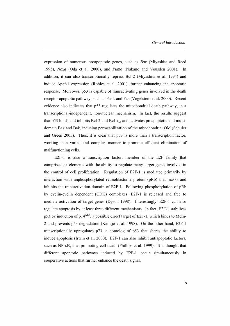
E2F-1 is also a transcription factor, member of the E2F family that
comprises six elements with the ability to regulate many target genes involved in
the control of cell proliferation. Regulation of E2F-1 is mediated primarily by
interaction with unphosphorylated retinoblastoma protein (pRb) that masks and
inhibits the transactivation domain of E2F-1. Following phosphorylation of pRb
by cyclin-cyclin dependent (CDK) complexes, E2F-1 is released and free to
mediate activation of target genes (Dyson 1998). Interestingly, E2F-1 can also
regulate apoptosis by at least three different mechanisms. In fact, E2F-1 stabilizes
p53 by induction of p14ARF, a possible direct target of E2F-1, which binds to Mdm-
2 and prevents p53 degradation (Kamijo et al. 1998). On the other hand, E2F-1
transcriptionally upregulates p73, a homolog of p53 that shares the ability to
induce apoptosis (Irwin et al. 2000). E2F-1 can also inhibit antiapoptotic factors,
such as NF-κB, thus promoting cell death (Phillips et al. 1999). It is thought that
different apoptotic pathways induced by E2F-1 occur simultaneously in
cooperative actions that further enhance the death signal.
19

Chapter 1 _________________________________________________________________________
Thus, after a toxic stimulus and under appropriate conditions, cells have
mechanisms to either arrest cell cycle and repair DNA, or trigger a complex and
efficient mechanism that culminates in death.
2. Role of bile acids in apoptosis
Bile acids are produced in the liver and secreted into the intestine, where they play
crucial biological roles such as the solubilization of lipids in the intestinal lumen,
among many others. However, certain hydrophobic bile acids are cytotoxic
molecules that can increase cell proliferation in the intestinal tract (Bayerdorffer et
al. 1993) and/or induce cell death by necrosis and apoptosis (Patel and Gores
1995). In contrast, more hydrophilic species can be cytoprotective (Heuman et al.
1991).
2.1. Bile acid biosynthesis and physiology
Bile acids are the major components of bile, synthesized in the liver from neutral
sterols by a complex series of chemical reactions (Russell and Setchell 1992).
They are a class of acidic steroids with a cyclopentanoperhydrophenanthrene
nucleus (ABCD-ring) containing 19 carbons, and most commonly a C5 side chain
with a terminal carboxylic acid (Rodrigues et al. 2004). In humans and most
animal species, bile acids are produced primarily from the cholesterol metabolic
pathway. The complete synthesis of bile acids requires approximately seventeen
enzymes. The expression of these enzymes is tightly regulated by nuclear
hormone receptors and other transcription factors, which ensure a steady supply of
bile acids to a highly demanding metabolic environment. Importantly, the initial
and rate-limiting step for the major bile acid biosynthetic pathway is the 7α-
hydroxylation of cholesterol, catalyzed by the cytochrome P450 enzyme,
20

General Introduction _________________________________________________________________________ cholesterol 7α-hydroxylase (CYP7A1). Different bile acid species have diverse
degrees of hydrophobicity, as determined by their biochemical and
physicochemical properties. The amphipathic structure allows these water-soluble
compounds to interact with proteins and insert into lipid bilayers. These effects
will have severe influences on cell function and structure, particularly when
intracellular concentrations of bile acids exceed certain limits, as it is the case in
cholestasis.
Primary bile acids are synthesized in the liver, conjugated with the amino
acids glycine or taurine, and then secreted via the bile ducts and gallbladder into
the lumen of small intestine (Russell and Setchell 1992). Bile acids act as
detergents to emulsify dietary lipids and fat-soluble vitamins, but they can also
solubilize bilirubin and other catabolites. Furthermore, the expression of genes
that synthesize cholesterol, fatty acids, and bile acids are regulated by
intermediates and/or end-products of the bile acid pathway itself (Repa and
Mangelsdorf 1999). While emulsified nutrients are taken up by enterocytes in the
proximal segments of the gut, bile acids re-enter the liver via the portal vein, and
are transported back into the gallbladder for use in the next feeding cycle (Russell
and Setchell 1992).
The biliary bile acid pool also includes secondary bile acids, such as
deoxycholic and lithocholic acids. These bile acids are not formed in the liver, but
rather result from the metabolism of primary bile acids by intestinal bacteria.
Biotransformations of primary bile acids include also the formation of
ursodeoxycholic acid (UDCA), by oxidation of chenodeoxycholic acid to 7-
oxolithocholic acid, followed by reduction yielding the 7β-isomer.
21
Chapter 1 _________________________________________________________________________
2.2. Bile acids and apoptosis
2.2.1. Bile acid-induced apoptosis
Accumulation of toxic bile acids is a common feature of several chronic human
liver diseases, resulting from interruption in bile flow. This pathological condition,
known as cholestasis, can promote liver cell death, leading to cirrhosis (Hofmann
2002). It was thought that hydrophobic bile acids, such as glycochenodeoxycholic
and taurochenodeoxycholic acids could induce cytotoxicity by acting as detergents
on cell membranes. However, other evidence suggests that basic cellular
mechanisms of hepatocyte injury might be primarily involved (Schmucker et al.
1990), ultimately causing cell death by either necrosis or apoptosis. Importantly,
the presence of classic Councilman bodies and cell failure suggests that apoptosis
may play a key role in cholestasis. Bile acid-induced apoptosis has been shown in
vivo as well as in primary rat hepatocytes and human hepatoma HuH-7 cells
(Rodrigues and Steer 2000). However, the predominant type of liver injury may
depend upon several factors, such as the cell type, level of exposure, and metabolic
status of the cell.
The mechanisms by which bile acids induce apoptosis in hepatocytes are still
not entirely known. Several studies have shown that caspase activation,
mitochondrial dysfunction, and cellular distribution of Bcl-2-related proteins
determine the fate of hepatocytes in models of cholestasis (Maher 2004). In
addition, toxic bile acids can also induce ligand-dependent and -independent death
receptor pathways, via Fas- and tumor necrosis factor-related apoptosis inducing
ligand (TRAIL) receptors (Faubion et al. 1999; Higuchi et al. 2003).
Subsequently, FADD is recruited and activates caspase-8 and Bid, which results in
downstream activation of effector caspases and cathepsin B (Roberts et al. 1999;
Sokol et al. 2001). The activation of death receptors invariably signals the
mitochondrial pathway of apoptosis in hepatocytes. In fact, deoxycholic acid
22

General Introduction _________________________________________________________________________ (DCA) was shown to induce the MPT pore formation in isolated mitochondria
(Rodrigues et al. 1998a), as well as mitochondrial depolarization, increased ROS
production, translocation of Bax to mitochondria and cytochrome c release
(Rodrigues et al. 1999; Rodrigues et al. 2003a). Further, MPT was prevented by
antioxidants and cyclosporine A, an inhibitor of the megapore channel (Botla et al.
1995; Rodrigues et al. 1998b; Sokol et al. 2001).
Curiously, the liver has the ability to limit apoptosis during cholestasis by
triggering specific mechanisms. Although it has been shown that Bcl-2, Bcl-xL,
and Bax are expressed in the liver, only cholangiocytes and not hepatocytes
normally express antiapoptotic Bcl-2. However, induction of cholestasis by bile
duct ligation leads to Bcl-2 expresion in hepatocytes, which may represent an
adaptative phenomenon to protect hepatocytes (Kurosawa et al. 1997). In addition,
the activation of NF-κB and subsequent regulation of antiapoptotic genes
(Schoemaker et al. 2003), as well as the cytoplasmic sequestration of p53 (Oh et al.
2002) are complementary mechanisms triggered by the liver to inhibit or modulate
apoptosis induced by toxic bile acids.
2.2.2. Inhibition of apoptosis by ursodeoxycholic acid
In contrast to toxic hydrophobic bile acids, UDCA improves liver function in
patients with hepatobiliary disorders (Lazaridis et al. 2001). It is normally present
in human bile in a low concentration, representing only 3% of total bile acids. In
black bears, however, UDCA is the major biliary bile acid. Bear bile has been
used for centuries in traditional Chinese medicine as a remedy for liver disorders
(Hagey et al. 1993). In the Western world, UDCA has been used for a few decades
as a therapeutic agent for chronic cholestatic liver diseases. At the present, it is the
only drug approved by the United States Food and Drug Administration for the
treatment of primary biliary cirrhosis (Lazaridis et al. 2001; Paumgartner and
Beuers 2004).
23

Chapter 1 _________________________________________________________________________
Both unconjugated UDCA and its amidated conjugates,
tauroursodeoxycholic acid (TUDCA) and glycoursodeoxycholic acid (GUDCA)
are effective modulators of toxicity induced by more hydrophobic bile acids
(Rodrigues and Steer 2000). The mechanisms of action of UDCA in cholestasis
may involve the protection of injured cholangiocytes, stimulation of impaired
biliary secretion, detoxification of hydrophobic bile acids, and/or inhibition of
hepatocyte apoptosis (Paumgartner and Beuers 2004). It is not clear which of these
mechanisms play a primary role for the beneficial therapeutic effects of UDCA.
Most likely, UDCA acts in a coordinated process involving several effects,
depending on the type and stage of the disease.
The role of UDCA in preventing apoptosis in cholestasis has been intensely
studied . In fact, toxic bile acids fed to rats induced apoptosis in the liver, while
UDCA inhibited this effect in vivo, in part by preventing translocation of
proapoptotic Bax from the cytosol to the mitochondria (Rodrigues et al. 1998b)
(Fig. 3). These studies were subsequently extended to show that UDCA plays a
unique role in modulating apoptosis in different cell types, in response to a variety
of agents, acting through different apoptotic pathways (Rodrigues et al. 1998a).
The antiapoptotic effect of UDCA appears to involve the mitochondrial membrane.
In fact, UDCA and its conjugates prevent the release of cytochrome c, caspase
activation, and PARP cleavage associated with mitochondrial depolarization and
Bax channel formation induced by apoptotic stimuli (Rodrigues et al. 1999).
Moreover, UDCA partially prevented apoptosis via the death receptor pathway in
primary mouse hepatocytes co-cultured with fibroblasts expressing the Fas ligand,
possibly by its direct effects at the mitochondrial membrane (Azzaroli et al. 2002)
Additional mechanisms of action for UDCA may also be engaged, where the
bile acid interferes with alternate molecular targets (Fig. 3). In fact, DNA
microarray analysis showed that UDCA can significantly modulate the expression
of 96 different genes, most of them involved in apoptosis, but also in cell cycle
24

General Introduction _________________________________________________________________________ regulation and proliferation (Castro et al. 2005). Apaf-1 was found to be
downregulated in rat hepatocytes in response to UDCA incubations. Importantly,
UDCA can also interfere with molecular targets upstream of the mitochondria.
UDCA inhibited TGF-β1-induced E2F-1 transcriptional activation, p53
stabilization and p53-associated Bax expression, independently of its effect on the
mitochondria and/or caspases (Solá et al. 2003b). UDCA also inhibited the
downregulation of Bcl-2 by TGF-β1, which is consistent with decreased p53
stabilization and/or of NF-κB degradation. Furthermore, recent evidence showed
that both UDCA and TUDCA reduce transcriptional activation and expression of
cyclin D1 in primary rat hepatocytes incubated with deoxycholic acid (Castro et al.
2005; Castro et al. 2007). The modulation of cyclin D1 expression appears to
contribute to the antiapoptotic effects of the bile acid, in part through a p53-
dependent mechanism. In fact, UDCA modulates the E2F-1/Mdm-2/p53 apoptotic
pathway in hepatocytes via a nuclear steroid receptor (NSR)-dependent mechanism
(Solá et al. 2004). UDCA upregulated both glucocorticoid (GR) and
mineralocorticoid (MR) receptor expression in hepatocytes during TGF-β1-
induced apoptosis. Moreover, it was shown that UDCA promotes GR/hsp90
dissociation, inducing subsequent NSR translocation (Solá et al. 2005). The
deletion of the C-terminal region of GR inhibited the capacity of UDCA to induce
GR/hsp90 dissociation, GR translocation and modulation of apoptosis, indicating
that the ligand binding domain of GR is required for the antiapoptotic function of
UDCA. These results strongly suggest that UDCA translocates to the nucleus, in a
complex with GR, where it may modulate apoptosis-related genes. In contrast,
recent evidence suggests that the modulation of induced neuronal apoptosis by
TUDCA requires an interaction with MR (Solá et al. 2006).
TUDCA was also shown to regulate the ER stress-mediated pathway in
HuH-7 cells, reducing the calcium efflux and the activation of caspases-12 (Xie et
al. 2002) (Fig. 3). Moreover, treatment of obese and diabetic mice with TUDCA
25

Chapter 1 _________________________________________________________________________
resulted in normalization of ER-induced hyperglycemia and restored systemic
insulin response (Ozcan et al. 2006). TUDCA was also responsible for resolution
of fatty liver disease and enhancement of insulin action in liver, muscle, and
adipose tissues, expanding its beneficial role to type 2 diabetes.
Fig. 3. Proposed mechanisms for the antiapoptotic actions of UDCA and TUDCA. UDCA
negatively modulates the mitochondrial pathway by inhibiting Bax translocation, ROS
formation, cytochrome c release and caspases-3 activation. UDCA can also interfere with
the death receptor pathway, inhibiting caspase-3 activation. Moreover, TUDCA inhibits
apoptosis associated with ER stress, by modulating intracellular calcium levels, and
inhibiting calpain and caspase-12 activation. UDCA interacts with NSR, leading to
NSR/hsp90 dissociation and nuclear translocation of NSR. The nuclear trafficking of
Fas L
Fas
Cytochrome c
E2F-1UDCA?
Nucleus
hsp90
p53
p14ARF Mdm-2
NSR
UDCA
UDCA
NSR
NSR
hsp90
EndoplasmicReticulum
Caspase-3UDCA
Bax
Bax
Bax
Bcl-2
UDCA
Ca2+Ca2+
Ca2+ Procaspase-12Calpain
Caspase-12
Mitochondria
TUDCA
APOPTOSISAPOPTOSIS
Cyclin D1
Apaf-1
26

General Introduction _________________________________________________________________________ UDCA allows it to modulate the E2F-1/p53/Bax pathway, preventing apoptosis. Finally,
UDCA downregulates cyclin D1 and Apaf-1, further inhibiting the mitochondrial apoptotic
cascade. See text for more complete description.
Finally, it is becoming increasingly evident that activation of survival
pathways may represent an important additional mechanism by which UDCA
inhibits apoptosis. One possible survival pathway involves the activation of NF-
κB by several molecules, including the inflammatory cytokines TNF-α and
interleukin-1β (Bradham et al. 1998). In addition, TUDCA was shown to protect
mitochondria-controlled apoptosis in primary rat hepatocytes by activating the
phosphatidylinositol 3-kinase (PI3K) and MAPK pathways (Schoemaker et al.
2004).
2.3. Bile acids for the treatment of neurodegenerative disorders
The therapeutic role of UDCA has been established in the treatment of certain liver
diseases. Importantly, UDCA has the ability to modulate apoptosis at several
levels, suggesting a common mechanism of cell survival regulation that is
independent of cell type. Thus, the use of UDCA to non-liver diseases in which
increased levels of apoptosis contribute to their pathogenesis is now a major
consideration.
Mitochondrial dysfunction and subsequent oxidative damage have been
implicated in several neurobiological disorders, such as acute stroke and chronic
neurodegenerative diseases. Moreover, increasing evidence suggests that apoptosis
plays a crucial role in the pathogenesis of such disorders (Mattson 2006).
Interestingly, after conjugation with taurine, UDCA administrated in high doses
can be delivered to other tissues, including the brain (Keene et al. 2001). In vitro,
27
Chapter 1 _________________________________________________________________________
TUDCA inhibits apoptosis induced by several stimuli in neuronal cells (Rodrigues
et al. 2000; Solá et al. 2003a). Furthermore, the protective role of TUDCA has
been extended to several models of neurological disorders, including
Hungtington’s disease (HD), Parkinson’s disease (PD), and acute ischemic and
hemorrhagic stroke.
HD is a genetically dominant neurological disorder caused by abnormal
expansion of the trinucleotide (CAG) repeat sequence in exon 1 for the gene Htt
encoding the huntingtin protein (Walker 2007). In HD, mitochondrial function is
compromised, which is demonstrated by abnormal energy metabolite levels,
impaired striatal mitochondrial respiratory chain complex II/III activity and
increased stress-induced mitochondrial depolarization, ROS production, and
associated oxidative damage. A consequence of mitochondrial dysfunction may be
the activation of certain apoptotic pathways, confirmed by activation of caspases
and DNA fragmentation in HD brains. Chronic administration of 3-nitropropionic
acid (3-NP), an irreversible inhibitor of succinate dehydrogenase provides a
relatively accurate animal model of HD, closely resembling the pathology and
symptoms of the disease. The systemic administration of TUDCA in the 3-NP rat
model of HD was shown to reduce the associated morphologic striatal lesions
(Keene et al. 2001). Moreover, behavioral studies correlated with
histopathological findings, since the significant neuroprotection resulted in almost
complete prevention of hyperactive behavior associated with 3-NP administration,
while maintaining neophobia characteristic of cognitively intact animals. TUDCA
also markedly reduced the mitochondrial perturbations associated with apoptosis
induction in cultured neuronal cells incubated with 3-NP (Rodrigues et al. 2000).
These data suggested that TUDCA could possibly act as a therapeutic agent for
regulating cell survival in HD. Thus, initial studies were extended to the R6/2
transgenic mouse model of HD, which express exon 1 of the human huntingtin
gene with ~ 150 CAG repeats and the human huntingtin promoter. Administration
28

General Introduction _________________________________________________________________________ of TUDCA induced a marked reduction in striatal cell apoptosis and degeneration
of R6/2 transgenics (Keene et al. 2002). In addition, TUDCA prevented striatal
atrophy, reduced the average size and number of individual characteristic
intracellular inclusions of huntingtin and ubiquitin, and, consequently, improved
the locomotor and sensorimotor abilities of R6/2 transgenic mice.
Transplantation of human embryonic dopamine neurons in patients with PD
is currently being evaluated in clinical trials. Although transplantation of nigral
tissue ameliorates deficiencies characteristic of PD, a major hurdle for successful
neural grafting is the poor survival of dopaminergic neurons implanted into the
brain of recipient patients. There is strong evidence showing that the majority of
cell death occurring in neuronal grafts results from apoptosis. Thus, based on its
antiapoptotic properties, TUDCA was added to cell suspensions prior to
transplantation to improve cell survival of nigral grafts in a rat model of PD (Duan
et al. 2002). The results showed that TUDCA reduced the levels of cultured cells
undergoing apoptosis, increasing the number tyrosine-hydroxylase-positive
neurons. In transplantation studies, where TUDCA treated-cell suspensions were
injected into the striatum of 6-hydroxydopamine lesioned rats, there was a
significant improvement of amphetamine-induced rotation scores, resulting from
increased survival of dopamine positive cells. More recently, TUDCA was shown
to partially rescue a PD model of C. elegans from mitochondrial dysfunction (Ved
et al. 2005).
Prompted by these results, the antiapoptotic role of TUDCA was extended to
acute conditions, particularly to a rat model of transient focal cerebral ischemia
(Rodrigues et al. 2002). Neuronal cell death resulting from acute stroke is a
complex process and appears to involve a variety of different pathways. Although
originally thought to be mainly necrotic, cell death can also occur via apoptosis in
both ischemic and hemorrhagic stroke (Friedlander et al. 1997; Matsushita et al.
2000). Intravenous administration of TUDCA resulted in improved neurologic
29

Chapter 1 _________________________________________________________________________
function and reduced infarct volumes by ~ 50%. In addition, TUDCA significantly
reduced the number of apoptotic cells, prevented mitochondrial swelling and
membrane disruption, and partially inhibited downstream caspases activation and
endogenous substrate cleavage associated with apoptosis. TUDCA also reduced
the levels of apoptosis, caspase-3-like activation and histological damage of the
peri-hematoma region in a collagenase-induced hemorrhagic model of stroke
(Rodrigues et al. 2003b). In addition, TUDCA inhibited NF-κB activation,
maintained elevated the levels of Bcl-2, and activated the PI3K survival pathway,
consequently improving neurological functions.
Cell death induced by glutamate may be involved in neuronal injury
associated with chronic neurodegenerative disorders, including HD, and with acute
conditions, such as hypoxia-reperfusion. The mechanisms by which neurons die
when exposed to glutamate can either be necrotic or apoptotic, depending on the
severity of the insult (Bonfoco et al. 1995). Interestingly, pretreatment with
TUDCA also significantly reduced glutamate-induced apoptosis of rat cortical
neurons (Castro et al. 2004). TUDCA inhibited the release of cytochrome c from
mitochondria as well as the activation of caspases-3. In addition, TUDCA induced
marked phosphorylation and translocation of Bad to cytosol, which was prevented
after inhibition of the PI3K survival pathway. These results suggested that
TUDCA modulates glutamate-induced apoptosis in part by activating a PI3K-
dependent Bad signaling pathway.
Taken together, these studies provide evidence for the antiapoptotic role of
UDCA and TUDCA. As a hydrophilic acid, TUDCA is readily water-soluble, can
be administrated orally and intravenously, and is associated with minimal toxicity.
These characteristics make TUDCA a potent therapeutic tool in several disorders
associated with higher susceptibility to apoptotic cell death. Thus, it would be
relevant and interesting to further extend its clinical applications to other
neurodegenerative diseases, such as Alzheimer’s disease (AD).
30

General Introduction _________________________________________________________________________
3. Alzheimer’s disease
In 1906, the physician Alois Alzheimer gave a lecture at a congress of psychiatry
in Tübingen, Germany. For the first time, he described a disease that Kraepelin
some years later named AD. Over several years, Alzheimer had been observing a
patient at the Frankfurt Asylum named Auguste D., who presented strange
behavioral symptoms, including a decline in short-term memory, confusion and
disorientation. After her death, staining techniques revealed the presence of
amyloid plaques and neurofibrillary tangles (NFT) in the brain, which are today
recognized as hallmarks of the disease. AD is the most common form of dementia.
However, after 100 years of its discovery, the triggering mechanisms of AD are not
entirely understood, and effective diagnosis and treatment are still lacking.
3.1. Epidemiology and risk factors
AD is a severe and common cause of dementia, accounting for 50-60% of all cases
(Ferri et al. 2005). This neurodegenerative disease targets specific brain regions
early in its course, especially the cholinergic basal forebrain and medial temporal
lobe structures, including the hippocampus, amygdale and entorhinal cortex. With
disease progression, the pathogenesis spreads to other regions, including the
posterior cingulate, temporal and parietal isocortical regions (Braak and Braak
1991). AD is characterized by progressive memory loss, anomia, constructional
apraxia, anasognosia, and variable degrees of personality changes (Katzman 1992).
The prevalence of AD is less than 1% in individuals aged 60-64 years, but the
probability of developing the disorder increases with age. In Western countries,
the prevalence reaches 24-33% in people aged 85 years or older. In 2001, more
than 24 million people in the world had dementia. With life expectancy increasing,
the numbers are predicted to double every 20 years, reaching up to 81 million in
31

Chapter 1 _________________________________________________________________________
2040 (Ferri et al. 2005). In Portugal, there are ~ 60,000 patients with AD, although
the numbers might be under estimated, given the difficulty of diagnosis.
In the vast majority of cases, the cause of AD remains unclear. Like most
other chronic conditions, AD probably develops as a result of multiple factors,
rather than a single cause. Undoubtedly, the greatest risk factor is advancing age,
but epidemiological studies have suggested several possible associations.
Decreased reserve capacity of brain, including reduced brain size, low levels of
education and occupation, low mental ability in early life, and reduced mental and
physical activities during late life, can increase the susceptibility to develop AD
(Mayeux 2003). In addition, it has been suggested that head injury can also be
linked to AD (Jellinger 2004). Other risk factors are hypo- and hyperthyroidism,
hypercholesterolemia, hyperthension, atherosclerosis, heart disease, smoking,
obesity, and diabetes (Mayeux 2003).
Although environmental influences might increase the susceptibility to
sporadic AD, genetic factors have a significant impact in the development of the
disease. The genetic factor that accounts for more cases of late-onset sporadic AD
is the apolipoprotein ε4 allele (APOE) located on chromosome 19. APOE ε4
increases the risk of developing AD 3-fold in heterozygotes and by 15-fold in
homozygotes, and also influences the age of disease onset (Farrer et al. 1997;
Meyer et al. 1998). The mechanisms by which APOE ε4 triggers AD are not
entirely understood, but may include enhanced rate of amyloid β (Aβ) deposition,
as well as reduced protection against oxidative stress and efficacy of neuronal
repair (Horsburgh et al. 2000). The search for additional susceptibility genes for
sporadic AD is still ongoing, but none has been verified with certainty.
A small percentage of AD patients have a familial form of the disease,
related to a specific hereditary mutation. Familial AD is rare, with a prevalence
below 0.1 % (Harvey et al. 2003). It is an autosomal-dominant disorder with early
onset, striking patients in their mid-thirties to mid-fifties. To date, more than 100
32

General Introduction _________________________________________________________________________ mutations of 3 genes have been identified to cause this specific form of AD. The
first mutation related to familial AD was identified in the amyloid precursor
protein (APP) gene on chromosome 21 (Goate et al. 1991). Since then, several
additional mutations have been described. The largest number of mutations,
however, is located in the presenilin 1 (PS1) gene, responsible for most cases of
familial AD (Sherrington et al. 1995). Finally, a small number of mutations have
also been identified in the presenilin 2 (PS2) gene in chromosome 1 (Levy-Lahad
et al. 1995). All mutations result in elevated Aβ levels. Although related to
hereditary factors, familial AD patients develop the same histopathological features
and cognitive deficits observed in sporadic cases.
3.2. Pathogenesis
Neurodegeneration in AD is estimated to begin 20-30 years before clinical onset
(Davies et al. 1988). During this preclinical phase and after diagnosis, several
pathologic mechanisms occur in specific areas of the brain. At the microscopic
level, Aβ and tau deposit and aggregate into characteristic external amyloid
plaques and internal NFT, respectively. At the same time and/or consequently,
other cellular abnormalities occur, such as neuronal dysfunction, cell cycle
deregulation, ROS production, inflammation and mitochondrial failure.
Ultimately, all these pathological mechanisms culminate in neuronal death and
cognitive decline.
3.2.1. Amyloid plaques
The main constituent of amyloid plaques found in AD brains is the Aβ peptide
(Glenner and Wong 1984). Initially, Aβ was thought to be an abnormal protein.
33

Chapter 1 _________________________________________________________________________
Extracellular Intracellular
COOH
APP
DAEFRHDSGYEVHHQKLVFFAEDVGSNKAIIGLMVGGVVIAT
γ-secretase
sAPPα
Nonamyloidogenic pathway
α-secretase
NH2
Extracellular Intracellular
NH2 COOH
APP
DAEFRHDSGYEVHHQKLVFFAEDVGSNKAIIGLMVGGVVIAT
β-secretase γ-secretase
Aβ
Amyloidogenic pathway
β-secretase
Fig. 4. Diagram of APP processing. In the nonamyloidogenic pathway, APP is cleaved by
β-, γ- and α-secretases, generating a soluble sAPPα fragment. In contrast, the cleavage of
APP by β- and γ-secretases produces Aβ, with high capacity to aggregate into amyloid
plaques. See text for more details.
However, it is also constitutively produced during normal cell metabolism
(Haass et al. 1992). Aβ is generated by proteolytic cleavage of APP, mediated by
the transmembrane proteins β- and γ-secretases (Vassar et al. 1999) (Fig. 4). The
β-site APP cleaving enzyme (BACE1), a member of the pepsin family of aspartyl
proteases, cleaves the ectodomain of APP to generate a membrane bound C-
terminal fragment, which is subsequently, cleavage by γ-secretase. Importantly,
although not entirely understood, the metabolism of APP appears to be modulated
both directly and indirectly by phosphorylation and phosphorylation-dependent
events (da Cruz e Silva and da Cruz e Silva 2003). Depending on the exact
cleavage site, peptides with 38 to 43 amino acids may be generated. Nevertheless,
34

General Introduction _________________________________________________________________________ the most common and toxic forms of Aβ contain 40 and 42 amino acids in length,
and are termed Aβ1-40 and Aβ1-42, respectively. These fragments of ~ 4 kDa
account for the majority of Aβ found in AD brains. Finally, APP can also be
cleaved by a third protease, the α-secretase (Allinson et al. 2003). α-secretase
cleavage is nonamyloidogenic and precludes subsequent BACE1 cleavage,
generating a soluble sAPPα fragment that appears to be neuroprotective (Meziane
et al. 1998). Under normal conditions, the brain also has the capacity to degrade
Aβ by the peptidases insulin-degrading enzyme, neprilysin, and by endothelin-
converting enzyme (Carson and Turner 2002). Moreover, Aβ is cleared from the
brain in a process balanced by a system of efflux and influx that allows the peptide
to cross the blood-brain barrier (Tanzi et al. 2004).
The amyloid cascade hypothesis is considered the major cause of AD (Hardy
and Selkoe 2002). According to this, an imbalance between production and
clearance of Aβ in the brain triggers neuronal degeneration and dementia, as the
initial step of the process. Support for this hypothesis includes the finding of
mutations in APP, and in PS1 and PS2, implicated in familial AD. Moreover, the
discovery of a third APP gene in people with Down’s syndrome, who develop AD
early in life, suggested that life-long APP overexpression triggers Aβ deposition.
Following cleavage, soluble Aβ undergoes conformational changes in its β-sheet
content. Next, the peptide self-aggregates by a complex process starting with self-
dimerization, followed by oligomerization, protofibril formation, and eventual
aggregation into large insoluble fibrillar structures (Urbanc et al. 2004).
Importantly, increasing evidence suggest that the ratio Aβ1-42/Aβ1-40 is important in
AD pathogenesis, rather than the total amount of Aβ produced. In fact, due to its
hydrophobic nature, Aβ1-42 has a higher tendency to aggregate and is believed to
initiate amyloid plaques formation, by triggering the misfolding of other Aβ
species (Jarrett et al. 1993; Iwatsubo et al. 1994).
35

Chapter 1 _________________________________________________________________________
Initially, only Aβ plaques were assumed to be neurotoxic. However, there
are several phenomena that can not be explained by fibrillar deposits alone.
Indeed, affected neurons do not necessarily colocalize with amyloid plaques in the
brain (Li et al. 1994). Similarly, amyloid plaques do not correlate with cognitive
decline, as it would be expected if plaques were the main toxic entity in AD
(Naslund et al. 2000). Thus, it has been suggested that intermediate soluble
oligomers of Aβ may be toxic entities (McLean et al. 1999). These protofibrils are
present in AD brain, and represent a distinct form of low molecular weight Aβ,
containing 9 to 50 molecules of the peptide. Interestingly, exposure of neuronal
cultures and mouse brains to soluble oligomers of Aβ resulted in higher levels of
toxicity and neuronal dysfunction, compared to fibrillar species, but the results are
still controversial (Dahlgren et al. 2002; Kirkitadze et al. 2002). Moreover, the
role of tau in AD pathogenesis can not be neglected and it possibly accounts for
other dysfunctions not directly correlated with Aβ aggregation.
3.2.2. Neurofibrillary tangles
Neurobrillary tangles, in addition to amyloid plaques, are another pathological
feature of AD, whose major component is the protein tau. Tau is a family of
microtubule associated proteins, abundant in the central nervous system and
predominantly expressed in axons (Binder et al. 1985). Through its microtubule-
binding domains, tau promotes microtubule assembly and stability. In addition, it
can also play a role in vesicular transport and axonal polarity. In adult human
brain, tau contains six different isoforms, ranging from 45 to 65 kDa, resulting
from alternative splicing. The isoforms differ by the presence of either three- (3R)
or four-repeat domains (4R) in the C-terminal region, the binding domains, and the
absence or presence of one or two inserts in the N-terminal region (Goedert et al.
1989). Each isoform is likely to have particular roles, since they are differentially
expressed during development. In fact, it has been demonstrated that adult tau
36

General Introduction _________________________________________________________________________ isoforms with 4R are more efficient at promoting microtubule assembly than the
fetal isoform with 3R (Goedert and Jakes 1990).
Tau is very hydrophilic, soluble, and natively unfolded. The functions of tau
are regulated by phosphorylation, which can occur in multiple sites. For instance,
it was demonstrated that tau is more efficient in promoting microtubule assembly
when in a more dephosphorylated state. Interestingly, the longest form of human
tau contains 80 serine or threonine residues and five tyrosine residues, which
represents almost 20% of the protein with potential for phosphorylation (Stoothoff
and Johnson 2005). Several kinases have been shown to phosphorylate tau. The
glycogen synthase kinase 3 (GSK3β) is the main kinase, responsible for
phosphorylation of tau at numerous sites. GSK3β may also play a role in
regulating the degradation of tau, via the proteasome (David et al. 2002). In
addition to GSK3β, the cycline-dependent kinase 5 (CDK5), and the microtubule
affinity-regulating kinase (MARK) are also known to regulate the physiological
role of tau through phosphorylation. In contrast, to counterbalance the action of
kinases, tau is readily dephosphorylated by numerous phosphatases, including
protein phosphatase 1 (PP1), 2A (PP2A), 2B (PP2B), and 5 (PP5) (Tian and Wang
2002).
The underlying mechanisms contributing to the pathological processing of
tau in neurodegenerative conditions have not been entirely elucidated.
Nevertheless, it is clear that tau becomes characteristically altered both
functionally and structurally in AD and other tauopathies. Supporting a central
role for tau in dementia, genetic studies showed that missense mutations in the tau
gene induce the development of neurodegenerative disorders such as the
frontotemporal dementia with parkinsonism linked to chromosome 17 (FTDP-17)
(Hutton et al. 1998). Interestingly, in FTDP-17 and other sporadic tauopathies,
pronounced tau pathology was observed, without amyloid plaque formation.
Moreover, the distribution and abundance of tau pathology in AD is well correlated
37
Chapter 1 _________________________________________________________________________
with neuronal degeneration and clinical symptoms, underscoring the relevance of
tau (Braak and Braak 1991). The majority of tau mutations are associated with
either reduced ability of the protein to interact with microtubules, or increased
capacity to aggregate (Hasegawa et al. 1998). Nevertheless, there are no identified
mutations associated with AD pathology.
The abnormal phosphorylation of tau has been proposed as the primary
modification of tau in AD brains. For example, phosphorylation of Ser205,
Ser396, Ser404, Thr205, and Thr212 enhance the polymerization of tau into
filaments (Necula and Kuret 2005). Proteolysis of tau is another posttranslational
event that may influence the aggregation in NFT. In fact, truncation at both the N-
and C-terminal regions directly influences fibril formation of tau. Cleavage of tau
by caspase-3 at Asp421 in the C-terminal region has been detected in AD brains
and was shown to promote tau assembly in vitro (Gamblin et al. 2003b). This
fragment apparently causes neuronal death, with a significant role in the
nucleation-dependent filament formation (Rissman et al. 2004). In contrast, the
removal of the N-terminal region greatly inhibits the aggregation of tau, although
this specific truncation occurs later in the NFT formation process (Gamblin et al.
2003a). Thus, aggregation of tau within neurons appears to involve an ordered
series of events. First, the microtubule binding function needs to be neutralized.
Then, tau molecules must self-associate to promote oligomerization, which leads to
conformational changes and increased insolubility of tau. Finally, these oligomers
induce nucleation and formation of NFT, which accumulate in neuron cell bodies.
Amyloid plaques and NFT are often present in the same area of the brain, but
the relationship between these two entities is not known. It was recently
demonstrated that Aβ-induced caspase-3 activation leads to tau cleavage, and
aggregation in NFT (Rissman et al. 2004). In addition, hyperphosphorylation of
tau was detected in mature hippocampal neurons treated with Aβ (Ferreira et al.
38
General Introduction _________________________________________________________________________ 1997). Thus, NFT pathology may represent a downstream effect of Aβ
accumulation probably triggered by apoptosis, among other toxic mechanisms.
3.2.3. Neuronal dysfunction and degeneration
In the AD brain, multiple mechanisms occur simultaneously and it is not always
easy to distinguish changes that promote the disease from the consequences of the
disease. It is established, however, that amyloid plaques and NFT are extra- and
intracellular aggregates that are toxic to neurons (Fig. 5).
Several reports have suggested that Aβ activates microglia, inciting an
inflammatory response and release of neurotoxic cytokines. In fact, prominent
activation of inflammatory processes and the innate immune response are observed
in AD brains (Wyss-Coray and Mucke 2002). Neuroinflammation would then
occur in the early stages of the disease, driving the pathology. However, other
studies suggest that inflammation may even be beneficial and responsible for
triggering survival pathways (Wyss-Coray 2006). Thus, the exact role of
inflammation in AD is still controversial.
Aβ can also trigger excessive release of excitatory amino acids such as
glutamate from glial cells, injuring nearby neurons and inducing the production of
ROS. Indeed, increasing evidence supports a role for oxidative stress in vulnerable
brain regions of AD patients. Oxidative damage begins early in the disease and
can even be responsible for a decline in proteasome function (Cecarini et al. 2007).
Moreover, Aβ may directly or indirectly injure mitochondria, thus decreasing the
brain metabolism. Aβ is involved in disruption of calcium homeostasis, via ER-
mediated stress, inducing mitochondrial dysfunction (Pereira et al. 2004). In
addition, Aβ blocks the respiratory complex I and the pyruvate dehydrogenase,
inducing a decline in ATP and enhancing ROS production (Casley et al. 2002). In
fact, as regulators of both energy metabolism and apoptotic pathways,
mitochondria and their consequent dysfunction have a key role in AD (Moreira et
39

Chapter 1 _________________________________________________________________________
al. 2006). Moreover, because synaptic function has a high energy demand,
mitochondrial failure is a major factor of synaptic failure in AD. In addition,
cholinergic transmission and synaptic density are considerably decreased in AD
patients and probably contribute to memory loss and cognitive deficits. Finally,
Aβ and tau may be responsible for paralyzing axonal and dendritic transport.
Fig. 5. Proposed mechanism of citotoxicity induced by Aβ and tau. Soluble and/or
aggregated Aβ interacts with glial cells, inducing the release of glutamate and
inflammatory cytokines. Aβ triggers signal transduction cascades that culminate in
increased production of ROS and disruption of calcium homeostasis. Moreover, Aβ may
directly or indirectly injure mitochondria, enhancing ROS production and inducing
Ca2+
NEURONAL DYSFUNCTION AND DEGENERATIONNEURONAL DYSFUNCTION AND DEGENERATION
Glial cell
Aβ Aβ
Aβ
Aβ
Cytokines
ROS
InflammationROS
ROSProteasome dysfunction
Tau Caspase-3/-6
P
PP
NFT
Glutamate
Tau
40

General Introduction _________________________________________________________________________ proteasome failure. Effector caspases are activated, which can be responsible for the
cleavage of tau. In addition, abnormal phosphorylation of tau might be enhanced by Aβ.
Tau aggregates into NFT and redistributes in neuron cell bodies, paralyzing axonal and
dendritic transport. All together, these abnormal mechanisms in AD brains are thought to
culminate in neuronal dysfunction and degeneration.
In conclusion, mitochondrial dysfunction and consequent energy deficits
contribute to impaired clearance of protein aggregates and neuronal dysfunction,
affecting neurotransmission and neuronal transport in AD brains. In this scenario,
neurons no longer have the ability to survive and massively die in the affected
brain regions. However, the primary mechanism(s) by which neurons die is still
not clear. Nevertheless, apoptosis has been the focus of intense research in the last
few years as a pivotal phenomenon in AD progression.
3.2.4. The role of apoptosis and cell cycle proteins in AD
First hints that apoptosis could be involved in AD came from studies performed in
the mid-90’s, where cultured neurons were incubated with fibrillar Aβ (Loo et al.
1993). Since then, both in vitro and in vivo models have supported apoptosis as a
general mechanism of cell death in AD. Analysis of post-mortem brain tissue has
also provided evidence for nuclear DNA fragmentation and activation of apoptotic
mechanisms in humans. Studies in the brain of AD patients showed an
upregulation of 50-fold in the levels of apoptosis, as compared with age-matched
controls (Colurso et al. 2003). Nevertheless, evidence for frank cellular apoptosis
in AD brains is controversial, since clear detection of apoptotic cells is difficult and
problematic. In this regard, apoptosis in the pathogenesis of AD is still an issue of
debate, and it has even been suggested that it provides a protective role in
removing damaged cells from the brain.
41
Chapter 1 _________________________________________________________________________
Studies of human AD tissue demonstrated that the expression of the
antiapoptotic Bcl-2 and proapoptotic Bax proteins are regulated in brain areas
showing increased apoptosis (Su et al. 1997). In addition, c-Jun, a protein of the
JNK-pathway and an immediate-early proapoptotic molecule was found to be
colocalized in neurons with fragmented DNA (Anderson et al. 1996). Moreover,
recent evidence suggests that mitochondrial dysfunction and ER-induced stress
have pivotal roles in the execution of apoptosis in AD (Takuma et al. 2005). In
fact, together with Aβ-induced toxicity, many other stimuli, such as oxidative
stress, neurotrophic factor deprivation, low energy metabolism, and mitochondrial
failure, known to induce apoptosis in cultured neurons, are also prominent in AD
brains.
Activation of caspases-3 in AD has been reported using specific antibodies
to the activated form of the enzyme (Su et al. 2001). Caspases-3 was detected in
more than 50% of hippocampal neurons showing granulovascular degeneration and
often colocalized with tau protein. In addition, an increase in brain fodrin
proteolysis in sprouting neurons was observed (Peterson et al. 1991). Caspase-6
was also found to be activated in AD brains (Guo et al. 2004). The activation of
caspase-6 occurs primarily in the frontal and temporal cortex, and is colocalized
with pre-tangles, suggesting that it may be an early event in AD pathogenesis.
Despite the clear activation of effector caspases, coincident morphologic evidence
of apoptosis are not always easy to detect and levels of apoptosis can not per se
explain the massive neuronal degeneration observed in AD brains. Nevertheless,
since AD is a chronic disease, and the apoptotic process short-lived, the detection
of substantial apoptotic neurons in AD brains could be difficult. Moreover, the
apoptotic mechanisms may play an important role in disease pathogenesis, even in
the absence of clear apoptosis. In fact, caspases-3 can cleave APP and presenilins,
and the product of APP cleavage, a C-terminal peptide called C31, is a potent
inducer of cell death (Lu et al. 2000). Importantly, caspases-3 appears to be
42

General Introduction _________________________________________________________________________ responsible for cleaving tau in its C-terminal region early in disease pathogenesis.
In AD brains, caspases-3-cleaved tau colocalizes with both intracellular Aβ and
activated caspases-3, mainly in tangle-bearing neurons (Rissman et al. 2004).
Recent studies have suggested that caspases-6 is also capable of cleaving tau and
promotes its aggregation in NFT (Guo et al. 2004). While caspases-6-cleaved tau
was found in amyloid plaques and NFT, caspases-6 was located primarily in
neurites. This suggests that apoptosis-like mechanisms can damage synapses,
axons and dendrites without causing overt neuronal death.
Expression of cell cycle markers in AD neurons suggests their potential
involvement in the disease process. In postmitotic neurons, which lack the
required components to complete the cell cycle, the presence of mitotic markers
indicates a breach of cell cycle check points. This activated but uncoordinated cell
cycle activity can induce deleterious effects and result in many pathological events
in AD, including apoptosis. For example, as a response to DNA damage and ROS
production, cyclin D-CDK was found to be upregulated in AD (Park et al. 1998).
In addition, phosphorylation and dysfunction of pRb resulting in E2F-1 release
may occur in neuronal cells (Phillips and Vousden 2001).
Evidence for a pivotal function of p53 in neuronal death has been provided
by data from cell culture models documenting increased p53 levels in affected
neurons (Morrison et al. 2003). Coincidently, accumulation of both Aβ1-42 and p53
was found in degenerating neurons in both transgenic mice and human AD brain.
Intracellular Aβ1-42 may cause p53-dependent neuronal apoptosis through activation
of the p53-promoter, thus demonstrating an alternative pathway of cell death in AD
(Ohyagi et al. 2005). The inhibition of p53 attenuates Aβ-induced apoptosis,
associated with preserved mitochondrial function and reduced activation of
caspases-3 (Culmsee et al. 2001). These findings suggest that p53 is an important
mediator of ROS-induced signaling upstream of the mitochondrial dysfunction.
Moreover, upregulation of p53 is also associated with indirect induction of tau
43

Chapter 1 _________________________________________________________________________
abnormal phosphorylation in HEK293a cells, underscoring the relevance of p53 in
AD pathology (Hooper et al. 2007).
Several lines of evidence confirm the pivotal role of apoptosis in AD. Either
by inducing effective neuronal death or enhancing mechanisms that culminate in
neuronal dysfunction, the importance of apoptosis in the pathogenesis of AD
should not be ignored. Indeed, therapeutic interventions targeting apoptosis and
specific molecular intervenients, such as p53, are expected to play a role in AD
treatment.
3.3. Clinical diagnosis and treatment of AD
After the first symptoms appear, the diagnosis of AD is based on the medical
history, together with clinical, neurological and psychiatric examinations. Given
the complexity of the disease and a possible overlapping of clinical and
pathological features with other dementias, the diagnosis of AD is not always
evident. Absolute diagnosis requires microscopic examination of the cerebral
cortex, and the identification of amyloid plaques and NFT (McKhann et al. 1984).
Cortical biopsy can provide efficient results during patient’s life but is not
routinely performed. Thus, a definitive diagnosis of AD is typically obtained in
post mortem analysis. Neuroioimaging, including magnetic resonance imaging
(MRI) and computed tomography (CT), is important to exclude alternative causes
of dementia, such as brain tumor and subdural hematoma. MRI and CT are
valuable tools to detect cerebral atrophy, specifically in the hippocampus and
entorhinal cortex. Positron emission tomography imaging of glucose is a
promising method and it can be used to differentiate with high sensitivity patients
with AD from cognitively normal elderly people or with other dementias
(Silverman et al. 2001). Furthermore, the identification of cerebrospinal fluid
44

General Introduction _________________________________________________________________________ biological markers, such as total and hyperphosphorylated tau and Aβ1-42, for the
prediction of incipient AD shows great promise (Blennow and Hampel 2003).
Unfortunately for the millions of patients that are diagnosed with AD, the
efficacy of the available treatment is far from satisfactory. Currently, approved
drugs can only slow progression of disease, albeit with some improvement in
quality of life. They typically ameliorate the neurotransmitter disturbances that
account for the characteristic memory decline and other cognitive symptoms.
Acetylcholinesterase inhibitors, including donopezil, galantamine, and
rivastigmine are designed to combat impairment of cholinergic neurons by slowing
degradation of acetylcholine after its release at synapses. Memantine is another
drug that is currently used in AD treatment, which prevents overstimulation of the
N-methyl-D-aspartate subtype of glutamate receptors. To manage the behavioral
signs, such as aggression, agitation and psychosis, many AD patients also receive
antipsychotics and antidepressants.
Over the last decade, many pathways have been identified that may
contribute to the pathogenesis of AD. Thus, substantial efforts are underway to
translate this knowledge into novel therapeutic interventions that prevent or arrest
the disease. The major focus has been to inhibit Aβ production and aggregation,
and to increase Aβ clearance from the brain. Currently, inhibitors of β- and γ-
secretases to reduce brain Aβ concentrations are being tested in transgenic mice as
well as phase I studies (Chang et al. 2004; Siemers et al. 2005). In addition, α-
secretase stimulation is also being tested for APP processing into non-
amyloidogenic peptides production and consequent Aβ reduction (Etcheberrigaray
et al. 2004). On the other hand, Aβ immunotherapy is being studied and has been
shown to markedly reduce AD pathology (Schenk et al. 2004). A
glycosaminoglycan mimetic and the metal chelator clioquinol have been shown to
reduce Aβ fibrillization in vitro and in transgenic mice (Cherny et al. 2001;
Sadowski et al. 2004). Inhibitors of tau phosphorylation are under investigation
45

Chapter 1 _________________________________________________________________________
despite the involvement of multiple kinases and phosphatases. Finally, different
types of drugs and supplements, such as anti-inflammatory and cholesterol-
lowering drugs, estrogens, and antioxidants have been suggested as having a
protective role on AD. Nevertheless, the results are controversial and the
beneficial effects have been difficult to prove.
It will probably take several decades before a cure for AD is discovered.
However, we now know that for an effective treatment, this devastating disease
requires a complete therapeutic intervention that targets several cellular and
molecular deregulated mechanisms. Given the growing evidence that apoptosis
contributes significantly to neuronal death and dysfunction in AD, a strategy of
antiapoptotic therapy is expected to provide some beneficial results.
46

General Introduction _________________________________________________________________________
References Allinson T. M., Parkin E. T., Turner A. J. and Hooper N. M. (2003) ADAMs family
members as amyloid precursor protein alpha-secretases. J Neurosci Res 74, 342-352. Anderson A. J., Su J. H. and Cotman C. W. (1996) DNA damage and apoptosis in
Alzheimer's disease: colocalization with c-Jun immunoreactivity, relationship to brain area, and effect of postmortem delay. J Neurosci 16, 1710-1719.
Ashkenazi A. and Dixit V. M. (1998) Death receptors: signaling and modulation. Science 281, 1305-1308.
Azzaroli F., Mehal W., Soroka C. J., Wang L., Lee J., Crispe N. and Boyer J. L. (2002) Ursodeoxycholic acid diminishes Fas-ligand-induced apoptosis in mouse hepatocytes. Hepatology 36, 49-54.
Baffy G., Miyashita T., Williamson J. R. and Reed J. C. (1993) Apoptosis induced by withdrawal of interleukin-3 (IL-3) from an IL-3-dependent hematopoietic cell line is associated with repartitioning of intracellular calcium and is blocked by enforced Bcl-2 oncoprotein production. J Biol Chem 268, 6511-6519.
Banner D. W., D'Arcy A., Janes W., Gentz R., Schoenfeld H. J., Broger C., Loetscher H. and Lesslauer W. (1993) Crystal structure of the soluble human 55 kd TNF receptor-human TNF beta complex: implications for TNF receptor activation. Cell 73, 431-445.
Barnhart B. C. and Peter M. E. (2003) The TNF receptor 1: a split personality complex. Cell 114, 148-150.
Basanez G., Sharpe J. C., Galanis J., Brandt T. B., Hardwick J. M. and Zimmerberg J. (2002) Bax-type apoptotic proteins porate pure lipid bilayers through a mechanism sensitive to intrinsic monolayer curvature. J Biol Chem 277, 49360-49365.
Baud V. and Karin M. (2001) Signal transduction by tumor necrosis factor and its relatives. Trends Cell Biol 11, 372-377.
Bayerdorffer E., Mannes G. A., Richter W. O., Ochsenkuhn T., Wiebecke B., Kopcke W. and Paumgartner G. (1993) Increased serum deoxycholic acid levels in men with colorectal adenomas. Gastroenterology 104, 145-151.
Bernardi P. (1999) Mitochondrial transport of cations: channels, exchangers, and permeability transition. Physiol Rev 79, 1127-1155.
Binder L. I., Frankfurter A. and Rebhun L. I. (1985) The distribution of tau in the mammalian central nervous system. J Cell Biol 101, 1371-1378.
Blagosklonny M. V. (2000) Cell death beyond apoptosis. Leukemia 14, 1502-1508. Blennow K. and Hampel H. (2003) CSF markers for incipient Alzheimer's disease. Lancet
Neurol 2, 605-613. Boldin M. P., Goncharov T. M., Goltsev Y. V. and Wallach D. (1996) Involvement of
MACH, a novel MORT1/FADD-interacting protease, in Fas/APO-1- and TNF receptor-induced cell death. Cell 85, 803-815.
Bonfoco E., Krainc D., Ankarcrona M., Nicotera P. and Lipton S. A. (1995) Apoptosis and necrosis: two distinct events induced, respectively, by mild and intense insults with N-methyl-D-aspartate or nitric oxide/superoxide in cortical cell cultures. Proc Natl Acad Sci U S A 92, 7162-7166.
Botla R., Spivey J. R., Aguilar H., Bronk S. F. and Gores G. J. (1995) Ursodeoxycholate (UDCA) inhibits the mitochondrial membrane permeability transition induced by
47
Chapter 1 _________________________________________________________________________
glycochenodeoxycholate: a mechanism of UDCA cytoprotection. J Pharmacol Exp Ther 272, 930-938.
Braak H. and Braak E. (1991) Neuropathological stageing of Alzheimer-related changes. Acta Neuropathol (Berl) 82, 239-259.
Bradham C. A., Plumpe J., Manns M. P., Brenner D. A. and Trautwein C. (1998) Mechanisms of hepatic toxicity. I. TNF-induced liver injury. Am J Physiol 275, G387-392.
Brunner T., Mogil R. J., LaFace D., Yoo N. J., Mahboubi A., Echeverri F., Martin S. J., Force W. R., Lynch D. H., Ware C. F. and et al. (1995) Cell-autonomous Fas (CD95)/Fas-ligand interaction mediates activation-induced apoptosis in T-cell hybridomas. Nature 373, 441-444.
Brustovetsky N. and Klingenberg M. (1996) Mitochondrial ADP/ATP carrier can be reversibly converted into a large channel by Ca2+. Biochemistry 35, 8483-8488.
Butt A. J., Harvey N. L., Parasivam G. and Kumar S. (1998) Dimerization and autoprocessing of the Nedd2 (caspase-2) precursor requires both the prodomain and the carboxyl-terminal regions. J Biol Chem 273, 6763-6768.
Carson J. A. and Turner A. J. (2002) Beta-amyloid catabolism: roles for neprilysin (NEP) and other metallopeptidases? J Neurochem 81, 1-8.
Casley C. S., Canevari L., Land J. M., Clark J. B. and Sharpe M. A. (2002) Beta-amyloid inhibits integrated mitochondrial respiration and key enzyme activities. J Neurochem 80, 91-100.
Castro R. E., Solá S., Ramalho R. M., Steer C. J. and Rodrigues C. M. P. (2004) The bile acid tauroursodeoxycholic acid modulates phosphorylation and translocation of bad via phosphatidylinositol 3-kinase in glutamate-induced apoptosis of rat cortical neurons. J Pharmacol Exp Ther 311, 845-852.
Castro R. E., Amaral J. D., Solá S., Kren B. T., Steer C. and Rodrigues C. M. P. (2007) Differential regulation of cyclin D1 and cell death by bile acids in primary rat hepatocytes. Am J Physiol Gastrointest Liver Physiol 293, G327-334.
Castro R. E., Solá S., Ma X., Ramalho R. M., Kren B. T., Steer C. J. and Rodrigues C. M. P. (2005) A distinct microarray gene expression profile in primary rat hepatocytes incubated with ursodeoxycholic acid. J Hepatol 42, 897-906.
Cecarini V., Ding Q. and Keller J. N. (2007) Oxidative inactivation of the proteasome in Alzheimer's disease. Free Radic Res 41, 673-680.
Chang W. P., Koelsch G., Wong S. et al. (2004) In vivo inhibition of Abeta production by memapsin 2 (beta-secretase) inhibitors. J Neurochem 89, 1409-1416.
Cherny R. A., Atwood C. S., Xilinas M. E. et al. (2001) Treatment with a copper-zinc chelator markedly and rapidly inhibits beta-amyloid accumulation in Alzheimer's disease transgenic mice. Neuron 30, 665-676.
Clem R. J., Fechheimer M. and Miller L. K. (1991) Prevention of apoptosis by a baculovirus gene during infection of insect cells. Science 254, 1388-1390.
Colurso G. J., Nilson J. E. and Vervoort L. G. (2003) Quantitative assessment of DNA fragmentation and beta-amyloid deposition in insular cortex and midfrontal gyrus from patients with Alzheimer's disease. Life Sci 73, 1795-1803.
Colussi P. A., Harvey N. L. and Kumar S. (1998) Prodomain-dependent nuclear localization of the caspase-2 (Nedd2) precursor. A novel function for a caspase prodomain. J Biol Chem 273, 24535-24542.
48

General Introduction _________________________________________________________________________ Cory S., Huang D. C. and Adams J. M. (2003) The Bcl-2 family: roles in cell survival and
oncogenesis. Oncogene 22, 8590-8607. Crompton M., Virji S. and Ward J. M. (1998) Cyclophilin-D binds strongly to complexes
of the voltage-dependent anion channel and the adenine nucleotide translocase to form the permeability transition pore. Eur J Biochem 258, 729-735.
Crook N. E., Clem R. J. and Miller L. K. (1993) An apoptosis-inhibiting baculovirus gene with a zinc finger-like motif. J Virol 67, 2168-2174.
Culmsee C., Zhu X., Yu Q. S., Chan S. L., Carmandola S., Guo Z., Greig N. H. and Mattson M. P. (2001) A synthetic inhibitor of p53 protects neurons against death induced by ischemic and excitotoxic insults, and amyloid beta-peptide. J Neurochem 77, 220-228.
da Cruz e Silva E. F. and da Cruz e Silva O. A. (2003) Protein phosphorylation and APP metabolism. Neurochem Res 28, 1553-1561.
Dahlgren K. N., Manelli A. M., Stine W. B., Jr., Baker L. K., Krafft G. A. and LaDu M. J. (2002) Oligomeric and fibrillar species of amyloid-beta peptides differentially affect neuronal viability. J Biol Chem 277, 32046-32053.
David D. C., Layfield R., Serpell L., Narain Y., Goedert M. and Spillantini M. G. (2002) Proteasomal degradation of tau protein. J Neurochem 83, 176-185.
Davies L., Wolska B., Hilbich C., Multhaup G., Martins R., Simms G., Beyreuther K. and Masters C. L. (1988) A4 amyloid protein deposition and the diagnosis of Alzheimer's disease: prevalence in aged brains determined by immunocytochemistry compared with conventional neuropathologic techniques. Neurology 38, 1688-1693.
De Pinto V. D. and Palmieri F. (1992) Transmembrane arrangement of mitochondrial porin or voltage-dependent anion channel (VDAC). J Bioenerg Biomembr 24, 21-26.
Degterev A., Boyce M. and Yuan J. (2003) A decade of caspases. Oncogene 22, 8543-8567.
Du C., Fang M., Li Y., Li L. and Wang X. (2000) Smac, a mitochondrial protein that promotes cytochrome c-dependent caspase activation by eliminating IAP inhibition. Cell 102, 33-42.
Duan W. M., Rodrigues C. M. P., Zhao L. R., Steer C. J. and Low W. C. (2002) Tauroursodeoxycholic acid improves the survival and function of nigral transplants in a rat model of Parkinson's disease. Cell Transplant 11, 195-205.
Dyson N. (1998) The regulation of E2F by pRB-family proteins. Genes Dev 12, 2245-2262.
Ellis H. M. and Horvitz H. R. (1986) Genetic control of programmed cell death in the nematode C. elegans. Cell 44, 817-829.
Eskes R., Desagher S., Antonsson B. and Martinou J. C. (2000) Bid induces the oligomerization and insertion of Bax into the outer mitochondrial membrane. Mol Cell Biol 20, 929-935.
Etcheberrigaray R., Tan M., Dewachter I. et al. (2004) Therapeutic effects of PKC activators in Alzheimer's disease transgenic mice. Proc Natl Acad Sci U S A 101, 11141-11146.
Farrer L. A., Cupples L. A., Haines J. L. et al. (1997) Effects of age, sex, and ethnicity on the association between apolipoprotein E genotype and Alzheimer disease. A meta-analysis. APOE and Alzheimer Disease Meta Analysis Consortium. JAMA 278, 1349-1356.
49

Chapter 1 _________________________________________________________________________
Faubion W. A., Guicciardi M. E., Miyoshi H., Bronk S. F., Roberts P. J., Svingen P. A., Kaufmann S. H. and Gores G. J. (1999) Toxic bile salts induce rodent hepatocyte apoptosis via direct activation of Fas. J Clin Invest 103, 137-145.
Ferreira A., Lu Q., Orecchio L. and Kosik K. S. (1997) Selective phosphorylation of adult tau isoforms in mature hippocampal neurons exposed to fibrillar A beta. Mol Cell Neurosci 9, 220-234.
Ferri C. P., Prince M., Brayne C. et al. (2005) Global prevalence of dementia: a Delphi consensus study. Lancet 366, 2112-2117.
Frey T. G. and Mannella C. A. (2000) The internal structure of mitochondria. Trends Biochem Sci 25, 319-324.
Friedlander R. M., Gagliardini V., Hara H. et al. (1997) Expression of a dominant negative mutant of interleukin-1 beta converting enzyme in transgenic mice prevents neuronal cell death induced by trophic factor withdrawal and ischemic brain injury. J Exp Med 185, 933-940.
Gamblin T. C., Berry R. W. and Binder L. I. (2003a) Tau polymerization: role of the amino terminus. Biochemistry 42, 2252-2257.
Gamblin T. C., Chen F., Zambrano A. et al. (2003b) Caspase cleavage of tau: linking amyloid and neurofibrillary tangles in Alzheimer's disease. Proc Natl Acad Sci U S A 100, 10032-10037.
Germain M., Mathai J. P., McBride H. M. and Shore G. C. (2005) Endoplasmic reticulum BIK initiates DRP1-regulated remodelling of mitochondrial cristae during apoptosis. EMBO J 24, 1546-1556.
Ghayur T., Banerjee S., Hugunin M. et al. (1997) Caspase-1 processes IFN-gamma-inducing factor and regulates LPS-induced IFN-gamma production. Nature 386, 619-623.
Glenner G. G. and Wong C. W. (1984) Alzheimer's disease: initial report of the purification and characterization of a novel cerebrovascular amyloid protein. Biochem Biophys Res Commun 120, 885-890.
Goate A., Chartier-Harlin M. C., Mullan M. et al. (1991) Segregation of a missense mutation in the amyloid precursor protein gene with familial Alzheimer's disease. Nature 349, 704-706.
Goedert M. and Jakes R. (1990) Expression of separate isoforms of human tau protein: correlation with the tau pattern in brain and effects on tubulin polymerization. EMBO J 9, 4225-4230.
Goedert M., Spillantini M. G., Jakes R., Rutherford D. and Crowther R. A. (1989) Multiple isoforms of human microtubule-associated protein tau: sequences and localization in neurofibrillary tangles of Alzheimer's disease. Neuron 3, 519-526.
Goonesinghe A., Mundy E. S., Smith M., Khosravi-Far R., Martinou J. C. and Esposti M. D. (2005) Pro-apoptotic Bid induces membrane perturbation by inserting selected lysolipids into the bilayer. Biochem J 387, 109-118.
Gottlieb R. A. (2001) Mitochondria and apoptosis. Biol Signals Recept 10, 147-161. Green D. R. and Kroemer G. (2004) The pathophysiology of mitochondrial cell death.
Science 305, 626-629. Griffiths G. J., Dubrez L., Morgan C. P., Jones N. A., Whitehouse J., Corfe B. M., Dive C.
and Hickman J. A. (1999) Cell damage-induced conformational changes of the pro-apoptotic protein Bak in vivo precede the onset of apoptosis. J Cell Biol 144, 903-914.
50
General Introduction _________________________________________________________________________ Guo H., Albrecht S., Bourdeau M., Petzke T., Bergeron C. and LeBlanc A. C. (2004)
Active caspase-6 and caspase-6-cleaved tau in neuropil threads, neuritic plaques, and neurofibrillary tangles in Alzheimer's disease. Am J Pathol 165, 523-531.
Haass C., Schlossmacher M. G., Hung A. Y. et al. (1992) Amyloid beta-peptide is produced by cultured cells during normal metabolism. Nature 359, 322-325.
Hagey L. R., Crombie D. L., Espinosa E., Carey M. C., Igimi H. and Hofmann A. F. (1993) Ursodeoxycholic acid in the Ursidae: biliary bile acids of bears, pandas, and related carnivores. J Lipid Res 34, 1911-1917.
Hardy J. and Selkoe D. J. (2002) The amyloid hypothesis of Alzheimer's disease: progress and problems on the road to therapeutics. Science 297, 353-356.
Harvey R. J., Skelton-Robinson M. and Rossor M. N. (2003) The prevalence and causes of dementia in people under the age of 65 years. J Neurol Neurosurg Psychiatry 74, 1206-1209.
Hasegawa M., Smith M. J. and Goedert M. (1998) Tau proteins with FTDP-17 mutations have a reduced ability to promote microtubule assembly. FEBS Lett 437, 207-210.
Heuman D. M., Mills A. S., McCall J., Hylemon P. B., Pandak W. M. and Vlahcevic Z. R. (1991) Conjugates of ursodeoxycholate protect against cholestasis and hepatocellular necrosis caused by more hydrophobic bile salts. In vivo studies in the rat. Gastroenterology 100, 203-211.
Higuchi H., Yoon J. H., Grambihler A., Werneburg N., Bronk S. F. and Gores G. J. (2003) Bile acids stimulate cFLIP phosphorylation enhancing TRAIL-mediated apoptosis. J Biol Chem 278, 454-461.
Hirata H., Takahashi A., Kobayashi S., Yonehara S., Sawai H., Okazaki T., Yamamoto K. and Sasada M. (1998) Caspases are activated in a branched protease cascade and control distinct downstream processes in Fas-induced apoptosis. J Exp Med 187, 587-600.
Hofmann A. F. (2002) Cholestatic liver disease: pathophysiology and therapeutic options. Liver 22 Suppl 2, 14-19.
Hofmann K., Bucher P. and Tschopp J. (1997) The CARD domain: a new apoptotic signalling motif. Trends Biochem Sci 22, 155-156.
Hooper C., Meimaridou E., Tavassoli M., Melino G., Lovestone S. and Killick R. (2007) p53 is upregulated in Alzheimer's disease and induces tau phosphorylation in HEK293a cells. Neurosci Lett 418, 34-37.
Horsburgh K., McCarron M. O., White F. and Nicoll J. A. (2000) The role of apolipoprotein E in Alzheimer's disease, acute brain injury and cerebrovascular disease: evidence of common mechanisms and utility of animal models. Neurobiol Aging 21, 245-255.
Hsu Y. T., Wolter K. G. and Youle R. J. (1997) Cytosol-to-membrane redistribution of Bax and Bcl-X(L) during apoptosis. Proc Natl Acad Sci U S A 94, 3668-3672.
Hutton M., Lendon C. L., Rizzu P. et al. (1998) Association of missense and 5'-splice-site mutations in tau with the inherited dementia FTDP-17. Nature 393, 702-705.
Irwin M., Marin M. C., Phillips A. C. et al. (2000) Role for the p53 homologue p73 in E2F-1-induced apoptosis. Nature 407, 645-648.
Iwakuma T. and Lozano G. (2003) MDM2, an introduction. Mol Cancer Res 1, 993-1000. Iwatsubo T., Odaka A., Suzuki N., Mizusawa H., Nukina N. and Ihara Y. (1994)
Visualization of A beta 42(43) and A beta 40 in senile plaques with end-specific A
51

Chapter 1 _________________________________________________________________________
beta monoclonals: evidence that an initially deposited species is A beta 42(43). Neuron 13, 45-53.
Jarrett J. T., Berger E. P. and Lansbury P. T., Jr. (1993) The carboxy terminus of the beta amyloid protein is critical for the seeding of amyloid formation: implications for the pathogenesis of Alzheimer's disease. Biochemistry 32, 4693-4697.
Jellinger K. A. (2004) Head injury and dementia. Curr Opin Neurol 17, 719-723. Kamijo T., Weber J. D., Zambetti G., Zindy F., Roussel M. F. and Sherr C. J. (1998)
Functional and physical interactions of the ARF tumor suppressor with p53 and Mdm2. Proc Natl Acad Sci U S A 95, 8292-8297.
Katzman R. (1992) Diagnosis and management of dementia. In: Principles of Geriatric Neurology, Katzman R., Rowe J.W. (eds), Philadelphia, PA: FA Davis, 167-206.
Keene C. D., Rodrigues C. M. P., Eich T., Chhabra M. S., Steer C. J. and Low W. C. (2002) Tauroursodeoxycholic acid, a bile acid, is neuroprotective in a transgenic animal model of Huntington's disease. Proc Natl Acad Sci U S A 99, 10671-10676.
Keene C. D., Rodrigues C. M. P., Eich T., Linehan-Stieers C., Abt A., Kren B. T., Steer C. J. and Low W. C. (2001) A bile acid protects against motor and cognitive deficits and reduces striatal degeneration in the 3-nitropropionic acid model of Huntington's disease. Exp Neurol 171, 351-360.
Kerr J. F., Wyllie A. H. and Currie A. R. (1972) Apoptosis: a basic biological phenomenon with wide-ranging implications in tissue kinetics. Br J Cancer 26, 239-257.
Kim T. H., Zhao Y., Barber M. J., Kuharsky D. K. and Yin X. M. (2000) Bid-induced cytochrome c release is mediated by a pathway independent of mitochondrial permeability transition pore and Bax. J Biol Chem 275, 39474-39481.
Kirkitadze M. D., Bitan G. and Teplow D. B. (2002) Paradigm shifts in Alzheimer's disease and other neurodegenerative disorders: the emerging role of oligomeric assemblies. J Neurosci Res 69, 567-577.
Kischkel F. C., Hellbardt S., Behrmann I., Germer M., Pawlita M., Krammer P. H. and Peter M. E. (1995) Cytotoxicity-dependent APO-1 (Fas/CD95)-associated proteins form a death-inducing signaling complex (DISC) with the receptor. EMBO J 14, 5579-5588.
Kothakota S., Azuma T., Reinhard C. et al. (1997) Caspase-3-generated fragment of gelsolin: effector of morphological change in apoptosis. Science 278, 294-298.
Krueger A., Baumann S., Krammer P. H. and Kirchhoff S. (2001) FLICE-inhibitory proteins: regulators of death receptor-mediated apoptosis. Mol Cell Biol 21, 8247-8254.
Kurosawa H., Que F. G., Roberts L. R., Fesmier P. J. and Gores G. J. (1997) Hepatocytes in the bile duct-ligated rat express Bcl-2. Am J Physiol 272, G1587-1593.
Kuwana T., Mackey M. R., Perkins G., Ellisman M. H., Latterich M., Schneiter R., Green D. R. and Newmeyer D. D. (2002) Bid, Bax, and lipids cooperate to form supramolecular openings in the outer mitochondrial membrane. Cell 111, 331-342.
Lakin N. D. and Jackson S. P. (1999) Regulation of p53 in response to DNA damage. Oncogene 18, 7644-7655.
Lamkanfi M., Kalai M. and Vandenabeele P. (2004) Caspase-12: an overview. Cell Death Differ 11, 365-368.
Lamkanfi M., D'Hondt K., Vande Walle L. et al. (2005) A novel caspase-2 complex containing TRAF2 and RIP1. J Biol Chem 280, 6923-6932.
52

General Introduction _________________________________________________________________________ Lavrik I. N., Golks A., Baumann S. and Krammer P. H. (2006) Caspase-2 is activated at the
CD95 death-inducing signaling complex in the course of CD95-induced apoptosis. Blood 108, 559-565.
Lazaridis K. N., Gores G. J. and Lindor K. D. (2001) Ursodeoxycholic acid 'mechanisms of action and clinical use in hepatobiliary disorders'. J Hepatol 35, 134-146.
Letai A., Bassik M. C., Walensky L. D., Sorcinelli M. D., Weiler S. and Korsmeyer S. J. (2002) Distinct BH3 domains either sensitize or activate mitochondrial apoptosis, serving as prototype cancer therapeutics. Cancer Cell 2, 183-192.
Levy-Lahad E., Wasco W., Poorkaj P.et al. (1995) Candidate gene for the chromosome 1 familial Alzheimer's disease locus. Science 269, 973-977.
Li L. Y., Luo X. and Wang X. (2001) Endonuclease G is an apoptotic DNase when released from mitochondria. Nature 412, 95-99.
Li P., Nijhawan D., Budihardjo I., Srinivasula S. M., Ahmad M., Alnemri E. S. and Wang X. (1997) Cytochrome c and dATP-dependent formation of Apaf-1/caspase-9 complex initiates an apoptotic protease cascade. Cell 91, 479-489.
Li S., Zhao Y., He X., Kim T. H., Kuharsky D. K., Rabinowich H., Chen J., Du C. and Yin X. M. (2002) Relief of extrinsic pathway inhibition by the Bid-dependent mitochondrial release of Smac in Fas-mediated hepatocyte apoptosis. J Biol Chem 277, 26912-26920.
Li Y. T., Woodruff-Pak D. S. and Trojanowski J. Q. (1994) Amyloid plaques in cerebellar cortex and the integrity of Purkinje cell dendrites. Neurobiol Aging 15, 1-9.
Lin X. Y., Choi M. S. and Porter A. G. (2000) Expression analysis of the human caspase-1 subfamily reveals specific regulation of the CASP5 gene by lipopolysaccharide and interferon-gamma. J Biol Chem 275, 39920-39926.
Loo D. T., Copani A., Pike C. J., Whittemore E. R., Walencewicz A. J. and Cotman C. W. (1993) Apoptosis is induced by beta-amyloid in cultured central nervous system neurons. Proc Natl Acad Sci U S A 90, 7951-7955.
Lu D. C., Rabizadeh S., Chandra S., Shayya R. F., Ellerby L. M., Ye X., Salvesen G. S., Koo E. H. and Bredesen D. E. (2000) A second cytotoxic proteolytic peptide derived from amyloid beta-protein precursor. Nat Med 6, 397-404.
Maher J. J. (2004) What doesn't kill you makes you stronger: how hepatocytes survive prolonged cholestasis. Hepatology 39, 1141-1143.
Mancini M., Machamer C. E., Roy S., Nicholson D. W., Thornberry N. A., Casciola-Rosen L. A. and Rosen A. (2000) Caspase-2 is localized at the Golgi complex and cleaves golgin-160 during apoptosis. J Cell Biol 149, 603-612.
Marchetti P., Castedo M., Susin S. A. et al. (1996) Mitochondrial permeability transition is a central coordinating event of apoptosis. J Exp Med 184, 1155-1160.
Martinon F., Burns K. and Tschopp J. (2002) The inflammasome: a molecular platform triggering activation of inflammatory caspases and processing of proIL-beta. Mol Cell 10, 417-426.
Martins L. M. (2002) The serine protease Omi/HtrA2: a second mammalian protein with a Reaper-like function. Cell Death Differ 9, 699-701.
Marzo I., Brenner C., Zamzami N. et al. (1998) Bax and adenine nucleotide translocator cooperate in the mitochondrial control of apoptosis. Science 281, 2027-2031.
53

Chapter 1 _________________________________________________________________________
Matsushita K., Meng W., Wang X., Asahi M., Asahi K., Moskowitz M. A. and Lo E. H. (2000) Evidence for apoptosis after intercerebral hemorrhage in rat striatum. J Cereb Blood Flow Metab 20, 396-404.
Mattson M. P. (2006) Neuronal life-and-death signaling, apoptosis, and neurodegenerative disorders. Antioxid Redox Signal 8, 1997-2006.
Mayeux R. (2003) Epidemiology of neurodegeneration. Annu Rev Neurosci 26, 81-104. McKhann G., Drachman D., Folstein M., Katzman R., Price D. and Stadlan E. M. (1984)
Clinical diagnosis of Alzheimer's disease: report of the NINCDS-ADRDA Work Group under the auspices of Department of Health and Human Services Task Force on Alzheimer's Disease. Neurology 34, 939-944.
McLean C. A., Cherny R. A., Fraser F. W., Fuller S. J., Smith M. J., Beyreuther K., Bush A. I. and Masters C. L. (1999) Soluble pool of Abeta amyloid as a determinant of severity of neurodegeneration in Alzheimer's disease. Ann Neurol 46, 860-866.
Metzstein M. M., Stanfield G. M. and Horvitz H. R. (1998) Genetics of programmed cell death in C. elegans: past, present and future. Trends Genet 14, 410-416.
Meyer M. R., Tschanz J. T., Norton M. C., Welsh-Bohmer K. A., Steffens D. C., Wyse B. W. and Breitner J. C. (1998) APOE genotype predicts when--not whether--one is predisposed to develop Alzheimer disease. Nat Genet 19, 321-322.
Meziane H., Dodart J. C., Mathis C., Little S., Clemens J., Paul S. M. and Ungerer A. (1998) Memory-enhancing effects of secreted forms of the beta-amyloid precursor protein in normal and amnestic mice. Proc Natl Acad Sci U S A 95, 12683-12688.
Mitchell P. and Moyle J. (1965a) Evidence discriminating between the chemical and the chemiosmotic mechanisms of electron transport phosphorylation. Nature 208, 1205-1206.
Mitchell P. and Moyle J. (1965b) Stoichiometry of proton translocation through the respiratory chain and adenosine triphosphatase systems of rat liver mitochondria. Nature 208, 147-151.
Miyashita T. and Reed J. C. (1995) Tumor suppressor p53 is a direct transcriptional activator of the human bax gene. Cell 80, 293-299.
Miyashita T., Krajewski S., Krajewska M., Wang H. G., Lin H. K., Liebermann D. A., Hoffman B. and Reed J. C. (1994) Tumor suppressor p53 is a regulator of bcl-2 and bax gene expression in vitro and in vivo. Oncogene 9, 1799-1805.
Moreira P. I., Cardoso S. M., Santos M. S. and Oliveira C. R. (2006) The key role of mitochondria in Alzheimer's disease. J Alzheimer's Dis 9, 101-110.
Morrison R. S., Kinoshita Y., Johnson M. D., Guo W. and Garden G. A. (2003) p53-dependent cell death signaling in neurons. Neurochem Res 28, 15-27.
Nakano K. and Vousden K. H. (2001) PUMA, a novel proapoptotic gene, is induced by p53. Mol Cell 7, 683-694.
Naslund J., Haroutunian V., Mohs R., Davis K. L., Davies P., Greengard P. and Buxbaum J. D. (2000) Correlation between elevated levels of amyloid beta-peptide in the brain and cognitive decline. JAMA 283, 1571-1577.
Necula M. and Kuret J. (2005) Site-specific pseudophosphorylation modulates the rate of tau filament dissociation. FEBS Lett 579, 1453-1457.
Oda E., Ohki R., Murasawa H., Nemoto J., Shibue T., Yamashita T., Tokino T., Taniguchi T. and Tanaka N. (2000) Noxa, a BH3-only member of the Bcl-2 family and candidate mediator of p53-induced apoptosis. Science 288, 1053-1058.
54

General Introduction _________________________________________________________________________ Oh S. H., Nan J. X., Sohn D. W., Kim Y. C. and Lee B. H. (2002) Salvia miltiorrhiza
inhibits biliary obstruction-induced hepatocyte apoptosis by cytoplasmic sequestration of p53. Toxicol Appl Pharmacol 182, 27-33.
Ohyagi Y., Asahara H., Chui D. H. et al. (2005) Intracellular Abeta42 activates p53 promoter: a pathway to neurodegeneration in Alzheimer's disease. FASEB J 19, 255-257.
Ozcan U., Yilmaz E., Ozcan L., Furuhashi M., Vaillancourt E., Smith R. O., Gorgun C. Z. and Hotamisligil G. S. (2006) Chemical chaperones reduce ER stress and restore glucose homeostasis in a mouse model of type 2 diabetes. Science 313, 1137-1140.
Parant J., Chavez-Reyes A., Little N. A., Yan W., Reinke V., Jochemsen A. G. and Lozano G. (2001) Rescue of embryonic lethality in Mdm4-null mice by loss of Trp53 suggests a nonoverlapping pathway with MDM2 to regulate p53. Nat Genet 29, 92-95.
Park D. S., Morris E. J., Padmanabhan J., Shelanski M. L., Geller I. M. and Green L. A. (1998) Cyclin-dependent kinases participate in death of neurons evoked by DNA-damaging agents. J Cell Biol 143, 457-467.
Patel T. and Gores G. J. (1995) Apoptosis and hepatobiliary disease. Hepatology 21, 1725-1741.
Paumgartner G. and Beuers U. (2004) Mechanisms of action and therapeutic efficacy of ursodeoxycholic acid in cholestatic liver disease. Clin Liver Dis 8, 67-81, vi.
Pereira C., Ferreiro E., Cardoso S. M. and de Oliveira C. R. (2004) Cell degeneration induced by amyloid-beta peptides: implications for Alzheimer's disease. J Mol Neurosci 23, 97-104.
Peter M. E. and Krammer P. H. (2003) The CD95(APO-1/Fas) DISC and beyond. Cell Death Differ 10, 26-35.
Peterson C., Vanderklish P., Seubert P., Cotman C. and Lynch G. (1991) Increased spectrin fragments in fibroblasts from aged and Alzheimer donors. Neurosci Lett 121, 239-243.
Phillips A. C. and Vousden K. H. (2001) E2F-1 apoptosis. Apoptosis 6, 173-182. Phillips A. C., Ernst M. K., Bates S., Rice N. R. and Vousden K. H. (1999) E2F-1
potentiates cell death by blocking antiapoptotic signaling pathways. Mol Cell 4, 771-781.
Porter A. G. and Janicke R. U. (1999) Emerging roles of caspase-3 in apoptosis. Cell Death Differ 6, 99-104.
Rao R. V., Hermel E., Castro-Obregon S., del Rio G., Ellerby L. M., Ellerby H. M. and Bredesen D. E. (2001) Coupling endoplasmic reticulum stress to the cell death program. Mechanism of caspase activation. J Biol Chem 276, 33869-33874.
Ray C. A., Black R. A., Kronheim S. R., Greenstreet T. A., Sleath P. R., Salvesen G. S. and Pickup D. J. (1992) Viral inhibition of inflammation: cowpox virus encodes an inhibitor of the interleukin-1 beta converting enzyme. Cell 69, 597-604.
Reed J. C. (2002) Apoptosis-based therapies. Nat Rev Drug Discov 1, 111-121. Repa J. J. and Mangelsdorf D. J. (1999) Nuclear receptor regulation of cholesterol and bile
acid metabolism. Curr Opin Biotechnol 10, 557-563. Ricci J. E., Gottlieb R. A. and Green D. R. (2003) Caspase-mediated loss of mitochondrial
function and generation of reactive oxygen species during apoptosis. J Cell Biol 160, 65-75.
55
Chapter 1 _________________________________________________________________________
Rissman R. A., Poon W. W., Blurton-Jones M., Oddo S., Torp R., Vitek M. P., LaFerla F. M., Rohn T. T. and Cotman C. W. (2004) Caspase-cleavage of tau is an early event in Alzheimer disease tangle pathology. J Clin Invest 114, 121-130.
Roberts L. R., Adjei P. N. and Gores G. J. (1999) Cathepsins as effector proteases in hepatocyte apoptosis. Cell Biochem Biophys 30, 71-88.
Robles A. I., Bemmels N. A., Foraker A. B. and Harris C. C. (2001) APAF-1 is a transcriptional target of p53 in DNA damage-induced apoptosis. Cancer Res 61, 6660-6664.
Rodrigues C. M. P., Castro R. E. and Steer C. J. (2004) The role of bile acids in the modulation of apoptosis. In: Principals of Medical Biology. The Liver in Biology and Disease. Bittar EE (ed), Vol. 15, Amsterdam: Elsevier Science, 119-146.
Rodrigues C. M. P. and Steer C. J. (2000) Mitochondrial membrane perturbations in cholestasis. J Hepatol 32, 135-141.
Rodrigues C. M. P., Fan G., Ma X., Kren B. T. and Steer C. J. (1998a) A novel role for ursodeoxycholic acid in inhibiting apoptosis by modulating mitochondrial membrane perturbation. J Clin Invest 101, 2790-2799.
Rodrigues C. M. P., Fan G., Wong P. Y., Kren B. T. and Steer C. J. (1998b) Ursodeoxycholic acid may inhibit deoxycholic acid-induced apoptosis by modulating mitochondrial transmembrane potential and reactive oxygen species production. Mol Med 4, 165-178.
Rodrigues C. M. P., Solá S., Sharpe J. C., Moura J. J. and Steer C. J. (2003a) Tauroursodeoxycholic acid prevents Bax-induced membrane perturbation and cytochrome C release in isolated mitochondria. Biochemistry 42, 3070-3080.
Rodrigues C. M. P., Ma X., Linehan-Stieers C., Fan G., Kren B. T. and Steer C. J. (1999) Ursodeoxycholic acid prevents cytochrome c release in apoptosis by inhibiting mitochondrial membrane depolarization and channel formation. Cell Death Differ 6, 842-854.
Rodrigues C. M. P., Stieers C. L., Keene C. D., Ma X., Kren B. T., Low W. C. and Steer C. J. (2000) Tauroursodeoxycholic acid partially prevents apoptosis induced by 3-nitropropionic acid: evidence for a mitochondrial pathway independent of the permeability transition. J Neurochem 75, 2368-2379.
Rodrigues C. M. P., Spellman S. R., Solá S., Grande A. W., Linehan-Stieers C., Low W. C. and Steer C. J. (2002) Neuroprotection by a bile acid in an acute stroke model in the rat. J Cereb Blood Flow Metab 22, 463-471.
Rodrigues C. M. P., Solá S., Nan Z., Castro R. E., Ribeiro P. S., Low W. C. and Steer C. J. (2003b) Tauroursodeoxycholic acid reduces apoptosis and protects against neurological injury after acute hemorrhagic stroke in rats. Proc Natl Acad Sci U S A 100, 6087-6092.
Rosen A. and Casciola-Rosen L. (1997) Macromolecular substrates for the ICE-like proteases during apoptosis. J Cell Biochem 64, 50-54.
Russell D. W. and Setchell K. D. (1992) Bile acid biosynthesis. Biochemistry 31, 4737-4749.
Sadowski M., Pankiewicz J., Scholtzova H., Ripellino J. A., Li Y., Schmidt S. D., Mathews P. M., Fryer J. D., Holtzman D. M., Sigurdsson E. M. and Wisniewski T. (2004) A synthetic peptide blocking the apolipoprotein E/beta-amyloid binding mitigates beta-
56

General Introduction _________________________________________________________________________
amyloid toxicity and fibril formation in vitro and reduces beta-amyloid plaques in transgenic mice. Am J Pathol 165, 937-948.
Sakahira H., Enari M. and Nagata S. (1998) Cleavage of CAD inhibitor in CAD activation and DNA degradation during apoptosis. Nature 391, 96-99.
Scaffidi C., Fulda S., Srinivasan A., Friesen C., Li F., Tomaselli K. J., Debatin K. M., Krammer P. H. and Peter M. E. (1998) Two CD95 (APO-1/Fas) signaling pathways. EMBO J 17, 1675-1687.
Schenk D., Hagen M. and Seubert P. (2004) Current progress in beta-amyloid immunotherapy. Curr Opin Immunol 16, 599-606.
Schmucker D. L., Ohta M., Kanai S., Sato Y. and Kitani K. (1990) Hepatic injury induced by bile salts: correlation between biochemical and morphological events. Hepatology 12, 1216-1221.
Schoemaker M. H., Conde de la Rosa L., Buist-Homan M., Vrenken T. E., Havinga R., Poelstra K., Haisma H. J., Jansen P. L. and Moshage H. (2004) Tauroursodeoxycholic acid protects rat hepatocytes from bile acid-induced apoptosis via activation of survival pathways. Hepatology 39, 1563-1573.
Schoemaker M. H., Gommans W. M., Conde de la Rosa L. et al. (2003) Resistance of rat hepatocytes against bile acid-induced apoptosis in cholestatic liver injury is due to nuclear factor-kappa B activation. J Hepatol 39, 153-161.
Schuler M. and Green D. R. (2005) Transcription, apoptosis and p53: catch-22. Trends Genet 21, 182-187.
Scorrano L., Ashiya M., Buttle K., Weiler S., Oakes S. A., Mannella C. A. and Korsmeyer S. J. (2002) A distinct pathway remodels mitochondrial cristae and mobilizes cytochrome c during apoptosis. Dev Cell 2, 55-67.
Scorrano L., Oakes S. A., Opferman J. T., Cheng E. H., Sorcinelli M. D., Pozzan T. and Korsmeyer S. J. (2003) BAX and BAK regulation of endoplasmic reticulum Ca2+: a control point for apoptosis. Science 300, 135-139.
Shen H. M. and Pervaiz S. (2006) TNF receptor superfamily-induced cell death: redox-dependent execution. FASEB J 20, 1589-1598.
Sherrington R., Rogaev E. I., Liang Y. et al. (1995) Cloning of a gene bearing missense mutations in early-onset familial Alzheimer's disease. Nature 375, 754-760.
Shimizu S., Konishi A., Kodama T. and Tsujimoto Y. (2000) BH4 domain of antiapoptotic Bcl-2 family members closes voltage-dependent anion channel and inhibits apoptotic mitochondrial changes and cell death. Proc Natl Acad Sci U S A 97, 3100-3105.
Siemers E., Skinner M., Dean R. A., Gonzales C., Satterwhite J., Farlow M., Ness D. and May P. C. (2005) Safety, tolerability, and changes in amyloid beta concentrations after administration of a gamma-secretase inhibitor in volunteers. Clin Neuropharmacol 28, 126-132.
Silverman D. H., Small G. W., Chang C. Y. et al. (2001) Positron emission tomography in evaluation of dementia: Regional brain metabolism and long-term outcome. JAMA 286, 2120-2127.
Slee E. A., Adrain C. and Martin S. J. (2001) Executioner caspase-3, -6, and -7 perform distinct, non-redundant roles during the demolition phase of apoptosis. J Biol Chem 276, 7320-7326.
57

Chapter 1 _________________________________________________________________________
Sokol R. J., Straka M. S., Dahl R., Devereaux M. W., Yerushalmi B., Gumpricht E., Elkins N. and Everson G. (2001) Role of oxidant stress in the permeability transition induced in rat hepatic mitochondria by hydrophobic bile acids. Pediatr Res 49, 519-531.
Solá S., Castro R. E., Laires P. A., Steer C. J. and Rodrigues C. M. P. (2003a) Tauroursodeoxycholic acid prevents amyloid-beta peptide-induced neuronal death via a phosphatidylinositol 3-kinase-dependent signaling pathway. Mol Med 9, 226-234.
Solá S., Castro R. E., Kren B. T., Steer C. J. and Rodrigues C. M. P. (2004) Modulation of nuclear steroid receptors by ursodeoxycholic acid inhibits TGF-beta1-induced E2F-1/p53-mediated apoptosis of rat hepatocytes. Biochemistry 43, 8429-8438.
Solá S., Ma X., Castro R. E., Kren B. T., Steer C. J. and Rodrigues C. M. P. (2003b) Ursodeoxycholic acid modulates E2F-1 and p53 expression through a caspase-independent mechanism in transforming growth factor beta1-induced apoptosis of rat hepatocytes. J Biol Chem 278, 48831-48838.
Solá S., Amaral J. D., Borralho P. M., Ramalho R. M., Castro R. E., Aranha M. M., Steer C. J. and Rodrigues C. M. P. (2006) Functional modulation of nuclear steroid receptors by tauroursodeoxycholic acid reduces amyloid beta-peptide-induced apoptosis. Mol Endocrinol 20, 2292-2303.
Solá S., Amaral J. D., Castro R. E., Ramalho R. M., Borralho P. M., Kren B. T., Tanaka H., Steer C. J. and Rodrigues C. M. P. (2005) Nuclear translocation of UDCA by the glucocorticoid receptor is required to reduce TGF-beta1-induced apoptosis in rat hepatocytes. Hepatology 42, 925-934.
Steele R. J., Thompson A. M., Hall P. A. and Lane D. P. (1998) The p53 tumour suppressor gene. Br J Surg 85, 1460-1467.
Stoothoff W. H. and Johnson G. V. (2005) Tau phosphorylation: physiological and pathological consequences. Biochim Biophys Acta 1739, 280-297.
Su J. H., Deng G. and Cotman C. W. (1997) Bax protein expression is increased in Alzheimer's brain: correlations with DNA damage, Bcl-2 expression, and brain pathology. J Neuropathol Exp Neurol 56, 86-93.
Su J. H., Zhao M., Anderson A. J., Srinivasan A. and Cotman C. W. (2001) Activated caspase-3 expression in Alzheimer's and aged control brain: correlation with Alzheimer pathology. Brain Res 898, 350-357.
Susin S. A., Lorenzo H. K., Zamzami N. et al. (1999) Molecular characterization of mitochondrial apoptosis-inducing factor. Nature 397, 441-446.
Takahashi A., Alnemri E. S., Lazebnik Y. A. et al. (1996) Cleavage of lamin A by Mch2 alpha but not CPP32: multiple interleukin 1 beta-converting enzyme-related proteases with distinct substrate recognition properties are active in apoptosis. Proc Natl Acad Sci U S A 93, 8395-8400.
Takuma K., Yao J., Huang J. et al. (2005) ABAD enhances Abeta-induced cell stress via mitochondrial dysfunction. FASEB J 19, 597-598.
Tanzi R. E., Moir R. D. and Wagner S. L. (2004) Clearance of Alzheimer's Abeta peptide: the many roads to perdition. Neuron 43, 605-608.
Thornberry N. A. and Lazebnik Y. (1998) Caspases: enemies within. Science 281, 1312-1316.
Tian Q. and Wang J. (2002) Role of serine/threonine protein phosphatase in Alzheimer's disease. Neurosignals 11, 262-269.
58

General Introduction _________________________________________________________________________ Tinel A. and Tschopp J. (2004) The PIDDosome, a protein complex implicated in
activation of caspase-2 in response to genotoxic stress. Science 304, 843-846. Tsujimoto Y. and Shimizu S. (2002) The voltage-dependent anion channel: an essential
player in apoptosis. Biochimie 84, 187-193. Tsujimoto Y., Gorham J., Cossman J., Jaffe E. and Croce C. M. (1985) The t(14;18)
chromosome translocations involved in B-cell neoplasms result from mistakes in VDJ joining. Science 229, 1390-1393.
Urbanc B., Cruz L., Yun S., Buldyrev S. V., Bitan G., Teplow D. B. and Stanley H. E. (2004) In silico study of amyloid beta-protein folding and oligomerization. Proc Natl Acad Sci U S A 101, 17345-17350.
Vahsen N., Cande C., Briere J. J. et al. (2004) AIF deficiency compromises oxidative phosphorylation. EMBO J 23, 4679-4689.
Vandenabeele P., Vanden Berghe T. and Festjens N. (2006) Caspase inhibitors promote alternative cell death pathways. Sci STKE 2006, pe44.
Varfolomeev E. E., Schuchmann M., Luria V. et al. (1998) Targeted disruption of the mouse Caspase 8 gene ablates cell death induction by the TNF receptors, Fas/Apo1, and DR3 and is lethal prenatally. Immunity 9, 267-276.
Vassar R., Bennett B. D., Babu-Khan S. et al. (1999) Beta-secretase cleavage of Alzheimer's amyloid precursor protein by the transmembrane aspartic protease BACE. Science 286, 735-741.
Ved R., Saha S., Westlund B. et al. (2005) Similar patterns of mitochondrial vulnerability and rescue induced by genetic modification of alpha-synuclein, parkin, and DJ-1 in Caenorhabditis elegans. J Biol Chem 280, 42655-42668.
Vogelstein B., Lane D. and Levine A. J. (2000) Surfing the p53 network. Nature 408, 307-310.
Walker F. O. (2007) Huntington's disease. Lancet 369, 218-228. Wolf B. B. and Green D. R. (1999) Suicidal tendencies: apoptotic cell death by caspase
family proteinases. J Biol Chem 274, 20049-20052. Wolter K. G., Hsu Y. T., Smith C. L., Nechushtan A., Xi X. G. and Youle R. J. (1997)
Movement of Bax from the cytosol to mitochondria during apoptosis. J Cell Biol 139, 1281-1292.
Wyss-Coray T. (2006) Inflammation in Alzheimer disease: driving force, bystander or beneficial response? Nat Med 12, 1005-1015.
Wyss-Coray T. and Mucke L. (2002) Inflammation in neurodegenerative disease--a double-edged sword. Neuron 35, 419-432.
Xie Q., Khaoustov V. I., Chung C. C., Sohn J., Krishnan B., Lewis D. E. and Yoffe B. (2002) Effect of tauroursodeoxycholic acid on endoplasmic reticulum stress-induced caspase-12 activation. Hepatology 36, 592-601.
Yuan J., Shaham S., Ledoux S., Ellis H. M. and Horvitz H. R. (1993) The C. elegans cell death gene ced-3 encodes a protein similar to mammalian interleukin-1 beta-converting enzyme. Cell 75, 641-652.
Zauberman A., Barak Y., Ragimov N., Levy N. and Oren M. (1993) Sequence-specific DNA binding by p53: identification of target sites and lack of binding to p53 - MDM2 complexes. EMBO J 12, 2799-2808.
59

Chapter 1 _________________________________________________________________________
Zheng T. S., Hunot S., Kuida K., Momoi T., Srinivasan A., Nicholson D. W., Lazebnik Y. and Flavell R. A. (2000) Deficiency in caspase-9 or caspase-3 induces compensatory caspase activation. Nat Med 6, 1241-1247.
Zhivotovsky B. and Orrenius S. (2005) Caspase-2 function in response to DNA damage. Biochem Biophys Res Commun 331, 859-867.
Zou H., Li Y., Liu X. and Wang X. (1999) An APAF-1.cytochrome c multimeric complex is a functional apoptosome that activates procaspase-9. J Biol Chem 274, 11549-11556.
60

Objectives
The research presented in this thesis was motivated by three major
objectives. First, our main goal was to characterize the role of apoptosis in AD.
By using in vitro and in vivo models of sporadic and familial AD, we confirmed
the relevance of apoptosis in a neurodegenerative disorder that affects millions of
people every year. Second, we investigated the potential antiapoptotic function of
TUDCA in AD. Given its efficacy at modulating several models of non-hepatic
diseases, such as neurological disorders, the role of TUDCA as modulator of
apoptosis was further expanded to AD models. Finally, we intended to
characterize the molecular pathways by which TUDCA inhibits apoptosis in AD.
In this regard, the role of apoptosis and cell cycle-related proteins in TUDCA
antiapoptotic effects has emerged as challenging targets for further therapeutic
intervention.
The specific questions addressed in this thesis are:
1. Is apoptosis a relevant mechanism of cell death in AD and how can it
contribute to the pathogenesis of the disease?
2. Is TUDCA capable of reducing the levels of apoptosis in several models
of AD?
3. Does modulation of apoptosis and cell-cycle related proteins play a role
during AD-associated apoptosis and can they be targeted for future
therapeutic intervention?
61

Chapter 1 _________________________________________________________________________
Ultimately, our overarching goal is to achieve a better understanding of the
pathologic function of apoptosis in AD and to characterize the antiapoptotic
mechanisms of TUDCA. The identification of specific targets and modulators of
the bile acid will help design effective therapeutic strategies.
62

2
Inhibition of the E2F-1/p53/Bax pathway by
tauroursodeoxycholic acid in amyloid β-peptide-induced
apoptosis in PC12 cells
Rita M. Ramalho1, Paulo S. Ribeiro1, Susana Solá1, Rui E. Castro1, Clifford J.
Steer2,3, and Cecília M. P. Rodrigues1
1Centro de Patogénese Molecular, Faculty of Pharmacy, University of Lisbon,
Lisbon, Portugal; and Departments of 2Medicine, and 3Genetics, Cell Biology, and
Development, University of Minnesota Medical School, Minneapolis, Minnesota,
U.S.A.
Journal of Neurochemistry 2004; 90: 567-575.

Reprinted from Journal of Neurochemistry, Vol. 90, Rita M. Ramalho, Paulo S.
Ribeiro, Susana Solá, Rui E. Castro, Clifford J. Steer, and Cecília M. P. Rodrigues,
Inhibition of the E2F-1/p53/Bax pathway by tauroursodeoxycholic acid in amyloid
β-peptide-induced apoptosis, 567-575, Copyright 2004, with permission from
International Society for Neurochemistry. All rights reserved.

TUDCA inhibition of Aβ-induced E2F _________________________________________________________________________
Abstract
Amyloid β-peptide (Aβ)-induced cell death may involve activation of the E2F-1
transcription factor and other cell cycle-related proteins. In previous studies, we
have shown that tauroursodeoxycholic acid (TUDCA), an endogenous bile acid,
modulates Aβ-induced apoptosis by interfering with crucial events of the
mitochondrial pathway. In this study, we examined the role of E2F and p53
activation in the induction of apoptosis by Aβ, and investigated novel molecular
targets for TUDCA. The results showed that despite Bcl-2 upregulation, PC12
neuronal cells underwent significant apoptosis after incubation with the active
fragment Aβ25−35, as assessed by DNA fragmentation, nuclear morphology and
caspase-3-like activation. In addition, transcription through the E2F-1 promoter
was significantly induced and associated with loss of the retinoblastoma protein.
In contrast, levels of E2F-1, p53 and Bax proteins were markedly increased.
Overexpression of E2F-1 in PC12 cells was sufficient to induce p53 and Bax
proteins, as well as nuclear fragmentation. Notably, TUDCA modulated Aβ-
induced apoptosis, E2F-1 induction, p53 stabilization, and Bax expression.
Further, TUDCA protected PC12 cells against p53- and Bax-dependent apoptosis
induced by E2F-1 and p53 overexpression, respectively. In conclusion, the results
demonstrate that Aβ-induced apoptosis of PC12 cells proceeds through a E2F-
1/p53/Bax pathway, which in turn can be specifically inhibited by TUDCA, thus
underscoring its potential therapeutic use.
65

Chapter 2 _________________________________________________________________________
Introduction
Alzheimer’s disease (AD) is a neurodegenerative disorder characterized by
progressive memory loss and deficit of cognitive skills. The pathological
hallmarks of AD include selective damage of synapses and neurons, neurofibrillary
tangles, activated glia, and presence of senile plaques (Selkoe 2001). Amyloid β-
peptide (Aβ) is the major constituent of senile or amyloid plaques found in the
brains of AD patients. Aβ is derived from the processing of the amyloid precursor
protein (Haas and Selkoe 1997), and is thought to play a critical role in the onset or
progression of AD. Previous studies have shown that Aβ-induced cytotoxicity
involves oxidative stress, inflammation and perturbation of calcium homeostasis
(Selkoe 2001). In fact, both necrosis and apoptosis are thought to occur in primary
neurons and neuronal cell lines after exposure to Aβ, as well as in brains of AD
patients (Yankner et al. 1990; Loo et al. 1993; Behl et al. 1994; Su et al. 1994;
Mark et al. 1995).
Cell cycle-related molecules are upregulated in post-mitotic neurons within
effected brain regions during AD (Mcshea et al. 1997; Vincent et al. 1997; Busser
et al. 1998). However, it is unclear whether deregulation of cell cycle events
contributes to neurodegeneration in AD. E2F-1 is the best-characterized member
of the E2F family of transcription factors that regulates genes involved in cell
cycle, proliferation and apoptosis (Phillips and Vousden 2001). Under certain
stress conditions and during the cell cycle, E2F-1 is released from the
retinoblastoma protein (pRb) (Mittnacht 1998), thus transactivating its target
genes. Interestingly, E2F-1 expression is increased in Aβ-treated cells suggesting
that this transcription factor may also promote neuronal apoptosis via E2F-1
transcriptional activation (Giovanni et al. 1999; Giovanni et al. 2000; Hou et al.
2000). Activation of E2F-1 induces cells to undergo apoptosis that may occur
66

TUDCA inhibition of Aβ-induced E2F _________________________________________________________________________
through stabilization of the tumor suppressor protein p53 via the transcription of
p14ARF, transcriptional activation of the p53 homologue p73, and inhibition of the
anti-apoptotic signaling of nuclear factor κB (Phillips and Vousden 2001).
Although the role of p53 in suppressing cell cycle progression has been
extensively described, less is known about the mechanism by which p53 induces
apoptosis. Nevertheless, both E2F-1 and p53 can upregulate apoptotic proteins
such as Bax and the apoptosis protease-activating factor 1 (Apaf-1), resulting in
caspase activation and death in several cell types, including neuronal cells
(Miyashita et al. 1994; Moroni et al. 2001; O´Hare et al. 2000; Fortin et al. 2001).
The involvement of p53 in Aβ-induced apoptosis was initially thought to be
negligible (Blasko et al. 2000). However, it has recently been demonstrated that
p53 participates in apoptosis of primary human neurons triggered by Aβ peptide,
probably through modulation of Bax expression (Zhang et al. 2002). During
apoptosis, cytosolic Bax is translocated to the mitochondrial membrane where it
induces cytochrome c release. Once in the cytosol, cytochrome c is a coactivator
of Apaf-1 in the cleavage of procaspase-9 and execution of apoptosis through the
mitochondrial pathway (Green 2000). Exposure of cells to Aβ peptide has been
shown to result in mitochondrial perturbation and subsequent caspase activation
(Paradis et al. 1996; Selznick et al. 2000; Xu et al. 2001; Luo et al. 2002; Solá et al.
2003). In addition, we have reported that Aβ induces cytochrome c release via
direct mitochondrial membrane permeabilization (Rodrigues et al. 2000a), which
appears to be associated with profound changes on membrane lipid and protein
structure (Rodrigues et al. 2001).
Ursodeoxycholic acid (UDCA) and its taurine-conjugated derivative
TUDCA are endogenous bile acids that increase the apoptotic threshold in several
cell types (Rodrigues et al. 1998). We have previously shown that TUDCA
stabilizes the mitochondrial membrane and prevents Aβ-induced apoptosis in
primary rat neurons (Solá et al. 2003). TUDCA acts by inhibiting mitochondrial
67
Chapter 2 _________________________________________________________________________
membrane depolarization and channel formation, production of reactive oxygen
species, release of cytochrome c, and caspase activation (Rodrigues et al. 2000b;
Rodrigues et al. 2003a). Interestingly, we have also demonstrated that TUDCA
inhibits E2F-1-induced apoptosis, in part, through a caspase-independent
mechanism (Solá et al. 2003). Finally, TUDCA is neuroprotective in a transgenic
mouse model of Huntington’s disease (Keene et al. 2002), and in rat models of
ischemic and hemorrhagic stroke (Rodrigues et al. 2002; Rodrigues et al. 2003b).
Here, we further characterized the anti-apoptotic effects of TUDCA in Aβ-induced
death of PC12 neuronal cells. Our results suggest that TUDCA specifically
inhibits the activation of E2F-1, p53 and Bax triggered by Aβ peptide, thus
modulating the apoptotic threshold.
Material and methods
Cell culture and induction of apoptosis
PC12 cells were grown in RPMI-1640 medium (Sigma Chemical Co., St. Louis,
MO, USA) supplemented with 10% heat-inactivated horse serum (Sigma Chemical
Co.), 5% fetal bovine serum (Invitrogen Corp., Grand Island, NY, USA) and 1%
penicillin/streptomycin, and maintained at 37ºC in a humidified atmosphere of 5%
CO2. Cells were plated at either 2 x 10⁵ cells/cm2 for morphologic assessment of
apoptosis and viability assays, or 4 x 10⁵ cells/cm2 for transfection assays and
protein extraction, and differentiated in the presence of nerve growth factor as
described previously (Park et al. 1998). PC12 neuronal cells were then incubated
in medium supplemented with 100 μM TUDCA (Sigma Chemical Co.), or no
addition (control) for 12 h, and exposed to 25 μM Aβ25-35 peptide active fragment
(Bachem AG, Bubendorf, Switzerland) for 1, 8, 24, and 48 h. Cells were fixed for
microscopic assessment of apoptosis or processed for cell viability assays. In
68

TUDCA inhibition of Aβ-induced E2F _________________________________________________________________________
addition, total and cytosolic proteins were extracted for immunoblotting and
caspase activity.
Evaluation of apoptosis and caspase activation
Viability of PC12 neuronal cells was analyzed by the metabolism of 3-(4,5-
dimethylthiazol-2-yl)-2,5-diphenyl tetrazolium (MTT) bromide (Sigma Chemical
Co.), and by trypan blue dye exclusion. Hoechst labeling of cells was used to
detect apoptotic nuclei. In brief, cells were fixed in 4% paraformaldehyde in
phosphate-buffered saline (PBS), pH 7.4, for 10 min at room temperature,
incubated with Hoechst dye 33258 (Sigma Chemical Co.) at 5 mg/ml in PBS for 5
min, and then washed with PBS and mounted using PBS:glycerol (3:1, v/v).
Fluorescent nuclei were scored blindly by laboratory personnel and categorized
according to the condensation and staining characteristics of chromatin. Normal
nuclei showed non-condensed chromatin dispersed over the entire nucleus.
Apoptotic nuclei were identified by condensed chromatin, contiguous to the
nuclear membrane, as well as nuclear fragmentation of condensed chromatin.
Three random microscopic fields per sample of ~ 150 nuclei were counted and
mean values expressed as the percentage of apoptotic nuclei. In addition, for the
terminal transferase-mediated dUTP-digoxigenin nick end-labeling (TUNEL)
assay, cells were fixed in 4% paraformaldehyde in PBS, pH 7.4, for 10 min at
room temperature, and post-fixed in precooled ethanol:acetic acid (2:1, v/v) for 5
min at -20ºC. Digoxigenin-nucleotide residues were added to 3’-OH ends of
double or single-stranded DNA by the terminal deoxynucleotidyl transferase.
Reactions were performed according to the manufacturer’s recommendations
(Serologicals Corp., Norcross, GA, USA), and the specimens were then
coverslipped with mounting medium before analysis by phase-contrast
microscopy.
69

Chapter 2 _________________________________________________________________________
Finally, caspase activity was determined in cytosolic protein extracts after
harvesting and homogenization of cells in isolation buffer, containing 10 mM Tris-
HCl buffer, pH 7.6, 5 mM MgCl2, 1.5 mM KAc, 2 mM DTT, and protease
inhibitor cocktail tablets (CompleteTM; Roche Applied Science, Mannheim,
Germany). General caspase-3-like activity was determined by enzymatic cleavage
of chromophore p-nitroanilide (pNA) from the substrate N-acetyl-Asp-Glu-Val-
Asp-pNA (DEVD-pNA; Sigma Chemical Co.). The proteolytic reaction was
carried out in isolation buffer containing 50 µg cytosolic protein and 50 µM
DEVD-pNA. The reaction mixtures were incubated at 37°C for 1 h, and the
formation of pNA was measured at 405 nm using a 96-well plate reader.
Transfections and CAT assays
Transfections were performed using reporter constructs E2F-1CAT and
4xE2FCAT, and four expression constructs, pCMVE2F-1, pCMVE2F-1Δ53,
pCMVp53 and pCMVp53(143Ala). E2F-1CAT consisted of the entire human
E2F-1 promoter fused to the chloramphenicol acetyltransferase (CAT) gene
(Johnson et al. 1993); and 4xE2FCAT was generated by insertion of a synthetic
promoter containing four E2F consensus binding sites (Ohtani and Nevins 1994)
upstream of the CAT reporter gene. Overexpression plasmids were generated by
cloning either wild type E2F-1 (pCMVE2F-1) and p53 (pCMVp53) or mutant
E2F-1 (pCMVE2F-1Δ53) and p53 (pCMVp53(143Ala)), all under CMV
enhancer/promoter control (Qin et al. 1992). PC12 cells at 40% confluence were
transfected with 4 and 8 μg of reporter and expression plasmids, respectively,
using LipofectamineTM 2000 (Invitrogen Corp.). To assess transfection efficiency,
cells were cotransfected with the luciferase construct, PGL3-Control vector
(Promega Corp., Madison, WI, USA). Based on this reporter, transfection
efficiencies were ~ 70% and did not differ between wild-type and dominant
negative plasmids. Twelve hours after E2F-1CAT or 4xE2FCAT transfection,
70

TUDCA inhibition of Aβ-induced E2F _________________________________________________________________________
vehicle or 100 μM of TUDCA was added to cells. After an additional 12 h, 25 μM
Aβ was included in the cultures. The cells were incubated with Aβ for 24 h after
which all cells were harvested for CAT ELISA (Roche Applied Science) and
luciferase assays (Promega Corp.), according to the manufacturers’ instructions.
Twelve hours prior to transfection with the expression plasmids, PC12 neuronal
cells were treated with vehicle or 100 μM of TUDCA. At 48 and 60 h post-
transfection, all cells were harvested, and total protein extracts analyzed for p53 or
Bax expression, respectively. In parallel experiments, cells were also fixed for
morphologic detection of apoptosis.
Immunoblotting
Steady-state levels of E2F-1, pRb, Mdm-2, p53, Bcl-2, Bax, and Bcl-xL proteins
were determined by Western blot analysis. Briefly, 150 μg of total protein extracts
were separated on 8 or 12% SDS-polyacrylamide electrophoresis gels. Following
electrophoretic transfer onto nitrocellulose membranes, immunoblots were
incubated with 15% H2O2 for 15 min at room temperature. After blocking with 5%
milk solution, the blots were incubated overnight at 4ºC with primary mouse
monoclonal antibodies reactive to E2F-1, Mdm-2, p53, Bax and Bcl-2, or primary
rabbit polyclonal antibodies to pRb and Bcl-xS/L (Santa Cruz Biotechnology, Santa
Cruz, CA, USA); and finally with secondary antibodies conjugated with
horseradish peroxidase (Bio-Rad Laboratories, Hercules, CA, USA) for 3 h at
room temperature. The membranes were processed for protein detection using
Super SignalTM substrate (Pierce, Rockford, IL, USA). β-actin was used as a
loading control. Protein concentrations were determined using the Bio-Rad protein
assay kit according to the manufacturer’s specifications.
71

Chapter 2 _________________________________________________________________________
Densitometry and statistical analysis
The relative intensities of protein bands were analyzed using the ImageMaster 1D
Elite v4.00 densitometric analysis program (Amersham Biosciences, Piscataway,
NJ, USA). All data were expressed as mean ± SEM from at least three separate
experiments. Statistical analysis was performed using GraphPad InStat version
3.00 for Windows 95 (GraphPad Software, San Diego, CA, USA) for the Student’s
t test. Values of p < 0.05 were considered significant.
Results
TUDCA inhibits Aβ-induced apoptosis in PC12 cells
Numerous studies have shown that both Aβ1−40 or Aβ1−42 and its active fragment
Aβ25−35 are highly toxic to primary neurons and a variety of neuronal cell lines. In
addition, TUDCA prevents cell death caused by apoptotic stimuli in a multiplicity
of cell types (Solá et al. 2003; Rodrigues et al. 1998; Rodrigues et al. 2000b),
suggesting that it could also modulate Aβ toxicity in a neuronal cell line. We used
an in vitro model of Aβ-induced death of neuronal PC12 cells which has provided
potentially valuable insight into Aβ signaling events. Apoptosis was assessed by
changes in nuclear and DNA fragmentation, and by caspase-3-like activation (Fig.
1). Increased levels of apoptosis were observed in PC12 cells after incubation with
active fragment Aβ25−35, with a maximum apoptotic response at 48 h (p < 0.01). In
contrast, the control reverse peptide Aβ35−25 was not toxic to PC12 cells (data not
shown). TUDCA prevented both nuclear fragmentation and caspase-3-like
activation by 60-75% (p < 0.05). A marked protection by TUDCA was also
confirmed using cell viability assays (data not shown). Taken together, these
findings indicate that PC12 neuronal cells undergo apoptosis when exposed to Aβ,
which in turn is markedly prevented by TUDCA.
72

TUDCA inhibition of Aβ-induced E2F _________________________________________________________________________
Fig. 1. TUDCA inhibits apoptosis
induced by Aβ in PC12 neuronal cells.
Cells were incubated with 25 μM
Aβ25−35, or no addition (control), ± 100
μM TUDCA for 1, 8, 24 and 48 h. In
coincubation experiments, TUDCA
was added 12 h prior to incubation
with Aβ. Cells were fixed and stained
for microscopic assessment of
apoptosis, and cytosolic proteins were
extracted for caspase activity as
described in “Materials and Methods”.
A: TUNEL staining in cells incubated
with Aβ ± TUDCA for 24 h. B:
Fluorescence microscopy of Hoechst
staining (top) and percentage of
apoptosis in cells exposed to Aβ ±
TUDCA at the indicated times
(bottom). C: DEVD-specific caspase
activity in cytosolic fractions after
incubation with Aβ ± TUDCA. Cells
pretreated with TUDCA showed less
nuclear fragmentation and caspase
activation compared to Aβ alone (p <
0.05). The results are expressed as
mean ± SEM of at least 3 different
experiments. *p < 0.01 from controls
at the same time point.
73

Chapter 2 _________________________________________________________________________
E2F-1 expression and pRb loss are modulated by TUDCA
The E2F-1 transcription factor controls cell cycle progression as well as induction
of apoptosis. It has been suggested that E2F-1 participates in Aβ-induced
apoptosis of neuronal cells (Giovanni et al. 2000). In addition, TUDCA inhibited
Aβ-associated apoptosis in PC12 cells, indicating that E2F-1 could be an important
regulatory factor targeted by TUDCA. PC12 cells were transfected with CAT
transcription reporter plasmids under the control of the E2F promoters, and
incubated with Aβ for 48 h. CAT activity assays showed that Aβ-induced
apoptosis was associated with a mild increase in transcription by E2F-1 at 24 h (p
< 0.01) (Fig. 2). Pretreatment with TUDCA caused a significant decrease in Aβ-
driven transcriptional activation of E2F-1 (p < 0.05). In contrast, CAT activity was
unchanged with the 4xE2F promoter at 24 h, suggesting that Aβ specifically
induces E2F-1 within the E2F family (data not shown).
Fig. 2. TUDCA effect on E2F-1-mediated transcription in PC12 cells incubated with Aβ.
Cells were cotransfected with a CAT transcription reporter plasmid under the E2F-1-
dependent promoter E2F-1CAT, and a luciferase control construct as described in
“Materials and Methods.” After 12 h, vehicle or 100 μM of TUDCA was added to cells.
At 24 h, 25 μM Aβ was included in the cultures and cells harvested for the CAT ELISA
and luciferase assays. E2F-1-mediated transcription in cells exposed to Aβ for 1, 8, 24 and
74
TUDCA inhibition of Aβ-induced E2F _________________________________________________________________________
48 h (left), or incubated with Aβ ± TUDCA for 24 h (right). CAT activity (absorbance/mg
protein) was normalized to control luciferase expression, and the results are expressed as
mean ± SEM arbitrary units of at least 3 different experiments. *p < 0.05 from control; †p
< 0.05 from Aβ.
Next, we evaluated E2F-1 protein levels by immunoblot analysis to assess
whether transcriptional changes were associated with altered protein expression.
PC12 cells exhibited a marked increase in E2F-1 protein levels, which was
significant at 1 h (2.5-fold, p < 0.05) through 48 h (3.5-fold, p < 0.01) of
incubation with Aβ peptide (Fig. 3A). Notably, TUDCA reduced Aβ-induced
E2F-1 upregulation by ~ 60% (p < 0.05) (Fig. 3B). Thus, Aβ-induced apoptosis in
PC12 neuronal cells appears to involve the E2F-1 transcription factor.
Interestingly, it appears that E2F-1 is required for reactivation of the cell cycle,
which is a prerequisite for Aβ-induced neuronal death (Copani et al. 1999).
The retinoblastoma protein regulates both the transactivation and function of
the E2F family of transcription factors. Thus, we determined whether Aβ induced
E2F-1 activation by modulating pRb function. Indeed, the results showed that Aβ
caused a marked loss of pRb by 30-50% throughout the time course of 48 h (p <
0.01) (Fig. 3A). Further, the inhibition of E2F-1 expression by TUDCA was also
dependent on changes in pRb expression. TUDCA prevented Aβ-associated loss
of pRb by ~ 65% at 24 h (p < 0.05) (Fig. 3B). Therefore, these data suggest that
TUDCA reduces E2F-1 activation induced by Aβ peptide not only by directly
modulating E2F-1 levels, but also by inhibiting loss of pRb.
75

Chapter 2 _________________________________________________________________________
Fig. 3. Modulation of E2F-1 and pRb expression in PC12 cells. Cells were incubated
with 25 μM Aβ, or no addition (control), ± 100 μM TUDCA, which was added to the
incubation media 12 h prior to incubation with Aβ. Total proteins were extracted for
Western blot analysis as described in “Materials and Methods”. A: Protein levels of E2F-1
and pRb in cells incubated with Aβ for 1, 8, 24 and 48 h. B: Protein levels of E2F-1 and
pRb in cells exposed to Aβ ± TUDCA for 24 h. The results are normalized to endogenous
β-actin production and expressed as mean ± SEM arbitrary units of at least 4 different
experiments. §p < 0.05 and *p < 0.01 from control; †p < 0.05 from Aβ.
76

TUDCA inhibition of Aβ-induced E2F _________________________________________________________________________
TUDCA inhibits Aβ-induced p53 stabilization via the Mdm-2 protein
Many cellular genes contain E2F sites that contribute to transcriptional regulation.
The p53/Mdm-2 pathway is a potential target of the E2F-1 transcription factor,
which activates p53 through inhibition of the Mdm-2 protein. Immunoblot
analysis of total protein extracts showed that Aβ induced a swift and stable
increase in p53 levels up to 5-fold at 24 h (p < 0.01) (Fig. 4A). This was
accompanied by increased activation of p53, since its inhibitor Mdm-2 was also
markedly diminished during Aβ exposure (~ 60% at 8 h, p < 0.01). Incubation
with TUDCA alone produced no significant changes in p53 and Mdm-2 protein
levels (Fig. 4B). However, coincubation with TUDCA reduced the observed Aβ
increase in p53 by 60%, and almost completely inhibited the decrease in Mdm-2 (p
< 0.05).
TUDCA modulates Aβ-induced expression of Bcl-2 family proteins
p53 has been shown to regulate the expression of Bcl-2 family proteins (Miyashita
et al. 1994). Our data also suggest that E2F-1-mediated p53 stabilization may
modulate Bcl-2 family protein expression. Exposure to Aβ led to a prompt
increase in pro-apoptotic Bax levels which was maintained throughout the time
course (Fig. 5A). At 24 h of incubation, Bax protein production was enhanced by
> 2-fold (p < 0.01). Anti-apoptotic Bcl-2 was also increased after incubation of
PC12 cells with Aβ (p < 0.05). In contrast, the anti-apoptotic Bcl-xL protein
remained relatively unchanged. Coincubation with TUDCA reduced Aβ-
associated Bax increase by ~ 80% at 24 h (p < 0.05) (Fig. 5B). Similarly, TUDCA
prevented Bcl-2 protein changes. These results suggest that TUDCA prevents Aβ-
induced changes in Bcl-2 family protein expression, restoring the equilibrium
between pro- and anti-apoptotic members.
77

Chapter 2 _________________________________________________________________________
Fig. 4. Effects of TUDCA on Aβ-induced modulation of Mdm-2 and p53. PC12 neuronal
cells were incubated with 25 μM Aβ, or no addition (control), ± 100 μM TUDCA. In
coincubation experiments, TUDCA was added to cells 12 h prior to incubation with Aβ.
Total proteins were extracted and subjected to Western blot analysis as described in
“Materials and Methods”. A: Protein levels of Mdm-2 and p53 in cells incubated with Aβ
78

TUDCA inhibition of Aβ-induced E2F _________________________________________________________________________
for 1, 8, 24 and 48 h. B: Protein levels of Mdm-2 and p53 in cells exposed to Aβ� ±
TUDCA for 24 h. The results are normalized to endogenous β-actin production and
expressed as mean ± SEM arbitrary units of at least 4 different experiments. §p < 0.05 and
*p < 0.01 from control; †p < 0.05 from Aβ.
Fig. 5. Modulation of Bcl-2 family members in PC12 neuronal cells. Cells were
incubated with 25 μM Aβ, or no addition (control), ± 100 μM TUDCA that was added to
hepatocytes 12 h prior to incubation with Aβ. Total proteins were extracted for Western
blot analysis as described in “Materials and Methods”. A: Representative immunoblots of
Bax, Bcl-2, and Bcl-xL proteins in cells exposed to Aβ for 1, 8, 24 and 48 h. B:
Immunoblots of Bax, Bcl-2, and Bcl-xL proteins in cells incubated with Aβ, ± bile acids for
24 h. The blots were normalized to endogenous β-actin protein levels.
79

Chapter 2 _________________________________________________________________________
The E2F-1/p53/Bax pathway is modulated by TUDCA
To further characterize the mechanism by which TUDCA modulates the p53-
regulated apoptosis pathway, we investigated its effects within the E2F-1/p53/Bax
pathway. PC12 cells were transfected with plasmids to overexpress either wild-
type or mutant E2F-1, or p53. In the absence of Aβ, cells transfected with wild-
type E2F-1 and p53 showed ~ 40 and 65% of apoptosis, respectively, compared
with only 10 and 19% of cells transfected with the corresponding mutant plasmids
(p < 0.01) (Fig. 6A). Interestingly, TUDCA markedly reduced apoptosis after
transfection with wild-type plasmids to almost control levels (p < 0.01).
Overexpression of E2F-1 led to an ~ 2-fold increase in p53 levels, when
compared with cells expressing the E2F-1 mutant plasmid (p < 0.05) (Fig. 6B).
Pretreatment with TUDCA completely prevented the increase in p53 induced by
E2F-1 (p < 0.01), suggesting a direct effect at the level of E2F-1. This action was
supported by a mild transcriptional repression of E2F-1 by TUDCA (data not
shown). However, because overexpressed wild-type E2F-1 is regulated differently
than native E2F-1, the protective effect of TUDCA could involve other
mechanisms, such as pRb modulation. Cells overexpressing wild-type p53 showed
a 2-fold direct increase in target protein Bax relative to cells expressing mutant p53
(p < 0.05). p53 overexpression was also accompanied by an increase in its
degradation, detected by several intense bands with a molecular weight < 53 kDa
(data not shown), which may have been because p53 induces its specific inhibitor,
Mdm-2. Nevertheless, TUDCA completely prevented the p53-induced Bax
expression (p < 0.01). Thus, our results indicate that TUDCA controls p53
function directly, and not only by interfering with upstream factors of the E2F-
1/p53/Bax pathway.
80

TUDCA inhibition of Aβ-induced E2F _________________________________________________________________________
Fig. 6. TUDCA specifically inhibits the E2F-1/p53/Bax pathway. Twelve hours after
incubation with 100 μM TUDCA, PC12 cells were transfected with the constructs
pCMVE2F-1 and pCMVE2F-1Δ53, or with pCMVp53 and pCMVp53(143Ala) plasmids.
At 48 and 60 h post-transfection, cells were fixed and stained for morphological detection
of apoptosis. In addition, total proteins were extracted and subjected to Western blot
81
Chapter 2 _________________________________________________________________________
analysis of p53 and Bax as described in “Materials and Methods”. A: Percentage of
apoptosis in cells exposed to bile acids and transfected with the constructs pCMVE2F-1
and pCMVE2F-1Δ53 (left), or with pCMVp53 and pCMVp53(143Ala) plasmids (right).
The results are expressed as mean ± SEM of at least 4 different experiments. B: p53
expression in cells transfected with the constructs pCMVE2F-1 and pCMVE2F-1Δ53 (left)
and Bax expression in cells tranfected with pCMVp53 and pCMVp53(143Ala) plasmids
(right). The results were normalized to luciferase expression and expressed as mean ±
SEM arbitrary units relative to mutant E2F-1 or p53 of at least 3 different experiments. §p
< 0.05 and *p < 0.01 from respective mutant; ‡p < 0.01 from respective wild-type.
Discussion
The precise molecular mechanisms responsible for AD-associated
neurodegeneration are not fully understood; however, it has been proposed that Aβ
peptide plays a crucial role in the pathogenesis of the disease. Aβ-induced toxicity
is a multifactorial process that is thought to involve generation of reactive oxygen
species, alteration of intracellular calcium homeostasis, mitochondrial perturbation,
and caspase activation. We have previously reported that TUDCA prevents Aβ-
induced apoptosis of cortical neurons by inhibiting the mitochondrial pathway of
cell death (Solá et al. 2003). The present study provides evidence that Aβ-induced
apoptosis of PC12 neuronal cells involves activation of the E2F-1/p53/Bax
pathway, which in turn is significantly altered by TUDCA. Thus, our results
suggest that TUDCA can interfere with alternate molecular targets.
Previous studies have reported that a variety of cyclins, cyclin-dependent
kinases, and pRb are activated in neurons following exposure to Aβ (Giovanni et
al. 1999) and in the brains of AD patients (McShea et al. 1997; Vincent et al. 1997;
Busser et al. 1998). Thus, proteins that normally control cell cycle progression in
82

TUDCA inhibition of Aβ-induced E2F _________________________________________________________________________
proliferating cells may also modulate neuronal death. Further, neurons lacking
E2F-1, a transcription factor regulated by pRb, are significantly protected from
death due to Aβ peptide (Giovanni et al. 2000). In the present study, we further
investigated the role of E2F-1 in Aβ-induced neurotoxicity. PC12 neuronal cells
treated with active fragment Aβ (25−35) showed elevated levels of apoptosis,
together with a significant increase in E2F-1 promoter-driven transcription and
E2F-1 protein production. The role of E2F-1 in apoptosis was also demonstrated
in overexpression experiments, where a significant percentage of cells showed
increased nuclear fragmentation. In addition, pRb levels were markedly decreased
after Aβ exposure, probably allowing release and activation of E2F-1. This is
consistent with previous findings where loss of pRb is associated with increased
cell death (Liu and Kitsis 1996; Shan et al. 1996) and overexpression of pRb
results in enhanced survival (Fan et al. 1996; Macleod et al. 1996; Berry et al.
1996). Finally, mice lacking pRb show massive neuronal loss during development
(Jacks et al. 1992).
Activation of E2F-1 may result in modulation of proteins involved in the
apoptotic process such as p53, Bax, and Apaf-1, or in the G1/S transition of the cell
cycle (Moroni et al. 2001; O´Hare et al. 2000; Phillips et al. 1997). Interestingly,
Aβ peptide appears to act as a proliferative signal for differentiated neurons,
driving cells into the cell cycle (Copani et al. 1999). It was suggested that neurons
must cross the G1/S transition before Aβ induces apoptosis. The mechanism by
which Aβ promotes transcription of E2F-1 is still not clear. Nevertheless, TUDCA
efficiently prevented E2F-1 upregulation and pRb loss, thus modulating activity of
the transcription factor. This effect may be related to a recently described
interaction between unconjugated UDCA and steroid nuclear receptors (Miura et
al. 2001), linking cytoprotection to modulation of transcription and gene
expression.
83
Chapter 2 _________________________________________________________________________
The tumor suppressor p53 appears to play an important role in E2F-1-
associated apoptosis. Given its importance, p53 is a tightly regulated molecule,
mainly at the level of protein stability. In normal cells, p53 activity is restrained
through a negative feedback loop in which the tumor suppressor induces its
specific inhibitor, Mdm-2, which in turn binds to p53 and targets its proteosomal
degradation (Haupt et al. 1997; Kubbutat et al. 1997). However, in cells under
stress, p53 is activated in part through inhibition of its degradation by Mdm-2
(Ashcroft et al. 2000). p53 stabilization can also be achieved through an E2F-1-
dependent mechanism, in which E2F-1 induces the tumor suppressor protein
p14ARF (Bates et al. 1998) that binds to Mdm-2, thus preventing p53 degradation
(Kamijo et al. 1998). Interestingly, it has been recently shown that Aβ can
promote p53 stability by activating the c-Jun N-terminal protein kinase 1 pathway,
and increasing the levels of p53 independent of its affect on phosphorylation
(Fogarty et al. 2003). Our results indicate that Aβ markedly induced E2F-1
activity, which then resulted in Mdm-2 degradation, p53 stabilization and
apoptosis. In contrast, TUDCA significantly suppressed the Aβ peptide effects on
p53 and Mdm-2 proteins, thereby inhibiting the Aβ-activated E2F-1/p53 pathway.
Further, TUDCA inhibited E2F-1-induced p53 activation and the marked levels of
apoptosis with E2F-1 overexpression. Our results suggest that TUDCA inhibition
of Aβ-induced apoptosis likely involves pathways for both E2F-1 transcription
factor and the tumor suppressor, pRb.
Aβ peptide appears to also modulate protein levels of Bcl-2 family members
in PC12 cells, resulting in upregulation of pro-apoptotic Bax and increased
expression of anti-apoptotic Bcl-2. This is not surprising since p53 is known to
modulate Bcl-2 family gene expression (Miyashita et al. 1994). p53 is highly
expressed in cells after accumulation of Aβ and in the brains of AD patients
(Zhang et al. 2002; LaFerla et al. 1996; Kitamura et al. 1997), and p53 inhibitors
can prevent neuronal cell death induced by Aβ (Culmsee et al. 2001). Further, Aβ
84
TUDCA inhibition of Aβ-induced E2F _________________________________________________________________________
has been shown to either upregulate pro-apoptotic Bax expression or require Bax to
mediate neurotoxicity (Paradis et al. 1996; Seznick et al. 2000; Culmsee et al.
2001). Although controversial, Bax protein levels have also been reported to
increase in AD brain (MacGibbon et al. 1997; Nagy et al. 1997; Su et al. 1999).
Finally, we have demonstrated that Bax protein increases in mitochondria during
Aβ-induced apoptosis of neurons, thus providing a mechanism for cytochrome c
release, and subsequent caspase-3 activation and nuclear fragmentation (Solá et al.
2003). In this study, the results of transgene overexpression suggest that
upregulation of Bax was indeed a result of p53 activation. TUDCA, by decreasing
E2F-1 transcriptional activation, prevented the downstream events of Aβ-induced
cell death associated with Bax production. Further, TUDCA can specifically
modulate the E2F-1/p53/Bax pathway, abrogating E2F-1-induced p53 and p53-
associated Bax expression. Of note is that phosphorylated Bad was upregulated
following Aβ incubation (data not shown), thereby possibly sequesting Bad from
its mitochondrial targets, and increasing the availability of Bcl-2.
In conclusion, this study suggests that TUDCA strongly abrogates Aβ-
induced apoptosis of PC12 neuronal cells. The bile acid specifically inhibited the
E2F-1/p53 apoptotic pathway, thus modulating the expression of Bcl-2 family
elements. The mechanism(s) by which TUDCA regulates the activation of E2F-1,
or modulates sections of the E2F-1/p53/Bax pathway remains to be elucidated.
Nevertheless, identification and validation of cellular targets that control life and
death decisions may ultimately prove useful at developing new therapeutic
interventions for diseases associated with deregulation of the apoptotic process.
Acknowledgments
The authors thank Dr. William G. Kaelin, Jr., Harvard University, Boston, MA
for the generous gift of pCMV overexpression plasmids. This work was supported
85

Chapter 2 _________________________________________________________________________
by grant POCTI/BCI/44929/2002 from Fundação para a Ciência e a Tecnologia
(FCT), Lisbon, Portugal (to CMPR). RMR, SS and REC were recipients of Ph.D.
fellowships (SFRH/BD/12641/2003, SFRH/BD/4823/2001 and SFRH/BD/12655/
2003, respectively) from FCT.
86

TUDCA inhibition of Aβ-induced E2F _________________________________________________________________________
References
Ashcroft M., Taya Y., and Vousden K. H. (2000) Stress signals utilize multiple pathways to
stabilize p53. Mol Cell Biol 20, 3224-3233. Bates S., Phillips A. C., Clark P. A., Stott F., Peters G., Ludwig R. L., and Vousden K. H.
(1998) p14ARF links the tumour suppressors RB and p53. Nature 395, 124−125. Behl C., Davis J. B., Klier F. G., and Schubert D. (1994) Amyloid beta peptide induces
necrosis rather than apoptosis. Brain Res 645, 253-264. Berry D. E., Lu Y., Schmidt B., Fallon P. G., O'Connell C., Hu S. X., Xu H. J., and Blanck
G. (1996) Retinoblastoma protein inhibits IFN-γ induced apoptosis. Oncogene 12, 1809-1819.
Blasko I., Wagner M., Whitaker N., Grubeck-Loebenstein B., and Jansen-Durr P. (2000) The amyloid β peptide Aβ (25-35) induces apoptosis independent of p53. FEBS Lett 470, 221-225.
Busser J., Geldmacher D. S., and Herrup K. (1998) Ectopic cell cycle proteins predict the sites of neuronal cell death in Alzheimer's disease brain. J Neurosci 18, 2801-2807.
Copani A., Condorelli F., Caruso A., Vancheri C., Sala A., Giuffrida Stella A. M., Canonico P. L., Nicoletti F., and Sortino M. A. (1999) Mitotic signaling by β-amyloid causes neuronal death. FASEB J 13, 2225-2234.
Culmsee C., Zhu X., Yu Q. S., Chan S. L., Camandola S., Guo Z., Greig N. H., and Mattson M. P. (2001) A synthetic inhibitor of p53 protects neurons against death induced by ischemic and excitotoxic insults, and amyloid β-peptide. J Neurochem 77, 220-228.
Fan G., Ma X., Kren B. T., and Steer C. J. (1996) The retinoblastoma gene product inhibits TGF-β1 induced apoptosis in primary rat hepatocytes and human HuH-7 hepatoma cells. Oncogene 12, 1909-1919.
Fogarty M. P., Downer E. J., and Campbell V. (2003) A role for c-Jun N-terminal kinase 1 (JNK1), but not JNK2, in the β-amyloid-mediated stabilization of protein p53 and induction of the apoptotic cascade in cultured cortical neurons. Biochem J 371, 789-798.
Fortin A., Cregan S. P., MacLaurin J. G., Kushwaha N., Hickman E. S., Thompson C. S., Hakim A., Albert P. R., Cecconi F., Helin K., et al. (2001) APAF1 is a key transcriptional target for p53 in the regulation of neuronal cell death. J Cell Biol 155, 207-216.
Giovanni A., Keramaris E., Morris E.J., Hou S. T., O’Hare M., Dyson N., Robertson G. S., Slack R. S., and Park D. S. (2000) E2F1 mediates death of β-amyloid-treated cortical neurons in a manner independent of p53 and dependent on Bax and caspase 3. J Biol Chem 275, 11553-11560.
Giovanni A., Wirtz-Brugger F., Keramaris E., Slack R., and Park D. S. (1999) Involvement of cell cycle elements, cyclin-dependent kinases, pRb, and E2F·DP, in β-amyloid-induced neuronal death. J Biol Chem 274, 19011-19016.
Green D. R. (2000) Apoptotic pathways: paper wraps stone blunts scissors. Cell 102, 1−4. Haas C., and Selkoe D. J. (1993) Cellular processing of beta-amyloid precursor protein and
the genesis of amyloid beta-peptide. Cell 75, 1039-1042.
87

Chapter 2 _________________________________________________________________________
Haupt Y., Maya R., Kazaz A., and Oren M. (1997) Mdm2 promotes the rapid degradation of p53. Nature 387, 296-299.
Hou S. T., Callaghan D., Fournier M.C., Hill I., Kang L., Massie B., Morley P., Murray C., Rasquinha I., Slack R., MacManus J.P. (2000) The transcription factor E2F1 modulates apoptosis of neurons. J Neurochem 75, 91-100.
Jacks T., Fazeli A., Schmitt E. M., Bronson R. T., Goodell M. A., and Weinberg R. A. (1992) Effects of an Rb mutation in the mouse. Nature 359, 295-300.
Johnson D. G., Schwarz J. K., Cress W. D., and Nevins J. R. (1993) Expression of transcription factor E2F1 induces quiescent cells to enter S phase. Nature 365, 349−352.
Kamijo T., Weber J. D., Zambetti G., Zindy F., Roussel M. F., and Sherr C. J. (1998) Functional and physical interactions of the ARF tumor suppressor with p53 and Mdm2. Proc Natl Acad Sci U S A 95, 8292-8297.
Keene C. D., Rodrigues C. M. P., Eich T., Chhabra M. S., Steer C. J., and Low W. C. (2002) Tauroursodeoxycholic acid, a bile acid, is neuroprotective in a transgenic animal model of Huntington's disease. Proc Natl Acad Sci U S A 99, 10671-10676.
Kitamura Y., Shimohama S., Kamoshima W., Matsuoka Y., Nomura Y., and Taniguchi T. (1997) Changes of p53 in the brains of patients with Alzheimer's disease. Biochem Biophys Res Commun 232, 418-421.
Kubbutat M. H., Jones S. N., and Vousden K. H. (1997) Regulation of p53 stability by Mdm2. Nature 387, 299-303.
LaFerla F. M., Hall C. K., Ngo L., and Jay G. (1996) Extracellular deposition of β-amyloid upon p53-dependent neuronal cell death in transgenic mice. J Clin Invest 98, 1626-1632.
Liu Y., and Kitsis R. N. (1996) Induction of DNA synthesis and apoptosis in cardiac myocytes by E1A oncoprotein. J Cell Biol 133, 325-334.
Loo D. T., Copani A., Pike C. J., Whittemore E. R., Walencewicz A. J., and Cotman C. W. (1993) Apoptosis is induced by beta-amyloid in cultured central nervous system neurons. Proc Natl Acad Sci U S A 90, 7951-7955.
Luo Y., Smith J. V., Paramasivam V., Burdick A., Curry K. J., Buford J. P., Khan I., Netzer W. J., Xu H., and Butko P. (2002) Inhibition of amyloid-β aggregation and caspase-3 activation by the Ginkgo biloba extract EGb761. Proc Natl Acad Sci U S A 99, 12197-12202.
MacGibbon G. A., Lawlor P. A., Sirimanne E. S. et al. (1997) Bax expression in mammalian neurons undergoing apoptosis, and in Alzheimer's disease hippocampus. Brain Res 750, 223-234.
Macleod K. F., Hu Y., and Jacks T. (1996) Loss of Rb activates both p53-dependent and independent cell death pathways in the developing mouse nervous system. EMBO J 15, 6178-6188.
Mark R. J., Hensley K., Butterfield D. A., and Mattson M. P. (1995) Amyloid beta-peptide impairs ion-motive ATPase activities: evidence for a role in loss of neuronal Ca2+ homeostasis and cell death. J Neurosci 15, 6239-6249.
McShea A., Harris P. L., Webster K. R., Wahl A. F., and Smith M. A. (1997) Abnormal expression of the cell cycle regulators P16 and CDK4 in Alzheimer's disease. Am J Pathol 150, 1933-1939.
Mittnacht S. (1998) Control of pRB phosphorylation. Curr Opin Genet Dev 8, 21-27.
88

TUDCA inhibition of Aβ-induced E2F _________________________________________________________________________
Miura T., Ouchida R., Yoshikawa N., Okamoto K., Makino Y., Nakamura T., Morimoto C., Makino I., and Tanaka H. (2001) Functional modulation of the glucocorticoid receptor and supression of NF-κB-dependent transcription by ursodeoxycholic acid. J Biol Chem 276, 47371-47378.
Miyashita T., Krajewski S., Krajewska M., Wang H.G., Lin H. K., Liebermann D. A., Hoffman B., and Reed J. C. (1994) Tumor suppressor p53 is a regulator of bcl-2 and bax gene expression in vitro and in vivo. Oncogene 9, 1799-1805.
Moroni M. C., Hickman E. S., Denchi E. L., Caprara G., Colli E., Cecconi F., Muller H., and Helin K. (2001) Apaf-1 is a transcriptional target for E2F and p53. Nat Cell Biol 3, 552-528.
Nagy Z. S., and Esiri M. M. (1997) Apoptosis-related protein expression in the hippocampus in Alzheimer's disease. Neurobiol Aging 18, 565-571.
O’Hare M. J., Hou S. T., Morris E. J., Cregan S. P., Xu Q., Slack R. S., and Park D. S. (2000) Induction and modulation of cerebellar granule neuron death by E2F-1. J Biol Chem 275, 25358-25364.
Ohtani K., and Nevins J. R. (1994) Functional properties of a Drosophila homolog of the E2F1 gene. Mol Cell Biol 14, 1603-1612.
Paradis E., Douillard H., Koutroumanis M., Goodyer C., and LeBlanc A. (1996) Amyloid β peptide of Alzheimer's disease downregulates Bcl-2 and upregulates Bax expression in human neurons. J Neurosci 16, 7533-7539.
Park D.S., Morris E.J., Stefanis L., Troy C.M., Shelanski M.L., Geller H.M., and Greene L.A. (1998) Multiple pathways of neuronal death induced by DNA-damaging agents, NGF deprivation, and oxidative stress. J Neurosci 18, 830-840.
Phillips A. C., and Vousden K. H. (2001) E2F-1 induced apoptosis. Apoptosis 6, 173-182. Phillips A. C., Bates S., Ryan K. M., Helin K., and Vousden K. H. (1997) Induction of
DNA synthesis and apoptosis are separable functions of E2F-1. Genes Dev 11, 1853-1863.
Qin X. Q., Chittenden T., Livingston D. M., and Kaelin W. G. (1992) Identification of a growth suppression domain within the retinoblastoma gene product. Genes Dev 6, 953-964.
Rodrigues C. M. P., Fan G., Ma X., Kren B. T., and Steer C. J. (1998) A novel role for ursodeoxycholic acid in inhibiting apoptosis by modulating mitochondrial membrane perturbation. J Clin Invest 101, 2790-2799.
Rodrigues C. M. P., Solá S., Silva R., and Brites D. (2000a) Bilirubin and amyloid-beta peptide induce cytochrome c release through mitochondrial membrane permeabilization. Mol Med 6, 936-946.
Rodrigues C. M. P., Stieers C. L., Keene C. D., Ma X., Kren B. T., Low W. C., and Steer C. J. (2000b) Tauroursodeoxycholic acid partially prevents apoptosis induced by 3-nitropropionic acid: evidence for a mitochondrial pathway independent of the permeability transition. J Neurochem 75, 2368-2379.
Rodrigues C. M. P., Solá S., Brito M. A., Brondino C. D., Brites D., and Moura J. J. (2001) Amyloid β-peptide disrupts mitochondrial membrane lipid and protein structure: protective role of tauroursodeoxycholate. Biochem Biophys Res Commun 281, 468-474.
89

Chapter 2 _________________________________________________________________________
Rodrigues C. M. P., Spellman S. R., Solá S., Grande A. W., Linehan-Stieers C., Low W. C., and Steer C. J. (2002) Neuroprotection by a bile acid in an acute stroke model in the rat. J Cereb Blood Flow Metab 22, 463-471.
Rodrigues C. M. P., Solá S., Sharpe J. C., Moura J. J. G., and Steer C. J. (2003a) Tauroursodeoxycholic acid prevents Bax-induced membrane perturbation and cytochrome c Release in isolated mitochondria. Biochemistry 42, 3070-3080.
Rodrigues C. M. P., Solá S., Nan Z., Castro R. E., Ribeiro P. S., Low W. C., and Steer C. J. (2003b) Tauroursodeoxycholic acid reduces apoptosis and protects against neurological injury after acute hemorrhagic stroke in rats. Proc Natl Acad Sci U S A 100, 6087-6092.
Selkoe D. J. (2001) Alzheimer's disease: genes, proteins, and therapy. Physiol Rev 81, 741-766.
Selznick L. A., Zheng T. S., Flavell R. A., Rakic P., and Roth K. A. (2000) Amyloid beta-induced neuronal death is bax-dependent but caspase-independent. J Neuropathol Exp Neurol 59, 271-279.
Shan B., Durfee T., and Lee W. H. (1996) Disruption of RB/E2F-1 interaction by single point mutations in E2F-1 enhances S-phase entry and apoptosis. Proc Natl Acad Sci U S A 93, 679-684.
Solá S., Castro R. E., Laires P. A., Steer C. J., and Rodrigues C. M. P. (2004) Tauroursodeoxycholic acid prevents amyloid β-peptide-induced neuronal death via a phosphatidylinositol 3-kinase-dependent signaling pathway. Mol Med 9, 226-234.
Solá S., Ma X., Castro R. E., Kren B. T., Steer C. J., and Rodrigues C. M. P. (2003) Ursodeoxycholic acid modulates E2F-1 and p53 expression through a caspase-independent mechanism in TGF-β1-induced apoptosis of rat hepatocytes. J Biol Chem 278, 48831-48838.
Su J. H., Anderson A. J., Cummings B. J., and Cotman C. W. (1994) Immunohistochemical evidence for apoptosis in Alzheimer's disease. Neuroreport 5, 2529-2533.
Su J. H., Deng G., and Cotman C. W. (1999) Bax protein expression is increased in Alzheimer's brain: correlations with DNA damage, Bcl-2 expression, and brain pathology. J Neuropathol Exp Neurol 56, 86-93.
Vincent I., Jicha G., Rosado M., and Dickson D. W. (1997) Aberrant expression of mitotic cdc2/cyclin B1 kinase in degenerating neurons of Alzheimer's disease brain. J Neurosci 17, 3588-3598.
Xu J., Chen S., Ku G., Ahmed S. H., Xu J., Chen H., and Hsu C. Y. (2001) Amyloid β peptide-induced cerebral endothelial cell death involves mitochondrial dysfunction and caspase activation. J Cereb Blood Flow Metab 21, 702-710.
Yankner B. A., Duffy L. K., and Kirschner D. A. (1990) Neurotrophic and neurotoxic effects of amyloid β protein: reversal by tachykinin neuropeptides. Science 250, 279-282.
Zhang Y., McLaughlin R., Goodyer C., and LeBlanc A. (2002) Selective cytotoxicity of intracellular amyloid β peptide 1–42 through p53 and Bax in cultured primary human neurons. J Cell Biol 156, 519-529.
90

3
Tauroursodeoxycholic acid modulates p53-mediated
apoptosis in Alzheimer’s disease mutant neuroblastoma
cells
Rita M. Ramalho1, Pedro M. Borralho1, Rui E. Castro1, Susana Solá1, Clifford J.
Steer2,3, Cecília M. P. Rodrigues1
1Centro de Patogénese Molecular, Faculty of Pharmacy, University of Lisbon,
Lisbon, Portugal; and Departments of 2Medicine, and 3Genetics, Cell Biology, and
Development, University of Minnesota Medical School, Minneapolis, Minnesota,
U.S.A.
Journal of Neurochemistry 2006; 98: 1610-1618.

Reprinted from Journal of Neurochemistry, Vol. 98, Rita M. Ramalho, Pedro M.
Borralho, Rui E. Castro, Susana Solá, Clifford J. Steer, and Cecília M. P.
Rodrigues, Tauroursodeoxycholic acid modulates p53-mediated apoptosis in
Alzheimer’s disease mutant neuroblastoma cells, 1610-1618, Copyright 2006,
with permission from the International Society for Neurochemistry. All rights
reserved.
TUDCA modulation of apoptosis in FAD _________________________________________________________________________
Abstract
Early onset familial Alzheimer’s disease (FAD) is linked to autosomal dominant
mutations in amyloid precursor protein (APP) and presenilin 1 and 2 (PS1, PS2)
genes. These are critical mediators of total amyloid β-peptide (Aβ) production,
inducing cell death through uncertain mechanisms. Tauroursodeoxycholic acid
(TUDCA) modulates exogenous Aβ-induced apoptosis by interfering with E2F-
1/p53/Bax. Here, we used mouse neuroblastoma cells that express either wild-type
APP, APP with the Swedish mutation (APPswe), or double-mutated human APP
and PS1 (APPswe/∆E9), all exhibiting increased Aβ production and aggregation.
Cell viability was decreased in APPswe and APPswe/∆E9, but partially reversed
by z-VAD.fmk. Nuclear fragmentation and caspase-2, -6, and -8 activation were
also readily detected. TUDCA reduced nuclear fragmentation as well as caspase-2
and -6, but not caspase-8 activities. p53 activity, and Bcl-2 and Bax changes were
also modulated by TUDCA. Overexpression of p53, but not mutant p53, in wild-
type and mutant neuroblastoma cells was sufficient to induce apoptosis, which in
turn was reduced by TUDCA. In addition, inhibition of the phosphatidylinositide
3’-OH kinase pathway reduced TUDCA protection against p53-induced apoptosis.
In conclusion, FAD mutations are associated with the activation of classical
apoptotic pathways. TUDCA reduces p53-induced apoptosis and modulates
expression of Bcl-2 family.
93

Chapter 3 _________________________________________________________________________
Introduction
Alzheimer’s disease (AD) is a neurodegenerative disorder usually classified into
sporadic, more frequent form, and familial AD (FAD), with rare gene mutations.
Both sporadic and familial AD, however, present the same histopathological
hallmarks, including deposition of amyloid β-peptide (Aβ), formation of neuritic
plaques and neurofibrillary tangles, and loss of synapses (Masters et al. 1985;
Selkoe 1990; Selkoe 1997). Aβ is derived from the processing of the amyloid
precursor protein (APP), an integral membrane glycoprotein (Kang et al. 1997). A
variety of APP mutations have been linked to autosomal FAD. In this regard, a
double mutation at codons 670 and 671 (APPswe) has been identified in a Swedish
family (Mullan et al. 1992). The β-secretase cleavage site of APP is mutated,
resulting in very strong β-secretase activity and, consequently, increased Aβ
production. The majority of early-onset FAD, however, is linked to mutations in
presenilins genes, PS1 and PS2. Since its discovery, more than 35 different
mutations have been described in the PS1 gene and all appear to be associated with
high levels of Aβ production and aggregation (Schellenberg et al. 1992).
Previous studies have shown that Aβ-induced cytotoxicity involves oxidative
stress, inflammation and perturbation of calcium homeostasis (Selkoe 2001). Both
necrosis and apoptosis are thought to occur in primary neurons and neuronal cell
lines after exposure to exogenous Aβ, as well as in brains of AD patients (Loo et
al. 1993; Behl et al. 1994; Su et al. 1994). Further, cell cycle-related molecules are
up-regulated in post-mitotic neurons within affected brain regions during AD
(McShea et al. 1997; Vincent et al. 1997; Busser et al. 1998), but the specific role
of these proteins in neurodegeneration is not clearly known. In fact, the
involvement of the tumor suppressor protein p53 in Aβ-induced apoptosis was
initially thought to be negligible (Blasko et al. 2000). It is now accepted that p53
94

TUDCA modulation of apoptosis in FAD _________________________________________________________________________
can up-regulate apoptotic proteins such as Bax and the apoptosis protease-
activating factor 1 (Apaf-1), resulting in caspase activation and death in several
cell types, including neuronal cells (Miyashita et al. 1994; Fortin et al. 2001). In
addition, we have demonstrated that p53 participates in Aβ-induced apoptosis of
PC12 neuronal cells through modulation of Bax expression (Ramalho et al. 2004).
Moreover, a recent study suggests a novel biological function of intracellular Aβ1-
42 as a transcription factor for the p53 promoter, enhancing p53-dependent neuronal
apoptosis in AD (Ohyagi et al. 2005).
Exposure of cells to Aβ has been shown to result in mitochondrial
perturbation and subsequent caspase activation (Xu et al. 2001; Luo et al. 2002;
Solá et al. 2003a). On the other hand, recent studies in primary neurons suggest
that activation of death receptors, such as the tumor necrosis factor type receptor 1
(TNF-R1) and Fas play a crucial role in Aβ-induced apoptosis (Li et al. 2004;
Ethell and Buhler 2005). At least 7 different caspases (caspase-1, -2, -3, -6, -8, -9,
and -12) have been implicated in regulating neuronal death in response to Aβ
exposure in vitro, in animal models of neurodegenerative diseases, and in the
human AD brain itself (Roth 2001). Nevertheless, despite all the data implicating
caspases and apoptosis as etiologic factors in AD, the direct involvement of
caspase-dependent neuronal apoptosis in AD pathogenesis remains uncertain. In
essence, features of caspase-mediated neuronal apoptosis have been difficult to
address in vivo, as well as extrapolating results from in vitro studies to animal
models.
Ursodeoxycholic acid (UDCA) and its taurine-conjugated derivative,
TUDCA, are endogenous bile acids that increase the apoptotic threshold in several
cell types (Rodrigues et al. 1998; Rodrigues et al. 2000; Rodrigues et al. 2003a).
Further, TUDCA inhibits E2F-1-induced apoptosis, upstream of the mitochondrial
commitment, in part through a caspase-independent mechanism (Solá et al. 2003b).
We have previously shown that TUDCA modulates mitochondrial function and
95

Chapter 3 _________________________________________________________________________
prevents Aβ-induced apoptosis (Ramalho et al. 2004; Solá et al. 2001a).
Importantly, TUDCA was neuroprotective in a transgenic mouse model of
Huntington’s disease (Keene et al. 2002), reduced lesion volumes in rat models of
ischemic and hemorrhagic stroke (Rodrigues et al. 2002; Rodrigues et al. 2003b),
improved the survival and function of nigral transplants in a rat model of
Parkinson's disease (Duan et al. 2002), and partially rescued a Parkinson’s disease
model of C. elegans from mitochondrial dysfunction (Ved et al. 2005). In
addition, the subcutaneous administration of TUDCA every three days from 6 to
12 weeks of age increased significantly the levels of bile acid in the brain of
transgenic mice with Huntington’s disease (Keene et al. 2002).
The incubation of cells with Aβ represents a relatively inaccurate model of
AD. In fact, the concentrations added to the culture medium are necessarily much
higher than those found in the brains of AD patients. Therefore, it is important to
carry out studies of Aβ-induced neurotoxicity under more physiological conditions
that mimick the natural process of AD. Herein, we used a neuroblastoma cell
model of AD, in which the cells were engineered to express wild-type APP, APP
with the Swedish mutation, or the double-mutated human APP and PS1, thereby
exhibiting endogenous Aβ aggregation at different levels (Thinakaran et al. 1996;
Xu et al. 1997; Luo et al. 2002). Our results suggest that FAD mutations are
associated with the activation of classical apoptotic pathways. In cell culture,
TUDCA can interfere with the apoptotic mitochondrial pathway via p53 and
inhibit the activation of caspase-2 and -6, thus modulating the apoptotic threshold.
Material and methods
Cell lines
N2a neuroblastoma control cells or the N2a cell lines with stable expression of
APP695 (APPwt), Swedish mutant APP (APPswe) or the exon-9 deletion mutant
96

TUDCA modulation of apoptosis in FAD _________________________________________________________________________
PS1 (APPswe/∆E9) were grown in culture medium containing 45% Dulbecco’s
modified Eagle’s medium (DMEM) and 50% OptiMEM (Invitrogen Corp., Grand
Island, NY, USA), supplemented with 5% fetal bovine serum (Invitrogen Corp.),
1% L-Glutamine 200 mM (Merck & Co. Inc., NJ, USA) and 1%
penicillin/streptomycin (Invitrogen Corp.). APPwt, APPswe and APPswe/∆E9
medium was also supplemented with 0.2 µg/ml G418, and 0.4 µg/ml hygromycin
for APPswe/∆E9 only. Cells were plated at either 1 x 10⁵ cells/cm2 for
morphologic assessment of apoptosis and viability assays or 2 x 10⁵ cells/cm2 for
protein extraction. Neuroblastoma cells were incubated in medium supplemented
with 100 μM TUDCA (Sigma Chemical Co., St. Louis, MO, USA), or no addition
for 12 h. The expression of the transgenes was then induced by the addition of 1
μM butyric acid (sodium salt) (Sigma Chemical Co.) for 12 h in 1% serum
medium, considered as non-toxic conditions (Kurita-Ochiai et al. 2001). In a
subset of experiments, cells were incubated with 50 µM z-VAD.fmk (Sigma
Chemical Co.), a general caspase inhibitor, for 1 h prior to transgene induction or
with 250 µM H2O2 for 1 h after transgene induction. Cells were fixed for
microscopic assessment of apoptosis or processed for cell viability assays. In
addition, total and cytosolic proteins were extracted for immunoblotting and
caspase activity assays, respectively.
Evaluation of apoptosis
Hoechst labeling of cells was used to detect apoptotic nuclei. In brief, cells were
fixed in 4% paraformaldehyde in phosphate buffer (PBS), pH 7.4, for 10 min at
room temperature, incubated with Hoechst dye 33258 (Sigma Chemical Co.) at 5
μg/ml in PBS for 5 min, and then washed with PBS and mounted using
PBS:glycerol (3:1, v/v). Fluorescent nuclei were scored and categorized according
to the condensation and staining characteristics of chromatin. Normal nuclei
showed non-condensed chromatin dispersed over the entire nucleus. Apoptotic
97
Chapter 3 _________________________________________________________________________
nuclei were identified by condensed chromatin, contiguous to the nuclear
membrane, as well as nuclear fragmentation of condensed chromatin. Three
random microscopic fields per sample of ~ 150 nuclei were counted and mean
values expressed as the percentage of apoptotic nuclei. In addition, for the
terminal tranferase-mediated dUTP-digoxigenin nick end-labeling (TUNEL) assay,
cells were fixed in 4% paraformaldehyde in PBS, pH 7.4, for 10 min at room
temperature, and post-fixed in pre-cooled ethanol:acetic acid (2:1, v/v) for 5 min at
-20ºC. Digoxigenin-nucleotide residues were added to 3’-OH ends of double- or
single-stranded DNA by the terminal deoxynucleotidyl transferase. Reactions
were performed according to manufacturer’s recommendations (Serological Corp.,
Norcross, GA, USA). Finally, cell viability was also analyzed by the metabolism
of 3-(4,5-dimethylthiazol-2-yl)-2,5-diphenyl tetrazolium (MTT) bromide (Sigma
Chemical Co.), and by trypan blue dye exclusion.
Transfections with expression plasmids
Transfections were performed using two expression constructs, pCMVp53 and
pCMVp53(143Ala). Overexpression plasmids were generated by cloning either
wild type p53 (pCMVp53) or mutant p53 (pCMVp53(143Ala)), both under CMV
enhancer/promoter control (Qin et al. 1992). Neuroblastoma cells at 40%
confluence were transfected with 4 μg of expression plasmids using
LipofectamineTM 2000 (Invitrogen Corp.). To assess transfection efficiency,
hepatocytes were cotransfected with the luciferase reporter construct, PGL3-
Control vector (Promega Corp., Madison, WI). Based on this control, transfection
efficiencies were approximately 70% and did not differ between wild-type and
dominant negative plasmids. Twelve hours prior to transfection with expression
plasmids, cells were treated with either vehicle or 100 μM of TUDCA. At 36 h
post-transfection, cells were fixed for morphologic detection of apoptosis. In
parallel experiments, wortmannin (Calbiochem, San Diego, CA, USA), an inhibitor
98

TUDCA modulation of apoptosis in FAD _________________________________________________________________________
of phosphatidylinositide 3’-OH kinase (PI3K) phosphorylation, was added to cells
1 h prior to TUDCA at a final concentration of 200 nM.
Caspase activity assays
Caspase activity was determined in cytosolic protein extracts after harvesting and
homogenization of cells in isolation buffer, containing 10 mM Tris-HCl buffer, pH
7.6, 5 mM magnesium chloride, 1.5 mM potassium acetate, 2 mM dithiothreitol,
and protease inhibitor cocktail tablets (CompleteTM; Roche Applied Science,
Mannheim, Germany). General caspase-2, -3, -6 and -8-like activities were
determined by enzymatic cleavage of chromophore p-nitroanilide (pNA) from the
substrates VDVAD-pNA, DEVD-pNA, VEID-pNA and IEPD-pNA (Sigma
Chemical Co.), respectively. The proteolytic reaction was carried out in isolation
buffer containing 50 µg cytosolic protein and 50 µM substrate. The reaction
mixtures were incubated at 37°C for 1 h, and the formation of pNA was measured
at 405 nm using a 96-well plate reader.
Immunoblot analysis
Steady-state levels of TNF-R1, Fas/CD95, p53, Bcl-2 and Bax proteins were
determined by Western blot analysis. Briefly, 100 μg of total protein extracts were
separated on 12% sodium dodecyl sulfate-polyacrylamide electrophoresis gels.
Following electrophoretic transfer onto nitrocellulose membranes, immunoblots
were incubated with 15% H2O2 for 15 min at room temperature. After blocking
with 5% milk solution, the blots were incubated overnight at 4ºC with primary
mouse monoclonal antibodies reactive to TNF-R1, Fas/CD95, p53, Bax and Bcl-2
(Santa Cruz Biotechnology, Santa Cruz, CA, USA); and finally with secondary
antibody conjugated with horseradish peroxidase (Bio-Rad Laboratories, Hercules,
CA, USA) for 3 h at room temperature. The membranes were processed for
protein detection using Super SignalTM substrate (Pierce, Rockford, IL, USA). β-
99

Chapter 3 _________________________________________________________________________
actin was used as a loading control. Protein concentrations were determined using
the Bio-Rad protein assay kit according to the manufacturer’s specifications.
Densitometry and statistical analysis
The relative intensities of protein bands were analyzed using the ImageMaster 1D
Elite v4.00 densitometric analysis program (Amersham Biosciences, Piscataway,
NJ, USA). All data were expressed as mean ± SEM from at least three separate
experiments done on different days. Statistical analysis was performed using
GraphPad InStat version 3.00 (GraphPad Software, San Diego, CA, USA) for the
Student’s t test. Values of p < 0.05 were considered significant.
Results
TUDCA reduces FAD-induced apoptosis
We investigated the role of apoptosis in FAD mutation-induced cytotoxicity and
the protective effects of TUDCA, using cells expressing APPwt, APPswe or
APPswe/∆E9. In each case, they exhibited Aβ production and aggregation as well
as cytotoxic cellular signaling pathways (Thinakaran et al. 1996; Xu et al. 1997;
Luo et al. 2002). Cell viability was comparable between parental N2a cells and
APPwt, APPswe and APPswe/∆E9 cell lines prior to transgene induction (data not
shown). Transgene expression resulted in at least 6-fold increased Aβ production
in cells expressing FAD mutations as determined by control immunoprecipitation
assays using the culture medium (data not shown). In addition, cell viability
decreased by ~ 10% in APPswe and 30% in APPswe/∆E9 compared with APPwt
cells (p < 0.05 and p < 0.01, respectively) (Fig. 1A). Pre-incubation with TUDCA
and z-VAD.fmk pan-caspase inhibitor for 12 h and 1 h, respectively, reduced cell
death by > 50% (p < 0.05) suggesting that caspase-dependent apoptosis may play a
role in FAD mutation-induced cytotoxicity. In fact, nuclear fragmentation
100
TUDCA modulation of apoptosis in FAD _________________________________________________________________________
characteristic of apoptosis was increased in APPswe and APPswe/ΔE9 cells by ~
15 and 20%, respectively (p < 0.01) (Fig. 1B). As expected, apoptosis was further
increased to 30% after additional oxidative stress by H2O2 (data not shown).
TUDCA prevented morphologic changes of apoptosis in both APPswe and
APPswe/ΔE9 overexpression (p < 0.01). These results were also confirmed using
the TUNEL assay, where TUDCA was similarly protective (Fig. 1C). Finally, pre-
incubation of cells with TUDCA for 1 h prior to transgene induction reduced cell
death by only 6%.
In agreement with the above findings, caspase-2 and -8 activities increased to
40% in APPswe and 60% in APPswe/ΔE9 compared with APPwt cells (p < 0.01)
(Fig. 2). Executioner caspase-6 activity was also increased in APPswe and
APPswe/ΔE9 cells by 35 and 65%, respectively (p < 0.01). TUDCA significantly
reduced caspase-2 and -6, but not caspase-8, activities. Surprisingly, caspase-3
was only marginally activated in neuroblastoma cells after transgene induction
suggesting the involvement of potentially alternative pathways.
TUDCA modulates FAD-induced mitochondrial-dependent apoptosis
To provide a possible link between endogenous Aβ production, FAD mutation-
induced cytotoxicity and activation of specific apoptotic pathways, we investigated
expression of the death receptors TNF-R1 and Fas in neuroblastoma cells
expressing APPswe or APPswe/ΔE9. FAD mutations resulted in significant up-
regulation of TNF-R1 and Fas production (p < 0.05) (Fig. 3). However, co-
incubation with TUDCA failed to modulate death receptor expression in
neuroblastoma cells.
101

Chapter 3 _________________________________________________________________________
Fig. 1. TUDCA inhibits apoptosis in
mutant APPswe and APPswe/ΔE9
neuroblastoma cells. Cells were treated with
either no addition (control), 100 μM TUDCA
for 12 h, or 50 µM z-VAD.fmk for 1 h prior
to transgene induction. Cell viability was
assessed by the MTT metabolism assay 12 h
after initiating transgene expression. In
addition, cells were fixed and stained for
morphological assessment of apoptosis. A:
MTT assay in cells after transgene induction
± TUDCA or z-VAD.fmk. B: Fluorescence
microscopy of Hoechst staining (top) in
APPwt (a), APPswe/ΔE9 (b), APPwt +
TUDCA (c) and APPswe/ΔE9 + TUDCA (d),
and percentage of apoptosis (bottom).
Apoptotic nuclei were identified by
condensed chromatin as well as nuclear
fragmentation (arrows). C: TUNEL staining
in APPwt (a), APPswe/ΔE9 (b), APPwt +
TUDCA (c) and APPswe/ΔE9 + TUDCA (d)
treated cells. Apoptotic cells were identified
by condensed nucleus with brownish staining
(arrows); normal cells were counterstained
with methyl green. The results are expressed
as mean ± SEM of at least 3 different
experiments. *p < 0.01 and §p < 0.05 from
APPwt; ‡p < 0.01 and †p < 0.05 from cells
without TUDCA or z-VAD.fmk.
102

TUDCA modulation of apoptosis in FAD _________________________________________________________________________
Fig. 2. TUDCA reduces caspase activity in mutant APPswe and APPswe/ΔE9
neuroblastoma cells. Cells were treated with either no addition (control) or 100 μM
TUDCA for 12 h before transgene induction. Cytosolic proteins were extracted for
caspase-2, -3, -6 and -8 activity assays 12 h after transgene expression. The results are
expressed as mean ± SEM of at least 3 different experiments. *p < 0.01 from APPwt; ‡p <
0.01 and †p < 0.05 from cells without TUDCA.
103
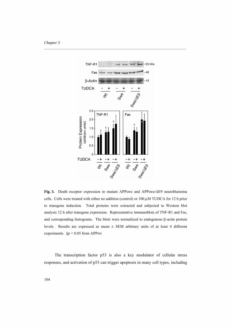
Chapter 3 _________________________________________________________________________
Fig. 3. Death receptor expression in mutant APPswe and APPswe/ΔE9 neuroblastoma
cells. Cells were treated with either no addition (control) or 100 μM TUDCA for 12 h prior
to transgene induction. Total proteins were extracted and subjected to Western blot
analysis 12 h after transgene expression. Representative immunoblots of TNF-R1 and Fas,
and corresponding histograms. The blots were normalized to endogenous β-actin protein
levels. Results are expressed as mean ± SEM arbitrary units of at least 4 different
experiments. §p < 0.05 from APPwt.
The transcription factor p53 is also a key modulator of cellular stress
responses, and activation of p53 can trigger apoptosis in many cell types, including
104

TUDCA modulation of apoptosis in FAD _________________________________________________________________________
neurons. Evidence for a pivotal function of p53 in neuronal death in many
neurodegenerative diseases, including AD, is provided by data from both in vitro
and in vivo models of increased p53 cell levels (Morrison et al. 2003). In fact, our
own studies with neuronal PC12 cells indicated that exogenous Aβ markedly
induced E2F-1 activity, which then resulted in Mdm-2 degradation, p53
stabilization and apoptosis (Ramalho et al. 2004). In the present study we
evaluated the levels of p53 in cells expressing FAD mutations. Interestingly, p53
expression increased ~ 20% in APPswe and 50% in APPswe/ΔE9 cells (p < 0.05)
(Fig. 4). Coincident with p53 up-regulation, anti-apoptotic Bcl-2 protein
expression decreased 25-50% in APPswe and APPswe/ΔE9 cells (p < 0.01) (Fig.
4). Transgene expression also led to increased pro-apoptotic Bax levels in both
APPswe and APPswe/ΔE9 cells (p < 0.05). In contrast, coincubation with TUDCA
reduced p53 protein expression to control levels and modulated Bcl-2 and Bax
protein changes. These results suggested that TUDCA prevents neuronal apoptosis
associated with FAD mutations by interfering with mitochondrial-mediated cell
death, perhaps through modulation of p53 and its downstream targets.
To further characterize the mechanism by which TUDCA modulates p53-
regulated mitochondrial apoptosis, we investigated its specific effects on p53-
induced cell death. Neuroblastoma cells were transfected with plasmids to
overexpress either wild-type or mutant p53. In the absence of transgene
expression, neuroblastoma cells transfected with wild-type p53 showed
morphologic signs of apoptosis after Hoechst staining in 15-20% of cells,
compared with only 3-5% in cells transfected with the mutant plasmid (p < 0.01)
(Fig. 5).
105

Chapter 3 _________________________________________________________________________
Fig. 4. Modulation of p53 and Bcl-
2 family members in mutant APPswe
and APPswe/ΔE9 neuroblastoma
cells. Cells were treated with either
no addition (control) or 100 μM
TUDCA for 12 h before transgene
induction. Total proteins were
extracted and subjected to Western
blot analysis 12 h after transgene
expression. Representative
immunoblots of p53, Bcl-2 and Bax,
and corresponding histograms are
shown. The blots were normalized
to endogenous β-actin protein levels.
Results are expressed as mean ±
SEM arbitrary units of at least 4
different experiments. *p < 0.01 and
§p < 0.05 from APPwt; †p < 0.05
from cells without TUDCA.
106

TUDCA modulation of apoptosis in FAD _________________________________________________________________________
Fig. 5. Modulation of p53 and Bcl-2 family members in mutant APPswe and
APPswe/ΔE9 neuroblastoma cells. Cells were treated with either no addition (control) or
100 μM TUDCA for 12 h before transgene induction. Total proteins were extracted and
subjected to Western blot analysis 12 h after transgene expression. Representative
immunoblots of p53, Bcl-2 and Bax, and corresponding histograms are shown. The blots
were normalized to endogenous β-actin protein levels. Results are expressed as mean ±
SEM arbitrary units of at least 4 different experiments. *p < 0.01 and §p < 0.05 from
APPwt; †p < 0.05 from cells without TUDCA.
Interestingly, TUDCA markedly reduced apoptosis after transfection with
wild-type p53 to almost control levels (p < 0.05). In addition, we have recently
shown that TUDCA can activate a PI3K survival pathway in neurons (Solá et al.
2003a). Wortmannin, a specific inhibitor of the PI3K pathway, blocked
serine/threonine protein kinase Akt activation, reducing the protective effect of
TUDCA against Aβ-induced cell death in neurons. Wortmannin alone was not
significantly cytototoxic in APPwt cells (data not shown). Notably, the PI3K
107

Chapter 3 _________________________________________________________________________
inhibitor reduced TUDCA protection in APPwt cells transfected with wild-type
p53 by almost 100% (p < 0.01). These results suggest that TUDCA interferes with
the activation of p53-related apoptotic events in a PI3K-dependent manner.
Discussion
The role of Aβ in the pathogenesis and progression of AD-associated
neurodegeneration has not been firmly established. Aβ-induced toxicity is a
multifactorial process that is thought to involve the generation of reactive oxygen
species, alteration of intracellular calcium homeostasis, mitochondrial perturbation,
and caspase activation (Selkoe 2001). Recent studies corroborated the
involvement of apoptosis by showing that Aβ alters expression of the Bcl-2 family
of apoptosis-related genes (Yao et al. 2005), and that survival signaling pathways
are required for protection of Aβ-mediated neuronal apoptosis (Watson and Fan
2005). In addition, we have previously reported that TUDCA prevents Aβ-induced
apoptosis by inhibiting the mitochondrial pathway of cell death and regulating cell
cycle-related proteins (Ramalho et al. 2004; Solá et al. 2003a). The present study
shows that TUDCA inhibits p53-induced toxicity in cells expressing FAD
mutations. In stably transfected neuroblastoma cells, the endogenous non-toxic
bile acid modulates p53 up-regulation, expression of Bcl-2 family members, and
caspase activation. These results indicate that attenuation of p53-mediated
apoptosis may underlie the protective effects of TUDCA, possibly via a PI3K-
dependent manner.
A major hallmark of AD is the progressive neuronal loss associated with
reduced memory function. Results from histopathological studies using post
mortem brain tissue of AD patients, as well as a variety of in vitro studies suggest
108

TUDCA modulation of apoptosis in FAD _________________________________________________________________________
that Aβ induces apoptosis and that cell death plays a relevant role in AD
pathogenesis (Loo et al. 2003; Su et al. 1994). In the present study, apoptosis
increased in mutant APPswe and APPswe/ΔE9 overexpressing cells. Moreover,
TUDCA inhibited apoptosis, confirming the importance of this bile acid in the
modulation of FAD mutation-induced cell death.
Apoptosis induction is an essential function of the transcription factor p53.
This protein is highly expressed in cells after accumulation of Aβ and in the brains
of AD patients. Furthermore, p53 inhibitors can prevent neuronal cell death
induced by Aβ (Culmsee et al. 2001). Our results indicate that FAD mutations,
particularly the expression of the APPswe/ΔE9 transgene, resulted in p53 up-
regulation. Moreover, overexpression of p53 in neuroblastoma cells induced
apoptosis, suggesting that it may have an important role in FAD mutation-induced
cell death. Interestingly, TUDCA prevented p53-induced apoptosis suggesting a
specific modulation of p53 expression and/or pathways in which p53 is involved.
Moreover, co-incubations with wortmannin resulted in inhibition of TUDCA
protection, suggesting an involvement of the PI3K pathway.
Studies in three different neuronal cell types showed that Aβ-induced cell
death requires the activation of caspase-2 (Troy et al. 2000). Caspase-2, an
initiator caspase that triggers apoptosis via activation of the mitochondrial
pathway, can be a target of the c-Jun N-terminal kinases signaling pathway, which
is known to be involved in Aβ-induced citotoxicity (Troy et al. 2000). In addition,
p53 is also known to activate caspase-2, via the PIDDosome (Tinel and Tschopp
2004). Our results showed that caspase-2 activity was increased in APPswe and
APPswe/∆E9 cells, and this was coincident with p53 up-regulation. Curiously,
TUDCA prevented caspase-2 activation; it remains to be determined as to whether
the effect was direct or indirect.
p53 has additional recognized apoptotic targets, including death receptors
located at the plasma membrane, such as Fas (Kannan et al. 2001). Fas and TNF-
109

Chapter 3 _________________________________________________________________________
R1 expression was increased in both APPswe and APPswe/ΔE9, compared with
APPwt cells. Moreover, caspase-8 was readily activated. TUDCA, however, did
not prevent these modifications, suggesting that the bile acid does not modulate
FAD mutation-induced apoptosis by the extrinsic pathway. Bcl-2 family members
represent other well-known targets of p53 (Miyashita et al. 1994). In addition, Aβ
has been shown to either up-regulate pro-apoptotic Bax expression or require Bax
to mediate neurotoxicity (Paradis et al. 1996; Selznick et al. 2000). Our results
demonstrated that expression of FAD mutations, Aβ production, and subsequent
p53 stabilization, resulted in up-regulation of pro-apoptotic Bax and decreased
expression of anti-apoptotic Bcl-2. Interestingly, TUDCA significantly tempered
the observed changes in Bcl-2 and Bax protein levels, confirming our previous
results in primary neurons (Solá et al. 2003a) and in PC12 neuronal cells (Ramalho
et al. 2004).
Effector caspase-3 was only slightly activated in cells expressing FAD
mutations. The precise role of caspase-3 in AD is not fully understood. There are
several evidences that caspase-3 might be involved in the neuronal death of
neurodegenerative diseases, including AD (Shimohama et al. 1999; Mashiah et al.
1998). Other results, however, suggest that caspase-3 activation is only slightly
up-regulated in AD patients, and therefore can not justify the massive loss of
neurons in this pathology. A study in human neurons showed that microinjection
with recombinant active caspase-3 did not induce cell death (Zhang et al. 2002). In
contrast, caspase-6 induced a protracted TUNEL-positive cell death. Another
study failed to detect the active fragment of caspase-3 in post mortem tissue from
AD patients but could confirm the presence of caspase-6 active protein (LeBlanc et
al. 1999). In contrast with caspase-3, caspase-6 was significantly up-regulated in
APPswe and APPswe/∆E9 cells, confirming the importance of caspase-6 in AD.
Caspase-6 can cleave the nuclear envelope protein lamin A, several transcription
factors, poly (ADP-ribose) polymerase, and APP generating potentially
110

TUDCA modulation of apoptosis in FAD _________________________________________________________________________
amyloidogenic fragments (Galande et al. 2001; Fernandes-Alnemri et al. 1995).
Nevertheless, the precise role of caspase-6 in the pathogenesis of AD remains to be
determined. In addition, although caspase-3 has been implicated, it is not clearly
known how caspase-6 is activated. Curiously, a recent study identified caspase-6
as a downstream target of p53 in the apoptotic process (MacLachlan and El-Deiry
2002). This may explain the marginal activation of caspase-3 but a robust
activation of caspase-6 in APPswe and APPswe/∆E9 cells, which in turn was
prevented by TUDCA.
Collectively, our studies further expand the neuroprotective effects of
TUDCA. The results demonstrate that TUDCA modulates p53 stabilization and
Bcl-2 family expression, reduces caspase-2 and -6 activation, and inhibits nuclear
fragmentation. Thus, modulation of apoptosis by TUDCA in APPswe and
APPswe/∆E9 cells appears to result from its specific effects at preventing p53
activation. Finally, identification and validation of more cellular targets are likely
to provide new therapeutic interventions for modulation of neuronal
apoptotic.death in Alzheimer’s disease.
Acknowledgments
We are grateful to Dr. Gopal Thinakaran, University of Chicago; Chicago, IL
for providing the neuroblastoma cells (N2a and cells expressing APPwt, APPswe
and APPswe/∆E9). We also thank Dr. William G. Kaelin Jr., Harvard University,
Boston, MA for the generous gift of pCMV overexpression plasmids. Supported
by grant L-V-595/2004 from Fundação Luso-Americana, Lisbon, Portugal (to
C.M.P.R.). R.M.R, R.E.C and S.S were recipients of Ph.D. fellowships
(SFRH/BD/12641/2003, SFRH/BD/12655/2003 and SFRH/BD/4823/2001,
respectively) from Fundação para a Ciência e a Tecnologia.
111

Chapter 3 _________________________________________________________________________
References Behl C., Davis J.B., Klier F. G. and Schubert D. (1994) Amyloid beta peptide induces
necrosis rather than apoptosis. Brain Res 645, 253-264. Blasko I., Wagner M., Whitaker N., Grubeck-Loebenstein B. and Jansen-Durr P. (2000)
The amyloid beta peptide abeta (25-35) induces apoptosis independent of p53. FEBS Lett 470, 221-225.
Busser J., Geldmacher D. S. and Herrup K. (1998) Ectopic cell cycle proteins predict the sites of neuronal cell death in Alzheimer's disease brain. J Neurosci 18, 2801-2807.
Culmsee C., Zhu X., Yu Q. S., Chan S. L., Camandola S., Guo Z., Greig N. H. and Mattson M. P. (2001) A synthetic inhibitor of p53 protects neurons against death induced by ischemic and excitotoxic insults, and amyloid beta-peptide. J Neurochem 77, 220-228.
Duan W. M., Rodrigues C. M. P., Zhao L. R., Steer C. J. and Low W. C. (2002) Tauroursodeoxycholic acid improves the survival and function of nigral transplants in a rat model of Parkinson's disease. Cell Transplant 11, 195-205.
Ethell D. W. and Buhler L. A. (2003) Fas ligand-mediated apoptosis in degenerative disorders of the brain. J Clin Immunol 23, 363-370.
Fernandes-Alnemri T., Litwack G. and Alnemri E. S. (1995) Mch2, a new member of the apoptotic Ced-3/Ice cysteine protease gene family. Cancer Res 55, 2737-2742.
Fortin A., Cregan S. P., MacLaurin J. G. et al. (2001) APAF1 is a key transcriptional target for p53 in the regulation of neuronal cell death. J Cell Biol 155, 207-216.
Galande S., Dickinson L. A., Mian I. S., Sikorska M. and Kohwi-Shigematsu T. (2001) SATB1 cleavage by caspase 6 disrupts PDZ domain-mediated dimerization, causing detachment from chromatin early in T-cell apoptosis. Mol Cell Biol 21, 5591-5604.
Kang J., Lemaire H. G.,Unterbeck A., Salbaum J. M., Masters C. L., Grzeschik K. H., Multhaup G., Beyreuther K. and Müller-Hill B. (1987) The precursor of Alzheimer's disease amyloid A4 protein resembles a cell-surface receptor. Nature 325, 733-736.
Kannan K., Amariglio N., Rechavi G., Jakob-Hirsch J., Kela I., Kaminski N., Getz G., Domany E. and Givol D. (2001) DNA microarrays identification of primary and secondary target genes regulated by p53.Oncogene 20, 2225-2234.
Keene C. D., Rodrigues C. M. P., Eich T., Chhabra M. S., Steer C. J. and Low W. C. (2002) Tauroursodeoxycholic acid, a bile acid, is neuroprotective in a transgenic animal model of Huntington's disease. Proc Natl Acad Sci U S A 99,10671-10676.
Kurita-Ochiai T., Ochiai K. and Fukushima K. (2001) Butyric acid-induced T-cell apoptosis is mediated by caspase-8 and -9 activation in a Fas-independent manner. Clin Diagn Lab Immunol 8, 325-332.
LeBlanc A., Liu H., Goodyer C., Bergeron C. and Hammond J. (1999) Caspase-6 role in apoptosis of human neurons, amyloidogenesis, and Alzheimer's disease. J Biol Chem 274, 23426-23436.
Li R., Yang L., Lindholm K., Konishi Y., Yue X., Hampel H., Zhang D. and Shen Y. (2004) Tumor necrosis factor death receptor signaling cascade is required for amyloid-beta protein-induced neuron death. J Neurosci 24, 1760-1771.
112

TUDCA modulation of apoptosis in FAD _________________________________________________________________________
Loo D. T., Copani A., Pike C. J., Whittemore E. R., Walencewicz A. J. and Cotman C. W. (1993) Apoptosis is induced by beta-amyloid in cultured central nervous system neurons. Proc Natl Acad Sci U S A 90, 7951-7955.
Luo Y., Smith J. V., Paramasivam V. et al. (2002) Inhibition of amyloid-beta aggregation and caspase-3 activation by the Ginkgo biloba extract EGb761. Proc Natl Acad Sci U S A 99, 12197-12202.
MacLachlan T. K. and El-Deiry W. S. (2002) Apoptotic threshold is lowered by 53 transactivation of caspase-6. Proc Natl Acad Sci U S A 99, 9492-9497.
Masliah E., Mallory M., Alford M., Tanaka S. and Hansen L. A. (1998) Caspase dependent DNA fragmentation might be associated with excitotoxicity in Alzheimer disease. J Neuropathol Exp Neurol 57, 1041-1052.
Masters C. L., Multhaup G., Simms G., Pottgiesser J., Martins R. N. and Beyreuther K. (1985) Neuronal origin of cerebral amyloid: neurofibrillary tangles of Alzheimer’s disease contain the same protein as the amyloid of plaque cores and blood vessels. EMBO J 4, 2757-2763.
McShea A., Harris P. L., Webster K. R., Wahl A. F. and Smith M. A. (1997) Abnormal expression of cell cycle regulators P16 and CDK4 in Alzheimer’s disease. Am J Pathol 150, 1933-1939.
Miyashita T., Krajewski S., Krajewska M., Wang H. G., Lin H. K., Liebermann D. A., Hoffman B. and Reed J. C. (1994) Tumor suppressor p53 is a regulator of bcl-2 and bax gene expression in vitro and in vivo. Oncogene 9, 1799-17805.
Morrison R. S., Kinoshita Y., Johnson M. D., Guo W. and Garden G. A. (2003) p53-dependent cell death signaling in neurons. Neurochem Res 28, 15-27.
Mullan M., Crawford F., Axelman K., Houlden H., Lillius L., Winblad B. and Lannfelt L. (1992) A pathogenic mutation for probable Alzheimer’s disease in the APP gene at the N-terminus of β-amyloid. Nature Genet 1, 345-347.
Ohyagi Y., Asahara H., Chui D. H. et al. (2005) Intracellular Aβ42 activates p53 promoter: a pathway to neurodegeneration in Alzheimer’s disease. FASEB J 19, 255-257.
Paradis E., Douillard H., Koutroumanis M., Goodyer C. and LeBlanc A. (1996) Amyloid beta peptide of Alzheimer's disease downregulates Bcl-2 and upregulates bax expression in human neurons. J Neurosci 16, 7533-7539.
Qin X. Q., Chittenden T., Livingston D. M. and Kaelin W. G. (1992) Identification of a growth suppression domain within the retinoblastoma gene product. Genes Dev 6, 953-964.
Ramalho R. M., Ribeiro P. S., Solá S., Castro R. E, Steer C. J. and Rodrigues C. M. P. (2004) Inhibition of the E2F-1/p53/Bax pathway by tauroursodeoxycholic acid in amyloid β-peptide-induced apoptosis in PC12 cells. J Neurochem 90, 567-575.
Rodrigues C. M. P., Fan G., Ma X., Kren B. T. and Steer C. J. (1998) A novel role for ursodeoxycholic acid in inhibiting apoptosis by modulating mitochondrial membrane perturbation. J Clin Invest 101, 2790-2799.
Rodrigues C. M. P., Stieers C. L., Keene C. D., Ma X, Kren B. T., Low W. C. and Steer C. J. (2000) Tauroursodeoxycholic acid partially prevents apoptosis induced by 3-nitropropionic acid: evidence for a mitochondrial pathway independent for the permeability transition. J Neurochem 75, 2368-2379.
113

Chapter 3 _________________________________________________________________________
Rodrigues C. M. P., Spellman S. R., Solá S., Grande A. W., Linehan-Stieers C., Low W. C. and Steer C. J. (2002) Neuroprotection by a bile acid in an acute stroke model in the rat. J Cereb Blood Flow Metab 22: 463-471.
Rodrigues C. M. P., Solá S., Sharpe J. C., Moura J. J. and Steer C. J. (2003a) Tauroursodeoxycholic acid prevents Bax-induced membrane perturbation and cytochrome C release in isolated mitochondria. Biochemistry 42, 3070-3080.
Rodrigues C. M. P., Solá S., Nan Z., Castro R. E., Ribeiro P. S., Low W. C. and Steer C. J. (2003b) Tauroursodeoxycholic acid reduces apoptosis and protects against neurological injury after acute hemorrhagic stroke in rats. Proc Natl Acad Sci U S A 100, 6087-6092.
Roth K. A. (2001) Caspases, apoptosis, and Alzheimer disease: causation, correlation, and confusion. J Neuropathol Exp Neurol 60, 829-838.
Schellenberg G. D., Bird T. D., Wijsman E. M. et al. (1992) Genetic linkage evidence for a familial Alzheimer’s disease locus in chromosome 14. Science 258, 668-671.
Selkoe D. J. (1990) Deciphering Alzheimer’s: the amylod precursor protein yelds new clues. Science 248, 1058-1060.
Selkoe D. J. (1997) Alzheimer’s disease: genotypes, phenotypes, and treatments. Science 275, 630-631.
Selkoe D. J. (2001) Alzheimer's disease: genes, proteins, and therapy. Physiol Rev 81, 741-766.
Selznick L. A., Zheng T. S., Flavell R. A., Rakic P. and Roth K. A. (2000) Amyloid beta-induced neuronal death is bax-dependent but caspase-independent. J Neuropathol Exp Neurol 59, 271-279.
Shimohama S., Tanino H. and Fujimoto S. (1999) Changes in caspase expression in Alzheimer's disease: comparison with development and aging. Biochem Biophys Res Commun 256, 381-384.
Solá S., Castro R. E., Laires P. A., Steer C. J. and Rodrigues C. M. P. (2003a) Tauroursodeoxycholic acid prevents amyloid β-peptide-induced neuronal death via a phosphatidylinositol 3-kinase-dependent signaling pathway. Mol Med 9, 226-234.
Solá S., Castro R. E., Kren B. T., Steer C. J. and Rodrigues C. M. P. (2003b) Ursodeoxycholic acid modulates E2F-1 and p53 expression through a caspase-independent mechanism in TGF-β1-induced apoptosis in rat hepatocytes. J Biol Chem 278, 48831-48838.
Su J. H., Anderson A. J., Cummings B. J. and Cotman C. W. (1994) Immunohistochemical evidence for apoptosis in Alzheimer's disease. Neuroreport 5, 2529-2533.
Thinakaran G., Borchelt D. R., Lee M. K. et al. (1996) Endoproteolysis of presenilin 1 and accumulation of processed derivatives in vivo. Neuron 17, 181-190.
Tinel A. and Tschopp J. (2004) The PIDDosome, a protein complex implicated in activation of caspase-2 in response to genotoxic stress. Science 304, 843-846.
Troy C. M., Rabacchi S. A., Friedman W. J., Frappier T. F., Brown K. and Shelanski M. L. (2000) Caspase-2 mediates neuronal cell death induced by beta-amyloid. J Neurosci 20, 1386-1392.
Ved R., Saha S., Westlund B. et al. (2005) Similar patterns of mitochondrial vulnerability and rescue induced by genetic modification of alpha-synuclein, parkin and DJ-1 in C. elegans. J Biol Chem 280, 42655-42668.
114
TUDCA modulation of apoptosis in FAD _________________________________________________________________________
Vincent I., Jicha G., Rosado M. and Dickson D. W. (1997) Aberrant expression of mitotic cdc2/cyclin B1 kinase in degenerating neurons of Alzheimer's disease brain. J Neurosci 17, 3588-98.
Watson K. and Fan G. H. (2005) Macrophage inflammatory protein 2 inhibits beta-amyloid peptide (1-42)-mediated hippocampal neuronal apoptosis through activation of mitogen-activated protein kinase and phosphatidylinositol 3-kinase signaling pathways. Mol Pharmacol 67, 757-765.
Xu H., Sweeney D., Wang R., Thinakaran G., Lo A. C., Sisodia S. S., Greengard P. and Gandy S. (1997) Generation of Alzheimer beta-amyloid protein in the trans-Golgi network in the apparent absence of vesicle formation. Proc Natl Acad Sci U S A 94, 3748-3752.
Xu J., Chen S., Ku G., Ahmed S. H., Xu J., Chen H. and Hsu C. Y. (2001) Amyloid beta peptide-induced cerebral endothelial cell death involves mitochondrial dysfunction and caspase activation. J Cereb Blood Flow Metab 21, 702-710.
Yao M., Nguyen T. V. and Pike C. J. (2005) Beta-amyloid-induced neuronal apoptosis involves c-Jun N-terminal kinase-dependent downregulation of Bcl-w. J Neurosci 25, 1149-1158.
Zhang Y., McLaughlin R., Goodyer C. and LeBlanc A. (2002) Selective cytotoxicity of intracellular amyloid beta peptide1-42 through p53 and Bax in cultured primary human neurons. J Cell Biol 156, 519-529.
115


4
Apoptosis in transgenic mice expressing the P301L
mutated form of human tau
Rita M. Ramalho1, Ricardo S. Viana1, Rui E. Castro1, Clifford J. Steer2, Walter C.
Low3, and Cecília M. P. Rodrigues1
1Centro de Patogénese Molecular, Faculty of Pharmacy, University of Lisbon,
Portugal; and Departments of 2Medicine, and Genetics, Cell Biology, and
Development, and 3Neurosurgery, University of Minnesota Medical School,
Minneapolis, U.S.A.
Submitted 2007

5

Apoptosis in transgenic model of tauopathy _________________________________________________________________________
Abstract
The rTg4510 mouse is a tauopathy model, characterized by massive
neurodegeneration in Alzheimer’s disease (AD)-relevant cortical and limbic
structures, deficits in spatial reference memory, and progression of neurofibrillary
tangles. In this study, we examined the role of apoptosis in neuronal loss and
associated tau pathology. The results showed that DNA fragmentation and
caspase-3 activation are common in the hippocampus and frontal cortex of young
rTg4510 mice. These changes were associated with cleavage of tau into smaller
intermediate fragments, which persist with age. Interestingly, active caspase-3 was
often co-localized with cleaved tau. In vitro, fibrillar Aβ1-42 resulted in nuclear
fragmentation, caspase activation, and caspase-3-induced cleavage of tau.
Notably, incubation with the antiapoptotic molecule tauroursodeoxycholic acid
abrogated apoptosis-mediated cleavage of tau in rat cortical neurons. In
conclusion, caspase-3-cleaved intermediate tau species precede cell loss in
rTg54510 brains and Aβ-exposed cultured neurons. These results underscore the
potential role of apoptosis in neurodegeneration.
119

Chapter 4 _________________________________________________________________________
Introduction
Alzheimer’s disease (AD) is a progressive neurodegenerative disease
characterized by accelerated neuronal death leading to dementia. Hallmark
pathologic features include extracellular plaques of amyloid β (Aβ) and
intracellular aggregations of tau protein (Selkoe 2001). Tau is a microtubule-
associated protein, abundant in the central nervous system, and predominantly
expressed in axons (Binder et al. 1985). It plays fundamental roles in both
stabilizing neuronal microtubules, and establishing cellular polarity and
intracellular transport (Drewes et al. 1998). Under physiological conditions, tau is
a highly soluble, hydrophobic and unfolded protein, tightly regulated by
phosphorylation at specific serine and threonine residues (Johnson and Stoothoff
2004). In AD and other tauopathies, tau looses its capacity to bind microtubules,
migrates to the cell body, and aggregates into neurofibrillary tangles (NFT)
(Mandelkow and Mandelkow 1998). Post-translational conformational changes of
tau, such as abnormal hyperphosphorylation and proteolysis increase its ability to
aggregate (Alonso et al. 2001, Gamblin et al. 2003a). Curiously, NFT accumulate
with well defined spatial and temporal progression (Santacruz et al. 2005).
The role of apoptosis in AD and other tauopathies is still controversial.
Indeed, synaptic loss and electrophysiological abnormalities typically precede cell
loss. Nevertheless, apoptosis is increased (Su et al. 1994) and caspase-3 is
activated (Selznick et al. 1999) in the brain of AD patients. Interestingly, tau can
be cleaved by caspase-3 at Asp421 in its C-terminal region, resulting in a N-
terminal product that can be detected in cultured neurons, in AD brains (Rissman et
al. 2004), and in other tauopathies (Newman et al. 2005). Truncated tau plays a
role in nucleation-dependent filament formation of tau and induces neuronal death.
Moreover, caspase-3-cleaved tau is often colocalized with Aβ peptide deposition.
Caspase-3 activation is a well established event in Aβ-induced neuronal apoptosis,
120

Apoptosis in transgenic model of tauopathy _________________________________________________________________________
suggesting a link between amyloid plaques and NFT formation (Gamblin et al.
2003b).
The rTg4510 mouse model expresses human tau containing the P301L
mutation associated with frontotemporal dementia with parkinsonism linked to
chromosome 17 (FTDP-17). It is thought to represent a robust model of tauopathy
(Santacruz et al. 2005, Ramsden et al. 2005, Spires et al. 2006). In fact, transgenic
mice showed massive neurodegeneration in specific cortical and limbic structures,
leading to forebrain atrophy and brain weight loss. In addition, the progression of
NFT pathology and neuronal loss was also correlated with deficits in spatial
reference memory. Nonetheless, the onset of memory deficits at 2.5-4 months
preceded NFT pathology and neuronal loss at 5.5 months. The suppression of
transgene expression, after initial NFT formation, prevented further loss of neurons
and enabled partial recovery of cognitive functions, but did not inhibit the
progression of NFT. These findings suggest that NFT formation is not directly
responsible for neurodegeneration and memory loss in rTg4510. Instead, toxic
intermediate tau species may trigger further neurodegeneration.
Ursodeoxycholic acid (UDCA) and its taurine-conjugated derivative,
tauroursodeoxycholic acid (TUDCA), are endogenous bile acids that increase the
apoptotic threshold in several cell types (Rodrigues et al. 2000, Rodrigues et al.
1998). We have previously shown that TUDCA stabilizes mitochondrial function
and prevents Aβ-induced apoptosis in primary rat neurons and in neuronal cell
lines (Solá et al. 2003, Ramalho et al. 2006, Ramalho et al. 2004). Furthermore,
TUDCA was neuroprotective in a transgenic mouse model of Huntington’s disease
(Keene et al. 2002), reduced lesion volumes in rat models of ischemic and
hemorrhagic stroke (Rodrigues et al. 2002, Rodrigues et al. 2003), improved the
survival and function of nigral transplants in a rat model of Parkinson’s disease
(Duan et al. 2002) and partially rescued a Parkinson’s disease model of
Caernohabditis elegans from mitochondrial dysfunction (Ved et al. 2005).
121

Chapter 4 _________________________________________________________________________
Using rTg4510 mice, we investigated the role of apoptosis in neuronal loss and
tau associated pathology. Our results suggest that apoptosis is an early event
associated with tau cleavage in the hippocampus and the frontal cortex. Cleaved
tau, in turn, appears to represent a toxic form of tau. In cultured cortical neurons,
apoptosis and caspase-3 cleavage of tau induced by fibrillar Aβ1-42 were
significantly inhibited by TUDCA. Thus, caspase-3-cleaved intermediate tau
species are responsible, in part, for cell loss in rTg4510 brains and Aβ-exposed
cultured neurons. These results underscore the potential role of apoptosis in
neurodegeneration.
Material and methods
Generation of transgenic mice
The rTg4510 is a recently developed mouse model of tauopathy in which
expression of human tau, containing the frontotempotal dementia-associated
P301L mutation can be suppressed with doxycycline administration (Santacruz et
al. 2005). Briefly, the method for generating rTg4510 mice utilized a system of
responder and activator transgenes. Mice expressing the activator transgene,
consisting of a four-repeat human tau with the P301L mutation placed downstream
of a tetracycline operon responsive element were successively backcrossed at least
five times onto a 129S6 background strain. Responder mice, consisting of a tet-off
open reading frame placed downstream of Ca2+-calmodulin kinase II promoter
elements were maintained in the FVB/N strain. From this, double transgenic mice
were generated in which expression of the P301L tau expression was restricted to
the forebrain structures. Mice were screened by PCR using the primer pairs 5’-
GAT TAA CAG CGC ATT AGA GCT G-3’ and 5´-GCA TAT GAT CAA TTC
AAG GCC GAT AAG-3’ for activator transgenes, and 5’-TGA ACC AGG ATG
GCT GAG CC-3’ and 5’-TTG TCA TCG CTT CCA GTC CCC G-3’ for responder
122

Apoptosis in transgenic model of tauopathy _________________________________________________________________________
transgenes. Tau-expressing mice and littermate control mice (lacking either the tau
responder transgene or the activator transgene) between 2.5 and 8.5 months were
used. Animals were sacrificed with an overdose of ketamine xylazine cocktail via
an intraperitoneal cavity injection. The chest cavity was then opened by cutting
through the diaphragm and rib cage laterally. The right atrium was cut to drain
blood; and the left ventricle punctured for cannula placement. PBS was flushed
through the circulatory system using a pressure of 60 mmHg for 3-4 min or until
the right atrium was cleared. The brain was removed and rapidly frozen in dry ice.
Cryostat brain sections of 10 µm were prepared for analysis of transgenic and
control mice. In addition, different areas of the brain were isolated, including the
olfactory bulb, frontal cortex, sensorimotor cortex, medial septal nucleus,
hippocampus, and entorhinal cortex.
All animals were housed and tested accordingly to standards established by the
American Association for the Accreditation of Laboratory Animal Care and
Institutional Animal Care and Use Committee guidelines, with every effort made to
minimize the number of animals used.
Isolation and culture of rat cortical neurons
Primary cultures of rat cortical neurons were prepared from 17- to 18-day-old
fetuses of Wistar rats as previously described (Brewer et al. 1993) with minor
modifications. In short, pregnant rats were ether-anesthetized and decapitated.
The fetuses were collected in Hank’s balanced salt solution (HBSS-1; Invitrogen,
Grand Island, NY, USA) and rapidly decapitated. After removal of meninges and
white matter, the brain cortex was collected in Hank’s balanced salt solution
without Ca2+ and Mg2+ (HBSS-2). The cortex was then mechanically fragmented,
transferred to a 0.025% trypsin in HBSS-2 solution, and incubated for 15 min at
37ºC. Following trypsinization, cells were washed twice in HBSS-2 containing
10% fetal calf serum (FBS) and re-suspended in Neurobasal medium (Invitrogen),
123

Chapter 4 _________________________________________________________________________
supplemented with 0.5 mM L-glutamine, 25 µM L-glutamic acid, 2% B-27
supplement (Invitrogen), and 12 mg/mL gentamicin. Neurons were then plated on
tissue culture plates, precoated with poly-D-lysine at 1 x 10⁶ cells/mL, and
maintained at 37ºC in a humidified atmosphere of 5% CO2. All experiments were
performed on cells cultured for 4 days in fresh medium without glutamic acid and
B-27 supplement. Cells were characterized by phase contrast microscopy and
indirect immunocytochemistry for neurofilaments and glial fibrillary acidic
protein. Neuronal cultures were >95% pure. After 4 days in culture, isolated rat
neurons were incubated with 20 µM Aβ1-42 (Bachem AG, Budendorf, Switzerland)
that had been induced to form fibrils by preincubation in culture medium. In short,
0.45 mg of Aβ1-42 peptide was dissolved in 20 µl of DMSO and diluted to a 100-
µM stock solution in medium, which was then incubated with gentle shaking at
room temperature for 4 days. Fibrillar Aβ1-42 was then diluted to 20 µM and
applied to neuron cultures; 0.2% DMSO was added to control cultures. Cortical
neurons were incubated with fibrillar Aβ1-42 for 24 h, with or without 100 μM
TUDCA (Sigma Chemical, St. Louis, MO, USA), or no addition. In co-incubation
experiments, TUDCA was added to neurons 12 h prior to incubation with Aβ1-42.
Evaluation of apoptosis and caspase-3 activity
DNA fragmentation in brain sections of both transgenic and control mice was
detected using an ApopTag® peroxidase in situ apoptosis detection kit
(Serologicals Corp., Norcross, GA) for transferase mediated dUTP-digoxigenin
nick-end labelling (TUNEL) staining. In brief, tissue sections were fixed in 4%
paraformaldehyde in PBS, pH 7.4, for 10 min at room temperature, post-fixed in
precooled ethanol:acetic acid (2:1, v/v) for 5 min at -20ºC, and treated with 3%
hydrogen peroxide to quench endogenous peroxidase activity. After adding the
equilibration buffer, sections were treated with terminal
deoxynucleotidyltransferase (TdT) and digoxigenin-dNTPs for 60 min at 37ºC.
124
Apoptosis in transgenic model of tauopathy _________________________________________________________________________
Specimens were then treated with anti-digoxigenin-peroxidase for 30 min at 37ºC,
colorized with 3,3’-diaminobenzidine (DAB) substrate, and counterstained with
0.5% methyl green. Finally, slides were rinsed, dehydrated, and mounted. A
negative control was prepared by omitting the TdT enzyme to control for non-
specific incorporation of nucleotides or binding of enzyme-conjugate. The
specimens were examined using a bright-field microscope (Zeiss Axioskop; Carl
Zeiss GmbH, Jena, Germany) and the data expressed as the number of TUNEL-
positive cells/high-power field (x400).
Cell viability of cortical neurons was assessed using trypan blue dye exclusion
and confirmed by lactate dehydrogenase viability assays (Sigma-Aldrich). In
addition, Hoechst labeling of cells was used to detect apoptotic nuclei. Briefly, the
medium was gently removed to prevent detachment of cells. Attached neurons
were fixed with 4% paraformaldehyde in phosphate-buffered saline (PBS), pH 7.4,
for 10 min at room temperature, incubated with Hoechst dye 33258 (Sigma-
Aldrich) at 5 μg/ml in PBS for 5 min, washed with PBS and mounted using
PBS:glycerol (3:1, v/v). Fluorescent nuclei were scored blindly and categorized
according to the condensation and staining characteristics of chromatin. Normal
nuclei showed non-condensed chromatin dispersed over the entire nucleus.
Apoptotic nuclei were identified by condensed chromatin, contiguous to the
nuclear membrane, as well as nuclear fragmentation of condensed chromatin.
Three random microscopic fields per sample of ~ 250 nuclei were counted and
mean values expressed as the percentage of apoptotic nuclei.
Finally, caspase activity was determined in cytosolic protein extracts from
brain tissue and cell cultures. Samples were homogenized in isolation buffer,
containing 10 mM Tris-HCl buffer, pH 7.6, 5 mM MgCl2, 1.5 mM KAc, 2 mM
DTT, and protease inhibitor cocktail tablets (CompleteTM; Roche Applied Science,
Mannheim, Germany). General caspase-3-like activity was determined by
enzymatic cleavage of chromophore p-nitroanilide (pNA) from the substrate N-
125

Chapter 4 _________________________________________________________________________
acetyl-Asp-Glu-Val-Asp-pNA (DEVD-pNA; Sigma Chemical Co.). The
proteolytic reaction was carried out in isolation buffer containing 50 µg cytosolic
protein and 50 µM DEVD-pNA. The reaction mixtures were incubated at 37°C for
1 h, and the formation of pNA was measured at 405 nm using a 96-well plate
reader.
Immunoblotting
Steady-state levels of total tau and caspase-3-cleaved tau proteins were determined
by Western blot analysis. Briefly, 50 μg of total protein extracts were separated on
12% SDS-polyacrylamide electrophoresis minigels. Following electrophoretic
transfer onto nitrocellulose membranes, immunoblots were incubated with 15%
H2O2 for 15 min at room temperature. After blocking with 5% nonfat milk
solution, the blots were incubated overnight at 4ºC with primary mouse
monoclonal antibodies reactive to total tau (Zymed Laboratories Inc., San
Francisco, CA, USA) and to caspase-3-cleaved tau (Chemicon, Millipore,
Billerica, MA, USA) and finally with a secondary antibody conjugated with
horseradish peroxidase (Bio-Rad Laboratories, Hercules, CA, USA) for 3 h at
room temperature. The membranes were processed for protein detection using
Super SignalTM substrate (Pierce, Rockford, IL, USA). β-actin was used as a
loading control. Protein concentrations were determined using the Bio-Rad protein
assay kit according to the manufacturer’s specifications.
Immunohistochemistry
Light-level immunohistochemistry was performed in fixed brain sections to detect
caspase-3-cleaved tau. Briefly, slides were soaked in 3% hydrogen peroxide, 10%
methanol for 10 min, washed, and incubated in serum blocking solution (Santa
Cruz Biotechnology, Santa Cruz, CA) with 0.3% Triton X-100 for 1 h. Specimens
were then incubated with primary antibody overnight at 4ºC. After rinsing,
126

Apoptosis in transgenic model of tauopathy _________________________________________________________________________
specimens were incubated with biotinylated secondary antibody and a horseradish
peroxidase-streptavidin complex, for 1 h each. Tissue samples were then colorized
with DAB substrate, counterstained, mounted and visualized in a bright-field
microscope (Zeiss Axioscope).
Immunofluorescence was performed to investigate the co-localization of
caspase-3-cleaved tau and active caspase-3 in brain tissue and cultured cells. Fixed
brain sections were soaked in 0.3% hydrogen peroxide for 5 min, washed and
blocked (0.3% Triton X-100, 10% donkey serum, 1% FBS) for 1 h. In addition,
cortical neurons were washed with PBS, incubated for 30 sec in microtubule
stabilization buffer, and fixed for 10 min in 4% paraformaldehyde at room
temperature. Fixed cultured cells were incubated with 1% nonfat milk in PBS
containing 1% bovine serum albumin and 1.5% Triton X-100 for 1 h at room
temperature. Specimens were then incubated with both monoclonal caspase-3-
cleaved tau and polyclonal caspase-3 (Santa Cruz Biotechnology) antibodies
overnight at 4ºC. After rinsing, specimens were then incubated with either
fluorescently labeled anti-mouse or anti-rabbit (CyTM2 and CyTM5, respectively;
Jackson ImmunoResearch Laboratories, Inc., West Grove, PA, USA) for 2 h at
room temperature. Samples were then mounted and visualized in a MRC1000
confocal microscope (Bio-Rad, Hercules, CA, USA). Finally, the percentage of
caspase-3-cleaved tau and active caspase-3-positive cortical neurons with apoptotic
nuclei was determined by staining with Hoechst 33258.
Densitometry and statistical analysis
The relative intensities of protein bands were analysed using the ImageMaster 1D
Elite v4.00 densitometric analysis program (Amersham Biosciences, Piscataway,
NJ, USA). All data were expressed as mean ± SEM from at least three separate
experiments. Statistical analysis was performed using GraphPad InStat version
127

Chapter 4 _________________________________________________________________________
3.00 for Windows 95 (GraphPad Software, San Diego, CA, USA) for the Student’s
t test. Values of p < 0.05 were considered significant.
Results
TUNEL-positive cells and caspase activity in the frontal cortex and
hippocampus of young rTg4510 animals
AD is a progressive neurodegenerative disease with well-defined spatial and
temporal lesions that correlate with neuronal loss. The entorhinal cortex and
hippocampus are particularly vulnerable (Braak and Braak 1991), while other
cortical and subcortical areas are influenced as the disease progresses. In
frontotemporal dementias, however, the most affected areas are the frontal and
temporal cortex (Mirra and Hyman 2002). Previous studies using rTg4510 mice
showed marked brain atrophy with massive neuronal loss, especially in the
hippocampus (Santacruz et al. 2005). The dental gyrus showed the earliest onset
of neuronal loss at 2.5 months of age (85% of total neuronal loss), followed by
CA1 and CA2/3 regions at 5.5 months (69-82%), and cortex at 8.5 months (52%)
(Spires et al. 2006). However, the role of apoptosis in neurodegeneration of
rTg4510 mice remained unclear.
In this study, levels of apoptosis in brain slices of control and rTg4510 mice at
2.5, 5.5, and 8.5 months were evaluated using the TUNEL assay. At 2.5 months,
transgenic animals showed significant levels of TUNEL-positive cells in specific
regions of the brain, including the frontal cortex (FC) (p < 0.05), and the medial
septal nucleus (MSN) (p < 0.01), subgranular zone (SGZ) (p < 0.01) and
subventricular zone (SVZ) (p < 0.05) of the hippocampus (Fig. 1A, upper
histogram). In addition, rTg4510 animals at 5.5 months showed increased
apoptosis only in the MSN (p < 0.05) and SGZ (p < 0.01) (Fig. 1A, middle
histogram). Finally, the number of apoptotic cells in transgenic animals at 8.5
128

Apoptosis in transgenic model of tauopathy _________________________________________________________________________
months was similar to controls, except in the sensorimotor cortex (SMC) and MSN
(p < 0.05) (Fig. 1A, lower histogram).
Fíg. 1. DNA fragmentation and caspase-3 activity occur early in the frontal cortex and
hippocampus of rTg4510 mice. Brain slices were analyzed from both control (white bars)
and transgenic animals (black bars) at 2.5, 5.5 and 8.5 months. DNA fragmentation was
129

Chapter 4 _________________________________________________________________________
assessed by the TUNEL assay, and the number of positive cells was counted in the
olfactory bulb (OB), frontal cortex (FC), sensorimotor cortex (SMC), medial septal
nucleus, subgranular zone (SGZ), subventricular zone (SVZ), CA1 and entorhinal cortex
(EC). Cytosolic proteins were extracted for caspase-3-like activity assays from OB, FC,
SMC, MSN, hippocampus (H) and EC of both control and transgenic animals at 2.5, 5.5
and 8.5 months. A: TUNEL staining in control FC (a); transgenic FC (b); control H (c);
and transgenic H (d) (top) and number of apoptotic cells (bottom). Apoptotic nuclei were
identified by a condensed nucleus with brown staining. B: DEVD-specific caspase
activity. The results are expressed as mean ± SEM of at least 3 different experiments. *p <
0.01 and §p < 0.05 from control animals. Scale bar: 100 µm.
As a hallmark of apoptosis, caspase-3 activation was investigated in both
control and transgenic animals. Consistent with TUNEL data, caspase-3-like
activity was increased in FC (p < 0.05) and hippocampus (p < 0.01) at 2.5 months
(Fig. 1B, upper histogram). At 5.5 months, despite the increase of TUNEL-
positive cells, caspase-3 activity was not significantly altered in the hippocampus
(Fig. 1B, middle histogram). Finally, no significant changes were detected at 8.5
months (Fig. 1B, lower histogram). Taken together, these findings indicate that
DNA fragmentation and caspase-3 activation are early events in the frontal cortex
and hippocampus of rTg4510, which precede massive neuronal loss (Santacruz et
al. 2005).
Caspase-3 activation is associated with tau cleavage in rTg4510 animals
Tau protein is functionally and structurally altered in AD and other tauopathies. In
fact, posttranslational changes such as abnormal phosphorylation and cleavage
result in altered solubility and conformation, as well as aggregation (Alonso et al.
2001, Gamblin et al. 2003a). Next, we focused our study in the frontal cortex and
hippocampus of rTg4510, which showed increased apoptosis, and investigated
130

Apoptosis in transgenic model of tauopathy _________________________________________________________________________
patterns of tau expression with aging. Using the antibody T14 to detect total
human tau, a fragment of ~ 50 kDa was evident in animals of all ages, although to
a less extent in the hippocampus at 2.5 months (Fig. 2). This fragment corresponds
to soluble and physiologic tau, which remains stable throughout the life of
transgenic animals (Ramsden et al. 2005). A fragment of 64 kDa was only present
in older animals, at 5.5 and 8.5 months, corresponding to the insoluble and
pathological form of tau. Finally, small fragments of ~ 30 and 25 kDa were only
present in the frontal cortex and hippocampus of younger animals. Cleaved
fragments may represent pathological forms of tau and are associated with
increased apoptosis. In fact, cleavage of tau at Asp421 of the C-terminal region by
caspase-3 has been detected both in neurons and AD brains (Rissman et al. 2004).
Fig. 2. Expression analysis of tau shows varying patterns with age of rTg4510 mice.
Total proteins of frontal cortex and hippocampus in both control and transgenic animals at
2.5, 5.5 and 8.5 months were extracted and subjected to Western blot analysis. A
representative immunoblot of total human tau is shown.
To determine if caspase-3 was responsible for cleavage of tau, we performed
immunohistochemistry assays using a specific antibody against Asp421-cleaved
131

Chapter 4 _________________________________________________________________________
tau. At 2.5 months, the number of caspase-3-cleaved tau cells was significantly
higher in FC and SGZ (p < 0.01) (Fig. 3A). The number of positive cells was also
significantly higher in the olfactory bulb (p < 0.01), SMC (p < 0.05), and
entorhinal cortex (p < 0.01) in transgenic animals. Consistently, western blot
analysis showed that caspase-3-cleaved tau was already detected at 2.5 months
(Fig. 3B). The ~ 50 kDa fragment was also evident in older animals, suggesting
that tau persists during tangle maturation and neurodegeneration in older animals
(Rissman et al. 2004, Santacruz et al. 2005, Spires et al. 2006).
Fig. 3. Tau cleavage is an early event modulated by caspase-3 activation in rTg4510 mice.
Brain slices were analyzed from both control (white bars) and transgenic animals (black
bars) at 2.5 months. Caspase-3-cleaved tau cells were identified by immunohistochemistry
and the number of positive cells was counted in the olfactory bulb (OB), frontal cortex
(FC), sensorimotor cortex (SMC), medial septal nucleus, subgranular zone (SGZ),
subventricular zone (SVZ), CA1 and entorhinal cortex (EC). Total proteins of frontal
132
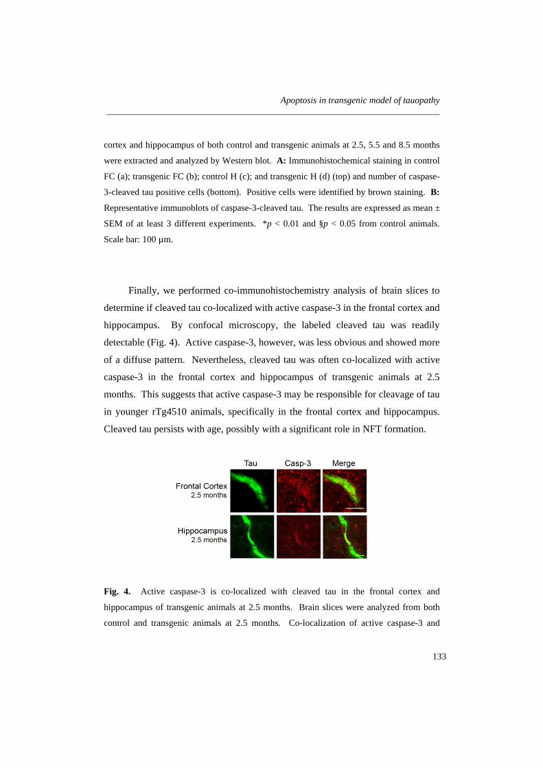
Apoptosis in transgenic model of tauopathy _________________________________________________________________________
cortex and hippocampus of both control and transgenic animals at 2.5, 5.5 and 8.5 months
were extracted and analyzed by Western blot. A: Immunohistochemical staining in control
FC (a); transgenic FC (b); control H (c); and transgenic H (d) (top) and number of caspase-
3-cleaved tau positive cells (bottom). Positive cells were identified by brown staining. B:
Representative immunoblots of caspase-3-cleaved tau. The results are expressed as mean ±
SEM of at least 3 different experiments. *p < 0.01 and §p < 0.05 from control animals.
Scale bar: 100 µm.
Finally, we performed co-immunohistochemistry analysis of brain slices to
determine if cleaved tau co-localized with active caspase-3 in the frontal cortex and
hippocampus. By confocal microscopy, the labeled cleaved tau was readily
detectable (Fig. 4). Active caspase-3, however, was less obvious and showed more
of a diffuse pattern. Nevertheless, cleaved tau was often co-localized with active
caspase-3 in the frontal cortex and hippocampus of transgenic animals at 2.5
months. This suggests that active caspase-3 may be responsible for cleavage of tau
in younger rTg4510 animals, specifically in the frontal cortex and hippocampus.
Cleaved tau persists with age, possibly with a significant role in NFT formation.
Fig. 4. Active caspase-3 is co-localized with cleaved tau in the frontal cortex and
hippocampus of transgenic animals at 2.5 months. Brain slices were analyzed from both
control and transgenic animals at 2.5 months. Co-localization of active caspase-3 and
133

Chapter 4 _________________________________________________________________________
caspase-3-cleaved tau was evaluated in the frontal cortex and hippocampus by fluorescent
immunohistochemistry and confocal microscopy. Scale bar: 20 µm.
Modulation of Αβ-induced toxicity and cleavage of tau in cortical neurons
Amyloid plaques and NFT have been largely categorized as independent
neuropathologic phenomena. In tauopathies, mutations in tau are thought to be
sufficient to induce its aggregation in NFT and the consequent neuronal
dysfunction, without the presence or aggregation of Aβ. However, in AD, there
are no described mutations in tau and studies have suggested that tau is necessary
for Aβ-induced cognitive dysfunction (Gotz et al. 2001). In fact, only a direct or
indirect interaction between Aβ and tau could explain the mechanistic development
of AD. A possible link would be Aβ-induced caspase-3 activation, followed by
cleavage and subsequent aggregation of tau (Gamblin et al. 2003b). Thus, we
examined this correlation in primary rat cortical neurons incubated with fibrillar
Aβ1-42 and investigated the potential modulation by incubation with the
antiapoptotic bile acid, TUDCA. General cell death increased after fibrillar Aβ1-42
incubation as evaluated by trypan blue dye exclusion and lactate dehydrogenase
viability assays (data not shown). Nuclear fragmentation characteristic of
apoptosis increased to ~ 30% (p < 0.01) in cells exposed to fibrillar Aβ1-42 (Fig.
5A). In contrast, TUDCA abrogated nuclear condensation and fragmentation (p <
0.01). Similar results were obtained in caspase-3 activity assays (p < 0.05) (Fig.
5B).
Next, we investigated if Aβ1-42-induced caspase activity was responsible for
cleavage of tau. Immunofluorescence assays showed that nuclear fragmentation
and active caspase-3 were often colocalized with Asp421 cleaved-tau in cortical
neurons undergoing apoptosis (Fig. 6A). Interestingly, cleaved-tau was
abnormally distributed within the neurons and detected in cell bodies. In addition,
134

Apoptosis in transgenic model of tauopathy _________________________________________________________________________
immunoblotting revealed that cleaved-tau was upregulated by almost 10-fold after
incubation with Aβ1-42 (p < 0.05) (Fig. 6B). Notably, TUDCA reduced caspase-3-
cleaved tau to control values (p < 0.05); and this was further confirmed by
immunofluorescence. These results showed that fibrillar Aβ1-42 decreased cell
viability, and induced apoptosis with subsequent cleavage of tau, which may
precede tau aggregation in NFT.
Fig. 5. TUDCA inhibits apoptosis and
caspase-3 activity in primary cortical
neurons incubated with fibrillar Aβ1-42.
Cells were incubated with 20 µM fibrillar
Aβ1-42, or no addition (control), ± 100 µM
TUDCA for 24 h. In coincubation
experiments, TUDCA was added 12 h
prior to incubation with Aβ1-42. Cells
were fixed and stained for microscopy
assessment of apoptosis. Cytosolic
proteins were extracted for caspase-3-like
activity assays. A: Fluorescence
microscopy of Hoechst staining in
controls (a); and in cells exposed to Aβ1-42
(b) (top) and percentage of apoptosis
(bottom). Apoptotic nuclei were
identified by condensed chromatin as well
as nuclear fragmentation. B: DEVD-
specific caspase activity. The results are
expressed as mean ± SEM of at least 3
different experiments. *p < 0.01 and §p <
0.05 from control; ‡p < 0.01 and †p <
0.05 from Aβ. Scale bar: 15 µm.
135

Chapter 4 _________________________________________________________________________
Fig. 6. TUDCA modulates cleavage of tau by caspase-3 in primary cortical neurons
incubated with fibrillar Aβ1-42. Cells were incubated with 20 µM fibrillar Aβ1-42, or no
addition (control), ± 100 µM TUDCA for 24 h. In coincubation experiments, TUDCA was
added 12 h prior to incubation with Aβ1-42. Colocalization of active caspase-3 and cleaved
tau was evaluated by confocal immunofluorescence microscopy. Apoptotic nuclei were
visualized with Hoechst staining. Total proteins were extracted and subjected to Western
blot analysis. A: Confocal microscopy and colocalization of apoptotic nuclei, active
caspase-3, and cleaved tau in cells exposed to Aβ1-42. B: Confocal microscopy and
colocalization of apoptotic nuclei and cleaved tau in cells exposed to Aβ (a) and Aβ +
TUDCA (b) (top); representative immunoblot of caspase-3 cleaved tau in cells after
treatment with Aβ ± TUDCA (bottom). The blot was normalized to endogenous β-actin
protein levels. Scale bar: 20 µm.
136

Apoptosis in transgenic model of tauopathy _________________________________________________________________________
Discussion
The precise molecular mechanisms involved in AD-related neuronal death and
cognitive decline are not entirely understood. Aggregation of tau into insoluble
NFT and formation of extracellular plaques by Aβ are the most characterized
pathological features in AD. Nevertheless, the correlation between these abnormal
entities and their role in neuronal death is still unclear. Implication of apoptosis as
a general mechanism in many neurodegenerative disorders including AD has been
supported by several studies (Fadeel and Orrenius 2005). In addition, we have
previously reported that antiapoptotic treatment with TUDCA prevents Aβ-induced
degeneration in neuronal cells (Solá et al. 2003, Ramalho et al. 2006, Ramalho et
al. 2004). In the present study, we provide evidence that apoptosis is an early
event in the frontal cortex and hippocampus of rTg4510 mice. Moreover, caspase-
3 activation is associated with cleavage of tau in its C-terminal region, which
precedes NFT formation. Cleaved tau represents potentially intermediate
neurotoxic species that are correlated with initial neuronal dysfunction. Finally,
antiapoptotic TUDCA prevents cleavage of tau from Aβ-induced apoptosis in
cultured neurons, underscoring the role of modulation of apoptosis in tau cleavage.
Previous studies have shown that the rTg4510 mouse is a remarkable model of
tauopathy. Tau expression induces age-dependent memory impairment, NFT
formation and memory loss (Ramsden et al. 2005, Santacruz et al. 2005, Spires et
al. 2006). Transgenic aged mice undergo massive neuronal loss, but the
mechanisms by which neurons die are still not clear. In addition, although
expressed in all forebrain structures, the transgene tau with the P301L mutation
affects only specific brain regions. Neuronal loss is > 80% in hippocampal area
CA1 and dentate gyrus, and ~ 50% in cortex by 8.5 months (Spires et al. 2006).
These results are consistent with the regional specificity observed in tauopathies,
including AD (Braak and Braak 1991).
137

Chapter 4 _________________________________________________________________________
Here, we investigated whether apoptosis is primarily involved in neuronal loss
of rTg4510 mice. DNA fragmentation and caspase activation were evident in
transgenic animals at 2.5 months. In fact, TUNEL-positive cells were easily
detected in the frontal cortex, subgranular zone of the dentate gyrus, and
subventricular zone. Interestingly, the subgranular zone gives rise to neural stem
cells that ultimately differentiate into granule cell neurons. Therefore, apoptosis
seen in the subgranular zone may account for the loss of granule cells. Levels of
apoptosis were still slightly elevated in the subgranular zone at 5.5 months, but
showed no changes at 8.5 months. Activation of caspase-3 was no longer evident
at 5.5 and 8.5 months. Interestingly, apoptosis was increased in the medial septal
nucleus at all ages, and in the sensorimotor cortex in older animals. We were also
unable to detect significant levels of apoptosis in hippocampal CA1 of rTg4510
mice. Thus, caspase-dependent and -independent cell death is confined to specific
areas, possibly signaling other brain regions.
Evidence of apoptosis in AD has been largely supported by tissue culture
studies (Fadeel and Orrenius 2005). Contradictory results came from observations
of human post-mortem brain tissue, where clear detection of apoptotic cells is
difficult (Migheli et al. 1994). This may be explained by the fact that cell death in
AD occurs over decades, while apoptosis is executed within a few hours. Thus,
synchronous detection of a substantial number of apoptotic cells at any given time
would be very difficult. In addition, there is strong evidence to suggest that
apoptotic mechanisms may play an important role in disease pathogenesis, even in
the absence of overt apoptosis (Dickson 2004). Our results suggest that apoptosis,
rather than being responsible for extensive neuronal loss in older animals, triggers
toxic events that ultimately contribute to cognitive deficits. In fact, activation of
caspase-3 in the frontal cortex and hippocampus resulted in cleavage of tau in
rTg4510 mice at 2.5 months. Neurons with active caspase-3 often showed
Asp421-cleaved tau. Caspase-3-cleavage of tau is thought to induce abnormal
138

Apoptosis in transgenic model of tauopathy _________________________________________________________________________
conformations by removing part of the C-terminal region, which is essential to
inhibit tau aggregation in vitro (Abraha et al. 2000, Berry et al. 2003). Although
early apoptosis may also represent a developmental-related phenomenon (Ramsden
et al. 2005), it is clear that the cleavage of tau is a toxic process with potentially
important neurodegenerative repercussions.
Importantly, the formation of NFT appears to be dissociated from neuronal loss
in rTg4510 mice. It has been shown that, at 4 months of age, ~ 50% of neurons in
the dentate gyrus were lost before any early conformational changes in tau were
detectable (Spires et al. 2006). In addition, after transgene suppression, neuronal
loss was prevented and memory decline reverted, but NFT continued to accumulate
(Spires et al. 2006, Santacruz et al. 2005). Interestingly, rT4510 mice developed
the first signs of cognitive decline at 2.5 months, coincident with cleavage of tau.
Thus, it is possible that deficits in neurons occur in the early stages of disease,
resulting from aberrant forms of tau, prior to massive neuronal loss and NFT
formation. In this scenario, apoptosis and subsequent cleavage of tau are strong
candidates to trigger the primary deleterious effects on neuronal function.
The identification of two forms of tau multimers with 140 and 170 kDa have
recently been described in rTg4510 mice (Berger et al. 2007). The molecular
weights suggest an oligomeric aggregate of the soluble and insoluble forms of tau,
respectively. The aggregates accumulate early in the pathogenesis and appear to
be associated with the development of functional deficits. Thus, it is tempting to
speculate that cleavage of tau occurs prior to formation of the first oligomers,
promoting their aggregation. Further, the fact that cleaved tau was found in older
animals suggests that although cleavage may occur early in NFT formation, the
resultant fragment persists throughout NFT evolution. It is possible that apoptosis
continues beyond 2.5 months, and that other proteases such as calpains are also
involved in tau cleavage (Park and Ferreira 2005). In this scenario, apoptosis may
139

Chapter 4 _________________________________________________________________________
represent the triggering event for the formation and aggregation of intermediate
species of tau, which ultimately result in massive non-apoptotic neuronal death.
The efficacy of the rTg4510 mouse model relies on the capacity of mutated tau
to induce neuronal modifications. Mutations of tau reduce its ability to interact
with microtubules and are associated with tauopathies (Goedert et al. 1998). These
disorders are distinct from AD because there is no evidence for Aβ, amyloid
precursor protein (APP) or presenilin involvement. In AD, however, Aβ plays an
important role in neuronal toxicity and cell death, and its involvement in tau
pathology cannot be neglected. Indeed, both mutations of APP with Aβ deposition
and intracranial injection of Aβ increased NFT formation in transgenic mice
expressing an FTDP-17-causing mutant (Gotz et al. 2001, Lewis et al. 2001).
Several evidences suggest that the link between Aβ and tau is predominantly, if not
exclusively, unidirectional. In fact, tau has no effect on the onset and progression
of Aβ accumulation (Oddo et al. 2007). Rather, tau accumulation occurs after
amyloid plaques formation, further exacerbating the neurotoxicity induced by Aβ.
In fact, it has been recently shown that overexpression of tau with the P301L
mutation induces mitochondria dysfunction, increasing the production of reactive
oxygen species and the vulnerability to Aβ insults (David et al. 2005). Moreover,
reduction of endogenous tau levels proved to inhibit behavioural deficits in
transgenic expressing human APP, without altering Aβ levels (Roberson et al.
2007).
Recent studies suggest that Aβ accumulation and caspase activation precedes
and induces cleavage of tau and NFT formation (Gamblin et al. 2003b, Rissman et
al. 2004). Our results confirmed that fibrillar Aβ1-42 induces toxicity, nuclear
fragmentation and caspase-3 activation, which was associated with cleavage of tau.
Moreover, cleaved tau was often co-localized with active caspase-3 in neurons
140

Apoptosis in transgenic model of tauopathy _________________________________________________________________________
undergoing nuclear modifications. Thus, apoptotic events and cleavage of tau
occur almost simultaneously in neurons exposed to aggregated Aβ.
TUDCA prevented apoptosis and caspase-3 activation after incubations of
neurons with fibrillar Aβ1-42, thus reducing caspase-3-cleaved tau. This suggests
that TUDCA may inhibit further aggregation of tau in NFT and promote cell
survival. It would be interesting to investigate whether administrations of TUDCA
to newborn or up to 2.5 months rTg4510 mice would prevent cleavage of tau, and
subsequent massive neuronal loss, memory decline and NFT formation.
Collectively, the present study provides evidence that apoptosis may represent
a crucial event in AD. Either by promoting further cell death or activating toxic
effectors, apoptosis interferes with the pathogenesis of the disease. In rTg4510
mice, apoptosis is an early event in the frontal cortex and hippocampus, that
preceeds NFT formation and massive neuronal death. Caspase-3-cleaved
intermediate tau species were responsible for cell loss in rTg54510 brains and Aβ-
exposed cultured neurons. These results underscore the importance of
antiapoptotic agents in treating neurodegeneration associated with AD and other
tauopathies.
Acknowledgments
We are grateful to Dr. Karen H. Ashe, University of Minnesota; Minneapolis,
MN for providing the rTg4510 mice.
This work was supported in part by grants POCI/SAU-MMO/57936/2004 and
PTDC/SAU-FCF/67912/2006 from Fundação para a Ciência e Tecnologia (FCT),
Lisbon, Portugal (to C. M. P. R.). R.M.R and R.S.V. were recipients of Ph.D.
fellowships (SFRH/BD/12641/2003 and SFRH/BD/30467/2006, respectively), and
R.E.C. was recipient of Postdoctoral fellowship (SFRH/BPD/30257/2006) from
FCT.
141

Chapter 4 _________________________________________________________________________
References
Abraha A., Ghoshal N., Gamblin T. C., Cryns V., Berry R. W., Kuret J. and Binder L. I.
(2000) C-terminal inhibition of tau assembly in vitro and in Alzheimer's disease. J Cell Sci, 113 Pt 21, 3737-3745.
Alonso A., Zaidi T., Novak M., Grundke-Iqbal I. and Iqbal K. (2001) Hyperphosphorylation induces self-assembly of tau into tangles of paired helical filaments/straight filaments. Proc Natl Acad Sci U S A, 98, 6923-6928.
Berger Z., Roder H., Hanna A. et al. (2007) Accumulation of pathological tau species and memory loss in a conditional model of tauopathy. J Neurosci, 27, 3650-3662.
Berry R. W., Abraha A., Lagalwar S., LaPointe N., Gamblin T. C., Cryns V. L. and Binder L. I. (2003) Inhibition of tau polymerization by its carboxy-terminal caspase cleavage fragment. Biochemistry, 42, 8325-8331.
Binder L. I., Frankfurter A. and Rebhun L. I. (1985) The distribution of tau in the mammalian central nervous system. J Cell Biol, 101, 1371-1378.
Braak H. and Braak E. (1991) Neuropathological stageing of Alzheimer-related changes. Acta Neuropathol (Berl), 82, 239-259.
Brewer G. J., Torricelli J. R., Evege E. K. and Price P. J. (1993) Optimized survival of hippocampal neurons in B27-supplemented Neurobasal, a new serum-free medium combination. J Neurosci Res, 35, 567-576.
David D. C., Hauptmann S., Scherping I. et al. (2005) Proteomic and functional analyses reveal a mitochondrial dysfunction in P301L tau transgenic mice. J Biol Chem, 280, 23802-23814.
Dickson D. W. (2004) Apoptotic mechanisms in Alzheimer neurofibrillary degeneration: cause or effect? J Clin Invest, 114, 23-27.
Drewes G., Ebneth A. and Mandelkow E. M. (1998) MAPs, MARKs and microtubule dynamics. Trends Biochem Sci, 23, 307-311.
Duan W. M., Rodrigues C. M., Zhao L. R., Steer C. J. and Low W. C. (2002) Tauroursodeoxycholic acid improves the survival and function of nigral transplants in a rat model of Parkinson's disease. Cell Transplant, 11, 195-205.
Fadeel B. and Orrenius S. (2005) Apoptosis: a basic biological phenomenon with wide-ranging implications in human disease. J Intern Med, 258, 479-517.
Gamblin T. C., Berry R. W. and Binder L. I. (2003a) Tau polymerization: role of the amino terminus. Biochemistry, 42, 2252-2257.
Gamblin T. C., Chen F., Zambrano A. et al. (2003b) Caspase cleavage of tau: linking amyloid and neurofibrillary tangles in Alzheimer's disease. Proc Natl Acad Sci U S A, 100, 10032-10037.
Goedert M., Crowther R. A. and Spillantini M. G. (1998) Tau mutations cause frontotemporal dementias. Neuron, 21, 955-958.
Gotz J., Chen F., van Dorpe J. and Nitsch R. M. (2001) Formation of neurofibrillary tangles in P301l tau transgenic mice induced by Abeta 42 fibrils. Science, 293, 1491-1495.
Johnson G. V. and Stoothoff,W. H. (2004) Tau phosphorylation in neuronal cell function and dysfunction. J Cell Sci, 117, 5721-5729.
142

Apoptosis in transgenic model of tauopathy _________________________________________________________________________
Keene C. D., Rodrigues C. M., Eich T., Chhabra M. S., Steer C. J. and Low W. C. (2002) Tauroursodeoxycholic acid, a bile acid, is neuroprotective in a transgenic animal model of Huntington's disease. Proc Natl Acad Sci U S A, 99, 10671-10676.
Lewis J., Dickson D. W., Lin W. L. et al. (2001) Enhanced neurofibrillary degeneration in transgenic mice expressing mutant tau and APP. Science, 293, 1487-1491.
Mandelkow E. M. and Mandelkow E. (1998) Tau in Alzheimer's disease. Trends Cell Biol, 8, 425-427.
Migheli A., Cavalla P., Marino S. and Schiffer D. (1994) A study of apoptosis in normal and pathologic nervous tissue after in situ end-labeling of DNA strand breaks. J Neuropathol Exp Neurol, 53, 606-616.
Mirra S. S. and Hyman B. T. (2002) Ageing and dementia/Alzheimer's disease. Greenfield's Neuropathology. Edited by Graham and PL Lantos, New York, Arnold, 200-206.
Newman J., Rissman R. A., Sarsoza F., Kim R. C., Dick M., Bennett D. A., Cotman C. W., Rohn T. T. and Head E. (2005) Caspase-cleaved tau accumulation in neurodegenerative diseases associated with tau and alpha-synuclein pathology. Acta Neuropathol (Berl), 110, 135-144.
Oddo S., Caccamo A., Cheng D., Jouleh B., Torp R. and Laferla F. M. (2007) Genetically augmenting tau levels does not modulate the onset or progression of Abeta pathology in transgenic mice. J Neurochem.
Park S. Y. and Ferreira A. (2005) The generation of a 17 kDa neurotoxic fragment: an alternative mechanism by which tau mediates beta-amyloid-induced neurodegeneration. J Neurosci, 25, 5365-5375.
Ramalho R. M., Borralho P. M., Castro R. E., Solá S., Steer C. J. and Rodrigues C. M. P. (2006) Tauroursodeoxycholic acid modulates p53-mediated apoptosis in Alzheimer's disease mutant neuroblastoma cells. J Neurochem, 98, 1610-1618.
Ramalho R. M., Ribeiro P. S., Solá S., Castro R. E., Steer C. J. and Rodrigues C. M. P. (2004) Inhibition of the E2F-1/p53/Bax pathway by tauroursodeoxycholic acid in amyloid beta-peptide-induced apoptosis of PC12 cells. J Neurochem, 90, 567-575.
Ramsden M., Kotilinek L., Forster C. et al. (2005) Age-dependent neurofibrillary tangle formation, neuron loss, and memory impairment in a mouse model of human tauopathy (P301L). J Neurosci, 25, 10637-10647.
Rissman R. A., Poon W. W., Blurton-Jones M., Oddo S., Torp R., Vitek M. P., LaFerla F. M., Rohn T. T. and Cotman C. W. (2004) Caspase-cleavage of tau is an early event in Alzheimer disease tangle pathology. J Clin Invest, 114, 121-130.
Roberson E. D., Scearce-Levie K., Palop J. J., Yan F., Cheng I. H., Wu T., Gerstein H., Yu G. Q. and Mucke L. (2007) Reducing endogenous tau ameliorates amyloid beta-induced deficits in an Alzheimer's disease mouse model. Science, 316, 750-754.
Rodrigues C. M. P., Fan G., Ma X., Kren B. T. and Steer C. J. (1998) A novel role for ursodeoxycholic acid in inhibiting apoptosis by modulating mitochondrial membrane perturbation. J Clin Invest, 101, 2790-2799.
Rodrigues C. M. P., Solá S., Nan Z., Castro R. E., Ribeiro P. S., Low W. C. and Steer C. J. (2003) Tauroursodeoxycholic acid reduces apoptosis and protects against neurological injury after acute hemorrhagic stroke in rats. Proc Natl Acad Sci U S A, 100, 6087-6092.
143

Chapter 4 _________________________________________________________________________
Rodrigues C. M. P., Spellman S. R., Solá S., Grande A. W., Linehan-Stieers C., Low W. C. and Steer C. J. (2002) Neuroprotection by a bile acid in an acute stroke model in the rat. J Cereb Blood Flow Metab, 22, 463-471.
Rodrigues C. M. P., Stieers C. L., Keene C. D., Ma X., Kren B. T., Low W. C. and Steer C. J. (2000) Tauroursodeoxycholic acid partially prevents apoptosis induced by 3-nitropropionic acid: evidence for a mitochondrial pathway independent of the permeability transition. J Neurochem, 75, 2368-2379.
Santacruz K., Lewis J., Spires T. et al. (2005) Tau suppression in a neurodegenerative mouse model improves memory function. Science, 309, 476-481.
Selkoe D. J. (2001) Alzheimer's disease: genes, proteins, and therapy. Physiol Rev, 81, 741-766.
Selznick L. A., Holtzman D. M., Han B. H., Gokden M., Srinivasan A. N., Johnson E. M., Jr. and Roth K. A. (1999) In situ immunodetection of neuronal caspase-3 activation in Alzheimer disease. J Neuropathol Exp Neurol, 58, 1020-1026.
Solá, S. Castro R. E., Laires P. A., Steer C. J. and Rodrigues C. M. P. (2003) Tauroursodeoxycholic acid prevents amyloid-beta peptide-induced neuronal death via a phosphatidylinositol 3-kinase-dependent signaling pathway. Mol Med, 9, 226-234.
Spires T. L., Orne J. D., SantaCruz K., Pitstick R., Carlson G. A., Ashe K. H. and Hyman B. T. (2006) Region-specific dissociation of neuronal loss and neurofibrillary pathology in a mouse model of tauopathy. Am J Pathol, 168, 1598-1607.
Su J. H., Anderson A. J., Cummings B. J. and Cotman C. W. (1994) Immunohistochemical evidence for apoptosis in Alzheimer's disease. Neuroreport, 5, 2529-2533.
Ved R., Saha S., Westlund B. et al. (2005) Similar patterns of mitochondrial vulnerability and rescue induced by genetic modification of alpha-synuclein, parkin, and DJ-1 in Caenorhabditis elegans. J Biol Chem, 280, 42655-42668.
144

5
Concluding Remarks

Concluding Remarks _________________________________________________________________________
The results presented in this thesis underscore the potential relevance of
apoptosis in the pathogenesis of Alzheimer’s disease (AD)-associated
neurodegeneration in cell culture and in vivo. We suggest that apoptosis results in
characteristic neuronal death and triggers additional neurotoxic mechanisms during
neurodegeneration. Moreover, the neuroprotective role of tauroursodeoxycholic
acid (TUDCA) has been extended to AD, which is considered the major cause of
dementia. In fact, we have established that TUDCA inhibits amyloid β (Aβ)-
induced neuronal death by modulating the transcription of genes involved in
apoptosis and cell cycle. Aβ resulted in E2F-1 and p53 activation, associated with
Bcl-2 family regulation, and further activation of effector caspases. TUDCA was a
strong modulator of these changes. In addition, caspase-3 activation was
responsible for cleavage of tau into a toxic fragment that facilitates aggregation in
neurofibrillary tangles (NFT). These effects were also inhibited by TUDCA.
Thus, our results further elucidate the signaling mechanisms involved in AD-
associated neurodegeneration and neuroprotection by TUDCA. In this final
Chapter, we integrate all findings, discuss some questions, and consider future
perspectives.
Ursodeoxycholic acid (UDCA) is an endogenous bile acid used in the
treatment of cholestatic liver diseases. The therapeutic effects of UDCA and its
taurine-conjugated form, TUDCA, have been mainly attributed to their unique role
in modulating the classical mitochondrial pathway of apoptosis (Botla et al. 1995;
Rodrigues et al. 1998; Rodrigues et al. 1999). In fact, UDCA was shown to
modulate mitochondrial membrane perturbation, channel formation, Bax
translocation, cytochrome c release, caspase activation and subsequent substrate
cleavage (Rodrigues et al. 1999). Importantly, UDCA appears to act in many cell
types and in response to different toxic stimuli. The protective role of TUDCA has
therefore been extended to non-hepatic diseases, including neurological disorders,
such as Huntington’s and Parkinson’s diseases, as well as acute and hemorrhagic
147

Chapter 5 _________________________________________________________________________
stroke (Rodrigues et al. 2004). However, despite the increased relevance and
applicability of UDCA and TUDCA, the precise mechanisms by which the bile
acids initiate and transduce signals to the mitochondria remain a matter of
discussion.
AD is a neurodegenerative disorder characterized by the presence of amyloid
plaques and NFT, increased production of reactive oxygen species, disruption of
calcium homeostasis, activation of inflammatory cytokines, and neuronal
dysfunction (Selkoe 2001). Ultimately, neurons lose their ability to survive and
undergo cell death in areas of the brain associated with memory and cognitive
skills. However, the primary mechanism responsible for neuronal death in AD is
still not clear. In the last several years, apoptosis has emerged as a key event in the
pathogenesis of the disease. Its occurrence has been confirmed in AD brains
(Colurso et al. 2003). Thus, the inhibition of apoptosis opens a new window of
opportunities for therapeutic intervention in AD.
Aggregated and/or soluble Aβ are thought to trigger apoptosis in AD.
Moreover, Aβ can also induce other cellular abnormalities, including cell cycle
deregulation. In fact, in AD brains, cell cycle-related proteins are often
upregulated, which may also be associated with increased levels of apoptosis
(Busser et al. 1998). Using PC12 neuronal cells incubated with Aβ, we
demonstrated increased DNA fragmentation, caspase-3 activation, as well as E2F-
1, p53, and Bax expression. Importantly, TUDCA modulated Aβ-induced
apoptosis by interfering with the E2F-1/p53/Bax pathway. These results suggest
that TUDCA modulates neuronal apoptosis by both promoting mitochondrial
stability, and regulating transcription of apoptosis- and cell cycle-related proteins
(Fig. 1). This transcriptional function of TUDCA has already been demonstrated
in DNA microarray analysis of bile acid-treated rat hepatocytes (Castro et al.
2005). Nevertheless, it remains to be determined how TUDCA regulates the
transcriptional activity of p53 and E2F-1. A recent study suggested that nuclear
148

Concluding Remarks _________________________________________________________________________
translocation of TUDCA, through a nuclear steroid receptor, transactivation-
independent mechanism, is crucial for its antiapoptotic function in primary rat
cortical neurons (Solá et al. 2006).
Fig. 1. Proposed model for the antiapoptotic function of TUDCA in AD. TUDCA
prevents Aβ-induced mitochondrial apoptosis by modulating the expression of E2F-1, p53,
and Bcl-2 family proteins, and subsequent activation of effector caspases. In addition,
TUDCA prevents c-Jun N-terminal kinase-mediated caspase-2 activation, further inhibiting
mitochondrial dysfunction. Finally, TUDCA promotes neuronal survival by reducing
caspase-3 activation and subsequent cleavage of tau. See text for more complete
description. JNK, c-Jun N-terminal kinase.
APOPTOSISAPOPTOSIS
Tau Caspase-3/-6PP
NFT
Tau
Aβ
Bcl-2
Bax
JNK
Cytochrome c
Caspase-2
E2F-1E2F-1 p53p53
AβAβ
TUDCA
TUDCA
P
P
149

Chapter 5 _________________________________________________________________________
The incubation of cells with Aβ, however, is far from the ideal model of AD.
In fact, the concentrations of Aβ added to culture media are usually much higher
than those found in AD brains. Therefore, we pursued our studies using a model of
familial AD consisting of neuroblastoma cells expressing hereditary mutations in
the amyloid precursor protein (APP) and in presenilin 1. These cells exhibited
endogenous Aβ aggregation, mimicking the natural process of AD. Similar to
other studies, including our own, p53 played a pivotal function in neuronal death
(Morrison et al. 2003). Indeed, apoptosis was associated with increased levels of
p53, upregulation of Bax, and downregulation of Bcl-2. Importantly, TUDCA
modulated p53 expression and Bcl-2 family changes. Moreover, we also showed
that TUDCA modulated Aβ-induced neuronal apoptosis, in part, by activating a
phosphatidylinositol 3-kinase (PI3K) signaling pathway. In fact, TUDCA
interfered with the activation of p53-related apoptotic events in a PI3K-dependent
manner, underscoring the pleiotropic effect of the bile acid.
As executioners of the apoptotic process, caspases have a pivotal role in AD-
associated neuronal death. In fact, active caspase-3 and -6 have been detected in
AD brains (Su et al. 2001; Guo et al. 2004). In our studies, caspase-3 was only
marginally activated in the cell culture model of familial AD, while, caspase-6 was
highly activated via a p53-dependent mechanism. These apparently contradictory
results reflect the complexity of neuronal apoptosis in AD and underscore the
importance of more accurate models of AD pathology. Interestingly, a recent
study suggested that Aβ-induced cell death also requires the activation of caspase-
2 (Troy et al. 200). Caspase-2 triggers apoptosis via activation of the
mitochondrial pathway. It has also been described as a target of the c-Jun N-
terminal kinase signaling pathway (Troy et al. 2000). Notably, TUDCA prevented
caspase-2 activation in neuroblastoma cells expressing hereditary mutations
associated with AD. Further investigations are warranted to elucidate the
150

Concluding Remarks _________________________________________________________________________
mechanism(s) by which TUDCA interferes with this pathway linked to Aβ-induced
toxicity.
The role of tau in AD and other tauopathies has recently attracted much
attention. Although essential for physiological microtubule stability, tau
distribution and abundance after posttranslational modifications is well correlated
with neuronal degeneration and clinical symptoms in AD (Braak and Braak 1991).
Using a transgenic model of tauopathy, the rTg4510 mice, with age-dependent
memory impairment, NFT formation and memory loss (Santacruz et al. 2005), we
further investigated the role of apoptosis in neurodegeneration. Our results showed
that apoptosis is not primarily responsible for massive neuronal death. Rather,
apoptosis triggers toxic mechanisms that precede NFT formation and memory loss.
In fact, DNA fragmentation and caspase-3 activation were evident in the frontal
cortex and hippocampus of young animals, which resulted in cleavage of tau into a
toxic fragment that enhances NFT formation. Moreover, we explored a possible
link between Aβ and tau, since tau is necessary for Aβ-induced cognitive
dysfunction (Gotz et al. 2001). In our cell culture study, Aβ treatment of rat
cortical neurons resulted in caspase-3 activation and subsequent cleavage of tau.
Importantly, TUDCA markedly reduced levels of cleaved tau, by inhibiting
apoptosis and, more specifically, caspase-3 activation. Thus, by interfering with
apoptotic pathways, TUDCA not only increased the survival of neurons, but also
prevented downstream conformation changes of tau, which may have positive
consequences in slowing cognitive decline. To characterize the role of TUDCA in
rTg4510 mice, it would be important to determine whether administration of the
bile acid to newborn or young rTg4510 mice prevents apoptosis and cleavage of
tau. In this regard, the effects of TUDCA on neuronal death, memory, and NFT
formation warrant further experiments.
In vivo models are valuable tools to investigate the pathogenic mechanisms of
a specific disease. However, a model that recapitulates all relevant aspects of AD
151

Chapter 5 _________________________________________________________________________
is not yet available. In an ideal model, Aβ and tau aggregation would mimic the
neuronal abnormalities that are characteristic of AD. In this regard, a triple-
transgenic model that expresses mutated forms of APP, presenilin 1, and tau has
already been developed (Oddo et al. 2003). This may represent an efficient tool for
understanding the pathology of AD and, more importantly, for developing effective
therapeutic approaches.
As a final note, it is important to underscore the antiapoptotic properties of
TUDCA. In this thesis, we showed that in addition to its direct effect at the
mitochondria, TUDCA also appears to act upstream of mitochondria regulating
apoptosis induced by Aβ at the level of transcription. Moreover, by modulating
neuronal cell death, TUDCA may also interfere with subsequent downstream
production of toxic factors responsible for AD-associated neuronal pathology. The
ultimate challenge will be to characterize and integrate all the separate pathways in
an orchestrated mechanism of TUDCA antiapoptic function, focusing on its
potential clinical application in AD.
152

Concluding Remarks _________________________________________________________________________
References Botla R., Spivey J. R., Aguilar H., Bronk S. F. and Gores G. J. (1995) Ursodeoxycholate
(UDCA) inhibits the mitochondrial membrane permeability transition induced by glycochenodeoxycholate: a mechanism of UDCA cytoprotection. J Pharmacol Exp Ther 272, 930-938.
Braak H. and Braak E. (1991) Neuropathological stageing of Alzheimer-related changes. Acta Neuropathol (Berl) 82, 239-259.
Busser J., Geldmacher D. S. and Herrup K. (1998) Ectopic cell cycle proteins predict the sites of neuronal cell death in Alzheimer's disease brain. J Neurosci 18, 2801-2807.
Castro R. E., Solá S., Ma X., Ramalho R. M., Kren B. T., Steer C. J. and Rodrigues C. M. P. (2005) A distinct microarray gene expression profile in primary rat hepatocytes incubated with ursodeoxycholic acid. J Hepatol 42, 897-906.
Colurso G. J., Nilson J. E. and Vervoort L. G. (2003) Quantitative assessment of DNA fragmentation and beta-amyloid deposition in insular cortex and midfrontal gyrus from patients with Alzheimer's disease. Life Sci 73, 1795-1803.
Gotz J., Chen F., van Dorpe J. and Nitsch R. M. (2001) Formation of neurofibrillary tangles in P301l tau transgenic mice induced by Abeta 42 fibrils. Science 293, 1491-1495.
Guo H., Albrecht S., Bourdeau M., Petzke T., Bergeron C. and LeBlanc A. C. (2004) Active caspase-6 and caspase-6-cleaved tau in neuropil threads, neuritic plaques, and neurofibrillary tangles in Alzheimer's disease. Am J Pathol 165, 523-531.
Morrison R. S., Kinoshita Y., Johnson M. D., Guo W. and Garden G. A. (2003) p53-dependent cell death signaling in neurons. Neurochem Res 28, 15-27.
Oddo S., Caccamo A., Kitazawa M., Tseng B. P. and LaFerla F. M. (2003) Amyloid deposition precedes tangle formation in a triple transgenic models of Alzheimer’s disease. Neurobiol Ageing 24, 1063-1070.
Rodrigues C. M. P., Fan G., Ma X., Kren B. T. and Steer C. J. (1998) A novel role for ursodeoxycholic acid in inhibiting apoptosis by modulating mitochondrial membrane perturbation. J Clin Invest 101, 2790-2799.
Rodrigues C. M. P., Ma X., Linehan-Stieers C., Fan G., Kren B. T. and Steer C. J. (1999) Ursodeoxycholic acid prevents cytochrome c release in apoptosis by inhibiting mitochondrial membrane depolarization and channel formation. Cell Death Differ 6, 842-854.
Rodrigues C.M. P., Castro R.E., Steer C.J. (2004) The role of bile acids in the modulation of apoptosis. In: Principles of Medical Biology. The Liver in Biology and Disease. Bittar EE (ed), Vol. 15, Amsterdam: Elsevier Science, 119-146.
Santacruz K., Lewis J., Spires T. et al.(2005) Tau suppression in a neurodegenerative mouse model improves memory function. Science 309, 476-481.
Selkoe D. J. (2001) Alzheimer's disease: genes, proteins, and therapy. Physiol Rev 81, 271-279.
Solá S., Amaral J. D., Borralho P. M., Ramalho R. M., Castro R. E., Aranha M. M., Steer C. J. and Rodrigues C. M P.. (2006) Functional modulation of nuclear steroid receptors by tauroursodeoxycholic acid reduces amyloid beta-peptide-induced apoptosis. Mol Endocrinol 20, 2292-2303.
153

Chapter 5 _________________________________________________________________________
Su J. H., Zhao M., Anderson A. J., Srinivasan A. and Cotman C. W. (2001) Activated caspase-3 expression in Alzheimer's and aged control brain: correlation with Alzheimer pathology. Brain Res 898, 350-357.
Troy C. M., Rabacchi S. A., Friedman W. J., Frappier T. F., Brown K. and Shelanski M. L. (2000) Caspase-2 mediates neuronal cell death induced by beta-amyloid. J Neurosci 20, 1386-1392.
154

Acknowledgements
As minhas primeiras palavras de agradecimento vão para a Prof. Doutora
Cecília Rodrigues, com quem tive a excelente oportunidade de aprender a fazer
ciência. A sua enorme capacidade de trabalho, determinação e energia foram, ao
longo destes 4 anos, imensamente inspiradoras. Gostaria de agradecer a forma
como acreditou em mim e como sempre me motivou a fazer mais e melhor. A sua
paixão e dedicação à investigação ensinaram-me que cada obstáculo pode e deve
ser um desafio e um mundo novo a descobrir. Obrigada por tudo e especialmente
pela experiência enriquecedora que me permitiu viver!
I am deeply thankful to Professor Clifford Steer for all the guidance and
great suggestions. Your constant support has enormously contributed to the studies
included in this thesis. I will never forget the first moments in your lab and how
warmly you received me in Minneapolis. Thank you for your friendship and for
making me feel at home when it was so far away.
I am also very grateful to Professor Walter Low for his precious help and for
allowing me to enter the world of transgenic mice, which made part of this work
possible.
I thank Betsy, Carol, Phill, Jung Mine, and Chang-Won for being such good
friends, for all the lunches, dinners, cultural and social encounters that made so
pleasant my stay in Minneapolis.

Aos meus colegas e amigos de laboratório, um obrigada muito especial.
Susana, Rui, Isabel, Pedro, Joana, Márcia, Ricardo e Mauro, sinto-me de facto uma
felizarda por ter partilhado convosco os dias mais inspiradores e também os menos
bons, ao longo destes 4 anos. A ajuda que me disponibilizaram, a motivação que
muitas vezes me transmitiram, as dúvidas que esclareceram, as gargalhadas que
partilhámos, a amizade que criámos e os momentos que vivemos dentro e fora do
laboratório são, de certeza, o que de melhor posso guardar deste desafio. Obrigada
por tudo e acreditem que todas as palavras serão sempre poucas.
A todos os colegas do subgrupo de Bioquímica, que num ou noutro momento
souberam sempre oferecer a sua disponibilidade, apoio e simpatia. Gostaria
também de agradecer a todos os elementos do subgrupo de Microbiologia, pela
entre-ajuda, permanente boa disposição e boa vizinhança que souberam criar.
Aos meus amigos, que sempre me ajudaram a encontrar o equilíbrio.
Obrigada pela amizade e por todos os encontros, momentos e ocasiões que
tornaram a minha vida mais colorida. Um especial agradecimento às “rizomas” da
Faculdade de Ciências, pelas inúmeras gargalhadas e boa disposição, e às amigas
alentejanas, com quem sempre partilhei mil e uma aventuras e alegrias.
Finalmente, à minha querida amiga Lena, pela amizade incondicional, apoio,
carinho e disponibilidade permanente.
À minha irmã Susana, companheira de casa e da vida. Obrigada por me
teres aturado nos dias em que estava mais mal humorada, pelas horas em que
estivémos enroladas no sofá, pelas confidências, por rires e chorares comigo e por
saber que, no meio de uma angústia e para trocar uma palavra amiga, era só bater
na porta do quarto ao lado.
Aos meus queridos pais, pelo amor incondicional. Saber que posso contar
permanetemente com o vosso apoio, ajuda-me e sempre me ajudará a superar os
obstáculos. Agradeço-vos por todos os valores que me transmitiram, pela

confiança que depositam em mim e, principalmente, pela paciência com que
sempre me ensinaram a desdramatizar os momentos menos bons da vida.
Ao Cláudio, por tudo e ainda mais alguma coisa. Sem ti, o teu amor e a tua
paciência infinita, tudo teria sido bem mais difícil. Apesar da distância e das
ausências, estiveste sempre ao meu lado. Sabes tudo o que quero dizer e não
consigo, mas nunca será demais agradecer-te por teres sido o meu porto de abrigo e
por tantas e tantas vezes me teres dito que tudo é possível, basta querermos...


Curriculum Vitæ
Rita Cruz Coelho de Mira Ramalho was born in Lisbon, Portugal, on 12
January 1979. In 1997, she graduated from High School and started her studies in
Biology at the Faculty of Sciences, University of Lisbon, Portugal. In 2002, she
received her B.Sc. degree, after completing a thesis on the identification of
apoptotic markers in several models, after toxic insults. These studies were
supervised by Professor Maria Celeste Lechner at the Unidade de Biologia
Molecular, Faculty of Pharmacy, University of Lisbon, Portugal. In 2003, she was
awarded a research fellowship supported by the project POCTI/BCI/44929/2002,
from Fundação para a Ciência e Tecnologia, Lisbon, Portugal. The studies were
directed to ascertain the role of tauroursodeoxycholic acid in amyloid β-induced
apoptosis and supervised by Professor Cecília M. P. Rodrigues at the Centro de
Patogénese Molecular, Faculty of Pharmacy, University of Lisbon, Portugal. In
the same year, she was awarded a Ph.D. scholarship (SFRH/BD/12641/2003) from
Fundação para a Ciência e Tecnologia. She started to work in the project that led
to this thesis, under the supervision of Professor Cecília M. P. Rodrigues at the
Centro de Patogénese Molecular, Faculty of Pharmacy, University of Lisbon,
Portugal. During her Ph.D. studies, she spent a period of 3 months at the
Departments of Medicine and Neurosurgery, University of Minnesota Medical
School, Minneapolis, USA, under the guidance of Professor Clifford J. Steer and
Professor Walter C. Low.

List of Publications
Articles in International Journals
1. Ramalho RM, Ribeiro PS, Solá S, Castro RE, Steer CJ, Rodrigues CMP.
Inhibition of the E2F-1/p53/Bax pathway by tauroursodeoxycholic acid in
amyloid β-peptide-induced apoptosis of PC12 cells. Journal of Neurochemistry
2004; 90: 567-575.
2. Castro RE, Solá S, Ramalho RM, Steer CJ, Rodrigues CMP.
Tauroursodeoxycholic acid modulates phosphorylation and translocation of
Bad through a phosphatidylinositol 3-kinase-dependent mechanism in
glutamate-induced apoptosis of rat neurons. Journal of Pharmacology and
Experimental Therapeutics 2004; 311: 845-852.
3. Ribeiro PS, Cortez-Pinto H, Solá S, Castro RE, Ramalho RM, Baptista A,
Moura MC, Camilo ME, Rodrigues CMP. Hepatocyte apoptosis, expression of
death receptors and activation of NF-κB in the liver of non-alcoholic and
alcoholic steatohepatitis patients. American Journal of Gastroenterology 2004;
99: 1708-1717.
[Editorial in American Journal of Gastroenterology 2004; 99: 1718-1719]
4. Castro RE, Solá S, Ma X, Ramalho RM, Kren BT, Steer CJ, Rodrigues CMP.
Distinct gene expression profile associated with ursodeoxycholic acid
treatment of primary rat hepatocytes using microarray analysis. Journal of
Hepatology 2005; 42: 897-906.

5. Solá S, Amaral JD, Castro RE, Ramalho RM, Borralho PM, Tanaka H, Kren
BT, Steer CJ, Rodrigues CMP. Nuclear translocation of UDCA by the
glucocorticoid receptor is required to reduce TGF-β1-induced apoptosis in rat
hepatocytes. Hepatology 2005; 42: 925-934.
6. Ramalho RM, Cortez-Pinto H, Castro RE, Solá S, Costa A, Moura MC,
Camilo ME, Rodrigues CMP. Apoptosis and Bcl-2 expression in the liver of
patients with steatohepatitis. European Journal of Gastroenterology and
Hepatology 2006; 18: 21-29.
7. Solá S, Amaral JD, Borralho PM, Ramalho RM, Castro RE, Aranha MM,
Steer CJ, Rodrigues CMP. Functional modulation of steroid nuclear receptors
by tauroursodeoxycholic acid reduces amyloid β-peptide-induced apoptosis.
Molecular Endocrinology 2006; 20: 2292-2303.
8. Ramalho RM, Borralho PM, Castro RE, Solá S, Steer CJ, Rodrigues CMP.
Tauroursodeoxycholic acid modulates of p53-mediated apoptosis Alzheimer’s
disease mutant neuroblastoma cells. Journal of Neurochemistry 2006; 98:
1610-1618.
9. Ferreira-Dias G, Mateus L, Costa AS, Solá S, Ramalho RM, Castro RE,
Rodrigues CMP. Progesterone and caspase-3 activation in equine cyclic
corpora lutea. Reproduction in Domestic Animals 2007; 42: 380-386.
10. Ramalho RM, Viana RS, Castro RE, Steer CJ, Low WC, Rodrigues CMP.
Apoptosis in transgenic mice expressing the P301L mutated form of human
tau. 2007 (submitted)

Articles in National Journals
1. Castro RE, Solá S, Ribeiro PS, Ramalho RM, Rodrigues CMP. Inibição da
apoptose no tratamento de doenças neurodegenerativas. Mundo Farmacêutico
2003; 7: 64-66.