rom j morphol embryol 2013, 54(4):1141–1145 r j m e · pdf filerom j morphol embryol...
TRANSCRIPT

Rom J Morphol Embryol 2013, 54(4):1141–1145
ISSN (print) 1220–0522 ISSN (on-line) 2066–8279
CCAASSEE RREEPPOORRTT
A case of non-Hodgkin lymphoma in a patient with chronic myeloid leukemia
AMELIA MARIA GĂMAN1,2), CAMELIA DOBREA3,4), IONELA ROTARU2,5)
1)Department of Pathophysiology, University of Medicine and Pharmacy of Craiova
2)Clinic of Hematology, “Filantropia” Municipal Hospital, Craiova
3)Department of Hematology, “Fundeni” Clinical Institute, Bucharest “Carol Davila” University of Medicine and Pharmacy, Bucharest
4)“Ştefan Berceanu” Centre of Hematology and Marrow Transplantation, “Fundeni” Clinical Institute, Bucharest
5)Department of Hematology, University of Medicine and Pharmacy of Craiova
Abstract Chronic myeloid leukemia is a clonal expansion of hematopoietic progenitor cells characterized by exaggerated proliferation of granulocytic lineage, with chronic phase, accelerated phase and blast crisis. Accelerated phase and blast crisis may be associated with extramedulary disease. Extramedullary transformation of CML can be determined both in nodal and extranodal sites. Non-Hodgkin lymphoma is rare in chronic myeloid leukemia and may be misdiagnosed as an extramedullary lymphoid blast transformation; the majorities are T-cell lymphomas with an immature thymic phenotype, while peripheral B-cell lymphomas are rarer. We report the case of a 79-year-old woman carrier Ph+ chronic myeloid leukemia who developed at eight months of diagnosis an accelerated phase of CML associated simultaneous with a tumor of soft palate, which was initial considering an extramedullary disease. The patient was treated with specific chemotherapy for accelerated phase of CML (Cytosinarabinoside) + Anagrelide, and reversed to secondary chronic phase of CML, but soft palate tumor persists. The immunohistochemical findings of bone marrow trephine biopsy examination showed chronic phase of CML (negativity for immature cells such as CD34, Tdt) and the biopsy of soft palate tumor and immunohistochemical findings revealed a primitive non-Hodgkin lymphoma (NHL) with medium B-cells (CD20, CD79a positive) and excluding an extramedullary blast crisis (CD34 negative, Tdt negative). Cytogenetic analysis in tumor revealed absence of Philadelphia chromosome. The patient was treated with local radiotherapy for NHL, with a favorable evolution and Hydroxyurea 1 g/day for CML with hematological remission. A localized lymphoid neoplasm may be an extramedullary localized blast crisis of CML or a distinct malignancy, with distinguished therapy and prognosis. A correct diagnosis based on a complex investigation: immunohistochemistry, conventional cytogenetic analysis and fluorescence in situ hybridization (FISH), molecular analysis (Southern blot and RT-PCR) is necessary. Further studies are required to clarify the pathogenetic relationship between chronic myeloid leukemia and non-Hodgkin lymphomas.
Keywords: chronic myeloid leukemia, non-Hodgkin lymphoma, extramedullary disease, distinct neoplasm.
Introduction
Chronic myeloid leukemia (CML) is a clonal expansion of hematopoietic progenitor cells characterized by exaggerated proliferation of granulocytic lineage. The cytogenetic marker of disease is Philadelphia (Ph) chromosome (present in granulocytes, monocyto-macro-phages, erythroblasts, megakaryocytes, B-lymphocytes) generated by a reciprocal translocation between the long arm of chromosome 9 and the long arm of chromosome 22 designated t(9;22)(q34;q11), recognized on the karyotype as a smaller chromosome 22, due to a shorter long arm. The translocation interests two cellular oncogene, c-ABL and c-SIS; c-ABL oncogene (which codifies a protein with tyrosine-kinase activity involved in normal cell growth and differentiation) is translocated on chromosome 22 in a small region called BCR (breakpoint cluster region) resulting a new fusion oncogene, the BCR-ABL. The product of this gene is bcr-abl P210 protein with enhanced tyrosine-kinase
activity compared with normal abl-kinase, resulting a growth advantage for the hematopoietic clone [1]. It had been identified three different types of bcr-abl: the classic 210 kDa protein seen in almost all CML patients and approximately one-third patients with Ph+ acute lympho-blastic leukemia; a 190 kDa type seen in the remainder of Ph+ acute lymphoblastic leukemia patients and rarely Ph+ chronic neutrophilic leukemia, and a 230 kDa type associated with the rare Ph+ chronic neutrophilic leukemia [2, 3]. The aberrant bcr-abl signaling is linked to five events that play key roles in leukemogenesis and disease progression: induces cell proliferation through activation of a number of mitogenic pathways, inhibits the adhesion of hematopoietic progenitor cells to stromal cells and the extracellular matrix in the bone marrow, inhibits apoptosis, causes the degradation of regulatory protein (potentially including p53), impairs DNA repair [1, 2, 4]. The latter two effects have been hypothesized to cause or contribute to genetic instability in Ph+ CML in blast
R J M ERomanian Journal of
Morphology & Embryologyhttp://www.rjme.ro/
Amelia Maria Găman et al.
1142
crisis (responsible for additional chromosomal aberrations or mutations frequently seen in blast crisis) [5]. The incidence of disease is 1–2 cases per 100 000 people every year, with a median age around 60 years. It affects men more frequently than women, but women have a survival advantage over men [6]. CML is characterized by two clinical phase: chronic phase [with a duration of 3–5 years, characterized by anemia, splenomegaly, fatigue, anorexia, weight loss, abdominal pain, low-grade fever, hyperleukocytosis, left shift, thrombocytosis, basophilia, increased granulopoiesis in bone marrow (G:E ratio >4:1), leukocyte alkaline phosphatase low or null, presence of t(9:22), abnormal gene BCR-ABL] and blastic phase, an acute leukemia with myeloid or lymphoid phenotype, dominated by signs and symptoms of bone marrow failure (anemia, bleedings, infections), weight loss, increasing splenomegaly, extramedullary disease, blasts >20% in peripheral blood or bone marrow. The evolution to the blastic phase may be done directly, or through accelerated phase (fatigue, weight loss, abdominal pain, progressive splenomegaly, difficult control of leukocytosis, increasing blast count in peripheral blood (15–30%), thrombocytopenia not related to therapy or persistent thrombocytosis, >20% basophils in peripheral blood, additional cytogenetic abnormalities, extramedullary disease). Numerous approaches to the therapy of newly diagnosed chronic phase CML is available: first generation tyrosine kinase inhibitors (Imatinib mesylate) or second generation tyrosine kinase inhibitors, active in case of Imatinib resistance (Nilotinib, Dasatinib, Bosutinib), allogeneic stem cell transplantation or other therapeutic options (Interferon alpha, conventional chemotherapy) [7–9].
Patient, Methods and Results
The patient was a 79-year-old woman presenting in March 2011 in the Clinic of Hematology, “Filantropia” Municipal Hospital, Craiova, Romania, with chronic phase Ph+ chronic myeloid leukemia.
Symptoms appeared two months ago, when the patient presented fatigue, dyspnea, weight loss, abdominal
pain, low-grade fever. Physical examination revealed splenomegaly (6 cm below costal margin), confirmed by abdominal ultrasound (16 cm mild splenomegaly, without any abnormality of the structure).
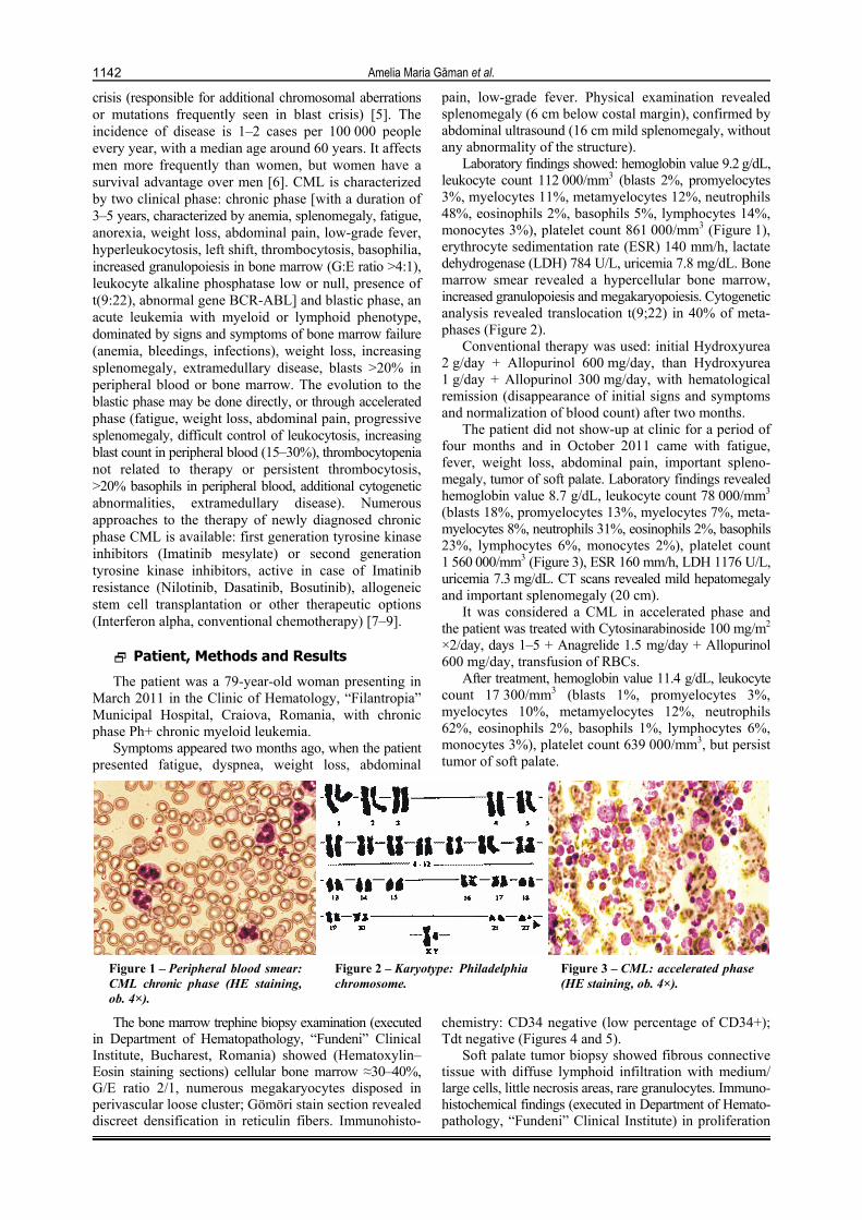
Laboratory findings showed: hemoglobin value 9.2 g/dL, leukocyte count 112 000/mm3 (blasts 2%, promyelocytes 3%, myelocytes 11%, metamyelocytes 12%, neutrophils 48%, eosinophils 2%, basophils 5%, lymphocytes 14%, monocytes 3%), platelet count 861 000/mm3 (Figure 1), erythrocyte sedimentation rate (ESR) 140 mm/h, lactate dehydrogenase (LDH) 784 U/L, uricemia 7.8 mg/dL. Bone marrow smear revealed a hypercellular bone marrow, increased granulopoiesis and megakaryopoiesis. Cytogenetic analysis revealed translocation t(9;22) in 40% of meta-phases (Figure 2).
Conventional therapy was used: initial Hydroxyurea 2 g/day + Allopurinol 600 mg/day, than Hydroxyurea 1 g/day + Allopurinol 300 mg/day, with hematological remission (disappearance of initial signs and symptoms and normalization of blood count) after two months.
The patient did not show-up at clinic for a period of four months and in October 2011 came with fatigue, fever, weight loss, abdominal pain, important spleno-megaly, tumor of soft palate. Laboratory findings revealed hemoglobin value 8.7 g/dL, leukocyte count 78 000/mm3 (blasts 18%, promyelocytes 13%, myelocytes 7%, meta-myelocytes 8%, neutrophils 31%, eosinophils 2%, basophils 23%, lymphocytes 6%, monocytes 2%), platelet count 1 560 000/mm3 (Figure 3), ESR 160 mm/h, LDH 1176 U/L, uricemia 7.3 mg/dL. CT scans revealed mild hepatomegaly and important splenomegaly (20 cm).
It was considered a CML in accelerated phase and the patient was treated with Cytosinarabinoside 100 mg/m2 ×2/day, days 1–5 + Anagrelide 1.5 mg/day + Allopurinol 600 mg/day, transfusion of RBCs.
After treatment, hemoglobin value 11.4 g/dL, leukocyte count 17 300/mm3 (blasts 1%, promyelocytes 3%, myelocytes 10%, metamyelocytes 12%, neutrophils 62%, eosinophils 2%, basophils 1%, lymphocytes 6%, monocytes 3%), platelet count 639 000/mm3, but persist tumor of soft palate.
Figure 1 – Peripheral blood smear: CML chronic phase (HE staining, ob. 4×).
Figure 2 – Karyotype: Philadelphia chromosome.
Figure 3 – CML: accelerated phase (HE staining, ob. 4×).
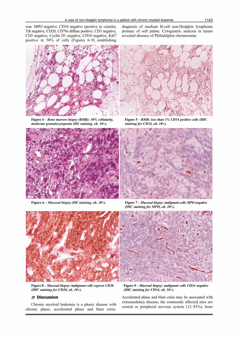
The bone marrow trephine biopsy examination (executed in Department of Hematopathology, “Fundeni” Clinical Institute, Bucharest, Romania) showed (Hematoxylin–Eosin staining sections) cellular bone marrow ≈30–40%, G/E ratio 2/1, numerous megakaryocytes disposed in perivascular loose cluster; Gömöri stain section revealed discreet densification in reticulin fibers. Immunohisto-
chemistry: CD34 negative (low percentage of CD34+); Tdt negative (Figures 4 and 5).
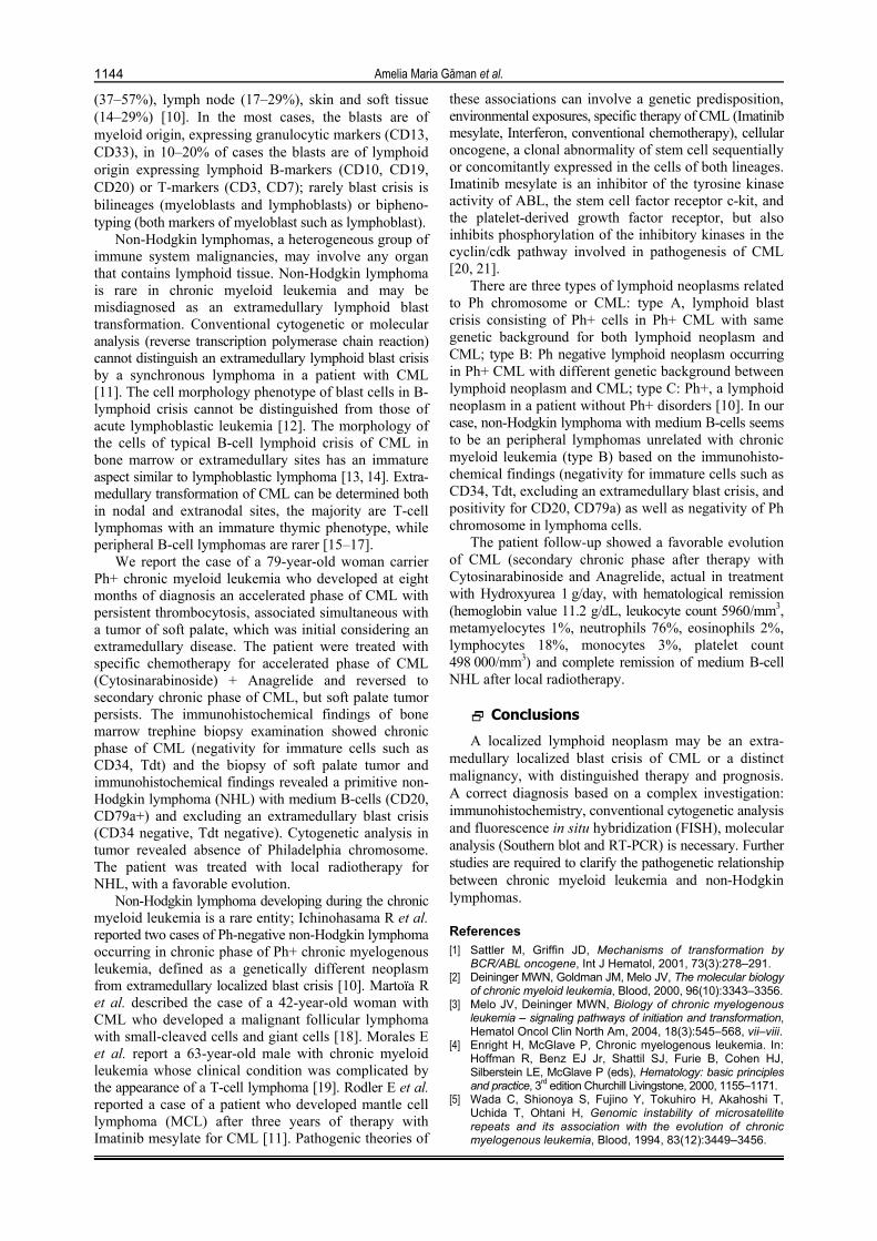
Soft palate tumor biopsy showed fibrous connective tissue with diffuse lymphoid infiltration with medium/ large cells, little necrosis areas, rare granulocytes. Immuno-histochemical findings (executed in Department of Hemato-pathology, “Fundeni” Clinical Institute) in proliferation
A case of non-Hodgkin lymphoma in a patient with chronic myeloid leukemia
1143
was: MPO negative, CD34 negative (positive in vessels), Tdt negative, CD20, CD79a diffuse positive, CD3 negative, CD5 negative, Cyclin D1 negative, CD10 negative, Ki67 positive in 50% of cells (Figures 6–9) establishing
diagnosis of medium B-cell non-Hodgkin lymphoma primary of soft palate. Cytogenetic analysis in tumor revealed absence of Philadelphia chromosome.
Figure 4 – Bone marrow biopsy (BMB): 30% cellularity, moderate granulocytopenia (HE staining, ob. 10×).
Figure 5 – BMB: less than 1% CD34 positive cells (IHC staining for CD34, ob. 10×).
Figure 6 – Mucosal biopsy (HE staining, ob. 20×). Figure 7 – Mucosal biopsy: malignant cells MPO negative (IHC staining for MPO, ob. 20×).
Figure 8 – Mucosal biopsy: malignant cells express CD20 (IHC staining for CD20, ob. 10×).
Figure 9 – Mucosal biopsy: malignant cells CD34 negative (IHC staining for CD34, ob. 10×).
Discussion
Chronic myeloid leukemia is a phasic disease with chronic phase, accelerated phase and blast crisis.
Accelerated phase and blast crisis may be associated with extramedulary disease; the commonly affected sites are central or peripheral nervous system (12–83%), bone
Amelia Maria Găman et al.
1144
(37–57%), lymph node (17–29%), skin and soft tissue (14–29%) [10]. In the most cases, the blasts are of myeloid origin, expressing granulocytic markers (CD13, CD33), in 10–20% of cases the blasts are of lymphoid origin expressing lymphoid B-markers (CD10, CD19, CD20) or T-markers (CD3, CD7); rarely blast crisis is bilineages (myeloblasts and lymphoblasts) or bipheno-typing (both markers of myeloblast such as lymphoblast).
Non-Hodgkin lymphomas, a heterogeneous group of immune system malignancies, may involve any organ that contains lymphoid tissue. Non-Hodgkin lymphoma is rare in chronic myeloid leukemia and may be misdiagnosed as an extramedullary lymphoid blast transformation. Conventional cytogenetic or molecular analysis (reverse transcription polymerase chain reaction) cannot distinguish an extramedullary lymphoid blast crisis by a synchronous lymphoma in a patient with CML [11]. The cell morphology phenotype of blast cells in B-lymphoid crisis cannot be distinguished from those of acute lymphoblastic leukemia [12]. The morphology of the cells of typical B-cell lymphoid crisis of CML in bone marrow or extramedullary sites has an immature aspect similar to lymphoblastic lymphoma [13, 14]. Extra-medullary transformation of CML can be determined both in nodal and extranodal sites, the majority are T-cell lymphomas with an immature thymic phenotype, while peripheral B-cell lymphomas are rarer [15–17].
We report the case of a 79-year-old woman carrier Ph+ chronic myeloid leukemia who developed at eight months of diagnosis an accelerated phase of CML with persistent thrombocytosis, associated simultaneous with a tumor of soft palate, which was initial considering an extramedullary disease. The patient were treated with specific chemotherapy for accelerated phase of CML (Cytosinarabinoside) + Anagrelide and reversed to secondary chronic phase of CML, but soft palate tumor persists. The immunohistochemical findings of bone marrow trephine biopsy examination showed chronic phase of CML (negativity for immature cells such as CD34, Tdt) and the biopsy of soft palate tumor and immunohistochemical findings revealed a primitive non-Hodgkin lymphoma (NHL) with medium B-cells (CD20, CD79a+) and excluding an extramedullary blast crisis (CD34 negative, Tdt negative). Cytogenetic analysis in tumor revealed absence of Philadelphia chromosome. The patient was treated with local radiotherapy for NHL, with a favorable evolution.
Non-Hodgkin lymphoma developing during the chronic myeloid leukemia is a rare entity; Ichinohasama R et al. reported two cases of Ph-negative non-Hodgkin lymphoma occurring in chronic phase of Ph+ chronic myelogenous leukemia, defined as a genetically different neoplasm from extramedullary localized blast crisis [10]. Martoïa R et al. described the case of a 42-year-old woman with CML who developed a malignant follicular lymphoma with small-cleaved cells and giant cells [18]. Morales E et al. report a 63-year-old male with chronic myeloid leukemia whose clinical condition was complicated by the appearance of a T-cell lymphoma [19]. Rodler E et al. reported a case of a patient who developed mantle cell lymphoma (MCL) after three years of therapy with Imatinib mesylate for CML [11]. Pathogenic theories of
these associations can involve a genetic predisposition, environmental exposures, specific therapy of CML (Imatinib mesylate, Interferon, conventional chemotherapy), cellular oncogene, a clonal abnormality of stem cell sequentially or concomitantly expressed in the cells of both lineages. Imatinib mesylate is an inhibitor of the tyrosine kinase activity of ABL, the stem cell factor receptor c-kit, and the platelet-derived growth factor receptor, but also inhibits phosphorylation of the inhibitory kinases in the cyclin/cdk pathway involved in pathogenesis of CML [20, 21].
There are three types of lymphoid neoplasms related to Ph chromosome or CML: type A, lymphoid blast crisis consisting of Ph+ cells in Ph+ CML with same genetic background for both lymphoid neoplasm and CML; type B: Ph negative lymphoid neoplasm occurring in Ph+ CML with different genetic background between lymphoid neoplasm and CML; type C: Ph+, a lymphoid neoplasm in a patient without Ph+ disorders [10]. In our case, non-Hodgkin lymphoma with medium B-cells seems to be an peripheral lymphomas unrelated with chronic myeloid leukemia (type B) based on the immunohisto-chemical findings (negativity for immature cells such as CD34, Tdt, excluding an extramedullary blast crisis, and positivity for CD20, CD79a) as well as negativity of Ph chromosome in lymphoma cells.
The patient follow-up showed a favorable evolution of CML (secondary chronic phase after therapy with Cytosinarabinoside and Anagrelide, actual in treatment with Hydroxyurea 1 g/day, with hematological remission (hemoglobin value 11.2 g/dL, leukocyte count 5960/mm3, metamyelocytes 1%, neutrophils 76%, eosinophils 2%, lymphocytes 18%, monocytes 3%, platelet count 498 000/mm3) and complete remission of medium B-cell NHL after local radiotherapy.
Conclusions
A localized lymphoid neoplasm may be an extra-medullary localized blast crisis of CML or a distinct malignancy, with distinguished therapy and prognosis. A correct diagnosis based on a complex investigation: immunohistochemistry, conventional cytogenetic analysis and fluorescence in situ hybridization (FISH), molecular analysis (Southern blot and RT-PCR) is necessary. Further studies are required to clarify the pathogenetic relationship between chronic myeloid leukemia and non-Hodgkin lymphomas.
References [1] Sattler M, Griffin JD, Mechanisms of transformation by
BCR/ABL oncogene, Int J Hematol, 2001, 73(3):278–291. [2] Deininger MWN, Goldman JM, Melo JV, The molecular biology
of chronic myeloid leukemia, Blood, 2000, 96(10):3343–3356. [3] Melo JV, Deininger MWN, Biology of chronic myelogenous
leukemia – signaling pathways of initiation and transformation, Hematol Oncol Clin North Am, 2004, 18(3):545–568, vii–viii.
[4] Enright H, McGlave P, Chronic myelogenous leukemia. In: Hoffman R, Benz EJ Jr, Shattil SJ, Furie B, Cohen HJ, Silberstein LE, McGlave P (eds), Hematology: basic principles and practice, 3rd edition Churchill Livingstone, 2000, 1155–1171.
[5] Wada C, Shionoya S, Fujino Y, Tokuhiro H, Akahoshi T, Uchida T, Ohtani H, Genomic instability of microsatellite repeats and its association with the evolution of chronic myelogenous leukemia, Blood, 1994, 83(12):3449–3456.
A case of non-Hodgkin lymphoma in a patient with chronic myeloid leukemia
1145
[6] Hehlmann R, Hochhaus A, Baccarani M, Chronic myeloid leukemia, Lancet, 2007, 370(9584):342–350.
[7] Baccarani M, Cortes J, Pane F, Niederwieser D, Saglio G, Apperley J, Cervantes F, Deininger M, Gratwohl A, Guilhot F, Hochhaus A, Horowitz M, Hughes T, Kantarjian H, Larson R, Radich J, Simonsson B, Silver RT, Goldman J, Hehlmann R; European LeukemiaNet, Chronic myeloid leukemia: an update of concepts and management recommendations of European LeukemiaNet, J Clin Oncol, 2009, 27(35):6041–6051.
[8] Hochhaus A, Management of newly diagnosed chronic myeloid leukemia patients, Hematology Education: the education program for the 16th Congress of the European Hematology Association, London, UK, June 9–12, 2001, 5(1):120–126.
[9] Hochhaus A, Educational session: managing chronic myeloid leukemia as a chronic disease, American Society of Hematology (ASH) Education Book, 2011, 2011(1):128–135.
[10] Ichinohasama R, Miura I, Takahashi N, Sugawara T, Tamate E, Endoh K, Endoh F, Naganuma H, DeCoteau JF, Griffin JD, Kadin ME, Ooya K, Ph-negative non-Hodgkin's lymphoma occurring in chronic phase of Ph-positive chronic myelogenous leukemia is defined as a genetically different neoplasm from extramedullary localized blast crisis: report of two cases and review of the literature, Leukemia, 2000, 14(1):169–182.
[11] Rodler E, Welborn J, Hatcher S, Unger K, Larkin E, Gumerlock PH, Wun T, Richman C, Blastic mantle cell lymphoma developing concurrently in a patient with chronic myelogenous leukemia and a review of the literature, Am J Hematol, 2004, 75(4):231–238.
[12] Griffin JD, Todd RF 3rd, Ritz J, Nadler LM, Canellos GP, Rosenthal D, Gallivan M, Beveridge RP, Weinstein H, Karp D, Schlossman SF, Differentiation patterns in the blastic phase of chronic myeloid leukemia, Blood, 1983, 61(1):85–91.
[13] Ohyashiki JH, Ohyashiki K, Shimizu H, Miki M, Kimura N, Mori S, Fujisawa K, Akatsuka J, Toyama K, Testicular tumor as the first manifestation of B-lymphoid blastic crisis in a case of Ph-positive chronic myelogenous leukemia, Am J Hematol, 1988, 29(3):164–167.
[14] Cervantes F, Villamor N, Esteve J, Montoto S, Rives S, Rozman C, Montserrat E, ‘Lymphoid’ blast crisis of chronic myeloid leukaemia is associated with distinct clinico-haematological features, Br J Haematol, 1998, 100(1):123–128.
[15] Specchia G, Palumbo G, Pastore D, Mininni D, Mestice A, Liso V, Extramedullary blast crisis in chronic myeloid leukemia, Leuk Res, 1996, 20(11–12):905–908.
[16] Van Dorpe J, Van Damme S, Jacobs V, Van den Berghe H, Criel A, Michielssen P, Louwagie A, T-lymphoid extramedullary (lymphadenopathic) blast crisis in CML, Acta Clin Belg, 1995, 50(2):121–125.
[17] Gaman G, Ignat F, Gaman A, Rotaru I, Ciurea M, Comanescu V, Ocular blast metamorphosis in a patient with chronic granulocytic leukemia, The Jubilee Meeting on Chronic Myeloid Leukemia (CML), 3rd International Conference Bologna, June 11–14, 2000, Haematologica, 2000, 85(Suppl):18.
[18] Martoïa R, Lamy T, Delmaire P, Algayres JP, Rougier Y, Laurens A, Occurrence of non-Hodgkin’s lymphoma in chronic myeloid leukemia, Rev Med Interne, 1987, 8(5):471–474.
[19] Morales E, Bancalari G, Fahrenkrog AM, Rossle A, Chronic myeloid leukemia and non Hodgkin lymphoma in the same patient. Clinical case, Rev Med Chil, 1999, 127(9):1105–1107.
[20] Druker BJ, Tamura S, Buchdunger E, Ohno S, Segal GM, Fanning S, Zimmermann J, Lydon NB, Effects of a selective inhibitor of the Abl tyrosine kinase on the growth of Bcr-Abl positive cells, Nat Med, 1996, 2(5):561–566.
[21] Braziel RM, Launder TM, Druker BJ, Olson SB, Magenis RE, Mauro MJ, Sawyers CL, Paquette RL, O’Dwyer ME, Hemato-pathologic and cytogenetic findings in imatinib mesylate-treated chronic myelogenous leukemia patients: 14 months’ experience, Blood, 2002, 100(2):435–441.
Corresponding author Amelia Maria Găman, Associate Professor, MD, PhD, Senior Specialist in Hematology and Internal Medicine, Department of Pathophysiology, Faculty of Medicine, University of Medicine and Pharmacy of Craiova, 2 Petru Rareş Street, 200349 Craiova, Romania; Phone +40770–684 146, e-mail: [email protected] Received: June 5, 2013
Accepted: November 20, 2013