ronald berezney and donald s. coffey
TRANSCRIPT

N U C L E A R M A T R I X
I so la t ion and C h a r a c t e r i z a t i o n o f a F r a m e w o r k S t r u c t u r e
F r o m R a t L i v e r N u c l e i
RONALD BEREZNEY and DONALD S. COFFEY
From the Department of Pharmacology and Experimental Therapeutics, the James Buchanan Brady Laboratory for Reproductive Biology and the Oncology Center, The Johns Hopkins University School of Medicine, Baltimore, Maryland 21205. Dr. Berezney's present address is the Division of Cell and Molecular Biology, State University of New York, Buffalo, New York 14214.
ABSTRACT
A nuclear framework structure termed the nuclear matrix has been isolated and characterized. This matrix forms the major residual structure of isolated nuclei and consists largely of protein with smaller amounts of RNA, DNA, carbohy- drate, and phospholipid. The nuclear matrix can be further resolved by combined treatment with DNase and RNase. The remaining nuclear protein structure, after extraction of 90% of the nuclear protein, 99.9% of the DNA, and 98% of the RNA and phospholipid, is termed the nuclear protein matrix. Electron micros- copy of this final nuclear protein matrix reveals an interior framework structure composed of residual nucleolar structures associated with a granular and fibrous internal matrix structure. The internal matrix framework is derived from the interchromatinic structures of the nucleus, and is connected to a surrounding residual nuclear envelope layer containing residual nuclear pore complex struc- tures.
Sodium dodecyl sulfate-acrylamide gel electrophoresis of the nuclear matrix proteins demonstrates three major polypeptide fractions, P-l, P-2, and P-3, with average molecular weights of -69,000, 66,000 and 62,000, as well as several minor polypeptides which migrate at -50 ,000 and at higher molecular weights (>100,000). Polypeptides with molecular weights identical to those of P-l, P-2, and P-3 are also components of isolated nuclear envelopes and nucleoli, whereas isolated chromatin contains no detectable matrix polypeptides. This suggests that the major matrix polypeptides are localized in specific structural regions of the nucleus, i.e., nuclear envelope, nucleoli, and interchromatinic structures. The presence of cytochrome oxidase activity in the isolated nuclear matrix indicates that at least some integral proteins of the nuclear membrane are associated with the matrix.
Preliminary communications from our laboratory (12-14) have reported the isolation of a residual framework structure derived from rat liver nuclei,
and termed the nuclear protein matrix, or more generally the nuclear matrix. Aside from serving as a skeletal framework, the nuclear matrix may
616 THE JOURNAL OF CELL BIOLOGY' VOLUME 73, 1977' pages 616-637
Dow
nloaded from http://rupress.org/jcb/article-pdf/73/3/616/1387723/616.pdf by guest on 22 January 2022

play an impor tan t role in nuclear funct ions. In this regard, newly repl icated D N A was recently re- por ted to be associated initially with the nuclear matr ix (15-17) . In this paper we present a more detailed study of the isolation, s t ructure, and bio- chemistry of isolated ra t liver nuclear matrix.
M A T E R I A L S A N D M E T H O D S
Nuclei
Liver nuclei were prepared from male rats (Sprague- Dawley, 300--350 g, Charles River) based on procedures reported by Berezney et al. (9, 19). Livers were quickly excised, minced with a scalpel, and homogenized with 10 strokes at 1,200 rpm in a Potter-Elvehjem apparatus with a tissue ratio of 1 vol of liver to 2 vol of TM buffer (0.25 M sucrose, 0.05 M Tris, pH 7.4, 5 mM MgCI2). After filtration through four layers of cheesecloth, the homogenate was centrifuged at 770g for 10 min (Sorvali SS-34 rotor, DuPont Instruments, Sorvall Operations, Newtown, Conn.) to yield a crude nuclear pellet. The pellet was resuspended in 2.2 M sucrose TM buffer and centrifuged at 40,000 g for 90 min in a Beckman 21 rotor (Beckman Instruments, Inc., Spinco Div., Palo Alto, Calif.). This purified nuclear pellet was washed two times and stored in 0.25 M sucrose TM buffer at 0~ Nuclei from 40-50 rat livers were isolated in this manner in - 6 - 8 h. The nuclei were used for isolating the nuclear protein matrix between 12 and 24 h after prepa- ration.
Nuclear Protein Matrix
The procedure for the isolation of the nuclear protein matrix is presented in detail in the Results section.
Residual Nucleoli
The isolation of nudeoli from rat liver nuclei was based on the sonication procedures described by Busch (25). Isolated rat liver nuclei were resuspended to a final protein concentration of 1 mg/ml in 0.25 M sucrose, 10 mM Tris-HCl, pH 7.4, 1 mM CaCI~. The nuclei were then sonicated in 30-s bursts at - 1 2 watts with a Bran- son sonicator (Heat Systems-Ultrasonics, Inc., Plain- view, N. Y., model W-140) while the temperature was maintained below 4~ The nucleoli were collected by centrifugation of 35 ml of nuclear sonicate through 20 ml of 0.9 M sucrose, 10 mM "Iris, pH 7.4 in a Beckman SW 25.2 swinging bucket rotor at 3,000 g for 20 min. The nucleolar pellet was resuspended in the initial buffer and the centrifugation through 0.9 M sucrose, 10 mM Tris HCI, pH 7.4 repeated twice. The final nucleolar pellet was resuspended in TM buffer for subsequent analysis. Electron microscope studies of the isolated nudeoli con- firmed the presence of relatively undamaged nudeoli and the virtual absence of visible contamination by other nuclear components.
Residual nucleoli were prepared by treating isolated
nudeoli in a manner identical to that for the preparation of the nuclear protein matrix from control nuclei (see Table II). All nudeolar fractions were centrifuged at 3,000 g for 60 min at each step.
Residual Nuclear Membranes
Nuclear membranes were isolated as described by Berezney et al. (9, 19) with slight modifications. The isolated nuclei were suspended in 0.25 M sucrose TM buffer at a protein concentration of 2 mg/ml, and di- gested for 12-14 h at 2~ with 25/~g of DNase (Worth- ington Biochemical Corp., Freehold, N. J.) per ml. After centrifugation at 770 g for 30 min, the DNase-treated nuclear pellet was resuspended in 0.25 M sucrose, 0.5 M MgCle in TM buffer to a final nuclear protein concentra- tion of 4 mg/ml and layered over a discontinuous high- salt sucrose gradient of 1.6 M and 2.2 M sucrose contain- ing 0.5 M MgCl~ in TM buffer. After centrifugation for 1 h at 75,000 g in a Beckman SW 25.2 rotor, the nuclear membrane fraction was collected at the 0.25 M sucrose- 1.6 M sucrose interface, suspended in 0.5 M MgCl~ TM buffer, and centrifuged at 200,000 g for 30 min in a Beckman 65 rotor. The nuclear membranes were then resuspended in TM buffer for subsequent analysis.
Residual nuclear membranes were prepared by treat- ing these isolated nuclear membranes exactly as in the preparation of the nuclear protein matrix from nuclei (see above). The nuclear membrane fractions were cen- trifuged at 200,000 g for 1 h at each step.
Chemical Fractionation o f
Nuclear Proteins
The nuclear proteins of isolated nuclei and the nuclear matrix were fractionated according to the sequential ex- traction procedures of Steele and Busch (71) as modified by Chung and Coffey (28). The general classes of nu- clear proteins that are fractionated in this manner are termed soluble or nudeoplasmic proteins S~ and Se, histones, acidic proteins, and residual proteins. All ex- tractions were performed at 0~ with 10 mg of nuclear protein in a total volume of 10 ml, followed by centrifu- gation at 15,000 g for 15 min.
Acrylamide Gel Electrophoresis
Sodium dodecyl sulfate (SDS) acrylamide gel electro- phoresis was performed according to the procedures of Weber and Osbom (76), using 10% acrylamide gels in 0.1% SDS, 0.05 M Tris, pH 7.4 run at 4 mA per gel with a solution of 0.1% SDS, 0.05 M Tris, pH 7.4 in both the lower and upper chambers. Lyophilized sam- ples were dissolved in 5% SDS, 0.05 M Tris, pH 7.4 containing 2% #-mercaptoethanol by heating at 100~ for 5 min, followed by incubation at room temperature for several hours. Addition of 6 M urea or guanidine HC1 to the SDS samples, or alkylafion of the polypepfides with sodium iodacetate (60) did not alter the polypeptide scans. Bromophenol blue was added to each sample to
R. BEREZNEY AND D. S. COFFEY Nuclear Matrix Isolation and Characterization 617
Dow
nloaded from http://rupress.org/jcb/article-pdf/73/3/616/1387723/616.pdf by guest on 22 January 2022

serve as a tracking dye for Rt determinations and molec- ular weight estimations as described by Weber and Os- born (76). Reference proteins for molecular weight cali- bration included thyroglobulin, phosphorylase a, col- lagenase, bovine serum albumin, catalase, ovalbumin, a-chymotrypsinogen and cytochrome c. All gels were stained with Coomassie Blue (R-250) and scanned at 600 nm with a Gilford Linear Transport (model 2410, Gilford Instrument Laboratories Inc., Oberlin, Ohio) equipped with a fixed slit width of 0.05-0.10 mm. Densitometric comparisons of various nuclear protein fractions were always done with an equal amount of protein on each gel (50 ttg).
Ure~-acrylamide gels were used to detect histones in the nuclear matrix proteins, according to the procedure of Panyim and Chalkley (59).
Amino Acid Analysis Protein samples were hydrolyzed in 5.7 N HCI for 24
h at 105~ in vacuo. Amino acid analysis of the protein hydrolysate was performed on a Beckman Model 120 Amino Acid Analyzer (Beckman Instruments, Inc., Ful- lerton, Calif.) by the procedures of Spackman et al. (68).
Chemical Determinations RNA and DNA were separated as described by
Munro and Fleck (57). DNA was determined by the Burton modification of the diphenylamine reaction (24), and RNA, protein, and phospholipid were analyzed as previously reported (19). Total neutral carbohydrate content was determined according to the phenolsulfuric acid procedure of Dubois et al. (32). Sialic acid was measured by the thiobarbituric acid method and cor- rected for deoxyribose as suggested by Warren (75). Protein was measured by the procedure of Lowry et al. (50).
Electron Transport Components Assays for various electron transport components
were performed as previously reported (19). NADH- cytochrome c reductase activity was assayed by following the reduction of cytochrome c at 550 nm. NADH-ferri- cyanide reductase activity was determined by measuring ferricyanide reduction at 420 nm. The enzymatic reduc- tion rates were corrected for nonenzymatic reduction. Succinate dehydrogenase activity was assayed by meas- uring the reduction of phenazine methosulfate spectro- photometrically at 600 nm coupled to 2,6-dichlorophen- olindophenol. All spectrophotometric determinations were performed with a Gilford Model 2000 recording spectrophotometer at 30~ Cytochrome bs was deter- mined by difference spectra in a Cary-15 double beam spectrophotometer (Cary Instruments, Monrovia, Calif.) after reduction by dithionite or NADH (19). Cyto- chrome c oxidase activity was measured polarographi- cally by a modification of the assay of Chuang et al. (27) as previously described (19). Lipid was isolated from
untreated nuclei by the procedure of Folch et al. (36). Lipid micelles for enzyme reconstitution studies were prepared in 10 mM Tris, pH 7.8 by sonication according to Rouser and Fleischer (63).
Estimation of Mitochondrial Contamination
Contribution of mitochondrial derived cytochrome c oxidase to the total activity of this enzyme in the nuclear matrix was routinely monitored by assaying for succinate dehydrogenase activity, or in some cases by the direct chemical determination of succinate dehydrogenase-as- sociated flavin as previously described (19). Succinate dehydrogenase activity indicated <0.1% mitoehondrial contamination on a protein basis. This correction indi- cated that <2% of the total matrix cytochrome c oxidase activity was due to mitochondrial contamination.
Electron Microscopy Samples were prepared for electron microscopy by
fixation in 4% glutaraldehyde (0.05 M cacodylate, pH 7.4, 5 mM MgCIa) and postfixation in 1% OsO4 in 0.05 M cacodylate, pH 7.4, 5 mM MgCI2 at 0~ The samples were embedded in Epon according to the method of Luft (51) or Spurr (70). Ultrathin sections were stained with both uranyl acetate and lead citrate according to Rey- nolds (61). To reveal nonchromatin structures in the nuclei, specimens were fixed in glutaraldehyde and treated by the regressive staining procedure described by Bernhard (20). All samples were observed and photo- graphed on an AEI 801 electron microscope operating at 60 kV.
Light Microscopy Sections of 1-2/xm were cut from the Epon-embed-
ded blocks and stained with a 0.2% solution of toluidine blue 0 with 0.5% sodium borate. Samples were stained for 1 rain at 60~ and rinsed in both distilled water and 70% ethanol.
Estimations of nuclear sphere radii were obtained by direct measurement on unfixed samples, utilizing Zeiss Nomarski interference optics. 100 random nuclear meas- urements were made for each reported value.
Materials DNase I (EC 3.1.4.5) from bovine pancreas (~3,000
Worthington Kuntz U/rag) and RNase A (EC 2.7.7.16) from bovine pancreas ( -3 ,000 Worthington Kalnitsky U/mg) were obtained from Worthington Biochemical Corp. Trypsin (3 x crystallized) and collagenase (Type III) were also obtained from Worthington. Hyaluroni- dase from bovine testes was a product of Schwarz/Mann Div., Becton, Dickinson & Co. (Orangeburg, N. Y.). Triton X-100, calf thymus DNA, yeast RNA, sialic acid (N-acetyl neurominic acid), glucose, and trizma base were obtained from Sigma Chemical Co. (St. Louis,
6 1 8 THE JOURNAL OF CELL BIOLOGY" VOLUME 73, 1977
Dow
nloaded from http://rupress.org/jcb/article-pdf/73/3/616/1387723/616.pdf by guest on 22 January 2022

Mo.). Sucrose, magnesium chloride, and sodium chlo- ride were reagent grade products of J. T. Baker Chemi- cal Co. (Phillipsburg, N. J.).
RESULTS
Chemical Treatment o f Isolated Nuclei
In the course of some survey experiments in our laboratory, we discovered that isolated rat liver nuclei maintain their general spherical shape as observed under the light microscope after a wide variety of chemical treatments which remove many of the major components of the nucleus (see Table I). Nuclear spheres, of approximate dimen- sions of nuclei, were observed after combined treatment with 2 M NaCl which so|ubilizes most of the nuclear chromatin (8, 54, 71), and Triton X- 100 which releases the nuclear envelope phospho- lipid as well as many of the membrane proteins (1, 2, 7, 12-14, 46, 74). In contrast, nuclear spheres were no longer observed after certain treatments known to solubilize or degrade proteins, such as 0.1 N NaOH, 5% SDS, or treatment with pro- teases such as pronase or trypsin (Table I). These observations suggested the presence of a suppor- tive framework structure in the cell nucleus of an apparent protein nature. This supportive frame- work was not due to the presence of either the chromatin or nuclear membrane phospholipids. Furthermore, since extraction of histones from nuclei with either 0.25 N HCl or 2 M NaCl solu- tions (Table I) still left nuclear spheres visible, it appeared that residual nonhistone proteins might be responsible for the nuclear framework structure.
Our approach was to remove as much of the nuclear components without destroying nuclear spheres, thus isolating the minimal residual com- ponents necessary for maintaining the constitutive structural framework of the nucleus. This was ac- complished by monitoring the progressive removal of various nuclear components in terms of chemi- cal composition, while observing the integrity of nuclear structure with light and electron micro- scope procedures.
Isolation Procedures
The procedure for the isolation of the nuclear protein matrix is composed of four consecutive extractions as summarized in Table II. Aliquots of nuclear spheres obtained after each treatment are resuspended in TM buffer for subsequent bio- chemical and morphological analyses. During the
TABLE I
Light Microscope Examination of Rat Liver Nuclei and Nuclear Matrix aider Chemical Treatment*
Presence of nuclear spheres
Nuclear Treatment Nuclei matrix
Distilled H20 + + 2 M NaCl + + 1% Triton X-100 + + 2 M NaCI + 1% Triton X-100 + + 0.25 N HCI + + Acetone (95%) + + 0.01 N HCl in ethanol (80%, + +
vol/vol) 1% ~Mercaptoethanol + + DNase-RNase + + Collagenase + + Hyaluronidase + +
0.1 N NaOH - - Trypsin or pronase - - 1% SDS + 1% (wt/vol)/~-Mer- - -
captoethanoi
* Nuclear pellets containing 2 mg of protein from either isolated nuclei or nuclear matrix were resuspended in 1 ml of the appropriate solution and incubated at 22~ for 60 min. All the enzyme digestions were performed with 100 /~g of enzyme per ml of suspension in TM buffer. The SDS-fl-mercaptoethanol solution was in 0.05 M Tris, pH 7.4. Although many of the procedures removed large amounts of nuclear material, nuclear spheres were still observed where indicated (+). Absence of observa- ble nuclear spheres is indicated as (-) .
treatments, the nuclear pellets are gently resus- pended with a glass stirring rod. Vortexing of the nuclear suspensions causes excessive damage to the nuclear structures and should be avoided. An important requirement for obtaining high yields of spheres is to centrifuge at low g-force (770 g). Higher g-forces cause progressive compression and disruption of nuclear spheres.
Step one consists of resuspension of isolated rat liver nuclei (see Materials and Methods) in low magnesium buffer (0.2 mM MgCIr 10 mM Tris- HCI, pH 7.4) to a final concentration of 2 mg of nuclear protein per ml. After incubation on ice for 10 min, the nuclear suspension is centrifuged at 770 g for 30 min in a Sorvall SS-34 rotor. The extraction was repeated twice with approximately one-half the initial volume of buffer. The final pellet obtained is termed the low magnesium- treated nuclear sphere fraction (LM).
R. BEREZNEY AND D. S. COFFEY Nuclear Matrix Isolation and Characterization 619
Dow
nloaded from http://rupress.org/jcb/article-pdf/73/3/616/1387723/616.pdf by guest on 22 January 2022

TABLE II Steps in the Isolation of the Nuclear Matrix
Total nuclear material extracted (cumulative)*
Step Consecutive extractions Resultant nuclear sphere Protein DNA RNA Phospholipid
%
0 Isolated nuclei Intact nucleus (N) 0 0 0 0 1 Low magnesium treatment Low Mg++-treated sphere 52.0 - 2.23 75.8 • 2.78 19.7 • 1.83 2.5 • 1.13
(0.2 mM MgClz) (LM) 2 High-salt treatment (2 M High salt-treated sphere 83.7 • 2.17 97.6 • 1.21 66.0 • 1.64 6.4 • 1.65
NaCI) (HS) 3 Detergent treatment (1% Tri- Nuclear matrix (NM) 90.0 • 1.57 97.7 • 1.21 71.0 • 1.68 97.8 • 1.51
ton X-100) 4 Nuclease treatment (DNase- Nuclear protein matrix 90.2 • 1.55 >99.9 98.0 +- 1.12 98.0 • 1.47
RNase) (NPM)
* Percent extracted determined relative to isolated nuclei centrifuged in 0.25 M sucrose TM buffer an equivalent number of times as each nuclear sphere fraction. SEM (---) for five separate preparations.
In step two, the low magnesium-treated nuclear spheres are suspended in high salt buffer (2 M NaC1, 0.2 mM MgCI2, 10 mM Tris-HC1, pH 7.4) to a volume corresponding to 4 mg of initial nu- clear protein per ml of suspension. After ten min on ice, the suspension is centrifuged at 770 g for 60 min. The nuclear pellets are extracted two more times with one-half the initial volume of high salt buffer. The final pellet is termed the high salt- treated nuclear sphere fraction (HS).
In step three, the high salt-treated nuclear spheres are resuspended in TM buffer to a final volume corresponding to 4 mg of initial nuclear protein per ml. Triton X-100 (10%, wt/vol) in TM buffer is added to a final concentration of 1% while stirring on ice. After 10 min on ice, the nuclear suspension is centrifuged at 770 g for 30 min. The pellets are washed two times in an equiv- alent volume of TM buffer. The final pellet is termed the nuclear matrix fraction (NM).
Step four involves extensive digestion with high concentrations of DNase and RNase. The nuclear matrix is resuspended in TM buffer to a final protein concentration of 2 mg/ml. Electrophoreti- cally purified DNase and RNase (200 /zg each) are added to each milliliter of the nuclear matrix suspension. After a 60-rain digestion at 22~ the nuclear suspension is centrifuged at 770 g for 30 min and washed two times in equal volumes of TM buffer. The final pellet is termed the nuclear pro- tein matrix fraction (NPM).
The final nuclear protein matrix represents the residual nuclear structure remaining after extract- ing 90% of the total nuclear proteins, 99.9% of DNA, and 98% of RNA and phospholipids (Ta- ble II).
The nuclear protein matrix isolation was devel-
oped for rat liver nuclei prepared in sucrose me- dium containing magnesium ions. The initial isola- tion procedures for nuclei are critical since endoge- nous nuclear endonuclease activity is an important factor for the subsequent removal of large amounts of DNA in the low magnesium extraction procedure of step one (6, 47, 48). In prelimi- nary attempts to prepare nuclear matrix from other mammalian tissues, we have found that the bulk DNA is not released during low magnesium treatment (step 1). In the absence of endogenous nuclease activity, the high-salt buffer (step 2) gen- erally leads to gel formation in the nuclear pellets. To overcome these difficulties we routinely pre- incubate isolated nuclei from various tissue sources (10 s nuclei/ml) with low levels of DNase I (2-5 ~g/ml) at 22~ for 15 min. This allows a more reproducible preparation of nuclear matrix from a variety of tissues including brain, prostrate, lung, spleen, thymus, and kidney. The details of these procedures and the properties of the isolated matrices will be reported elsewhere. (R. Berez- ney. Unpublished experiments.)
Gross Composi t ion
A summary of the macromolecular composition of the various nuclear sphere fractions at each of the four extraction steps is presented in Table III. The final chemical composition of the nuclear pro- tein matrix is 97.6% protein with traces of RNA (1.2%), phospholipid (1.1%) and <0.1% DNA. This is based on the assumption that these compo- nents represent 100% of the nuclear protein ma- trix. However, recently, small amounts of carbo- hydrate of nonnudeic acid origin have been de- tected in the nuclear protein matrix. No sialic acid (<0.1%) but significant amounts of neutral sugars
620 THE JOURNAL OF CELL BIOLOGY" VOLUME 73 , 1 9 7 7
Dow
nloaded from http://rupress.org/jcb/article-pdf/73/3/616/1387723/616.pdf by guest on 22 January 2022
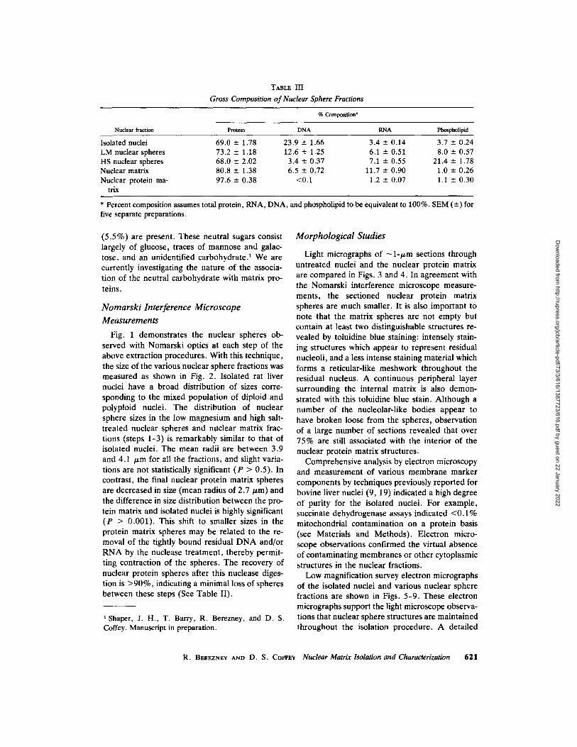
TABLE III
Gross Composition of Nuclear Sphere Fractions
% Composition*
Nuclear fraction Protein DNA RNA Phospholipid
Isolated nuclei 69.0 -+ 1.78 23.9 • 1.66 3.4 • 0.14 3.7 • 0.24 LM nuclear spheres 73.2 - 1.18 12.6 -+ 1.25 6.1 -+ 0.51 8.0 • 0.57 HS nuclear spheres 68.0 - 2.02 3.4 -+ 0.37 7.1 - 0.55 21.4 -+ 1.78 Nuclear matrix 80.8 -+ 1.38 6.5 -+ 0.72 11.7 • 0.90 1.0 • 0.26 Nuclear protein ma- 97.6 -+ 0.38 <0.1 1.2 • 0.07 1.1 -+ 0.30
trix
* Percent composition assumes total protein, RNA, DNA, and phospholipid to be equivalent to 100%. SEM (-+) for five separate preparations.
(5.5%) are present. These neutral sugars consist largely of glucose, traces of mannose and galac- tose, and an unidentified carbohydrate? We are currently investigating the nature of the associa- tion of the neutral carbohydrate with matrix pro- teins.
Nomarski Interference Microscope
Measurements
Fig. 1 demonstrates the nuclear spheres ob- served with Nomarski optics at each step of the above extraction procedures. With this technique, the size of the various nuclear sphere fractions was measured as shown in Fig. 2. Isolated rat liver nuclei have a broad distribution of sizes corre- sponding to the mixed population of diploid and polyploid nuclei. The distribution of nuclear sphere sizes in the low magnesium and high salt- treated nuclear spheres and nuclear matrix frac- tions (steps 1-3) is remarkably similar to that of isolated nuclei. The mean radii are between 3.9 and 4.1 /~m for all the fractions, and slight varia- tions are not statistically significant (P > 0.5). In contrast, the final nuclear protein matrix spheres are decreased in size (mean radius of 2.7/~m) and the difference in size distribution between the pro- tein matrix and isolated nuclei is highly significant (P > 0.001). This shift to smaller sizes in the protein matrix spheres may be related to the re- moval of the tightly bound residual DNA and/or RNA by the nuclease treatment, thereby permit- ting contraction of the spheres. The recovery of nuclear protein spheres after this nuclease diges- tion is >90%, indicating a minimal loss of spheres between these steps (See Table II).
1 Shaper, J. H., T. Barry, R. Berezney, and D. S. Coffey. Manuscript in preparation.
Morphological Studies
Light micrographs of ~l-/~m sections through untreated nuclei and the nuclear protein matrix are compared in Figs. 3 and 4. In agreement with the Nomarski interference microscope measure- ments, the sectioned nuclear protein matrix spheres are much smaller. It is also important to note that the matrix spheres are not empty but contain at least two distinguishable structures re- vealed by toluidine blue staining: intensely stain- ing structures which appear to represent residual nucleoli, and a less intense staining material which forms a reticular-like meshwork throughout the residual nucleus. A continuous peripheral layer surrounding the internal matrix is also demon- strated with this toluidine blue stain. Although a number of the nucleolar-like bodies appear to have broken loose from the spheres, observation of a large number of sections revealed that over 75% are still associated with the interior of the nuclear protein matrix structures.
Comprehensive analysis by electron microscopy and measurement of various membrane marker components by techniques previously reported for bovine liver nuclei (9, 19) indicated a high degree of purity for the isolated nuclei. For example, succinate dehydrogenase assays indicated <0.1% mitochondrial contamination on a protein basis (see Materials and Methods). Electron micro- scope observations confirmed the virtual absence of contaminating membranes or other cytoplasmic structures in the nuclear fractions.
Low magnification survey electron micrographs of the isolated nuclei and various nuclear sphere fractions are shown in Figs. 5-9. These electron micrographs support the light microscope observa- tions that nuclear sphere structures are maintained throughout the isolation procedure. A detailed
R. BEREZr~EY AND D. S. Corr~Y Nuclear Matrix Isolation and Characterization 621
Dow
nloaded from http://rupress.org/jcb/article-pdf/73/3/616/1387723/616.pdf by guest on 22 January 2022

FIGURE 1 Nomarski interference light micrographs of nuclear sphere fractions. (A) Control nudeus, untreated; (B) low magnesium-treated nuclear sphere; (C) high salt-treated nuclear sphere; (D) nuclear matrix; and (E) nudear protein matrix, x 870.
20"
CONTROL NUCLEI
10
I I LM NUCLEAR t0 I L
W
=) �9 I HS NUCLEAR < ~ I I SPHERES LU tO
CO LIJ rY Iii I Q_ G)
NUCLEAR MATRIX " '
. _ J . . O 10. Z LL o
20. NUCLEAR PROTEIN MATRIX
(2.72 + O,08tum I
10
I 2 3 4 5 6 7 8
RADIUS IN MICRONS FIGURE 2 Size distribution of nuclear sphere fractions. The average radius of 100 nuclear spheres was measured for each fraction with Nomarski interference optics.
study of the structure of the various nuclear sphere fractions will be presented elsewhere .2 In this anal- ysis, we will concentrate on the structural organi- zation of the final nuclear protein matrix, and the identification of the in situ nuclear components from which the nuclear matrix is derived.
The structural organization of isolated rat liver nuclei closely resembles the in situ appearance. Previous studies of nuclear morphology in liver and other mammalian cells have clearly defined several distinct regions in the nuclei (21, 22, 55, 73). Similarly, four main structural regions can be visualized in the nuclei of intact liver cells (Fig. 10a) and isolated nuclei (Fig. 10b): (1) a sur- rounding nuclear envelope (NE); (2) nucleoli (N); (3) dense chromatin (heterochromatin) patches beneath the nuclear envelope (PC) and surrounding the nucleoli (perinucleolar chroma- tin) (PNC), as well as in other interior regions of the nuclei; and (4) interchromatinic areas (IC), between condensed chromatin regions, which con- tain various granular and fibrous components, such as the interchromatinic granules and the peri- chromatinic fibers and granules (21, 22, 55, 73). The interchromatinic area is also presumed to contain the diffuse chromatin (euchromatin) of the nucleus (43).
The structure of the nuclear protein matrix con- sists of three main components (Fig. 11): (a) a residual nuclear envelope (RE) which forms a continuous structure surrounding the nuclear sphere, (b) highly condensed and electron-dense residual nucleoli (RN), and (c) an extensive gran- ular and fibrous internal matrix structure (1M) which extends throughout the interior of the nu- clear sphere from the residual nucleoli to the sur- rounding residual nuclear envelope (Fig. 12). This internal matrix framework appears to be derived primarily from the interchromatinic structures of the nucleus.
Higher magnification of the nuclear protein ma- trix interior (Fig. 13) demonstrates internal matrix
2 Berezney, R., and D. S. Coffey. Manuscript in prepa- ration.
6 2 2 THE JOURNAL OF CELL BIOLOGY' VOLUME 73, 1977
Dow
nloaded from http://rupress.org/jcb/article-pdf/73/3/616/1387723/616.pdf by guest on 22 January 2022

FmURES 3 and 4 Light microscope sections of untreated nuclei (Fig. 3) and the nuclear protein matrix (Fig. 4). 1-2-/~m section stained with toluidine blue. x 1,450.
structures in close association with a residual nu- cleolus. The empty perinucleolar areas, which may correspond to sites of perinucleolar chroma- tin in isolated nuclei, are bordered by the residual nucleolus and the internal matrix structures. The internal matrix structures consist of densely packed fibers (matrix fibers) associated with elec- tron-dense particulate structures termed matrix particles (Fig. 15). The fine structure of the inter- nal matrix (Fig. 15) shows close resemblance to that of certain interchromatinic structures of iso- lated nuclei (Fig. 14) as well as nuclei in situ (Fig. 10a and references 21, 22, 55, 73). Although the internal matrix structures appear more condensed than the interchromatinic structures of isolated nuclei, both consist of distinct electron-dense par- ticles of similar dimensions (150-250/~ in diame- ter) associated with a less electron-dense, tightly packed, fibrous substance (Figs. 14 and 15). Fi- bers as thin as 50/~ in width can be seen in the matrix of both the intact nuclei and the nuclear protein matrix.
The derivation of the internal matrix framework from the interchromatinic structure of the nucleus was also indicated by application of the Bernhard EDTA regressive staining technique (21, 55) to the various nuclear sphere fractions. With this
procedure, the chromatin areas of the nucleus appear as unstained or slightly stained areas while the interchromatinic structures of the nucleus are highly electron dense. It was therefore possible to monitor the degree of preservation of the inter- chromatinic structure during isolation of the nu- clear matrix. Our preliminary results indicate that components of the interchromatinic structures are still present in the internal matrix structure .2
The residual nuclear envelope of the nuclear matrix consists of a continuous electron-dense layer -100-150 A in thickness which completely surrounds the nuclear matrix. Associated with this continuous layer are distinct residual nuclear pore complex structures. In lateral view, characteristic annular structures -800-900 A, in diameter and central granules of the nuclear pore complexes are visible (Figs. 16 and 17).
Chemical Treatment o f
Nuclear Matrix
In these experiments, isolated nuclear matrix (before final treatment with nucleases, see Table II) was exposed to a variety of chemical treat- ments as described earlier for isolated nuclei. Us- ing Nomarski interference microscopy, we then determined whether the particular treatment re-
R. BEREZNEY AND D. S. COFFEY Nuclear Matrix Isolation and Characterization 623
Dow
nloaded from http://rupress.org/jcb/article-pdf/73/3/616/1387723/616.pdf by guest on 22 January 2022

FIGURES 5, 6, 7, 8, and 9 Low magnification comparison of electron microscope sections of the nuclear sphere fractions. (Fig. 5), untreated nuclei; (Fig. 6), low magnesium-treated nuclear spheres; (Fig. 7), high salt-treated nuclear spheres; (Fig. 8), nuclear matrix; and (Fig. 9), nuclear protein matrix, x 3,100.
6 2 4
Dow
nloaded from http://rupress.org/jcb/article-pdf/73/3/616/1387723/616.pdf by guest on 22 January 2022

I~GURE 10 Electron microscope sections of rat liver nuclei/n s/tu in liver tissue (Fig. 10a) and in the isolated nuclear fraction (Fig. 10b). The isolated rat liver nuclei maintain many of the structural features characteristic of nuclei/n situ. These include: NE, nuclear envelope; N, nucleolus; PNC, perinucleolar condensed chromatin; PC, peripheral condensed chromatin; IC, interchromatinic areas. The interchroma- tinic areas contain electron-dense particles 150-250 tlt in diameter termed interchromatinic granules (ig) as well as less electron-dense fibrous material (f). Clusters of interchromatinic granules are enclosed by a broken line. x 44,000.
suits in visible disruption of the matrix nuclear sphere structure. As indicated in Table I, the pattern is identical for isolated nuclei and nuclear matrix.
Protein Fract ionat ion
Since the nuclear protein matrix is predomi- nantly protein, it was important to establish what
R. BEREZNEY AND D. S. COFFEr Nuclear Matrix Isolation and Characterization 625
Dow
nloaded from http://rupress.org/jcb/article-pdf/73/3/616/1387723/616.pdf by guest on 22 January 2022

626 "I~E JOURNAL OF CELL BIOLOGY" VOLUME 73, 1977
Dow
nloaded from http://rupress.org/jcb/article-pdf/73/3/616/1387723/616.pdf by guest on 22 January 2022

classes of nuclear proteins were present in this residual structure. 10 mg of the isolated nuclear protein matrix was subjected to a series of extrac- tions which are commonly used to separate the major classes of nuclear proteins (28, 71). This extraction procedure was applied to the isolated nuclear protein matrix (NPM) and the results were compared to those obtained with isolated nuclei (N). The results are presented in Fig. 18 and are expressed as the percent of total proteins solubilized with each step of the fractionation. It is apparent that the nuclear matrix proteins fraction- ate predominantly as acidic proteins.
To determine whether acidic amino acids pre- dominated in the nuclear matrix proteins, an amino acid analysis was performed and is pre- sented in Table IV. The acidic to basic amino acid ratio is 1.46, and is consistent with an acidic na- ture for these proteins. Moreover, an earlier re- port by Steele and Busch (72) of the analysis of a fraction of residual acidic proteins extracted from rat liver nuclei is also presented for comparison.
The absence of histones as a significant compo- nent of the nuclear matrix proteins was established by resolution on urea acrylamide gels by the pro- cedure of Panyim and Chalkley (59).
SDS-Acrylamide Gel Electrophoresis
o f Matrix Proteins
SDS gels of nuclear matrix and other nuclear fractions are compared in Fig. 19. Total nuclear proteins (gel 1) show a typical heterogeneous pat- tern with a multitude of hands. The low molecular weight histone-containing bands are particularly prominent. Nuclear matrix proteins (gels 3 & 4) are characterized by three major polypeptide bands in the molecular weight region of 60,000- 70,000 and the absence of stained bands in the
low molecular weight regions. Polypeptide gel pat- terns of the nuclear matrix (gel 3) or the nuclear protein matrix (gel 4) were compared in six sepa- rate preparations, and no significant differences were detected. This is consistent with the composi- tional data (see Table III) which demonstrate a high recovery of total protein in step 3 from nu- clear matrix to step 4 in the nuclear protein ma- trix.
Comparison of total chromatin proteins (gel 5) prepared by the method of Shaw and Huang (65) as modified by Arnold and Young (5), and nu- clear matrix proteins (gels 3 & 4) suggests the absence or chromatin polypeptides in the nuclear matrix and the corresponding absence of matrix polypeptides in chromatin protein. One excep- tion, however, is a high molecular weight compo- nent whose molecular weight can only be roughly approximated as 200,000, which is found in all five of the gels.
Approximate molecular weights of the polypep- tide bands were determined from gel scans by the method of Weber and Osborn (76). In each exper- iment, appropriate molecular weight protein stan- dards (see Materials and Methods) were run in parallel gels or were comigrated in the same gel with the nuclear matrix proteins. The gel scans were also divided into five zones, e.g. Fig. 20 A - E, and the percent of total stained area in each region was determined. The three major polypep- tide peaks in region B of the nuclear protein ma- trix are termed P-I, P-2, and P-3 and represent - 5 0 % of the total stained area (Fig. 20). These polypeptide bands have approximate molecular weights of 69,000, 66,000 and 62,000, respec- tively. Other minor polypeptide bands are ob- served, primarily at ~ 50,000 daltons (zone C) and in the higher molecular weight regions >100,000 daltons (zone A).
FIGURE 11 Electron microscope section through the nuclear protein matrix revealing the internal structural components of the matrix. RN, residual nudeolus; IM, internal matrix framework; RE, residual nuclear envelope layer. Note the empty spaces surrounding the residual nucleoli and along the periphery (arrows). These may correspond to regions previously occupied by the perinudeolar and peripheral condensed chromatin in untreated nuclei (compare with Fig. 10a and b). • 21,000.
FIGURE 12 Higher magnification electron microscope section of the nuclear protein matrix in the region of the residual nuclear envelope. A close association of the internal matrix framework (IM) with the residual nuclear envelope (RE) is evident (white arrows). A residual nuclear pore complex structure is projecting through the residual nuclear envelope layer (black arrows). Regions of the internal matrix framework (enclosed in broken line) resemble dusters of interchromatinic granules seen in both isolated and in situ nuclei (compare with the regions enclosed by broken lines in Fig. 10a and b. x 50,000.
R. Bm~EZN~.V AND D. S. COVFEy Nuclear Matrix Isolation and Characterization 6 2 7
Dow
nloaded from http://rupress.org/jcb/article-pdf/73/3/616/1387723/616.pdf by guest on 22 January 2022

6 2 8 ThE JOURNAL OF CELL BIOLOGY �9 VOLUME 73 , 1977
Dow
nloaded from http://rupress.org/jcb/article-pdf/73/3/616/1387723/616.pdf by guest on 22 January 2022

80-
7'0-
60-
50-
40-
30-
20-
I0-
N N N
NPM
NPM NPM . . I I _ I I - - I I I I .
NPM
S I Soluble S 2 Soluble Histone$ Acidic Pro~i'~$ Residual Protein~l PrOteins Proteins
FmU]~E 18 The chemical fractionation of the isolated nuclear protein matrix (black bars, NPM) com- pared to that of the total protein of isolated nuclei (white bars, N). The general classes of nuclear proteins were fractionated by the extraction procedures of Steele and Busch (71).
Comparisons o f Nuclear Matrix,
Nuclear Membrane, and
Nucleolar Proteins
Our morphological studies indicated that the nuclear matrix contains components of the resid- ual nuclear envelope which are continuous with an internal nuclear matrix connecting to residual nu- cleolar elements. This led us to determine whether these residual nuclear matrix protein components
(P- l , P-2, and P-3) were present in isolated nu- cleoli or nuclear membrane preparations which had been subsequently extracted by the four steps used in the isolation of the nuclear protein matrix. Nucleoli were isolated by the method of Busch (25) and nuclear membranes by the procedure of Berezney et al. (9, 19). The final residual protein fractions of the nucleoli and nuclear membranes were subjected to solubilization and electrophore- sis parallel with the nuclear protein matrix. The
FIGURE 13 High magnification of the nuclear protein matrix in the area of the residual nucleolus. The residual nucleolus, RN appears continuous with the internal matrix framework (IM). Empty spaces (arrows) surrounding the residual nudeolus may correspond to regions previously occupied by the perinucleolar condensed chromatin (compare with Fig. 10a and b). Note that these empty areas are bordered by the nucleolus and the internal matrix framework, x 102,000.
FIGURE 14 High magnification section of a region in the interior of an isolated nucleus. Distinct areas of condensed chromatin, C, are seen in the upper-center and lower-left regions of the micrograph. The regions between these condensed chromatin areas are the interchromatinic regions which contain electron- dense interchromatinic granules (ig) and less electron-dense fibrous structures (f). x 116,000.
FIGURE 15 High magnification of a section through the internal matrix framework of the nuclear protein matrix. This residual framework structure consists of electron-dense matrix particles (mp) and matrix fibers (f) which bear a close similarity to the interchromatinic structures of isolated as well as in situ liver nuclei (compare with Figs. 14 and 10a and b). x 116,000.
FIGURm 16 and 17 High magnification sections through the residual nuclear envelope layer of the nuclear protein matrix. Distinct residual nuclear pore complex structures are observed which still retain their characteristic annular structure (arrows). Central granules are often visible in tangential sections through the residual pore complex structures (white arrow in Fig. 17). x 158,000.
R. BEREZNEY AND D. S. COFFEr Nuclear Matrix Isolation and Characterization 629
Dow
nloaded from http://rupress.org/jcb/article-pdf/73/3/616/1387723/616.pdf by guest on 22 January 2022

TABLE IV
Comparison of Amino Acid Analyses of the Nuclear Protein Matrix with the Nonchromosomal Acidic Nuclear Protein Fraction of Steele and Busch*
(B) Nonchro- (A) Nuclear mosomal alkali- matrix pro- soluble nuclear Ratio
Amino acid reins proteins* A/B
, mol / lO0 mol o f amino acids
Lysine 6.55 6.27 1.04 Arginine 6.61 5.67 1.17 Histidine 2.26 2.29 0.99 Aspartic acid 9.23 9.25 1.00 Glutamic acid 13.34 12.04 1.11 Threonine 5.02 5.67 0.89 Serine 6.96 7.36 0.95 Proline 5.54 5.47 1.01 Glycine 9.34 7.96 1.17 Alanine 7.50 7.36 1.02 Valine 6.42 6.07 1.06 Isoleucine 3.94 4.58 0.86 Leucine 9.45 9.35 1.01 Tyrosine 2.56 2.79 0.92 Methionine 1.81 2.59 0.70 1/2 Cystine 0.24 1.29 0.19 Phenylalanine 3.21 3.98 0.81
Ratio acidic 1.46 1.50 0.97 basic
* Values under (B) from Steele and Busch (71).
residual nuclear membrane contains the three ma- jor polypeptide peaks (P-l, P-2, and P-3) of the nuclear matrix and, in addition, a prominent in- crease in the polypeptide peak at -50 ,000 daltons (Fig. 21). In contrast, the residual nucleoli exhibit a wider variety of polypeptide peaks. Polypeptide bands were observed on the acrylamide gel of the residual nucleoli in the area of P-1 and P-2, but the P-3 band was deficient (Fig. 21).
Analysis o f Electron Transport
Components
Previous studies have shown that isolated nu- clear membranes contain electron transport com- ponents including NADH cytochrome b~ reduc- tase, cytochrome bs, and cytochrome c oxidase (9, 18, 19, 78). We therefore assayed the total nu- clear matrix for these electron transport compo- nents. The specific activities of these factors in isolated nuclei increase from two- to fivefold in the high salt-treated nuclear sphere fraction, when expressed on a total milligram protein basis (Table V). These results are anticipated since the high-salt nuclear spheres are depleted in chromatin but still
retain both the outer and inner nuclear mem- branes. When these membranes are disrupted with Triton X-100 and the final nuclear protein matrix is isolated, the specific activities of the NADH-ferricyanide and NADH-cytochrome c re- ductase are essentially abolished, as is the pres- ence of cytochrome bs. In contrast, considerable activity of cytochrome c oxidase is retained and the activity can be further increased by the addi- tion of exogenous phospholipid. This indicates that at least some proteins of the nuclear mem- brane, such as cytochrome c oxidase, are still associated with residual components of the nu- clear matrix.
DISCUSSION
Organization o f the Nuclear Matrix
A number of investigators have previously re- ported the existence of residual nuclear structures after extraction of chromatin with high ionic strength solutions (26, 37, 58, 67, 80, 81). Re-
FIGURE 19 SDS-acrylamide gel electrophoresis of the nuclear matrix polypeptides. Approximately 80 tLg of protein was placed on the top of each gel and resolved by electrophoresis. (1) Untreated control nudei, (2) high salt-treated nuclear spheres, (3) nuclear matrix, (4) nu- clear protein matrix, and (5) isolated chromatin. Stan- dard proteins were (a) thyroglobulin (167,500); (b) phosphorylase a (95,000); (c) bovine serum albumin (68,000); (d) catalase (60,000); (e) ovalbumin (43,000); (f) chymotrypsinogen (25,700); and (g) cyto- chrome c (11,700). The high molecular weight compo- nent common to all the gels has an approximate molecu- lar weight of 200,000.
630 "DIE JOURNAL OF CELL BIOLOGY' VOLUME 73, 1977
Dow
nloaded from http://rupress.org/jcb/article-pdf/73/3/616/1387723/616.pdf by guest on 22 January 2022

,, a C T D T E 1.0
0 8 ,
8 ~o 0 6
0 4 .
0 , 2 -
P : 2 p . 3
I I I
I I I
l i I
I I m ~ N -
0
F]oux~ 20
I I
G!
I I t I ! 0 I. t I
0.2 0 3 0.4 0.5 0,6 7 0.8 0.9
Rf
SDS-acrylamide gel scan of the nuclear protein matrix polypeptides. The gel was run with 50 /xg of protein, stained with Coomassie Blue, and scanned with a Gilford Linear Transport at 600 rim. Molecular weight values were determined from a standard curve according to the procedures of Weber and Osborn (76) using the proteins listed in Fig. 19.
!
I O
cently, we reported preliminary results on the iso- lation of a residual protein framework in the rat liver nucleus (12, 13) which we later termed the nuclear protein matrix (14). Particular emphasis is placed on understanding the structural derivation of the matrix from isolated nuclei. It might be argued, for example, that the nuclear protein ma- trix represents a fortuitous association of residual nuclear proteins, and bears no definitive relation- ship to the structure of the nucleus in situ. High magnification electron micrographs of the nuclear protein matrix (Figs. 11-17), however, clearly demonstrate that the structural components of the isolated matrix bear a remarkable resemblance to well-defined structures of the intact cell nucleus. These results are summarized in the following points: ( a) A residual nuclear envelope layer which still retains residual nuclear pore complexes. High magnification of the residual pore complex (Figs. 16 and 17) demonstrates features typical of the intact nuclear pore complex such as the character- istic annular structure, central rodlets, and radial filaments connecting the annular and central rod- lets (78). These findings agree with the results of
Aaronson and Blobel (3) and Scheer et al. (64), who have demonstrated morphologically recogniz- able nuclear pore structures in isolated residual nuclear envelope layers, and support an earlier speculation by Wunderlich (77) that nuclear pore complexes contain permanent protein compo- nents. (b) A residual internal matrix framework derived from the interchromatinic structure o f the nucleus (21, 22, 55, 73). Use of the Bernhard regressive staining procedure has revealed that the interchromatinic structure forms a reticulum or matrix throughout the nuclear interior (20, 55). In its structural organization the residual internal ma- trix observed in the nuclear protein matrix is strik- ingly similar to this in situ matrix as indicated by comparing Figs. 14 and 15. This structure consists of electron-dense matrix particles - 1 5 0 - 2 5 0 / ~ in diameter, associated with less electron-dense but tightly packed matrix fibers. The electron-dense matrix particles appear identical to the interchro- matinic particles previously observed in nuclei in situ (21, 73). The matrix fibers which have a diameter of - 5 0 /~ also have potential in situ structural derivatives in the interchromatinic re-
R. BEREZNEY AND D. S. COI~EY Nuclear Matrix Isolation and Characterization 631
Dow
nloaded from http://rupress.org/jcb/article-pdf/73/3/616/1387723/616.pdf by guest on 22 January 2022

P -I P-$
/III A s,DuA, .uc,Ec,_us
i
I 3 5 7 DISTANCE FROM ORIGIN (cm)
FmURE 21 Comparative SDS-acrylamide gel scans of the nudear protein matrix, residual nucleolar and residual nuclear membrane polypeptides. Treatment of isolated nucleoli and nuclear membranes was performed in the same way as treatment of isolated nuclei for the preparation of nuclear matrix. 50 p.g of residual protein was placed on each gel.
TABLE V
Electron Transport Components
High salt- treated nuclear Nuclear protein
Assay Nuclei spheres matrix NPM/N ratio
$p act
NADH-cytochrome c reductase (~mol cytochrome c/rain per mg protein)
+ Rotenone (10 -4 M) NADH-ferrieyanide reductase
(/.~mol K3Fe(CN)dmin per mg protein) Cytochrome bs
(AA 425-410 nm/mg protein) Cytochrome c oxidase
(/zmol O2/min per mg protein)* + Phospholipid
0.0604 0.278 0.001 0.017
0.0589 0.273 0.001 0.017 0.379 1.97 0.04 0.106
0.016 0.067 0.001 0.063
0.015 0.034 0.013 0.867
0.017 0.037 0.025 1.47
* 0.5-1.0% of the total cytochrome c oxidase activity in the liver homogenate was recovered in the isolated nuclei.
6 3 2 ThE JOURNAL OF CELL BIOLOGY" VOLUME 73, 1977
Dow
nloaded from http://rupress.org/jcb/article-pdf/73/3/616/1387723/616.pdf by guest on 22 January 2022

gions of nuclei, but it has always been difficult to distinguish euchromatin fibers from "other inter- chromatinic substances".
Monneron and Bernhard (55), however, using the EDTA-regressive staining technique, identi- fied 30-50-A fibers as a basic component of var- ious extrachromatinic structures. These include perichromatinic fibers, perichromatinic granules, coiled bodies, fibers associated with the nuclear pore complexes, and fibers which interconnect interchromatinic granules. Monneron and Bern- hard further stressed similar staining properties of the fibers, and suggested a structural linkage be- tween perichromatinic fibers and interchromatinic granules, as well as occasional connections be- tween perichromatinic fibers and granules. The 50-/~ matrix fibers may therefore represent resid- ual components of these 30-50-/~, fibers observed in intact nuclei. Recently, Comings and Okada (30) have suggested that these residual matrix fibers be termed matrixin. (c) Residual nucleoli. The identification of the highly condensed and electron-dense bodies as residual nucleoli is based on the monitoring of structural alternations which the nucleolus undergoes during the extraction pro- cedures for the nuclear protein matrix. Recently, we have confirmed this interpretation by electron microscope examination of the isolated residual nucleoli prepared in this study. (R. Berezney. Unpublished experiments.)
The above considerations indicate that the iso- lated matrix is derived from well-defined struc- tures of the intact nucleus. The question then arises as to what extent this isolated residual matrix corresponds to the matrix of intact nuclei. It is likely that the large decrease in the radius of the final nuclear protein matrix (r = 2.72 --- 0.08/~m) in comparison to isolated nuclei (r = 3.95 +-- 0.07 /zm) (see Fig. 2) is a reflection of massive configu- rational changes in specific components of the matrix. Further studies are therefore necessary to more precisely relate the structure of the isolated matrix to its in situ organization.
There is no doubt, however, that specific com- ponents of the matrix can be separated. This must occur, for example, in the isolation of nucleoli (25) or nuclear membrane (9, 19, 56). The ef- fectiveness of 0.5 M MgCI2 for the isolation of nuclear membranes (9, 19, 56, 78) suggests that high concentrations of the divalent cation mag- nesium may be critical in the separation of nuclear membranes from the matrix.
It is important to consider whether the nuclear
matrix can be isolated from other types of eucar- yotic cells. Recently, Herlan and Wunderlich (44) reported the isolation of a nuclear matrix from the macronuclei of the ciliate protozoan Tetrahymena pyriformis, using a modification of the procedure reported for rat liver (14). The isolated Tetrahy- mena matrix displayed a structural organization similar to that of the rat liver matrix. Comings and Okada (30) recently reported a similar structure for mouse liver nuclear matrix, and Hildebrand et al. (45) have reported the isolation of a nuclear matrix from cultured Chinese hamster ovary (CHO) cells.
The ability of the nuclear matrix to contract after nuclease treatment (Fig. 2) suggests that the matrix is not a rigid skeletal structure, but rather a flexible framework capable of vast changes in or- ganization. In support of this view, Wunderlich and his co-workers have recently discovered that isolated Tetrahymena nuclear matrix can reversi- bly expand and contract when the concentrations of Mg § and Ca +* are varied in the medium.~ The presence of a flexible nuclear matrix framework in the cell nucleus is consistent with the observed swelling of the cell nucleus during the S period of the cell cycle (52) and the well-known phenome- non of nuclear swelling as a prerequisite for the initiation of RNA and/or DNA synthesis (23, 29, 39, 41, 42, 53).
It is therefore suggested that the nuclear matrix in situ is a dynamically changing structure closely coupled with nuclear functioning. Recently, we have found that newly replicated DNA is closely associated with the nuclear matrix in regenerating liver (15-17). Moreover the matrix proteins have been shown to phosphorylate to a maximal level at a time just preceding the onset of DNA replication in the regenerating liver (4, 10).
It has also been speculated that the nuclear matrix plays an important role in the transcription and intranuclear transport of RNA (26, 78). The association of residual nuclear pore complexes as an integral component of the nuclear matrix may provide a structural linkage for intranuclear trans- port to the cytoplasm. In accord with this view, Faiferman and Pogo (34) recently demonstrated that RNP particles containing rapidly labeled, heterogeneous nuclear RNA (hn-RNA) are asso- ciated with the nuclear matrix. In addition, we
a Wunderlich, F., and G. Herlan. 1977. A reversi- bly contractile nuclear matrix. Its isolation, structure, and composition. J. Cell Biol. 73:271-278.
R. BEREZNEY AND D. S. COFFEy Nuclear Matrix Isolation and Characterization 633
Dow
nloaded from http://rupress.org/jcb/article-pdf/73/3/616/1387723/616.pdf by guest on 22 January 2022

have recently observed that polyribonucleotides bind tightly to the isolated nuclear protein matrix .2 These polyribonucleotides alter nuclear structure and function (29). Recently, Goidl et al. (38) have reported that certain of these polyribonucleotides release preformed polyribosomes from isolated nuclei.
Nuclear Matrix Proteins
The presence of the major matrix polypeptides P- l , P-2, and P-3 in isolated nucleoli and nuclear envelopes suggests that the 60,000-70,000-dalton polypeptides are distributed throughout the nu- clear matrix as a major component of the frame- work structure. In support of these findings, Aaronson and Blobel (3), Dwyer and Blobel (33), Riley et al. (62), and Shelton et al. (66) have demonstrated similar 60,000-70,000-dalton poly- peptides, and Comings and Okada (30) have re- cently reported that mouse liver nuclear matrix contains three major polypeptides with molecular weights similar to those of polypeptides of rat liver nuclear matrix.
The presence of nuclear membrane cytochrome c oxidase activity in the nuclear matrix suggests that at least some protein components of the nu- clear membrane are still associated with the resid- ual nuclear envelope layer of the matrix. This conclusion is in agreement with a view recently proposed by Scheer et al. (64). Our results, how- ever, also demonstrate that some tightly bound nuclear membrane proteins are specifically re- moved during the isolation of the matrix. For example, cytochrome bs, a tightly bound, integral membrane protein of both the endoplasmic reticu- lum (69) and the nuclear membrane (18), is largely extracted (Table V).
Since the nuclear membrane enzymes analyzed in this study make up only a small percentage of the total protein of the nuclear membrane, the total amount of membrane protein associated with the residual nuclear envelope layer cannot be de- termined from this analysis. A number of studies, however, have demonstrated that certain mem- brane proteins can maintain residual structures, albeit altered, after extraction of lipids with deter- gents (31, 49, 79), phospholipases (11), or or- ganic solvents (35, 40). The large extraction of phospholipid (98%) from the nuclear envelope during matrix isolation, therefore, does not neces- sarily imply that a correspondingly large propor- tion of nuclear membrane proteins are also ex- tracted.
Recent studies of the matrix polypeptides from other cells indicate possible differences in molecu- lar weights. For example, Herlan and Wunderlich (44) reported three major polypeptides of 70,000, 57,000 and 54,000 in isolated matrix from the more primitive Tetrahymena pyriformis macronu- cleus, and Hildebrand et al. (45) found major polypeptide bands between 58,000 and 71,000 in nuclear matrix from CHO cells. It will be neces- sary to examine the matrix in cells from a large variety of organisms before interpreting any simi- larities or differences in matrix proteins, phyloge- netically. It is important to emphasize, however, that other polypeptides aside from the 60,000- 70,000-dalton fractions are components of the liver matrix such as the 50,000 and the ~200,000- dalton peptides.
The authors wish to thank Dr. Ann M. Benson for performing the amino acid analysis. These studies were supported by U. S. Public Health Service Grant CA 13745. Dr. Berezney was supported by National Insti- tutes of Health Training Grant GM 01183.
Received for publication 21 May 1976, and in revised form 28 January 1977.
REFERENCES
1. AARONSON, R. P., and G. BLOBEL. 1973. On the mode of attachment of the nuclear pore complex to the nucleus.J. Cell Biol. 59(2, Pt. 2):1 a. (Abstr).
2. AARONSOr% R. P., and G. BLOBEL. 1974. On the attachment of the nuclear pore complex. J. Cell Biol. 62:746-754.
3. AARONSON, R. P., and G. BLOBEL. 1975. Isolation of nuclear pore complexes in association with a lamina. Proc. Natl. Acad. Sci. U. S. A. 72:1007- 1011.
4. ALLEN, S., R. BEREZNEY, and D. S. COVFEV. 1977. Phosphorylation of nuclear matrix proteins during rat liver regeneration. Biochem. Biophys. Res. Commun. In press.
5. ARNOLD, E. A., and K. E. YOUNO. 1972. Isolation and partial electrophoretic characterization of total protein from non-sheared rat liver chromatin. Biochim. Biophys. Acta. 257:482-496.
6. BARRACK, E. R. 1975, Models for Studying the Interactions of Acidic Polymers with Eukaryotic Nuclei. Ph.D. Dissertation. The Johns Hopkins University School of Medicine, Baltimore.
7. BARTON, A. D., W. E. KISIELESKI, F. WASSER- MANN, and F. MACKEVnJS. 1971. Experimental modification of structures at the periphery of the liver cell nucleus. Z. Zellforsch Mikrosk. Anat. 115:299-306.
8. BEKI-IOR, I., G. M. KUNG, and J. BONNER. 1969.
634 THE JOURNAL OF CELL BIOLOGY" VOLUME 73, 1977
Dow
nloaded from http://rupress.org/jcb/article-pdf/73/3/616/1387723/616.pdf by guest on 22 January 2022

Sequence specific interaction of DNA and chromo- somal protein. J. Mol. Biol. 39:351-364.
9. BE~ZNEV, R. 1974. Large scale isolation of nu- clear membranes from bovine liver. Methods Cell Biol. 7:205-228.
10. BEREZNEY, R., S. ALLEN, and D. S. COFFEY. 1976. Phosphorylation of the nuclear protein matrix. J. Cell Biol. 70(2, Pt. 2):305 a. (Abstr.).
11. BEREZNEY, R., Y. C. AWASTHI, L. K. FUNK, and F. L. CRANE. 1970. The relation of phospholipid and membrane structure in mitochondrial electron transport particles. Bioenergetics. 1:445-456.
12. BER~ZNEV, R., and D. S. Cora~EV. 1973. Isolation of a nuclear structural complex from mammalian nuclei. J. Cell Biol. 59(2, Pt. 2):22 a. (Abstr.).
13. BEREZNEV, R., and D. S. COVFEV. 1974. Identifica- tion of a nuclear structural protein network in rat liver nuclei. Fed. Proc. 33:1395.
14. BEREZNEY, R., and D. S. CorrEY. 1974. Identifica- tion of a nuclear protein matrix. Biochem. Biophys. Res. Commun. 60:1410-1417.
15. BEREZNEY, R., and D. S. COWEY. 1975. The nu- clear matrix: association with rapidly labeled DNA. Fed. Proc. 34:494.
16. BEaEzNEY, R., and D. S. COFfEe. 1975. Nuclear protein matrix: association with newly synthesized DNA. Science (Wash. D. C. ) 189:291-293.
17. B~m~zNEv, R., and D. S. ComEr. 1975. Associa- tion of newly replicated DNA with the nuclear pro- tein matrix. J. Cell Biol. 67(2, Pt. 2):29 a. (Abstr.).
18. BE~ZNEV, R., and F. L. CRANE. 1972. Characteri- zation of electron transport activity in bovine liver nuclear membranes. J. Biol. Chem. 247:5562- 5568.
19. BEREZNEY, R., L. K. MACAULAY, and F. L. CRANE. 1972. The purification and biochemical characterization of bovine liver nuclear membranes. J. Biol. Chem. 247:5549-5561.
20. BERN~Ud~D, W. 1969. A new staining procedure for electron microscopical cytology. J. Ultrastruct. Res. 27:250-265.
21. BERNr~RD, W., and N. GRANBONLAN. 1963. The fine structure of the cancer cell nucleus. Exp. Cell Res. Suppl. 9:19-53.
22. BowrEILLE, M., M. LAVAL, and A. M. DuPuY- COIN. 1974. Localization of nuclear functions as revealed by ultrastructural autoradiography and cy- tochemistry. In The Cell Nucleus. H. Busch, editor. Academic Press, Inc., New York. 1:3-71.
23. BROWN, D. G., and D. S. COVFEY. 1972. Effects of polyinosinic acid and polycytidylic acid on the de- oxyribonucleic acid template activity of isolated nu- clei and soluble chromatin from rat liver. J. Biol. Chem. 247:7674-7683.
24. BURTON, K. 1968. Determination of the DNA con- centration with diphenylamine. Methods Enzymol. 12B:163-166.
25. BuscrL H. 1967. Isolation and purification of nu- cleoli. Methods Enzymol. 12A:448-464.
26. Buscl~, H., and K. SIdETANA. 1970. The nuclear ribonucleoprotein network and the nuclear residue. In The Nucleolus. H. Busch and K. Smetana, edi- tors. Academic Press, Inc., New York. 361-379.
27. CHUANG, T. F., Y. C. AwmTm, and F. L. CRANE. 1970. A model mosaic membrane: cytochrome oxi- dase. Proc. Indiana Acad. Sci. 79:110-120.
28. CHUNO, L. W. K., and D. S. COFI~V. 1971. Bio- chemical characterization of prostatic nuclei. I. An- drogen-induced changes in nuclear proteins. Biochim. Biophys. Acta. 247:570-583.
29. COFFEY, D. S., E. R. BARRACK, and W. D. W. HESTON. 1974. The regulation of nuclear DNA template restrictions by acidic polymers. Adv. En- zyme Regul. 12:219-266.
30. COMINGS, D. E., and T. A. OKADA. 1976. The fibrillar nature of the nuclear matrix. J. Cell Biol. 70(2, Pt. 2):119a. (Abstr.).
31. CRANE, F. L., J. W. STILES, K. S. PREZBINDOWSKI, F. J. RUZlCKA, and F. F. SUN. 1968. The molecular organization of mitochondrial cristae. In Regulatory Functions of Biological Membranes. J. Jarnefelt, editor. Elsevier Publishing Co., New York. 21-56.
32. Dunols, M., K. A. GILES, J. K. HAMILTON, P. k . REnERS, and F. SMrrH. 1956. Colorimetric method for determination of sugars and related substances. Anal. Chem. 28:350-356.
33. DwYE~, N., and G. BLOnEL. 1976. A modified procedure for the isolation of a pore complex-lam- ina fraction from rat liver nuclei. J. Cell Biol. 70:581-591.
34. FAIFERMAN, I., and O. Poc, o. 1975. Isolation of a nuclear ribonucleoprotein network that contains heterogeneous RNA and is bound to the nuclear envelope. Biochemistry 14:3808-3816.
35. FLEISCHER, S., B. FLEISCHER, and W. STOECKEN- IUS. 1969. Fine structure of lipid-depleted mito- chondria. J. Cell Biol. 32:193-208.
36. FOLCH, J., M. LEES, and G. H. SLOANE STANLEY. 1957. A simple method for the isolation and purifi- cation of total lipids from animal tissues. J. Biol. Chem. 226:497-509.
37. GEORGIEV, G. P., and CRENTSOV. 1962. On the structural organization of nucleolochromosomal ri- bonucleoproteins. Exp. Cell Res. 27:570-572.
38. GOIDL, J. A., D. CANAANI, M. BOUBLIK, H. WEISSRACH, and H. DICKERMAN. 1975. Polyanion induced release of polyribosomes from HeLa cell nuclei. J. Biol. Chem. 250:9198-9205.
39. GRAHAU, C. F., K. ARStS, and J. B. GUIDON. 1966. The induction of DNA synthesis by egg cyto- plasm. Dev. Biol. 14:349-381.
40. HALL, J. D., and F. L. CRANE. 1971. Disruption of mitochondrial membrane by acetone extraction. Biochim. Biophys. Acta. 241:682-686.
41. HAms, H. 1970. Nucleus and Cytoplasm. Claren- don Press, Oxford.
42. HAms, H. 1970. Cell Fusion. Clarendon Press, Oxford.
R. BEREZNEY AND D. S. COFFEY Nuclear Matrix Isolation and Characterization 635
Dow
nloaded from http://rupress.org/jcb/article-pdf/73/3/616/1387723/616.pdf by guest on 22 January 2022

43. Hay, E. D., and J. P. Revel. 1963. The fine struc- ture of the DNP component of the nucleus. J. Cell Biol. 16:29-51.
44. HERLAN, G., and F. WUNDERLICH. 1976. Isolation of a nuclear protein matrix from tetrahymena ma- cronuclei. Cytobiologie. 13:291-296.
45. HILDEBRAND, C. E., R. T. OKINAKA, and L. R. GUaLEY. 1975. Existence of a residual nuclear pro- tein matrix in cultured Chinese hamster cells. J. Cell Biol. 6"/(2, Pt. 2):169a. (Abstr.).
46. KARTENBECK, J., E. D. JARASCH, and W. W. FRANKE. 1973. Nuclear membranes from mamma- lian liver. VI. Glucose 6-phosphatase in rat liver, a cytochemical and biochemical study. Exp. Cell Res. 81:175-194.
47. KAY, R. R., D. FRASER, and I. R. JOHNSTON. 1972. A method for the rapid isolation of nuclear mem- branes from rat liver. Eur. J. Biochem. 30:145-154.
48. KRAEMER, R. J., and D. S. COFFEY. 1970. The interaction of natural and synthetic polyanions with mammalian nuclei. II. Nuclear swelling. Biochim. Biophys. Acta. 224:568-578.
49. K~EmlCH, G., B. ULRICH, and D. S.~mATn~I. 1975. Polypeptide, compositional differences between rough and smooth microsomal membranes. J. Cell Biol. 67(2, Pt. 2):225 a. (Abstr.).
50. LowRY, O. H., N. J. ROSEBROUGrl, A. L. FARR, and R. J. RANDALL. 1951. Protein measurement with the Folin phenol reagent. J. Biol. Chem. 193:265-275.
51. LuFr, J. H. 1961. Improvements in epoxy resin embedding methods. J. Biophys. Biochem. Cytol. 9:409-414.
52. MAUL, G. G., H. M. MAUL, J. E. SCOGNA, M. W. LtEnERMAN, G. S. STEIN, B. Y. Hsu, and T. W. BORUN. 1972. Time sequence of nuclear pore for- mation in phytohemagglutinin-stimulated lympho- cytes and in HeLa cells during the cell cycle. J. Cell Biol. 55:433-447.
53. MEn ,M, R. W. 1969. Movement of cytoplasmic proteins into nuclei induced to enlarge and initiate DNA or RNA synthesis. J. Cell Sci. 5:333-349.
54. MmsKv, A. E., and A. W. POLLISTER. 1946. Chro- mosin, a deoxyribonucleoprotein complex of the cell nucleus. J. Gen. Physiol. 30:117-147.
55. MONNERON, A., and W. BERNHARD. 1969. Fine structural organization of the interphase nucleus in some mammalian cells. J. Ultrastruct. Res. 27:266- 288.
56. MONNERON, A., G. BLOBEL, and G. E. PALAOE. 1972. Fractionation of the nucleus by divalent cat- ions. Isolation of nuclear membranes. J. Cell Biol. 55:104-125.
57. MUNRO, H. N., and A. FLECK. 1965. The determi- nation of nucleic acids. Methods Biochem. Anal. 14:113-176.
58. NARAYAN, K. S., W. J. STEELE, K. SMETANA, and H. BuscH. 1967. Ultrastructural aspects of the ri-
bonucleoprotein network in nuclei of Walker tumor and rat liver. Exp. Cell Res. 46:65-77.
59. PANYIra, S., and R. CaALKLEY. 1969. High resolu- tion acrylamide gel electrophoresis of histories. Arch. Biochem. Biophys. 130:337-346.
60. RENAUD, F. L., A. J. ROWE, and I. R. GmBONS, 1968. Some properties of the protein forming the outer fibers of cilia. J. Cell Biol. 36:79-90.
61. REYNOLOS, E. S. 1963. The use of lead citrate at high pH as an electron-opaque stain in electron microscopy. J. Cell Biol. 17:208-212.
62. RILEY, D. E., J. M. KELLER, and B. BYE~s. 1975. The isolation and characterization of nuclear ghosts from cultured HeLa cells. Biochemistry. 14:3005- 3013.
63. ROUSER, G., and S. FLEISCHER. 1967. Isolation, characterization and determination of polar lipids of mitochondria. Methods Enzymol. 10:385-433.
64. SCHEER, O., J. KARTENBECK, i . F. TRENDELEN- BURG, J. STADLER, and W. W. FRANKE. 1976. Experimental disintegration of the nuclear enve- lope. Evidence for pore-connecting fibrils. J. Cell Biol. 69:1-18.
65. SHAW, L. M. J., and R. C. C. HUANO. 1970. A description of two procedures which avoid the use of extreme pH conditions for the resolution of compo- nents isolated from chromatins prepared from pig cerebellar and pituitary nuclei. Biochemistry. 9:4530-4541.
66. SHELTON, K. R., S. W. LINDSEY, C. S. COBB, and J. T. POVLISHOCK. 1975. Isolation of nuclear struc- tural proteins. J. Cell Biol. 67(2, Pt. 2):395 a. (Abstr.).
67. SMETANA, K., W. J. STEELE, and H. BUSCH. 1963. A nuclear ribonucleoprotein network. Exp. Cell Res. 31:198-201.
68. SPACKMAN, D. H., W. H. STEIN, and S. MOORE. 1958. Automatic recording apparatus for use in the chromatography of amino acids. Anal. Chem. 30:1190-1206.
69. SPATZ, L., and P. STmTrMarrER. 1971. A form of cytochrome b5 that contains an additional hydro- phobic sequence of 40 amino acid residues. Proc. Natl. Acad. Sci. U. S. A. 611:1042-1046.
70. SeuRi~, A. R. 1969. A low viscosity epoxy resin embedding medium for electron microscopy. J. Ul- trastruct. Res. 26:31-43.
71. STEELE, W. J., and H. BUSCH. 1963. Studies on acidic nuclear proteins of the Walker tumor and liver. Cancer Res. 23:1153-1163.
72. STEELE, W. J., and H. Bosch. 1964. Studies on the composition of nuclear residual proteins from rat liver and Walker 256 Carcinosarcoma. Exp. Cell Res. 33:68-73.
73. SwtFr, H. 1963. Cytochemical studies of nuclear fine structure. Exp. Cell Res. Suppl. 9:54-67.
74. TATA, J. R., M. J. HAMILTON, and R. D. COLE. 1972. Membrane phospholipids associated with nu-
636 THE JOURNAL OF CELL BIOLOGY' VOLUME 73, 1977
Dow
nloaded from http://rupress.org/jcb/article-pdf/73/3/616/1387723/616.pdf by guest on 22 January 2022

clei and chromatin: melting profile, template activ- ity and stability of chromatin. J. Mol. Biol. 67:231- 246.
75. WARREN, L. 1959. The thiobarbituric acid assay of sialic acids. J. Biol. Chem. 234:1971-1975.
76. W~BER, K., and M. OsBo~. 1969. The reliability of molecular weight determinations by dodecyl sui- fate-polyacrylamide gel electrophoresis. J. Biol. Chem. 244:4406-4412.
77. WUNDERUCH, F. 1972. The macronuclear envelope of Tetrahymena pyriformis GL in different physio- logical states. V. Nuclear pore complexes-a control- ling system in protein biosynthesis. J. Membr. Biol. 7:220-230.
78. WUNDERLICH, F., R. BEREZNEY, and H. KLEINIG. 1976. The nuclear envelope: an interdisciplinary
analysis of its morphology, composition and func- tions. In Biological Membranes. D. Chapman and D. F. H. Wallach, editors. Academic Press, Inc., New York. 3:241-333.
79. Yu, J., D. A. FISCHMAN, and T. L. STECK. 1973. Selective solubilization of proteins and phospholip- ids from red blood cell membranes by nonionic detergents. Y. Supramol. Struct. 1:233-248.
80. ZBAXSKY, I. B., and G. P. GEORGIEV. 1959. Cyto- logical characteristics of protein and nucleoprotein fractions of cell nuclei. Biochim. Biophys. Acta. 32:301-302.
81. ZBARSKY, I. B., N. P. DMITmEVA, and L. P. YER- MOLAYEVA. 1962. On the structure of tumor cell nuclei. Exp. Cell Res. 27:573-576.
R. BEREZNEY AND D. S. COFFEr Nuclear Matrix Isolation and Characterization 637
Dow
nloaded from http://rupress.org/jcb/article-pdf/73/3/616/1387723/616.pdf by guest on 22 January 2022