ruf - student sourcingstudent-sourcing.com/wp-content/uploads/2013/12/chapter-12-17... · exercise...
TRANSCRIPT

rUF
SnrPSSS '̂S;;-^:
;^v:>v*:^';v^ar'r::::.;iV;:;:
mmmmm.^mmgmmm
mmmmi • —•..: •-. .•.J''-.X-/A-^"."' .' I";

C. Functions of Brain Regions
Give the brain region for the functions described
CoMhdl
Exercise 14 Structure and Fanetloa of the flraiu 291
Vital centers regulate heartbeat,breathing, blood pressure, vomiting, coughing.
2. Smoothes and coordinates skilled skeletal muscle movement; also posture andbalance or equilibrium.
3. Secretes melatonin; sleep-wake qrcie.
Controls and integrates the ANS; regulates hormones, emotional behavior, temperature, eating, and drinking behavior.
5. Interprets sensoryinput, controlsskilledskeletal muscle movements, and is involved in emotioiial and intellectual processes.
6. Helpscontrolbreathing; conducts impulses to and from the cerebellum, midbrain,and medulla.
7. Relays all sensory input to the cerebral cortex; involved in skeletal muscle actionsprocessing.
• l ,yU-^Of /> g. Coordinates vivisual and auditory reflexes.
i\ •\\.
Coordinates gross, automatic muscle movements; also involved with limbic✓ I • cvcf«»mn r jfJ L
White fiber tracts communicating between hemispheres.

8 Exercise 14 Stracture and Fucciioe of the Qratn
ACTIVITY 6l
FIfiURE 14.8 External features of the cerebrum.
MW
w^BS
DRggy
fSi

Exercise 14 SlriictBre and Fuaction ol the Brain
3. Functional Areas of
the Cerebral Cortex
The cerebral cortex is composed of three types of functional areas; motor, sensory, and a^ociaiion areas. Sensory areas receive and interpret impulses from sensoryreceptors while motor areas initiate impulses to skeletalmuscles. Association areas» which perform complex inie-grative functions, receive and send information to multiple areas of the cortex via association fibers. The majorityof the cortex is composed of association areas.
Motor Areas
* primary motor cortex—located in the precentralgyms of each frontal lobe; initiates impulses toskeletal muscles
• Broca's speech area—located superior to the lateral sulcus and anterior to the primary motor cortex,usually in the left hemisphere; initiates impulsesthat result in speech
Sensory Areas
* primary somatosensory cortex—located in thepostcentral gyms of each parietal lobe; receives impulses from cutaneous receptors, proprioceptors,and determines body location stimulated
• primary auditory area—located in each temporallobe across the lateral sulcus from the gustatoryarea; receives impulses from auditory receptors.
• Broca's speech area• puriiaty dUdlloty uurlex -
• primaiy gustatory cortex (GUS-tah-tory)
5omcu^ St/M .Co
udCuJ
r
POSTERIOR
FIBURE 14.7 Functional areas of the cerebral cortex.
• primary gustatory area—in each postcentralgyms, Just superior to the lateral sulcus; receivesimpulses from taste receptors
• primary olfactory area—located on the medialside of each temporal lobe; cannot be seen from thelateral view; receives impulses from olfactory receptors
• primary visual area—in the posterior occipitallobe; receives impulses from photoreceptors of eye
Selected Association Areas
• Wemicke's area—recognizes spoken words, translates words into thoughts, and possibly helps ussound out strange or new words
• somatosensory, visual, and auditory associationareas—larger areas adjacent to the correspondingsensory cortex; integrate sensory information fromsensory cortex with past experiences allowing us,for example, to identify objects by touch or to identify sound as music or speech
f'lHli'ilt'W Functional Areas of
Cerebral Cortex
1 Label the brain cortical area with its function in
Figure 14.7.
2 Pronounce the terms as you point to the area on a human brain model.
2 3 4
iW.. -
Right lateral view Af^TTERIOR

Anaton^ (^Pl^siolog^Chester 14: Paris ofthe Brain 'Review Name.
DURA MATER ARACHNOID MATER PIA MATERCEREBROSPINAL FLUID WHITE MATTER GRAY MATTERGVRI LONGITUDINAL FISSURE SULCI
CEREBELLUM CEREBRUM
PONS MEDULLA OBLONGATA OPTIC TRACTOLFACTORY BULB OPTIC NERVES OPTIC CHUSMAMAMMILARY BODIES PITUITARY GLAND THALAMUSHYPOTHALAMUS FOURTH VENTRICLE PINEAL GLANDCORPORA QUADRIGEMINA CORPUS CALLOSUM INFUNDIBULUM
1. associated with reflexes involving the sense of smell0
2. shallow grooves found in the cerebrum
. Receives and transmits sensory impulses for smell to the brain
Oph'CuMpY] __4. region where optic nerves crosslAjhrkjmHt£__5. myelinated areas of the brain containing axons (sending branches)
gjrdq ma iler 6. unmyelinated areas ofthe brain containing dendrites &cell bodieso' <_J (receiving branches)
7. outer layerof fibrous connective tissuecoveringthe brain;containsblood vessels & nerves ("tough mother")
'ThJa.fntiS _8. receives all sensory information (except smell) from the brain stem anddirects it to the correct regions ofthe cerebrum
9. regulates heart rate, blood pressure, body temperature, hunger,thirst, body weight, sleep, emotional response & behavior
(j-uhoisdnaL 10. liquid cushion for brain &spinal cord; maintains astable ionicTiiJZ concentration; provides apathway to the blood for wastesfons 11. part ofthe brain stem and regulates breathing
Qiyht. nerve, 12. conducts impulses between the eye and the optic chiasm
nryhc -hact 13. Bundles ofnerves associated with vision from the chiasm into the' cerebrum
ofthe brain stem that regulates heart rate and blood pressure
15. Middle meninx layer that covers the brainmater
} ina i 16. deep groove that divides the two hemispheres
17.Part ofthe midbrain-made of4 parts; upper2 control visual reflexesandlower 2 control auditory reflexes

^Yinkidt. 18. one offour cavities filled with cerebrospinal fluidVia 0/\eick^ _19. thin layer that adheres to the brain; contains minute blood vessels;
clings tightly to the brain ("gentle mother")
[ treJarwtn ^20. regions interpret sensory and motor impulses and are also responsible forproblem solving, memory and speech
akes and secretes hormones- ex. Growth hormone
Pi t\jLa.f~b>l&ndjn.. secretes melatonin
ftrfusCaHmmn. holds the two hemispheres together and allows them to communicate24. regulates and coordinates muscle activity
25. raised ridges of the cerebrum3^Jxtiundilu/tim 26. stalk attaching pituitary gland to the base ofthe brain
Match the lobes ofthe brain to their functions
a. Parietal b. Frontal c. Occipital d. Temporal
^ 27. Associated with vision1^ 28. Associated withhearingR 29. Associated with speaking/Broca's
Ai> 30.Associated withWemicke's area(choose 2)E> 31. Associated with the motor cortex^ 32. Associated with the sensory cortex
A__33. Analysis ofsomething bytouchA" 34. Spatial visualization andanalysis... includes recognizing faces
Match the hemispheres ofthe brain to the functions below. Assume left brain dominance
a. Left b. Right
^ 35. Language- reading and writing36. Solving math problems
3 37.Spatial awarenessft 38.Logic
39. Analysis by touch40. Writing

Exercise 15 iaatame el the Spiaai Card
The spinal cord and the brain comprise the central nervous system. Being continuous with the brain, the spinalcord begins at the foramen magnum and terminates be
tween vertebrae LI and L2. It is suspended within thevertebral canal, an area formed by the vertebral foramen ofthe vertebral column. The spinal cord has two functions:(1) carrying sensory information to the brain and motoroutput to nerves and (2) mediating spinal reflexes. Spinalreflexes process sensory input from and convey motor output to the spinal nerves.
A. Protective Structuresand Spinal Meninges
The spinal cord is protected by the bony vertebrae, adi-pose tissue, spinal meninges, and cerebrospinal fluid. Adipose tissue cushions the spinal cord and is found withinthe space between the vertebrae and the meninges, theepidural space. The three meninges (meninx, singular) or
• arachnoid mater withweb-like projection• dura mater
• epidural space• pia mater• subarachnoid space
, Pi a NVaierDiA^ra 'N\ck~\tx~ Denticulate'
ligament
connective tissue membranes cover the spinal cord and arecontinuous with the cranial meninges that protect the brain.Dura mater, the outer meninx, isa tough, single-layeredmembrane that is deep to the epidural space and su^rfi-cial to the spider web-like arachnoid' mater. The innermeninx, the pia mater is delicate and hugs the spinal cord.Denticulate ligaments are lateral extensions of pia materthat fuse with arachnoid mater and secure the spinal cord-Between the pia and arachnoid mater is the subarachnoidspace that contains cerebrospinal fluid, which also cushions the spinal cord.
HrtU'ilt'rf Spinal Meninges
1 Label Figure 15.1 with dieparts listed below.2 Identify the spinal meninges on a spirial cord model or
chart.
3 Pronounce the terms as you point to them.
-Splnous processof vertebra
- -Body of vertebra
FIGURE 15.1 Spinal cord transverse section showing meninges.

Cell boA
FIGOflE 15.4 Tra
anterior gr^homanterior median fissureanterior (ventral).-«wtanterior whita^unfecentral canai \
• laierai wme column . I
I• posteri^(dorsal) root 1Wposte^r (dorsal) root I\ g^gfion\£9«ferior gray horn• posterior white column• posterior median solcus
W-
.... —
ese^n of spinal cord with areas of gray and white matter.
%7 8
1'R-.9 10 It
FIGURE 15.5 Photomicrograph of spinal cord cross-section withspinal nerve.
lUu/0^
Posterior
- Anterior

^ IS—^
LEFT HAND RIGHT HAND
Pfeffontal Pf^frontal1 cortex
-- Aniertof
comm^ure
SpatialvisuaHz5il!or>and ah^/ais
(languagemalhematicai
Visual cortex(right visual fieic?) left RIGHT
HEMISPHERE HEMISPHERE
00(>ifri9h6pSXI5A P+WK" 1^.. pX?iMVii>-M-Uifi4T"« ClK'/Trf^g^,,
-Visual cortex(lart visual tialdj

Motor areas involved with the controlof voluntary muscles
Concentration, planning,problem solving
Central sulcus
Auditory area IFSiaBllBBSsn®
Sensory areas involved withcutaneous and other senses
— Understanding speech, using words
Parietal lobe
. General interpretative
Frontal
lobe
Motor speech area(Broca's area)
Lateral sulcus
Interpretation of sensory experiences,memory of visual and auditory patterns
Bj ^— Occipital lobe
—« Combiningvisual images,
wSr visual recognitionof objects
^ Visual area
Cerebellum
Temporal lobe
HOC Sensory, Motor, and Association AreasI Ow Fiaure 11.11
Brain stem
From K0nt M. Van De Graad, Human Anatomy. 4th ed. Copyright© 1995 Wm. C. BrownComrnunicalions, inc.. Dubuque, Iowa. Reprinted by permission of Times Mirror HigherEducation Group, Inc., Dubugue, Iowa. All Rights Reserved.Shier, et al,. Hole'sHumanAnatomyand Physiology. 7th ed. Copyright©1995 Wm.C. BrownCommunications, Inc.. Dubugue, Iowa. All Rights Resen/ed.

(b) Sensory area

Anatomy & Physiology
Chapter 14: Brain Labels
Anatomy
^rO'̂ beALob>e
rO^Ufht
Inferior View
tOc'rvJc
'irc*d
Lobes-' Cerc.b'-^/ri
^^\5(AS
' L!^ ^ Lobe
il.!
' /
•'ors
^ V.
SEx^m
StVpf?
Ocl^ iPi-fiftlLo\^
(zrebeUwi^
Op+i^ ^hitfisnawhere optic nerves cross
Attaches pituitary gland


Anatomy & PhysiologyChapter 14- Brain Function Name|-|(/t'̂ lcC -CiVirPOf all of the organs in the human body, the brain is the only one involved in regulating all humanphysiological, behavioral and emotional functions. Functions such as breathing are oftenassociated with the lungs, however, few people consider the brain in coordinating the activity.The following two activities analyze the areas of the brain that are active during certainactivities.
Part I: Functions ofthe Cerebral Cortex
Directions: Using the information discussed in classroom lecture, and the provided diagram ofthe brain. Identify the brain areas that are involved in each of the activities listed on the datatable below. Some of the functions may stimulate more than one area of the brain.
Activity General Functions Involved
(ex. AutonomiCy speaking,skeletal motor, vision,problem-solving, etc)
Part(s) ofthe Brainassociated with the task
Breathing Po^5)Waving hands in the air
Hopping up and down on theright foot
\ -"0r.prc.W \iiAivA
Walking around theclassroom
Looking out the windowr 0 \ -
Oreipi'+BilReciting the "Pledge of .Allegience"
\ '1 ' \ v-i
Doing an Algebra Problem J }
\0fiA \C1 \
"^\CRemembering how to getfrom class to class
> 0 / \
Reading a sentence out loud
W Ca(L (XTemperature regulation
hij\06Throwing free throws inbasketball DrtcjAl-rJ qrifu^Listening to music
V \J
0

Part 11: PET
One of the ways that scientists investigate the function of the living human brain is by usingPositron Emission Tomography (PET). The PET images are examined using radioactiveglucose. Active brain areas use more glucose than less active areas and therefore, more ofthe labeled glucose is taken up by the active areas. PET scans are color-coded. The mostactive brain areas are shown in red and the least active areas are shown in blue or purple.
Directions: Analyze the PET images and answer the questions on the "Interpreting PET Images"worksheet. Use the list below to identify the tasks that the subjects were asked to perform duringeach set.
Set #1: Subject is restingSet #2: Subject is listening to musicSet #3: Subject is looking at a picture showing both pattern and colorSet U4: Subject is performing a thinking taskSet #5: Subject must remember an image for later recallSet #6: Subject is hopping up and down on the right foot
1. What is the significance of the images in Set #1?
2. Compare the images in Set #1 to the other five sets of images
Set Identify theNumber image that shows
the greatestchange
( a, b, c or d)
What part of the What are the functions of this part ofbrain is this?
Hippocampus
the brain
ViriKtiil
/uhfcb
HMtool morvtotofloodoAM tviHobbs — juacok*Mi bicife> rigM mtwi* my tnpM U.*
yinatomy <2^ Vfysiolo^Chapter 13:ReJIex Notes
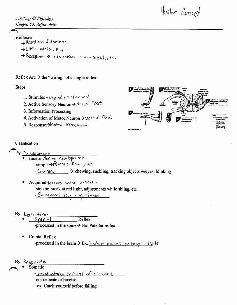
Reflexes
t'f'fc'c h'>r
ReflexArc^ the "wiring" of a single reflex
Steps
1. Stimulus-j)\v^S\riK\ Of2. Active Sensory Neuron->
3. Information Processing
4.Activation of Motor Neuron^ '̂ C/H rc'ot.5.Response->^^oVdt
Classification
II
'y HiO .i/ri^p>rY\guA4-Innate-^i^c/r^j
-simple^f^"^ ^- fr>iA-\p\r\ chewing, suckling, tracking objects w/eyes, blinking
• Acquired-U^fn?^?^
-step on break at red light, adjustments while skiing,etc
®y L<n^rt4-iReflex
-processed in the spine-^ Ex. Patellar reflex
• Cranial Reflex
-processed in the brain-> Ex. i>jJthyy at brigu)- )rs
By Ref>p<?KNS^• Somatic
- np '.V'- riC ^-not delicate or precise- ex. Catch yourself before falling

Visceral
- AutonOt
By CflrAyi^Xi'Vu oF r.irC:tA>4-• MonosynSptic
-
-simplest & r^piol
Polysynaptic
-sensory neuron synapses with
-complex-^ can control vvif.tkp^ '-longer delay between SfiK-' 1*1-
- Ci' Vc and/or inhibit
which synapse with motor neurons
ir^liSrlg- fifu ^
Examples of Monosynapttc
1. Stretch-> r,Y.i ftC. regulation of muscle length
-Many associated with maintaining |V/vj-orcEx. Patellar Reflex^ triggered by stretching the quads-> rapid increase of muscle tone of quads
*stretches sensory structures called f\)i,
Examples of Polysynaptic
1. Tendon Reflex- monitors to prevent tearing
- the greater the tension, the greater the inhibitory effect on muscles
Reflex- move away from stimulus (pictured below)
-pain, hiAcV) pressure tomcor
-flexor reflex- step on a tack
-cross-over (pictured to the right) / ./
pressure To motor neurons in other segments of the spinal cont
Psinlui
7 stimulus
Oletrlbutlon within grey horns to other segments el the spinel cord4 4
Flexors ^—stimuleted -J'
KEY
— SsnsorynMsm ----lAeler neuRIslknulawe} (InNbilsd)
• Motor notson
(uiiiwIalrC)
B Extensors —•JmiKi-.f inhibited
fsUmutated tea if• Extensors
stimulated
' Bexois
inhibited
I KEYj Senseryncuren! {sUimilaled)
ExcHelorykilemeuron
—Molar rteuion
(inhlMed)
— tnhlblloryimerrMtiton

Uynl
^IB
•E
sIS
nn
UJ
UJ
uu
nn
Uu
nn
uu
uunto
•u
ni
1-
<f
u1
Y-
OJ
ST
UD
EN
TS
INS
TR
UC
TO
RS
"l!SE
S<)
2P
E^
II.OM
.^T
Ti
•E
XA
MP
LE
;=
*=
=b
==
0=
t=e=
•M
AK
ED
4/?
KM
AR
KS
•E
RA
SE
CO
MP
LE
TE
LY
TO
CH
AN
GE
•M
AK
EN
OS
TR
AY
MA
RK
S
KE
YM
AR
KIN
GIN
ST
RU
CT
ION
SK
EY
:
VE
RIF
Y:
NAMFljf/^kr-
]?\
nn
CD
mu
u
nn
<<
uu
In
1n
n
4U
J1
UJ
UJ
fu
1u
u
nn
ni
nQ
oQ
1Q
Uu
U1
U
n»
nn
nu
uu
uu
1u
uu
nn
nn
n0
1<
0C
Om
CD
uu
uu
U
nn
nn
1<
<<
<•1
uu
uu
•
lOC
Oh
.C
OO
)
SU
BJE
CT
.M
ustmark
thisbo*
onK
eysheet.
Prin
tsco
rrectresp
on
senext
toincorrect
answers,
ifVerify
isnot
marked,
ad
ashw
iiiprint
nextto
inco
rrect
an
swers.
RE
SCO
RE
:Rescoras
apreviously
scoredtest.
Autom
aticafiyprints
correctresponses.
DA
TEv
nu
^P
ER
IOD
.
Ap
perso
nR
EO
RD
ER
(f2S420-RR
06/t1&
1SQZ
U.S.Patent
No.6,695,216
06/13w
ww
.apD
erson
.com
80
0.8
27
.92
19
L
nn
nn
nW
lUU
JlU
liju
uu
uu
nn
no
uu
uu
u
nn
nn
UJ
UJ
UJ
UJ
uu
uu
nI
n1
Q1
oI
U1
u1
•n
nn
1u
uo
1u
uu
nn
n1
CO
mID
fu
uU
1n
n•
n<
<1
<u
u1
u
CO
r-YC
Or—
T-
•>
-
\
ni
nn
•<
<4
uu
I
[1n
nn
nn
nn
nn
nn
nn
nn
nn
Ui
UJ
UJ
UJ
UJ
UJ
UJ
UJ
UJ
UJ
UJ
UJ
UJ
UJ
UI
UJ
U)
UJ
uu
uu
uu
uu
uu
uu
uu
uu
uu
nn
nn
nn
nn
nn
nn
nn
nn
nn
oo
oa
Qo
Q•
oo
oQ
•Q
oQ
UU
uu
uu
Uu
Uu
uu
uU
uU
uU
nn
nn
nn
nn
nn
nn
nn
nn
nn
uu
oL
>o
uu
uu
uo
oo
ou
ou
ou
uu
Uu
uu
uu
uu
uu
uu
uu
u
nn
nn
nn
nn
nn
nn
nn
nn
nn
IDC
DC
DC
DC
DC
DC
DC
DID
CD
CD
00
CD
CD
CD
CD
so
CD
Uu
UU
uU
UU
UU
uu
uU
UU
uU
nn
nn
nn
nn
nn
nn
nn
nn
nn
<<
<<
<<
<<
<<
<<
<<
<<
<<
uu
uu
uu
uu
uu
uu
uu
uu
uu
CO
mC
Dr-Y
CO
o>
ocy
CO
mC
Oh
-C
OC
7>o
pj
C\J
CM
cv
C\J
CM
CV
JC
OC
OC
OC
OC
OC
OC
OC
OC
OC
O"f.
nn
nn
nn
nn
nn
UJ
UJ
UJ
UJ
UJ
UJ
UJ
UJ
UJ
UI
uu
uu
uu
uu
uu
nn
nn
nn
nn
nn
Q•
aQ
oo
oo
Uu
uu
uU
uu
uu
nn
nn
nn
nn
nn
ou
uu
uu
uo
uu
uu
uu
uu
uu
u
nn
nn
nn
nn
nn
CD
IDID
CD
CD
CD
CD
IDC
DC
Ou
UU
uU
uU
UU
u
nn
nn
nn
nn
nn
<<
<<
<<
<<
<<
uu
Uu
uu
Uu
uu
C<
JC
O3
lOC
OC
O0
>o
M-
M-
'TM
-rr
fM
-•y
in
ISI
1KII
IIM
1II
IIII
IIII
IIII
II(,
IIIII
IIII
I!III
IIII
III
Sco
re-
-Resco
re-
17
/85
.0^

•^KN
AM
E.
c
nn
nn
nn
nn
nn
nn
nn
nn
nn
nn
nn
nn
nn
nn
nn
nn
nn
nn
nn
nn
nn
nn
nn
nn
nn
ulU
Ul
lUlU
UJ
UJ
Ul
UJ
Ui
UJ
UJ
UJ
Ul
UJ
Ui
UJ
UJ
UJ
UJ
UJ
UJ
UJ
UJ
UJ
UJ
UJ
UJ
UJ
UJ
UJ
UJ
UJ
Ui
Ul
UJ
Ul
UJ
UJ
Ul
Ul
Ul
Ul
Ul
UJ
UJ
Ul
UJ
UJ
Ul
uu
L)
uu
uu
uu
uL
Iu
uu
uu
uu
uu
uu
uu
uu
uu
uu
uu
uu
uu
uu
uu
uu
uu
uu
uu
uu
nn
nn
nn
nn
nn
nn
nn
nn
nn
nn
nn
nn
nn
nn
nn
nn
nn
nn
nn
nn
nn
nn
nn
nn
nn
Q•
oo
a•
aQ
Do
aQ
Qo
oo
Q•
Oo
oQ
ao
oo
oo
CD
Qo
oo
oo
aQ
oo
QU
uu
uu
uu
uU
UU
uU
Uu
UU
uU
Uu
uU
uU
Uu
uu
Uu
uu
uu
uu
Uu
uu
uu
UU
uU
uu
U
nn
nn
nn
nn
nn
nn
nn
nn
nn
nn
nn
nn
nn
nn
nn
nn
nn
nn
nn
nn
nn
nn
nn
nn
nn
uo
uo
uu
oo
uo
uu
oo
ou
Oo
ou
uC
Ju
CJ
uC
Jo
(j
(j
(J
u<
jo
oo
uo
oo
u(J
ou
uu
uu
(J
o(J
uu
uu
uu
uu
uu
uu
uu
uu
uu
uu
uu
uu
uu
uu
uu
uu
uu
uu
uu
uu
uu
uu
uu
uu
uu
nn
nn
nn
nn
nn
nn
nn
nn
nn
nn
nn
nn
nn
nn
nn
nn
nn
nn
nn
nn
nn
nn
nn
nn
nn
CQ
OQ
CD
ffiC
OC
Dm
mC
DC
DC
DC
DC
DC
O03
03C
D03
CD
CD
CD
03
CD
CD
mC
D03
CD
CD
CD
CD
CD
CD
CD
CD
CD
CD
CD
CD
CD
CD
CD
CD
03C
DC
OC
DC
DG
OC
Ou
uu
uu
uu
uU
UU
UU
uu
uU
Uu
uU
UU
Uu
Uu
UU
UU
uu
UU
UU
uu
Uu
UU
UU
uU
uU
u
nn
nn
nn
nn
nn
nn
nn
nn
nn
nn
nn
nn
nn
nn
nn
nn
nJ1
nn
nn
nn
nn
nn
nn
nn
nn
<<
<<
4<
44
44
44
44
44
44
44
44
44
44
44
44
44
44
44
44
44
44
44
44
44
44
uu
uu
Uu
uU
uu
uu
uu
uu
uu
uu
Uu
uU
UU
UU
uu
uu
Uu
uu
uu
uu
uu
uu
uu
uU
uu
CM
CO
i/>C
D1
^C
OC
7)o
CM
CO
•cr
in
CO
N.
CO
a>
oC
MC
OT
in
CO
h.
CO
O)
oC
MC
O't
in
CD
r«.
CO
c:5
oC
MC
Oin
CO
N.
CO
CD
olO
mm
in
in
in
in
in
CO
I.CDC
DC
DC
OC
OC
OC
OC
OC
O1
^h
"h
-C
O.C
DC
DC
OC
OC
OC
OC
OC
OC
Oo
>U
iC
DC
DC
DO
)(D
<D
O)
CD
o
III
II
II
II
II
II
II
II
II
II
II
II
II
II
II
II
II
II
II
II
II
II
II
II
II
I
c•Sco
re-R
esco
re-

y\natomy PhysiologjiChapter 13 SpineQui:^
Name
''̂ Nfatch the labels to the picture on the right and below. Answers are only used
a. Conus Medullaris b. Cervical Enlargementc. Lumbar Enlargement d. Cauda Equinae. Filum Terminale
b 1Match the labels to the picture below. Answers only used once.
a. Arachnoid Mater b. Subarachnoid Spacec. Pia Mater d. Dura Mater
e. Epidural space
6 U
<:/»'»• Tij»* r -
q 2
c ^
'lO(between #6 and #9)
Mark"a" for monosynaptic or "b" for polysynaptic11. Involves an interneuronb12. The patelJar tendon reflex
13. Can result in several different muscle groups tocontract
Match the following parts of the spine to the diagram below:^f^^entral Rootle Co\t/lv^h-U]V\fcteTTateral Hornyiy^ —' ^
Dorsal Column^ gsterior Median Sulcus D
^BT^orsal Root Ganglionjfy \ \l^pinal Nerve DK A. \(20- PosteriorHorn(i
P I)
Anterior

uS
TU
DE
NT
SIN
ST
RU
CT
OR
S
KE
YM
AR
KIN
GIN
ST
RU
CT
ION
SK
EY
:M
ustm
arl(this
boxon
Key
sbeet.
VE
RIFY
:P
rintsco
rrectresp
on
senext
toIncorrect
answ
ers.If
Verity
isnot
mark
ed,
ad
ash
will
printn
ext
toIn
co
rrect^
sw
ers.
RE
SC
OR
E:R
escores
apreviously
scored
test.A
utomahcally
printscorrect
respo
nses.
MA
MF
RUBJECTrVYTp4i:ir
/?w
uniV
fD'
Es
IS
?lisE
W>."2"l'l-.N
Cn.
OM
.Y'T
T
•E
XA
MP
LE
:=
«=
=b
=^
=0
==
e=
•M
AK
E0
AH
KM
AR
KS
•E
RA
SE
CO
MP
LE
TE
LY
TO
CH
AN
GE
•M
AK
EN
OS
TR
AY
MA
RK
S
tnn
nU
JlU
UJ
uu
u
nn
nn
Q•
OQ
uu
uu
nn
oo
uu
,n
in
i(D
§m
•u
•u
1
nn
<<
CM
CO
V
\
nn
nn
nU
JU
JU
JliJ
UJ
uu
uu
u
nn
Q•
uun
n
ou
uu
nn
nO
uu
u
nn
nn
nffl
CD
uu
uu
u
•u
ui
in
CO
h-
CO
<35
o
nn
nn
nn
nn
nn
UJU
JU
JU
JU
JU
JU
JU
JU
JU
Ju
uu
uu
uu
uu
u
nn
UU
nn
nQ
OO
uu
u
nn
o(J
uu
I^
^I
•U
Li
1
nn
ou
uu
fn
an
lintn
n(d
Aid
biS
id
Acq
iq
utu
Rlu
lu
u
\'-C
Mco
TT
inco
r^^
o
H
II
Il|(
III
IIII
IIII
II1
IIII
IIII
I(.
IIII
IIII
IIII
IIII
IIII
I(
DA
TE7
,/M}
MP
ER
IOD
.
Ap
perso
nR
EO
RD
ER
#2
54
20
-RR
06
/11
A1
80
7U
.S.P
atent
No.
8,6
95
,21
6
ww
w.apperson.com
80
0.8
27
.92
19
nn
nn
nU
JU
JU
JU
JU
Ju
uu
uu
n1
nt
n
0V
QJ
au
1U
1u
nn
•n
nu
uf
uu
uu
1u
u
nn
nn
n
mm
<0
CD
CD
uu
uU
U
In
n1
t{
<<
{1
•u
u1
1in
CO
00
CM
CM
CM
CM
CM
nn
nn
nn
nn
nn
UJU
IU
JU
JU
JL
UU
JL
UU
JU
JU
UU
UU
UU
UU
U
nn
QO
uu
nn
nO
uu
uI
nn
nn
nn
in
fn
uu
uo
ou
fu
fu
uu
uu
uu
lu
lu
(ftn
nin
nin
nS
mcD
acD
caA
cD
mlu
ulu
ulu
u
nI
IL
>U
uu
if
^$
fi-
nn
nn
nn
nU
JU
JU
JU
JU
JU
JU
J
uu
uu
uu
u
nn
nn
nn
n
••
0Q
00
Qu
uu
uu
uu
In
nn
nn
n
10
00
UU
uf
uu
uu
uu
nn
nn
nn
n
CD
CD
CD
CD
CD
CD
CD
uU
uU
uU
U
nI
nn
nn
n<
<<
<<
<u
1u
uu
uu
tin
<0
r>.
CD
o>
0'S
-•M
-in
ea
lOu
j0
5<
/><
3<
o§

NA
ME
.<
nn
nn
nn
nn
nn
nn
nn
nn
nn
nn
nn
nn
nn
nn
nn
nn
nn
nn
nn
nn
nn
nn
nn
nn
nn
lUU
JlU
tuU
JU
JU
JU
JU
JU
JU
JU
JU
JU
JU
Jm
UJ
UJ
UJ
UJ
UJ
UJ
Ul
UJ
UJ
UJ
UJ
UJ
UJ
UJ
UJ
UJ
cu
UJ
UJ
UJ
UJ
UJ
UJ
cu
UJ
UJ
UJ
UJ
UJ
UJ
UJ
UJ
UJ
UJ
uu
uu
uu
uu
uu
uu
uu
uu
uu
uL
iu
uu
uu
uu
uu
uu
uu
uu
uu
uu
uu
uu
uu
uu
uu
u
nn
nn
nn
nn
nn
nn
nn
nn
nn
nn
nn
nn
nn
nn
nn
nn
nn
nn
nn
nn
nn
nn
nn
nn
nn
Qo
••
oo
oo
oQ
oQ
oQ
Qo
•a
oo
Q•
Qo
•o
Q•
Qo
•a
oQ
ao
Qo
ao
uu
uL
iu
uu
uu
UL
iU
uu
Uu
UU
uu
uu
Uu
Li
uu
uU
uU
UU
uu
uu
Uu
uU
UU
uU
uu
uL
iu
nn
nn
nn
nn
nn
nn
nn
nn
nn
nn
nn
nn
nn
nn
nn
nn
nn
nn
nn
nn
nn
nn
nn
nn
nn
oc
uo
oo
ou
oo
ou
ou
oo
uo
ou
oO
uo
uu
uo
uu
uo
uu
uo
uu
OU
CJ
uo
uu
uU
uu
LI
uu
uu
uu
uu
uu
uu
uu
uU
uu
uu
uu
uu
uu
uu
uL
iu
uu
uu
uu
uu
uu
uu
uu
uu
Li
u
„n
nn
nn
nn
nn
nn
nn
nn
nn
nn
nn
nn
nn
nn
nn
nn
nn
nn
nn
nn
nn
nn
nn
nn
nn
nIL
CO
0)
IDC
DC
DC
Dm
mC
Dm
CD
IDC
DID
CD
CD
IDC
DC
DC
DC
DC
DC
Dm
CD
CD
CD
CD
CD
CD
CD
CD
CD
CD
CD
CD
CD
CD
CD
IDC
DC
DC
DC
DC
DC
DC
DC
Dm
CD
uu
Uu
uU
uu
Uu
uU
uU
Uu
UU
uU
uu
Uu
UU
Uu
UU
UU
UU
UU
UU
UU
UU
Uu
Uu
Uu
uu
nn
nn
nn
nn
nn
nn
nn
nn
nn
nn
nn
nn
nn
nn
'nn
nn
nn
nn
nn
nn
nn
nn
nn
nn
nn
£<
<<
<<
<<
<<
<<
<<
<<
<<
<<
<<
<<
<4
44
44
44
44
44
44
44
44
44
44
44
44
4u
uu
uu
uu
uu
uu
uu
uu
uu
LJ
uu
uu
uU
UU
UU
Uu
UU
UU
Uu
UU
uU
Uu
uu
uU
uu
U
CM
CO
in
(D
h-
CO
O)
o<
MC
Oin
CO
r-
CO
o>
oC
MC
Oin
CD
CO
O)
oT
-C
MC
O4
-in
CD
h-
CO
05
oC
MC
O"ir
mC
DN
CO
O)
oto
to
in
in
in
in
inin
in
CO
.CO
CO
CO
CO
CO
CO
co
CO
CD
h-
r«.
r«-
CO
CO
CO
CO
CO
CO
CO
CO
CO
CO
a>J
o>
05
C3)
05
CJ>
O)
a>
O)
05
o
III
II
II
II
II
II
II
II
II
II
II
iI
II
II
II
II
II
II
II
II
II
II
II
II
II
I
•Sco
re-R
esco
re-

00011AtiatofTiy wv/v.Chapter 12Quest Name
Match the following eventsto the parts of the nervous system below. Answersmay he used once,morethan once or not at all
a. Autonomic b. Somatic Sensory c. Somatic Motor d. Speciale. Visceral ab.Sympathetic ac.Parasympathetic
1. The stretch receptorsin the bladdertelling the brain that it is filling2. Yourheart rate dramatically increase aftera fiiendjumpsout of thebushes andscares you3. You push on the brake pedal to stopa car in response to seeing a traffic light turnfrom yellow to red.4. You feel a mosquito biting you
Match the following types of neurons to the descriptions/diagrams below. Answers may be used once,more than once or not at all
a. Anaxonic b. Bipolar c. Multipolar d.Unipolar
5. What type ofneuron is pictured to the right in Figure A?6. Typically only found in the brain7. Sensory neurons (not special senses)8. Neurons that wouldform a synapse with a muscle cell
Match the following types ofneurons to the characteristics below.Answers may be used once, more than once or not at all
Wm
-Syna^itetfnaW*;
Figure A
a. Type A b. TypeB ^ c. TypeC wnnm^W:9. Myeiinated multipolar neurons Figure A10. Unmyelinated special sensory neurons11.Sensory neurons (non-^ecial senses)12. Transmit impulsesas fast as SOOmph
Match the following neuroglial cells to the descriptions below. Answers may be used once, more thanonce or not at all:
a. Astrocytes b. Ependymal c. Microglial d. Oligodendrocytese. Satellite ab. Schwann
13.These cells are foundin the CNS only (chooseall that apply)14. These cells form the blood brain barrier in order to guard brain cells15. These cells form the myelin sheath for PNS neurons
16. These cells form cerebrospinal fluid
17. A neuron at rest...
.a. Isdepolanzing,'
a trah^embrane potential'of'-55'c. Is experiencing a graded potential- ,
d.. None ofthe aboved. None ofthe above

0001118. Resting potential is established by... ww-a. More potassium ions being pumped in than sodium being pumped outb. More potassium ions diJSusmg out than sodium inc. More Sodium ions gettingpumped in that oittd. More Sodium ions getting pumped out than in
Match the followmg ion channels to their descriptions below.Answers can be used once, more than once,or not at all:
a. Chemically gated b. Leak c. Sodium/Potassium pump d. Voltage-sensitive19. Is foimd ONLY in the dendrites and axons (jJjL.
20. Found ONLY in the axon hillock and axon
21. Found throughout the entire membrane ofthe neuron (choose aUthat appl^)22. Associated with resting potential (choose-hU that apply)23. Help to re-establish resting potenti^ gradients24. Associated with graded potentials
25. Graded potentials..:a. Can lead to action potentialsb- Depend upon the intensity ofthe stimulusc. Are localized
d. All ofthe above
26. If acetylcholine binds to its receptor ontheneuron cellmembrane...a. Sodiumchannelsopenand sodium moves out ofthe neuronb. Potassium channelsopen and potassium moves out ofthe neuronc. ^Sodium channels open and sodium moves into the neurond. None ofthe above
' . 27. A.transmei^iane potentialof-55 mV (choose2)..,a. Is con^deiedthresholdb. Wouldopen chemicaUy-gated ion channelsat the axon hillockc. Is considered resting potentiald. Wouldopenvoltage-sensitive ion channels at the axonhillock
Match the following descriptions to the answers below. Answers may be used once, more th^ once or notat all.
a. VoltageSensitiveSodiumChannel b. VoltageSensitive Potassium Channel
28. Opens vihen threshold is reached' ' 29. Opens at+30 mV '
Closes 8t .
32. Leadsto hypeipolar^tion ofa neuron
•K. > > ^ V

33. An action potential...a. Is triggeredby a gradedpotentialb. Begins at the axon hillock
c. Involves depolarization of the axond. All ofthe above
00011
Match the followingquestions to the graph on the right Answersmay be used once,more than once or not at all
34. Resting Potential35. Threshold
36. Sodium Channels Close
37. Depolarization38. Graded potential39. Hyperpolarization40. Potassium Channels Open
• w.
AB
Match the following '^potenti^" to the description hidow. Answers can be used onc^ more than once or
a. Actionpotential b. Graded potential c. Restingpotential d. Noneof the above
41. Results from movement ofions throu^ leak channels42. Begins at an axon hillock43. Can be stimulated to depolarization OR hyperpolarization44. Occurs throughout the entire neuron45. Associated with a''domino effect**

mmm
Section
.^'«SiiSi:>r'>JS,-5ft3iSiWS»^MsSS''W»»> •-~i
REVIEWING YOUR KNOWLEDGE
A. Meninges
Match the terms with the correct description.
0< 1. Arachnoid mater
~ 2. Denticulate ligament
middle meninx; weblike
J&r tough, outer meninxspace filled with adipose tissuethin meninx intimate with spinal cordcontains cerebrospinal fluid
F. extension of pia mater attaching to dura3. Dura mater
4. Epidural space
5. Pia mater
Subarachnoid space
B. Identification of Spinal Cord Structures
foot.
FIGURE 15.7 Anterior view of spinal cord Withmeninges and gray and white matter.

306 Exercise 15 Anatoroy ot the Spinal Card
C. Spinal Cord Structures
Write the terms by the correct description.
anteriorgray horn conusmedullaris posterior (dorsal) rootanterior median fissure filum terminale ' posterior (dorsal) root ganglionanterior (ventral) root gray commissure posterior gray horncauda equina gray matter posterior median sulcuscentral canal lumbar enlargement white mattercervical enlargement
1. contains neuron cell bodies that receive impulses from sensory neurons
2. contains neuron cell bodies and unmyelinated processes
—yy")^^3. shallow groove on dorsal side
(7 t/| Cr^lV^>Y> 4..;connects right and left halves of gray matter in spinal cord5. i sensory branch entering spinal cord
COf\ ^6, tapered end of spinal cord(\l/^hCyCiOC ^T. motor branch exiting spinal cord
—Coek ^ contains sensory neuron cell bodies
9, collection of spinal nerves that arise from inferior end of spinal cord
contains myelinated axons
11. contains somatic motor neuron cell bodies
ryiAffA\ _ 12. contains cerebral spinal fluid
r^rVlfA)—13. bulge in spinal cord containing cell bodieS of motor neurons supplying upper limb
15. extension ofpiamater that attaches spinal cord tococcyx
16. bulge in spinal cord at T9-T12
r#
It"

USE NO. 2 pevai only
NAMEjju
SUBJECT.
• EXAMPLE: =*= =b= mtm e:o= =e=
• MAKE DARK MARKS
• ERASE COMPLETELY TO CHAUGE
: • MAKE NO STRAY MARKS
KEY MARKING INSTRUCTIONSKEY: Must marlt this t>Ox on Key sheet.
VERIFY: Prints correct response next toIncorrect answers. If Verify is notmarked, a dash will print next toincorrect answers.
RESCORE: Rescores a previously scored test.Automatically prints correct responses.
PERIOD.
APPERSON EDUCATION PRODUCTS 800.827.9219»25420-RR 06/tl A1807 US Patent No. 8.695,216 09/11
n i n n n
lU i lU UJ lU
u 1 u u u
n n n n n
O o o o ou u u u u
n n n 1 1O O U i 9u u u 1 1
1 n • n n
a m If m ffiV u 1 u u
1 n n n n
J UJ UJ UJ UJ
1 u u u u
n n 1 1 1o a 1 i {u u 1 1 n
n n n n n
U O O O uu u u u u
n n n n n
m a m m CDu u u u u
n_nniinitinnn
U UUlfutlBUlJU>
S '-ojcO'^-mcoNcooJOi-
n n nUJ UJ UJu u u
n n n
Q Q Ou u u
n n no O cLI u u
n n n
< < <u u u
CO in
n n n t " t n n n n n n n n n n n n n n n n n n n n n n n n n
UJ UJ UJ If UJ UJ q UJ UJ UJ UJ UJ UJ UJ UJ UJ UJ Ul UJ UJ UJ UJ UJ UJ UJ UJ UJ UJ UJ UJ UJ UJ
u u u 1 u u 1 u u u 11 u u u u u u u u u u u u u u u LJ u u u u u
n n n n n n n n n n n n n n n n n n n n n n n n n n n n n n n no o O O Q Q Q Q Q Q o Q o Q Q o Q o o Q Q o Q a Q Q o Q Q Q Q QU U u u u u u Li LJ Li u U Li U U u U u u U U u U u U U u U U U U U
1 1 n n n I f n n n n n n n n n n n n n n n n n n n n n n n n n
I f u o o 4 1 O u u u o u o o u o u o u u o o o o u o o o o o O1 »• u u u 1 1 u u u u u ^u Li u u u u LJ u u u ' u u u u u u u u u u
1 " 1 " 1 1 " n n n n n ! n n n n n n n n n n n n n n n n n n n n
q o J n q q CO CD CD CD CD CO CD CD CD CD CD CD CD DO DD CD CO CD ffi ffi ffi ffi ffi CD ffi ffi1 u f u 9 1 Li U U U u Li LJ u u LI U u U U u u . u U u u u u LJ u U u
n n n « 1 n n n n n n n ri n n n rt n n n n n n n n n n n n n n n
< < < T a < < < < < < < < < < < < < < < < < < < < < < < < < < <u u u 9 1 u u u u u u u u Li u u u u LJ u u U u u u u LJ u u u Li u
o> o CM 05 ^ m CO r- 00 cn o CM CO in to !»• CO 05 o CM CO m (O r- CD <51 oCM. CM CM CM CM CM CM CM CM CM 05 .m 05 CO CO CO CO CO CO CO •M- •M" in
Score —y—Rescore-
-s: /•! Af-iV
t?: Si
U°" 52 g;
I III IIIIII I!II II II II M II II II II II II M I II II II II II II II I

jjEs
a
NA
ME
nn
nn
nn
nn
nn
nn
nn
nn
nn
nn
nn
nn
nn
nn
nn
nn
nn
nn
nn
nn
nn
nn
nn
nn
nn
lUU
JU
JU
JU
JU
JU
JU
JU
JU
JU
JU
JU
JU
JU
JU
JU
JU
JU
JU
JU
JU
JU
JU
JU
JU
JU
JU
JU
JU
JU
JU
JU
JU
lU
lU
JU
JU
JU
JU
lU
JU
JU
lU
JU
JU
JU
JU
JU
JU
J
uu
uu
uu
uu
uu
uu
uu
uu
uu
Li
uu
uu
uL
iu
uu
uu
uu
uu
uu
uu
uu
uu
uu
uL
Ju
uu
u
nn
nn
nn
nn
nn
nn
nn
nn
nn
nn
nn
nn
nn
nn
nn
nn
nn
nn
nn
nn
nn
nn
nn
nn
nn
Q0
00
00
Q0
0Q
Q0
Q0
0Q
•o
aQ
o0
00
00
DQ
00
0Q
Q0
C3
Q0
Q0
Q0
Q0
0u
uu
uu
uu
uu
Uu
uU
uu
Uu
uu
UU
Uu
uu
uu
Li
uu
uu
uu
uu
UU
uU
uL
JL
IU
Li
Uu
UU
u
nn
nn
nn
nn
nn
nn
nn
nn
nn
nn
nn
nn
nn
nn
nn
nn
nn
nn
nn
nn
nn
nn
nn
nn
nn
uU
00
uu
00
(J
uu
uu
u0
uu
uu
uu
00
00
aU
UU
00
0U
U0
00
a0
00
u0
UU
u0
u0
uu
uu
uu
Li
uu
uL
iu
uu
uu
Uu
uu
uu
uu
uL
iu
uu
Li
uu
uu
uu
uu
uu
uu
uu
uu
uL
iu
uu
_n
nn
nn
nn
nn
nn
nn
nn
nn
nn
nn
nn
nn
nn
nn
nn
nn
nn
nn
nn
nn
nn
nn
nn
nn
nLX.
ffi0
3(0
CO
nC
BC
Om
CD
IDC
DC
DC
DC
DC
DC
DC
OC
DC
DC
DC
DC
Dffl
CO
mC
DC
DC
DC
DC
DC
DC
DC
DC
DC
OC
DC
DC
DC
Dm
CD
CD
CD
CD
CD
CD
CD
CD
CD
CD
^u
uu
uu
uu
uu
Uu
UU
Uu
Uu
Uu
uu
uu
uu
Uu
UU
uU
Uu
IJL
JU
IJU
LJ
uu
UU
uL
JU
Uu
LJ
U
rn
nn
nn
nn
nn
nn
nn
nr
nn
nn
nn
nn
nn
nn
nn
nn
nn
nn
nn
nn
nn
nn
nn
nn
nn
b<
<<
<<
<<
<<
<<
<<
<<
<<
<<
<<
<<
<<
<<
<<
<<
<<
<<
<<
<<
<<
<<
<<
<<
<<
<^
uu
uu
uu
uu
uu
uu
uu
uu
uu
uu
uu
uu
Li
uu
uu
uu
uu
uu
uu
uu
uu
uL
iu
uu
Li
uu
u
OJ
rt
'»m
CD
r-~C
DO
)0
r-
DJ
CO
in
CO
r».
CO
O)
0O
JC
O•*r
in
CO
CO
o>
0cv
jC
Oin
<0
N®
O)
0O
JC
Oin
CO
h-
Oi
0in
in
min
min
in
in
in
CD
.<D
CO
CO
CO
CO
CO
<0
CO
CO
r--r--
r--r-
r-
r-
h-
CO
®0
0C
O(O
®as
OiJ
o>
Oi
CJ>
o>
Oi
O)
O)
Oi
Oi
0
I1
1I
II
II
II
II
II
II
II
II
II
II
II
II
II
II
II
II
II
II
II
II
II
II
II
iI
Sco
re-
-Resco
re-
u°"
55
a
oto
SO
"lu
^s
mo
KO
3

Nen/ows System. 6ZuLz N«kvte:
Fill in the following llowchart on divisions of the nervous system with the choices below.Answers are only used once:
a. Autonomic b. Central Nervous System c. Efferent Divisiond. Parasympathetic e. Peripheral Nervous System ab. Afferent Divisionac. Visceral/Somatic/Special ad. Sympathetic Division ae. Somatic MotorIII
Nervous System
Match the following parts of a neuron to the diagram below. Answers may be used once, more thanonce or not at ail
a. Axon b. Axon Hillock c. Cell Body d. Dendritese. Nodes of Ranvier ab. Synaptic Bulb
AT T
41
7 JiL \sv ',-hM
14. The cell above is... a. myelinated b. unmyeiinated

Match the following types of neurons to the descriptions below. Answers may be used once, morethan once or not at all:
^>^^4paxonic "^b^ipolar c. Multipolar d. Unipolar
15. Neurons that would be found in the special senses16. Are often motor neurons
17. Are located only in the brain18. Are sensory neurons (excluding special senses)
Match the neurons to the descriptions below. Answers may be used once, more than once or not atall.
a. Type A b. Type B c. Type C
19. Sensory neurons can be of this type? (Choose 2)20. Unmyelinated21. Impulses 40mph on average
Match the following neuroglial cells to the descriptions below: Cells can be used once, more thanonce or not at all
a. Oligodendrocytes b. Astrocytes c. Satellite Cells d. Microgliale. Schwann Cells ab. Ependymal
22. The cells involved in myelinating a neuron (choose 2)23. The cells responsible for cerebrospinal fluid production24. Cells that provide communication between the nerve cells and their environments (choose 2)25. Accessory cells found in the PNS (choose all that apply)

Anatomy & PhysiologyChapter 12: Merve Impulse Review
fransmembrane Potential
1. Define transmembrane potential-
+ 0 OCA+.
"3%'"'li.EP0imi.i-Vk> vw.(id\«s bejpj Meolt -lOwV
3. Describe the relationship between resting potential and transmembrane potential.
VJhm l-hc pofcjAh'o^ '̂3a)A(j (iM- W^z. I'S l^f e^l.-~lO^\j.
4. What is the typical resting potential for aneuron? _ O fv\\I
5. Howis this resting potential created? Be sure to include Na+andK+ in your explanation.
/\a of OfiAit^to (Yc CVKO/x vjfiW 44'X or^
h OLlf of Itc g-toVN n.i/voi tw m (4i:;. C,X/->y\
Graded Potential
6. Where in the neuron does a graded potential typically occur?
0]to\
7. What type of gated ion channels are associated graded potentials?
8. Describe what threshold is for an axon?
/it Vhc
"3

f
>9. What is the relationship between a graded potential and threshold?
orcl^ c)x>vy\e)^ )"G> -ho(or po^eyNHcl V\>rc-5l-.o\el
10. What types of gated ion channels are typically associated with threshold?
\C)f^
11. Where in the neuron would you find the ion channel that is associated with threshold?
Owteicl^ bv Crj\ ^ '-ic b'.\\oc)^Action Potential
12. Which specific ion channels first open when threshold is reached?
Ncv''
13. Which direction does this ion move?
fft-14. What happens to the transmembrane potential ofthe axon? ^
|y3ovV\v/c KJca' ^̂ 0^\J
15. What is the term that is used to describe this change in transmembrane potential?
16. When does this ion channel close? What is theC^nsipambra^^
[f-lVciA i" fcu.&e'̂ ia\J /i'S
17. Which ion channel opens next?
18. Which direction does this ion move?
Vco\i^ 0Uv\:5\A«-fo iv>?.ide .19. What happens to the transmembrane potential as a result ofthis ion movement?
7?. yv^e^rt, ey\h<j-s, ^(rc

20. What is the term used to describe this change in transmembrane potential?
21. When does this ion channel close? What is the ^mSnembrane potenti^
iV ' lOfwVj. r<LpQWi"TL£^,22. What occurs to the transmembranepotential as this ion channel closes?
23. Explain how action potentials along an axon behavelike a "dominoeffect."
Ql^cc- NCa"^ l i'S }->n2_C l^-^P
i/rr.\ d^/cd pohne '̂-^.
24. Is this an example of a positive or negative feedback mechanism? Explain
t>cC(A{A6c. ^ Cijc\c 0\r£^ »'SCcMVliW-d Ipo-byV-^e fagfe^VoxgW mchp^xSv '̂̂
25. Inwhat type ofneurons would anaction potential move along the axon through continuous propagation?Saltatory propogation?
M'oa Dfcf^- '̂̂ vKcjA-— CfviAhiAUOtAo, pffopcv(^<Al^c.<A •^C\(rc/?>6<^v\hrtu
^ Pfop&ACAbia^VN ^ F(X<?.Ve<- iw\^u\^eCfow f PjC^z. (5-P ro'Aie' '<i^ ( deOo)/rPpoi (la-'h'Ai^e)
- ><TfiviiAueJ node ho v^oc^ C?»\ooo)
6(x\bo|-6J»^(A' jUK\piir^ irrow^ pen-so I,'A bo ^^cr"3€4A.

\
o
(\
fljps c?r \NC?"^

Anatomy Physio/o^ J1Nen/ous System Organisation and l^enrons
A. Organization of the Nervous System- Match the following terms to the appropriate blanksAfferent Efferent Central SomaticAutonomic Special Visceral
1. The brain and spinal cord are part of the
2- be divided into two divisions. List Ap hvo divisions below:a. b. Cl^A/>{)r\r^
nervous system
3. The part of the PNS is directly responsible for control of skeletal muscle contractions- both voluntary andinvoluntary S^rmKg.
directly divided into the sympathetic and parasympathetic divisions.
5. Pain, temperature and pressure receptors fall into which immediate division ofthe PNS*?- -
6. Stretch receptors in the bladder communicating to the brain that the bladder is full would be part of theV division ofthe afferent division ofthe PNS
f^\ B. Structure ofaneuron -Label the diagram below with the following terms
Axon Axon hillock ^
/ "Pf^S(M!34c;r^
Axon terminals
Pfindritftfr
of Rftfmcr
CelLBodySchwann cells
a '̂i^ for on
BiAbs
Ax6n
cord)
^Chcu0/inCrll5
SLn^n\A]d

C. Structural Classification of Neurons- match the following labels to the diagrams belowa. Multipolar b. unipolar c. anaxonic d. bipolar
Dendritic
branches
Dendrite
- Cell body
Axon
Dendrites
Initial
segment
Synapticterminals j^Axon
—'?»cyec\a\ Q
Synapticterminals
(Sc.A5o^^5- f ^ Most commonly found in the motor division ofthe PNS
6. lA rare.. .associated with sight, smell and sound7. P Associate with the sensory division of the PNS
8. C . Typically found in the brain
D. Functional Classification ofNeurons- Match the type ofneuron to the description belowa. interneuron b. sensoryneuron c. motor neurona. interneuron b. sensory neuron c. motor neuron
r A1• ^ Functional neuron that is structurally multipolar2. A Most often found in the CNS
3. Neuron type whosecell body is found in the PNS4- (K type whose cell body if found in the CNS
I'u VT-Ceil
X-body
Synapticterminals
E. Gray and White Matter
a. Gray b. white
1. Contains myelinated fibers
c. myelinated d. unmyelinated e. Nodes of Ranvier
1- U Contains mvel inated fibers i. , rV-'J2- _0\_ Contains neuron cell bodies and unmyelinated fibers -C W3- Large diameter axons transmit impulses the fastest4. cA Nerve fibers that are gray in color "vHZ*5. ^ Gaps in the myelin sheath 0\

Chapter12:NervousSysternOrganizationandCellsHandout
Crania!nerves
(ChapWf14)
Spinalcord-Spinaloorves-(Chapter13)
WghorOfdoffunction*CENTRA!.NERVOUSmmcn>ory.teirn-ng
SYSTEM(Chapiof16)
}n!(xma*Jooprocess
Sensory^{ormauon
withinotfereotdMslon
(Chapter1Q
•PERIPHERALNERVOUS
iSYSTEM
SpocUdsensoryreceptor*
prc\^sensationsofsmoB.usto.
vision.boiorKO.andhearing(C^vapter17)
Somaticsensoryreceptorsmcnccr
stetoutmxKlos,
)3<rc,piinsi.rtK<<:pro-iia®pwcicnMrtioandtouch,prearo.
parvandlompcraliroScriMtjOns{Cvscwr1V)
VrscorNsensoryreceptorsmorvior|Worm)<rgjnj.inc4.>dr>9thoseofi
canPorascuty,respracor/.di^rro.urinvy.|andrepodoci*i'»5>"st<ms(Chi>pr«15)]
RECEPTORS
Motorcooimaod;
withincfforontdMstort
SomaDcAulonomicnervousnervoussystemsystem.(Chapteri«)
(Chapter15)jL-JZI
ParasympatheticSympatheticdVislonrevision
SKeletfllmuscle
Smoothmuscfo
Cardiacmuscle
EFFECTORS
Ccev^OiOirP««w»£0*»M'vW;,tuM5'v>J»*e*nw*<nCtrv*r»}*
f
X\V/
Dendriticbranches
-NissIbodies(RERandfreeribosomes)
-Mitochondrion
>—Axonhillock
Initialsegment.,ofaxonAxolemma
Goigiapparatus
Neurofilament
Nucleus
Nucleolus
Dendrite
Axon
Telodendria
Synapticterminals
SeeFigure12-2
PRESYNAPTICCELLAnunderstandingofneuronfunctionrequiresknowingitsstructuralcomponents.
POSTSYNAPTICCELL
O20I2PaattcnEducalcn.tnc.

Anaxonic neuron Bipolar neuron
' Dendritic
branches
' Cell body
Synapticterminals
Unipolar neuron
Initial
segment
/) A ^Synaptic^ terminals
Q Anaxonic neurons • Bipolarneurons • Unipolarneuronshave more than two
processes, but axonscannot be
distinguished fromdendrites.
have two
processes
separated by thecell body.
have a singleelongate process,with the cell l>odysituated off to the
side.
Muitipoiar neuron
•i^Cell
4 Synaptic^ terminals
• Muitipoiar neuronshave more than two
processes; there is a ^single axon andmultiple deridrites.
Ncuroglia^
are found in
j Line ventricles; (brain) and central ^i canal (spinalcord);,; assist In
i producing,r circulating, andj monitoring; cerebrosptnal fluid
' Maintain biood-biairi '; barrier; provide stru^ral: support; regulate Ion,i nutrient, and dissotved-I gas concentrations;: absorb and recyclej neurotransmitters; form: scar tissue after injury
Myeiinate CNSaxons; providestructural
framework
f^I'lcroglia ''JI Remove cell t
debris, :: wastes, and 'i pathogens >iby Ii phagocytosis [
iil#
©2012 Hlr%SA tCKMW. mc.
li
o
• A 0*n*«i>al Hum hi Iht CNS,tnowins rclsllon»hlp> belWMn
Peripheral Nervous System
: Sntcllito cells 1
Surround neuron |cell bodies In 1ganglia; regulate O2' !CO2, nutrient, and ineurotransmitter ;levels around
neurons in ganglia :
Schwann cells
Surround all axons 1inPNS; responsible [ r ifor myelination of pperipheral axons; ; |-participate in repair |process after Injury 1

Anatomy &Physiology IINeuroglia! Cells
.W/9I
A. Neurogli^ cells of the CNS and PNS- In the space below, iden.ify .he 4neuroglial cells found in .he CNSand the two neurglial cells found in the PNS
cm
2.
B. Neuroglial Cell Functions- Match the neuroglial cells to the functions belowa. Astrocyte b. Ependymal c. Oligodendrocytes d. Microgliale. Satellite cells ab. Schwann cells
^ —Entire cell forms myelin sheath around axonForms and circulates cerebrospinal fluid
—d— Engulfs invading microbes, clears debris— Covers sensory neuron cell bodies to maintain the neuron's environment—Extensions from these cells form the myelin sheath around CNS neurons
Help to form the blood-brain barrier
C. Label the following neuroglial cells in the pictures below...Aetpocyte OligedeHdrocyTS" Uiefogh'Ql C-gth Satellite ceils
Neuron Cell Bodies
Blood vessel
NodO of RftryrtOr
Sebvfann,colls
A*oo