safety factors in mesenteric ligations* by norman s
TRANSCRIPT

SAFETY FACTORS IN MESENTERIC LIGATIONS*BY NORMAN S. ROTHSCHILD, M.D.
OF PHILADELPHIA, PA.FROM THE LABORATORY OF RESEARCH SURGERY, UNIVERSITY OF PENNSYLVANIA. AIDED BY A GRANT FROM THE
HARRIET MI. FRAZIER FUND FOR RESEARCH IN SURGERY
THE surgeon is often confronted at the operating table with the problemof the viability of the intestine after damage to its cirdulation in such clinicalcases as laceration of the mesentery, mesenteric thrombosis, and strangulatedhernia. In lacerations of the mesentery, which are usually accompanied bysevere haemorrhage, one wishes to do the least possible surgical procedure-that is, ligation, of the bleeding points and closing of the wound of the mesen-tery. Radical procedures such as resection of the bowel increase the dangerto the patient's life. Recovery from wounds of the mesentery after mereligation of the bleeding vessels has been reported by Bost.1 In three casesoccurring on the service of Dr. John H. Jopson, although death eventuallyresulted from complications, autopsy revealed a normal intestine at the siteof the injury.
Ross 2 and Klein 3 have reported cases of mesenteric thrombosis whichhave recovered without surgical interference other than simple laparotomy.In incarcerated hernia where the intestine is somewhat cedematous and doubtmay exist as to the competence of the circulation, the bowel is replaced withthe hope that the circulation will be reestablished or a collateral circulationwill form and so maintain the viability of the gut. Experience has taught usthat this conservative measure is justified.
Anatomical studies of Dwight,4 Mall,5 and Eisberg 6 of the arterial supplyto the intestine and, especially, Eisberg's study of the arterial supply to theintestinal coats, have given us a clear understanding of the vascular supplyof this structure. Eisberg observed that "the blood supply consists of vasarecta arising from the last series of mesenteric arcades and passing directlyto the intestine. These vessels generally alternate, one passing in front of, theother behind, the intestine. The vasa recta in passing between the serosaand the muscularis, give off numerous lateral offshoots which unite with simi-lar branches from adjacent arteries. They pierce the muscle coat in the mes-enteric quarters. They branch out in tree-like fashion as they approach theanterior mesenteric border and anastomose freely with the similar branchesof the arteries of the opposite side. Numerous branches are given off fromvasa recta at right angles to the vertical axis of the gut. These branches inturn divide and inosculate with similar branches above and below, as well aslaterally, in the submucosa and mucosa. From the plexuses in the latter situ-ation, arteries also arise from the terminal arcades and directly from the vasa
* Read before the Philadelphia Academy of Surgery, March 4, I929.878

SAFETY FACTORS IN MESENTERIC LIGATIONS
recta before the latter reach the muscularis." He believes that there is a well-defined mesenteric border arterial anastomosis in addition to the vasa recta.
Monks 7 has drawn attention in his exhaustive study of the mesentericvessels, to the variations of arcade of the mesenteric vessels to the differentportions of gut. He has suggested that a segment may be localized from astudy of the vascularization. In the duodenum there is an occasional arcade;these arcades increase in number in the jejunum until a plexus formation isfound in the terminal ileum.
In the intestine of the dog the blood supply is considerably different. Com-
FIG. i.-Section of intestine of dog with injection of the arteries with oxychloride of bismuth. A, BC, D show the points of severance of the blood vessels. This specimen does not show the arcades,although that was one of the points at which severance was made.
ing off from the mesenteric artery we, as a rule, have numerous brancheswhich at -times form one, but rarely more than two, arcades, from which thevasa recta arise. In the majority of instances there is a distinct marginalartery running along the mesenteric attachment to the intestine. This vesselvaries in size and at times is so small as to be hardly recognizable.
From an anatomical study one would expect a greater margin of safetyin the human because of the extensive vascular plexus formation in the mes-
entery. (Figs. i and 2.) Our experiments were performed upon dogs underamytal an2esthesia (fifty milligrams per kilo), using aseptic precautions. Ineach instance the vessels, veins and arteries were severed between ligatures,and the opening thus formed was closed. In one case the site of the openingwas covered with a portion of omentum. Several conditions were noted con-
stantly. After severing of the vessels, that portion of the intestine supplied bythe severed vessels contracted and became purple. Mall 5 made the same
879
NORMAN S. ROTHSCHILD
observation with the exception that the intestine became ischaomic. It was alsonioted by me that the pulsation of all vessels distal to the ligation ceased. Sev-erance of the vessels was performed at five points: (i) Along the mesentericattachment; (2) through the vasa recta; (3) through the arcade; (4) throughthe main branches; and (5) through the mesenteric artery. Several of theabove series were carried on in the same dog.
Series A. Severing of the mesentery along its attachment to the small intestine.Dog No. I.-Severance of the mesentery for two inches along its attachment; died sixdays later with gangrene of the intestine.
A.. s _ --.
a.. ......E' L \ t djj
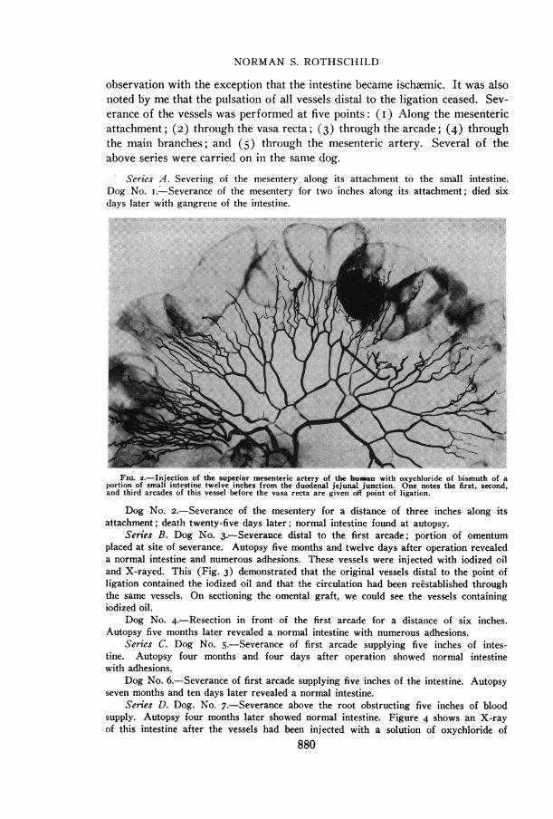
FioG. 2.-Injection of the super-ior mesenteric artery of the huml!an with oxychloride of bismuth of aportion of smnall intestine twelve in'ches from the duodenal jejunal junction. One notes the first, second,and third arcades of this vessel before the vasa recta are given off point of ligation.
Dog No. 2.-Severance o-f -the mes'entery for a distance of three inches along itsattachment; death twenty-five days later; normal intestine found at autopsy.
Series B. Dog No. 3.7-Severance. distal to the. first arcade; portion of omentumplaced at-site of severance. Autopsy five months and twelve days after operation revealeda normal intestine and numerous adhesions. These vessels were injected with iodized oiland X-rayed. This (Fig. 3) -demonstrated that the originial vessels distal to the point ofligation contained the iodized oil and that the circulation had been reestablished throughthe same vessels. On sectioni'ng the omental graft, we could see the vessels containingiodized oil.
Dog NO. 4.-Resection in front of the first arcade for a distance of six inches.Autopsy five months later revealed a normal intestine with numerous adhesions.
Series C. Dog NO. 5.-Severance of first arcade supplying five inches of intes-tine. Autopsy four months and four days after operation showed normal intestinewith adhesions.
Dog No. 6.-Severance of first arcade supplying five inches of the intestine. Autopsyseven months and ten days later revealed a normal intestine.
Series D. Dog. No. 7.-Severance above the root obstructing five inches of bloodsupply. Autopsy- four months later showed normal intestine. Figure 4 shows an X-rayof this intestin'e after the vessels had been injec'ted with a solution of oxychloride of
880

SAFETY FACTORS IN MESENTERIC LIGATIONS
bismuth. Here we find that the marginal artery of the segment adjacent to-the ligatedportion is well developed, and that there are a few small arteries appearing at the siteof ligation.
FIG. 3.-Section of intestine found at autopsy of dog five months and twelve days -after operation.Vessels were injected with iodized oil. Picture shows the reestablishment of the circulation through thesame vessels.
Series E. Dog No. 8.-Severance at the root, four to five inches of intestine involved;died three days later. Autopsy showed gangrene of intestine.
Dog No. 9.-Severance at the root, supplying six inches of intestine; died two dayslater,-showing jangrene of the intestine.
56 881

NORMAN S. ROTHSCHILD
Dog No. IO.-Severance at the root, supplying five inches of initestine; died five dayslater; gangrene of the intestine.
In the large intestine, there is a distinct marginal artery. It was our desire to resectthe mesentery, leaving the marginal artery attached.
Series F. Dog No. II.-Severance of the meso of the large intestine, distance oftwo inches. Autopsy three months later showed a normal large intestine.
Dog No. I2.-Severance of the meso of large intestine for a distance of five inches,leaving the marginal artery intact. Autopsy five months later showed normal intestinewith adhesions.
From the foregoing experiments one can readily see that severanceof the blood supply of the intestine between the mesentric attachment and
m ,.... ... 0- A : ;0ff fSa
FIG. 4.-Vessels of the dog's intestine after injection with a solution of oxychloride of bismuth.These vessels were severed at the root. Autopsy four months later. The reEstablishment of the circula-tion in this specimen was by means of the marginal arteries. Severance indicated by arrow.
the roots offers a great margin of safety, none of the dogs having diedfrom- such operative procedure. Involvement of the superior mesentericartery, per se, produced gangrene in each instance. Severance of the mesen-
tery from its attachment to the intestine produced gangrene in but one ofthe two cases.
The question naturally arises, by what means is the circulation re~stab-lished? In Figure 3 we have shown by means of the X-ray picture that thecirculation has been reE3stablished through an omental graft. Thus a com-munication was afforded between the intact blood supply and the originalvessels which had been severed. In Figure 4 we have shown that a collateralcirculation was established by the blood vessels adjacent to~the severed vesselsand that the collateral circulation was established through the marginal artery.
Numerous adhesions were found in all the cases. Some of these adhesionswere found at the site of the severance while others were found along theintestinal wall. They were as a rule very filmy in character and grossly did
882

SAFETY FACTORS IN MESENTERIC LIGATIONS
not show blood vessels of any considerable size. Eisberg 8 believes that thedevelopment of adhesions between loops of gut and the omentum are bene-ficial to the recovery of the affected gut. He also states that there is no evi-dence of the formation of new blood vessels through these adhesions. I amfully in accord with the first statement. As to the latter one, I feel sure thatmicroscopic sections of the omentum at the site of adhesion would show vas-
FIG. 5.-Vessels of the dog's intestine injected with iodized oil. This shows marginal artery conveyingmaterial to the part supplied by the separate vessels. Severance indicated by arrow.
cilarizationl since Bothe 9 has demonstrated vasctularization of even free graftsin a very short time after transplantationi. Lanz,'0 Scudder," and( \NVilkie 1 2
wrapl)ed omenttum around portions of the intestine from which the blood suip-ply had been ligated. Wilkie found intestine intact over three and one-halfcentimetres long which had had its blood supply ligated. However, it was ofno avail over larger areas.
CONCLUSIONS
i. Interference with the circulation of the small intestine between the mes-enteric attachment and the superior mesenteric artery i-s not usually accompa-nied by gangrene of the intestine. Interference with the superior mesentericartery resuilts in gangrene of the bowel. Detachment of the mesentery from
883

NORMAN S. ROTHSCHILD
the intestine may not result in gangrene of the bowel. Severance of the- mes-entery of the large bowel, permitting the marginal artery to be left intact,does not interfere with the viability of the large intestine.
2. The reestablishment of the circulation in the dog by means of the miar-ginal artery of the segment and by means of formation of new vessels com-municating with the vessels severed has been demonstrated. The adhesions inall probability do not play an important part in this. Adhesions of the omen-tum to the intestine may play a part in the vascularization of small areas ofdevitalized gut.
3. The degree of safety is far greater in man than in the animal.
BIBLIOGRAPHY
1 Bost, T. C.: ANNALS OF SURGERY, vol. lxxxix, p. 219, 1929.2 Ross, G.: ANNALS OF SURGERY, vol. xxii, p. I21, I920.'Klein, E.: Surg., Gyn. and Obst., vol. xxxiii, p. 385, I921.'Dwight, T.: Proc. Tenth Annual Sess. Assoc. Amer. Anat., I897.'Mall, F. P.: Abhandl. d. Math., phys. Cl. d. k. sachs. Gellsch. d. Wissensh., I887.6 Eisberg, H. B.: Anat. Record, vol. xxviii, p. 4, I924.'Monks, G. A.: ANNALS OF SURGERY, vol. xxxviii, p. 574, 1903.8 Eisberg, H. B.: ANNALS OF SURGERY, VOl. lxxXi, p. 926, 1925.9Bothe, F.: The Fate of Omental Grafts. ANNALS OF SURGERY, vol. lxxxix, p. 886, i929.10 Lanz: Centralblatt f. Chir., vol. xxxiv, p. 6I7, I907.1 Scudder, C. L.: Boston Med. and Surg. Jour., vol. clix, p. 338, I908.1Wilkie, D. P. D.: Brit. Med. Jour., vol. ii, p. II03, 19II.
DISCUSSION: DR. JOHN H. JOPSON said that this paper which DoctorRothschild has read opens up the field for a good deal of thought and dis-cussion in connection with accidental and purposeful lesions following opera-tions on the intestine or injuries to its blood supply. Some of his observationsoffer an explanation for what we have for a long while known clinically. Oneof the earliest lessons in the treatment of strangulated hernia is that there isa point in the progress of strangulation where we know that gangrene of thebowel will occur if not resected, and there is another group in which we arereasonably sure the viability has been preserved; and then, a large middlegroup in which experience many years ago taught us that return of the bowelis usually followed by recovery. The speaker recalled hearing Doctors Whar-ton and Deaver tell about their own experiences when young operators andthe lessons they learned from the teaching of D. Hayes Agnew, who, whenwatching operations of this type and asked for advice, advised them the bowelbe put back; the patients got well. The experience of most surgeons has beenthat many of the cases which looked doubtful, but in which the bowel wasreturned, did preserve their viability.
Doctor Rothschild's experiments explain why these cases did not go onto gangrene and how the circulation was reestablished, by one or the othermeans which he has demonstrated experimentally. He mentioned the cases of
884

SAFETY FACTORS IN MESENTERIC LIGATIONS
mesenteric thrombosis reported by Ross and others in which nothing was donebut a simple laparotomy.
Doctor Jopson had one such case in which he found extensive mesentericthrombosis, in which the bowel was returned and nothing further done, andthe patient recovered. Doctor Deaver has also had such a case. This problemhas been brought home to- all of us by operations on the large intestine andespecially operations for carcinoma of the rectum in two stages. -DoctorRothschild and the speaker have had unfortunate experiences with ligationsof the inferior mesentery artery above the point where it should have been.In fat subjects it is sometimes hard to define just where the line of safety isand they have had some patients go to gangrene. The suggestion of the inter-position of a mesenteric graft, or use of it as a covering of the bowel, seems adistinct contribution. The percentage of cases in which it may be used issmall, and although we must not apply his conclusions too radically (remem-bering that the vascularity in dogs is different from that in humans), it isto be hoped at the same time that this contribution will in the future offera means of overcoming or getting around this question of gangrene in smallpercentage of doubtful cases.
885