schlemm scanalandtrabecularmeshwork …€¦ · schlemm’scanalandtrabecularmeshwork...
TRANSCRIPT
RESEARCH ARTICLE
Schlemm’s Canal and Trabecular Meshworkin Eyes with Primary Open Angle Glaucoma: AComparative Study Using High-FrequencyUltrasound BiomicroscopyXiaoqin Yan, Mu Li, Zhiqi Chen, Ying Zhu, Yinwei Song*, Hong Zhang*
Department of Ophthalmology, Tongji Hospital, Tongji Medical College, Huazhong University of Science andTechnology, Wuhan, China
* [email protected] (YS); [email protected] (HZ)
AbstractWe investigated in vivo changes in Schlemm’s canal and the trabecular meshwork in eyes
with primary open angle glaucoma (POAG). Relationships between Schlemm’s canal diam-
eter, trabecular meshwork thickness, and intraocular pressure (IOP) were examined. Forty
POAG patients and 40 normal individuals underwent 80-MHz Ultrasound Biomicroscopy
examinations. The Schlemm’s canal and trabecular meshwork were imaged in superior,
inferior, nasal and temporal regions. Normal individuals had an observable Schlemm’s
canal in 80.3% of sections, a meridional canal diameter of 233.0±34.5 μm, a coronal diame-
ter of 44.5±12.6 μm and a trabecular meshwork thickness of 103.9±11.1 μm, in POAG
patients, Schlemm’s canal was observable in 53.1% of sections, a meridional canal diame-
ter of 195.6±31.3 μm, a coronal diameter of 35.7±8.0 μm, and a trabecular meshwork
thickness of 88.3±13.2 μm, which significantly differed from normal (both p <0.001).
Coronal canal diameter (r = -0.623, p < 0.001) and trabecular meshwork thickness
(r = -0.663, p < 0.001) were negatively correlated with IOP, but meridional canal diameter
was not (r = -0.160, p = 0.156). Schlemm’s canal was observable in 50.5% and 56.6% of
POAG patients with normal (<21 mmHg) and elevated (>21 mmHg) IOP, respectively
(χ = 1.159, p = 0.282). Coronal canal diameter was significantly lower in the elevated IOP
group (32.6±4.9 μm) than in the normal IOP group (35.7±8.0 μm, p < 0.001). This was also
true of trabecular meshwork thickness (81.9±10.0 μm vs. 97.1±12.0 μm, p < 0.001). In con-
clusion, eyes with POAG had fewer sections with an observable Schlemm’s canal. Canal
diameter and trabecular meshwork thickness were also lower than normal in POAG
patients. Schlemm’s canal coronal diameter and trabecular meshwork thickness were neg-
atively correlated with IOP.
PLOS ONE | DOI:10.1371/journal.pone.0145824 January 4, 2016 1 / 15
OPEN ACCESS
Citation: Yan X, Li M, Chen Z, Zhu Y, Song Y, ZhangH (2016) Schlemm’s Canal and TrabecularMeshwork in Eyes with Primary Open AngleGlaucoma: A Comparative Study Using High-Frequency Ultrasound Biomicroscopy. PLoS ONE 11(1): e0145824. doi:10.1371/journal.pone.0145824
Editor: Fan Yuan, Duke University, UNITED STATES
Received: August 12, 2015
Accepted: December 9, 2015
Published: January 4, 2016
Copyright: © 2016 Yan et al. This is an open accessarticle distributed under the terms of the CreativeCommons Attribution License, which permitsunrestricted use, distribution, and reproduction in anymedium, provided the original author and source arecredited.
Data Availability Statement: All relevant data arewithin the paper and its Supporting Information files.
Funding: Support was provided by grants No81170842 from the Natural Science Foundation ofChina [http://isisn.nsfc.gov.cn/egrantindex/funcindex/prjsearch-list] to HZ, and by grants No 81300760from the Natural Science Foundation of China [http://isisn.nsfc.gov.cn/egrantindex/funcindex/prjsearch-list]to ZC. The funders had no role in study design, datacollection and analysis, decision to publish, orpreparation of the manuscript.

IntroductionGlaucoma is a leading cause of irreversible blindness worldwide [1]. It is characterized byprogressive visual field defects and optic atrophy. The most important risk factor for glau-coma is elevated intraocular pressure (IOP) [2], with aqueous outflow cycling playing animportant role in IOP regulation. After being secreted by the ciliary body, aqueous humorarrives in anterior chamber, then through trabecular meshwork (TM) draining intoSchlemm’s canal (SC), and then next to the collector channels and the intrascleral plexus,entering the episcleral veins which finally back to the blood circulation to maintain adynamic balance [3]. To maintain IOP within the normal physiological range of 10-21mmHg, the aqueous humor outflow rate through the conventional TM pathway mustequal the aqueous humor production rate. Maepea and Bill [4] showed that, in monkey eyes,nearly 90% of the outflow resistance was located in the sub-endothelial region of SC. Addi-tionally, Grant [5] concluded that approximately 75% of the resistance to flow in enucleatedhuman eyes was located internal to SC, within the TM, when perfusion pressure was25 mmHg. Following complete trabeculectomy [6], 71% of the outflow resistance was elimi-nated at a perfusion pressure of 25 mmHg, but only 49% of the resistance was eliminated at anormal perfusion pressure of 7 mmHg. Gong et al. [7] reported that one third to one half ofthe outflow resistance is located distal to the inner wall of SC. These results suggested thatpressure-dependent changes in outflow resistance occurred in the TM, SC, and distal to SC[4–7]. Abnormal aqueous humor outflow pathway resistance can result in excess aqueoushumor and elevated IOP.
In 1973, an opinion was formed based on the observations of Jonestone and Grant [8]regarding the relationship between SC and IOP. They showed that acute elevation of IOPmight cause the SC to collapse and the TM to compression, both of which would furtherincrease resistance of the aqueous outflow pathway and begin a vicious cycle of progressivelyincreasing IOP. The major aqueous outflow resistance located in the JCT region and the innerwall of Schlemm’s canal [9]. In 1996, a clinical study revealed that SC cross-sectional area,perimeter, and inner wall length were smaller in eyes with POAG than in normal eyes. It alsodemonstrated the reduction in SC dimensions accounting for approximately 50% of thedecrease in aqueous outflow facility in POAG eyes, which suggested that a significant correla-tion between SC size and aqueous outflow capacity, [10]. Based on above findings, surgical pro-cedures on SC (e.g., canaloplasty, iStent and Eypass implantation) and the TM (e.g.,trabectome) have been launched worldwide. The features of SC and the TM have played animportant role in the development of these new techniques. Bull et al. [11] showed that canalo-plasty successfully reduced IOP from 23.0 ± 4.3 mmHg before surgery to 15.1 ± 3.1 mmHgthree years after surgery in eyes with POAG, demonstrating that changes in SC could affectIOP. The effects of changes in the TM have also been shown, with trabectome now beingapplied in some developed countries. This procedure generally results in a mean IOP reductionof approximately 30–40% [12, 13].
Previous conclusions about SC and the TM were made in vitro, not in vivo, studies thanexamined ocular structures using micro-CT, light microscopy, or electron microscopy. To bet-ter understand these results and to acquire a more theoretical foundation for new surgical tech-niques, the physiological activity of the SC and TM need to be directly examined in vivostudies.
Advancements in medical imaging make it convenient and possible to study SC and the TMin vivo. Asrani et al. [14] were the first to use Fourier-domain optical coherence tomography(OCT) to visualize SC and the TM, allowing them to measure SC and TM size. The high-den-sity OCT was more recently used to show that acute IOP elevations in healthy eyes resulted in
Schlemm's Canal and Trabecular Meshwork in POAG
PLOSONE | DOI:10.1371/journal.pone.0145824 January 4, 2016 2 / 15
Competing Interests: The authors have declaredthat no competing interests exist.

a reduced SC cross-sectional area [15]. In addition, Hong et al. [16] used spectral-domain OCTto show that SC size was significantly different in eyes with and without POAG and that SCarea was negatively correlated with IOP. The above studies showed that the in vivo research onthe SC and TM was possible, but these studies were limited in that they only examined the SCand TM in one or two quadrants. Therefore, the data may not represent overall changes inthese structures. Using 80-MHz ultrasound biomicroscopy, a noninvasive, real-time, dynamic,continuous observation technique, high resolution in vivo images of the SC can be obtained. In2010, Irshad [17] prospectively measured in vivo variations in SC diameter and location using80-MHz ultrasound biomicroscopy, with measurements taken at 12’o clock in 94 subjects thatdid or did not have glaucoma. However, the population examined had wide variations in raceand surgical history [17]. No previous study has examined changes in SC and TM features inpatients with POAG using 80-MHz ultrasound biomicroscopy, which can continuously anddynamically observe and record anterior chamber structures in detail. Owing to new theoriesregarding the role of TM and SC changes in POAG, new surgical techniques specifically treat-ing TM and SC abnormalities were developed. New methods to image SC and the TM allowedthese structures to be thoroughly studied to better understand this physiological channel. Thepurpose of the current study was to evaluate and compare SC and TM parameters in normalindividuals and in patients with POAG using 80-MHz ultrasound biomicroscopy. Main out-come measurements included the percentage of sections with an observable SC, SC diameter,and TM thickness.
Materials and MethodsThis study was approved by the ethics committee of the Tongji Hospital, part of the MedicalCollege of Huazhong University of Science and Technology Institute. All study conductadhered to the tenets of the Declaration of Helsinki and all subjects provided written informedconsent to participate in the study.
SubjectsThis observational, comparative study included 44 patients with POAG who visited theDepartment of Ophthalmology, Tongji Hospital, Tongji medical college, Huazhong Universityof Science and Technology, between March and May of 2014. Additionally, 42 age- and gen-der-matched normal subjects were enrolled into a control group. Subjects were included in thePOAG group if all of the following were true: (1) at least 18 years of age, (2) cup-to-disc (C/D)ratio� 0.6 with an interocular C/D ratio difference� 0.2, (3) retinal nerve fiber layer defectwas present, (4) glaucomatous visual field defects corresponding to optic nerve changes werepresent, (5) normal anterior chamber depth with an open angle, and (6) refractive errorbetween +3.0 and -6.0 diopters (D). Patients who had prior ocular surgeries or a history of eyedisease (except for POAG) were excluded from participation. Patients with systemic diseasewere also excluded. Normal subjects were included if all of the following were true: (1) at least18 years of age, (2) normal fundus, (3) normal visual field, (4) normal anterior chamber depthwith an open angle, and (5) a refractive error between +3.0 and -6.0 D. Potential controlpatients were excluded from participation if they had a family history of glaucoma, a history ofophthalmic disease or surgery, or systemic disease.
Subjects with POAG were divided into two groups based on IOP measurements of each eye.If IOP was>21 mmHg (24 right eyes, 22 left eyes) the eye was placed in the elevated IOP sub-group and if IOP was<21 mmHg (16 right eyes, 18 left eyes) the eye was placed in the normalIOP subgroup and it was assumed that the subject used anti-glaucoma eye drops.
Schlemm's Canal and Trabecular Meshwork in POAG
PLOSONE | DOI:10.1371/journal.pone.0145824 January 4, 2016 3 / 15
Study ExaminationsAll subjects underwent a comprehensive ophthalmologic examination, which included mea-surement of visual acuity, refractive error, IOP (non-contact tonometer), and axial length(AL). Slit-lamp examination, gonioscopy, 80-MHz ultrasound biomicroscopy (iScience Inter-ventional, Inc., Menlo Park, CA), optic nerve and fundus photography, and visual field testing(Humphery perimetry with the 30–2 threshold test protocol). Anterior (Visante OCT) andposterior (SD-OCT Heidelberg Engineering GmbH, Heidelberg, Germany) segment OCT werealso performed to measure central corneal thickness (CCT) and retinal nerve fiber layer thick-ness, respectively.
Schlemm’s Canal and Trabecular Meshwork BiomicroscopyMeasurementsImaging of SC and the TM was conducted by the same experienced technician using the iUltra-sound imaging system. The observer remained masked to patient group assignment. Imageswere obtained using the following iUltrsound system settings: transducer frequency = 80-MHz,axial resolution = 25 μm, lateral resolution = 50 μm, electronic resolution = 10 μm, caliper posi-tioning limit = 10μm, tissue penetration depth = 2 mm, scan rate = 7 frames/s, and imagingwindow size = 4.0 × 4.0 mm. Before iUltrasound measurements were made, eyes were anesthe-tized with topical 1% oxybuprocaine and a low-viscosity ultrasound gel was placed on the ocu-lar surface. The iUltrasound probe was directly placed on the eye and SC and TM parameterswere directly measured from images using the built-in electronic and digital caliper applicationof the iUltraSound imaging system. Each eye had measurements taken at the 12, 3, 6 and 9o’clock positions (Fig 1).
The SC was defined as observable when a thin, black, lucent space was found in two images.Optimum image contrast and magnification and previous histology manifestations were sub-jectively defined to maximize SC visualization. The percentage of sections with an observableSC (eyes with a completely observable SC/total number of eyes × 100), the longest SC meridio-nal diameter (diameter of the white oval space measured from the posterior to the anterior SCend point), and the SC coronal diameter (maximum distance from the inner to the outer wallof SC) were measured. To determine SC coronal diameter, we manually drew a vertical lineacross the canal to obtain two intersection points (points c and d). We then measured the max-imum distance between c and d. The TM thickness was calculated as the average of two mea-surements made at the anterior end point of SC and halfway down the SC. The posterior endpoint of SC was not chosen, the measurement of TM at this region might not truly representthe thickness of TM itself, but the measurement of ciliary muscle behind the sclera spur [18–20]. Each TM thickness measurement was made perpendicular to the inner layer of the mesh-work (Fig 2).
Statistical AnalysesAll analyses were performed using the SPSS software package version 19.0. Data were pre-sented as mean ± standard deviation where applicable. The Mann–Whitney U test, the Krus-kal-Wallis H test, and the Chi-square test were used for comparing differences between groups.Nonparametric Spearman correlation analyses were performed to statistically examine the rela-tionships between IOP and SC and TM parameters. All tests were two-tailed and statistical sig-nificance was defined as p< 0.05.
Schlemm's Canal and Trabecular Meshwork in POAG
PLOSONE | DOI:10.1371/journal.pone.0145824 January 4, 2016 4 / 15

ResultsA total of 44 POAG patients and 42 normal individuals were enrolled in this study. Of these, 4POAG patients and 2 normal individuals were excluded because of poor image quality.
Fig 2. Example of Schlemm’s Canal and Trabecular Meshwork Measurements Made Using theiUltrasound Imaging System. The black oval space shows Schelmm’s canal (SC). The meridional diameterof SC was measured from the anterior (a) to the posterior (b) end point of SC. To measure the coronaldiameter of SC, we drew a vertical line across the canal to get two intersection points (c and d). The maximumdistance between c and d was taken as the coronal diameter of SC. Lines 1, 2 indicated where trabecularmeshwork thickness was measured and the dotted line shows the meshwork inner layer.
doi:10.1371/journal.pone.0145824.g002
Fig 1. An 80-MHz Ultrasound Biomicroscopy Image of Schlemm’s Canal and the Trabecular Meshwork in a Normal Individual. Schlemm’s canal (redarrow) and the trabecular (white arrow) are apparent in the image.
doi:10.1371/journal.pone.0145824.g001
Schlemm's Canal and Trabecular Meshwork in POAG
PLOSONE | DOI:10.1371/journal.pone.0145824 January 4, 2016 5 / 15
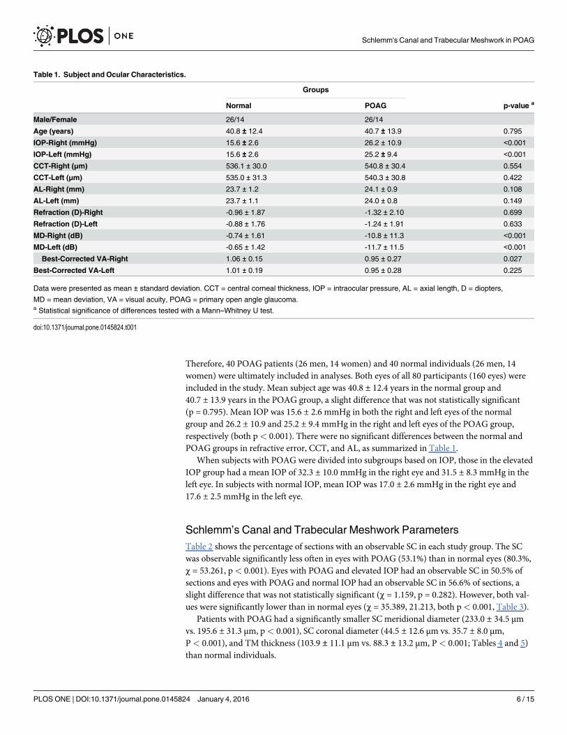
Therefore, 40 POAG patients (26 men, 14 women) and 40 normal individuals (26 men, 14women) were ultimately included in analyses. Both eyes of all 80 participants (160 eyes) wereincluded in the study. Mean subject age was 40.8 ± 12.4 years in the normal group and40.7 ± 13.9 years in the POAG group, a slight difference that was not statistically significant(p = 0.795). Mean IOP was 15.6 ± 2.6 mmHg in both the right and left eyes of the normalgroup and 26.2 ± 10.9 and 25.2 ± 9.4 mmHg in the right and left eyes of the POAG group,respectively (both p< 0.001). There were no significant differences between the normal andPOAG groups in refractive error, CCT, and AL, as summarized in Table 1.
When subjects with POAG were divided into subgroups based on IOP, those in the elevatedIOP group had a mean IOP of 32.3 ± 10.0 mmHg in the right eye and 31.5 ± 8.3 mmHg in theleft eye. In subjects with normal IOP, mean IOP was 17.0 ± 2.6 mmHg in the right eye and17.6 ± 2.5 mmHg in the left eye.
Schlemm’s Canal and Trabecular Meshwork ParametersTable 2 shows the percentage of sections with an observable SC in each study group. The SCwas observable significantly less often in eyes with POAG (53.1%) than in normal eyes (80.3%,χ = 53.261, p< 0.001). Eyes with POAG and elevated IOP had an observable SC in 50.5% ofsections and eyes with POAG and normal IOP had an observable SC in 56.6% of sections, aslight difference that was not statistically significant (χ = 1.159, p = 0.282). However, both val-ues were significantly lower than in normal eyes (χ = 35.389, 21.213, both p< 0.001, Table 3).
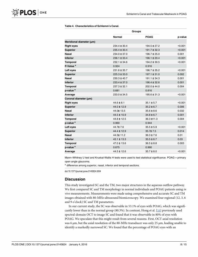
Patients with POAG had a significantly smaller SC meridional diameter (233.0 ± 34.5 μmvs. 195.6 ± 31.3 μm, p< 0.001), SC coronal diameter (44.5 ± 12.6 μm vs. 35.7 ± 8.0 μm,P< 0.001), and TM thickness (103.9 ± 11.1 μm vs. 88.3 ± 13.2 μm, P< 0.001; Tables 4 and 5)than normal individuals.
Table 1. Subject and Ocular Characteristics.
Groups
Normal POAG p-value a
Male/Female 26/14 26/14
Age (years) 40.8 ± 12.4 40.7 ± 13.9 0.795
IOP-Right (mmHg) 15.6 ± 2.6 26.2 ± 10.9 <0.001
IOP-Left (mmHg) 15.6 ± 2.6 25.2 ± 9.4 <0.001
CCT-Right (μm) 536.1 ± 30.0 540.8 ± 30.4 0.554
CCT-Left (μm) 535.0 ± 31.3 540.3 ± 30.8 0.422
AL-Right (mm) 23.7 ± 1.2 24.1 ± 0.9 0.108
AL-Left (mm) 23.7 ± 1.1 24.0 ± 0.8 0.149
Refraction (D)-Right -0.96 ± 1.87 -1.32 ± 2.10 0.699
Refraction (D)-Left -0.88 ± 1.76 -1.24 ± 1.91 0.633
MD-Right (dB) -0.74 ± 1.61 -10.8 ± 11.3 <0.001
MD-Left (dB) -0.65 ± 1.42 -11.7 ± 11.5 <0.001
Best-Corrected VA-Right 1.06 ± 0.15 0.95 ± 0.27 0.027
Best-Corrected VA-Left 1.01 ± 0.19 0.95 ± 0.28 0.225
Data were presented as mean ± standard deviation. CCT = central corneal thickness, IOP = intraocular pressure, AL = axial length, D = diopters,
MD = mean deviation, VA = visual acuity, POAG = primary open angle glaucoma.a Statistical significance of differences tested with a Mann–Whitney U test.
doi:10.1371/journal.pone.0145824.t001
Schlemm's Canal and Trabecular Meshwork in POAG
PLOSONE | DOI:10.1371/journal.pone.0145824 January 4, 2016 6 / 15
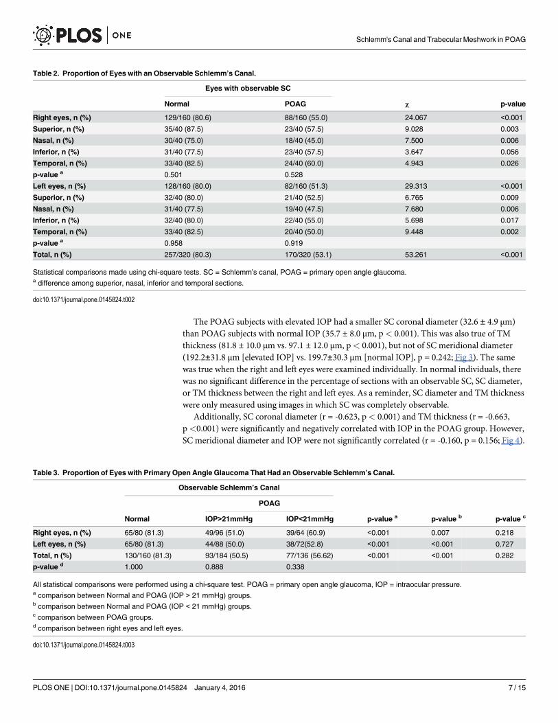
The POAG subjects with elevated IOP had a smaller SC coronal diameter (32.6 ± 4.9 μm)than POAG subjects with normal IOP (35.7 ± 8.0 μm, p< 0.001). This was also true of TMthickness (81.8 ± 10.0 μm vs. 97.1 ± 12.0 μm, p< 0.001), but not of SC meridional diameter(192.2±31.8 μm [elevated IOP] vs. 199.7±30.3 μm [normal IOP], p = 0.242; Fig 3). The samewas true when the right and left eyes were examined individually. In normal individuals, therewas no significant difference in the percentage of sections with an observable SC, SC diameter,or TM thickness between the right and left eyes. As a reminder, SC diameter and TM thicknesswere only measured using images in which SC was completely observable.
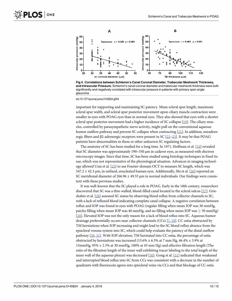
Additionally, SC coronal diameter (r = -0.623, p< 0.001) and TM thickness (r = -0.663,p<0.001) were significantly and negatively correlated with IOP in the POAG group. However,SC meridional diameter and IOP were not significantly correlated (r = -0.160, p = 0.156; Fig 4).
Table 2. Proportion of Eyes with an Observable Schlemm’s Canal.
Eyes with observable SC
Normal POAG χ p-value
Right eyes, n (%) 129/160 (80.6) 88/160 (55.0) 24.067 <0.001
Superior, n (%) 35/40 (87.5) 23/40 (57.5) 9.028 0.003
Nasal, n (%) 30/40 (75.0) 18/40 (45.0) 7.500 0.006
Inferior, n (%) 31/40 (77.5) 23/40 (57.5) 3.647 0.056
Temporal, n (%) 33/40 (82.5) 24/40 (60.0) 4.943 0.026
p-value a 0.501 0.528
Left eyes, n (%) 128/160 (80.0) 82/160 (51.3) 29.313 <0.001
Superior, n (%) 32/40 (80.0) 21/40 (52.5) 6.765 0.009
Nasal, n (%) 31/40 (77.5) 19/40 (47.5) 7.680 0.006
Inferior, n (%) 32/40 (80.0) 22/40 (55.0) 5.698 0.017
Temporal, n (%) 33/40 (82.5) 20/40 (50.0) 9.448 0.002
p-value a 0.958 0.919
Total, n (%) 257/320 (80.3) 170/320 (53.1) 53.261 <0.001
Statistical comparisons made using chi-square tests. SC = Schlemm’s canal, POAG = primary open angle glaucoma.a difference among superior, nasal, inferior and temporal sections.
doi:10.1371/journal.pone.0145824.t002
Table 3. Proportion of Eyes with Primary Open Angle Glaucoma That Had an Observable Schlemm’s Canal.
Observable Schlemm’s Canal
POAG
Normal IOP>21mmHg IOP<21mmHg p-value a p-value b p-value c
Right eyes, n (%) 65/80 (81.3) 49/96 (51.0) 39/64 (60.9) <0.001 0.007 0.218
Left eyes, n (%) 65/80 (81.3) 44/88 (50.0) 38/72(52.8) <0.001 <0.001 0.727
Total, n (%) 130/160 (81.3) 93/184 (50.5) 77/136 (56.62) <0.001 <0.001 0.282
p-value d 1.000 0.888 0.338
All statistical comparisons were performed using a chi-square test. POAG = primary open angle glaucoma, IOP = intraocular pressure.a comparison between Normal and POAG (IOP > 21 mmHg) groups.b comparison between Normal and POAG (IOP < 21 mmHg) groups.c comparison between POAG groups.d comparison between right eyes and left eyes.
doi:10.1371/journal.pone.0145824.t003
Schlemm's Canal and Trabecular Meshwork in POAG
PLOSONE | DOI:10.1371/journal.pone.0145824 January 4, 2016 7 / 15
DiscussionThis study investigated SC and the TM, two major structures in the aqueous outflow pathway.We first compared SC and TMmorphology in normal individuals and POAG patients using invivomeasurements. Measurements were made using comprehensive and accurate SC and TMimages obtained with 80-MHz ultrasound biomicroscopy. We examined four regional (12, 3, 6and 9 o’clock) SC and TM parameters.
In our current study, the SC was observable in 53.1% of eyes with POAG, which was signifi-cantly lower than in the normal group (80.3%). In contrast, Hong et al. [16] previously usedspectral-domain OCT to image SC and found that it was observable in 80% of eyes withPOAG. We speculate that this might result from several reasons. First, OCT axial resolutionwas 6 μm, but the axial resolution of the 80-MHz transducer was only 25 μm, leading unable toidentify a markedly narrowed SC. We found that the percentage of POAG eyes with an
Table 4. Characteristics of Schlemm’s Canal.
Groups
Normal POAG p-value
Meridional diameter (μm)
Right eyes 234.4 ± 30.4 194.5 ± 27.2 <0.001
Superior 235.4 ± 30.4 191.7 ± 32.3 <0.001
Nasal 234.0 ± 37.0 196.7 ± 25.0 0.001
Inferior 236.1 ± 33.4 196.1 ± 20.4 <0.001
Temporal 232.1 ± 34.6 194.2 ± 30.5 <0.001
P-Value a 0.924 0.916
Left eyes 231.6 ± 35.7 196.7 ± 35.2 <0.001
Superior 225.3 ± 33.0 197.1 ± 31.0 0.002
Nasal 230.3 ± 40.7 191.1 ± 34.3 0.001
Inferior 233.4 ± 37.0 196.4 ± 32.6 0.001
Temporal 237.3 ± 32.1 202.0 ± 44.0 0.004
p-value a 0.681 0.816
Average 233.0 ± 34.5 195.6 ± 31.3 <0.001
Coronal diameter (μm)
Right eyes 44.6 ± 8.1 35.1 ± 5.7 <0.001
Superior 44.0 ± 12.9 35.2 ± 6.7 0.006
Nasal 44.0± 13.5 35.6 ± 8.6 0.032
Inferior 44.5 ± 10.9 34.8 ± 6.7 0.001
Temporal 44.8 ± 12.5 36.3 ±11.3 0.004
p-value a 0.960 0.944
Left eyes 44.7± 7.6 35.5 ± 5.9 <0.001
Superior 44.4 ± 12.9 35.7± 7.5 0.014
Nasal 44.5± 11.2 36.3 ± 7.6 0.01
Inferior 43.1 ± 13.3 35.5 ± 6.7 0.03
Temporal 47.0 ± 13.6 36.5 ± 8.8 0.003
p-value a 0.675 0.990
Average 44.5 ± 12.6 35.7 ± 8.0 <0.001
Mann–Whitney U test and Kruskal-Wallis H tests were used to test statistical significance. POAG = primary
open angle glaucoma.a difference among superior, nasal, inferior and temporal sections.
doi:10.1371/journal.pone.0145824.t004
Schlemm's Canal and Trabecular Meshwork in POAG
PLOSONE | DOI:10.1371/journal.pone.0145824 January 4, 2016 8 / 15

observable SC on high-frequency ultrasound biomicroscopy was slightly, but not significantly,higher when IOP was normal (56.6%) than when IOP was elevated (50.5%). If the axial resolu-tion would be higher, different SC closure rate might be detected between two IOP subgroups.Second, observations with ultrasound biomicroscopy were continuous and dynamic, but obser-vations with spectral-domain OCT were static. Third, elevated IOP was not the only factorresponsible for SC collapse in eyes with POAG and SC anatomical variations and other factorsregulating SC were likely involved. Swain et al. [19] demonstrated that scleral spurs were
Table 5. Trabecular Meshwork Thickness.
TM thickness (μm)
Normal POAG p-value
Right eyes 105.4 ± 10.0 86.1 ± 13.0 <0.001
Superior 105.1 ± 17.5 85.9± 18.2 <0.001
Nasal 106.5 ± 13.1 86.4 ± 16.2 <0.001
Inferior 103.4 ± 20.2 86.5 ± 16.1 0.004
Temporal 107.9 ± 13.5 87.7 ± 14.6 <0.001
p-value a 0.702 0.966
Left eyes 102.3 ± 12.1 90.6 ± 13.2 <0.001
Superior 101.4 ± 14.8 91.0 ± 11.1 0.005
Nasal 106.1 ± 18.7 91.8 ± 17.7 0.011
Inferior 99.2 ± 18.3 90.2 ± 13.6 0.06
Temporal 104.8 ± 17.1 87.9 ± 17.6 0.001
p-value a 0.372 0.863
Average 103.9 ± 11.1 88.3 ± 13.2 <0.001
Mann–Whitney U test and Kruskal-Wallis H tests were used to test statistical significance. TM = trabecular
meshwork, POAG = primary open angle glaucoma.a difference among superior, nasal, inferior and temporal sections.
doi:10.1371/journal.pone.0145824.t005
Fig 3. Schlemm’s Canal Meridional and Coronal Diameter and Trabecular Meshwork Thickness in Patients with Primary Open Angle Glaucoma.Schlemm’s canal (SC) and trabecular meshwork (TM) measurements for patients with primary open angle glaucoma (POAG) and normal IOP (< 21 mmHg)and patients with POAG and elevated IOP (> 21 mmHg). *indicates a statistically significant difference.
doi:10.1371/journal.pone.0145824.g003
Schlemm's Canal and Trabecular Meshwork in POAG
PLOSONE | DOI:10.1371/journal.pone.0145824 January 4, 2016 9 / 15
important for supporting and maintaining SC patency. Mean scleral spur length, maximumscleral spur width, and scleral spur posterior movement upon ciliary muscle contraction weresmaller in eyes with POAG eyes than in normal eyes. They also showed that eyes with a shorterscleral spur posterior movement had a higher incidence of SC collapse [19]. The ciliary mus-cles, controlled by parasympathetic nerve activity, might pull on the conventional aqueoushumor outflow pathway and prevent SC collapse when contracting [21]. In addition, noradren-ergic fibers and β2-adrenergic receptors were present in SC [22–23]. It may be that POAGpatients have abnormalities in these or other unknown SC regulating factors.
The anatomy of SC has been studied for a long time. In 1971, Hoffman et al. [24] revealedthat SC diameter was approximately 190–350 μm in cadaver eyes, as measured with electronmicroscopy images. Since that time, SC has been studied using histology techniques in fixed tis-sue, which was not representative of the physiological situation. Advances in imaging technol-ogy allowed Usui et al. [25] to use Fourier-domain OCT to measure SC length, which was347.2 ± 42.3 μm, in unfixed, enucleated human eyes. Additionally, Shi et al. [26] reported anSC meridional diameter of 266.96 ± 49.55 μm in normal individuals. Our findings were consis-tent with these previous studies.
It was well-known that the SC played a role in POAG. Early in the 18th century, researchersdiscovered that SC was a thin-walled, blood-filled canal located in the scleral sulcus [27]. Grie-shaber et al. [28] assessed SC status by observing blood reflux from collector channels to SC,with a lack of refluxed blood indicating complete canal collapse. A negative correlation betweenreflux and IOP was found in eyes with POAG (regular filling when mean IOP was 30 mmHg,patchy filling when mean IOP was 40 mmHg, and no filling when mean IOP was� 50 mmHg)[28]. Elevated IOP was not the only reason for a lack of blood reflux into SC. Aqueous humordrainage preferentially occurs near collector channels (CCs) [7, 29]. CC ostia obstructed byTM herniations when IOP increasing and might lead to the SC blood reflux absence from theepiscleral venous system into SC, which could help evaluate the patency of the distal outflowpathway [30, 31]. With IOP elevation, TM herniated into CC ostia, the percentage of ostiaobstructed by herniations was increased (15.6% ± 6.5% at 7 mmHg, 46.4% ± 3.9% at15mmHg, 95% ± 2.3% at 30 mmHg, 100% at 45 mm Hg) and effective filtration length (Theratio of the filtration length of the inner wall exhibiting tracer labeling to the total length of theinner wall of the aqueous plexus) was decreased [30]. Gong et al. [31] indicated that weakenedand interrupted blood reflux into SC from CCs was consistent with a decrease in the number ofquadrants with fluorescein egress into episcleral veins via CCs and that blockage of CC ostia
Fig 4. Correlations between Schlemm’s Canal Coronal Diameter, Trabecular Meshwork Thickness,and Intraocular Pressure. Schlemm’s canal coronal diameter and trabecular meshwork thickness were bothsignificantly and negatively correlated with intraocular pressure in patients with primary open angleglaucoma.
doi:10.1371/journal.pone.0145824.g004
Schlemm's Canal and Trabecular Meshwork in POAG
PLOSONE | DOI:10.1371/journal.pone.0145824 January 4, 2016 10 / 15
existed in vivo in POAG. Using three-dimensional micro-computed tomography (CT) recon-struction of the SC and collector channel, a recent study showed that SC was more discontinu-ous and had fewer anastomosing channels in eyes with POAG than in normal eyes at the sameperfusion pressure [32]. Using spectral-domain OCT, Kagemann et al. [15] reported that acuteIOP elevation (12.5 to 36.1 mm Hg) in healthy eyes significantly reduced SC cross-sectionalarea. Imaging revealed that SC compression occurred because of inner wall movement towardsthe outer wall [15]. Shi et al. [26] used swept source OCT to measure SC meridional diameter,which was significantly larger in normal individuals (272.83 ± 49.39 μm) than in patients withPOAG (190.91 ± 46.47 μm) [26]. Wang et al. [33] also used swept source OCT to measure SCcircumference, area, and long diameter in POAG patients. All parameters were smaller inPOAG patients than in normal individuals [33]. Our results were in agreement with these pre-vious studies and we found that both meridional and coronal SC diameter were significantlysmaller in eyes with POAG than in normal eyes. We also found that the SC coronal, but notmeridional diameter was correlated with IOP and was smaller in eyes with POAG and elevatedIOP than in eyes with POAG and normal IOP. We speculated that SC meridional diameter wasonly one indicator and did not reflect SC collapse.
The TM consists of anterior and posterior regions. The anterior portion of TM is known as“nonfiltering”meshwork which is not adjacent to SC and has no aqueous humor filtration, theposterior portion of TM is known as “filtering”meshwork which leads to SC. Yang et al. [20]reported that outflow facility increased after perfusion with Y27632 (a Rho-kinase inhibitor),the increase in outflow facility correlated positively with an increase in effective filtrationlength, which was associated with expansion in JCT. Meanwhile, TM and JCT thickness wasthicker in high-tracer regions than low-tracer regions in human eyes. They postulated that theincrease in outflow facility by Y27632 due to the effect of increased TM and JCT expansion, inwhich JCT expansion played the leading role. Besides, previous studies have confirmed thatTM could be observed and measured in vivo with medical imaging [25, 34]. Therefore, we mea-sured the thickness of TM below the SC (“filtering”meshwork) and compared the TM thick-ness between POAG patients and normal individuals with 80-MHz ultrasound biomicroscopy.The TM thickness was smaller in glaucoma patients (88.3 ± 13.2 μm) than in normal individu-als (103.9±11.1 μm) and was negatively correlated with IOP significantly in this study. Thedirect effect of increased IOP, TM fibrosis, TM stiffness, and TM atrophy could all lead to TMalternations. Grant et al. [8] found that the TMmight compress with acute elevation in IOP.Filla et al. [35] reported that increased extracellular matrix expression and deposit in the TMresulted in an increase in IOP. Additionally, a number of studies have shown that fibrous gran-ular material deposition and increased TM electron density contributed to increasing outflowsystem resistance in eyes with glaucoma [36–41]. Unfortunately, the composition of the extra-cellular matrix remained unclear, but collagen fibers are a main extracellular matrix compo-nent and play an important role in aqueous outflow [42]. Types I, II, III, and VI collagen havebeen found in the TM, but the amount of type VI collagen increased in eyes with glaucoma[43]. Millard et al. [44] showed that the amount of type I collagen increased in the TM. Thedysregulation of the extracellular matrix caused fibrosis and increased stiffness of the TM. TMstiffness that could influence the extent of deformation of TM and the inner wall of SC [45–47]. Using atomic force microscopy, Last et al. [48] found that the elastic modulus of the TMwas higher in eyes with glaucoma than in normal eyes, suggesting an increased TM stiffness ineyes with glaucoma. Last et al. [48] also mathematically demonstrated that an increase in TMstiffness would theoretically lead to an increase in juxtacanalicular region flow resistance.These results indicated that changes in TM biomechanics might be involved in IOP elevationand glaucoma development [45–48]. Additionally, Gabelt et al. [49] showed that a reduction incellularity and loss of TM cells in glaucomatous eyes resulted in a reduced outflow capacity and
Schlemm's Canal and Trabecular Meshwork in POAG
PLOSONE | DOI:10.1371/journal.pone.0145824 January 4, 2016 11 / 15
an increased IOP. The degeneration of the trabecular cells might cause more serious changes,including fusion of the trabecular meshwork [50]. A decreased TM thickness might be relatedto the increased outflow resistance and elevated IOP in glaucomatous eyes. Perhaps we can tryto use the TM thickness evaluate the function of TM in vivo.
This study had several limitations. First, the majority patients with POAG were not visitingour clinic for the first time and had been using one or more antiglaucomatous drugs to lowerIOP. Therefore, IOP was higher than 21 mmHg in only 46 eyes. Additionally, the use of theantiglaucomatous agents might have affected SC and the TM and the effects of these drugswere not evaluated. Second, our study sample size was relatively small, which made it impossi-ble to investigate SC and TMmorphology at different POAG stages. Future research is neededwith a larger group of patients. Third, we only obtained canal measurements from each patientonce. It is well known that IOP fluctuated throughout the day and, because SC anatomy wasinfluenced by IOP, the effects of circadian rhythms on our measurements were not evaluated.Fourth, it is possible that the ultrasound biomicroscopy axial resolution (25 μm) affected SCcoronal diameter measurements, if the resolution is higher, in POAG eyes smaller coronaldiameter may be detected and the actual coronal diameter may be smaller than the value inthis study.
ConclusionsIn conclusion, SC and the TM can be noninvasively, dynamically, and continuously imaged invivo using 80-MHz ultrasound biomicroscopy. These images allow SC and TM parameters tobe evaluated. Using such measurements, we found that patients with POAG have a less observ-able SC, smaller SC diameter, and decreased TM thickness than normal individuals. In addi-tion, both SC coronal diameter and TM thickness were correlated with IOP. These resultsconfirm that outflow structures are visible on 80-MHz ultrasound biomicroscopy images andthat there is more than one indicator of SC status. Additionally, TM thickness may be a usefulclinical measure for evaluating physiologic TM changes in patients with POAG.
Supporting InformationS1 STROBE Checklist. STROBE_checklist_v4_combined_PlosMedicine.(DOCX)
Author ContributionsConceived and designed the experiments: HZ. Performed the experiments: XY YS. Analyzedthe data: XY YS. Contributed reagents/materials/analysis tools: HZ ZC YZ XY YS ML. Wrotethe paper: XY YS ML.
References1. Quigley HA, Broman AT. The number of people with glaucoma worldwide in 2010 and 2020. Br J
Ophthalmol. 2006 Mar; 90(3):262–7. PMID: 16488940
2. Caprioli J, Coleman AL. Intraocular pressure fluctuation a risk factor for visual field progression at lowintraocular pressures in the advanced glaucoma intervention study. Ophthalmology. 2008 Jul; 115(7):1123–9.e3. PMID: 18082889
3. Grant WM. Clinical measurements of aqueous outflow. Am J Ophthalmol. 1951 Nov; 34(11):1603–5.PMID: 14885362
4. Mäepea O, Bill A. Pressures in the juxtacanalicular tissue and Schlemm's canal in monkeys. Exp EyeRes. 1992 Jun; 54(6):879–83 PMID: 1521580
5. Grant WM. Experimental aqueous perfusion in enucleated human eyes. Arch Ophthalmol. 1963 Jun;69:783–801. PMID: 13949877
Schlemm's Canal and Trabecular Meshwork in POAG
PLOSONE | DOI:10.1371/journal.pone.0145824 January 4, 2016 12 / 15

6. Rosenquist R, Epstein D, Melamed S, Johnson M, Grant WM. Outflow resistance of enucleated humaneyes at two different perfusion pressures and different extents of trabeculotomy. Curr Eye Res. 1989Dec; 8(12):1233–40. PMID: 2627793
7. Gong H, Francis A. Schlemm’s Canal and Collector Channels as Therapeutic Targets. In Innovations inGlaucoma Surgery, Samples JR and Ahmed I eds. Springer New York; 2014. Chapter 1, pp 3–25.
8. Johnstone MA, Grant WM. Pressure dependent changes in the structures of the aqueous outflow sys-tem of human and monkey eyes. Am J Ophthalmol. 1973 Mar; 75(3):365–83. PMID: 4633234
9. Mäepea O, Bill A. Pressures in the juxtacanalicular tissue and Schlemm's canal in monkeys. Exp EyeRes. 1992 Jun; 54(6):879–83. PMID: 1521580
10. AllinghamRR, de Kater AW, Ethier CR. Schlemm's Canal and Primary Open Angle Glaucoma: Correla-tion Between Schlemm's Canal Dimensions and Outflow Facility. Exp Eye Res. 1996 Jan; 62(1):101–9.PMID: 8674505
11. Bull H, vonWolff K, Körber N, Tetz M. Three-year canaloplasty outcomes for the treatment of open-angle glaucoma: European study results. Graefes Arch Clin Exp Ophthalmol. 2011 Oct; 249(10):1537–45. doi: 10.1007/s00417-011-1728-3 PMID: 21732110
12. Minckler DS, Baerveldt G, Alfaro MR, Francis BA. Clinical results with the Trabectome for treatment ofopen-angle glaucoma. Ophthalmology. 2005 Jun; 112(6):962–7. Erratum in: Ophthalmology. 2005Sep;112(9):1540. PMID: 15882909
13. Maeda Maeda M, Watanabe M, Ichikawa K, et al. Evaluation of Trabectome in Open-Angle Glaucoma.J Glaucoma. 2013 Mar; 22(3):205–8. doi: 10.1097/IJG.0b013e3182311b92 PMID: 23429629
14. Asrani S, Sarunic M, Santiago C, Izatt J. Detailed visualization of the anterior segment using fourier-domain optical coherence tomography. Arch Ophthalmol. 2008 Jun; 126(6):765–71. doi: 10.1001/archopht.126.6.765 PMID: 18541838
15. Kagemann L, Wang B, Wollstein G, Ishikawa H, Nevins JE, Nadler Z, et al. IOP Elevation ReducesSchlemm's Canal Cross-Sectional Area. Invest Ophthalmol Vis Sci. 2014 Mar 25; 55(3):1805–9. doi:10.1167/iovs.13-13264 PMID: 24526436
16. Hong J, Xu J, Wei A, WenW, Chen J, Yu X, et al. Spectral-domain optical coherence tomographicassessment of Schlemm's canal in Chinese subjects with primary open-angle glaucoma. Ophthalmol-ogy. 2013 Apr; 120(4):709–15. doi: 10.1016/j.ophtha.2012.10.008 PMID: 23352198
17. Irshad FA, Mayfield MS, Zurakowski D, Ayyala RS. Variation in Schlemm's canal diameter and locationby ultrasound biomicroscopy. Ophthalmology. 2010 May; 117(5):916–20. doi: 10.1016/j.ophtha.2009.09.041 PMID: 20079926
18. Dietlein TS, Jacobi PC, Luke C, Krieglstein GK. Morphological variability of the trabecular meshwork inglaucoma patients: implications for non-perforating glaucoma surgery. Br J Ophthalmol. 2000 Dec; 84(12):1354–9. PMID: 11090472
19. Swain DL, Ho J, Lai J, Gong H. Shorter scleral spur in eyes with primary open-angle glaucoma. InvestOphthalmol Vis Sci. 2015 Feb; 56(3):1638–48. doi: 10.1167/iovs.14-15593 PMID: 25670488
20. Yang CY, Liu Y, Lu Z, Ren R, Gong H. Effects of Y27632 on aqueous humor outflow facility withchanges in hydrodynamic pattern and morphology in human eyes. Invest Ophthalmol Vis Sci. 2013Aug; 54(8):5859–70. doi: 10.1167/iovs.12-10930 PMID: 23920374
21. Li G, Farsiu S, Chiu SJ, Gonzalez P, Lütjen-Drecoll E, Overby DR, et al. Pilocarpine-induced dilation ofSchlemm's canal and prevention of lumen collapse at elevated intraocular pressures in living mice visu-alized by OCT. Invest Ophthalmol Vis Sci. 2014 Mar 4; 55(6):3737–46. doi: 10.1167/iovs.13-13700PMID: 24595384
22. Akagi Y, Ibata Y, Sano Y. The sympathetic innervation of the ciliary body and trabecular meshwork ofthe cat. Fluorescence histochemistry and electron microscopy. Cell Tissue Res. 1976 Oct 6; 173(2):261–9 PMID: 991239
23. Zhou EH, Krishnan R, Stamer WD, Perkumas KM, Rajendran K, Nabhan JF, et al. Zhou E H. Mechani-cal responsiveness of the endothelial cell of Schlemm's canal: scope, variability and its potential role incontrolling aqueous humour outflow. J R Soc Interface. 2012 Jun 7; 9(71):1144–55. doi: 10.1098/rsif.2011.0733 PMID: 22171066
24. Hoffman F, Dumitrescu L. Schlemm’s canal under the scanning electron microscope. Ophthalmic Res1971 Nov 5; 2:37–45.
25. Usui T, Tomidokoro A, Mishima K, Mataki N, Mayama C, Honda N, et al. Identification of Schlemm'scanal and its surrounding tissues by anterior segment fourier domain optical coherence tomography.Invest Ophthalmol Vis Sci. 2011 Sep 1; 52(9):6934–9. doi: 10.1167/iovs.10-7009 PMID: 21757587
26. Shi G, Wang F, Li X, Lu J, Ding Z, Sun X, et al. Morphometric measurement of Schlemm's canal in nor-mal human eye using anterior segment swept source optical coherence tomography. J Biomed Opt.2012 Jan; 17(1):016016. doi: 10.1117/1.JBO.17.1.016016 PMID: 22352666
Schlemm's Canal and Trabecular Meshwork in POAG
PLOSONE | DOI:10.1371/journal.pone.0145824 January 4, 2016 13 / 15
27. Winkelmann A. Schlemm, the body snatcher? Ann Anat 2008; 190(3):223–9. doi: 10.1016/j.aanat.2007.12.002 PMID: 18396022
28. Grieshaber MC, Pienaar A, Olivier J, Stegmann R. Clinical Evaluation of the Aqueous Outflow Systemin Primary Open-Angle Glaucoma for Canaloplasty. Invest Ophthalmol Vis Sci. 2010 Mar; 51(3):1498–504. doi: 10.1167/iovs.09-4327 PMID: 19933180
29. Hann CR, Fautsch MP. Preferential fluid flow in the human trabecular meshwork near collector chan-nels. Invest Ophthalmol Vis Sci 2009 Apr; 50(4):1692–7. doi: 10.1167/iovs.08-2375 PMID: 19060275
30. Battista SA, Lu Z, Hofmann S, Freddo T, Overby DR, Gong H. Reduction of the available area for aque-ous humor outflow and increase in meshwork herniations into collector channels following acute IOPelevation in bovine eyes. Invest Ophthalmol Vis Sci 2008 December; 49(12):5346–52. doi: 10.1167/iovs.08-1707 PMID: 18515571
31. Gong H, Huang R, Zhu J, Stegmann R. Blockages of collector channel ostia exist in patients with Pri-mary Open Angle Glaucoma (POAG). In: American Glaucoma Society 19th annual meeting,SanDiego, CA, 5–8 March 2009.
32. Hann CR, Vercnocke AJ, Bentley MD, Jorgensen SM, Fautsch MP. Anatomic Changes in Schlemm’sCanal and Collector Channels in Normal and Primary Open-Angle Glaucoma Eyes Using Low andHigh Perfusion Pressures. Invest Ophthalmol Vis Sci. 2014 Aug 19; 55(9):5834–41. doi: 10.1167/iovs.14-14128 PMID: 25139736
33. Wang F, Shi G, Li X, Lu J, Ding Z, Sun X, et al. Comparison of Schlemm's canal's biological parametersin primary open-angle glaucoma and normal human eyes with swept source optical. J Biomed Opt.2012 Nov; 17(11):116008. doi: 10.1117/1.JBO.17.11.116008 PMID: 23117803
34. Tun TA, Baskaran M, Zheng C, Sakata LM, Perera SA, Chan AS, et al. Assessment of trabecular mesh-work width using swept source optical coherence tomography. Graefes Arch Clin Exp Ophthalmol.2013 Jun; 251(6):1587–92. doi: 10.1007/s00417-013-2285-8 PMID: 23436037
35. Filla MS, Schwinn MK, Sheibani N, Kaufman PL, Peters DM. Regulation of cross-linked actin network(CLAN) formation in human trabecular meshwork (HTM) cells by convergence of distinct beta1 andbeta3 integrin pathways. Invest Ophthalmol Vis Sci. 2009 Dec; 50:5723–31. doi: 10.1167/iovs.08-3215PMID: 19643963
36. Rohen JW,Witmer R. Electron microscopic studies on the trabecular meshwork in glaucoma simplex.Albrecht Von Graefes Arch Klin Exp Ophthalmol. 1972; 183(4):251–66. PMID: 4111808
37. Rodrigues MM, Spaeth GL, Sivalingam E, Weinreb S. Histopathology of 150 trabeculectomy speci-mens in glaucoma. Trans Ophthalmol Soc U K. 1976 Jul; 96(2):245–55. PMID: 1070878
38. Segawa K. Electron microscopic changes of the trabecular tissue in primary open angle glaucoma. AnnOphthalmol. 1979 Jan; 11(1):49–54. PMID: 84545
39. Orsida BE, Rolland JM, Werkmeister JA, West RH. Trabecular meshwork changes in glaucoma. AustN Z J Ophthalmol. 1996 May; 24(2 Suppl):21–4. PMID: 8811235
40. Pescosolido N, Cavallotti C, Rusciano D, Nebbioso M. Trabecular Meshwork in Normal and pathologi-cal eyes. Ultrastruct Pathol. 2012 Apr; 36(2):102–7. doi: 10.3109/01913123.2011.634090 PMID:22471432
41. Acott TS, Kelly MJ. Extracellular matrix in the trabecular meshwork. Exp Eye Res. 2008 Apr; 86(4):543–61. doi: 10.1016/j.exer.2008.01.013 PMID: 18313051
42. Tengroth B, Ammitzbøll T. Changes in the content and composition of collagen in the glaucomatouseye—basis for a new hypothesis for the genesis of chronic open angle glaucoma—a preliminary report.Acta Ophthalmol (Copenh). 1984 Dec; 62(6):999–1008.
43. Luetjen-Drecoll E. Functional Morphology of the Trabecular Meshwork in Primate Eyes. Prog Retin EyeRes. 1999 Jan; 18(1):91–119. PMID: 9920500
44. Millard CB, Tripathi BJ, Tripathi RC. Age-related changes in protein profiles of the normal human tra-becular meshwork. Exp Eye Res. 1987 Oct; 45(4):623–31. PMID: 3428388
45. Huang J, Camras LJ, Yuan F. Mechanical analysis of rat trabecular meshwork. Soft Matter2015 Mar25; 11(14):2857–65. doi: 10.1039/c4sm01949k PMID: 25710888
46. Stamer WD, Braakman ST, Zhou EH, Ethier CR, Fredberg JJ, Overby DR, et al. Biomechanics ofSchlemm's canal endothelium and intraocular pressure reduction. Prog Retin Eye Res2015 Jan;44:86–98. doi: 10.1016/j.preteyeres.2014.08.002 PMID: 25223880
47. Kagemann L, Wang B, Wollstein G, Ishikawa H, Mentley B, Sigal I, et al. Trabecular MeshworkResponse to Pressure Elevation in the Living Human Eye. J Vis Exp2015 Jun 20;(100: ):e52611 doi:10.3791/52611 PMID: 26132890
48. Last JA, Pan T, Ding Y, Reilly CM, Keller K, Acott TS, et al. Elastic m odulus determination of normaland glaucomatous human trabecular meshwork. Invest Ophthalmol Vis Sci2011 Apr 5; 52(5):2147–52.doi: 10.1167/iovs.10-6342 PMID: 21220561
Schlemm's Canal and Trabecular Meshwork in POAG
PLOSONE | DOI:10.1371/journal.pone.0145824 January 4, 2016 14 / 15

49. Gabelt BAT, Kaufman PL.Changes in aqueous humor dynamics with age and glaucoma. Prog RetinEye Res. 2005 Sep; 24(5):612–37. PMID: 15919228
50. Hamanaka T, Kasahara K, Takemura T. Histopathology of the Trabecular Meshwork and Schlemm’sCanal in Primary Angle-Closure Glaucoma. Invest Ophthalmol Vis Sci. 2011 Nov 17; 52(12):8849–61.doi: 10.1167/iovs.11-7591 PMID: 21960557
Schlemm's Canal and Trabecular Meshwork in POAG
PLOSONE | DOI:10.1371/journal.pone.0145824 January 4, 2016 15 / 15