school children in the three senatorial districts of
TRANSCRIPT

Page 1/14
Detecting Schistosoma Haematobium Infection byMicroscopy and Polymerase Chain Reaction (Pcr) InSchool Children In The Three Senatorial Districts ofCross River State, NigeriaRosemary Kaiso Esiere
University of Calabar Teaching HospitalEmmanuel onyekachukwu Ibeneme
University of CalabarEmmanuel O�ong Effanga
University of CalabarEdema Enogiomwan Imalele ( [email protected] )
University of Calabar https://orcid.org/0000-0003-3774-5121Miracle Kaiso Esiere
University of CalabarPaul Columba Inyang-Etoh
University of CalabarAmbrose Anyanwu Alaribe
University of Calabar
Research Article
Keywords: schistosomiasis, microscopy, PCR, school-age children, Nigeria, Cross River State
Posted Date: May 28th, 2021
DOI: https://doi.org/10.21203/rs.3.rs-560563/v1
License: This work is licensed under a Creative Commons Attribution 4.0 International License. Read Full License

Page 2/14
AbstractAs a result of the poor sensitivity and speci�city of the standard parasitological diagnostic methodscurrently being used, this study was conducted to compare the standard parasitological diagnosticmethods and Polymerase Chain Reaction (PCR) in determining the prevalence of urinary schistosomiasisin Cross River State (CRS). The study was conducted between April 2015 and March 2016. Sevenhundred and seventy seven (777) urine samples were randomly collected from selected school-agechildren. The urine samples were subjected to standard parasitological and molecular examinations. Chi-square test was used to test the differences between the data on subgroups and the results fromspecimen examinations. An overall prevalence of 1.7% was recorded using microscopy and 34.7%recorded using PCR. The highest prevalence of infection by microscopy occurred in the SouthernSenatorial District (2.3%), while the Northern Senatorial District recorded the highest prevalence ofinfection by PCR (53.2%) (p < 0.05). Males were more infected (2.4%) than females (0.6%) usingmicroscopy. With PCR, males were also more infected (35.7%) compared to females (33.3%) (p < 0.05).The highest prevalence of infection using microscopy and PCR both occurred in school-age children aged5–8 years (3.6% and 47.8% respectively), while the lowest prevalence for both methods occurred inparticipants aged 17–20 years (0% for both methods) (p < 0.05). This study has shown PCR to beeffective in detecting schistosomiasis infection and also re-a�rms the endemicity of urinaryschistosomiasis in the three Senatorial Districts of CRS.
IntroductionSchistosomiasis is a worldwide problem a�icting about 249 million persons and 97% of the infectedpersons reside in Africa (Anazaku et al. 2017). Approximately 779 million people are at risk of beinginfected in 76 endemic countries globally, 85% of these reside on the African continent (Utzinger et al.2009). Every year schistosomiasis is responsible for about 1.7 million disability adjusted life years(DALYs) (Chitsulo et al. 2000). Schistosoma mansoni and S. haematobium cause intestinal and urinaryschistosomiases respectively in many African countries where they may occur concurrently.Schistosomiasis causes health, labour loss, hypertension and signi�cant reduction in socioeconomicbene�ts, particularly, late complications such as irreversible urinary tract obstruction and risk of renalfailure are often associated with urinary schistosomiasis (King, 2009).
Control programmes for schistosomiasis are mainly founded on treating of infected people, henceadequate-case �nding is essential. Hitherto, the existing protocols for the diagnosis of schistosomiasisare search for ova of the schistosomes in urine or stool samples and detection of ova or adult wormantigens in sera and urine of infected persons. Detecting antigens of ova or adult worm in sera/urine ofdiseased persons can distinguish current infections from past ones with almost 100% speci�city. Eggcounts and haematuria are the only parameters presently employed in surveillance. However, Poggenseeet al. (1998) reported the presence of morbidity of the urinary tract in some women who wereschistosomiasis-infected in Tanzania who had scanty or no egg output and no haematuria. This impliesthat there is need for methods of detection with more speci�city and sensitivity of the disease in humans.

Page 3/14
Several studies have looked at the prevalence and intensity of S. haematobium in communities in Nigeriausing methods like the detection of haematuria and ova in urine (Okon et al. 2009, 2010; Adie et al. 2013).Though they are useful in the detection of heavy infections, their low sensitivity in detecting light/earlyinfections is a drawback that cannot be overlooked. As a result of the poor speci�city and sensitivity ofthe standard parasitological diagnostic methods currently being used, the real estimate of the prevalenceof schistosomiasis in sub-Saharan endemic areas of Africa is not clear (Hotez and Fenwick, 2009). Earlierstudies have shown that detection of schistosomal parasite-speci�c DNA can be done in urine of personswho are infected (Lodh et al. 2013). Adult schistosome worms are the source of DNA because they shedteguments at regular intervals. Therefore if parasite-speci�c DNA is detected in urine it con�rms theparasites’ presence (Ibironke et al. 2012). In recent years, multiple authors have demonstratedSchistosome parasite-speci�c DNA in urine (Lodh et al. 2013). This study, therefore, sought to comparemicroscopy and PCR in the characterization of S. haematobium in Cross River State (CRS), Nigeria.
Materials And Methods
Study area and populationThis study was carried out in Cross River State with Calabar as the capital. The state is divided into threeSenatorial Districts (North, South and Central) with 18 Local Government Areas (LGAs). Samples werecollected from Central, Southern and Northern Senatorial Districts and were analysed at the University ofCalabar Teaching Hospital (UCTH), Calabar and Molecular Biology Laboratory, Niger Delta University,Bayelsa State. The study population consisted of children/adolescents aged 5–20 years attendingprimary and secondary schools in the study area.
Ethical considerationSubjects for this study were enrolled after approval was sought and obtained from Ministry of HealthEthical Committee, Cross River State (REC No.: RP/REC/2016/423). The village heads were informed ofthe study through advocacy visits to their palaces and local school authorities. Written informed consentwas obtained from parents/guardians of participants.
Sampling and sample size determinationSeven hundred and seventy seven (777) samples were randomly obtained from children/adolescents insecondary/primary schools in the study area. Selection of children from schools in the study area wasdone randomly by picking one out of every four pupil within the range of the study.
Sample collectionThe study was conducted between April 2015 and March 2016. Seven hundred and seventy seven (777)clean appropriately labelled universal containers were issued to the randomly selected children forcollection of urine samples. The urine samples from these individuals were used forparasitological/molecular examinations. Subjects were instructed to include the last drops of urine

Page 4/14
(Cheesbrough, 2005). The samples were collected between 10.00am and 2.00pm, on each collection dayto ensure maximum yield of schistosome eggs (Cheesbrough, 2005). All samples collected werepreserved with 2 drops of 10% formalin in 25mL capacity (WHO, 2003) and transported to theparasitology laboratory, University of Calabar Teaching Hospital.
Parasitological examinationA modi�ed �ltration system adopted by Useh and Ejezie, (1999) was used. Brie�y a funnel holding aWhat-man No. 1 �lter paper was suspended on a conical �ask to act as the �ltration system. 10mL of theurine sample was �ltered for the eggs of Schistosoma. The thoroughly agitated sample was allowed topass through the �ltration system. Using a blunt-ended forceps, the �lter paper was removed with greatcare and placed on a slide. The �lter paper was placed upwards (eggs on surface) on the slide using ×10objective, with the condenser iris closed adequately to give a good contrast. A systematic examination ofthe entire �lter paper was conducted for S. haematobium ova. Any urine sample with ova was recorded aspositive. The number of eggs counted was recorded per 10ml of urine samples collected.
Molecular examination for schistosome infection
Urine cell pellet preparationThe urine sample was spun at 5,000 rpm for 10 min, the supernatant decanted and cell pellets washedthrice with 25mL phosphate buffer saline (0.8% NaCl, 2.7mM KCl, 1.8mM KH2P04, 8.mM Na2HP04 pH
7.4). These were immediately stored at -80oC until used.
Genomic DNA extraction from urine cell pelletThe urine cell-pellets were re-suspended using 150µL of DNA elution buffer, then transferred intoindividual Eppendorf tubes, followed by 200µL bio-�uid - cell buffer solution (red) and 20µL proteinase Kenzyme to digest the sample. Vortexing was done to have a thorough mix, and then incubated at 55oC for10 minutes on a heating block. Exactly 420 µL of genomic binding buffer was added to the digestedsample and mixed thoroughly. The mixture was then transferred to a Zymo-Spin IIC – XL column in acollection tube and centrifuged at 14,000 rpm for one minute. The columns were then placed in newcollection tubes, where 400µL DNA of pre-wash buffer was added and spun for one minute at 14,000 rpmand the collection tubes emptied. Seven hundred (700µL) g-DNA wash buffer was then added and spunat 14,000 rpm for one minute, and the collection tube emptied again. Furthermore, 200µL of g-DNA washbuffer was added and centrifuged again at 14,000 rpm for one minute and the collection tube discardedwith the �ow through. The columns were transferred to clean micro-centrifuge tubes and 50µL of DNAelution buffer was added, incubated at room temperature for 5 minutes, and then spun at 14,000 rpm forone minute to elute the DNA. Finally, the columns were discarded and the g-DNA, already extracted in themicro-centrifuge tubes, stored at -20oC until further use for PCR reactions.
DNA quanti�cation
Page 5/14
NanoDrop ND-1000 spectrophotometer (NanoDrop Technologies, California, USA) was used to determinethe quantity and purity of DNA. The equipment was initialized and blanked before readings. DNAconcentrations of the samples were recorded, in ng/µL while the absorbance 260/280 was used todetermine the purity. Samples with absorbance ranging from 1.70 to 2.10 were selected for ampli�cationby PCR.
Ampli�cation of Schistosoma haematobium DNA repeatfragment from urine samplesPolymerase chain reaction was carried out on all the 13 positive samples and 108 negative samples,respectively, by microscopy, using 0.4µL of species-speci�c primers ShDra1 F (forward: 5′-GATCTCACCTATCAGACGAAAC-3′ and ShDra1 R (reverse: 5′-T CACAACGATACGACCAAC-3′), which werepreviously designed by Hamburger et al., (2001) for speci�c ampli�cation of 121 bp Dra1 tandem repeatsof S. haematobium. Ampli�cation reactions were carried out in 20µL volume. Two microlitre (2µL) of DNA(concentration: 4-6ng/ µL) served as PCR template in the reaction volume, 10µL of 2X Quick-Load Master-mix containing 200µM of dNTPs, 1.5µM of Taq polymerase (Inqaba Biotech, Johannesburg, SouthAfrica) and 1.5µM of MgCl. The PCR conditions were as follows: initial denaturation at 95oC for 5minutes, followed by 30 cycles of denaturation at 95oC for one minute, annealing at 55oC for 90 secondsand extension at one minute at 72oC for 5 minutes.
Agarose gel electrophoresesOne gram and half (1.5gm) of agarose powder was measured and poured in 100ml of 1X TBE buffer (TrisBoric Ethylene diamine Tetra-acetic acid.) then heated in a microwave for 5 minutes to allow the agarosepowder dissolve and make a clear solution. It was allowed to cool and 4µL of ethidium bromide wasadded. The solution was poured in an electrophoretic casting tray, the comb was placed and then thesolution was allowed to cool and solidi�ed for about 30 minutes then placed in an electrophoresis tank.Exactly 10µL of PCR product was pipetted, already mixed with DNA loading dye, and then loaded in theagarose gel lane after removing the comb. DNA ladder and negative control were also loaded and the gelwas subjected to an electric �eld at 120V for 25 minutes. The gel was visualized on a UV trans-illuminatorfor band detection.
Data analysisAll analyses were performed using the SPSS package (version 22). Descriptive statistical analysis wasused to calculate the prevalence and intensity of infections. Chi-square test was used to test thedifferences between the data on subgroups and the results from specimen examinations. Differenceswere considered signi�cant at a p-value less than 0.05 (p < 0.05).
ResultsSeven hundred and seventy seven (777) school children/adolescents randomly selected from eightschools in the three Senatorial Districts of Cross River State were screened to determine the current status

Page 6/14
of urinary schistosomiasis in these districts. The Northern Senatorial District had the highest number ofparticipants (348/777; 44.8%) followed by the Southern Senatorial District (220/777; 28.6%) and theCentral Senatorial District (209/777; 26.9%). The school children/adolescents also comprised of 450(57.9%) males and 327 (42.1%) females. The age distribution of the participants in the study was 5–8years (248/777; 31.9%), 9–12 years (339/777; 43.6%), 13–16 years (116/777; 14.9%) and 17–20 years(74/777; 9.5%).
Table 1 shows the prevalence of S. haematobium infection by microscopy and PCR in the SenatorialDistricts. The highest prevalence of infection by microscopy occurred in the Southern Senatorial Districts(2.3%), while the Northern Senatorial District recorded the highest prevalence of infection by PCR (53.2%);there was a statistically signi�cant difference (p < 0.05) in the prevalence of infection in the SenatorialDistricts using microscopy and PCR.
Table 2 shows the prevalence of S. haematobium infection by microscopy and PCR with respect togender. Males were more infected than females using microscopy (2.4% for males and 0.6% for females)and PCR (35.7% for males and 33.3% for females); there was a statistically signi�cant difference inprevalence of infection by gender, using microscopy and PCR (p < 0.05).
Table 3 shows the prevalence of S. haematobium infection by microscopy and PCR in participants withrespect to age. The highest prevalence of infection using PCR occurred in participants aged 5–8 years(47.8%), while the lowest prevalence occurred in participants aged 17–20 years (0%). Using microscopy,the highest and lowest prevalence occurred in age groups 5–8 years (3.6%) and 17–20 years (0%). Therewas a statistically signi�cant difference (p < 0.05) in the prevalence of infection among the different age-groups using microscopy and PCR.
Table 1Prevalence of Schistosoma haematobium infection by microscopy and PCR by Senatorial Districts
SenatorialDistrict
No.
Examined bymicroscopy
No. (%) positiveby microscopy
Numberexamined byPCR
Number(%)positive byPCR
χ2 p-value
South 220 5 (2.3) 31 5 (16.1) 11.082 0.004
Central 209 1 (0.5) 46 14 (30.4)
North 348 7 (2.0) 44 23 (52.3)
Total 777 13 (1.7) 121 42 (34.7)

Page 7/14
Table 2Prevalence of Schistosoma haematobium infection by microscopy and PCR by gender
Gender No.examined
bymicroscopy
No. (%) positive bymicroscopy
No. examinedby PCR
No. (%)positive byPCR
χ2 p-value
Males 450 11(2.4) 70 25(35.7) 0.257 0.012
Females 327 2(0.6) 51 17(33.3)
Total 777 13(1.7) 121 42(34.7)
Table 3Prevalence of S. haematobium infection by microscopy and PCR of subjects examined by age
Age group(years)
No.
Examined
bymicroscopy
No. (%)positive by
microscopy
No. Examinedby PCR
No. (%) positiveby PCR
χ2 p-value
5–8 248 9 (3.6) 46 22 (47.8) 8.106 0.044
9–12 339 3 (0.9) 58 18 (31.0)
13–16 116 1 (0.9) 13 2 (15.4)
17–20 74 0 (0.0) 4 0 (0.0)
Total 777 13 (1.7) 121 42 (34.7)
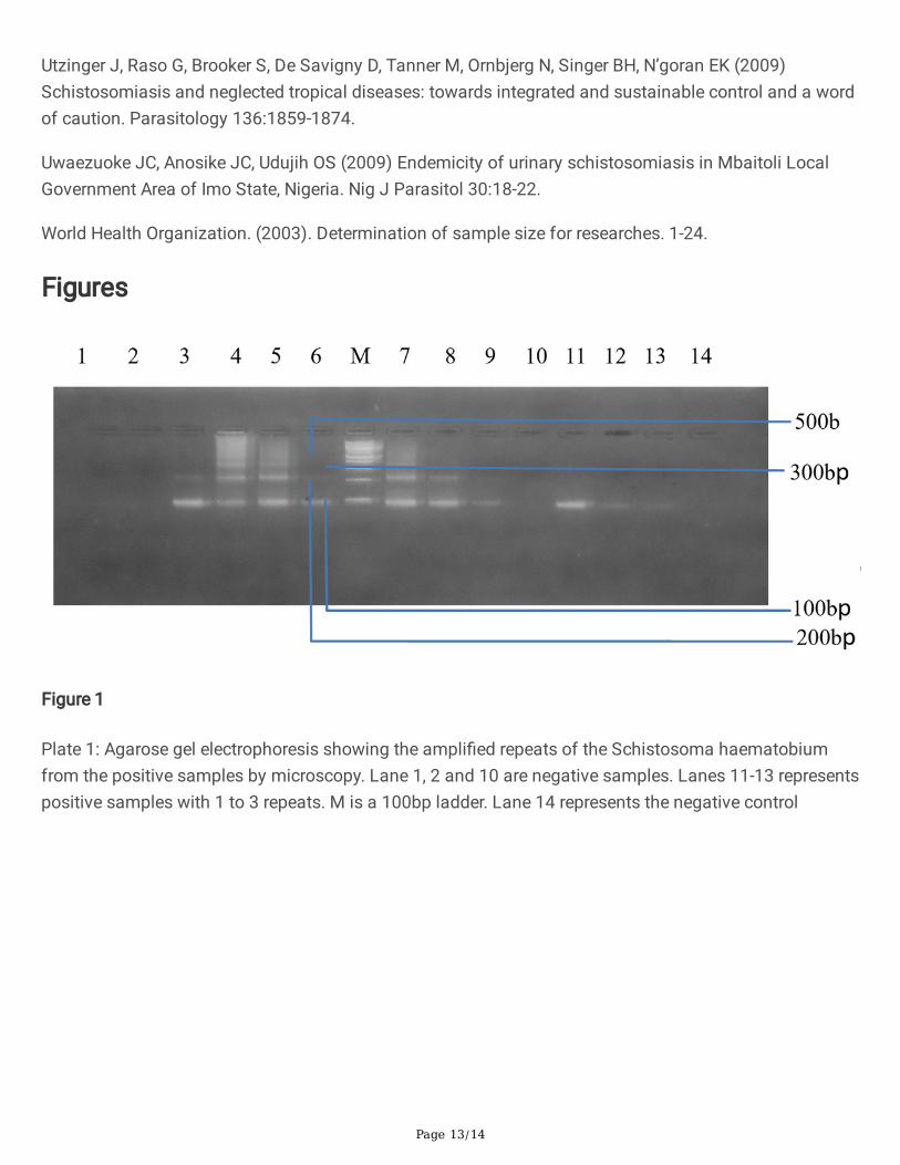
Plate 1 shows an agarose gel electrophoresis of the ampli�ed repeats of Schistosoma haematobiumfrom positive samples by microscopy. Lane 1, 2, and 10 are negative samples, Lanes 39, 11–13represents positive samples with 1 to 3 repeats. M is a 100bp ladder. Lane 14 represents the negativecontrol.
Plate 2 shows an agarose gel electrophoresis showing the ampli�ed repeats of the Schistosomahaematobium from selected samples negative by microscopy. Lanes showing 1 band or more representpositive samples for S. haematobium while lanes with no bands represent negative samples. L is 100bpladder.
DiscussionStudies have compared different diagnostic procedures used to detect schistosomiasis. These includetests for speci�c antibody, circulating antigens, haematuria, egg detection and ultrasound scans of theurinary tract. Nonetheless, it is di�cult to �nd a “gold” standard because of the variability of thediagnostic performance of these techniques especially in areas with variable prevalence of S.

Page 8/14
haematobium infection (Koukounari et al. 2009). However, assays based on polymerase chain reaction(PCR) have recently begun to show potentials as effective methods in detecting DNA of parasites in urineand saliva (Mharakurwa et al. 2006).
In this study, the 121bp Dra1 tandem repeat PCR developed by Hamburger and colleagues (Hamburger etal. 2001) was adapted to detect S. haematobium DNA in urine. The Dra1 fragments were detected insamples from both children and adolescents when ova of schistosome were detected, and also inspecimens where no ova were detected. However, these fragments were not detected in some of thesamples. Out of the 777 urine samples, 13 (1.7%) were positive by microscopy, but PCR showed that 42(34.7%) out of 121 randomly selected samples showed parasite-speci�c DNA for S. haematobium. Thisspeaks volume of the sensitivity of PCR over microscopy. This agrees with results reported byGasmelseed et al. (2014).
The overall prevalence (1.7%) recorded from parasitological examination of urine sample is lower thanthat reported for Cross River State (CRS) in earlier studies. Ejezie et al. (1991) established aschistosomiasis prevalence value of 43.5% in Adim, CRS, while Useh and Ejezie (1996) recorded aprevalence of 53.8% in the same study area. Ekanem et al. (1995) recorded a prevalence of 44% in Ijiman,Yakurr Local Government Area (LGA), a community close to Adim. Recent studies have shown that theurinary schistosomiasis prevalence in Adim community increased from 53.8% reported by Useh andEjezie (1996) to 57.4% (Okon et al. 2009). In Adim, Etim et al. (2012) reported a 42.2% prevalence ofurinary schistosomiasis, while Okon et al. (2007, 2009) reported 35% and 45.3% prevalence ratesrespectively from parasitological examination of urine samples. The present study has re-a�rmed theendemicity of S. haematobium in the three Senatorial Districts; Southern (Biase LGA), Central (YakurrLGA) and Northern (Bekwarra LGA). According to Useh (2013), the extensive, unrestrained and uncheckedincrease in irrigation, water impoundment schemes, and widespread ignorance of schistosomiasis in themajority of the endemic areas have elevated the disease to a very serious health predicament in the sub-Saharan part of the continent, including Nigeria. This disease is also considered as one of the mostfrequently occurring parasitic infections globally (Okon et al. 2007). In CRS, the agrarian population andsuitable conditions that enable the snail intermediate host to thrive has added to the prevalence ofschistosomiasis (Ejezie et al. 1991).
It is likely that the reduction in prevalence and intensity of urinary schistosomiasis recorded in thesecommunities, is as a result of treatments carried out by previous researchers; Okon et al. (2010),evaluated the status of urinary schistosomiasis in Ogoja LGA, where he went ahead to treat infectedpersons with Artesunate and found 64% cure rate while Inyang-Etoh et al. (2004) had a 70% cure rate intheir work in Adim, CRS, using Artesunate. Another work, in Adim, recorded a cure rate of 88% afterutilizing Artesunate and Praziquantel combination (Inyang-Etoh et al. 2009). This reduction in morbidityof urinary schistosomiasis could also be as a result of the integrated health interventions introducedabout eight years ago by the Neglected Tropical Diseases Control Unit in collaboration with theschistosomiasis/soil transmitted helminths unit of the Federal Ministry of Health, CRS, in November 2012(Adie et al. 2015). Integrated control measures put in place included chemotherapy of infected individuals

Page 9/14
with Praziquantel and health education on the predisposing factors responsible for the transmission ofurinary schistosomiasis (Adie et al. 2015).
Infection pattern varied according to gender with more male participants (11; 2.4%) being infected thanfemales (2; 0.6%). This difference varied signi�cantly at (p < 0.05). Similar �ndings were reported in otherstudies (Inyang-Etoh et al. 2004; Ugbomoiko et al. 2010). This difference may be explained by the factthat males are more involved in water contact activities than females due to social inhibitions like beingrestricted from activities such as �shing, public bathing and swimming (Etim et al. 2012). However, thisdeviates from other studies that reported no signi�cant difference in gender-related prevalence. Okon etal. (2007), Bala et al. (2012) and Morenikeji et al. (2014) all reported both genders to be equally at risk.
Prevalence of S. haematobium varied signi�cantly across age groups with the least (0% for bothmicroscopy and PCR methods) and highest (3.6% for microscopy and 47.8% for PCR) recorded in agegroup 17–20 and 5–8 years, respectively (p 0.05). This is similar to previous result reported in Biase andYakurr LGAs by Adie et al. (2015), but disagrees with other �ndings which recorded highest infection ratesoccurring in age brackets 12–15 (Morenikeji et al. 2014) and 10–14 (Ugbomoiko et al. 2010 and Deribe etal. 2011). The prevalence of urinary schistosomiasis varied with age in this study which is in contrast to�ndings reported by Ekwunife et al. (2009), while it was supported by Sam-Wobo et al. (2009) andUwazuoke et al. (2009).
ConclusionThis study has re-a�rmed the endemicity of urinary schistosomiasis in the three Senatorial Districts ofCRS. The prevalence of 34.7% recorded in this study is still within the WHO recommended threshold of ≥ 20%, for mass drug administration (MDA). This study has shown that PCR is more sensitive thanstandard parasitological methods for the diagnosis of S. haematobium infection.
DeclarationsAcknowledgements
The authors wish to thank the staff of the University of Calabar Teaching Hospital (UCTH), Calabar andMolecular Biology Laboratory, Niger Delta University, Bayelsa State, for their assistance during the courseof this study
Funding
The authors did not receive support from any organization for the submitted work.
Con�icts of interest
The authors declare no con�ict of interest exist.
Page 10/14
Availability of data and material
Not applicable
Code availability
Not applicable
Authors’ contributions
Esiere, Rosemary Kaiso: Conceptualization, Data curation, Investigation, Methodology, Writing - originaldrafting. Ibeneme, Emmanuel Onyekachukwu: Investigation, Methodology, Writing – review and editing.Imalele, Edema Enogiomwan: Methodology, Writing – review and editing. Effanga, Emmanuel O�ong:Data curation, Methodology. Esiere, Miracle Kaiso: Investigation, Data curation. Inyang-Etoh, PaulColumba: Formal analysis, Project administration, Validation. Alaribe, Ambrose Andrew Anyanwu:Conceptualization, Validation, Supervision
Ethics approval
Subjects for this study were enrolled after approval was sought and obtained from Ministry of HealthEthical Committee, Cross River State.
Consent to participate
The village heads were informed of the study through advocacy visits to their palaces and local schoolauthorities. Written informed consent was obtained from parents/guardians of participants.
ReferencesAdie HA, Oyo-Ita A, Okon OE, Arong GA, Ating IA, Braide EI, Nebe O, Emanghe UE, Otu, AA (2015)Evaluation intensity of urinary schistosomiasis in Biase and Yakurr Local Government Area of Cross RiverState, Nigeria, after two years of Integrated Control Measures. Res J Parasitology 10:58-65.
Adie HA, Okon OE, Arong, GA, Braide EI, Ekpo UF (2013) Spatial distribution of urinary schistosomiasis inCross River State, Nigeria, using geographical information system and school based questionnaire.Pakistan J Bio Sci 16:1166 – 1172.
Anzaku AA, Oche OD, Ishaku A, Ishaku A (2017) Prevalence of urinary schistosomiasis and water contactactivities as risk factor in Wowyen community. J Ann Toxicology Applications 1(1):7-10.
Bala AY, Ladan MU, Mainasara M (2012) Prevalence and intensity of urinary Schistosomiasis in Abarmavillage, Gusau, Nigeria: a preliminary investigation. Science World J 7(2):1-4.
Cheesbrough M (2005) District laboratory practice in Tropical countries. (2ndedn) Tropical HealthTechnology, London.

Page 11/14
Chitsulo L, Engels D, Montresor A, Savioli L (2000) The global status of schistosomiasis and its control.Acta Tropica,77: 41–51.
Deribe K, Eldaw A, Hadziabduli S, Kailie E, Omer MD, Mohammed AE, Jamshed T, Mohammed EA,Mergani A, Ali GA, Babikir K, Adem A, Hashim F (2011) High prevalence of urinary schistosomiasis in twocommunities in South Darfur: implication for interventions. Parasites and Vectors. 4(14):1-5.
Ejezie GC, Uko IE, Braide EI (1991) Schistosomiasis in Cross River State, Nigeria: Prevalence and intensityof infection in Adim, Akamkpa Local Government Area. J Hyg Epidemiol Microbiol Immunol 35:141-147.
Ekanem EE, Ejezie GC, Asindi AA, Antia-Obong OE (1995) Urinary symptoms and blood pressure ofchildren with Schistosoma haematobium infection in South-Eastern Nigeria. East African Med J 72:486-489.
Ekwunife CA, Agbor VO, Ozumba AN, Eneanya CI, Ukaga CN (2009) Prevalence of urinary schistosomiasisin Iyede-Ame community and environ in Ndokwa East Local Government Area, Delta State, Nigeria. Nig JParasitol 30:27-31.
Etim SE, Okon OE, Oku EE, Ukpong GI, Ohioma ME, Uttah CE (2012) Urinary schistosomiasis in a rice-farming community in Biase Area of Cross River State. Nig J Parasitol 33: 197-201.
Gasmelseed N, Karamino NE, Abdelwahed MO, Hamdoun AO, Elmadani AE (2014) Genetic diversity ofSchistosoma haematobium parasite IS NOT associated with severity of disease in epidemic area inSudan. Bio Med Central Infect Dis, 14:469.
Hamburger J, He N, Abbasi I, Ramzy RM, Jourdane J, Ruppel A (2001) Polymerase chain reaction assaybased on a highly repeated sequence of Schistosoma haematobium: A potential tool for monitoringSchistosome-infested water. Am J Trop Med Hyg 65:907-911.
Hotez PJ, Fenwick A (2009) Schistosomiasis in Africa: An Emerging Tragedy in Our New Global HealthDecade. Plos Negl Trop Dis 3(9):485.
Ibironke O, Koukounari A, Asaolu S, Moustaki I, Shiff C (2012) Validation of a new test for Schistosomahaematobium based on detection of Dra1 DNA fragments in urine: evaluation through latent classanalysis. Pub Lib Sci Negl Trop Dis 6:e1464.
InyangEtoh PC, Essien UC, Amama M, Useh F (2004) Prevalence of urinary schistosomiasis amongschool in Ukwolo – Obudu and Abini communities in Cross River State, Nigeria. Port-Harcourt Med J 3:13.
Inyang-Etoh PC, Ejezie GC, Useh MF, Inyang-Etoh EC (2009) E�cacy of a combination of Praziquantel andArtesunate in the treatment of urinary schistosomiasis in Nigeria. Trans R Soc Trop Med Hyg 103:38–44.
King CH (2009) Toward the elimination of schistosomiasis. New England J Med 360 (2):106–109.

Page 12/14
Koukounari A, Webster JP, Donnelly CA, Bray BC, Naples J, Bosompem K, Shiff C (2009) Sensitivities andSpeci�cities of diagnostic tests and infection prevalence of Schistosoma haematobium estimated fromdata on adults in villages Northwest of Accra, Ghana. Am J Trop Med Hyg 80:435-441.
Lodh N, Mwansa JCL, Mutengo MM, Shiff CJ (2013) Diagnosis of Schistosoma mansoni without theStool: Comparison of Three Diagnostic Tests to Detect Schistosoma mansoni Infection from FilteredUrine in Zambia. Am Soc Trop Med Hyg 89: 46–50.
Mharakurwa S, Simoloka C, Thuma PE, Shiff CJ, Sullivan DJ (2006) PCR detection of Plasmodiumfalciparum in human urine and saliva samples. Malaria J 5:103.
Morenikejji O, Quazim J, Omoregie C, Hassan A, Nwuba R, Anumudu C, Adejuwon S, Salawu O, Jegede A,Odaibo A (2014) A Cross sectorial study on urogenital schistosomiasis in children, haematuria in anendemic rural area of Nigeria. Afri Health Sci, 14(2): 390-396.
Okon OE, Udoutun MF, Oku EE, Nta AI, Etim SE, Abraham JJ, Akpan PA (2007) Prevalence of urinaryschistosomiasis in Abini community, Biase local government area, Cross river State, Nigeria. Nig JParasitol 28(1):28–31.
Okon OE, Ememayom A, Opara K (2009) Reliability of self-reported blood in urine for diagnosis ofSchistosoma haematobium in a community in South-Eastern Nigeria. The Internet J Epidemiol 7(2).
Okon OE, Obi A, Opara K (2010) The e�cacy of Artesunate in the treatment of urinary schistosomiasis inOgoja, Cross River State, Nigeria. Internet J Trop Med 6(2).
Poggensee G, Kiwelu I, Saria M, Richter J, Krantz I, Feldmeir H (1998) Schistosomiasis of the lowerreproductive tract without egg excretion in urine Am J Trop Med Hyg 59: 782–783.
Sam-Wobo SO, Ekpo UF, Ameh IG, Osileye OT (2009) Continued high endemicity of urinaryschistosomiasis in Ogun State, Nigeria. Nig J Parasitol 30(1):48-52.
Ugbomoiko US, Ofoezie IE, Okoye IC, Heukelbach J (2010) Factors associated with uriinaryschistosomiasis in two Peri-urban communities in South- Western Nigeria. Ann Trop Med Parasitol104(5): 409-419.
Useh MF, Ejezie GC (1996) Prevalence and morbidity of S. haematobium infection in Adim community ofNigeria. JMedical Lab Sci 5:10-15.
Useh MF, Ejezie GC (1999) Modi�cation of behaviour and attitude in the control of schistosomiasis. 1.Observations on water-contact patterns and perceptions of infection. Ann Trop Med Parasitol 93(7):711-720.
Useh MF (2013). Parasitic diseases-schistosomiasis. Creative Commons Attribution License, Geneva,Switzerland, pp: 82-83.
Page 13/14
Utzinger J, Raso G, Brooker S, De Savigny D, Tanner M, Ornbjerg N, Singer BH, N’goran EK (2009)Schistosomiasis and neglected tropical diseases: towards integrated and sustainable control and a wordof caution. Parasitology 136:1859-1874.
Uwaezuoke JC, Anosike JC, Udujih OS (2009) Endemicity of urinary schistosomiasis in Mbaitoli LocalGovernment Area of Imo State, Nigeria. Nig J Parasitol 30:18-22.
World Health Organization. (2003). Determination of sample size for researches. 1-24.
Figures
Figure 1
Plate 1: Agarose gel electrophoresis showing the ampli�ed repeats of the Schistosoma haematobiumfrom the positive samples by microscopy. Lane 1, 2 and 10 are negative samples. Lanes 11-13 representspositive samples with 1 to 3 repeats. M is a 100bp ladder. Lane 14 represents the negative control

Page 14/14
Figure 2
Plate 2: Agarose gel electrophoresis showing the ampli�ed repeats of the Schistosoma haematobiumfrom selected samples negative by microscopy. Lanes showing 1 band or more represent positivesamples for S. haematobium while lanes with no bands represent negative samples. L is a 100bp ladder.