scientific illustration and reconstruction of a …jpaleontologicaltechniques.org/pasta3/jpt...
TRANSCRIPT
Number 17, Mar 2017
SCIENTIFIC ILLUSTRATION AND RECONSTRUCTION OF A
SKULL OF THE DIPLODOCID SAUROPOD DINOSAUR
GALEAMOPUS
Simão Mateus1-5 & Emanuel Tschopp3,4,6
1 - Museu de História Natural e da Ciência da Universidade do Porto, Porto, Portugal
2 - Departamento de Ciências e Técnicas do Património da Faculdade de Letras da Universidade do Porto (DCTP-FLUP), Porto, Portugal 3 - Museu da Lourinhã, Rua João Luís de Moura, 2530-157 Lourinhã, Portugal
4 - GeoBioTec, Faculdade de Ciências e Tecnologia, Universidade Nova de Lisboa, Caparica, Portugal
5 - Universidade de Évora, Largo dos Colegiais, 7004-516 Évora, Portugal
6 - Dipartimento di Scienze della Terra, Università degli Studi di Torino, Via Valperga Caluso 35, 10125 Torino, Italy
Emails: [email protected] (SM), [email protected] (ET)
ABSTRACT
High-quality scientific illustration is an important visualization tool for natural sciences. In paleontology, drawings help to guide the reader to important features of the fossils under study, and to remove irrelevant information or strong shadows that might obscure parts of photographs. Furthermore, drawings allow for the deformation of the fossils to be corrected. However, for an accurate interpretation of these reconstruction drawings, it is important to provide a detailed report about the creation of the drawings. Herein, we describe the methodology of the reconstruction drawing of a skull of the sauropod dinosaur Galeamopus. After preparation and reconstruction of the skull in the laboratory, illustrations were needed to correct natural deformations, restore missing parts, and highlight critical features for anatomical recognition of the several bones. The illustrations were successful thanks to the collaborative work between the paleontologist and the illustrator. Keywords: illustration report; drawing; reconstruction; diplodocid skull
RESUMO [in Portuguese]
Ilustrações científicas de alta qualidade são uma ferramenta importante de visualização nas ciências naturais. Na paleontologia ajudam o leitor a perceber as estruturas anatómicas importantes dos fósseis em estudo, removendo informação irrelevante, ou eliminar zonas escuras que escondam pormenores dos ossos nas fotografias. Além disso, as ilustrações permitem corrigir de ossos deformados. Para a correcta interpretação das reconstruções efectuadas, é importante existirem relatórios detalhados do processo da ilustração. Vimos descrever a metodologia de ilustração de um crânio de dinossauro saurópode Galeamopus que foi reconstruído. Após a preparação e montagem do crânio no laboratório, as ilustrações tiveram de reajustar as deformações naturais, repor partes em falta, e realçar características essenciais necessárias à compreensão dos diversos ossos. As ilustrações são bem sucedidas graças à colaboração entre o paleontólogo e o ilustrador.
How to cite this paper: Mateus, S. and Tschopp, E. (2017). Scientific illustration and reconstruction of a skull of the diplodocid sauropod
dinosaur Galeamopus. Journal of Paleontological Techniques, 17:1-11.
Copyright (c) 2017 by Mateus and Tschopp. This work is made available under the terms of the Creative Commons
Attribution 3.0 Unported License, http://creativecommons.org/licenses/by-sa/3.0/.
www.jpaleontologicaltechniques.org ISSN: 1646-5806
Mateus & Tschopp 2017: ILLUSTRATION OF A SKULL OF GALEAMOPUS
2 ● Journal of Paleontological Techniques
INTRODUCTION
Paleontological research is often based on
reconstructions (Benton, 2005). Initial
descriptions of new species or specimens often
include photographs or line drawings of the
actual fossils, made by the paleontologist. In a
further step, reconstructions can be produced,
often together with an artist, by adding missing
parts and restoring deformed portions.
Reconstructions like these are more clear and
appealing, because they omit information that might be confusing at first sight.
Generally, the paleoartist is not a
paleontologist, and does not have the necessary
knowledge about the extinct animals and
environments he or she must portray (Ghilardi
and Ribeiro, 2010). In order to prepare an
accurate paleoreconstruction, it is thus
important that the basic scientific data is
compiled and simplified by the paleontologist
supervising the work. Without a solid scientific
knowledge the paleoartist will support him- or
herself on deduction, and the artwork will be
less consistent and could be more erroneous
(Ghilardi et al., 2007) and, therefore, lead to
mistakes.
CHALLENGES IN PALEORECONSTRUCTIONS
During the taphonomical process, nearly all
fossils undergo some degree of damage and
deformation (Benton, 2005; Tschopp et al.,
2013). Such changes include both pre-burial
(physical damage, scavenging), or post-burial
events (compression, chemical alterations,
erosion). It is the task of the paleontologist to
recognize such alterations, and try to account
for them in the studies based on deformed
material (Benton, 2005). A first briefing helps
the paleoartist to understand the goal of the
paleontologist and the latter to understand the
difficulties of the artist (Ghilardi and Ribeiro,
2010).
One of the most challenging problems
encountered when reconstructing a fossil is that
frequently no single complete skeleton exists
for reference and assorted partial skeletons of
the same or similar species differ in size.
Sometimes, what is missing on one side of a
specimen can be found on its other side (Paul
and Chase, 1989), but if that is not the case,
assumptions have to be made based on closely
related species, where the bones lacking in the
species in question are preserved. In cases,
where information from more than one
specimen is available to restore a single
individual, it remains possible that no or only
few parts are shared among the specimens
used (Paul and Chase, 1989). In order to
produce the most accurate reconstruction
possible, careful guesstimates must be made of
the animal’s proportions, preferentially based
on closely related taxa, where such information
is not available from the fossils under study. A
bibliography should be provided at the briefing
to illustrate how missing portions in the fossil to
be reconstructed look like in closely related taxa
(Ghilardi and Ribeiro, 2010).
In vertebrates, one of the most complex
structures of the skeleton is the skull. In
sauropod dinosaurs like Galeamopus - the study
object of this paper - the skull is composed of
more than 25 bones per side. Being so
complex, skulls should preferentially be
represented in five views (Correia, 2010):
frontal, lateral (most commonly used), posterior
(occipital), dorsal, and ventral. If the mandible
is preserved, it should be either drawn
articulated with the skull and slightly open so
that no detail is obliterated, or isolated (in
lateral, dorsal and ventral views). Each kind of
tooth should be represented isolated and in
apical, labial, and lingual views. Even though
any illustrator should attempt to complete such
an extensive work, we acknowledge that this
can be highly dependent on the time and
publication space available, especially when no
additional funds can be found for the time the
illustrator has to spend at the institution where
the specimen is housed (as was the case here).
MATERIAL
History
After an invitation by the Sauriermuseum
Aathal (SMA) to the illustrator (SM) to study
their collection, the idea of making an
illustration of a diplodocid sauropod skull (SMA
0011) emerged. The specimen is informally
known as “Max”, and was at the time still
classified as Apatosaurus, although preliminary
studies indicated that it might belong to a new
genus. ET was preparing the description of SMA
0011, and was the scientific supervisor of the
illustration process.

Mateus & Tschopp 2017: ILLUSTRATION OF A SKULL OF GALEAMOPUS
3 ● Journal of Paleontological Techniques
The SMA is a natural history museum focusing
on dinosaurs. It is located 20 km east of Zurich,
Switzerland, and has a substantial collection of
dinosaurs from Howe Ranch, an abandoned
ranch north of Shell, Wyoming, USA
(Brinkmann and Siber, 1992; Ayer, 2000;
Michelis, 2004; Siber and Möckli, 2009;
Tschopp and Mateus, 2013; Foth et al., 2015; Tschopp et al., 2015).
In 1995, the SMA team found a new site on the
ranch, now called Howe-Scott quarry (Ayer,
2000). The specimen SMA 0011 was one of the
first and most complete dinosaurs recovered
from this site and included a disarticulated
skull. It was excavated in 1995, and the bones
were spread over an area of 80 m2 with the
numerous skull elements spread over an area of
9 m2 (Figure 1). Preparation of the postcranial
skeleton was completed for the 10th
anniversary exhibition in 2002 at SMA by Y.
Schicker-Siber, M. Siber, E. Wolfensberger, and
ET. The skull was entirely prepared and
reconstructed by B. Pabst for a new display in
2004. During the preparation, some bones were
glued and replaced, and lacking elements were
reconstructed based on the preserved element
from the other side of the skull (B. Pabst, pers.
comm., 2011). The original bones included in
the mount are both premaxillae, the right
maxilla and nasal, both prefrontals, frontals,
postorbitals, jugals, and quadratojugals, the
dorsal half of the left lacrimal, the right quadrate, both squamosals and parietals, the
Figure 1: Quarry map of SMA 0011. Note how wide the single elements of the skull were spread among the quarry. Drawn by
Esther Premru (Mönchaltorf, Switzerland), copyright Sauriermuseum Aathal, Switzerland.
Mateus & Tschopp 2017: ILLUSTRATION OF A SKULL OF GALEAMOPUS
4 ● Journal of Paleontological Techniques
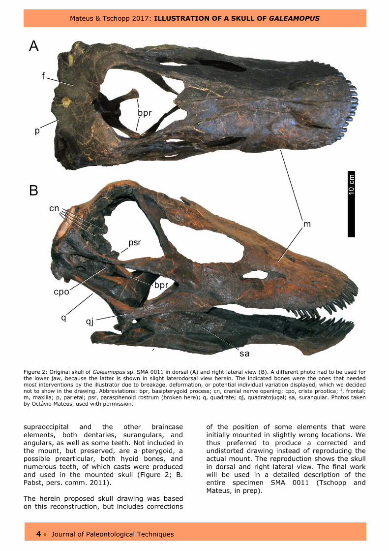
Figure 2: Original skull of Galeamopus sp. SMA 0011 in dorsal (A) and right lateral view (B). A different photo had to be used for the lower jaw, because the latter is shown in slight laterodorsal view herein. The indicated bones were the ones that needed most interventions by the illustrator due to breakage, deformation, or potential individual variation displayed, which we decided not to show in the drawing. Abbreviations: bpr, basipterygoid process; cn, cranial nerve opening; cpo, crista prootica; f, frontal; m, maxilla; p, parietal; psr, parasphenoid rostrum (broken here); q, quadrate; qj, quadratojugal; sa, surangular. Photos taken by Octávio Mateus, used with permission.
supraoccipital and the other braincase
elements, both dentaries, surangulars, and
angulars, as well as some teeth. Not included in
the mount, but preserved, are a pterygoid, a
possible prearticular, both hyoid bones, and
numerous teeth, of which casts were produced
and used in the mounted skull (Figure 2; B.
Pabst, pers. comm. 2011).
The herein proposed skull drawing was based
on this reconstruction, but includes corrections
of the position of some elements that were
initially mounted in slightly wrong locations. We
thus preferred to produce a corrected and
undistorted drawing instead of reproducing the
actual mount. The reproduction shows the skull
in dorsal and right lateral view. The final work
will be used in a detailed description of the
entire specimen SMA 0011 (Tschopp and
Mateus, in prep).

Mateus & Tschopp 2017: ILLUSTRATION OF A SKULL OF GALEAMOPUS
5 ● Journal of Paleontological Techniques
METHODS
Following Ghilardi and Ribeiro (2010), an
introductory briefing between illustrator (SM)
and scientific supervisor (ET) was held, where
methods, necessary views, access to original
material, deadlines, and purpose of the artwork
was discussed. Right lateral and dorsal views
were decided to be drawn. The limitation to
these two views was necessary due to time
constraints. Given that an undistorted,
hypothetical, perfect lateral view should be
produced, the chosen side does not actually
matter. The right side was chosen here because
it is more complete than the left, where e.g. the
maxilla is lacking. The dorsal view was added
for two reasons: 1) many earlier
reconstructions included a dorsal view, and 2)
many typical diplodocid features are best visible
in this view, as are some peculiar features in
the skull of SMA 0011 (ET, unpublished data).
Finally, a portfolium with photos and
illustrations of diplodocid sauropod skulls was
provided to the illustrator (e.g. Wilson and
Sereno, 1998, fig. 6; Whitlock, 2011, fig. 3).
As a first step, the illustrator took new pictures
of the skull at the SMA that served as a basis
for a first raw pencil sketch. In order to avoid
lens distortion, a focal length of 50 mm was
used for photography and the camera was
oriented such that the fossil fit on the central
area of the photograph when imagining the
picture divided into a grid of nine equal parts.
The inclusion of a scale bar is crucial at this
stage, especially in case the illustrator has no
access anymore to the original material
afterwards. An inclusion of the scale bar here
will also allow to add a more accurate scale bar in the final drawing.
For the first sketch (Figure 3), soft pencils (B, 2B or higher) were used, because they are
Figure 3: Initial pencil drawing of the skull of SMA 0011.

Mateus & Tschopp 2017: ILLUSTRATION OF A SKULL OF GALEAMOPUS
6 ● Journal of Paleontological Techniques
easier to see and to erase if needed. Hard
pencils (e.g. 3H) produce a more precise, but
less dark line. Although levels and curves can
be used in Photoshop to increase their
darkness, this will also increase the visibility of
slight blurs resulting from the drawing process
or erasing. Therefore, scanning of a sketch
made with soft pencils is less likely to miss a
pencil line, and less work is needed afterwards
in Photoshop. The first drawing was then
compared to the original skull, in order to
correct it for possible optical distortions. An
additional briefing with the scientist was
necessary while correcting the first sketch in
order to point out deformed or wrongly
mounted bones in the restored skull.
Subsequently, the pencil drawing was scanned
and revised with a graphic tablet device
(hardware) on Photoshop (software) in order to
obtain a cleaner drawing.
There are several graphic tablets on the
market. One of the most important features for
illustrators is the size of the so-called active
area, which is the working area of the tablet. To
have a better control and definition of the
drawing, we preferred an active area of at least
10 per 15 centimeters.
We used a resolution of 300 pixel/inch (dpi) for
an initial area of 20 per 30 centimeters, which
results on 2362 x 3543 pixels. These values
guarantee a file of a resolution high enough to
produce optimal quality printing on a DIN A4
page, because printers usually work with a
resolution of 150-300 dpi. Given that the
drawing was intended to be published in online
journals, it was not necessary to use a higher
resolution, and computing time could be
reduced considerably. It is important to specify
the dimensions of the working area, as it is also
possible to have a 6 pixel drawing with 300 dpi, thus measuring only 0,3 x 0,2 mm.
For the working steps in Photoshop, the
working document was split into several layers.
The background layer was always left white.
Different layers were created for each
photograph (dorsal and lateral views of the
skull, and lateral view of the mandible). All of
these images were resized to the same scale
and their layer were locked, such that they
could not be changed accidentally. The
photograph, the pencil sketch, and the final
working drawing were placed in different layers.
Finally, a layer with a reconstructed skull of the
closely related Diplodocus (Wilson and Sereno,
1998; fig. 6) was added for comparative
purposes and to help understand the shape of
distorted or incomplete bones. We used folders
to organize the several layers, in order to keep
track more easily in which layer we were
supposed to work, and which layer was not
necessary to see and could be hidden at that
time. It is also useful to have a notebook - or
an additional layer - to write some information
about the brush or pencils tools used,
specifically the master diameter and hardness
used for outlines or for texture details.
A first version of the computer drawing was
saved as “Max_skull_v1.psd” and sent to the
scientific supervisor for corrections and comments.
Eight changes were proposed by the scientific
supervisor, and directly highlighted and
sketched in a copy of the original first drawing
(Figure 4). All these proposed changes were
discussed with the illustrator with the original
skull at hand. Some of the necessary
corrections concerned additional shape changes
because of deformed or fractured parts of the
fossil skull of SMA 0011: deletion of lines on the
lateral side of the braincase that were based on
features that were due to breakage or
deformation (Figure 4, number 1); correction of
the lateral outline of the braincase, which was
necessary because some parts of the anterior
edge and the parasphenoid rostrum were
broken off during diagenesis (Figure 4, number
2); deletion of a line indicating a feature on the
surangular bone that was due to deformation
(Figure 4, number 4); changes to the outline of
the frontal due to deformation (Figure 4,
number 5); closure of what appears to be a
large pineal foramen and a smaller postparietal
foramen, but which have broken edges on the
frontal and parietal bones, indicating that the
presence of these foramina is due to
taphonomic breakage (Figure 4, number 6);
and the deletion of two wavy lines indicating a
deformation in the posterior process of the
maxilla (Figure 4, number 8). Other proposed
corrections aimed for a clearer visualization of
the single bones, and other morphological
features: addition of the major foramina for the
cranial nerves visible in lateral view (Figure 4,
number 1); and the substitution of the lines
illustrating three-dimensional morphology of the
articular ramus of the quadrate by the outline
of the quadratojugal, in order to show the exact
shapes of the single bones (Figure 4, number 3).

Mateus & Tschopp 2017: ILLUSTRATION OF A SKULL OF GALEAMOPUS
7 ● Journal of Paleontological Techniques
Figure 4: The digitized drawing of the skull of SMA 0011 with the comments of the scientist. The numbers indicate the changes
requested: 1) deletion of lines due to breakage and major foramina for cranial nerves; 2) broken parasphenoid process; 3)
outline of quadratojugal; 4) deletion of line indicating a feature on surangular bone that is due to breakage; 5) adaption of
frontal outline due to deformation; 6) closure of openings due to taphonomic breakage; 7) wrong orientation of basipterygoid
processes; 8) wavy lines indicating a deformation on posterior process of maxilla.
Finally, one correction was necessary because
the broken off basipterygoid processes were
erroneously mounted in a position dorsal to the
crista prootica (Figure 4, number 7). The input
of the supervisor were integrated in the second
version of the drawing, and saved as
“Max_skull_v2.psd”. Duplicate copies of the
work steps were saved on an external hard
drive as a safety backup. Small details were
corrected in another meeting between
illustrator and scientific supervisor (e.g. the
orientation of the reconstructed basipterygoid
processes). During this third meeting, an
additional layer was created in the drawing,
adding the grey gradients. These gradients
significantly increased the three-dimensional
Mateus & Tschopp 2017: ILLUSTRATION OF A SKULL OF GALEAMOPUS
8 ● Journal of Paleontological Techniques
understanding in the two views of the skull
(Figures 5-6). We preferred these gradients
over weighted lines because thin lines were
already used for bone textures that are at the
same level as the edges of the bone they mark.
Using the same line width for these textures
and for elements that lie below others could
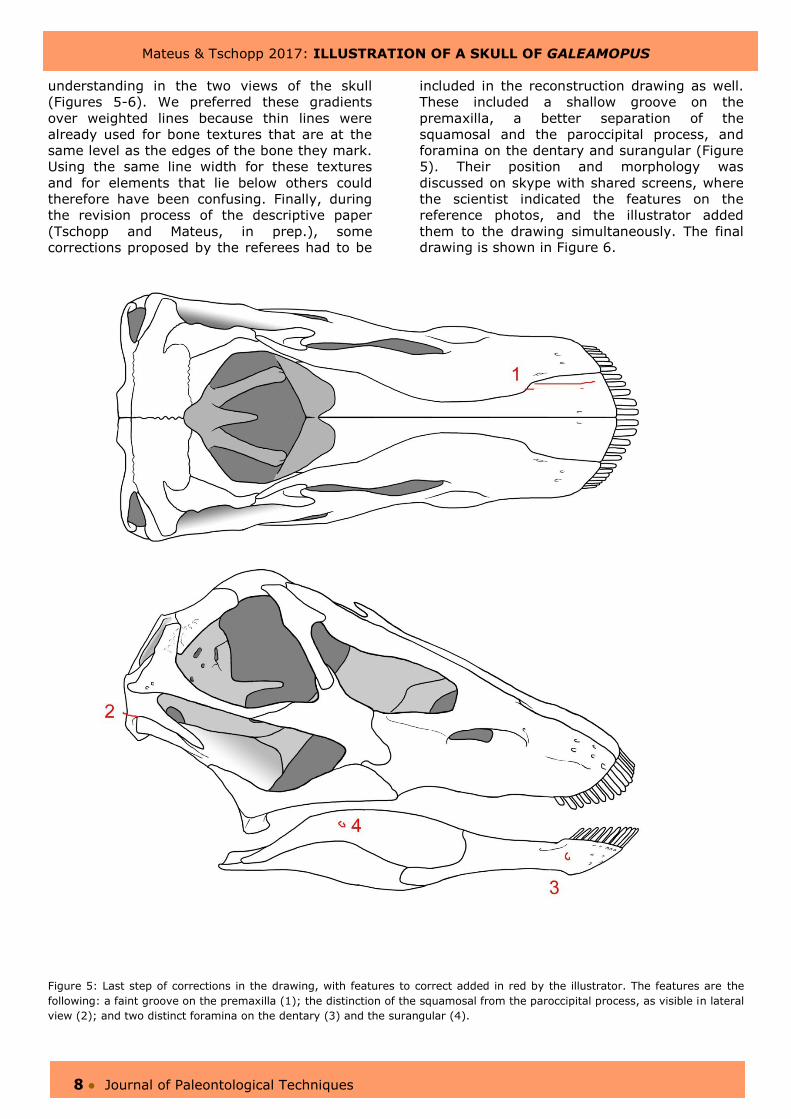
therefore have been confusing. Finally, during
the revision process of the descriptive paper
(Tschopp and Mateus, in prep.), some
corrections proposed by the referees had to be
included in the reconstruction drawing as well.
These included a shallow groove on the
premaxilla, a better separation of the
squamosal and the paroccipital process, and
foramina on the dentary and surangular (Figure
5). Their position and morphology was
discussed on skype with shared screens, where
the scientist indicated the features on the
reference photos, and the illustrator added
them to the drawing simultaneously. The final drawing is shown in Figure 6.
Figure 5: Last step of corrections in the drawing, with features to correct added in red by the illustrator. The features are the
following: a faint groove on the premaxilla (1); the distinction of the squamosal from the paroccipital process, as visible in lateral
view (2); and two distinct foramina on the dentary (3) and the surangular (4).
Mateus & Tschopp 2017: ILLUSTRATION OF A SKULL OF GALEAMOPUS
9 ● Journal of Paleontological Techniques
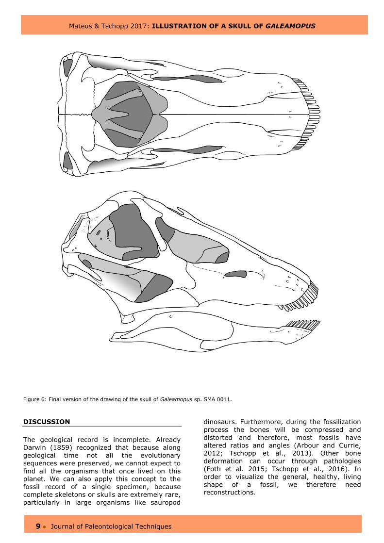
Figure 6: Final version of the drawing of the skull of Galeamopus sp. SMA 0011.
DISCUSSION
The geological record is incomplete. Already
Darwin (1859) recognized that because along
geological time not all the evolutionary
sequences were preserved, we cannot expect to
find all the organisms that once lived on this
planet. We can also apply this concept to the
fossil record of a single specimen, because
complete skeletons or skulls are extremely rare,
particularly in large organisms like sauropod
dinosaurs. Furthermore, during the fossilization
process the bones will be compressed and
distorted and therefore, most fossils have
altered ratios and angles (Arbour and Currie,
2012; Tschopp et al., 2013). Other bone
deformation can occur through pathologies
(Foth et al. 2015; Tschopp et al., 2016). In
order to visualize the general, healthy, living
shape of a fossil, we therefore need reconstructions.

Mateus & Tschopp 2017: ILLUSTRATION OF A SKULL OF GALEAMOPUS
10 ● Journal of Paleontological Techniques
In the current reconstruction, adding missing
parts, or accounting for distortion was
facilitated by the large amount of skulls known
from very similar taxa (see reviews in Whitlock
et al. 2010; Whitlock, 2011). In other species,
however, the reconstruction can be more
difficult because skulls from closely related taxa
are lacking (see e.g. the changes in the
reconstruction of Nemegtosaurus or Euhelopus;
Upchurch 1999, fig. 2, and Wilson, 2005, fig.
16; Mateer & McIntosh 1985, fig. 6, and
Poropat and Kear 2013, fig. 1). In such cases,
illustrators usually use dotted lines or different
shading to indicate the hypothetical shapes of
unpreserved elements (e.g. Madsen et al.,
1995; Wilson, 2005; Sereno et al., 2007;
Tschopp and Mateus, 2013).
The biggest advantage of an illustration
(compared to photographs or 3D renderings) is
that it can be used to highlight important
details and hide irrelevant ones, as for instance
the grooves resulting from distortion in our case
(see Figure 4, numbers 4, 8). Two of the
biggest disadvantages are the time needed to
produce a good illustration and the costs of
hiring an illustrator. The supervision of the
illustrator by the paleontologists is essential but
not always easy, because visible features have
to be reinterpreted in the light of deformation.
In some cases, these differences between
actual occurrence and interpretation are
significant, and can result in long discussions
between illustrator and paleontologist. One
example of such a significant difference in the
present artwork of the skull of SMA 0011 was
the drawing of the parasphenoid rostrum that is
visible through the orbit (Figures 4, 5). The
parasphenoid rostrum is broken and lost on the
fossil skull (Figure 2). During the illustration
process, on the second sketch, the scientific
supervisor added by hand the missing part.
However, it was not easy for the illustrator to
understand the size, shape or the orientation of
the rostrum. Also the orientation of the
basipterygoid processes (Figures 2-4) and
therefore the interpretation of how much of
them was visible on the drawing (Figure 4) was
quite controversial. The basipterygoid processes
pass in part behind the postorbital, and are
thus partly obscured, depending on the exact
angle of the view. In order to solve these
issues, a good dialog between illustrator and
scientific supervisor was essential and beneficial
for both persons and the final drawing.
ACKNOWLEDGMENTS
We thank Hans-Jakob "Kirby" Siber (SMA) for
the invitation to come to the SMA, and the
entire staff for helping with logistics while
producing the first drawings. Many thanks also
to Octávio Mateus (Univ. Nova da Lisboa,
Portugal) for the photos we used as a base for
the reconstruction drawings. Last but not least,
the two referees, Greg Paul (Baltimore, USA)
and Carol Abraczinskas (Univ. of Michigan,
USA), are thanked for providing useful
comments to improve the manuscript, and
Christophe Hendrickx and Femke Holwerda for
editorial handling.
We also thank the Curry Fund of the Geologist’s
Association, which covered the costs of the
article production.
REFERENCES CITED
Arbour, V. M., and P. J. Currie. 2012.
Analyzing taphonomic deformation of
ankylosaur skulls using retrodeformation and
Finite Element Analysis. PLoS ONE 7:e39323.
Ayer, J. 2000. The Howe Ranch Dinosaurs.
Sauriermuseum Aathal, Aathal, Switzerland, 96
pp.
Benton, M. J. 2005. Vertebrate Palaeontology,
3rd ed. Blackwell Publishing, Malden, MA, USA,
472 pp.
Brinkmann, W., and H.-J. Siber. 1992.
Dinosaurier in Aathal. Sauriermuseum Aathal,
Aathal, 37 pp.
Correia, F. 2010. Ilustração Paleontologica –
Existências Riscadas; pp. 459–558 in I. de S.
Carvalho (ed.), Paleontologia: Conceitos e
métodos. vol. 1. Editora Interciência, Rio de
Janeiro, Brazil.
Darwin, C. 1859. On the Origin of Species by
Means of Natural Selection. Murray, London,
574 pp.
Foth, C., S. W. Evers, B. Pabst, O. Mateus,
A. Flisch, M. Patthey, and O. W. M. Rauhut.
2015. New insights into the lifestyle of
Allosaurus (Dinosauria: Theropoda) based on
another specimen with multiple pathologies.
PeerJ 3:e940.
Mateus & Tschopp 2017: ILLUSTRATION OF A SKULL OF GALEAMOPUS
11 ● Journal of Paleontological Techniques
Ghilardi, R. P., and R. N. S. Ribeiro. 2010.
The briefing in paleodesign: selection and
arrangement of data for the reconstitution of
paleovertebrates. Brazilian Geographical
Journal: Geosciences and Humanities Research
Medium 1.
Ghilardi, R. P., R. N. S. Ribeiro, and F. A.
Elias. 2007. Paleodesign: uma nova proposta
metedológica e terminológica aplicada à
reconstituição em vida de espécies fósseis; pp.
61–70 in I. de S. Carvalho (ed.), Paleontologia:
cenários de vida. Editora Interciência, Rio de
Janeiro, Brazil.
Madsen, J. H., J. S. McIntosh, and D. S.
Berman. 1995. Skull and atlas-axis complex of
the Upper Jurassic sauropod Camarasaurus
Cope (Reptilia: Saurischia). Bulletin of Carnegie
Museum of Natural History 31:1–115.
Mateer, N. J., and J. S. Mcintosh. 1985. A
new reconstruction of the skull of Euhelopus
zdanskyi (Saurischia: Sauropoda). Bulletin of
the Geological Institution of the University of
Upsala 11:125–131.
Michelis, I. 2004. Taphonomie des Howe
Quarry’s (Morrison-Formation, Oberer Jura),
Bighorn County, Wyoming, USA. Ph.D.
dissertation, Institute of Palaeontology,
University of Bonn, Bonn, Germany, 41 pp.
Paul, G. S., and T. L. Chase. 1989.
Reconstructing extinct vertebrates; pp. 239–
256 in E. R. S. Hodges (ed.), The Guild
Handbook of Scientific Illustration, 1st ed. John
Wiley & Sons Inc, New York, USA.
Poropat, S. F., and B. P. Kear. 2013.
Photographic atlas and three-dimensional
reconstruction of the holotype skull of
Euhelopus zdanskyi with description of
additional cranial elements. PLoS ONE
8:e79932.
Sereno, P. C., J. A. Wilson, L. M. Witmer, J.
A. Whitlock, A. Maga, O. Ide, and T. A.
Rowe. 2007. Structural extremes in a
Cretaceous dinosaur. PLoS ONE 2:e1230.
Siber, H. J., and U. Möckli. 2009. The
Stegosaurs of the Sauriermuseum Aathal.
Sauriermuseum Aathal, Aathal, 56 pp.
Tschopp, E., and O. Mateus. 2013. The skull
and neck of a new flagellicaudatan sauropod
from the Morrison Formation and its implication
for the evolution and ontogeny of diplodocid
dinosaurs. Journal of Systematic Palaeontology
11:853–888.
Tschopp, E., J. Russo, and G. Dzemski.
2013. Retrodeformation as a test for the
validity of phylogenetic characters: an example
from diplodocid sauropod vertebrae.
Palaeontologia Electronica 16:1–23.
Tschopp, E., O. Wings, T. Frauenfelder, and
W. Brinkmann. 2015. Articulated bone sets of
manus and pedes of Camarasaurus (Sauropoda,
Dinosauria). Palaeontologia Electronica 18:1–
65.
Tschopp, E., O. Wings, T. Frauenfelder, and
B. Rothschild. 2016. Pathological phalanges in
a camarasaurid sauropod dinosaur and
implications on behaviour. Acta Palaeontologica
Polonica 61:125–134.
Upchurch, P. 1999. The phylogenetic
relationships of the Nemegtosauridae
(Saurischia, Sauropoda). Journal of Vertebrate
Paleontology 19:106–125.
Whitlock, J. A. 2011. Inferences of
diplodocoid (Sauropoda: Dinosauria) feeding
behavior from snout shape and microwear
analyses. PLoS ONE 6:e18304.
Whitlock, J. A., J. A. Wilson, and M. C.
Lamanna. 2010. Description of a nearly
complete juvenile skull of Diplodocus
(Sauropoda: Diplodocoidea) from the Late
Jurassic of North America. Journal of Vertebrate
Paleontology 30:442–457.
Wilson, J. A. 2005. Redescription of the
Mongolian sauropod Nemegtosaurus
mongoliensis Nowinski (Dinosauria: Saurischia)
and comments on Late Cretaceous sauropod
diversity. Journal of Systematic Palaeontology
3:283–318.
Wilson, J. A., and P. C. Sereno. 1998. Early
evolution and higher-level phylogeny of
sauropod dinosaurs. Journal of Vertebrate
Paleontology 18:1–79.
Additional images and material can be downloaded at http://www.jpaleontologicaltechniques.org/