scientific trends in clinical research on zirconia dental
TRANSCRIPT
materials
Review
Scientific Trends in Clinical Research on ZirconiaDental Implants: A Bibliometric Review
Felice Lorusso 1,2 , Sammy Noumbissi 1,2 , Inchingolo Francesco 3, Biagio Rapone 4 ,Ahmad G. A. Khater 5 and Antonio Scarano 1,2,*
1 Department of Medical, Oral and Biotechnological Sciences, University of Chieti-Pescara, Via dei Vestini, 31,66100 Chieti, Italy; [email protected] (F.L.); [email protected] (S.N.)
2 Zirconia Implant Research Group (Z.I.R.G), International Academy of Ceramic Implantology,Silver Spring, MD 20901, USA
3 Department of Interdisciplinary Medicine, University of Bari Aldo Moro, 70121 Bari, Italy;[email protected]
4 Department of Basic Medical Sciences, Neurosciences and Sense Organs, University of Bari Aldo Moro,70121 Bari, Italy; [email protected]
5 Faculty of Oral and Dental Medicine, Ahram Canadian University, 6th of October City, 8655 Giza, Egypt;[email protected]
* Correspondence: [email protected]; Tel.: +39-0871-355-4084; Fax: +39-0871-355-4099
Received: 18 October 2020; Accepted: 2 December 2020; Published: 4 December 2020�����������������
Abstract: Background: The clinical use of zirconia implants has been shown to increase steadily due totheir biological, aesthetic, and physical properties; therefore, this bibliometric study aimed to reviewthe clinical research and co-authors in the field of zirconia dental implant rehabilitation. Methods:We searched Scopus and Web of Science databases using a comprehensive search strategy to 5 October2020, and independently paired reviewers who screened studies, and collected data with inclusioncriteria restricted to clinical research only (either prospective or retrospective). Data on article title,co-authors, number of citations received, journal details, publication year, country and institutioninvolved, funding, study design, marginal bone loss, survival rate, failure, follow-up, and the author’sbibliometric data were collected and evaluated. Results: A total of 29 clinical studies were publishedbetween 2008 and 2020 as 41.4% were prospective cohort studies and 48.3% originated from Germany.Most of the included studies had been published in Clinical Oral Implant Research (n = 12), and themost productive institution was the Medical Center of University of Freiburg. The author with thelargest number of clinical studies on zirconia implants was Kohal R.J. (n = 10), followed by Spies B.C.(n = 8). Conclusions: This study revealed that zirconia implants have been more prominent in the lastten years, which is a valuable option for oral rehabilitation with marginal bone loss and survival ratecomparable to titanium dental implants.
Keywords: zirconia implant; bibliometrics; citations; scientometric
1. Introduction
The clinical application of dental implant rehabilitation represents consolidated effectiveness in theliterature due to long-term predictability and high-level satisfactory functioning and aesthetics [1–4].Titanium alloys are the most widely used biomaterials for dental implant fixtures due to their physical,chemical, and thermal properties, which produce the osseointegrating ability of the fixture placed toreplace the natural teeth [2,5–12].
Recently, the use of zirconia as an implant material has become more prevalent due to its highaesthetic characteristics, particularly in the rehabilitation of the compromised anterior jaw area,where there is fine soft-tissue biotype and the metal sensibility of the patients [13–15].
Materials 2020, 13, 5534; doi:10.3390/ma13235534 www.mdpi.com/journal/materials

Materials 2020, 13, 5534 2 of 19
In fact, the literature reports that the titanium ion dissolution related to the implant corrosion couldalter the natural oral microbiome and the homeostatic functional balance of the oral tissues [16–20].
On the contrary, it has been shown in vitro that the zirconia surface can lead to a significantdecrease in periodontal pathogen adhesion compared to the titanium surface [21], alongside similarbone–implant contact compared to the titanium fixture with an almost overlapping range [22].
Additionally, Scarano et al. reported in a rabbit study that zirconia implants had about 68.4%bone–implant contact with evidence of contact osteogenesis without fibrous tissue interposition [23].
Zirconia material is distinguished by its clear ivory appearance, which is very similar to thenatural color of the teeth and is characterized by an intrinsic strength and physical resistance to theloading [24–26]; as a result, it has been introduced as a restorative material for dental crowns, bars,abutments, and specially designed drills and burs [26–34]. Therefore, zirconia has recently gainedfurther attention in the scientific community by growing research activities to confirm the clinicaleffectiveness of zirconia as a dental implant material.
Although citations are not an infallible metric to determine whether research is beneficial toresearchers and clinicians, citations and citation analysis can quantify an article’s influence, author,subject of debate, country, journal, or a specialty [35,36]. Based on citation analysis, the bibliometricanalysis aims to provide information about the trend in a research field and demonstrates its growthand development [37]; the number of citations received, researcher H-index, and journal impact factorare the most common bibliometric evaluation variables and considered as a scientific productivityscore for the scientometric evaluation [38].
With the significant increase in the published articles on dental implants, recognizing trendsand advances in a research field is critical and relevant to the needs of dental practitioners andresearchers [39,40]. In this sense, bibliometric analysis is a useful tool for this purpose [41,42].
As far as we know, the trends and advances in zirconia dental implants have not been studiedbefore; hence this study aimed to evaluate the bibliometric output of clinical research and co-authorsin the field of zirconia dental implant rehabilitations.
2. Materials and Methods
We reported this bibliometric study in compliance with the Standards for Reporting QualitativeResearch (SRQR) [43] and the Preferred Reporting Items for Systematic Reviews and Meta-Analyses(PRISMA) guidelines [44].
2.1. Search Strategy
An online literature search was conducted in Elsevier’s Scopus and Clarivate Analytics’ Web ofScience (WoS) until 5 October 2020. We used the medical terms (MeSH) feature in the Cochrane Libraryto obtain the available synonyms for our search terms to create a detailed search strategy (Table 1).
Table 1. Search strategy used for each database.
Scopus
TITLE-ABS-KEY (“Zirconia” OR “Zirconium” OR “Zircon*”) AND TITLE-ABS-KEY(“Dental implant” OR “Dental implants” OR “Oral implant” OR “Oral implants” OR“Implant dentistry” OR “Dental implantology” OR “Dental Implantation” OR“Osseointegrated” OR “Osseointegrated Dental Implantation”) AND TITLE-ABS-KEY(“Intervention Study” OR “Clinical Trial” OR “Controlled Clinical Trial” OR “RandomizedControlled Trials OR “Non-Randomized Clinical Trial” OR “Nonrandomized Clinical Trial”OR “Quasi-Experimental” OR “Observational Study” OR “Prospective Study” OR“Prospective” OR “Retrospective Study” OR “Retrospective” OR “Comparative Study” OR“Multicenter Studies” OR “Epidemiologic Study” OR “Epidemiological Studies” OR“Cohort Study” OR “Case Studies” OR “Follow-Up Study” OR “Case-Control Study” OR“Case Report” OR “Case Series” OR “Pilot Study”)

Materials 2020, 13, 5534 3 of 19
Table 1. Cont.
Web of Science
TS = (“Zirconia” OR “Zirconium” OR “Zircon*”) AND TS = (“Dental implant” OR“Dental implants” OR “Oral implant” OR “Oral implants” OR “Implant dentistry” OR “Dentalimplantology” OR “Dental Implantation” OR “Osseointegrated” OR “Osseointegrated DentalImplantation”) AND TS = (“Intervention Study” OR “Clinical Trial” OR “Controlled ClinicalTrial OR “Randomized Controlled Trials” OR “Non-Randomized Clinical Trial” OR“Nonrandomized Clinical Trial” OR “Quasi-Experimental” OR “Observational Study” OR“Prospective Study” OR “Prospective” OR “Retrospective Study” OR “Retrospective” OR“Comparative Study” OR “Multicenter Studies” OR “Epidemiologic Study” OR“Epidemiological Studies” OR “Cohort Study” OR “Case Studies” OR “Follow-Up Study” OR“Case-Control Study” OR “Case Report” OR “Case Series” OR “Pilot Study”)Timespan: All years. Databases: WOS, ARCI, BCI, KJD, MEDLINE, RSCI, SCIELO, ZOOREC.Search language = Auto.
2.2. Data Extraction and Bibliometric Parameters
We used a specially built Excel file (Microsoft, Redmond, WA, USA) to collect the findings of theliterature search. The file contained the following information: abstracts, year of publication, indexedkeywords, journal name, citations as well as all co-author bibliometric data (H-index, number of papersrelated to zirconia implant, the total number of papers, citation of paper regarding zirconia implant,and citation of paper regarding zirconia implant). Authors with the highest quantity of clinical studiesregarding zirconia dental implants were evaluated and measured the average, the standard deviation,minimum and maximum of topic paper, total papers, topic citations, overall citations, and H-index.Moreover, we evaluated the scientific trend of the included study according to the year of publicationand journal details (full title, the impact factor (IF), and rank) based on the Clarivate Analytics reportfor 2019 with selected categories: “Dentistry, Oral Surgery & Medicine”, study design, number ofcitations received, marginal bone loss, survival rate, failure, and study follow-up.
2.3. Study Selection
We screened the literature search results in two steps, where the first phase was the screening of thetitle and abstract by paired reviewers separately. Then, the second phase was a full-text assessment bytwo expert reviewers (L.F and A.S). The reference list of the studies included in the full-text screeningwas hand-screened for potential additional studies. In this bibliometric study, inclusion criteria wereonly clinical studies (either prospective or retrospective) without time restrictions. Exclusion criteriawere animal studies, in vitro studies, literature reviews, systematic reviews, short communications,personal opinion, letters, book chapters, and non-English studies.
2.4. Data Analysis
We used VOSviewer software (version 1.6.8; Leiden University, Leiden, The Netherlands) tovisualize a term map analyzing keywords from the data obtained. “Create Map” function was used toanalyze the data by using the “Citation” type and setting the unit of analysis as a “number of citations.”In the keyword map, the node’s size reflects the number of received citations, as the larger size indicatesthe author with the highest citations. Furthermore, keywords that often appeared together wereclassified as the same color in network visualization mode [45,46].
3. Results
3.1. Study Selection
A total of 1159 references were collected from electronic databases in which (n = 185) were omitteddue to duplication. By title and abstract, 968 articles were screened and 841 excluded as irrelevant topics.By the full-text screening of 127 papers, 29 studies were included in this bibliometric study [47–75]excluding the remaining 98 articles because they did not meet our inclusion criteria (Figure 1).

Materials 2020, 13, 5534 4 of 19
Materials 2020, 13, x FOR PEER REVIEW 4 of 21
Figure 1. Preferred Reporting Items for Systematic Reviews and Meta-Analyses (PRISMA) flow chart demonstrates the process of literature search and study selection.
3.2. Study Characteristics
The included studies showed wide variability in the study design, presence/absence of a control group, experimental site, type of prosthetic rehabilitation, prosthetic connection (one-piece or two-piece), follow-up period, and different methods for evaluating the effectiveness of research. Although these differences exist, most studies reported favorable outcomes for the use of zirconia implants in oral rehabilitation. The main characteristics of the included studies are summarized in Table 2.
A total of 21 studies evaluated monolithic or one-piece zirconia implants [47–49,51–54,56–58,60,62,64,66,70–74,76], two of which had titanium implants as their control and showed no significant difference in survival rate and marginal bone loss between groups (p > 0.05) [64,70]. Two studies evaluated the immediate loading of zirconia implants [53,64]: one study compared it to the non-occlusal loading procedure [64], while the other study compared it with the standard loading protocol [57]. Furthermore, 26 papers assessed the cylindrical microgeometry of zirconia implants [47–65,67,70–75], while three studies evaluated the root-analog zirconia implants obtained by a three-dimensional scan [66,68,69]. However, Akça et al. and Pirker et al. reported the lowest marginal bone loss after two years (0.31 ± 0.24 and 0.5 ± 0.7 mm, respectively), in which Akça et al. used specially designed titanium–zirconia alloy implants [47], and Pirker et al. used specially designed root-analog zirconia implants with a micro-retention surface in a fresh extraction socket [69].
Figure 1. Preferred Reporting Items for Systematic Reviews and Meta-Analyses (PRISMA) flow chartdemonstrates the process of literature search and study selection.
3.2. Study Characteristics
The included studies showed wide variability in the study design, presence/absence of a controlgroup, experimental site, type of prosthetic rehabilitation, prosthetic connection (one-piece or two-piece),follow-up period, and different methods for evaluating the effectiveness of research. Although thesedifferences exist, most studies reported favorable outcomes for the use of zirconia implants in oralrehabilitation. The main characteristics of the included studies are summarized in Table 2.
A total of 21 studies evaluated monolithic or one-piece zirconia implants [47–49,51–54,56–58,60,62,64,66,70–74,76], two of which had titanium implants as their control and showed no significantdifference in survival rate and marginal bone loss between groups (p > 0.05) [64,70]. Two studiesevaluated the immediate loading of zirconia implants [53,64]: one study compared it to the non-occlusalloading procedure [64], while the other study compared it with the standard loading protocol [57].Furthermore, 26 papers assessed the cylindrical microgeometry of zirconia implants [47–65,67,70–75],while three studies evaluated the root-analog zirconia implants obtained by a three-dimensionalscan [66,68,69]. However, Akça et al. and Pirker et al. reported the lowest marginal bone loss aftertwo years (0.31 ± 0.24 and 0.5 ± 0.7 mm, respectively), in which Akça et al. used specially designedtitanium–zirconia alloy implants [47], and Pirker et al. used specially designed root-analog zirconiaimplants with a micro-retention surface in a fresh extraction socket [69].

Materials 2020, 13, 5534 5 of 19
Table 2. Main characteristics of the clinical research included (Zir: Zirconia implant group, Tit: Titanium implant group, IF: impact factor, RCT: Randomizedcontrolled trial).
Authors(Year) [Ref]
JournalCited By Study
DesignPatients
(Implants) Test ControlMarginal Bone Loss
(Mean ± SD) Survival Rate FailureFollow
UpFull Title Rank IF
Pirker et al.(2008) [68]
International Journal ofOral and Maxillofacial
Surgery33 2.068 50 Case report 1 (1 Implant)
Microretention andsandblasted
root-analoguezirconia implant
- - 100% - 2 years
Pirker et al.(2009) [69]
International Journal ofOral and Maxillofacial
Surgery33 2.068 58 Prospective
Case Series 18 (18 Implants)
Microretention andsandblasted
root-analoguezirconia implants
Sandblastedroot-analogue
zirconia implants0.5 ± 0.7 mm Test: 92%
Control: 0%
Test:1 implant
Control: All implants (6)2 years
Cannizzaro et al.(2010) [54]
European Journal ofImplantology - - 69 Multicenter
RCT 40 (40 Implants)Immediate occlusal
loading zirconiaImplants
Immediatenon-occlusal loading
zirconia Implants
Test:0.90 ± 0.48 mm
Control:0.72 ± 0.59 mm
88.50%5 implants (12.5%): Test:
3 Implants Control:2 Implants
1 year
Borgonovo et al.(2011) [51] Minerva Stomatologica - - 21 Prospective
Case Series 16 (26 Implants)One-piece yttriumstabilized zirconia
implants- - 96.16% 1 Implant
osseointegration failure 2 years
Payer et al.(2012) [66]
Clinical Oral ImplantsResearch 8 3.723 61 Prospective
Case Series 20 (20 Implants) One-piece zirconiaimplants - 1.29 ± 0.73 mm 95% 1 Implant
osseointegration failure 2 years
Akça et al.(2013) [47]
International Journal ofOral and Maxillofacial
Implants24 2.32 8 Prospective
Case Series 23 (52 Implants) - - 0.32 ± 0.24 mm 100% No failure 2 years
Borgonovo et al.(2013) [52] Minerva Stomatologica - - 10 Prospective
Case Series 6 (14 Implants)One-piece yttriumstabilized zirconia
implants- 0.67 ± 0.51 mm 100% No failure 4 years
Kohal et al.(2013) [59]
Journal of ClinicalPeriodontology 2 5.241 47 Prospective
Case Series 28 (56 Implants)
One-pieceyttria-stabilized
tetragonal zirconiaimplants
- 1.95 ± 0.65 98.20% 1 Implantosseointegration failure 1 year
Osman et al.(2013) [63]
International Journal ofProsthodontics 61 1.49 6 Pilot study 4 (28 Implants)
One-piece zirconiaimplants for ball
abutment- - 85.70% 4 Implants 1 year
Osman et al.(2014) [64]
Clinical Oral ImplantsResearch 8 3.723 34 RCT 19 (129
Implants)
One-piece zirconiaimplants for
ball-abutment
One-piece titaniumimplants for
ball-abutment
Zir:0.42 ± 0.40
Tit:0.18 ± 0.47
Zir: 90.9%Tit:95.8%
Zir:21Implants(3 fractured)
Tit:10Implants
1 year
Becker et al.(2015) [50]
Clinical Oral ImplantsResearch 8 3.723 15 Prospective
Cohort Study 52 (52 Implants) Two-piece zirconiaimplants - - 95.80% 2 Implants 2 years
Cionca et al.(2015) [55]
Clinical Oral ImplantsResearch 8 3.723 43 Prospective
Case Series 32 (49 Implants) Two-piece zirconiaimplants - - 87% 6 Implants 1 year

Materials 2020, 13, 5534 6 of 19
Table 2. Cont.
Authors(Year) [Ref]
JournalCited By Study
DesignPatients
(Implants) Test ControlMarginal Bone Loss
(Mean ± SD) Survival Rate FailureFollow
UpFull Title Rank IF
Jung et al.(2015) [56]
Clinical Oral ImplantsResearch 8 3.723 27 Prospective
Cohort Study 60 (71 Implants) Immediate one-piecezirconia implants - 0.78 ± 0.79 mm 98.30% 1 implant
osseointegration failure 1 year
Payer et al.(2015) [67]
Clinical Oral ImplantsResearch 8 3.723 41 RCT 22 (31 Implants) Two-piece zirconia
implantsTwo-piece titanium
implants
Zir:1.48 ± 1.05
Tit:1.43 ± 0.67
Zir: 93.3%Tit:
100%
Zir:1 Implant
Tit:No failure
2 years
Siddiqi et al.(2015) [70]
Clinical ImplantDentistry and Related
Research9 3.396 17 RCT 22 (150
Implants)
One-piece zirconiaimplants for
ball-abutment
Titanium implantsfor one-pieceball-abutment
Zir:2.23 ± 0.69
Tit:1.59 ± 0.33
Zir: 67.6%Tit:
66.7%
Zir:16 Implants
Tit:7 Implants
1 year
Spies et al.(2015) [73]
Journal of DentalResearch 3 4.914 22 Prospective
Cohort Study 40 (53 Implants)One-piece
alumina-toughenedzirconia implant
- 0.79 ± 0.47 mm 94.2% 3 Implantsosseointegration failure 3 years
Patankar et al.(2016) [65]
Journal of Maxillofacialand Oral Surgery - - 3 Case report 1 (1 Implant)
Microretention andsandblasted
root-analoguezirconia implant
- - 100% - 1.5 year
Spies et al.(2016) [74]
Clinical Oral ImplantsResearch 8 3.723 13 Prospective
Cohort Study 27 (27 Implants)Immediate one-piecealumina-toughened
zirconia implant- 0.77 ± 0.31 mm 88.90% 3 Implants
osseointegration failure 1 year
Kniha et al.(2017) [58]
International Journal ofOral and Maxillofacial
Surgery33 2.068 9 Prospective
Cohort Study81 (105
Implants) Zirconia implants - 0.66 ± 0.33 mm 100% No failure 3 years
Kniha et al.(2017) [57]
International Journal ofOral and Maxillofacial
Surgery33 2.068 9 Prospective
Cohort Study 78 (82 Implants)Immediate loadingone-piece zirconia
implants
Delayed one-piecezirconia implants
Immediate:0.76 ± 1.13 mm
Delayed:0.83 ± 0.65 mm
Immediate:100%
Delayed:100%
No failure 1 year
Spies et al.(2017) [71] Journal of Dentistry 10 3.242 6 Prospective
Case Series 60 (71 Implants) One-piece zirconiaoral implants - - 100% No failure 3 years
Spies et al.(2017) [75]
Clinical Oral ImplantsResearch 8 3.723 11 Prospective
Case Series 13 (26 Implants) One-piece zirconiaimplants - - 100% No failure 5 years
Balmer et al.(2018) [49]
Clinical Oral ImplantsResearch 8 3.723 11
ProspectiveMulticenter
Cohort Study60 (71 Implants)
One-piece immediateloading zirconia
implants- 0.70 ± 0.72 mm 98.50% 1 Implant
osseointegration failure 3 years
Bormann et al.(2018) [53] BMC Oral Health 38 1.911 7
ProspectiveMulticenter
Cohort Study44 (44 Implants) Zirconia implants - 0.97 ± 0.88 mm 97.50% 1 Implant 3 years
Kohal et al.(2018) [60]
Journal of ClinicalPeriodontology 2 5.241 5 Prospective
Cohort Study 65 (65 Implants)Immediate loadingone-piece zirconia
implants- 1.45 ± 1.96 mm 90.80% 6 Implants 3 years

Materials 2020, 13, 5534 7 of 19
Table 2. Cont.
Authors(Year) [Ref]
JournalCited By Study
DesignPatients
(Implants) Test ControlMarginal Bone Loss
(Mean ± SD) Survival Rate FailureFollow
UpFull Title Rank IF
Lorenz et al.(2019) [62]
Clinical ImplantDentistry and Related
Research9 3.396 4 Prospective
Cohort Study 28 (83 Implants) Zirconia implants Natural teeth 1.2 ± 0.76 mm 100%No failure one
peri-implantitis resistantto therapies
7.8 years
Spies et al.(2019) [72]
Clinical Oral ImplantsResearch 8 3.723 5
ProspectiveMulticenter
Cohort Study45 (45 Implants) Zirconia implants - - 97.5 ± 2.47%.
Chipping (n = 19)occlusal roughness
(n = 35)5 years
Balmer et al.(2020) [48]
Clinical Oral ImplantsResearch 8 3.723 4
ProspectiveMulticenter
Cohort Study60 (71 Implants)
Single crownone-piece zirconia
implant
Multiple prosthesesone-piece zirconia
implant0.7 ± 0.6 mm 98.4% 1 Implant 5 years
Koller et al.(2020) [61]
Clinical Oral ImplantsResearch 8 3.723 0 Pilot RCT 22 (31 Implants) Two-piece zirconia
implantsTwo-piece titanium
implants
Zir:1.38 ± 0.81 Tit:1.17 ± 0.73 mm
Zir: 87.5%Tit:93.3%
Zir:2 Implants
Tit:1 Implant
6.67 years

Materials 2020, 13, 5534 8 of 19
3.3. Growth of Publications
In total, 29 clinical studies were published between 2008 and 2020, in which 19 papers (65.5%) werepublished in the last five years and ten papers published before 2015. The highest number of publishedstudies was in 2015 (n = 6, 20.6%) followed by 2013 and 2017 (n = 4, 13.7% for each) (Figure 2).
Materials 2020, 13, x FOR PEER REVIEW 10 of 21
3.3. Growth of Publications
In total, 29 clinical studies were published between 2008 and 2020, in which 19 papers (65.5%) were published in the last five years and ten papers published before 2015. The highest number of published studies was in 2015 (n = 6, 20.6%) followed by 2013 and 2017 (n = 4, 13.7% for each) (Figure 2).
Figure 2. Publication trend of the clinical studies on the zirconia implants.
3.4. Journal of Publication
The clinical studies on the use of zirconia dental implants for oral rehabilitation were published across ten peer-reviewed journals. The journal with the largest number of publications was “Clinical Oral Implants Research” (n = 12, 41%), followed by “International Journal of Oral and Maxillofacial Surgery” (n = 4, 13.7%) (Figure 3).
The majority of publications were published in Q1 journals (n = 25, 86%), while the journal with the highest impact factor was “Journal of Clinical Periodontology” (IF = 5.241), which had two articles.
2008
2009
2010
2011
2012
2013
2014
2015
2016
2017
2018
2019
2020
0
1
2
3
4
5
6
Papers
Year
Growth of publications
Figure 2. Publication trend of the clinical studies on the zirconia implants.
3.4. Journal of Publication
The clinical studies on the use of zirconia dental implants for oral rehabilitation were publishedacross ten peer-reviewed journals. The journal with the largest number of publications was “Clinical OralImplants Research” (n = 12, 41%), followed by “International Journal of Oral and Maxillofacial Surgery”(n = 4, 13.7%) (Figure 3).
The majority of publications were published in Q1 journals (n = 25, 86%), while the journal withthe highest impact factor was “Journal of Clinical Periodontology” (IF = 5.241), which had two articles.

Materials 2020, 13, 5534 9 of 19Materials 2020, 13, x FOR PEER REVIEW 11 of 21
Figure 3. Contribution journals in clinical research on zirconia implants.
3.5. Study Design and Level of Evidence
All included studies were prospective, while the most common study design of clinical research on zirconia implants was cohort study (n = 12, 41.4%), followed by case series (n = 9, 31%), and RCT (n = 5, 17%). According to the hierarchy of evidence levels (Is) [77,78], the available evidence supporting the use of zirconia implants is 17% level II, 41.4% EL IV, and the remaining EL VI.
3.6. Contribution of Countries and Institutions
The majority of the studies originated from institutions in Germany (n = 14, 48.3%), followed by Switzerland, (n = 6, 20.7%), and Austria (n = 5, 17%), where the most productive institution was the Medical Center of University of Freiburg (n = 8, 27.6%), followed by the Center of Dental Medicine, University of Zürich (n = 5, 17%). While many of the included studies were funded, the most funding support for included research was provided by VITA Zahnfabrik—H. Rauter GmbH & Co. KG, Bad Säckingen, Germany (n = 5, 17%) (Table 3).
Minerva Stomatologica
Journal of Maxillofacialand Oral Surgery
Journal of Dentistry
Journal of Dental Research
Journal of Clinical Periodontology
International Journal of Prosthodontics
International Journal of Oral and Maxillofacial Surgery
International Journal of Oral and Maxillofacial Implants
European Journal of Implantology
Clinical Oral Implants Research
Clinical Implant Dentistry and Related Research
BMC Oral Health
0 2 4 6 8 10 12
Distribution of scientific journals
Figure 3. Contribution journals in clinical research on zirconia implants.
3.5. Study Design and Level of Evidence
All included studies were prospective, while the most common study design of clinical research onzirconia implants was cohort study (n = 12, 41.4%), followed by case series (n = 9, 31%), and RCT (n = 5,17%). According to the hierarchy of evidence levels (Is) [77,78], the available evidence supporting theuse of zirconia implants is 17% level II, 41.4% EL IV, and the remaining EL VI.
3.6. Contribution of Countries and Institutions
The majority of the studies originated from institutions in Germany (n = 14, 48.3%), followedby Switzerland, (n = 6, 20.7%), and Austria (n = 5, 17%), where the most productive institutionwas the Medical Center of University of Freiburg (n = 8, 27.6%), followed by the Center of DentalMedicine, University of Zürich (n = 5, 17%). While many of the included studies were funded, themost funding support for included research was provided by VITA Zahnfabrik—H. Rauter GmbH &Co. KG, Bad Säckingen, Germany (n = 5, 17%) (Table 3).

Materials 2020, 13, 5534 10 of 19
Table 3. Contribution of countries and institutions to clinical studies on zirconia implants.
Country Institution Study [Ref] Funding
Germany
Universitätsklinikum Düsseldorf, Düsseldorf Becker et al., 2015 [50] ZV3 Zircon Vision GmbH, Wolfratshausen, Germany
University Hospital Aachen, Aachen Kniha et al., 2017 [57]
No FundingFriedrich-Alexander-University Erlangen-Nürnberg Kniha et al., 2017 [58]
Johann-Wolfgang Goethe University, Frankfurt/Main Lorenz et al., 2019 [62]
Hannover Medical School, Hannover Bormann et al., 2018 [53] Institut Straumann AG, Basel, Switzerland
School of Dentistry, Albert-Ludwigs University, Freiburg Kohal et al., 2013 [59]Nobel Biocare AB, Göteborg, Sweden
Medical Center of University of Freiburg, Freiburg
Kohal et al., 2018 [60]
Spies et al., 2015 [73] Metoxit AG (Thayngen, Switzerland)Spies et al., 2016 [74]
Spies et al., 2017 [75] Ivoclar Vivadent
Germany and Switzerland Medical Center of University of Freiburg, Freiburg andCenter of Dental Medicine, University of Zürich, Zürich
Spies et al., 2017 [71]
VITA Zahnfabrik—H. Rauter GmbH & Co. KG, Bad Säckingen, GermanyBalmer et al., 2018 [49]
Spies et al., 2019 [72]
Balmer et al., 2020 [48]
SwitzerlandSchool of Dental Medicine, University of Geneva, Geneva Cionca et al., 2015 [55] Dentalpoint AG, Zürich, Switzerland
Center of Dental Medicine, University of Zürich, Zürich Jung et al., 2015 [56] VITA Zahnfabrik—H. Rauter GmbH & Co. KG, Bad Säckingen, Germany
Austria
Alfred Kocher, Medical University Vienna, ViennaPirker et al., 2008 [68]
No FundingPirker et al., 2009 [69]
School of Dentistry, Medical University Graz, Graz
Payer et al., 2012 [66] Bredent medical GmbH, Senden, Germany
Payer et al., 2015 [67]Ziterion GmbH, Uffenheim, Germany
Koller et al., 2020 [61]
Italy
Private practice Cannizzaro et al., 2010 [54] Partially supported by Z-systems
School of Dentistry, University of Milan, MilanBorgonovo et al., 2011 [51]
Not reportedBorgonovo et al., 2013 [52]
New ZealandOral Implantology Research Group, Sir John Walsh
Research Institute, School of Dentistry, University of Otago
Osman et al., 2013 [63]Oral Implantology Research Group, Sir John Walsh Research Institute,
School of Dentistry, University of Otago and Southern ImplantsOsman et al., 2014 [64]
Siddiqi et al., 2015 [70]
India BV Dental College and Hospital, Pune Patankar et al., 2016 [65] No Funding
Turkey Faculty of Dentistry, Hacettepe University Akça et al., 2013 [47] No Funding
Materials 2020, 13, 5534 11 of 19
3.7. Bibliometric Assessment
A total of 29 articles with total citations[Scopus] ranged from 0 to 176 (mean 57.28 ± 42.18), while thenumber of citations[Scopus] received by each paper ranged from 0 to 69 (mean 21.3 ± 20). The top-citedstudy was the RCT of Cannizzaro et al. (2010) (n[Scopus] = 69) [54], followed by the prospective caseseries of Payer et al. (2012) (n[Scopus] = 61) [66], and Pirker et al. (2009) (n[Scopus] = 58) [69].
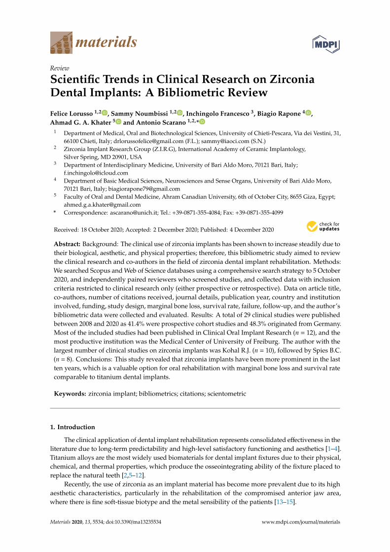
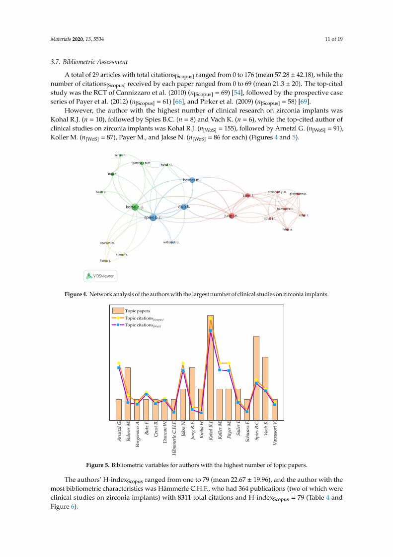
However, the author with the highest number of clinical research on zirconia implants wasKohal R.J. (n = 10), followed by Spies B.C. (n = 8) and Vach K. (n = 6), while the top-cited author ofclinical studies on zirconia implants was Kohal R.J. (n[WoS] = 155), followed by Arnetzl G. (n[WoS] = 91),Koller M. (n[WoS] = 87), Payer M., and Jakse N. (n[WoS] = 86 for each) (Figures 4 and 5).
Materials 2020, 13, x FOR PEER REVIEW 13 of 21
3.7. Bibliometric Assessment
A total of 29 articles with total citations[Scopus] ranged from 0 to 176 (mean 57.28 ± 42.18), while the number of citations[Scopus] received by each paper ranged from 0 to 69 (mean 21.3 ± 20). The top-cited study was the RCT of Cannizzaro et al. (2010) (n[Scopus] = 69) [54], followed by the prospective case series of Payer et al. (2012) (n[Scopus] = 61) [66], and Pirker et al. (2009) (n[Scopus] = 58) [69].
However, the author with the highest number of clinical research on zirconia implants was Kohal R.J. (n = 10), followed by Spies B.C. (n = 8) and Vach K. (n = 6), while the top-cited author of clinical studies on zirconia implants was Kohal R.J. (n[WoS] = 155), followed by Arnetzl G. (n[WoS] = 91), Koller M. (n[WoS] = 87), Payer M., and Jakse N. (n[WoS] = 86 for each) (Figures 4 and 5).
Figure 4. Network analysis of the authors with the largest number of clinical studies on zirconia implants.
Figure 5. Bibliometric variables for authors with the highest number of topic papers.
The authors’ H-indexScopus ranged from one to 79 (mean 22.67 ± 19.96), and the author with the most bibliometric characteristics was Hämmerle C.H.F., who had 364 publications (two of which were clinical studies on zirconia implants) with 8311 total citations and H-indexScopus = 79 (Table 4 and Figure 6).
Arn
etzl
G.
Balm
er M
.
Borg
onov
o A
.
Butz
F.
Cen
si R
.
Dun
can
W.
Häm
mer
le C.
H.F
.
Jaks
e N.
Jung
R.E
.
Kni
ha H
.
Koha
l R.J.
Kolle
r M.
Paye
r M.
Saile
r I.
Schw
arz
F.
Spies
B.C
.
Vach
K.
Vava
ssor
i V.
Topic papers
Topic citations[Scopus]
Topic citations[WoS]
Figure 4. Network analysis of the authors with the largest number of clinical studies on zirconia implants.
Materials 2020, 13, x FOR PEER REVIEW 13 of 21
3.7. Bibliometric Assessment
A total of 29 articles with total citations[Scopus] ranged from 0 to 176 (mean 57.28 ± 42.18), while the number of citations[Scopus] received by each paper ranged from 0 to 69 (mean 21.3 ± 20). The top-cited study was the RCT of Cannizzaro et al. (2010) (n[Scopus] = 69) [54], followed by the prospective case series of Payer et al. (2012) (n[Scopus] = 61) [66], and Pirker et al. (2009) (n[Scopus] = 58) [69].
However, the author with the highest number of clinical research on zirconia implants was Kohal R.J. (n = 10), followed by Spies B.C. (n = 8) and Vach K. (n = 6), while the top-cited author of clinical studies on zirconia implants was Kohal R.J. (n[WoS] = 155), followed by Arnetzl G. (n[WoS] = 91), Koller M. (n[WoS] = 87), Payer M., and Jakse N. (n[WoS] = 86 for each) (Figures 4 and 5).
Figure 4. Network analysis of the authors with the largest number of clinical studies on zirconia implants.
Figure 5. Bibliometric variables for authors with the highest number of topic papers.
The authors’ H-indexScopus ranged from one to 79 (mean 22.67 ± 19.96), and the author with the most bibliometric characteristics was Hämmerle C.H.F., who had 364 publications (two of which were clinical studies on zirconia implants) with 8311 total citations and H-indexScopus = 79 (Table 4 and Figure 6).
Arn
etzl
G.
Balm
er M
.
Borg
onov
o A
.
Butz
F.
Cen
si R
.
Dun
can
W.
Häm
mer
le C.
H.F
.
Jaks
e N.
Jung
R.E
.
Kni
ha H
.
Koha
l R.J.
Kolle
r M.
Paye
r M.
Saile
r I.
Schw
arz
F.
Spies
B.C
.
Vach
K.
Vava
ssor
i V.
Topic papers
Topic citations[Scopus]
Topic citations[WoS]
Figure 5. Bibliometric variables for authors with the highest number of topic papers.
The authors’ H-indexScopus ranged from one to 79 (mean 22.67 ± 19.96), and the author with themost bibliometric characteristics was Hämmerle C.H.F., who had 364 publications (two of which wereclinical studies on zirconia implants) with 8311 total citations and H-indexScopus = 79 (Table 4 andFigure 6).

Materials 2020, 13, 5534 12 of 19
Table 4. General bibliometric variables for authors with the largest number of topic papers.
Author TopicPapers Total Papers Topic/Total
Papers %
TopicCitations[Scopus]
TopicCitations
[WoS]
TotalCitations[Scopus]
Total Citation[WoS]
Topic/TotalCitations %
[Scopus-WoS]
H-index[Scopus]
H-index[WoS]
Kohal R.J. 10 109 9.17% 176 155 3053 2975 [5.76–5.21%] 30 29Spies B.C. 8 47 17.02% 70 64 452 448 [15.49–14.29%] 14 14
Vach K. 6 73 8.22% 54 51 661 820 [8.17–6.21%] 15 16Balmer M. 5 13 38.46% 44 31 256 130 [17.19–23.84%] 7 5Jung R.E. 5 202 2.48% 23 19 8359 9126 [0.28–0.21%] 47 57Jakse N. 3 70 4.29% 99 86 1114 981 [8.89–8.77%] 18 18Kniha H. 3 28 10.71% 21 13 664 726 [3.16–1.779%] 11 13Koller M. 3 9 33.33% 99 87 166 312 [59.64–27.88%] 5 5Payer M. 3 48 6.25% 99 86 770 1484 [12.86–5.79%] 15 23Sailer I. 3 113 2.65% 35 31 6027 5713 [0.58–0.54%] 34 33
Arnetzl G. 2 40 5.00% 99 91 437 183 [22.65–49.72%] 11 6Butz F. 2 24 8.33% 48 45 1248 1441 [3.85–3.12%] 18 19
Censi R. 2 18 11.11% 31 29 169 95 [18.34–30.52%] 8 4Duncan W. 2 87 2.30% 38 35 2163 1356 [1.76–2.58%] 20 19
Hämmerle C.H.F. 2 364 0.55% 16 14 18,311 16,032 [0.09–0.08%] 79 72Schwarz F. 2 261 0.77% 17 15 9093 9169 [0.19–0.16%] 57 57
Vavassori V. 2 12 16.67% 31 27 136 96 [22.79–28.12%] 7 5Borgonovo A. 2 51 3.92% 31 28 535 449 [5.79–6.23%] 12 11
Summary(Mean ± SD) 3.61 ± 2.33 87.17 ± 96.37 10.07 ± 0.11% 57.28 ± 42.18 50.39 ± 37.68 2978.56 ± 4750.05 2863.11 ± 4386.90 - 22.67 ± 19.96 22.56 ± 20.14

Materials 2020, 13, 5534 13 of 19Materials 2020, 13, x FOR PEER REVIEW 15 of 21
Figure 6. Box plots summarize the bibliometric variables of the authors with the largest number of studies.
4. Discussion
The present study carried out a bibliometric evaluation of clinical research on zirconia implant rehabilitation, highlighting the significant heterogeneity of the included studies, which revealed considerable variations in methodology, technical approaches, follow-up, and control group involvement. Our findings indicate that there is a trend for zirconia implants in oral rehabilitation as there has been an increase in about 180% of the studies published in the last five years.
The included studies reported a survival rate for zirconia implants ranging from 87% to 100% with follow-up periods from one to 7.8 years, while the least survival rate reported in RCT by Siddiqi et al. was 67.6% after one-year follow-up (i.e., 16 zirconia implants failed out of 68) [70]. This RCT aimed to study the effectiveness of zirconia vs. titanium implants restored with one-piece ball-abutment in mandibular and maxillary overdentures, while this high decrease in the survival rate was for both groups (i.e., 67.6% for zirconia implants and 66.7% for titanium implants); the outcomes of maxillary rehabilitation were worse than the mandible, while no mechanical fractures of the fixtures were reported [70].
Although one-piece and two-piece zirconia implants have been evaluated, the lower marginal bone loss and higher survival rates were observed in studies of one-piece zirconia implant rehabilitation on a single tooth or three element prosthetic rehabilitation [59,61]. However, the studies did not report any differences in the marginal bone loss and survival rate between the single crown and the fixed multiple zirconia implant recovery, while the prosthetic connection appears to have no apparent effect on these parameters [48]. Additionally, Lorenz et al. showed no significant difference in marginal bone loss with a total of 83 zirconia implants compared to natural teeth after 7.8 years of
Topic pap
ers
Total p
apers
Topic/
Total pap
ers%
Topic
Citatio
ns [Sc
opus}
Topic
Citatio
ns [W
oS]
H-index
[Sco
pus]
H-index
[WoS
]
0
20
40
60
80
100
120
140
160
180
200
220
240
260Range
25%~75% Range within 1.5IQR Median Line Mean Outliers
Figure 6. Box plots summarize the bibliometric variables of the authors with the largest number of studies.
4. Discussion
The present study carried out a bibliometric evaluation of clinical research on zirconia implantrehabilitation, highlighting the significant heterogeneity of the included studies, which revealedconsiderable variations in methodology, technical approaches, follow-up, and control groupinvolvement. Our findings indicate that there is a trend for zirconia implants in oral rehabilitation asthere has been an increase in about 180% of the studies published in the last five years.
The included studies reported a survival rate for zirconia implants ranging from 87% to 100% withfollow-up periods from one to 7.8 years, while the least survival rate reported in RCT by Siddiqi et al.was 67.6% after one-year follow-up (i.e., 16 zirconia implants failed out of 68) [70]. This RCT aimedto study the effectiveness of zirconia vs. titanium implants restored with one-piece ball-abutment inmandibular and maxillary overdentures, while this high decrease in the survival rate was for bothgroups (i.e., 67.6% for zirconia implants and 66.7% for titanium implants); the outcomes of maxillaryrehabilitation were worse than the mandible, while no mechanical fractures of the fixtures werereported [70].
Although one-piece and two-piece zirconia implants have been evaluated, the lower marginalbone loss and higher survival rates were observed in studies of one-piece zirconia implant rehabilitationon a single tooth or three element prosthetic rehabilitation [59,61]. However, the studies did not reportany differences in the marginal bone loss and survival rate between the single crown and the fixedmultiple zirconia implant recovery, while the prosthetic connection appears to have no apparent effecton these parameters [48]. Additionally, Lorenz et al. showed no significant difference in marginal boneloss with a total of 83 zirconia implants compared to natural teeth after 7.8 years of function [47], and themarginal bone loss was similar in the other studies, which was less than 1 mm in the first year and

Materials 2020, 13, 5534 14 of 19
stabilized in subsequent functional loading [47–49,52,56–58,64,69,73,74]. Moreover, the prospectivestudy by Kniha et al. contained the largest sample size of the included studies involving 81 patientswith 105 implants for fixed rehabilitation, who reported a significant decrease of 0.66 ± 0.30 mm with asurvival rate of 100% after three years [58].
However, the most common complication (70%) was the failure of implant osseointegration as17 studies reported a loss of at least one implant in the first six months [48–51,53–56,59–61,63,64,66,67,69,70,72–74].
As previously reported for titanium dental implant threads, microgeometry appears to have asignificant effect on the osseointegration of zirconia implants [79,80], whereas a more retentive surfaceresulted in an increased survival rate compared to a sandblasted surface only [68,69].
Although all clinical research included in this analysis was screened and selected from the Scopusand Web of Science databases, which may avoid restriction in each database [39,81], our investigationhas further limitations. First, the year of publication, which is a reliable indicator of the numberof citations received, as older papers receive more citations than recent publications because thereis more time to cite them, regardless of their impact [82,83]. Second, open access policies have asignificant influence on the citations received in the evaluated papers [84–86], as a result, we foundlarge heterogeneity in Topic/Total Citations% and co-authors’ H-index.
5. Conclusions
This was the first study highlighting bibliometric output of clinical research and co-authors in thefield of zirconia dental implants and shows a strong interest in the development of research into theclinical application of zirconia dental implants, as evidenced by the increase in the number of scientificpapers published in the last ten years.
Author Contributions: Conceptualization, F.L. and A.S.; Methodology, F.L. and A.G.A.K.; Software, F.L.;Validation, F.L. and A.S.; Formal analysis, F.L. and A.G.A.K.; Investigation, A.S., F.L. and B.R.; Data curation, F.L.,A.G.A.K. and A.S.; Writing—original draft preparation, F.L. and A.S.; Writing—review and editing, F.L., A.G.A.K.and S.N.; Visualization, A.S., B.R. and I.F.; Supervision, A.S. and I.F. All authors have read and agreed to thepublished version of the manuscript.
Funding: This research received no external funding.
Conflicts of Interest: Authors declare no conflicts of interest.
References
1. Albrektsson, T.; Wennerberg, A. On osseointegration in relation to implant surfaces. Clin. Implant Dent.Relat. Res. 2019, 21 (Suppl. 1), 4–7. [CrossRef]
2. Buser, D.; Janner, S.F.M.; Wittneben, J.-G.; Brägger, U.; Ramseier, C.A.; Salvi, G.E. 10-Year Survival andSuccess Rates of 511 Titanium Implants with a Sandblasted and Acid-Etched Surface: A Retrospective Studyin 303 Partially Edentulous Patients. Clin. Implant Dent. Relat. Res. 2012, 14, 839–851. [CrossRef] [PubMed]
3. Degidi, M.; Piattelli, A. A 7-year Follow-up of 93 Immediately Loaded Titanium Dental Implants. J. OralImplantol. 2005, 31, 25–31. [CrossRef] [PubMed]
4. Scarano, A.; Inchingolo, F.; Murmura, G.; Traini, T.; Piattelli, A.; Lorusso, F. Three-Dimensional Architectureand Mechanical Properties of Bovine Bone Mixed with Autologous Platelet Liquid, Blood, or PhysiologicalWater: An In Vitro Study. Int. J. Mol. Sci. 2018, 19. [CrossRef] [PubMed]
5. Barros, R.R.M.; Degidi, M.; Novaes, A.B.; Piattelli, A.; Shibli, J.A.; Iezzi, G. Osteocyte Density in thePeri-Implant Bone of Immediately Loaded and Submerged Dental Implants. J. Periodontol. 2009, 80, 499–504.[CrossRef]
6. Gehrke, S.; Mazón, P.; Del Fabbro, M.; Tumedei, M.; Aramburú Júnior, J.; Pérez-Díaz, L.; De Aza, P.Histological and Histomorphometric Analyses of Two Bovine Bone Blocks Implanted in Rabbit Calvaria.Symmetry 2019, 11, 641. [CrossRef]
Materials 2020, 13, 5534 15 of 19
7. Scarano, A.; Carinci, F.; Lorusso, F.; Festa, F.; Bevilacqua, L.; Santos de Oliveira, P.; Maglione, M. Ultrasonicvs Drill Implant Site Preparation: Post-Operative Pain Measurement Through VAS, Swelling and CrestalBone Remodeling: A Randomized Clinical Study. Mater. Basel 2018, 11, 2516. [CrossRef]
8. Scarano, A.; Crincoli, V.; Di Benedetto, A.; Cozzolino, V.; Lorusso, F.; Podaliri Vulpiani, M.; Grano, M.;Kalemaj, Z.; Mori, G.; Grassi, F.R. Bone Regeneration Induced by Bone Porcine Block with Bone MarrowStromal Stem Cells in a Minipig Model of Mandibular “Critical Size” Defect. Stem. Cells Int. 2017, 2017,9082869. [CrossRef]
9. Scarano, A.; De Oliveira, P.S.; Traini, T.; Lorusso, F. Sinus Membrane Elevation with Heterologous CorticalLamina: A Randomized Study of a New Surgical Technique for Maxillary Sinus Floor Augmentation withoutBone Graft. Mater. Basel 2018, 11, 1457. [CrossRef]
10. Piattelli, A.; Scarano, A.; Piattelli, M. Detection of alkaline and acid phosphatases around titanium implants:A light microscopical and histochemical study in rabbits. Biomaterials. 1995, 16, 1333–1338. [CrossRef]
11. Comuzzi, L.; Tumedei, M.; Piattelli, A.; Iezzi, G. Short vs. Standard Length Cone Morse Connection Implants:An In Vitro Pilot Study in Low Density Polyurethane Foam. Symmetry 2019, 11, 1349. [CrossRef]
12. Tumedei, M.; Savadori, P.; Del Fabbro, M. Synthetic Blocks for Bone Regeneration: A Systematic Review andMeta-Analysis. Int. J. Mol. Sci. 2019, 20, 4221. [CrossRef] [PubMed]
13. Afrashtehfar, K.I.; Del Fabbro, M. Clinical performance of zirconia implants: A meta-review. J. Prosthet. Dent.2020, 123, 419–426. [CrossRef] [PubMed]
14. Hanawa, T. Zirconia versus titanium in dentistry: A review. Dent. Mater. J. 2020, 39, 24–36. [CrossRef][PubMed]
15. Hashim, D.; Cionca, N.; Courvoisier, D.S.; Mombelli, A. A systematic review of the clinical survival ofzirconia implants. Clin. Oral Investig. 2016, 20, 1403–1417. [CrossRef]
16. Cantore, S.; Mirgaldi, R.; Ballini, A.; Coscia, M.F.; Scacco, S.; Papa, F.; Inchingolo, F.; Dipalma, G.; De Vito, D.Cytokine gene polymorphisms associate with microbiogical agents in periodontal disease: Our experience.Int. J. Med. Sci. 2014, 11, 674–679. [CrossRef]
17. Noronha Oliveira, M.; Schunemann, W.V.H.; Mathew, M.T.; Henriques, B.; Magini, R.S.; Teughels, W.;Souza, J.C.M. Can degradation products released from dental implants affect peri-implant tissues?J. Periodontal Res. 2017, 53, 1–11. [CrossRef]
18. Noumbissi, S.; Scarano, A.; Gupta, S. A Literature Review Study on Atomic Ions Dissolution of Titanium andIts Alloys in Implant Dentistry. Mater. Basel 2019, 12, 368. [CrossRef]
19. Ottria, L.; Lauritano, D.; Andreasi Bassi, M.; Palmieri, A.; Candotto, V.; Tagliabue, A.; Tettamanti, L.Mechanical, chemical and biological aspects of titanium and titanium alloys in implant dentistry. J. Biol.Regul. Homeost. Agents 2018, 32 (Suppl. 1), 81–90.
20. Rodrigues, D.C.; Valderrama, P.; Wilson, T.G.; Palmer, K.; Thomas, A.; Sridhar, S.; Adapalli, A.; Burbano, M.;Wadhwani, C. Titanium Corrosion Mechanisms in the Oral Environment: A Retrieval Study. Mater. Basel2013, 6, 5258–5274. [CrossRef]
21. Scarano, A.; Piattelli, M.; Caputi, S.; Favero, G.A.; Piattelli, A. Bacterial Adhesion on Commercially PureTitanium and Zirconium Oxide Disks: An In Vivo Human Study. J. Periodontol. 2004, 75, 292–296. [CrossRef][PubMed]
22. Mihatovic, I.; Golubovic, V.; Becker, J.; Schwarz, F. Bone tissue response to experimental zirconia implants.Clin. Oral Investig. 2016, 21, 523–532. [CrossRef] [PubMed]
23. Scarano, A.; Di Carlo, F.; Quaranta, M.; Piattelli, A. Bone Response to Zirconia Ceramic Implants:An Experimental Study in Rabbits. J. Oral Implantol. 2003, 29, 8–12. [CrossRef]
24. Agustín-Panadero, R.; Serra-Pastor, B.; Roig-Vanaclocha, A.; Fons-Font, A.; Solá-Ruiz, M.F. Fracture resistanceand the mode of failure produced in metal-free crowns cemented onto zirconia abutments in dental implants.PLoS ONE 2019, 14, e0220551. [CrossRef] [PubMed]
25. Bethke, A.; Pieralli, S.; Kohal, R.-J.; Burkhardt, F.; von Stein-Lausnitz, M.; Vach, K.; Spies, B.C. FractureResistance of Zirconia Oral Implants In Vitro: A Systematic Review and Meta-Analysis. Mater. Basel 2020, 13,562. [CrossRef] [PubMed]
26. Sailer, I.; Asgeirsson, A.G.; Thoma, D.S.; Fehmer, V.; Aspelund, T.; Özcan, M.; Pjetursson, B.E. Fracturestrength of zirconia implant abutments on narrow diameter implants with internal and external implantabutment connections: A study on the titanium resin base concept. Clin. Oral Implant. Res. 2018, 29, 411–423.[CrossRef]

Materials 2020, 13, 5534 16 of 19
27. Cao, Y.; Yu, C.; Wu, Y.; Li, L.; Li, C. Long-Term Survival and Peri-Implant Health of Titanium Implants withZirconia Abutments: A Systematic Review and Meta-Analysis. J. Prosthodont. 2019, 28, 883–892. [CrossRef]
28. Fanali, S.; Tumedei, M.; Pignatelli, P.; Inchingolo, F.; Pennacchietti, P.; Pace, G.; Piattelli, A. Implantprimary stability with an osteocondensation drilling protocol in different density polyurethane blocks.Comput. Methods Biomech. Biomed. Eng. 2020, 1–7. [CrossRef]
29. Fujiwara, S.; Kato, S.; Bengazi, F.; Urbizo Velez, J.; Tumedei, M.; Kotsu, M.; Botticelli, D. Healing at implantsinstalled in osteotomies prepared either with a piezoelectric device or drills: An experimental study in dogs.Oral Maxillofac. Surg. 2020. [CrossRef]
30. Kermanshah, H.; Geramy, A.; Ebrahimi, S.F.; Bitaraf, T. IPS-Empress II inlay-retained fixed partial denturereinforced with zirconia bar: Three-dimensional finite element andin-vitrostudies. Acta Odontol. Scand. 2012,70, 569–576. [CrossRef]
31. Kotsu, M.; Urbizo Velez, J.; Bengazi, F.; Tumedei, M.; Fujiwara, S.; Kato, S.; Botticelli, D. Healing at implantsinstalled from ~ 70- to <10-Ncm insertion torques: An experimental study in dogs. Oral Maxillofac. Surg.2020. [CrossRef]
32. Scarano, A.; Di Carlo, F.; Piattelli, A. Effect of sterilization and cleansing on implantology drills: Zirconia vssteel. Ital. Oral Surg. 2008, 3, 61–72.
33. Scarano, A.; Valbonetti, L.; Marchetti, M.; Lorusso, F.; Ceccarelli, M. Soft Tissue Augmentation of the Facewith Autologous Platelet-Derived Growth Factors and Tricalcium Phosphate. Microtomography Evaluationof Mice. J Craniofac. Surg. 2016, 27, 1212–1214. [CrossRef] [PubMed]
34. Scarano, A.; Piattelli, A.; Quaranta, A.; Lorusso, F. Bone Response to Two Dental Implants with DifferentSandblasted/Acid-Etched Implant Surfaces: A Histological and Histomorphometrical Study in Rabbits.BioMed Res. Int. 2017, 2017, 8724951. [CrossRef] [PubMed]
35. Ibrahim, G.M.; Carter Snead, O.; Rutka, J.T.; Lozano, A.M. The most cited works in epilepsy: Trends in the“Citation Classics”. Epilepsia 2012, 53, 765–770. [CrossRef]
36. Parker, J.N.; Lortie, C.; Allesina, S. Characterizing a scientific elite: The social characteristics of the mosthighly cited scientists in environmental science and ecology. Scientometrics 2010, 85, 129–143. [CrossRef]
37. Park, K.M.; Park, B.S.; Park, S.; Yoon, D.Y.; Bae, J.S. Top-100 cited articles on headache disorders: A bibliometricanalysis. Clin. Neurol. Neurosurg. 2017, 157, 40–45. [CrossRef]
38. Lorusso, F.; Inchingolo, F.; Scarano, A. Scientific Production in Dentistry: The National Panorama through aBibliometric Study of Italian Academies. BioMed Res. Int. 2020, 2020, 3468303. [CrossRef]
39. Jayaratne, Y.S.N.; Zwahlen, R.A. The evolution of dental journals from 2003 to 2012: A bibliometric analysis.PLoS ONE 2015, 10, e0119503. [CrossRef]
40. Pommer, B.; Valkova, V.; Ubaidha Maheen, C.; Fürhauser, L.; Rausch-Fan, X.; Seeman, R. Scientific Interestsof 21st Century Clinical Oral Implant Research: Topical Trend Analysis. Clin. Implant Dent. Relat. Res. 2015,18, 850–856. [CrossRef]
41. Gutiérrez-Vela, M.M.; Díaz-Haro, A.; Berbel-Salvador, S.; Lucero-Sánchez, A.; Robinson-García, N.;Cutando-Soriano, A. Bibliometric analysis of research on regenerative periodontal surgery during thelast 30 years. J. Clin. Exp. Dent. 2012, 4, e112–e118. [CrossRef] [PubMed]
42. Tetè, S.; Zizzari, V.L.; De Carlo, A.; Lorusso, F.; Di Nicola, M.; Piattelli, A.; Gherlone, E.; Polimeni, A.Characterizing scientific production of Italian Oral Surgery professionals through evaluation of bibliometricindices. Ann. Di Stomatol. 2014, 5, 23. [CrossRef]
43. O’Brien, B.C.; Harris, I.B.; Beckman, T.J.; Reed, D.A.; Cook, D.A. Standards for Reporting Qualitative Research.Acad. Med. 2014, 89, 1245–1251. [CrossRef] [PubMed]
44. Liberati, A.; Altman, D.G.; Tetzlaff, J.; Mulrow, C.; Gøtzsche, P.C.; Ioannidis, J.P.A.; Clarke, M.; Devereaux, P.J.;Kleijnen, J.; Moher, D. The PRISMA statement for reporting systematic reviews and meta-analyses of studiesthat evaluate health care interventions: Explanation and elaboration. PLoS Med. 2009, 6, e1000100. [CrossRef]
45. Van Eck, N.J.; Waltman, L. Software survey: VOSviewer, a computer program for bibliometric mapping.Scientometrics 2010, 84, 523–538. [CrossRef]
46. Van Eck, N.J.; Waltman, L. VOSviewer manual. Leiden Univeristeit Leiden 2013, 1, 1–53.47. Akça, K.; Cavusoglu, Y.; Uysal, S.; Cehreli, M.C. A Prospective, Open-Ended, Single-Cohort Clinical Trial on
Early Loaded Titanium-Zirconia Alloy Implants in Partially Edentulous Patients: Up-to-24-Month Results.Int. J. Oral Maxillofac. Implant. 2013, 28, 573–578. [CrossRef]

Materials 2020, 13, 5534 17 of 19
48. Balmer, M.; Spies, B.C.; Kohal, R.J.; Hämmerle, C.H.F.; Vach, K.; Jung, R.E. Zirconia implants restored withsingle crowns or fixed dental prostheses: 5-year results of a prospective cohort investigation. Clin. OralImplant. Res. 2020, 31, 452–462. [CrossRef]
49. Balmer, M.; Spies, B.C.; Vach, K.; Kohal, R.-J.; Hämmerle, C.H.F.; Jung, R.E. Three-year analysis of zirconiaimplants used for single-tooth replacement and three-unit fixed dental prostheses: A prospective multicenterstudy. Clin. Oral Implant. Res. 2018, 29, 290–299. [CrossRef]
50. Becker, J.; John, G.; Becker, K.; Mainusch, S.; Diedrichs, G.; Schwarz, F. Clinical performance of two-piecezirconia implants in the posterior mandible and maxilla: A prospective cohort study over 2 years. Clin. OralImplant. Res. 2015, 28, 29–35. [CrossRef]
51. Borgonovo, A.; Censi, R.; Dolci, M.; Vavassori, V.; Bianchi, A.; Maiorana, C. Use of endosseous one-pieceyttrium-stabilized zirconia dental implants in premolar region: A two-year clinical preliminary report.Minerva. Stomatol. 2011, 60, 229–241. [PubMed]
52. Borgonovo, A.; Vavassori, V.; Censi, R.; Calvo, J.; Re, D. Behavior of endosseous one-piece yttrium stabilizedzirconia dental implants placed in posterior areas. Minerva. Stomatol. 2013, 62, 247–257. [PubMed]
53. Bormann, K.-H.; Gellrich, N.-C.; Kniha, H.; Schild, S.; Weingart, D.; Gahlert, M. A prospective clinical studyto evaluate the performance of zirconium dioxide dental implants in single-tooth edentulous area: 3-yearfollow-up. BMC Oral Health 2018, 18, 181. [CrossRef] [PubMed]
54. Cannizzaro, G.; Torchio, C.; Felice, P.; Leone, M.; Esposito, M. Immediate occlusal versus non-occlusalloading of single zirconia implants. A multicentre pragmatic randomised clinical trial. Eur. J. Oral Implant.2010, 3, 111–120.
55. Cionca, N.; Müller, N.; Mombelli, A. Two-piece zirconia implants supporting all-ceramic crowns:A prospective clinical study. Clin. Oral Implant. Res. 2015, 26, 413–418. [CrossRef]
56. Jung, R.E.; Grohmann, P.; Sailer, I.; Steinhart, Y.-N.; Fehér, A.; Hämmerle, C.; Strub, J.R.; Kohal, R. Evaluationof a one-piece ceramic implant used for single-tooth replacement and three-unit fixed partial dentures:A prospective cohort clinical trial. Clin. Oral Implant. Res. 2015, 27, 751–761. [CrossRef]
57. Kniha, K.; Kniha, H.; Möhlhenrich, S.C.; Milz, S.; Hölzle, F.; Modabber, A. Papilla and alveolar crest levels inimmediate versus delayed single-tooth zirconia implants. Int. J. Oral Maxillofac. Surg. 2017, 46, 1039–1044.[CrossRef]
58. Kniha, K.; Schlegel, K.A.; Kniha, H.; Modabber, A.; Hölzle, F.; Kniha, K. Evaluation of peri-implant bonelevels and soft tissue dimensions around zirconia implants—A three-year follow-up study. Int. J. OralMaxillofac. Surg. 2017, 47, 492–498. [CrossRef]
59. Kohal, R.-J.; Patzelt, S.B.M.; Butz, F.; Sahlin, H. One-piece zirconia oral implants: One-year results from aprospective case series. 2. Three-unit fixed dental prosthesis (FDP) reconstruction. J. Clin. Periodontol. 2013,40, 553–562. [CrossRef] [PubMed]
60. Kohal, R.-J.; Spies, B.C.; Bauer, A.; Butz, F. One-piece zirconia oral implants for single-tooth replacement:Three-year results from a long-term prospective cohort study. J. Clin. Periodontol. 2018, 45, 114–124.[CrossRef] [PubMed]
61. Koller, M.; Steyer, E.; Theisen, K.; Stagnell, S.; Jakse, N.; Payer, M. Two-piece zirconia versus titanium implantsafter 80 months: Clinical outcomes from a prospective randomized pilot trial. Clin. Oral Implant. Res. 2020,31, 388–396. [CrossRef] [PubMed]
62. Lorenz, J.; Giulini, N.; Hölscher, W.; Schwiertz, A.; Schwarz, F.; Sader, R. Prospective controlled clinical studyinvestigating long-term clinical parameters, patient satisfaction, and microbial contamination of zirconiaimplants. Clin. Implant Dent. Relat. Res. 2019, 21, 263–271. [CrossRef] [PubMed]
63. Osman, R.B.; Payne, A.; Duncan, W.; Ma, S. Zirconia implants supporting overdentures: A pilot study withnovel prosthodontic designs. Int. J. Prosthodont. 2013, 26, 277–281. [CrossRef]
64. Osman, R.B.; Swain, M.V.; Atieh, M.; Ma, S.; Duncan, W. Ceramic implants (Y-TZP): Are they a viablealternative to titanium implants for the support of overdentures? A randomized clinical trial. Clin. OralImplant. Res. 2014, 25, 1366–1377. [CrossRef] [PubMed]
65. Patankar, A.; Kshirsagar, R.; Patankar, S.; Pawar, S. Immediate, non submerged root analog zirconia implantin single rooted tooth replacement: Case report with 2 years follow up. J. Maxillofac. Oral Surg. 2016, 15,270–273. [CrossRef]

Materials 2020, 13, 5534 18 of 19
66. Payer, M.; Arnetzl, V.; Kirmeier, R.; Koller, M.; Arnetzl, G.; Jakse, N. Immediate provisional restoration ofsingle-piece zirconia implants: A prospective case series-results after 24 months of clinical function. Clin. OralImplant. Res. 2012, 24, 569–575. [CrossRef]
67. Payer, M.; Heschl, A.; Koller, M.; Arnetzl, G.; Lorenzoni, M.; Jakse, N. All-ceramic restoration of zirconiatwo-piece implants—A randomized controlled clinical trial. Clin. Oral Implant. Res. 2015, 26, 371–376.[CrossRef]
68. Pirker, W.; Kocher, A. Immediate, non-submerged, root-analogue zirconia implant in single tooth replacement.Int. J. Oral Maxillofac. Surg. 2008, 37, 293–295. [CrossRef]
69. Pirker, W.; Kocher, A. Immediate, non-submerged, root-analogue zirconia implants placed into single-rootedextraction sockets: 2-year follow-up of a clinical study. Int. J. Oral Maxillofac. Surg. 2009, 38, 1127–1132.[CrossRef]
70. Siddiqi, A.; Kieser, J.A.; De Silva, R.K.; Thomson, W.M.; Duncan, W.J. Soft and Hard Tissue Response toZirconia versus Titanium One-Piece Implants Placed in Alveolar and Palatal Sites: A Randomized ControlTrial. Clin. Implant Dent. Relat. Res. 2015, 17, 483–496. [CrossRef]
71. Spies, B.C.; Balmer, M.; Jung, R.E.; Sailer, I.; Vach, K.; Kohal, R.-J. All-ceramic, bi-layered crowns supported byzirconia implants: Three-year results of a prospective multicenter study. J. Dent. 2017, 67, 58–65. [CrossRef][PubMed]
72. Spies, B.C.; Balmer, M.; Jung, R.E.; Sailer, I.; Vach, K.; Kohal, R.J. All-ceramic single crowns supported byzirconia implants: 5-year results of a prospective multicenter study. Clin. Oral Implant. Res. 2019, 30, 466–475.[CrossRef] [PubMed]
73. Spies, B.C.; Balmer, M.; Patzelt, S.B.M.; Vach, K.; Kohal, R.J. Clinical and Patient-reported Outcomes of aZirconia Oral Implant: Three year Results of a Prospective Cohort Investigation. J. Dent. Res. 2015, 94,1385–1391. [CrossRef] [PubMed]
74. Spies, B.C.; Sperlich, M.; Fleiner, J.; Stampf, S.; Kohal, R.-J. Alumina reinforced zirconia implants: 1-yearresults from a prospective cohort investigation. Clin. Oral Implant. Res. 2016, 27, 481–490. [CrossRef][PubMed]
75. Spies, B.C.; Witkowski, S.; Vach, K.; Kohal, R.J. Clinical and patient-reported outcomes of zirconia-basedimplant fixed dental prostheses: Results of a prospective case series 5 years after implant placement. Clin. OralImplant. Res. 2017, 29, 91–99. [CrossRef] [PubMed]
76. Borgonovo, A.-E.; Fabbri, A.; Vavassori, V.; Censi, R.; Maiorana, C. Multiple teeth replacement withendosseous one-piece yttrium-stabilized zirconia dental implants. Med. Oral Patol. Oral Cir. Bucal. 2012, 17,e981–e987. [CrossRef] [PubMed]
77. Greenhalgh, T. How to read a paper: Getting your bearings (deciding what the paper is about). BMJ 1997,315, 243–246. [CrossRef]
78. Guyatt, G.H.; Sackett, D.L.; Sinclair, J.C.; Hayward, R.; Cook, D.J.; Cook, R.J.; Bass, E.; Gerstein, H.; Haynes, B.;Holbrook, A. Users’ guides to the medical literature: IX. A method for grading health care recommendations.JAMA 1995, 274, 1800–1804. [CrossRef]
79. Scarano, A.; Crocetta, E.; Quaranta, A.; Lorusso, F. Influence of the thermal treatment to address a betterosseointegration of Ti6Al4V dental implants: Histological and histomorphometrical study in a rabbit model.BioMed Res. Int. 2018. [CrossRef]
80. Scarano, A.; Degidi, M.; Perrotti, V.; Degidi, D.; Piattelli, A.; Iezzi, G. Experimental Evaluation in Rabbits ofthe Effects of Thread Concavities in Bone Formation with Different Titanium Implant Surfaces. Clin. ImplantDent. Relat. Res. 2013, 16, 572–581. [CrossRef]
81. Van Eck, N.J.; Waltman, L. Accuracy of citation data in Web of Science and Scopus. arXiv 2019, arXiv:1906.07011.82. Feijoo, J.F.; Limeres, J.; Fernández-Varela, M.; Ramos, I.; Diz, P. The 100 most cited articles in dentistry.
Clin. Oral Investig. 2014, 18, 699–706. [CrossRef] [PubMed]83. Ugolini, D.; Neri, M.; Cesario, A.; Bonassi, S.; Milazzo, D.; Bennati, L.; Lapenna, L.M.; Pasqualetti, P. Scientific
production in cancer rehabilitation grows higher: A bibliometric analysis. Supportive Care Cancer 2012, 20,1629–1638. [CrossRef] [PubMed]
84. Davis, P.M. Open access, readership, citations: A randomized controlled trial of scientific journal publishing.FASEB J. 2011, 25, 2129–2134. [CrossRef] [PubMed]

Materials 2020, 13, 5534 19 of 19
85. Gargouri, Y.; Hajjem, C.; Larivière, V.; Gingras, Y.; Carr, L.; Brody, T.; Harnad, S. Self-selected or mandated,open access increases citation impact for higher quality research. PLoS ONE 2010, 5, e13636. [CrossRef][PubMed]
86. Hua, F.; Shen, C.; Walsh, T.; Glenny, A.-M.; Worthington, H. Open Access: Concepts, findings,and recommendations for stakeholders in dentistry. J. Dent. 2017, 64, 13–22. [CrossRef] [PubMed]
Publisher’s Note: MDPI stays neutral with regard to jurisdictional claims in published maps and institutionalaffiliations.
© 2020 by the authors. Licensee MDPI, Basel, Switzerland. This article is an open accessarticle distributed under the terms and conditions of the Creative Commons Attribution(CC BY) license (http://creativecommons.org/licenses/by/4.0/).