second revised edition: 2014 - himpub.com
TRANSCRIPT


Second Revised Edition: 2014
rDNATECHNOLOGY
Dr. Arun Dev SharmaHOD of Biotechnology,
Lyallpur Khalsa College,Jalandhar, Punjab.
MUMBAI NEW DELHI NAGPUR BENGALURU HYDERABAD CHENNAI PUNE LUCKNOW AHMEDABAD ERNAKULAM BHUBANESWAR INDORE KOLKATA GUWAHATI

© AUTHOR
No part of this publication may be reproduced, stored in a retrieval system, or transmitted in any form orby any means, electronic, mechanical, photocopying, recording and/or otherwise without the prior writtenpermission of the publishers.
First Edition : 2011Second Revised Edition : 2014
Published by : Mrs. Meena Pandey for Himalaya Publishing House Pvt. Ltd.,“Ramdoot”, Dr. Bhalerao Marg, Girgaon, Mumbai - 400 004.Phone: 022-23860170/23863863, Fax: 022-23877178E-mail: [email protected]; Website: www.himpub.com
Branch Offices :New Delhi : “Pooja Apartments”, 4-B, Murari Lal Street, Ansari Road, Darya Ganj,
New Delhi - 110 002. Phone: 011-23270392, 23278631; Fax: 011-23256286
Nagpur : Kundanlal Chandak Industrial Estate, Ghat Road, Nagpur - 440 018.Phone: 0712-2738731, 3296733; Telefax: 0712-2721216
Bengaluru : No. 16/1 (Old 12/1), 1st Floor, Next to Hotel Highlands, Madhava Nagar,Race Course Road, Bengaluru - 560 001.Phone: 080-22286611, 22385461, 4113 8821, 22281541
Hyderabad : No. 3-4-184, Lingampally, Besides Raghavendra Swamy Matham, Kachiguda,Hyderabad - 500 027. Phone: 040-27560041, 27550139
Chennai : 8/2 Madley 2nd street, T. Nagar, Chennai - 600 017. Mobile: 09320490962
Pune : First Floor, "Laksha" Apartment, No. 527, Mehunpura, Shaniwarpeth(Near Prabhat Theatre), Pune - 411 030. Phone: 020-24496323/24496333;Mobile: 09370579333
Lucknow : House No 731, Shekhupura Colony, Near B.D. Convent School, Aliganj,Lucknow - 226 022. Phone: 0522-4012353; Mobile: 09307501549
Ahmedabad : 114, “SHAIL”, 1st Floor, Opp. Madhu Sudan House, C.G. Road, Navrang Pura,Ahmedabad - 380 009. Phone: 079-26560126; Mobile: 09377088847
Ernakulam : 39/176 (New No: 60/251) 1st Floor, Karikkamuri Road, Ernakulam, Kochi – 682011.Phone: 0484-2378012, 2378016; Mobile: 09387122121
Bhubaneswar : 5 Station Square, Bhubaneswar - 751 001 (Odisha).Phone: 0674-2532129, Mobile: 09338746007
Indore : Kesardeep Avenue Extension, 73, Narayan Bagh, Flat No. 302, IIIrd Floor,Near Humpty Dumpty School, Indore - 452 007 (M.P.). Mobile: 09303399304
Kolkata : 108/4, Beliaghata Main Road, Near ID Hospital, Opp. SBI Bank,Kolkata - 700 010, Phone: 033-32449649, Mobile: 7439040301
Guwahati : House No. 15, Behind Pragjyotish College, Near Sharma Printing Press,P.O. Bharalumukh, Guwahati - 781009, (Assam).Mobile: 09883055590, 08486355289, 7439040301
DTP by : Pooja S.Printed at : M/s Sri Sai Art Printer Hyderabad. On behalf of HPH.

Preface
As we know, that current era is the time of biotechnology and especiallyI would say rDNA technology. In this context, there is key need to understand theconcept of rDNA technology as such. Keeping in mind the basic need of students,this book is designed. All the chapters are in very simple language. The Chapter1 deals with key DNA manipulating enzymes followed by Chapter 2 which dealswith the most important part of rDNA and that is, vectors. The labeling of thenucleic acids have been described in Chapter 3 followed by Chapter 4 and 5which overall deals with cDNA/genomic libraries, PCR and applications.Sequencing of the nucleic acid has been described in Chapter 6. Nevertheless,to gain insight into the applications of rDNA technology, Chapter 7 has beendesigned. The whole content is simply presented, and orderly arranged. I amfully sure that the students will gain a lot from this book and I encourage them toread and suggest modifications in the coming time. I feel satisfied if the bookserves the purpose for which it is intended. I have tried to minimize typographicalerrors but still some might have crept in. If they are brought to my notice, I will berectifying them in the next edition. Constructive criticism is always welcome.
Author

Preface to Second Revised Edition
A substantial amount of achievements have been made using many prokary-otic and eukaryotic organisms to understand the basic principles that govern life.Many achievements made by biotechnologists have helped in developing newbranches of science such as molecular biology, rDNA technology, proteomics andgenomics. Hence, biotechnology is one of most exciting and challenging field inteaching and research. Among all branches of biotechnology rDNA technology hasattracted worldwide at tent ion. It is the field that can end the world hunger in alesser duration. Even newspapers too are giving information on all fields of biotech-nology to educate people. Hence, the present book is intended to be an introduc-tion to the subject for students.
As we know that current era is the time of biotechnology and especially, I wouldsay rDNA technology. In this context, there is key need to understand the conceptof rDNA technology as such. Keeping in mind the basic need of students, this bookis designed. All the chapters are in very simple language. The Chapter 1 deals withkey DNA manipulating enzymes followed by Chapter 2 which deals with the mostimportant part of rDNA and that is, vectors. The labeling of the nucleic acids havebeen described in Chapter 3 followed by Chapters 4 and 5 which overall deals withcDNA/genomic libraries, PCR and applications. Sequencing of the nucleic acid hasbeen described in Chapter 6. Nevertheless, to gain insight into the applications ofrDNA technology, Chapter 7 has been designed. Chapter 8 has been designed forsite directed mutagenesis. The whole content is simply presented, and orderly ar-ranged. I am fully sure that students will gain a lot from this book and I encouragethem to read and suggest modifications in the coming time. I feel satisfied if thebook serves the purpose for which it is intended. I have tried to minimize typographicalerrors but still some might have crept in. If they are brought to my notice, I will berectifying them in the next edition. Constructive criticism is always welcome.
Author

1. rDNA Technology: DNA Manipulating Enzymes 1 – 32
2. Cloning Vectors 33 – 110
3. Labeling of Nucleic Acids 111 –128
4. Polymerase Chain Reaction (PCR)Other Blotting Techniques, Introductionto Genomics, Proteomics and Bioinformatics 129 – 180
5. Transformation, cDNA and Genomic Libraries 181 – 216
6. Sequencing of Nucleic Acid 217 – 233
7. Applications of rDNA Technology 234 – 242
8. Changing Genes: Site Directed Mutagenesis 243 – 267
Contents

rDNA TECHNOLOGY: DNA MANIPULATING ENZYMES -----------------------------------------------G 1
Chapter
rDNA Technology:DNA Manipulating
Enzymes
Until the early 1970s, DNA was the most difficult cellular molecule for the biochemist toanalyze. Enormously long and chemically monotonous, the nucleotide sequence of DNA couldbe approached only by indirect means, such as by protein or RNA sequencing or by geneticanalysis. Today, the situation has changed entirely. From being the most difficult macromoleculeof the cell to analyze, DNA has become the easiest. It is now possible to excise a specific regionof DNA, to produce a virtually unlimited number of copies of it, and to determine the sequenceof its nucleotides at a rate of hundreds of nucleotides a day. By variations of the sametechniques, an isolated gene can be altered (engineered) atwill and transferred back into cells inculture. With more difficulty, the redesigned gene can be inserted into the germ line of an animalor plant, so as to become a functional and heritable part of the organism’s genome. Thesetechnical breakthroughs have had a dramatic impact on all aspects of cell biology by allowingthe study of cells and their macromolecules in previously unimagined ways. They have led to thediscovery of whole new classes of genes and proteins and have revealed that many proteinshave been much more highly conserved in evolution than had been suspected. They haveprovided new means to determine the functions of proteins and of individual domains withproteins, revealing a host of unexpected relationships between them. By making available large
1

2 G----------------------------------------------------------------------------------- rDNA TECHNOLOGY
Fig. 1.1
amounts of any protein (fig.1.1), they have shown the way to efficient mass production ofprotein hormones and vaccines. Finally, by allowing the regulatory regions of genes to bedissected, they have provided biologists with an important tool for unraveling the complexmechanisms by which eucaryotic gene expression is regulated. Recombinant DNA technologycomprises a mixture of techniques, some new and some borrowed from other fields such asmicrobial genetics. The most important of these techniques are the following:
mRNAfromreticulocytes
A A A.....An3’
Poly A tail
Hybridize withpoly t primer
A A A AT T T T
Reverse transcriptasetranscribes RNA intocDNA
A A A A 3’T T T T
T T T T 5’
Remove RNA withalkali; add poly C tail to3’-end of cDNA
Hybridize with poly Gprimer
T T T T 5’
T T T T 5’
T T T T 5’
A A A A 3’
A A A A C C C C 3’
DNA polymerase
Add single-stranded tailto both 3’-end
Complementary tailshybridize: seal withDNA ligase
Mix
Add single-stranded tailscomplementary to duplexcDNA tails to both 3’-end
DNA plasmid bearingampicillin resistancegene
Cut withrestrictionenzyme
Transform E, coil cellsthat DO NOT carryampr genes
Globin cDNA
3’ C C C C
5’ G G G G
5’ G G G G3’ C C C C
5’

rDNA TECHNOLOGY: DNA MANIPULATING ENZYMES -----------------------------------------------G 31. Cleavage of DNA at specific sites by restriction nucleases, which greatly facilitates the “isolation”
and manipulation of individual genes.
2. Rapid sequencing of all the nucleotides in a purified DNA fragment, which makes it possible todetermine the boundaries of a gene and the amino acid sequence it encodes.
3. Nucleic acid hybridization, which makes it possible to find a specific sequence of DNA orRNA with great accuracy and sensitivity on the basis of its ability to bind a complementarynucleic acid sequence.
4. DNA cloning, whereby a single DNA molecule can be copied to generate many billions ofidentical molecules.
5. DNA engineering, by which DNA sequences are altered to make modified versions of genes,which are reinserted back into cells or organisms.
SOME MAJOR STEPS IN THE DEVELOPMENT OF RECOMBINANT DNATECHNOLOGY
1869 Miescher isolated DNA for the first time.
1944 Avery provided evidence that DNA, rather than protein, carries the genetic informationduring bacterial transformation.
1953 Watson and Crick proposed the double-helix model for DNA structure based on x-rayresults of Franklin and Wilkins.
1957 Kornberg discovered DNA polymerase, the enzyme now used to produce labeled DNAprobes.
1961 Marmur and Doty discovered DNA renaturation, establishing the specificity and feasibilityof nucleic acid hybridization reactions.
1962 Arber provided the first evidence for the existence of DNA restriction nucleases, leadingto their later purification and use in DNA sequence characterization by Nathans and H.Smith.
1966 Nirenberg, Ochoa, and Khorana elucidated the genetic code.
1967 Gellert discovered DNA ligase, the enzyme used to join DNA fragments together.
1972-1973 DNA cloning techniques were developed by the laboratories of Boyer, Cohen, Berg, andtheir colleagues at Stanford University and the University of California at San Francisco.
1975 Southern developed gel-transfer hybridization for the detection of specific DNA sequences.
1975-1977 Sanger and Barrell and Maxam and Gilbert developed rapid DNA-sequencing methods.
1981-1982 Palmiter and Brinster produced transgenic mice; Spradling and Rubin producedtransgenic fruit flies.
1985 Mullis and co-workers invented the polymerase chain reaction (PCR).
The greatest advances in molecular cell biology in the recent past have been in the analysisand manipulation of macromolecules, particularly DNA. For years, it was clear that many deep

4 G----------------------------------------------------------------------------------- rDNA TECHNOLOGY
biological secrets were locked up in the sequence of DNA. However, obtaining the sequences oflong regions of DNA — not to mention altering the sequences at will — seemed a distant dream.An avalanche of technical advances has drastically changed that perspective. First came thediscovery of enzymes that cut the DNA from any organism at specific short nucleotidesequences, thus generating a reproducible set of pieces. The availability of these enzymes thengreatly facilitated two other important developments, DNA cloning and DNA sequencing.
Through the use of advanced enzymatic and micro logical techniques, pure pieces of anyDNA can be inserted into bacteriophage DNA or other carrier DNA to produce recombinantDNA. The recombinant molecule can be introduced into bacteria or yeast cells, and cells bearingspecific recombinant molecules can be selected. These can then be grown in unlimitedquantities. This procedure is referred to as cloning a particular DNA sequence; the joining ofsequences from two DNA molecules to form a recombinant DNA molecule is accomplished bythe use of recombinant DNA technology.
In addition to procedures that allow the selection and production of large amounts of pureDNA, a number of other techniques have been developed. For example, rapid DNA sequencing,which came into being in the late 1970s, was made possible by advances in certain chemical andenzymatic techniques: fragments of DNA can be labeled with radioactive tracers, and then anyfragments containing up to about 500 nucleotides could be separated by gel electrophoresis, atechnique that uses an electric field to separate nucleotide chains on the basis of their length.There was no longer any obstacle to obtaining the sequence of a DNA molecule containing10,000 or more nucleoties. Suddenly, any DNA was accessible to isolation and to sequencing.Techniques were soon developed for modifying and rearranging DNA sequences. Theseprocedures have been coupled with others representing advances in cell biology to test therecombinant DNA molecules for changed biological activity. Finally, selected DNA fragmentsthat encode proteins of particular interest have been transferred to bacteria and to other cells,where the transferred DNA has caused the production of these proteins. Almost overnight, thesetechniques of molecular genetics have become the dominant approach to the study of manybasic biological questions particularly questions concerning the nature of genes and how theywork in eukaryotic cells. The power and the success of the new technology have given birth tomany hopes for the practical use of our ever increasing biological knowledge to benefit humanbeings.
Various steps of molecular genetics are:
1. Identification of Gene2. Selection of a suitable vector: As per the mode of cloning, various vectors are available which
can be used to clone the DNA, e.g., plasmids, cosmids, BAC, YAC etc.3. Ligation of desired DNA: DNA can be ligated by suitable enzymes into the vector. The rDNA
molecule then can be amplified. Large number of clones can be generated.

rDNA TECHNOLOGY: DNA MANIPULATING ENZYMES -----------------------------------------------G 5
4. Transfer of Desired Gene: The rDNA molecule can be transferred to the host selected, multipliedand produced transgenic plants.
5. Expression of DNA: The inserted rDNA can be altered for the expression of new product.
Nowadays keeping in mind the potential of genetic engineering, many potential applicationcan be done for mankind.
Important Applications of rDNA Technology
The Gene can be modified or changed as per the desire of scientist, e.g.,
1. Induce of silent gene.2. Gene can be silenced by RNAi.3. Gene can be overexpressed.4. One can change the spatial and temporal expression of a gene.5. Gene can be altered as per the environmental condition.
Hence, rDNA technology can do immense potential for the Science and Technology asshown in the Fig. 1.2 below:
rDNA Technology
Industrial producede.g., antibiotics,enzyme scp etc.
Genetic Research e.g.,cloning, RFLP, etc.
Petroleum Industry,e.g., superbugBio-chips
Gene Therapy
Vitamins, e.g., Golden RiceVit c, B12 E
Hormones e.g.,insulin, HGH
somatostatin etc.
Fig. 1.2

6 G----------------------------------------------------------------------------------- rDNA TECHNOLOGY
DNA Manipulating Enzymes
Nucleases
Nucleases degrade DNA by breaking the phosphodiester bonds. There are two differentkinds of nucleases. (Fig. 1.3).
Fig. 1.3: (a) an exonuclease which cuts from the end of DNA, (b) an endonuclease which removes with in theDNA strand
(a) Exonucleases that nucleotides one at a time from the end of a DNA molecule, e.g., BAL31,exonucleaseIII.
(b) Endonucleases are able to break internal phosphodiester bonds, e.g., Sl endonuclease,deoxyribonuclease (DNase I).
EXONUCLEASES
The difference among different exonucleases lies in the number of strands that aredegraded, when a double-stranded molecule is attacked. The enzyme called BAL31 (from thebacterium Alteromonas espejiana) is an example of an exonuclease that removes nucleotides
Cleavage
Hydrogen bondNucleotidePhosphodiester bondCleavage
(a) An exonucleaseCleavage
(b) An endonuclease

rDNA TECHNOLOGY: DNA MANIPULATING ENZYMES -----------------------------------------------G 7from both strands of double-stranded molecule (Fig. 1.4). The more the length for which BAL31is allowed to act on a group of DNA molecules, the shorter the resulting DNA fragments will be.However, enzymes such as E.coli exonuclease will degrade just one strand of a double-strandedmolecule, leaving single-stranded DNA as the product.
(a) BAL31
(b) Exonuclease III
Fig. 1.4: The reactions catalyzed by different types of exonuclease. (a) BAL31, which removes nucleotides fromboth strands of a double-stranded molecule. (b) exonuclease III, which removes nucleotides only
from the 3’-terminus.
ENDONUCLEASES
Endonuclease (from the fungus Aspergillus oryzae) only cleaves single strands (Fig. 1.4)whereas deoxyribonuclease I (DNase I), which is prepared from cow pancreas, cuts both singleand double-stranded molecules (Fig. 1.4). DNase I is non-specific in that it cuts DNA at anyinternal phosphodiester bond and therefore DNase produces a mixture of mononucleotides andvery short oligonucleotides.

8 G----------------------------------------------------------------------------------- rDNA TECHNOLOGY
Fig. 1.5: Nuclease: (a) S1 nuclease, which cleaves only single-stranded DNA including single-stranded nicksmainly in double-stranded molecules. (b) DNAse I, which cleaves both single and doublestranded DNA.(c) A restriction endonuclease, which cleaves double-stranded DNA, but only at a limited number of sites.
The other group of enzymes called restriction endonucleases cleave double-stranded DNAonly at specific sites having certain recognition sequences.
EXONUCLEASES
(a) 3'-5 exonuclease: E.coli exonuclease III
This is a monomeric multifunctional enzyme that removes 5'-mononucleotides from the 3'-hydroxyl end leaving protruding 5'-termini. The second activity is the DNA 3'-phosphataseactivity which hydrolyses phosphatase. Third activity degrades the RNA strand in DNA-RNAheteroduplex thus the RNase H activity. The fourth activity of Exo III is ‘an AP-Endonuclease’which cleaves phosphodiester bonds at apurinic or apurimidinic sites.
The exonuclease will not degrade single-stranded DNA with a protruding 3'-terminus.Linear double-stranded DNAs containing nicks or gaps are substrated as depicted below.
ssDNA dsDNA
(a). S1 nuclease
(b). DNase
(c) Restriction endonuclease
(a) S1 nuclease
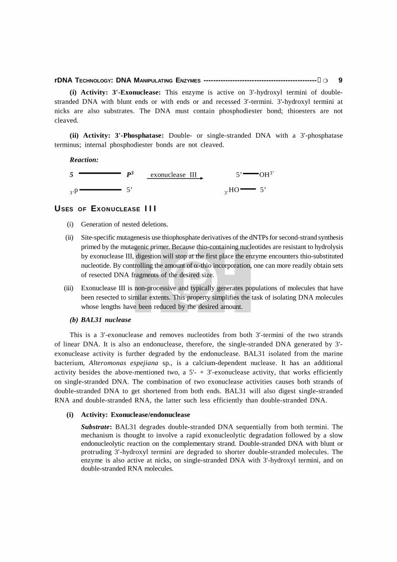
rDNA TECHNOLOGY: DNA MANIPULATING ENZYMES -----------------------------------------------G 9(i) Activity: 3'-Exonuclease: This enzyme is active on 3'-hydroxyl termini of double-
stranded DNA with blunt ends or with ends or and recessed 3'-termini. 3'-hydroxyl termini atnicks are also substrates. The DNA must contain phosphodiester bond; thioesters are notcleaved.
(ii) Activity: 3'-Phosphatase: Double- or single-stranded DNA with a 3'-phosphataseterminus; internal phosphodiester bonds are not cleaved.
Reaction:
5 P3 exonuclease III 5’ OH3’
3’p 5’ 3’HO 5’
USES OF EXONUCLEASE III
(i) Generation of nested deletions.
(ii) Site-specific mutagenesis use thiophosphate derivatives of the dNTPs for second-strand synthesisprimed by the mutagenic primer. Because thio-containing nucleotides are resistant to hydrolysisby exonuclease III, digestion will stop at the first place the enzyme encounters thio-substitutednucleotide. By controlling the amount of -thio incorporation, one can more readily obtain setsof resected DNA fragments of the desired size.
(iii) Exonuclease III is non-processive and typically generates populations of molecules that havebeen resected to similar extents. This property simplifies the task of isolating DNA moleculeswhose lengths have been reduced by the desired amount.
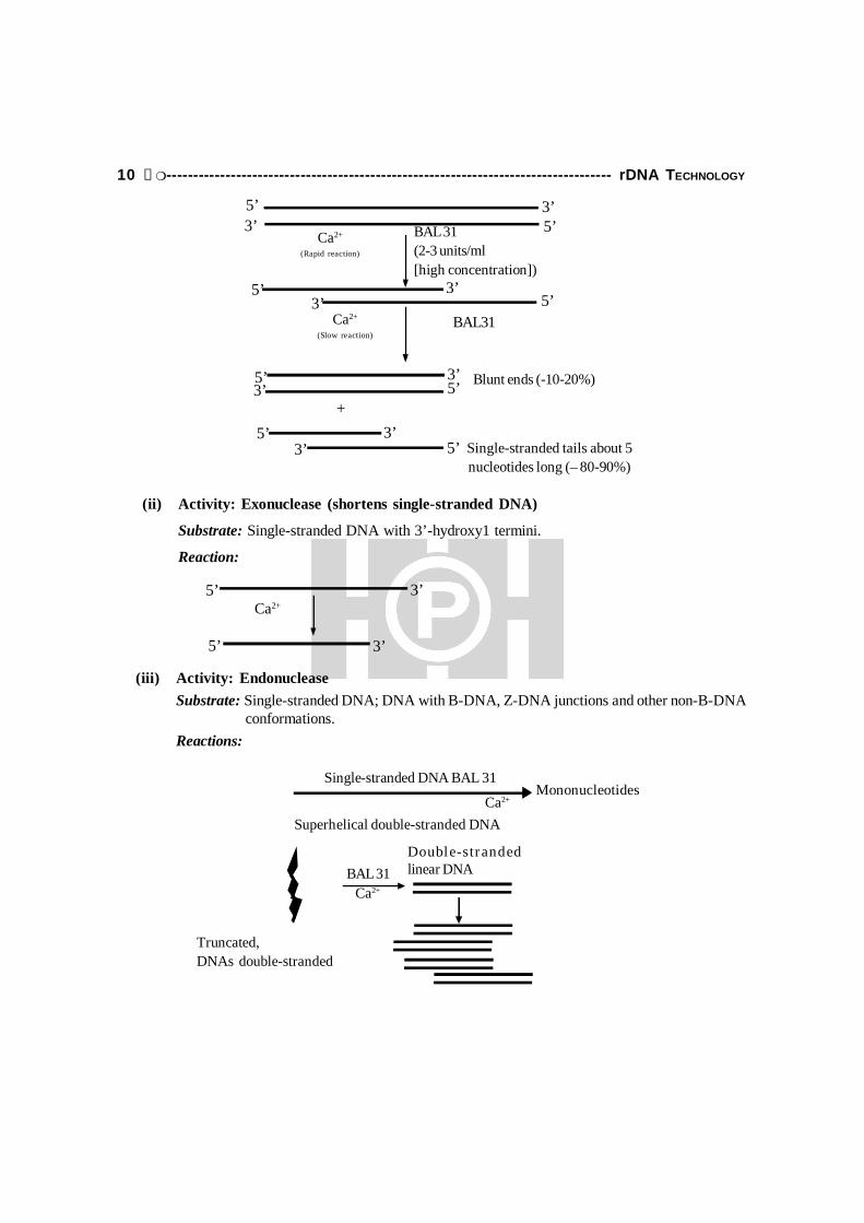
(b) BAL31 nuclease
This is a 3'-exonuclease and removes nucleotides from both 3'-termini of the two strandsof linear DNA. It is also an endonuclease, therefore, the single-stranded DNA generated by 3'-exonuclease activity is further degraded by the endonuclease. BAL31 isolated from the marinebacterium, Alteromonas espejiana sp., is a calcium-dependent nuclease. It has an additionalactivity besides the above-mentioned two, a 5'- + 3'-exonuclease activity, that works efficientlyon single-stranded DNA. The combination of two exonuclease activities causes both strands ofdouble-stranded DNA to get shortened from both ends. BAL31 will also digest single-strandedRNA and double-stranded RNA, the latter such less efficiently than double-stranded DNA.
(i) Activity: Exonuclease/endonuclease
Substrate: BAL31 degrades double-stranded DNA sequentially from both termini. Themechanism is thought to involve a rapid exonucleolytic degradation followed by a slowendonucleolytic reaction on the complementary strand. Double-stranded DNA with blunt orprotruding 3'-hydroxyl termini are degraded to shorter double-stranded molecules. Theenzyme is also active at nicks, on single-stranded DNA with 3'-hydroxyl termini, and ondouble-stranded RNA molecules.
10 G----------------------------------------------------------------------------------- rDNA TECHNOLOGY
5’
5’
3’
3’
Ca2+
Mononucleotides
Superhelical double-stranded DNA
BAL 31Ca2+
Double-str andedlinear DNA
Truncated,DNAs double-stranded
Ca2+
Single-stranded DNA BAL 31
3’5’
5’3’
Ca2+
(Rapid reaction)
BAL 31(2-3 units/ml[high concentration])
5’5’
3’3’
Ca2+
(Slow reaction)BAL31
3’
+3’5’ Blunt ends (-10-20%)
3’3’
5’5’
5’
Single-stranded tails about 5nucleotides long (– 80-90%)
(ii) Activity: Exonuclease (shortens single-stranded DNA)
Substrate: Single-stranded DNA with 3’-hydroxy1 termini.
Reaction:
(iii) Activity: EndonucleaseSubstrate: Single-stranded DNA; DNA with B-DNA, Z-DNA junctions and other non-B-DNA conformations.Reactions:

rDNA TECHNOLOGY: DNA MANIPULATING ENZYMES -----------------------------------------------G 11
Restriction Endonucleases (RE):
History
RE were initially discovered in the early 1950’s as a part of restriction (R) and modification(M) systems that bacteria operate for their protection against invading bacteriophages.
Phage inject DNAinto a bacterium
Restrictionendonuclease bindto the phage DNA
Phage DNA iscleaved andinactivated
(a) Restriction of phage DNA
Bacterial DNA
Me
Me
Recognition sequences aremethylated
Restriction endonucleasecannot bind to therecognition sequence
(b) Bacterial DNA is not cleaved
Fig. 1.6: The function of a restriction endonuclease in a bacterial cell: (a) phage DNA is cleaved, but(b) bacterial DNA is not.
RM systems consist of two enzymatic activities: (a) a site-specific “restriction” endonuclease(RE) that is responsible for digesting exogenous DNA and (b) a DNA “modification” methylase(or methy-transferase) with identical sequences specificity which is responsible for modifyingand protecting endogenous DNA from similar digestions by RE.
The preparation of restriction and modification can be explained by the behavior of phage on two E.coli host strains.
If a preparation of phage is prepared by growth upon E.coli strain C and this stock is thentitred upon E.coli C and E.coli K, the efficiency of growth of phage is bigger by several ordersof magnitude on E.coli C than that on E.coli K. The phage is said to be restricted by the second

12 G----------------------------------------------------------------------------------- rDNA TECHNOLOGY
host strain (E.coli K). When those phage that do grow on E.coli K are now used to reinfectE.coli K, they are no longer restricted, but if they are again first grown on E.coli C then onE.coli K, they are once again restricted. Thus, the efficiency with which phage plates upon aparticular host strain depends upon the strain on which it was last propagated. This noninheritablechange brought about in phage by the second host strain (E.coli K) that allows it be replated(grown) on that strain without further restriction is called modification. Restriction occursbecause the bacterium produces an enzyme that degrades phage DNA before it has time toreplicate and direct synthesis of new phage particles. The bacterium’s own DNA, the destructionof which would of course be lethal is protected from attack because it carries’ additional methylgroups; positioned and added by methyl transferases, that block the degradative enzyme actionof restriction endonuclease.
This explains why phage that survive one cycle of growth upon the restrictive host cansubsequently reinfect that host efficiently; their DNA bas been replicated in the presence ofmodifying methylase and so it like the host DNA becomes methylated and protected from therestriction system.
WHAT ARE RESTRICTION ENDONUCLEASES (RE)?
These are the enzymes which break the strands of DNA at internal portions but in aspecific way. The site where the RE attached are called as Restriction sites which may rangefrom 4-8 nucleotides. These RE cleave the DNA by cleaving two phosphodiester bonds withineach strands of DNA.
Naming of Restriction Endonuclease(RE)
As per the proposal given by Smith and Nathan in 1973, the RE can be nomeclatured as perthe following:
1. Every enzyme is named in 3 letter code.2. The first letter of the code is derived from first alphabet of source (genus).3. The second and third letter are from species.
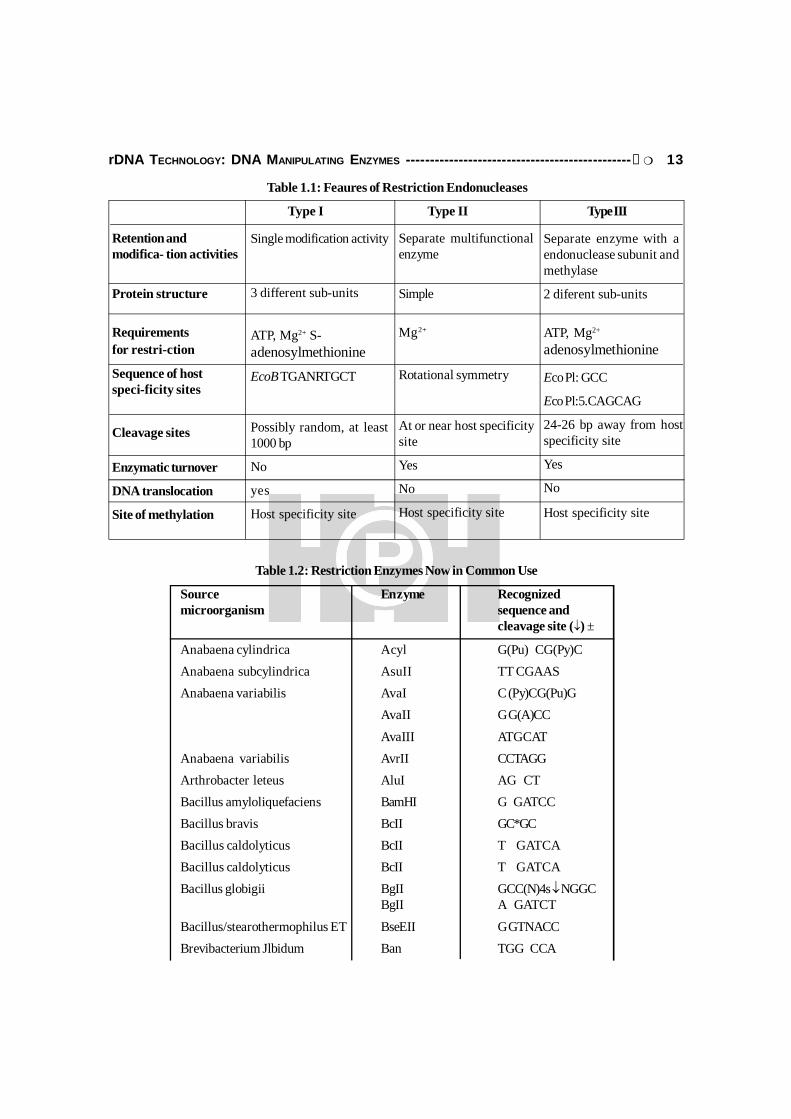
Restriction enzyme can be classified (Table 1.1) in the 3 different classes as per theirfeatures as given below:
Type I RE: These RE interact with unmodified target site in dsDNA. After traveling longdistance between 1000 to 2000 nucleotides, the RE cleaves only one strand. This enzyme is notof much used in rDNA technology.
Type II RE: These are mostly used enzymes in genetic engineering. These RE are highlyspecific and cleave within or very near to the recognition sequence.
Type III RE: These enzyme cleave dsDNA at defined positions and need ATP, Mg2+.
rDNA TECHNOLOGY: DNA MANIPULATING ENZYMES -----------------------------------------------G 13
Table 1.1: Feaures of Restriction Endonucleases
Type I Type II Type III
Retention andmodifica- tion activities
Protein structure
Requirementsfor restri-ction
Sequence of hostspeci-ficity sites
Cleavage sites
Enzymatic turnover
DNA translocation
Site of methylation
Single modification activity
3 different sub-units
ATP, Mg2+ S-adenosylmethionineEcoB TGANRTGCT
Possibly random, at least1000 bp
No
yes
Host specificity site
Separate multifunctionalenzyme
Simple
Mg2+
Rotational symmetry
At or near host specificitysite
Yes
No
Host specificity site
Separate enzyme with aendonuclease subunit andmethylase
2 diferent sub-units
ATP, Mg2+
adenosylmethionine
Eco Pl: GCC
Eco Pl:5.CAGCAG
24-26 bp away from hostspecificity site
Yes
No
Host specificity site
Table 1.2: Restriction Enzymes Now in Common Use
Source Enzyme Recognizedmicroorganism sequence and
cleavage site () ±
Anabaena cylindrica Acyl G(Pu) CG(Py)CAnabaena subcylindrica AsuII TT CGAASAnabaena variabilis AvaI C (Py)CG(Pu)G
AvaII G G(A)CC
AvaIII ATGCATAnabaena variabilis AvrII CCTAGGArthrobacter leteus AluI AG CTBacillus amyloliquefaciens BamHI G GATCCBacillus bravis BcII GC*GCBacillus caldolyticus BcII T GATCABacillus caldolyticus BcII T GATCABacillus globigii BgII GCC(N)4s NGGC
BgII A GATCT
Bacillus/stearothermophilus ET BseEII G GTNACCBrevibacterium Jlbidum Ban TGG CCA
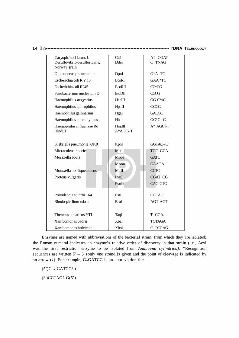
14 G----------------------------------------------------------------------------------- rDNA TECHNOLOGY
CaryophJnoll Iatun. L ClaI AT CGATDesulfovibrio desulfuricans, DdeI C TNAGNorway srainDiplococcus pneumoniae DpnI G*A TCEscherichia coli R Y 13 EcoRI GAA *TCEscherichia coli R245 EcoRII CC*GGFusubacterium nucleatum D fnuDII CGCGHaemophilus aegyptius HaeIII GG C*sC
Haemophilus aphrophilus HpaII ClCGGHaemophilus gallinarum HgaI GACGCHaemophilus haemolyticus Hhal GC*G CHaemophilus influenzae Rd HindII A* AGCTHindIII A*AGCT
Klebsiella pneumonia. OK8 Kpnl GGTAC CMicracoleus species Mstl TGC GCAMoraxella bovis Mbol GATC
Mbon GAAGAMoraxella nonliquefaciens MnII CCTCProteus vulgaris PvuI CGAT CG
PvuII CAG CTG
Providencia stuartii 164 PstI CGCA GRhodospiriIium rubrum RruI AGT ACT
Thermus aquaticus YTI Taql T CGA.
Xantbomonas badrii Xbal TCTAGAXantbomonas holcicola Xbol C TCGAG
Enzymes are named with abbreviations of the bacterial strain, from which they are isolated;the Roman numeral indicates an enzyme’s relative order of discovery in that strain (i.e., Acylwas the first restriction enzyme to be isolated from Anabaena cylindrica). *Recognitionsequences are written 5' – 3' (only one strand is given and the point of cleavage is indicated byan arrow (). For example, GGATCC is an abbreviation for:
(5’)G GATCC3')
(3')CCTAG G(5’)

rDNA TECHNOLOGY: DNA MANIPULATING ENZYMES -----------------------------------------------G 15
When no arrow appears, the precise cleavage site has not been determined. The symbol Pu(purine) indicates that either A or G will be recognized; Py (pyrimidine) indicates that either C orT will be recognized. Two bases appearing in parentheses signify that either base may occupythat position in the recognition sequence. Thus, AvaIl cleaves the sequence GGACC or GGTCC.Where known, the base modified by the corresponding specific methylase is indicated by anasterisk: A. is N-methyladenosine; C is 5-methylcytosine. (Source: R.J. Robens, 1981, Nuc.Acids Res. 9:75.)
What are Target Sites?
These are the 4-6 nucleotide long sequences and the DNA which are being recognized byRE Type II and these sequences exhibit palindromic symmetry from central axis, we can readthe base as in same orientation as shown below:
Nature of Cuts: Two type cuts are being produced by RE Type II. These are of twotypes.
(i) Blunt cuts: No free hanging base pairs. These cuts are also known as flush cuts.
(ii) Cohesive cuts: These are also called as staggered cuts, producing free base longs at each endof cleaved DNA.
ATT AAT
TAA TTA
Axis of symmetry
Sticky/Cohesive ends
R-EDNABlunt Ends
Fig. 1.7: Blunt and cohesive ends upon digestion with Restriction enzyme
Isochizomers: These are the enzymes which are isolated from different sources butrecognize same target site., e.g., Asp 718 and KpnI have identical sites.
Role of RE
1. RE are particularly used in Gene cloning experiments to cut insert DNA and vector.
2. RE are used in DNA Restriction digestion during southern blotting in order to detect copynumber of Gene.

16 G----------------------------------------------------------------------------------- rDNA TECHNOLOGY
Star activity: Several enzyme show relaxation in specificity when the optional conditionsare being altered. In such cases, nucleases even recognize other alternative base instead of aspecific base. Some conditions like High Ionic buffers, High glycerol conch or high even alteredTemperature can change the cutting nature of RE.
RestrictionenzymeEcoRI
EcoRI will notcleave methylaseDNA
RestrictionenzymeEcoRI
G A A T T C G A A T T CC T T A A G
G A A T T CC T T A A G
MethylatedDNA
G A A T T CC T T A A G
C T T A A GUnmethylatedDNA
Cleavage
Stickyends
UnmethylatedDNA
CH3
CH3
3’5’
5’3’5’
3’5’3’5’
3’
5’3’ 5’
3’ 5’3’
EcoRImethylase
Fig. 1.8: EcoRI and many other restriction enzymes cleave DNA so that the fragments have shortcomplementary single-stranded segments at the ends. These “sticky ends” are important in recombinant DNA
techniques because they readily combine with the ends of other cleavage fragments produced by the samerestriction enzyme. EcoRI recognizes the six-base-pair sequence. (b) Most restriction enzymes exist in cells
along with modification enzyme. The modification enzyme EcoRI methylase catalyzes the methylation of twoadenylates (asterisked) in the six-base-pair sequence that is normally cleaved by EcoRI. If a methylated sampleis then exposed to EcoRI, the added methyl groups protect the restriction site so that the restriction enzyme
does not cut the DNA.
Ligases:
In rDNA technology, Ligases are used to join two DNA molecules. These enzymes alsoplay a key role in replication, recombination and cloning. These enzymes are also called asmolecular structures.
How Ligases Act?
In the presence of Ligase enzyme when two DNA fragments are mixed, base pairingbetween two fragments occurs which results in sealing of two different DNA fragments. Itoccurs due to covalent bonds formation between 2'-PO4 gp and 3'-OH gp of adjustment strands(Fig. 1.9).

rDNA TECHNOLOGY: DNA MANIPULATING ENZYMES -----------------------------------------------G 17
Fig. 1.9: Showing Activity of E.coli and T4 DNA ligases
Two Types of Ligases:
1. E.coli ligases which is generally used to join two sticky or cohesive ends.
2. T4 DNA ligase which is generally used to join two blunt-end DNA molecules. All the reactionsare generally carried out at 16°C. Sometimes PEG (polyethylene glycol) can be added whichacts as volume excluders. PEG increases the frequency of collision between two DNA molecules.
How to put Sticky end to blunt-end DNA?
Sometimes, vector molecule has sticky ends but DNA to be cloned is having blunt ends.So, under these conditions, Linkers and Adaptors play a key role in cloning.
1. Linker: Linker is a short piece of dsDNA of known sequence. It is blunt-ended butcontains one restriction site. The structure of a Linker and attachment of Linker to DNA isshown below.
2. Adaptors: One major problem with linkers is that if DNA to be contains one or moreBamHI sites, the enzymes will also cut the DNA molecules along with linker molecules. Hence,adaptor molecules have been synthesized as shown below so unlike linkers, an adaptor issynthesized having one end sticky (Fig. 1.10).
Problem with adaptor: Sticky free ends of adaptors can ligase to form dimmers butdimmer formation can be frequented by removing 5'-P group from sticky ends as shown below:
DNA Fragment
R.E
Sticky endsE.coli. ligase
R.E
Blunt ends
T4 LigaseATP
PEGs
Ligation occurs
Ligation occurs

18 G----------------------------------------------------------------------------------- rDNA TECHNOLOGY
(a) Linker
(b) Use of Linkers
Linker attachedBaH1 Sticky ends
Cleaved Linkers
Blunt-end DNAs
Ligase
BamH1
Ready to be cloned into vector
A—T—G—C—T—A—G—C—A—T
T—A—C—G—A—T—C—G—T—A
Fig. 1.10: A decameric linker molecule containing an ecoRI target site is joined by T4 DNA ligase to both endsof flush-ended foreign DNA. Cohesive ends are then generated by EcoRI. This DNA can then be incorporated
into a vector that has been treated with the same restriction endonuclease.
Decamericlinkermolecule
C C G A A T T CG G C A A T T C
3’5’
T4 DNAligases
+
ForeignDNA
Linkermolecules
Vector
EcoRI
DNAligases

rDNA TECHNOLOGY: DNA MANIPULATING ENZYMES -----------------------------------------------G 19
OVERVIEW:
Kinases and phosphatases, are common reagents in modern day molecular biologylaboratories. Although there are a variety of sources for these enzymes, the most common arecalf intestinal alkaline phosphatase (CIAP) and T4 polynucleotide kinase (T4 PNK). Their mostfrequent use is to modify the phosphorylation state of the 5'-ends of DNA molecules (fig. 1.11).ClAP is most commonly used to remove 5'-phosphates from vector DNA to prevent self-ligationduring cloning. Only one strand of a DNA duplex must be joined prior to bacterial transformation;the other will remain nicked until it is repaired inside the bacteria. While the vector DNA isdephosphorylated, the insert DNA should not be dephosphorylated as 5'- phosphates are requiredfor a successful ligation reaction. ClAP is also used to end-label DNA fragments by removing5'phosphates, making the DNA fragments better T4 PNK substrates. Synthetic DNA, usually inthe form of custom-made oligonucleotides, is devoid of 5'-phosphates and is therefore a lessthan ideal template for ligation reactions. T4 PNK is routinely used to transfer a -phosphatefrom a nucleotide triphosphate (usually A TP) to the 5'-end of oligonucleotides to facilitateligation. For blotting, gel-shift or sequencing procedures, [-32P]A TP is used as the phosphatedonor, resulting in a radiolabeled species. The 5'-end of a DNA molecule generated byrestriction endonuclease cleavage can also be labeled, even though a phosphate already exists atthat position. This can be achieved either by making use of the exchange activity of T4 PNK to
Fig. 1.11: Use of a BamHI adaptor molecule. A synthetic adaptor molecule is lighted to the foreign DNA.The adaptor is used in the 5'-hydroxyl form to prevent self-polymerization. The foreign DNA plus ligated
adaptors is phosphorylated at the 5'-termini and ligated into the vector previously cut with BamHI.
KINASES AND PHOSPHATASES
5’ -HO-GATCCCCGGG-HO3, – HO-GGGCCC-P
adaptor molecule
Vector
OHOHOH
OHOH
OHP
POHP
OH P +
OHP
OHP
OHOH
OHOH
POH
OHP
PNK ATP
20 G----------------------------------------------------------------------------------- rDNA TECHNOLOGY
exchange the existing phosphate with a radiolabeled phosphate from the phosphate donor H, orby first treating the DNA with ClAP to remove the existing phosphates, then adding theradiolabeled phosphate with PNK via the forward reaction, which will result in a high specificactivity. Finally, T4 PNK has a 3'-phosphatase activity that can be used to remove phosphategroups from the 3'-terminus of DNA and RNA. Although both enzymes are most commonlyused for cloning purposes, they have other activities and are also used for other types ofstudies. These other activities will be listed in further detail in the following section. Therobustness and versatility of ClAP and T4 PNK have made them staples in today’s molecularbiology applications.
T4 Polynucleotide Kinase
Composed of identical sub-units. The relative mobility of the monomers as measured bySDSPAGE is 33kDa, by centrifugation; 33.2kDa. From the sequence of T4 PNK, the monomerconsists of 301 amino acids with a predicted molecular weight of 34kDa. The molecular weightof the tetramer as estimated by gel filtration is 140kDa, by centrifugation, 147.3kDa. Monomersand dimmers are not enzymatically active.
Description
T4 Polynucleotide Kinase (polynucleotide 5'-hydroxyl-kinase) or ATP: 5'-dephosphopoly-nucleotide 5' phosphatase) or T4 Polynucleotide Kinase (T4 PNK) is a tetramer composed ofidentical sub-units and has multiple activities. The 5'-kinase activity of T4 PNK catalyzes thetransfer of the -phosphate from ATP to the 5'-OH terminus of mono- or polynucleotides. Thereaction is reversible and in the presence of a nucleotide diphosphate such as ADP the enzymehas 5'-phosphatase activity. Dephosphorylation and subsequent rephosphorylation allow theenzyme to transfer phosphates between A TP and a 5'-phosphate group on an acceptor moleculein an exchange reaction. T4 PNK also has 3'phosphate activity. T4 PNK can be used tophosphorylate RNA, DNA and synthetic oligonucleotides prior to subsequent manipulations suchas ligation. Radioactive phosphate can be used as a label for DNA sequencing, gel shift analysis,footprinting, primer extension, and restriction mapping. Labeling the 5'-ends of DNA and RNAmay be done using a dephosphorylated template (5'-OH) using the forward or 5'-kinase reaction.Alternatively, labeling of 5'-ends can be achieved without removal of the existing 5'-phosphateusing the exchange reaction. The reaction conditions for the forward and exchange reactions arenot the same. The forward reaction generally results in better incorporation. T4 PNK can also beused to remove 3'-phosphatase from DNA and RNA.

rDNA TECHNOLOGY: DNA MANIPULATING ENZYMES -----------------------------------------------G 21
Forward Reaction
5’—OH—DNA+ [y32—P]ATP P32–O4—DNA+ADP
Exchange Reaction
5’—PO—DNA+ADP5’—OH—DNA+ATP
[y32–P] ATP
5PO32—DNA+ADP
Fig. 1.12(a): Describes forward and exchange reactions by kinases
Source: An E.coli strain that carries a plasmid encoding the modified T4 Polynucleotide Kinase gene.
Applications
5' end-labeling of ss- and dsDNA and RNA
Phosphorylation of insert DNA prior to ligation.
Phosphorylation of oligonucleotides.
Removal of 3'-phosphates 5'-phosphorylation of DNA/RNA for subsequent ligation.
End-labeling of DNA or RNA.
5'-phosphorylation of 3'-phosphorylated mononucleotides to generate a substrate (pNp) thatcan be added to the 3'-end of DNA or RNA by ligase activity.
5'-end labeling of 3'- phosphorylated oligos.
Cofactor Concentration: For the 5'-kinase reaction, 10mM Mg2+ optimal at pH 7.6. Forthe 3'-phosphatase reaction, SmM Mg2+ optimal.
Optimal Substrate
5'-Kinase Activity: ss- and dsDNA, ss- and dsRNA, synthetic oligonucleotides andnucleotide 3'-monophosphates. 5'-OH groups on ssDNA overhangs in dsDNA are phosphorylatedmore efficiently that 5'-OH groups on blunt or 5'-recessed ends. With increased concentrationof ATP or enzyme, blunt- and 5'-recessed ends can be completely phosphorylated. Phosphorylationof 5'-OH groups located at nicks in dsDNA is 10-30X slower than for ssDNA. The reactiondoes not differ significantly for substrates. T4 PNK can also phosphorylate a variety of modifiednucleotides and non-nucleotide substrates. Nucleotides (adenosine), nucleotide 2'-phosphates,3'-termini, or 5'-termini bearing phosphomonoesterase are not substrates.
Exchange Reaction: ss-and dsDNA, 5'-overhangs of dsDNA and single-stranded oligonucleotidesare more efficient substrates than 5'-recessed or blunt ends. 5'-recessed and blunt ends arelabeled 15-25% as efficiently as 5'-overhangs. 5'-phosphatase groups at nicks are the most

22 G----------------------------------------------------------------------------------- rDNA TECHNOLOGY
difficult to exchange (30X less efficient than 5'-overhangs). Some tRNA species can act assubstrates for phosphatase exchange.
Stimulators
5'-Kinase Activity: Spermidine (1.7mM optimal) can increase the rate of the reactionthreefold (24). Spermidine promotes tetramer formation. Salts such as NaCl, KCL and CsCL(125mM optimal) can increase activity up to 5X. LiCL and NH4Cl give similar stimulation. Thiseffect is the same for 5'-overhangs on dsDNA and ssDNA, oligo and mononucleotides.Conversely, KCl decreases phosphorylation of 5'-recessed ends and at nicks.
A sulfhydryl compound such as DTT (5mM DTT optimal) is essential for activity. 10mM2mercaptoethanol and IOmM glutathione result in 80% and 70% respectively, of the activityobserved with DTT. In the absence of a sulfhydryl compound, only 2% of the optimal activity isobserved. Several anions (at I 25/lM) are stimulatory, with Cl–, Br– > F–, N03
–, S042-. PEG 8000
at 4-6% improves the efficiency of labeling 5'- and 3'-overhangs, blunt ends and at nicks.
Inhibitors
5'-Kinase Activity: Phosphate (Pi) and pyrophosphate (PPi) anions are inhibitors of T4PNK. Ammonium ions are strong inhibitors of T4 PNK. DNA should not be dissolved in orprecipitated from buffers containing ammonium salts prior to treatment with kinase. Sulfatecontaining polymers (e.g., agar, dextran sulfate and heparin) are inhibitors of T4 PNK. However,addition of cationic compounds such as spermine or polylysine can counteract the inhibition.Nonsulfate polysaccharides have no effect on T4 PNK.
Typical Working Conditions: A TP at 2-5 fold molar excess over DNA ends results in95% phosphorylation of 5'-overhangs. Increasing concentration of A TP to 100-fold molarexcess will allow complete labeling of blunt ends or recessed 5'-ends. Protocols for using T4PNK are available from Promega.
Storage Conditions: Store at -20°C. T4 Polynucleotide Kinase is supplied in 20mM Tris-HCL (pH 7.5), 25mM KCL, 2mM DTT, 0.1mM EDTA, 0.1mM ATP and 50% (v/v) glycerol.
Unit Definition: One unit is defined as the amount of T4 PNK required to catalyze thetransfer of I nanomole of phosphate to the 5'-OH end of a polynucleotide from [-32P] A TP in30 minutes at 37°C. The reaction conditions are: 40mM Tris-HCL (pH 7.5), 10mM MgCI2, 5mMDTT, 0.lmM [ -32P]A TP, 0.5mM 5'-OH polynucleotide end concentration.
Activity Assays
End Labeling: To test for activity, 5pmol of dephosphorylated primer is incubated with 8units of T4 PNK for I hour at 37°C in IX Kinase Buffer containing [y-32P]A TP. Followingincubation, the amount of [-32P]A TP converted to TCA-insoluble material is determined. Theminimal passing specification is > 20% incorporation.

rDNA TECHNOLOGY: DNA MANIPULATING ENZYMES -----------------------------------------------G 23Sequencing Assay: This enzyme has been tested and qualified for performance in the
TaqTrack Sequencing Systems.
Contaminant Assays
Endonuclease Assay: To test for endonucleases activity, 1/lg of supercoiled plasmid DNAis incubated with 25 units of T4 PNK for 5 hours at 37°C in IX Kinase Buffer. Followingincubation, the supercoiled DNA is visualized on an ethidium bromide-stained agarose gel toverify the absence of visible nicking or cutting.
DNase and RNase Assay: To test for nuclease activity, 50ng of radiolabeled DNA orradiolabeled RNA is incubated with 25 units of T4 Polynucleotide Kinase in IX Kinase Buffer for3 hours at 37°C, and the release of radiolabeled nucleotides is monitored by scintillation countingof TCA-soluble material. Minimum passing specification is < 3% release for both DNase andRNase.
Calf Intestinal Alkaline Phosphatase E.C.3.1.3.1
Description
Calf Intestinal Alkaline Phosphatase is a phosphomonoesterase that catalyzes the hydrolysisof 5'- and 3'-phosphate of DNA, RNA. ClAP is primarily used to dephosphorylate vector DNAprior to cloning and for removal of 5'-phosphates from RNA and DNA before labeling with [‘-32P]NTP and T4 polynucleotide kinase. ClAP has also been widely utilized to dephosphorylateproteins. In contrast to acid phosphatases, ClAP can be utilized for in vitro dephosphorylation ofproteins under conditions that do not denature the substrate protein. ClAP effectively dephosphorylatesproteins containing phosphoserine, phosphothreonine and phosphotyrosine, although it can showpreferential dephosphorylation of phosphotyrosine under certain conditions.
The process of removing the phosphate group is called dephosphorylation. As the namesuggests, alkaline phosphatases are most effective in an alkaline environment. It is sometimesused synonymously as basic phosphatase.
Bacterial
In bacteria, alkaline phosphatase is located in the periplasmic space, external to the cellmembrane. Since this space is much more subject to environmental variation than the actualinterior of the cell, bacterial alkaline phosphatase is comparatively resistant to inactivation,denaturation, and degradation, and also has a higher rate of activity. Although the actual purposeof the enzyme is still not fully understood, the simple hypothesis, that it is a means for thebacteria to generate free phosphate groups for uptake and use is supported by the fact thatalkaline phosphatase is usually only produced by the bacteria during phosphate starvation and notwhen phosphate is plentiful. However, other possibilities exist; for instance, the presence ofphosphate groups usually prevents organic molecules from passing through the membrane,

24 G----------------------------------------------------------------------------------- rDNA TECHNOLOGY
therefore dephosphorylating them may be important for bacterial uptake of organic compoundsin the wild. Some complexities of bacterial regulation and metabolism suggest that other moresubtle purposes for the enzyme may also play a role for the cell. In the laboratory, however.mutant Escherichia coli lacking alkaline phosphatase survive quite well, as do mutants unable toshut off alkaline phosphatase production.
The optimal pH for the activity of the E.coli enzyme is 8.0 while the bovine enzymeoptimum pH is slightly higher at 8.5.
Uses in Research
Common alkaline phosphatases used in research include:
For removing phosphate monoesterase to prevent self-ligation.
Shrimp alkaline phosphatase (SAP), from a species of Arctic shrimp (Pandalus borealis).
Calf Intestinal Alkaline Phosphatase (CIP).
Placental alkaline phosphatase (P ALP) and its C terminally truncated version that lacks the last24 amino acids (constituting the domain that targets membrane anchoring) — the secretedalkaline phosphatase (SEAP).
Alkaline phosphatase has become a useful tool in molecular biology laboratories, since DNAnormally possesses phosphatase groups on the 5'-end. Removing these phosphates prevents theDNA from ligating (the 5'-end attaching to the 3'-end), thereby keeping DNA molecules linearuntil the next step of the process for which they are being prepared; also, removal of thephosphate groups allows radiolabeling (replacement by radioactive phosphatase groups) in orderto measure the presence of the labeled DNA through further steps in the process or experiment.For these purposes, the alkaline phosphatase from shrimp is the most useful, as it is the easiestto inactivate once it has done its job.
Another important use of alkaline phosphatase is as a label for enzyme immunoassays.
One common use in the dairy industry is as a marker of pasteurisation in cows’ milk. Thismolecule is denatured by elevated temperatures found during pasteurisation, and can be testedfor via colour change of a para-Nitrophenylphosphate substrate in a buffered solution (AschaffenburgMullen Test). Raw milk would typically produce a yellow colouration within a couple of minutes,whereas properly pasteurised milk should show no change. There are of course exceptions tothis in the case of heat stable alkaline phophatases produced by some bacteria.
Inhibitors
All mammalian alkaline phosphatases, except placental (P ALP and SEAP), are inhibited byhomoarginine and similarly all, except the intestinal and placental ones are blocked by levamisole.Heating for 2 hours at 65°C inactivated most is enzymes except Placental is of forms (P ALPand SEAP).

rDNA TECHNOLOGY: DNA MANIPULATING ENZYMES -----------------------------------------------G 25
Applications
Dephosphorylation of 5'-phosphorylated ends of cloning vectors.(Fig. 1.12b)
Dephosphorylation of 5'-phosphorylated ends of DNA or RNA tor subsequent labeling with32P using [-32P]NTP and T4 Polynucleotide Kinase.
Dephosphorylation of proteins.
Source: Calf intestinal mucosa.
Molecular Weight: 68kDa.
Typical Working Conditions: 50mM Tris-HCL (pH 9.3 at 25°C), ImM MgCL2, 0.lmMZnCl2 and 1mM spermidine.
Storage Conditions: Store at -20°C. ClAP is supplied in storage buffer containing 10mMTris-HCI (pH 8.0), ImM MgCL2, 0.lmM ZnLI2, 50mM KCL and 50% (v/v) glycerol amount ofenzyme required to catalyze the hydrolysis of 1 ~lmol of 4-nitrophenyl phosphate per minute at37°C in 1M diethanolamine, 10.9mM 4-nitrophenyl phosphate, 0.5mM MgCl2 (pH 9.8).
CONTAMINANT ASSAYS
Endonuclease Assay: 1g of supercoiled plasmid DNA is incubated with 5 units of CIAP inIX Reaction Buffer for one hour at 37°C. Following incubation, the supercoiled DNA isvisualized on an ethidium bromide-stained agarose gel to verify the absence of visible nicking orcutting.
Exonuclease Assay: To test for DNase and RNase activity, 50ng of radiolabeled DNA orradiolabeled RNA is incubated with 5 units of Calf Intestinal Alkaline Phosphatase in IX ReactionBuffer for one hour at 37°C, and the release of radiolabeled nucleotides is monitored byscintillation counting of TCA-soluble material. Minimum passing specification is 3% release forboth DNase and RNase.
Blue/White Cloning Assay: Five micrograms of pGEM-3Zf(+) Vectore(d) is digestedwith representative restriction enzymes (leaving 5'-termini, 3'-termini or blunt ends). The terminiare treated with 5 units of Calf Intestinal Alkaline Phosphatase for 1 hour at 37°C, kinased andligated. The religated plasmid is then transformed into JMI09 cells that are plated on X-Gal/lPTG/Amp plates. A minimum of 200-400 colonies are counted. White colonies result fromtransformation with ligated plasmids with damaged ends. These white colonies represent thenumber of false positives expected in a typical cloning experiment. Enzymes that generateoverhangs, such as EeaR I. Hind III or Kpn I, must produce fewer than 2% white colonies, andblunt-cutting enzymes, such as Hinc II must produce fewer than 5% white colonies (I).Transformation efficiency must be 1 × 10⁵ cfu/g DNA.

26 G----------------------------------------------------------------------------------- rDNA TECHNOLOGY
ANTARCTIC PHOSPHATASE (ANTP)
Antartic phosphatse has been derived from Antarctic strain TAB5 which is a psychrophilicstrain being isolated at the Dumont d’Urville antarctic station. Antarctic Phosphatase catalyzesthe removal of 5´ phosphate groups from DNA and RNA. Since phosphatase-treated fragmentslack the 5´ phosphoryl termini required by ligases, they cannot self-ligate . This property can beused to decrease the vector background in cloning strategies. AnTP is a superior choice becauseit is 100% heat inactivated in 5 minutes at 65°C; as a result you can proceed directly to theligation reaction without further purification of vector DNA. Since, neither CIP or ShrimpAlkaline Phosphatase (SAP) can be completely inactivated at 65°C in 30 minutes, it may benecessary to purify the DNA after the phosphatase reaction. It is important to inactivate thephosphatase because residual active phosphatase can cause failure in subsequent ligation/transformation experiments.
Main features:
Heat inactivated in 5 minutes at 65°C
Ligate without purifying vector DNA
Isolated from a recombinant source
Removes 5' phosphates from DNA, RNA, rNTPs and dNTPs
Prevents recircularization of cloning vectors
Supplied with 10X Reaction Buffer
Source:
An E. coli strain that carries the TAB5 AP gene, originally cloned in plasmid pNI, reclonedin plasmid pEGTAB7-4.1(new England biolabs)
Applications:
Removing 5´ phosphates from DNA, RNA, rNTPs and dNTPs
Preparation of templates for 5´ end labeling
Prevention of recircularization of cloning vectors
Removal of dNTPs and pyrophosphate from PCR reactions
Dephosphorylation of proteins
Some 5'-End Labeling Tips
The forward reaction is much more efficient than the exchange reaction for labeling ofphosphorylated ends.
The template used should be gel-purified. Contamination by short oligonucleotides or tRNAwill lower the efficiency of labeling. Consider the use of glycogen instead of tRNA for efficientprecipitation of oligonucleotides.
rDNA TECHNOLOGY: DNA MANIPULATING ENZYMES -----------------------------------------------G 27
Oligonucleotides are synthesized with a 5'-hydroxyl group. They can be labeled directly in theforward reaction without prior dephosphorylation.
Ammonium ions are strong inhibitors of T4 Polynucleotide Kinase; therefore, DNA should notbe dissolved in, or precipitated from, buffers containing ammonium salts prior to treatmentwith T4 PNK.
The final A TP concentration in the forward reaction should be at least 1M.
Equal concentrations of A TP and 5'-ends result in about 50% labeling of oligonucleotides. Toobtain higher specific activity, the A TP:oligonucleotide ratio should be increased to 10: 1. Only10% of the label will be transferred but virtually every oligonucleotide molecule will belabeled (I).
0.5 pmol of linear DNA equals Ipmol of 5'-ends. To convert g DNA to pmol:
For dsDNA:
g × 106 pg/g x pmol/660pg × 1/N = pmolwhere N is the number of nucleotide pairs and 660pg/pmol is the conversion factor for a
nucleotide pair.
For ssDNA:
g × 106 pg/g × pmol/330pg × 1/N = pmolwhere N is the number of nucleotides and 330pg/pmol is the conversion factor for a single
nucleotide.
Single-stranded DNA and double-stranded DNA containing 5'-overhangs are labeledmore efficiently than 3'-overhangs (5'-recessed) or blunt ends. Labeling of blunt or recessedends can be improved by increasing the concentration of ATP in the reaction or bydenaturing the template with heat or NaOH prior to labeling. Alternative protocols, involvingthe addition of PEG 8000 to the reaction may also be used to increase the labeling efficiencyof these templates.

28 G----------------------------------------------------------------------------------- rDNA TECHNOLOGY
POH
OHP
Ligase
Alkaline Phosphatase
OHOH
OHOH
OHOHP
P
Ligase
No reaction
Foreign DNAFragment
Ligase
Nick
Transformation
Nick
Host repair onenick at each join
Fig. 1.12(b): Phosphatase treatment to prevent recircularization of vector plasmid without insertion offoreign DNA
NUCLEASE
SI Nuclease is a single-stranded specific endonuclease that cleaves DNA to release5'-mono and 5'-oligo nucleotides. sDNA, dsRNA and DNA-RNA hybrids are resistant, however,very large amounts of enzyme can completely hydrolyze double-stranded nucleotides. SInuclease can also cleave single-stranded areas of Superhelical DNA at torsional points whereDNA may be unpaired or ‘weakly hydrogen bonded’. Once the superhelical DNA is nicked, Slcan cleave the second strand near the nick to generate linear DNA.
SI nuclease is a monomeric protein with molecular weight. It need Zn2+ for its activity,and is relatively stable against denaturing agents such as urea, SDS and formamide. Theoptimum pH lies between 4-4.5.

rDNA TECHNOLOGY: DNA MANIPULATING ENZYMES -----------------------------------------------G 29
Applications of SI Nuclease1. It is used to analyse DNA-RNA hybrid structures.2. It can be used to remove single-stranded tails from DNA fragments to produce blunt ends.3. Hairpin loop structures formed during synthesis of double-stranded cDNA can be digested.
RNASES
Generally, RNase A and RNase TI are used in genetic engineering procedures. Both cleavethe phosphodiester bond between adjacent ribonucleotides. However, RNase A cleaves next touracils and cytosine such that phosphate remains with these pyrimidines. The nucleotide presenton the other side of phosphatase is dephosphorylated. RNase A is obtained from the bovinepancreas.
RNase Tl cleaves specifically next to guanine. The phosphatase group at the 3'-end of thenucleotide remains with the cut end. This enzyme is derived from Aspergillus oryzae.
RIBONUCLEASE (RNASE H)
RNase H is an endoribonuclease that degrades the RNA portion of the RNA-DNA hybrids.It cuts the RNA into short fragments.
Applications of RNase H
1. RNase H is the key enzyme in the cDNA cloning procedure. Here, it is used to remove themRNA from the RNA-DNA hybrid.
2. Used to detect the presence of RNA-DNA hybrids.
3. Used to remove poly (A) tails on mRNA.
DEOXYRIBONUCLEASE I
Deoxyribonuclease I (DNase I) is an endonuclease which digest either single-or double-stranded DNA, producing a mixture of mono and oligonucleotides. DNase I hydrolyzes eachstrand of double-stranded DNA independently and at random. Addition of Mg2+ensures randomcleavage while Mn2+ gives cleavage nearly at the same place on both strands. The enzyme isderived mostly from bovine pancreas.
Applications of DNase I
This enzyme is useful for a variety of applications including nick translation, DNAfootprinting, bisulfite mediated mutagenesis and RNA purification.

30 G----------------------------------------------------------------------------------- rDNA TECHNOLOGY
Reverse Transcriptase
In the fields of molecular biology and biochemistry, a reverse transcriptase, also known asRNAdependent DNA polymerase, is a DNA polymerase enzyme that transcribes singlestrandedRNA into double-stranded DNA. It also helps in the formation of a double helix DNA once theRNA has been reverse transcribed into a single-stranded cDNA. Normal transcription involves thesynthesis of RNA from DNA; hence, reverse transcription is the reverse of this. Reversetranscriptase first purified from retrovirus-infected cells, produces a cDNA copy from an mRNAmolecule if first provided with an antisense primer (oligo dT or a random primer). This enzyme iscritical for converting mRNA into cDNA for purposes of cloning, PCR amplification, or theproduction of specific probes. Reverse transcriptase was discovered by Howard Temin at theUniversity of Wisconsin-Madison, and independently by David Baltimore in 1970 at MIT. The twoshared the 1975 Nobel Prize in Physiology or Medicine with Renato Dulbecco for their discovery.
Well-studied reverse transcriptases include:
HIV-I reverse transcriptase from human immunodeficiency virus type I (PDB I HMV).
M-ML V reverse transcriptase from the Moloney murine leukemia virus.
AMV reverse transcriptase from the avian myeloblastosis virus.
Telomerase reverse transcriptase that maintains the telomeres of eukaryotic chromosomes.
Terminal Deoxynucleotidyl Transferase (TDT)
It also known as DNA nucleotidylexotransferase (DNTT) or terminal transferase, is aspecialized DNA polymerase expressed in immature, pre-B, pre-T lymphoid cells, and acutelymphoblastic leukemia/lymphoma cells. TDT adds N-nucleotides to the variable, diversity, andjoining exons during antibody gene recombination. In humans, terminal transferase is encoded by theDNTT gene. TDT catalyzes the addition of nucleotides to the 3'-terminus of a DNA molecule. Unlikemost DNA polymerases, it does not require a template. The preferred substrate of this enzyme is a 3'-overhang, but it can also add nucleotides to blunt or recessed 3'-ends. Cobalt is a necessary co-factor. However, the enzyme catalyzes reaction upon Mg and Mn administration in vitro (Fig. 1.13).
Fig. 1.13
TerminalTransferase
+daTP
TerminalTransferase
+ TTP5’
5’ AAAHybridisation and ligation gap willfilled by E.coli when transformed
5’3’
5’3’
AA ATTT
5’5’ AAA
TTTT 3’5’
AA A TTTTTTTT
Separate preparations of DNA to be joined to each other

rDNA TECHNOLOGY: DNA MANIPULATING ENZYMES -----------------------------------------------G 31
POLYMERASES
DNA polymerase: The enzyme that synthesizes DNA from a DNA template. The intactenzyme purified from bacteria (termed the holoenzyme) has both synthetic and editing functions.The editing function results from nuclease activity.
Klenow fragment: A modified version of bacterial DNA polymerase that has been modifiedso that only the polymerase function remains; the 5'3' exonuclease activity has beeneliminated.
Thermostabile polymerases: The prototype polymerase, Taq, and newer versions such asVent and Tth Jolymerase are derived from microorganisms that normally reside at hightemperature. Consequently, their DNA polymerase enzymes are quite stable to heat denaturation.making them ideal enzymes for use, in the polymerase chain reaction. It is a thermostable DNApolymerase named after the thermophilic bacterium Thermus aquaticus from which it wasoriginally isolated hy Thomas D. Brock in 1965. It is often abbreviated to “Taq Pol” (or simply“Taq”) and is frequently used in polymerase chain reaction OCR), methods for greatlyamplifying short segments of DNA. T aquaticus is a bacterium that lives in Hot springs andhvdrothermal vents, and Taq polymerase was identified as an enzyme able to withstand theprotein-denaturing conditions (high temperature) required during PCR. Therefore, it replaced theDNA polymerase from E.coli originally used in PCR. Taq’s optimum temperature for activity is75-80°C with a half life of 9 minutes at 97.5°C, and can replicate a 1000 basepair strand of DNAin less than 10 seconds. One of Taq’s drawbacks is its relatively low replication fidelity. It lacksproof reading activity Hl, and has an error rate measured at about 1 in 9,000 nucleotides. Somethermostable DNA polymerases have been isolated from other thermophilic bacteria and archaea,such as Pfu DNA polymerase, possessing a proof reading activity and are being used instead of(or in combination with) Taq for high-fidelity amplification. Taq makes DNA products that haveA (adenine) overhangs at their 3'-ends. This may be useful in TA cloning, whereby a cloningvector (such as a plasmid) is used which has a T (thymine) 3'-overhang, which complementswith the A overhang of the PCR product, thus enabling ligation of the PCR product into theplasmid vector.
RNA polymerase II: This enzyme is used by mammalian cells to transcribe structuralgenes that result in mRNA. The enzyme interacts with a number of other proteins to correctlyinitiate transcription including a number of general factors, and tissue-specific and induction-specific enhancing proteins.
RNA polymerase III: This enzyme is used by the cell to transcribe ribosomal RNA genes.
Applications of Klenow Fragment
1. In DNA sequencing by sanger method.
2. For production of second strand of cDNA.
3. Radiolabelling by filling in 5'-single stranded extension on double-stranded DNA.

32 G----------------------------------------------------------------------------------- rDNA TECHNOLOGY
4. “Mutagenesis of” DNA with oligonucleotides.
5. In labelling the DNA by random primer method.
Table 1.3: Active Properties of Some DNA Synthesizing Enzymes
Enzymes 5'-.3' synthesis 5’-Exonuclease 3'-Exonuclease
DNA Polymerase I (E.coli)
Klenow Fragment
DNA Polymerase
stands for presence of particular activity by the enzyme. Blank presents absence of suchactivity.
Table 1.4: Comparative Uses of Some DNA Synthesizing Enzymes
Nick Trans- Fill DNA 3'-end Second In Vitrolation in Sequencing Labelling strand of Mutagenesis
cDNA
DNA PolymeraseI (E.col).
Klenow Fragment
T-4 DNA Polymerase
stands for presence of particular activity by the enzyme. Blank presents absence of such activity.