secretion and endocytosis - yale...
TRANSCRIPT
Secretion and Endocytosis
Peter TakizawaCell Biology

• Vesicular transport between organelles
• Glycosylation
• Protein sorting in the Golgi
• Endocytosis

Secretory pathway delivers proteins and lipids to plasma membrane protein and organelles.
Outside cell
Cytosol
Endoplasmic Reticulum
Golgi
Cell Membrane
Secretory Vesicles
Endosomes
Lysosomes
The secretory pathway allows cells to release proteins to the external environment (e.g. antibodies, hormones). It also delivers proteins and lipids to the cell membrane and to several intracellular organelles, such as endosomes, lysosomes and sectory granules. Proteins that are delivered to different locations via the secretory pathway contain signal sequences. Proteins without a signal sequence follow the default pathway which delivers protein and lipids to the cell membrane. Sorting of proteins with signal sequence occurs in the Golgi.

Vesicles mediate transport between membrane-bound compartments.
GolgiER Secretory Vesicle
Endosomes
Lysosomes
Secretory pathway consists of several separate membrane-bound compartments: ER, Golgi, endosomes, etc. Proteins must move through these organelles before reaching their final destination. Lipids and proteins are shuttled between compartments via small vesicles. Vesicles are membrane-bound structures that form from one compartment and fuse with next compartment.
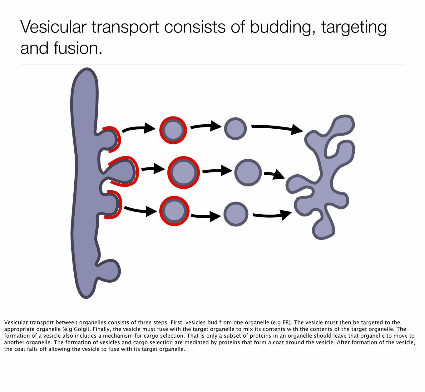
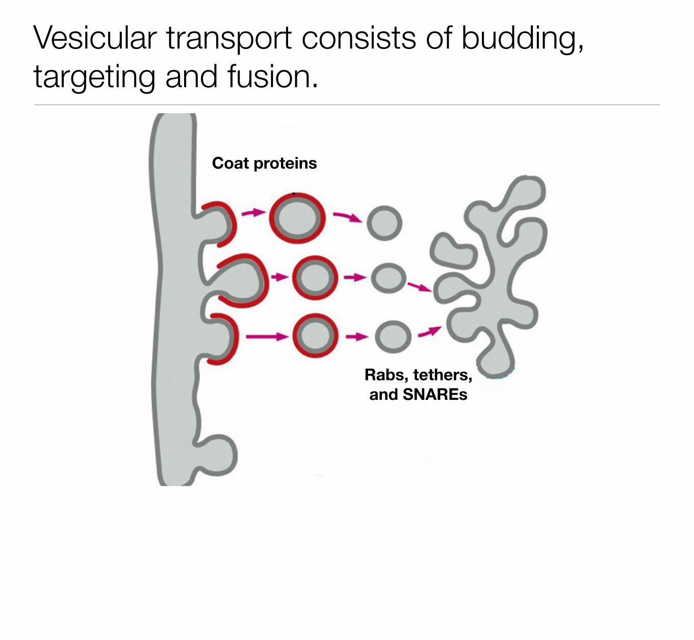
Vesicular transport consists of budding, targeting and fusion.
Vesicular transport between organelles consists of three steps. First, vesicles bud from one organelle (e.g ER). The vesicle must then be targeted to the appropriate organelle (e.g Golgi). Finally, the vesicle must fuse with the target organelle to mix its contents with the contents of the target organelle. The formation of a vesicle also includes a mechanism for cargo selection. That is only a subset of proteins in an organelle should leave that organelle to move to another organelle. The formation of vesicles and cargo selection are mediated by proteins that form a coat around the vesicle. After formation of the vesicle, the coat falls off allowing the vesicle to fuse with its target organelle.
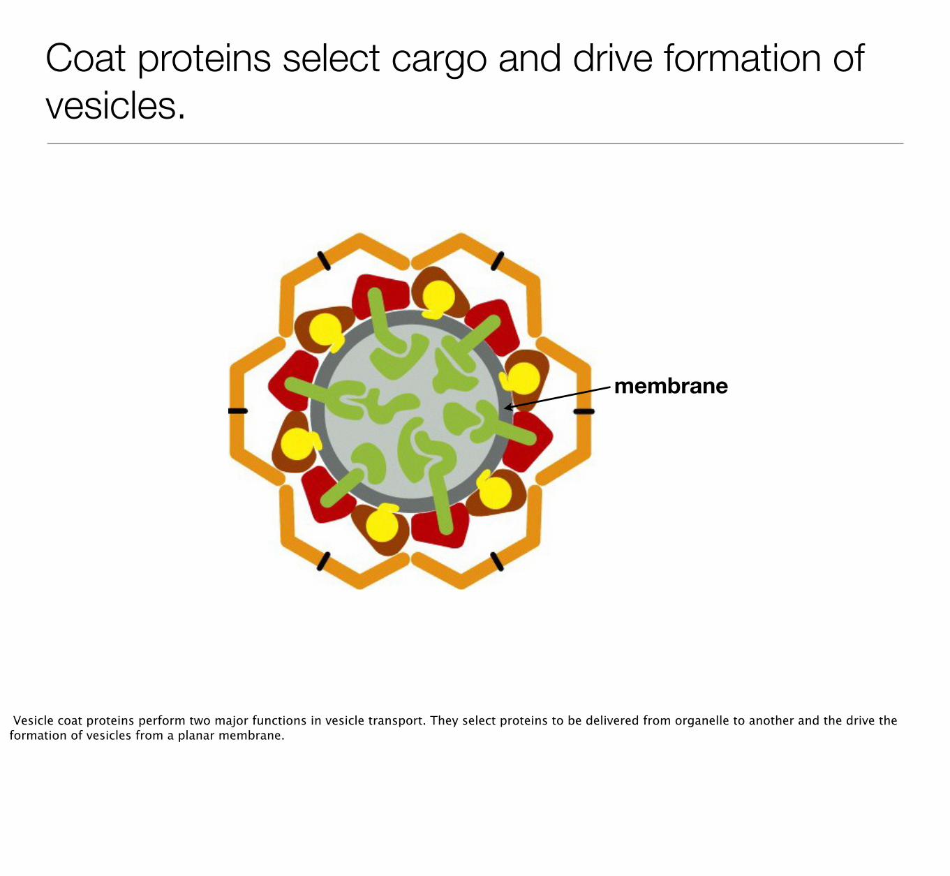
Coat proteins select cargo and drive formation of vesicles.
membrane
Vesicle coat proteins perform two major functions in vesicle transport. They select proteins to be delivered from organelle to another and the drive the formation of vesicles from a planar membrane.

Coat proteins select cargo and drive formation of vesicles.
Vesicle coat proteins perform two major functions in vesicle transport. They select proteins to be delivered from organelle to another and the drive the formation of vesicles from a planar membrane.

Vesicle formation triggered by small GTP-binding proteins.
Endoplasmic Reticulum
Sar1
Sar1-GEF
To describe how coat proteins mediate cargo selection and vesicle formation we will focus on process of vesicle formation from ER. The initial step in the formation of a vesicle from the ER is the binding of the small GTP-binding protein Sar1. When Sar1 is bound to GDP it remains in the cytosol. A guanine nucleotide exchange factor in the ER causes Sar1 to release GDP allowing it to bind GTP. In its GTP-bound form Sar1 extends a small helix that inserts into the outer leaflet of the ER membrane. The presence of the helix expands the outer leaflet producing a curvature in the ER membrane.

Coat proteins select cargo through interactions with cytosolic tails of ER proteins.
Sar1 Cytosol
ER Lumen
Selecting Soluble Proteins Selecting Integral Membrane Proteins
Sar1 recruits two proteins, Sec 23 and Sec 24, that select protein for inclusion into the transport vesicle. Sec24 contains binding sites for proteins that should exit the ER. Transmembrane proteins that are slated to leave the ER contain a signal sequence on a domain that faces the cytoplasm. The signal sequence binds Sec 24 tethering the protein to the site of vesicle formation. Soluble proteins that need to leave the ER are recognized by a receptor in the ER membrane. The cytoplasmic domain of the receptor is bound by Sec24.

Assembly of coat proteins induces curvature in membrane and eventual budding.
A second set of proteins is recruited to the site of vesicle formation. These proteins, Sec13 and Sec31, assemble onto the surface of the ER membrane and increase the deformation of the membrane leading to the formation of a vesicle.

How do vesicles fuse with the correct organelle?
GolgiER Secretory Vesicle
Once vesicle form from one organelle, they need to fuse with another organelle or plasma membrane to deliver their cargo. How do vesicle find the correct organelle within the cytoplasm?

Rabs, tethers and SNAREs target vesicles to their destination.
Rab-GTP
Tether
t-SNARE
v-SNARE
There are many potential membrane-bound organelles to which a vesicle could fuse. Two interactions increase probability that vesicle fuse with the correct organelle. The first interaction involves a family of small GTP-binding proteins called Rabs and their binding partners, tethers. Rabs bind membranes only when they are in a GTP-bound state. There are over 60 different types of Rabs and each type of organelle contains a unique set of Rabs. In this way Rabs help identify the different membrane-bound organelles in the secretory pathway. A vesicle that forms from one organelle will contain a unique Rab. This Rab will interact preferentially with a tether protein on the appropriate target organelle.
The second interaction that mediates fusion between vesicles and organelle is between SNARE proteins. SNAREs are transmembrane proteins that reside in organelles and vesicles. SNAREs in vesicles are called v-SNAREs and SNAREs in organelles are called t-SNAREs. There are over 35 different SNARE proteins allowing the cells to mark different types of organelles and vesicles with a unique set of SNAREs. Certain v-SNAREs and t-SNAREs interact to generate an accurate interaction between a vesicle with its target organelle. The energy of pairing between SNAREs is thought to drive the fusion of the vesicle membrane and organelle membrane.

Mutations in Rab27 prevent fusion of vesicles with cell membrane, causing hemophagocytic syndrome.
Normal Rab27 defective
To illustrate the role of Rabs in targeting vesicles to their target membranes, one can look at the effect of mutations in Rabs. Rab27 is a member of the Rab family that localizes to a special organelle called the secretory lysosome. These are common in a type of T-cell that kills other cells that are infected with a virus. Rab27 is required to link the secretory lysosome with the plasma membrane, and in its absence the secretory lysosomes are not targeted to the cell membrane and the fusion of the secretory lysosomes with the plasma membrane is inhibited. Consequently, the T-cells don’t effectively kill infected cells and a prolonged immune response ensues. The over activation of the immune system triggers macrophages to ingest red blood cells and platelets, leading to the symptoms of the disease which include fever, enlarged spleen and anemia.
Vesicular transport consists of budding, targeting and fusion.
Coat proteins
Rabs, tethers, and SNAREs

The sweet life: glycosylation of proteins
One important process that occurs in the secretory pathway is glycosylation which is the covalent addition of sugars to proteins. About half the proteins that enter the ER are covalently modified with sugars. The sugar groups help proteins fold, prevent digestion of proteins, target proteins to their final destinations and mediate cell adhesion.

Glycosylation occurs in the ER and Golgi.
There are tow types of glycosylation: N-linked with occurs in the ER and O-linked which occurs in the Golgi. The Golgi contains enzymes that modify the composition of the sugars added in the ER.
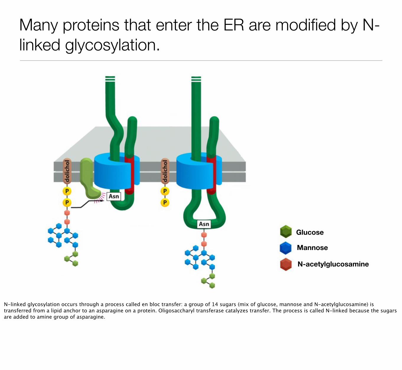
Many proteins that enter the ER are modified by N-linked glycosylation.
Glucose
Mannose
N-acetylglucosamine
N-linked glycosylation occurs through a process called en bloc transfer: a group of 14 sugars (mix of glucose, mannose and N-acetylglucosamine) is transferred from a lipid anchor to an asparagine on a protein. Oligosaccharyl transferase catalyzes transfer. The process is called N-linked because the sugars are added to amine group of asparagine.

Carbohydrate side chains sequentially modified by Golgi enzymes.
One of the major functions of Golgi is to modify sugar side chains. The Golgi comprises a set of flattened membranes called cisternae. Proteins from the ER are transported sequentially through each cisterna. Proteins enter the cis-cisterna and exit the Golgi at the trans-Golgi network. Each cisterna contains a unique set of enzymes that remove or add sugars to the sugar groups that were added in the ER. Usually, the enzymes that remove sugars are found toward the cis side and those that add sugars are found towards the trans side.

Carbohydrate side chains sequentially modified by Golgi enzymes.
Mannosidase I N-acetylglucosamine transferase
Mannosidase II Various transferases
Cis Medial Trans
One of the major functions of Golgi is to modify sugar side chains. The Golgi comprises a set of flattened membranes called cisternae. Proteins from the ER are transported sequentially through each cisterna. Proteins enter the cis-cisterna and exit the Golgi at the trans-Golgi network. Each cisterna contains a unique set of enzymes that remove or add sugars to the sugar groups that were added in the ER. Usually, the enzymes that remove sugars are found toward the cis side and those that add sugars are found towards the trans side.

Golgi enzymes catalyze O-linked glycosylation of proteins.
Protein
Rest of sugar side chain
Protein
Rest of sugar side chain
A smaller fraction of proteins are modified by O-linked glycosylation. O-linked differs from N-linked in that the sugars are added to hydroxyl group of serines, threonines. In addition, the sugars are not added as an entire block but grow as a chain of disaccharides (two sugar groups) from an amino acid. The sugar side chains in O-linked can become very long (100s of disaccharides).

Golgi enzymes catalyze O-linked glycosylation of proteins.
A smaller fraction of proteins are modified by O-linked glycosylation. O-linked differs from N-linked in that the sugars are added to hydroxyl group of serines, threonines. In addition, the sugars are not added as an entire block but grow as a chain of disaccharides (two sugar groups) from an amino acid. The sugar side chains in O-linked can become very long (100s of disaccharides).

Sorting proteins in the Golgi

Proteins are sorted in trans-Golgi network to their final destinations.
cis medial
trans
TGN
ER Secretory vesicle
lysosome
Proteins in secretory pathway need to find their final destinations. All proteins in the secretory pathway proceed from the ER through the Golgi and are sorted in the trans-Golgi network (TGN). Proteins with signal sequences will sent to the lysosome or secretory vesicle. Proteins without a signal sequence will be delivered to the cell membrane.

Protein sorting to lysosomes

Lysosomes contain many digestive enzymes that are active at low pH.
Lysosomes digest cellular and foreign material and are often referred to as the garbage dump of cell. Lysosomes contain many hydrolases that digest different macromolecules. Many of theses hydrolases are targeted to the lysosome via a unique sugar modification called mannose 6-phosphate.

Mannose-6-Phosphate targets proteins to lysosomes.
A receptor in the TGN recognizes proteins with mannose-6-phosphate and recruits those proteins into vesicles that will be delivered to endosomes. These endosomes will eventually mature into lysosomes.

Mannose-6-Phosphate targets proteins to lysosomes.
cis-Golgi network
trans-Golgi network
Mannose
Phosphate
A receptor in the TGN recognizes proteins with mannose-6-phosphate and recruits those proteins into vesicles that will be delivered to endosomes. These endosomes will eventually mature into lysosomes.

GlcNAC-phosphotransferase phosphorylates mannose on lysosomal proteins.
GlcNAC-phosphotransferase
Cis-Golgi
Trans-Golgi
LysosomeProteins destined for the lysosome contain a signal sequence that is recognized by an enzyme in the Golgi called GlcNAC-phosphotransferase. The enzymes catalyzes the transfer of an N-acetylglucosamine with a phosphate onto the mannose residue of the sugar side chain of the lysosomal protein. The N-acetylglucoamine is eventually removed, leaving mannose-phosphate. This mannose-phosphate is recognized by the receptor in the TGN and the protein delivered to the lysosome.

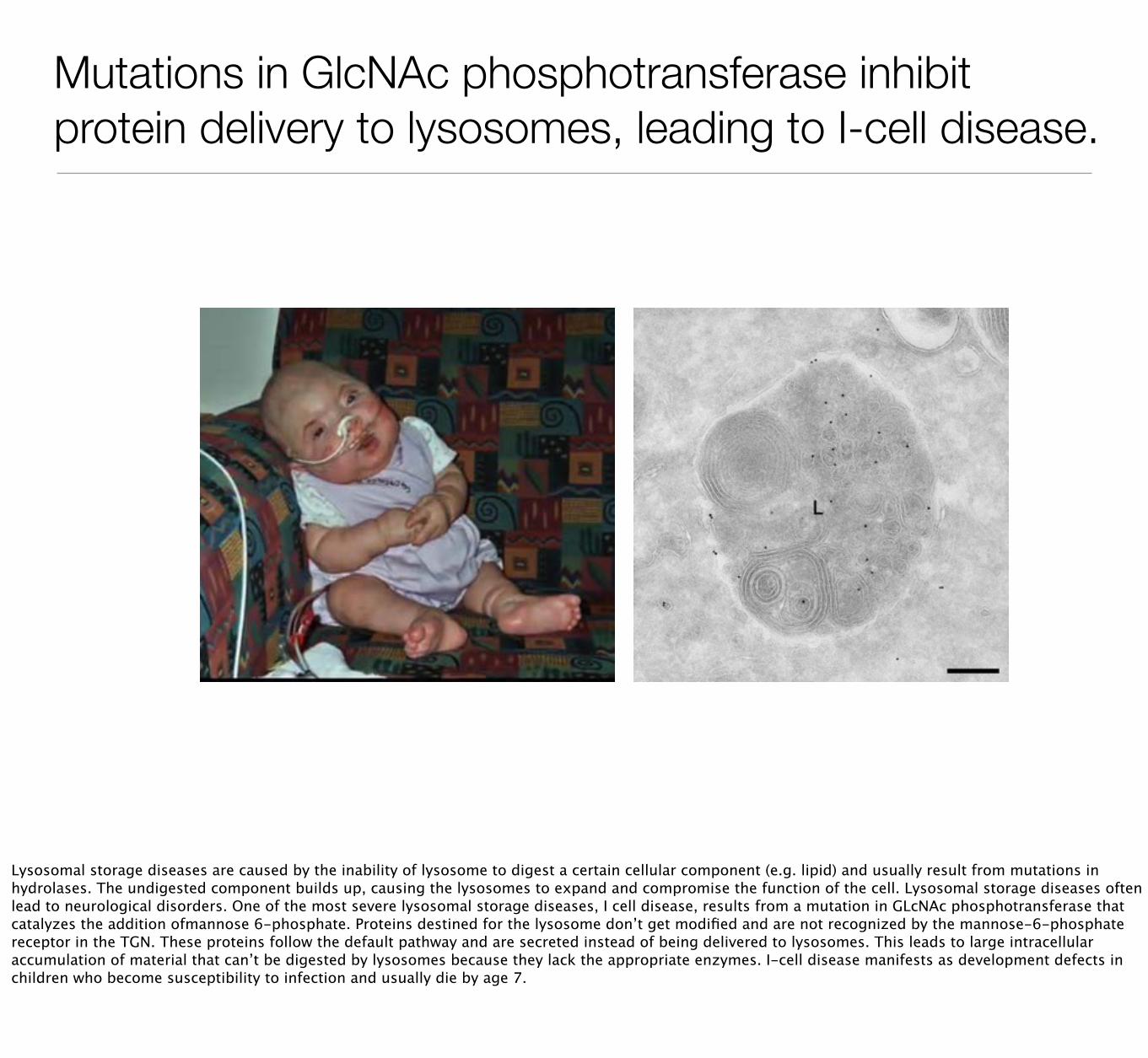
Mutations in GlcNAc phosphotransferase inhibit protein delivery to lysosomes, leading to I-cell disease.
Lysosomal storage diseases are caused by the inability of lysosome to digest a certain cellular component (e.g. lipid) and usually result from mutations in hydrolases. The undigested component builds up, causing the lysosomes to expand and compromise the function of the cell. Lysosomal storage diseases often lead to neurological disorders. One of the most severe lysosomal storage diseases, I cell disease, results from a mutation in GLcNAc phosphotransferase that catalyzes the addition ofmannose 6-phosphate. Proteins destined for the lysosome don’t get modified and are not recognized by the mannose-6-phosphate receptor in the TGN. These proteins follow the default pathway and are secreted instead of being delivered to lysosomes. This leads to large intracellular accumulation of material that can’t be digested by lysosomes because they lack the appropriate enzymes. I-cell disease manifests as development defects in children who become susceptibility to infection and usually die by age 7.

Proteins are sorted in trans-Golgi network to their final destinations.
cis medial
trans
TGN
ER Secretory vesicle
lysosome

Secretory vesicles accumulate aggregated proteins in TGN and undergo maturation.
The mechanism of targeting protein to secretory vesicles is less clear. The process appears to involve a signal patch on proteins that induces them to cluster in the TGN. The aggregated protein is somehow selected by vesicles budding from the TGN that will develop into secretory granules. Secretory granules do not immediately fuse with the cell membrane but remain in the cytoplasm until an appropriate signal to fuse is received by the cell.

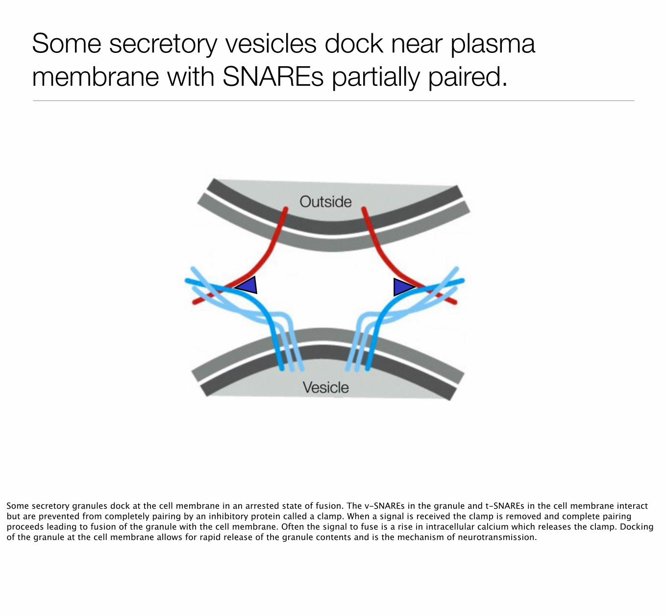
Some secretory vesicles dock near plasma membrane with SNAREs partially paired.
Some secretory granules dock at the cell membrane in an arrested state of fusion. The v-SNAREs in the granule and t-SNAREs in the cell membrane interact but are prevented from completely pairing by an inhibitory protein called a clamp. When a signal is received the clamp is removed and complete pairing proceeds leading to fusion of the granule with the cell membrane. Often the signal to fuse is a rise in intracellular calcium which releases the clamp. Docking of the granule at the cell membrane allows for rapid release of the granule contents and is the mechanism of neurotransmission.

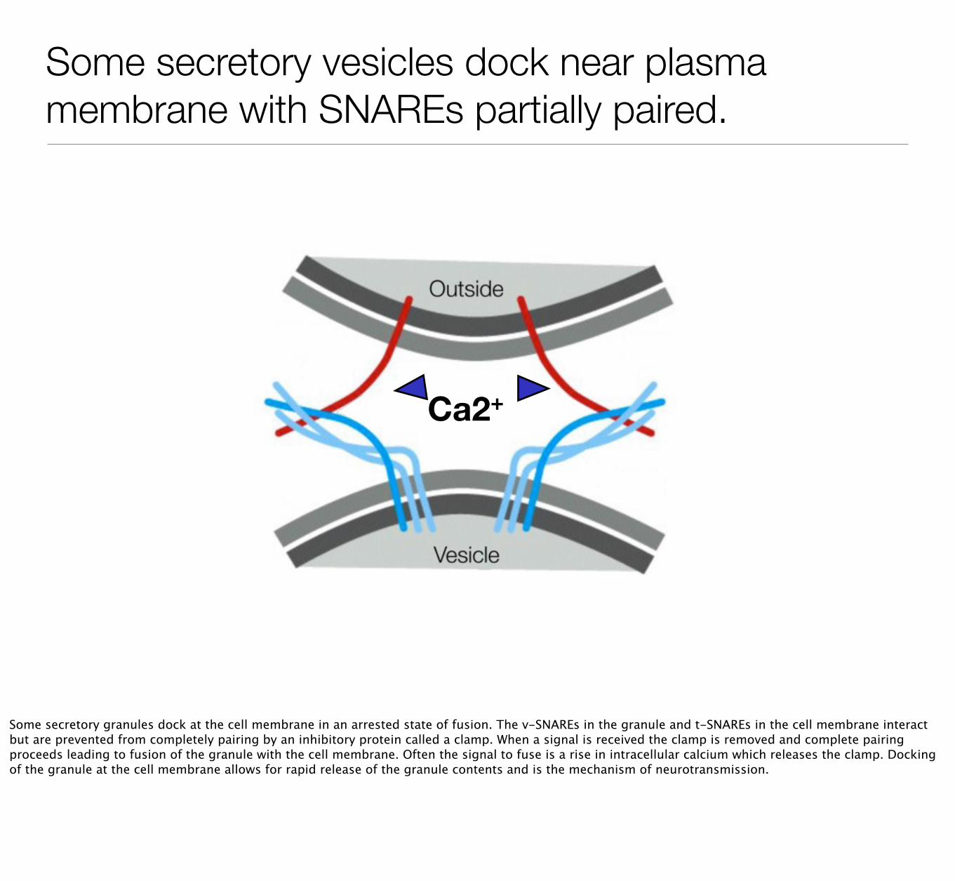
Some secretory vesicles dock near plasma membrane with SNAREs partially paired.
Ca2+
Some secretory granules dock at the cell membrane in an arrested state of fusion. The v-SNAREs in the granule and t-SNAREs in the cell membrane interact but are prevented from completely pairing by an inhibitory protein called a clamp. When a signal is received the clamp is removed and complete pairing proceeds leading to fusion of the granule with the cell membrane. Often the signal to fuse is a rise in intracellular calcium which releases the clamp. Docking of the granule at the cell membrane allows for rapid release of the granule contents and is the mechanism of neurotransmission.

Some secretory vesicles dock near plasma membrane with SNAREs partially paired.
Vesicle
Outside
Some secretory granules dock at the cell membrane in an arrested state of fusion. The v-SNAREs in the granule and t-SNAREs in the cell membrane interact but are prevented from completely pairing by an inhibitory protein called a clamp. When a signal is received the clamp is removed and complete pairing proceeds leading to fusion of the granule with the cell membrane. Often the signal to fuse is a rise in intracellular calcium which releases the clamp. Docking of the granule at the cell membrane allows for rapid release of the granule contents and is the mechanism of neurotransmission.

Endocytosis: the uptake of extracellular material.
Endocytosis allows cells to take up material from external environment. Most cells take up small amounts of external fluid but some cells, macrophages and neutrophils, can engulf entire cells (bacteria, dead cells). Endocytosis also allows cells to recycle their cell membrane and remove specific proteins from the cell membrane.

Phagocytosis engulfs large particles and microorganisms.
Uptake of large objects is called phagocytosis and is usually found only in macrophages and neutrophils. Phagocytosis is triggered when a macrophage or neutrophil binds a foreign object on its plasma membrane. The cell then pushes its cell membrane around object to engulf it. This is an actin dependent process. The engulfed object will be delivered to the lysosome for degradation.

Pinocytosis ingests small amount of fluid and plasma membrane.
All cells continuously take up smaller portions of plasma membrane into 100 nm vesicles in process called pinocytosis. Through pinocytosis the cell recycles 1 to 3% of plasma membrane per minute. Pinocytosis usually relies on a set of proteins called clathrin that form a basket-like coat around the vesicles.
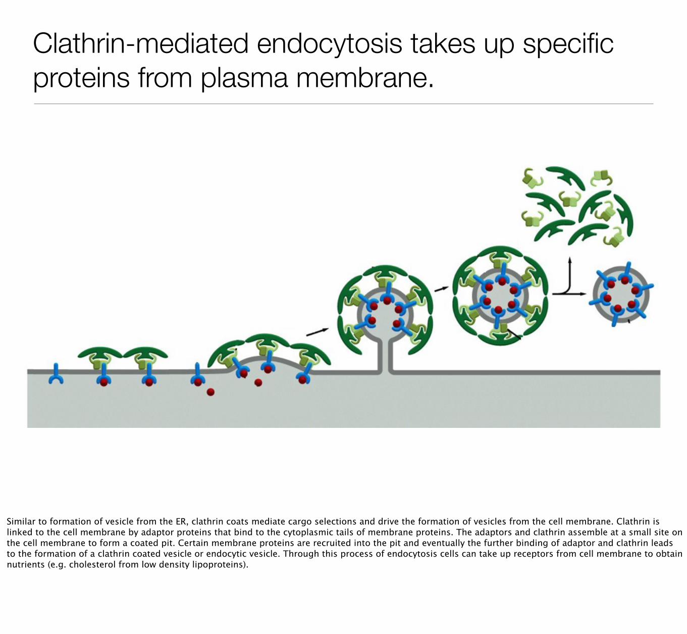
Clathrin-mediated endocytosis takes up specific proteins from plasma membrane.
Similar to formation of vesicle from the ER, clathrin coats mediate cargo selections and drive the formation of vesicles from the cell membrane. Clathrin is linked to the cell membrane by adaptor proteins that bind to the cytoplasmic tails of membrane proteins. The adaptors and clathrin assemble at a small site on the cell membrane to form a coated pit. Certain membrane proteins are recruited into the pit and eventually the further binding of adaptor and clathrin leads to the formation of a clathrin coated vesicle or endocytic vesicle. Through this process of endocytosis cells can take up receptors from cell membrane to obtain nutrients (e.g. cholesterol from low density lipoproteins).
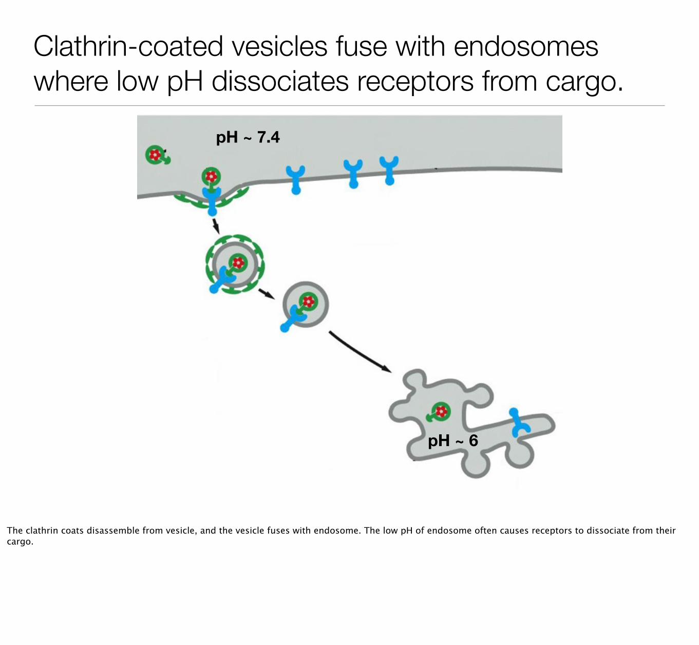
Clathrin-coated vesicles fuse with endosomes where low pH dissociates receptors from cargo.
pH ~ 7.4
pH ~ 6
The clathrin coats disassemble from vesicle, and the vesicle fuses with endosome. The low pH of endosome often causes receptors to dissociate from their cargo.

Receptors returned to plasma membrane and cargo delivered to lysosome.
pH ~ 7.4
pH ~ 6
pH ~ 5
Receptors in the endosome can be returned to cell membrane, whereas their cargo can be delivered to lysosome for degradation and processing.

Multivesicular bodies process receptors for degradation in lysosomes.
Receptors on the cell membrane that function in signaling pathways are degraded by a similar mechanism but are processed through a different type of organelle called the multivesicular body. These receptors are taken up into clathrin coated vesicles. After clathrin is removed, the membrane surrounding the vesicle invaginates to form internal vesicles that contain receptors. Multivesicular bodies appears to contain multiple internal vesicles. These organelles usually fuse with lysosomes.

• Vesicles move proteins and lipids between organelles in the secretory pathway.
• Proteins are sorted in the Golgi and delivered to cell membrane, lysosomes or storage granules.
• Glycosylation is the addition of sugars to proteins and occurs in the ER and Golgi.
• Endocytosis allows cells to take up external material and portions of the cell membrane for degradation or recycling.
Take home points...