seff amalia-laura analysis of enzyme-catalyzed reactions...
TRANSCRIPT
BABES-BOLYAI UNIVERSITY, CLUJ-NAPOCA BUDAPEST UNIVERSITY OF TECHNOLOGY AND ECONOMICS
Ph. D. Thesis Abstract
Seff Amalia-Laura
ANALYSIS OF ENZYME-CATALYZED REACTIONS BY
COMPUTATION
Scientific Advisers: Prof. Dr. Ioan Silaghi-Dumitrescu† Prof. Dr. László Poppe Acad. Prof. Dr. Ionel Haiduc Jury President Conf. Dr. Cornelia Majdik Reviewers: Prof. Dr. Paul Mezey, Memorial University of Newfoundland. Canada CR1 (CNRS) Dr. Dragoş Horvath, University of Louis Pasteur Strasbourg, France Conf. Dr. Radu Silaghi-Dumitrescu, Babes-Bolyai University, Cluj-Napoca Public Defense: July, 9th 2010 Cluj-Napoca
2
Table of Contents
Abbreviations................................................................................................................................................ 3
Keywords ...................................................................................................................................................... 3
Introduction................................................................................................................................................... 4
1. Literature data ........................................................................................................................................... 5
1.1. Ammonia-lyases ................................................................................................................................ 5
1.1.1 Ammonia-lyase structures........................................................................................................... 5
1.1.2 Mechanism of PAL, HAL and TAL reactions ............................................................................ 7
ORIGINAL CONTRIBUTIONS .................................................................................................................. 9
2. Models and methods ................................................................................................................................. 9
2.1. Homology modeling .......................................................................................................................... 9
2.2. Conformational analysis within the rigid enzyme ............................................................................. 9
2.2.1 Conformational analysis within the 1W27mod partially modified rigid parsley PAL structure ... 9
2.2.2 2nd Type of conformational analysis for the covalently bound MIO-substrate intermediates
within the partial 1GKMmod structure ................................................................................................. 10
2.3. Geometry optimization of the covalent intermediates, L-histidine and (E)-urocanate within the
active site of HAL................................................................................................................................... 12
2.3.1 Geometry optimization after the 2nd type of conformational analysis....................................... 12
2.4. DFT calculations on ligands involved in HAL reactions ................................................................ 13
3. Results and discussion ............................................................................................................................ 15
3.1. The active ammonia-lyase structures............................................................................................... 15
3.1.1 Modeling the active conformation of PAL................................................................................ 15
3.1.2 Modeling the active conformation of the HAL structures......................................................... 17
3.2. Computational investigation of the histidine ammonia-lyase: a modified loop conformation and the
role of Zn (II) ion.................................................................................................................................... 19
3.2.1 Construction of a closed 1GKM HAL active site environment for calculations....................... 19
3.2.2 Comparison of the conformation of the covalent reaction intermediates of the HAL reaction
with the arrangements of the substrate and product ........................................................................... 20
3.2.3 The role of Zn(II) in the HAL reaction ..................................................................................... 25
Conclusions................................................................................................................................................. 29
List of Publications ..................................................................................................................................... 31
Selected references...................................................................................................................................... 33
3
Abbreviations 1B8Fmod Partially modified structure of the histidine ammonia-lyase (1B8F)
1GKMmod Partially modified structure of the L-cysteine inhibited histidine ammonia-lyase
(1GKM)
1W27mod Partially modified structure of the phenylalanine ammonia-lyase (1W27)
B3LYP Becke’s three parameter hybrid functional combined with the Lee-Yang-Parr
correlation functional
CS Systematic conformational search
DFT Density functional theory
E1cB ammonia elimination mechanism by Michael addition
EC Enzyme Commission number system
FC Friedel-Crafts type mechanism
HAL histidine ammonia-lyase
MIO 3,5-dihidro-5-metilidén-4H-imidazol-4-on group
MM Molecular mechanics
N-MIO Covalent intermediate of the HAL reaction bound to MIO via the amino group of
L-histidine
PAL phenylalanine ammonia-lyase
PDB Brookhaven Protein Data Bank
PI 2-aminoindane phosphonate inhibitor
TAL tyrosine ammonia-lyase
TAM Tyrosine-2,3-aminomutase
QM Quantum mechanics method
QM/QM hybrid quantum mechanical methods
Keywords: phenylalanine ammonia-lyase • histidine ammonia-lyase • homology modeling •
conformational analysis • docking • DFT • Zn2+
4
Introduction Investigation of the mechanisms of enzyme-catalyzed reactions by computational modeling has
advanced significantly in the last years. Computer modeling methods can investigate some
important questions about enzyme mechanism and catalysis that cannot be easily studied by
experiment.
The PAL, HAL and TAL enzymes catalyze the elimination of ammonia from L-
phenylalanine, L-histidine and from L-tyrosine when (E)-cinnamic, (E)-urocanic and (E)-
coumaric acids are formed. These ammonia-lyases require the presence of the MIO electrophilic
prosthetic group. The crystal structures found for PAL and HAL have the mechanistically
significant Tyr loop region out/missing (PAL, PDB code: 1W27/1T6P) or as a partially opened
(HAL) conformation in which the mechanism of the reaction could not be investigated by
computation.
It was observed that a number of different metal ions, like Cd2+, Mn2+ or Zn2+, can increase
the activity of the HAL enzyme. This experimental information was the reason why during this
research we analyzed the active center of the partially modified crystal structure of HAL in
presence of a Zn2+ ion.
During this Ph.D. research, we wanted to verify the following aspects included in the
analysis of the mechanism of reactions catalyzed by ammonia-lyases using different
computational tools:
- the first step was to construct in silico partially modified PAL and HAL structures starting from
related known crystal structures where the active centers are totally closed and compact;
- a systematic conformational analysis of the covalently bound substrate to the MIO group in the
active centers of the partially modified crystal structures was another part of this research;
- determination of the possible geometrical orientation of substrates and products within the
closed active center of PAL/HAL using geometry optimization and ligand docking;
- calculation of the Th/Tbp complexation mode of Zn2+ in the presence of the substrate L-
histidine within the closed conformation of the active site of HAL;
- determination of the acidity of pro-(S) β-hydrogen from L-histidine, L-4nitro-histidine and from
Zn2+ complex through calculations at density functional theory (DFT) level. Here the zinc ion
could affect the acidity of the pro-(S) β-hydrogen, allowing it to be abstracted by a base more
easily.
5
1. Literature data
1.1. Ammonia-lyases
1.1.1 Ammonia-lyase structures
Because PAL, HAL and TAL catalyze almost the same reaction (in which elimination of
ammonia happens from amino acids with aromatic rings forming α,β – unsaturated acids), these
enzymes are similar in their structures. The similarity between the crystal structures is shown in
Figure 1A, B, C and D, and Figure 2A, B and C).
HAL, PAL and TAL contain 4 active sites in the own homotetramer. Every monomer from
the enzyme structure has an inner and an outer loop in the important Tyr region. Each inner loop
from the monomers is in the same region with the outer loop from the next monomer which has
the opposite direction.
The published1 crystal structure of tyrosine ammonia-lyase – (wild type Rhodobacter
sphaeroides, PDB code: 2O7B, Figure 1D) – has a more closed and compact conformation of the
interested loop region (Figure 1H), as in case of the crystal structure found for bacterial HAL
enzyme (Pseudomonas putida, PDB code: 1B8F, Figure 1A)2, where the inner loop from the
active center (Figure 1E) is not fully closed and we cannot identify an outer loop part. In case of
parsley PAL X-ray structure3 (Petroselinum crispum, PDB code: 1W27, Figure 3C), the
conformation of the significant Y110 loop region from the active site (Figure 1G, 2A) is opened
and inactive, in comparison with the X-ray structure4 of yeast PAL (Rhodosporidium toruloides,
PDB code: 1T6P, Figure 1B), where the catalytically essential Y110 loop region (from Q104 to
T122 sequence in the inner part and H345 to R359 in the outer loop) is missing (Figure 1F).
The bacterial PpHAL (PDB code: 1GKM) structure5 reveals a partially opened, solvent
accessible active site (Figure 2B). All the six crystal structures determined for HAL so far
contain the catalytically essential Tyr53 in a partially open loop conformation. This may be the
reason why these structures could not retain substrate or product related ligands. Only the 1GKM
crystal structure of HAL (inhibited with L-cysteine) was determined in the presence of an
inhibitor in the active center.
The nonoxidative elimination of ammonia from the substrates, catalyzed by HAL, PAL and
TAL requires the presence of 3,5-dihydro-5-methylidene-4H-imidazol-4-one (MIO)6,7
electrophilic prosthetic group.
6
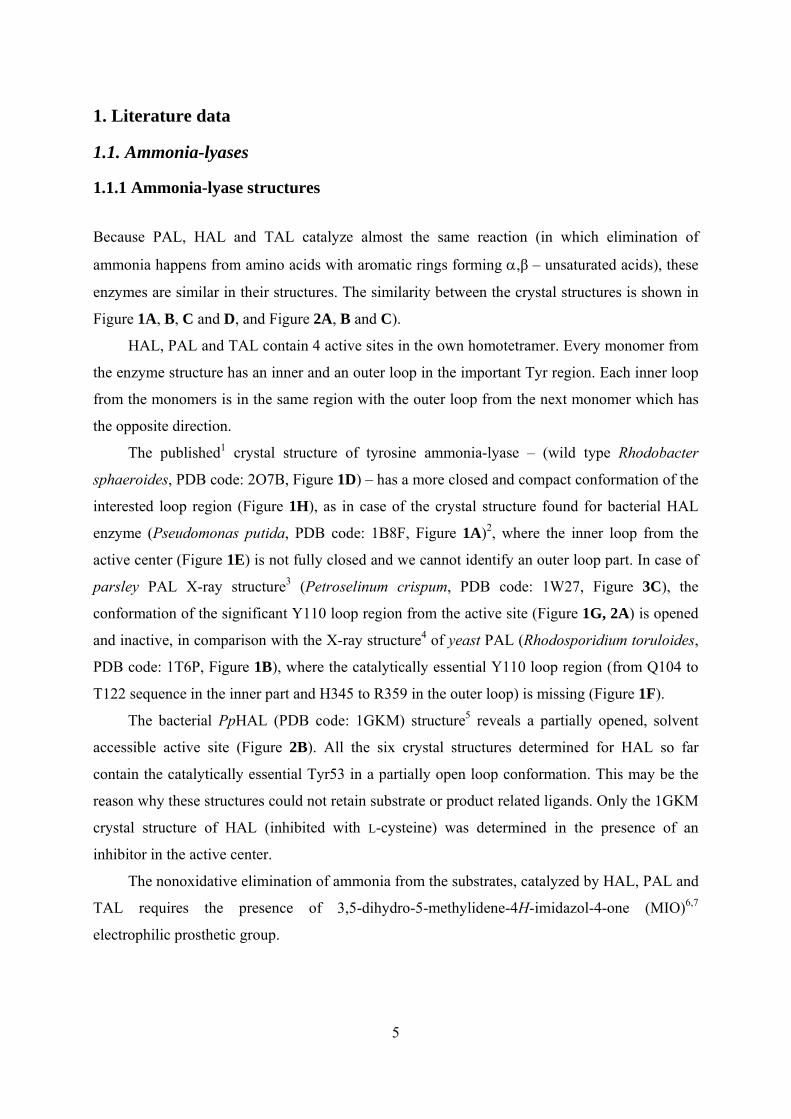
A B C D
E F G H
Figure 1. Ribbon representation of PpHAL (PDB code 1B8F) homotetramer (A), RtPAL (PDB code 1T6P) homotetramer (B), PcPAL (PDB code 1W27) homotetramer (C) and RsTAL (PDB code 2O7B) homotetramer (D) showing subunits A (yellow), B (violet), C (green) and D (red); Ribbon
plot around the important Tyr amino acids (stick model) in (E) PpHAL (PDB code 1B8F), (F) RtPAL (PDB code 1T6P), (G) PcPAL (PDB code 1W27) and (H) RsTAL (PDB code 2O7B) crystal structures including a part of D (red) and C (green) chains.
7
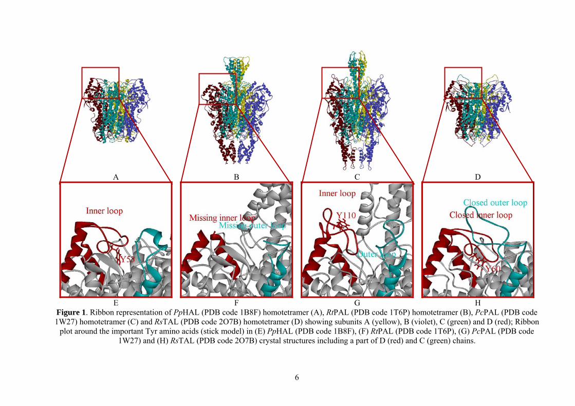
A
B C
Figure 2. Comparison of the substrate entrance channel towards MIO group (red) in several ammonia-lyase structures. Representation of molecular surface of (A) PcPAL (PDB code
1W27), (B) PpHAL (PDB code 1GKM) and (C) AvPAL (PDB code 3CZO) crystal structures. The analogous Tyr110 (A) and Tyr53 (B) residues are seen as stick models in the partially
opened ammonia-lyase structures.
The recently published ammonia-lyase crystal structure (PDB code: 3CZO),8 determined
for Anabaena variabilis PAL (AvPAL), contains the most compact active center in which the
essential Tyr78 and the MIO prosthetic group are deeply buried and not solvent accessible
(Figure 2C).
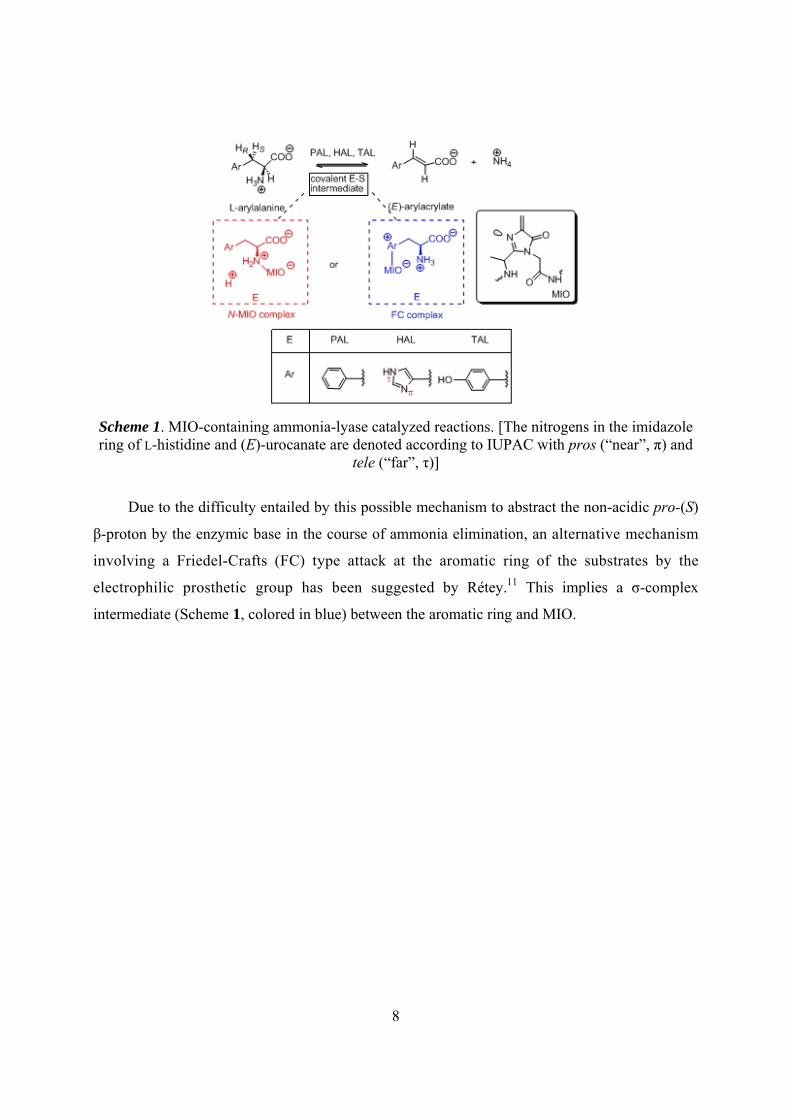
1.1.2 Mechanism of PAL, HAL and TAL reactions
The MIO group similarity suggests that the HAL (EC 4.3.1.3), PAL (EC 4.3.1.24) and TAL (EC
4.1.3.23) behave similarly during the reaction of ammonia-lyases. HAL, PAL and TAL should
remove the non-acidic pro-(S) β-proton from their substrates, without extracting the more acidic
protons from the ammonium moiety of the corresponding L-amino acid. On the basis of the
biochemical data two significantly different mechanisms were proposed for the reaction (Scheme
1) of these ammonia-lyases.
According to Hanson and Havir,9 the Michael addition (Scheme 1, colored in red) of the
amino group from the substrate to the MIO electrophilic prosthetic group of the enzyme takes
place.
Typical for this mechanism is the formation of a covalent E-S intermediate (i.e. an N-MIO
intermediate, Scheme 1), where the amino group of the substrate is covalently bounded to the
methylene part of the MIO prosthetic electrophilic group which facilitates the reaction owing to
the formation of a better leaving group.9,10
8
Scheme 1. MIO-containing ammonia-lyase catalyzed reactions. [The nitrogens in the imidazole ring of L-histidine and (E)-urocanate are denoted according to IUPAC with pros (“near”, π) and
tele (“far”, τ)]
Due to the difficulty entailed by this possible mechanism to abstract the non-acidic pro-(S)
β-proton by the enzymic base in the course of ammonia elimination, an alternative mechanism
involving a Friedel-Crafts (FC) type attack at the aromatic ring of the substrates by the
electrophilic prosthetic group has been suggested by Rétey.11 This implies a σ-complex
intermediate (Scheme 1, colored in blue) between the aromatic ring and MIO.
9
ORIGINAL CONTRIBUTIONS
2. Models and methods
2.1. Homology modeling
The homology modeling method was used by us because we needed the closed form
(catalytically active form) of the important Y-containing inner loop region of ammonia-lyase
structures and the X-ray structures (PAL 1W27,3 HAL 1B8F,2 1GKM5) of PAL and HAL were
determined with an opened or partially opened active site (with a non-active conformation of the
inner loop). The 1W27 determined for PAL, 1B8F and 1GKM crystal structures determined for
HAL weren’t able for computational investigation within the active site. Our aim was to modify
only just a small amino acid sequence at the inner loop region which is in opened/partially
opened form. For homology modeling, the Swiss-Model automated homology modeling
service12,13,14,15,16,17 was used. We took into account the amino acid sequence identity between
the interested crystal structure and the template.
2.2. Conformational analysis within the rigid enzyme
2.2.1 Conformational analysis within the 1W27mod partially modified rigid parsley PAL structure
The initial ligand structure for systematic conformational search (CS) was built up from the 2-
aminoindan-2-phosphonic acid inhibitor (PI) bound to MIO via its N-atom (from structure
2O7E).
In the 1W27mod PAL structure the PI ligand was introduced and for our study a 15 Å sphere
around the MIO prosthetic group was cut off from the active site model. Next, by using
HyperChem18 standard procedure hydrogen atoms were added to the amino acid residues of this
raw active site model. In this way the C- and N-termini at cutting were completed to neutral
aldehyde and amino moieties. The MIO group was manually corrected.18 During CS on the
covalently bound phenylalanine and the heterocyclic ring of the MIO in rigid enzymatic
environment (no water), 3 torsion angles of the ligand were varied for the N-MIO model. The
conformational searches were performed by using the CS module18 implemented in HyperChem
10
using default settings (MM+ forcefield; gradient: 0.1 kcal/mol; Polak-Ribiere method; limits:
300 iterations, 150 optimizations, 15 conformations; test options: "skip if atoms are closer than
0.3 Å").
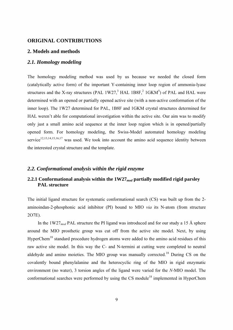
Figure 3. The conformational analysis results for the N-MIO intermediate of the natural substrate in PAL model. Tube model shows the best energy conformation of the N-MIO
intermediate and the other conformations are depicted as wireframe model.
For each covalent intermediate model, the best ligand energy conformation was selected
(Figure 3) which was also in correspondence with the overall arrangement (aromatic moiety
points towards Leu138 and carboxylate is in the close vicinity of Arg354) found in the
experimental inhibited structures of TAL. The initial N-MIO active site model was built by
replacing the PI-MIO part of the raw PcPAL(1W27)/PI active site construct with the ligand
arrangements resulted from the CS's.
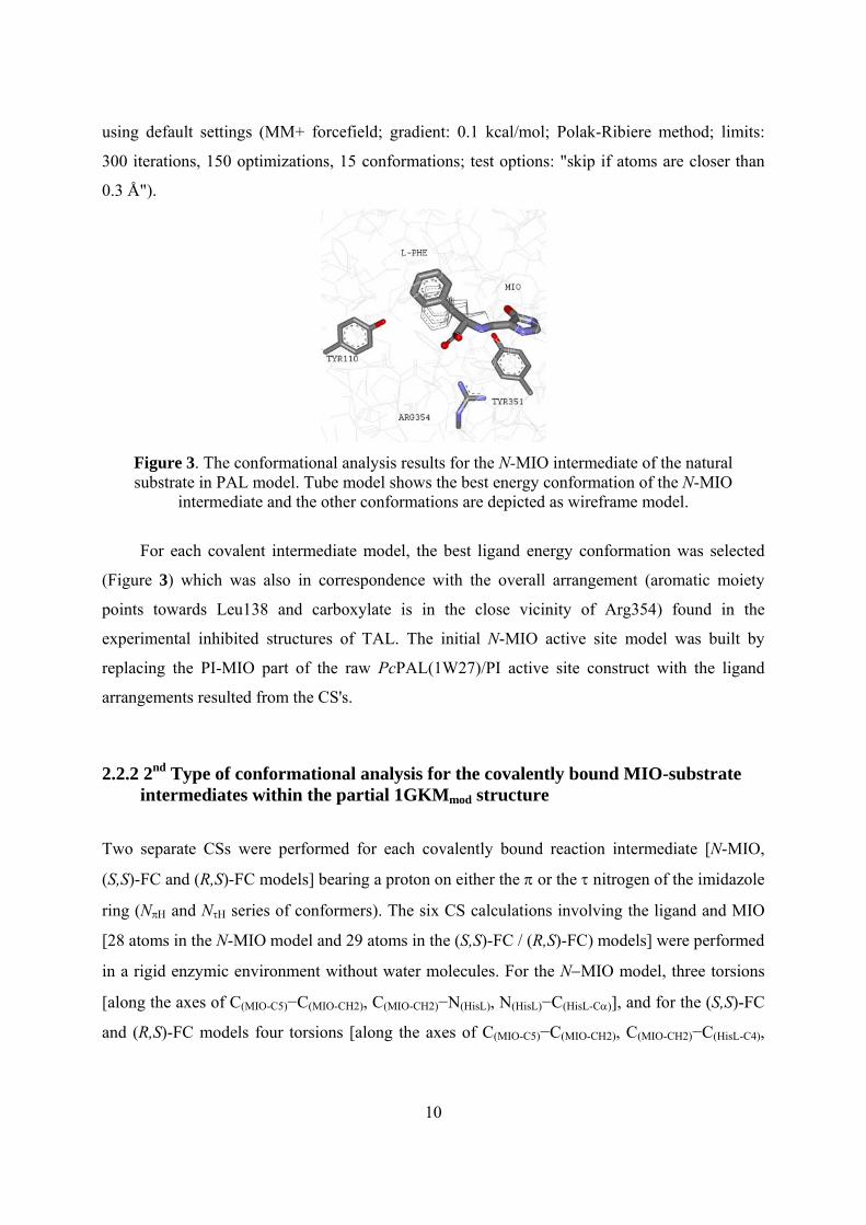
2.2.2 2nd Type of conformational analysis for the covalently bound MIO-substrate intermediates within the partial 1GKMmod structure
Two separate CSs were performed for each covalently bound reaction intermediate [N-MIO,
(S,S)-FC and (R,S)-FC models] bearing a proton on either the π or the τ nitrogen of the imidazole
ring (NπΗ and NτΗ series of conformers). The six CS calculations involving the ligand and MIO
[28 atoms in the N-MIO model and 29 atoms in the (S,S)-FC / (R,S)-FC) models] were performed
in a rigid enzymic environment without water molecules. For the N−MIO model, three torsions
[along the axes of C(MIO-C5)−C(MIO-CH2), C(MIO-CH2)−N(HisL), N(HisL)−C(HisL-Cα)], and for the (S,S)-FC
and (R,S)-FC models four torsions [along the axes of C(MIO-C5)−C(MIO-CH2), C(MIO-CH2)−C(HisL-C4),
11
C(HisL-C5)−C(HisL-Cβ) and C(HisL-Cβ)−C(HisL-Cα)] were varied during the CSs. The CSs were performed
by the HyperChem implemented CS module18 using the default settings (MM+ forcefield;
gradient: 0.1 kcal/mol; Polak-Ribiere method; limits: 300 iterations, 150 optimizations, 15
conformations; test options: "skip if atoms are closer than 0.3 Å").
The resulted conformers of the covalent intermediates after systematic conformational
search are presented in the Figure 4.
N-MIO Nπ N-MIO Nτ
(S,R)-FC Nπ (S,R)-FC Nτ
(S,S)-FC Nπ (S,S)-FC Nτ
Figure 4. Results obtained after 2nd Systematic Conformational Search (CS) of the N-MIO, (S,R)-FC and (S,S)-FC covalent intermediates.
12
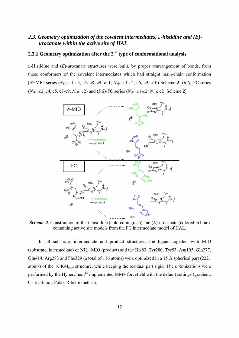
2.3. Geometry optimization of the covalent intermediates, L-histidine and (E)-urocanate within the active site of HAL
2.3.1 Geometry optimization after the 2nd type of conformational analysis L-Histidine and (E)-urocanate structures were built, by proper rearrangement of bonds, from
those conformers of the covalent intermediates which had straight main-chain conformation
[N−MIO series (NτΗ: c1-c3, c5, c8, c9, c11; NπΗ: c1-c4, c6, c9, c10) Scheme 2; (R,S)-FC series
(NτΗ: c2, c4, c5, c7-c9; NπΗ: c2) and (S,S)-FC series (NτΗ: c1-c2; NπΗ: c2) Scheme 2].
Scheme 2. Construction of the L-histidine (colored in green) and (E)-urocanate (colored in blue)
containing active site models from the FC intermediate model of HAL.
In all substrate, intermediate and product structures, the ligand together with MIO
(substrate, intermediate) or NH2−MIO (product) and the His83, Tyr280, Tyr53, Asn195, Gln277,
Glu414, Arg283 and Phe329 (a total of 116 atoms) were optimized in a 15 Å spherical part (2221
atoms) of the 1GKMmod structure, while keeping the residual part rigid. The optimizations were
performed by the HyperChem18 implemented MM+ forcefield with the default settings (gradient:
0.1 kcal/mol; Polak-Ribiere method).
N-MIO
FC
13
For the docking of different ligands within 1W27mod PAL active site we used the
Arguslab19 software.
In case of 1GKMmod for docking zwitterionic L-histidine, Tyr53 and Tyr280 were kept
deprotonated and four torsion angles (along the axes of C5-Cβ, Cβ-Cα, Cα-N and Cα-CCOO-) were
varied, whereas for docking (E)-urocanate, protonated forms of Tyr53 and Tyr280 were used.
Gasteiger charges were added to the atoms of the binding interfaces used for docking by
AutoDock software.
2.4. DFT calculations on ligands involved in HAL reactions
DFT calculations were carried out on L-histidine and L-4-nitrohistidine models (both with
protonated amino groups) for conformations corresponding to the HAL bound state, on a
truncated model of the N−MIO-intermediate (by replacing the MIO ring of the calculated
structure with a hydrogen atom at the exocyclic methylene carbon of MIO) and on a partial
active site model including the truncated model of the N-MIO-intermediate in coordination with
a Zn2+ ion, which is also coordinated to representative parts of His83 and Met382 and to a water.
The DFT optimizations were performed using the GAUSSIAN 09 (rev. A.1)20 and GaussView21 as
front-end. Two positively charged L-histidine structures (with –COOH and –NH3+) were
constructed from the N−MIO models (from conformations c5 and c4 of the NτH and NπH series of
CSs, respectively). Two positively charged L-nitrohistidine models were built from these two L-
histidine structures by replacing the hydrogen at C4 of the imidazole with a nitro group.
In the Zn2+ complex models, the Zn2+ ion had four or five ligands: the imidazole of the
N−MIO (NπH) ligand at the Nτ−atom (i.e. the c4 conformer of the N−MIO NπH series, with a –
NH2(CH3)+ group as a model of the amino moiety bound to MIO, truncated between the MIO
ring and the exocyclic carbon), a 4-methyl-1H-imidazole (coordinated at Nτ, representing His83),
a dimethyl sulfide (coordinated at its S-atom, representing Met382) and one (in the tetrahedral
case) or two (in the trigonal bipyramidal case) water molecules. Proper constraints were used to
maintain the conformation of the MIO-bound histidine ligand (HisL) as allowed within the HAL
active site. For the Zn2+ complex models, several atomic positions were frozen [in Model 1: an
oxygen atom of carboxylic acid moiety of the MIO-bound histidine ligand (the one which was
closer to Arg283), the carbon atom of the methyl group of 4-methylimidazole (truncated His83);
in Model 2: as in Model 1 + a carbon atom of the dimethyl sulfide (representing the Cγ atom of
14
Met382); in Model 3: as in Model 1 + methylene carbon atom of MIO; and in Model 4: as in
Model 1 + a carbon atom of the dimethyl sulfide (representing the Cγ atom of Met382) and
methylene carbon atom of MIO].
Full geometry optimizations for the two types of L-histidine, L-4-nitrohistidine and the
tetrahedral or trigonal bipyramidal Zn2+ complexes were carried out by the DFT method using
Becke’s three parameter hybrid functional combined with the Lee-Yang-Parr correlation
functional (B3LYP)22,23 with the 6-31G or 6-31G(d,p) basis sets. After optimizations, vibrational
frequencies were computed at the same level of theory and single point energies were calculated
with a larger basis [6-311+G(d,p) for Zn2+, for the two imidazole rings, for the –S-CH3 part of
Met382 and for the water molecule(s); and 6-31G(d) for the other parts of the Zn2+ complex
models].
15
3. Results and discussion
3.1. The active ammonia-lyase structures
3.1.1 Modeling the active conformation of PAL
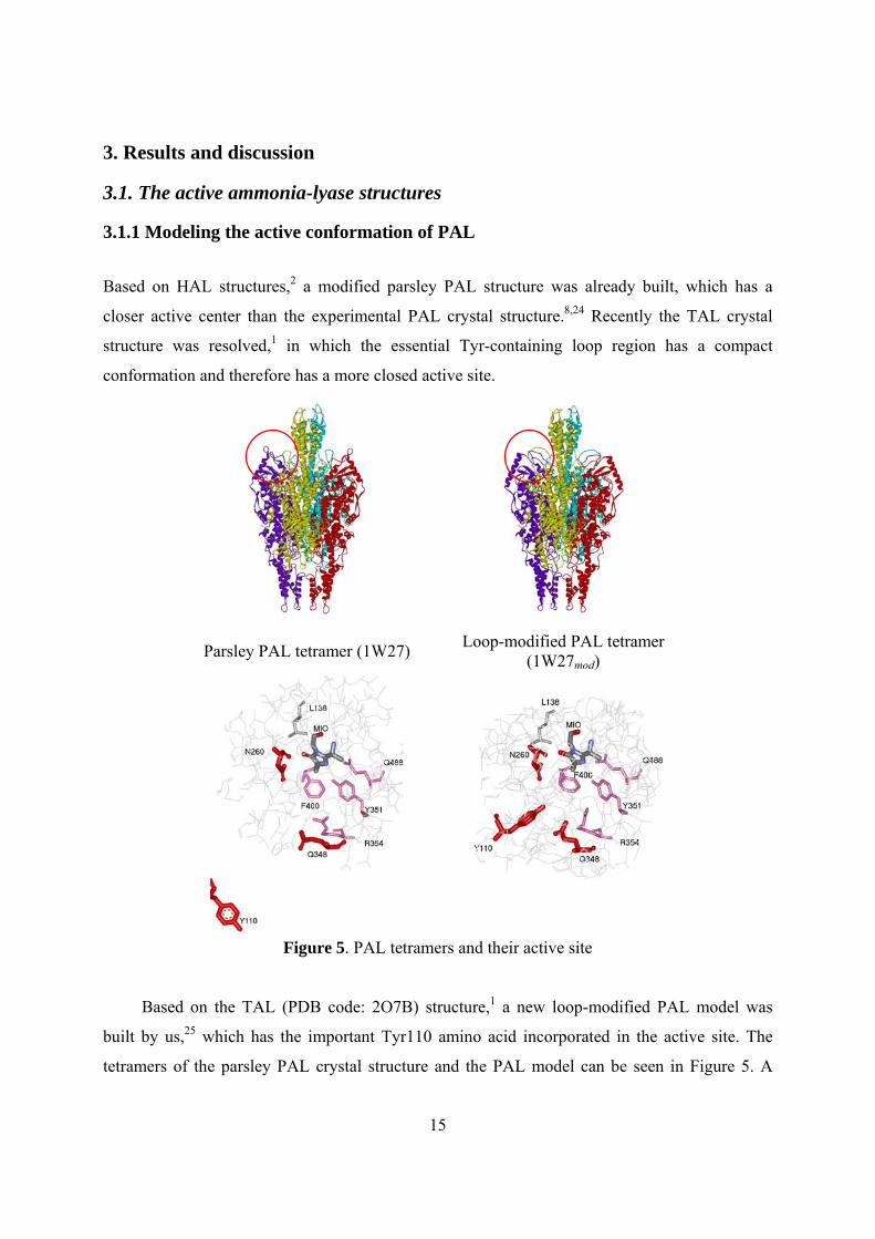
Based on HAL structures,2 a modified parsley PAL structure was already built, which has a
closer active center than the experimental PAL crystal structure.8,24 Recently the TAL crystal
structure was resolved,1 in which the essential Tyr-containing loop region has a compact
conformation and therefore has a more closed active site.
Parsley PAL tetramer (1W27) Loop-modified PAL tetramer (1W27mod)
Figure 5. PAL tetramers and their active site
Based on the TAL (PDB code: 2O7B) structure,1 a new loop-modified PAL model was
built by us,25 which has the important Tyr110 amino acid incorporated in the active site. The
tetramers of the parsley PAL crystal structure and the PAL model can be seen in Figure 5. A
16
more detailed comparison of the two PAL active sites shows the Tyr110 residue in two different
orientations.
Tyr110 in our model points in the direction of MIO, whereas in the crystal structure was far
from the active site (Figure 5).
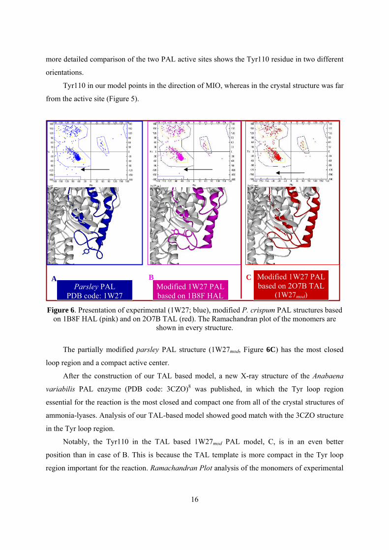
Figure 6. Presentation of experimental (1W27; blue), modified P. crispum PAL structures based
on 1B8F HAL (pink) and on 2O7B TAL (red). The Ramachandran plot of the monomers are shown in every structure.
The partially modified parsley PAL structure (1W27mod, Figure 6C) has the most closed
loop region and a compact active center.
After the construction of our TAL based model, a new X-ray structure of the Anabaena
variabilis PAL enzyme (PDB code: 3CZO)8 was published, in which the Tyr loop region
essential for the reaction is the most closed and compact one from all of the crystal structures of
ammonia-lyases. Analysis of our TAL-based model showed good match with the 3CZO structure
in the Tyr loop region.
Notably, the Tyr110 in the TAL based 1W27mod PAL model, C, is in an even better
position than in case of B. This is because the TAL template is more compact in the Tyr loop
region important for the reaction. Ramachandran Plot analysis of the monomers of experimental
Parsley PAL PDB code: 1W27
Modified 1W27 PAL based on 1B8F HAL
Modified 1W27 PAL based on 2O7B TAL
(1W27mod)
A B C
17
parsley PAL (1W27), modified PAL based on 1B8F HAL and the 1W27mod PAL indicated that
from the 716 amino acid residues of a single subunit of the experimentally 1W27 structure 12
amino acids (six in the Y110 loop region), in the Y110 loop region of the 1B8F HAL-based
modified 1W27 structure eight amino acids (only two in the Y110 loop region), but in the Y110
loop region of 1W27mod structure only four amino acids (neither of them) are outside the likely
Phi/Psi combinations (Figure 6).
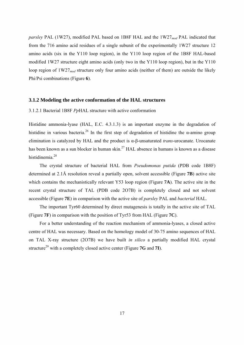
3.1.2 Modeling the active conformation of the HAL structures 3.1.2.1 Bacterial 1B8F PpHAL structure with active conformation
Histidine ammonia-lyase (HAL, E.C. 4.3.1.3) is an important enzyme in the degradation of
histidine in various bacteria.26 In the first step of degradation of histidine the α-amino group
elimination is catalyzed by HAL and the product is α-β-unsaturated trans-urocanate. Urocanate
has been known as a sun blocker in human skin.27 HAL absence in humans is known as a disease
histidinemia.28
The crystal structure of bacterial HAL from Pseudomonas putida (PDB code 1B8F)
determined at 2.1Å resolution reveal a partially open, solvent accessible (Figure 7B) active site
which contains the mechanistically relevant Y53 loop region (Figure 7A). The active site in the
recent crystal structure of TAL (PDB code 2O7B) is completely closed and not solvent
accessible (Figure 7E) in comparison with the active site of parsley PAL and bacterial HAL.
The important Tyr60 determined by direct mutagenesis is totally in the active site of TAL
(Figure 7F) in comparison with the position of Tyr53 from HAL (Figure 7C).
For a better understanding of the reaction mechanism of ammonia-lyases, a closed active
centre of HAL was necessary. Based on the homology model of 30-75 amino sequences of HAL
on TAL X-ray structure (2O7B) we have built in silico a partially modified HAL crystal
structure29 with a completely closed active center (Figure 7G and 7I).
18
A) The essential Y53 loop region in HAL structure
B) Molecular surface representation
C) Active site amino acids of HAL enzyme
D) The essential Y60 loop region in TAL structure
E) Molecular surface representation
F) Active site amino acids of TAL enzyme
G) The essential Y53 loop region in 1B8Fmod HAL
H) Molecular surface representation
I) Active site amino acids of 1B8Fmod HAL model
Figure 7. X-ray structures of bacterial HAL (1B8F), bacterial TAL (2O7B) and the partially modified HAL structure (1B8Fmod)
19
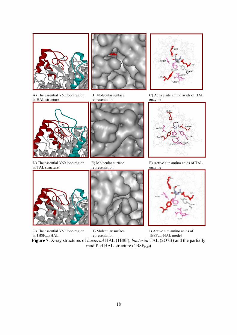
3.2. Computational investigation of the histidine ammonia-lyase: a modified loop conformation and the role of Zn (II) ion
3.2.1 Construction of a closed 1GKM HAL active site environment for calculations
The 1GKM is the only crystal structure of PpHAL in which an inhibitor is present. Importantly,
this is also the structure where the side chain of Met382 has a different conformation from the
side chain arrangement of the other five unliganded PpHAL structures.2,30
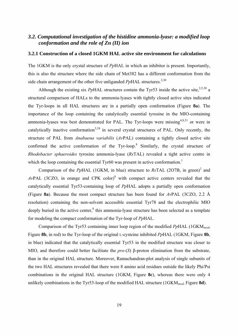
Although the existing six PpHAL structures contain the Tyr53 inside the active site,2,5,30 a
structural comparison of HALs to the ammonia-lyases with tightly closed active sites indicated
the Tyr-loops in all HAL structures are in a partially open conformation (Figure 8a). The
importance of the loop containing the catalytically essential tyrosine in the MIO-containing
ammonia-lyases was best demonstrated for PAL. The Tyr-loops were missing4,8,31 or were in
catalytically inactive conformation3,24 in several crystal structures of PAL. Only recently, the
structure of PAL from Anabaena variabilis (AvPAL) containing a tightly closed active site
confirmed the active conformation of the Tyr-loop.8 Similarly, the crystal structure of
Rhodobacter sphaeroides tyrosine ammonia-lyase (RsTAL) revealed a tight active centre in
which the loop containing the essential Tyr60 was present in active conformation.1
Comparison of the PpHAL (1GKM, in blue) structure to RsTAL (2O7B, in green)1 and
AvPAL (3CZO, in orange and CPK color)8 with compact active centers revealed that the
catalytically essential Tyr53-containing loop of PpHAL adopts a partially open conformation
(Figure 8a). Because the most compact structure has been found for AvPAL (3CZO, 2.2 Å
resolution) containing the non-solvent accessible essential Tyr78 and the electrophilic MIO
deeply buried in the active center,8 this ammonia-lyase structure has been selected as a template
for modeling the compact conformation of the Tyr-loop of PpHAL.
Comparison of the Tyr53 containing inner loop region of the modified PpHAL (1GKMmod,
Figure 8b, in red) to the Tyr-loop of the original L-cysteine inhibited PpHAL (1GKM, Figure 8b,
in blue) indicated that the catalytically essential Tyr53 in the modified structure was closer to
MIO, and therefore could better facilitate the pro-(S) β-proton elimination from the substrate,
than in the original HAL structure. Moreover, Ramachandran-plot analysis of single subunits of
the two HAL structures revealed that there were 8 amino acid residues outside the likely Phi/Psi
combinations in the original HAL structure (1GKM, Figure 8c), whereas there were only 4
unlikely combinations in the Tyr53-loop of the modified HAL structure (1GKMmod, Figure 8d).
20
Figure 8. The mobile Tyr-loops in the active site of MIO-containing ammonia-lyases. a. Comparison of two mobile regions (including the MIO stabilizing Asn and the catalytically
essential Tyr residues) of four different ammonia-lyases: Anabaena variabilis PAL (3CZO, in orange and CPK color); Petroselinum crispum PAL (1W27, in bright green); Rhodobacter
sphaeroides TAL (2O7B, in dark green); Pseudomonas putida HAL (1GKM, in blue). The Asn residue is numbered according to AvPAL (3CZO). b. Overlay of the essential Tyr53 loop regions
of PpHAL (1GKM inhibited with L-cysteine, colored by CPK, blue chain) and the Tyr-loop modified PpHAL (1GKMmod, red chain). c. and d. Ramachandran plots for monomeric units of
PpHAL (1GKM) and the partially modified PpHAL (1GKMmod), respectively.
3.2.2 Comparison of the conformation of the covalent reaction intermediates of the HAL reaction with the arrangements of the substrate and product
Some early reports have indicated that treatment of HAL at high pH in the presence of L-cysteine
and oxygen leads to an irreversible inactivation of the enzyme.32,33 On denaturation, the L-
cysteine inhibited HAL, followed by pronase digestion resulted in two main chromophoric
products.34 In one product, the exocyclic methylene of the MIO was substituted by the amino
a b
c d
21
groups of L-cysteine. When L-cysteine inhibited HAL was first digested with trypsin, two
chromophoric 24-residue peptides were isolated and identified as N−MIO fragments.35 This was
later supported by the L-cysteine inhibited structure of PpHAL (PDB code: 1GKM5). The
inhibited HAL contains the inhibitor with its amino moiety close to the exocyclic methylene of
the electrophilic MIO prosthetic group. This fact can be considered as a further proof for the
presence of an amino-enzyme intermediate in the HAL reaction demonstrated by Peterkofsky.36
Structures of the HAL with L-cysteine,5 TAL with 2-aminoindan-2-phosphonate inhibitor5
and TAM co-crystallized with α,α-difluoro-β-tyrosine37 or p-fluorocinnamate epoxide38
provided strong evidence for reactions via N−MIO intermediates (in which the substrate is
connected to MIO through its amino group) for the ammonia-lyase and aminomutase reactions.
An alternative covalently bound intermediate was proposed by Rétey11 and coworkers. In this
case, a σ-complex would be formed between the aromatic part of the substrate and the MIO
prosthetic group by Friedel-Crafts-like mechanism (FC).
Analysis of the active site residues surrounding the ligand in the L-cysteine-inhibited HAL5
indicated that the pro-(S) β-proton from the L-histidine substrate can be abstracted by one of the
three residues (Tyr53, Tyr280, Glu414) which might be considered as enzymatic bases (Figure
18). Mutagenesis experiments also demonstrated that Tyr53, Glu41439 and Tyr2805,39 are
important residues for the catalysis. The remarkably reduced catalytic activity of the analogous
tyrosine (Tyr 60 and Tyr300) mutants of TAL implies the importance of Tyr53 and Tyr280 in the
HAL reaction.
When PAL was investigated with the phenylalanine analogues D- and L-2-aminooxy-3-
phenyl-propionic acid, it was deduced that ammonia elimination approximated the least-motion
course.40 In several RsTAL structures, products of the elimination reaction were found in the
active site in a zig-zag orientation.1 The least-motion course principle and the similar straight
chain zig-zag shape of the (E)-urocanate product of the HAL reaction determine a straight chain
zig-zag arrangement of the covalently bound intermediate and the L-histidine substrate as well.
Irrespectively of the nature of the covalently bound reaction intermediate, four reaction paths can
fulfill these requirements (Figure 9).
22
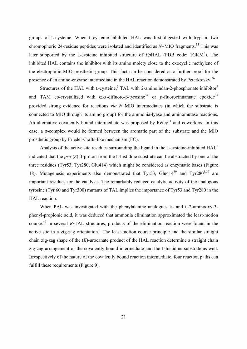
Figure 9. Four possible arrangements of L-histidine and (E)-urocanate along the reaction pathways (A-1, A-2, B-1 and B-2) assuming the least-motion course in the HAL active site
containing the MIO prosthetic group, His83, Arg283 and three possible enzymatic bases: Tyr53, Tyr280 and Glu414.
Along the A-1 reaction path for transformation of the substrate to product, both N−MIO
and FC intermediates are possible but only the Tyr53 amino acid could abstract the pro-(S) β-
proton from the substrate. Only an FC-like mechanism is possible via the A-2 path involving
Tyr280 as the base for pro-(S) β-proton abstraction. Along the B-1 path, deamination of L-
23
histidine may take place by the FC mechanism involving Glu414 as an enzymic base. Along the
B-2 path, both types of the mechanism (N−MIO and FC) can be taken into account involving
Tyr53 as the base for abstraction of the pro-(S) β-proton.
In addition to the substrate and product states (Figure 9), the covalently bound reaction
intermediate should also fulfill the requirements of least-motion course in the HAL active site. If
the HAL reaction proceeded via the N−MIO intermediate, the amino moiety of the L-histidine
substrate would be bound to MIO. If the HAL reaction proceeded via a Friedel-Crafts type
intermediate, the C4 carbon of the aromatic ring of L-histidine substrate would form a σ-complex
with MIO. However, in this case the reaction may take place via two diastereomeric
intermediates [(S,S)-FC and (R,S)-FC] due to a newly forming center of asymmetry at the C4
carbon of the aromatic ring in the σ-complex.
As in cases of all possible intermediates the substrate was anchored to the enzyme by a
covalent bond, the systematic conformational search (CS) with the alternative reaction
intermediates was a powerful tool to find their possible arrangements within the HAL active site.
Because none of the six HAL2,5,30 crystal structures indicated significant variations at the most
part of the active center, the CSs were performed in rigid enzyme environment. This approach
was also supported by the analysis of the B-factors of the active site amino acid residues
indicating low mobility (with exception of the residues of the mobile Tyr53-loop).
Because the imidazole ring allowed two different protonation states (NτΗ or NπΗ) for each
reaction intermediates, six CSs were performed for the three principal alternatives [N-MIO-NτΗ,
N-MIO-NπΗ, (R,S)-FC-NτΗ, (R,S)-FC-NπΗ, (S,S)-FC-NτΗ, (S,S)-FC-NπΗ]. Only those
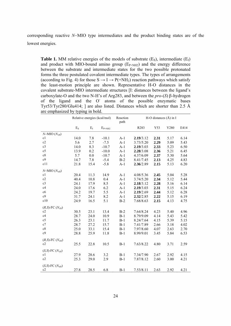
conformations were kept which had a straight chain zig-zag arrangement within the lowest 10
kcal/mol range (Table 1). From the retained conformations of the covalent intermediates of the
six CSs, L-histidine and (E)-urocanate containing active site models were constructed. The
substrate and product and the surrounding eight catalytically relevant amino acid residues were
optimized within the closed HAL (1GKMmod) active site. In this way, comparative analysis of the
full S → I → P(+NH2) reaction pathways become feasible (Table 1).
Although the energies obtained at the MM level of theory are usually not accurate enough
for reliable enzyme mechanistic studies, several observations are worth mentioning. According
to our calculations (Table 1), energetic results favor the reaction via N−MIO (NτH) type
intermediates. In this case, the substrate binding states are of substantially higher energy than the
24
corresponding reactive N−MIO type intermediates and the product binding states are of the
lowest energies.
Table 1. MM relative energies of the models of substrate (ES), intermediate (EI) and product with MIO-bound amino group (EP+NH2) and the energy difference between the substrate and intermediate states for the two possible protonated forms the three postulated covalent intermediate types. The types of arrangements (according to Fig. 4) for those S → I → P(+NH2) reaction pathways which satisfy the least-motion principle are shown. Representative H-O distances in the covalent substrate-MIO intermediate structures [I: distances between the ligand’s carboxylate-O and the two N-H’s of Arg283, and between the pro-(S) β-hydrogen of the ligand and the O- atoms of the possible enzymatic bases Tyr53/Tyr280/Glu414; ] are also listed. Distances which are shorter than 2.5 Å are emphasized by typing in bold.
Relative energies (kcal/mol) Reaction path
H-O distances (Å) in I
ES EI EP+NH2 R283 Y53 Y280 E414
N−MIO (NτH) c1 14.0 7.8 -10.1 A-1 2.19/3.12 2.31 5.17 6.14 c2 5.6 2.7 -7.5 A-1 3.73/5.20 2.29 5.09 5.43 c3 14.0 8.3 -10.7 A-1 2.19/3.03 2.33 5.23 6.50 c5 13.9 0.2 -10.0 A-1 2.28/3.08 2.36 5.21 6.45 c8 5.7 0.0 -10.7 A-1 4.37/6.09 2.37 5.30 5.64 c9 14.7 7.8 -5.4 B-2 8.41/7.45 2.13 4.25 4.83 c11 21.8 15.4 -5.8 A-1 2.36/2.89 2.15 5.13 6.20
N−MIO (NπH) c1 20.4 11.3 14.9 A-1 4.08/5.36 2.45 5.04 5.28 c2 40.4 10.8 0.4 A-1 3.74/5.20 2.34 5.12 5.44 c3 24.1 17.9 8.5 A-1 2.18/3.12 2.28 5.16 6.14 c4 24.0 17.6 6.2 A-1 2.19/3.03 2.31 5.15 6.24 c6 24.2 19.7 5.5 A-1 2.19/2.69 2.44 5.12 6.28 c9 31.7 24.1 8.2 A-1 2.32/2.85 2.22 5.15 6.19 c10 24.9 16.5 5.1 B-2 7.68/8.63 2.15 4.13 4.75
(R,S)-FC (NτH) c2 30.5 23.1 13.4 B-2 7.64/8.24 4.23 5.40 4.96 c4 28.7 24.0 10.9 B-1 8.79/9.09 4.14 5.43 5.42 c5 26.3 23.1 11.7 B-1 8.24/7.64 4.15 5.39 5.15 c7 28.7 27.2 15.7 B-1 7.41/7.89 2.66 3.18 4.02 c8 25.0 33.1 15.4 B-1 7.97/8.60 4.07 2.63 2.70 c9 28.8 25.9 11.8 B-1 8.99/9.01 3.45 5.84 6.53
(R,S)-FC (NπH) c2 25.5 22.8 10.5 B-1 7.63/8.22 4.80 3.71 2.59
(S,S)-FC (NτH) c1 27.9 28.6 3.2 B-1 7.34/7.90 2.67 2.92 4.15 c2 25.3 29.0 2.9 B-1 7.87/8.12 2.60 3.00 4.21
(S,S)-FC (NπH) c2 27.8 28.5 6.8 B-1 7.53/8.11 2.63 2.92 4.21
25
Thus, the calculated energy profile of the reaction via an N−MIO (NτH) type intermediate is
in full agreement with experimental results since MIO-containing ammonia-lyases catalyze,
under normal conditions, ammonia elimination from the L-amino acids in a practically
irreversible manner.26,41 All the other intermediate structures had higher energies (10.8-33.1
kcal/mol) than the lowest energy N−MIO (NτH) intermediate conformer.
Next, the H-O distances in the optimized structures of the MIO-bound intermediate models
between the ligand’s pro-(S) β-hydrogen and oxygen atoms of the possible enzymic bases
(Tyr53, Tyr280 and Glu414) and between ligand’s carboxylate oxygen group and Arg283 were
analyzed (Table 1). The most decisive result of this analysis was the observation that there was
no enzymic base in any of the FC-intermediate conformations which was close enough to the
pro-(S) β-hydrogen to abstract it. Therefore, the A-1 pathway involving an N−MIO covalent
intermediate is the most plausible for the HAL reaction but a B-2 type orientation would also be
allowed.
The optimized structures containing L-histidine and (E)-urocanate ligands in the active site
of the closed HAL structure were compared to the arrangements of these ligands obtained by a
docking procedure. The analysis of the AutoDock results revealed a well conserved orientation
of the product (the carboxylate of the ligand is in the vicinity of the Arg283 while the imidazole
moiety points towards His83) and agreed with the results obtained from the MM optimizations.
Taking into account the orientation of the product within the active site, the docking results
corresponded only to the A-1 pathway.
3.2.3 The role of Zn(II) in the HAL reaction
It was observed that Zn2+ or a number of different divalent cations, like Cd2+ or Mn2+, increase
the activity of HAL.42 On the other hand, there is no Zn2+ containing crystal structure for
HAL.2,5,30 This apparent contradiction can be resolved by assuming that Zn2+, which is necessary
for the catalytic activity, interacts during the HAL reaction with the HAL-specific His83 residue
and with the imidazole of the substrate.39 Therefore, the reason why no Zn2+ containing HAL
structure is known is that no substrate or product containing HAL structure has been determined
so far.2,5,30 Interaction of a Zn2+ with the HAL-specific His83 and with the substrate during the
catalysis39 can also rationalize why HAL accepts only L-histidine,26 L-4-fluorohistidine43 or L-4-
nitrohistidine11,44 as substrates.
26
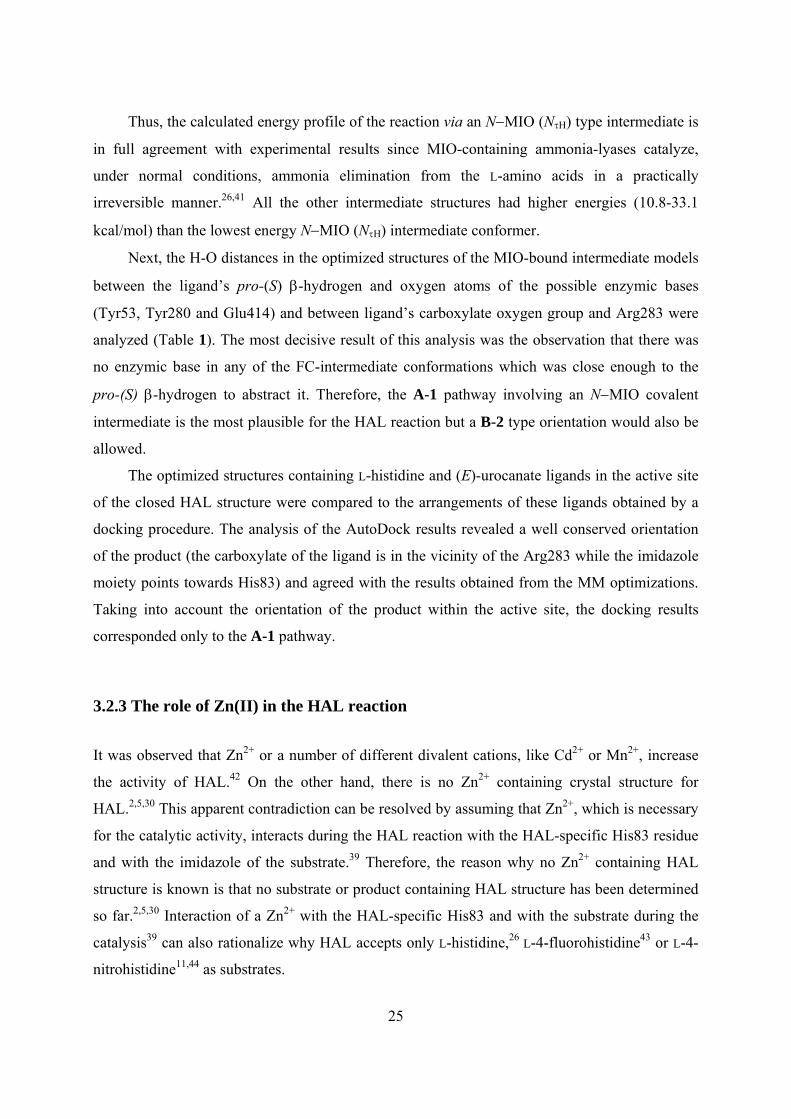
To examine the contribution of the Zn2+ in HAL, model studies were performed on the
conformations found for the N−MIO intermediate in the 1GKMmod active site. Analysis of the
existing Zn-containing protein crystal structures45,46,47,48,49,50 and the calculated N−MIO
intermediate conformations led to the conclusion that Zn could be coordinated at Nτ atoms of
His83 and of the substrate’s imidazole (Figure 10a), similarly as found in Adamalysin II, a zinc
endopeptidase from the snake venom of Crotalus adamanteus51 (Figure 10b).
Figure 10 a. Arrangement of the covalent N-MIO intermediate (NπΗ-c4) in the active site of HAL (1GKMmod). b. The tetrahedral coordination of Zn (in pink) in the Adamalysin II, a zinc
endopeptidase from the snake venom of Crotalus adamanteus51 (PDB code: 1IAG). c. Fit of the tetrahedral Zn-complex model (Model 4, by DFT calculation) into the ligand-free active site of
HAL (1GKMmod).
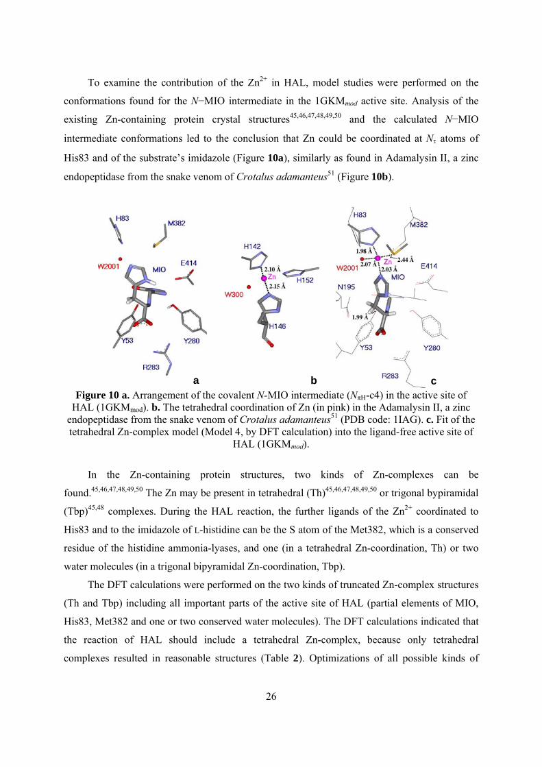
In the Zn-containing protein structures, two kinds of Zn-complexes can be
found.45,46,47,48,49,50 The Zn may be present in tetrahedral (Th)45,46,47,48,49,50 or trigonal bypiramidal
(Tbp)45,48 complexes. During the HAL reaction, the further ligands of the Zn2+ coordinated to
His83 and to the imidazole of L-histidine can be the S atom of the Met382, which is a conserved
residue of the histidine ammonia-lyases, and one (in a tetrahedral Zn-coordination, Th) or two
water molecules (in a trigonal bipyramidal Zn-coordination, Tbp).
The DFT calculations were performed on the two kinds of truncated Zn-complex structures
(Th and Tbp) including all important parts of the active site of HAL (partial elements of MIO,
His83, Met382 and one or two conserved water molecules). The DFT calculations indicated that
the reaction of HAL should include a tetrahedral Zn-complex, because only tetrahedral
complexes resulted in reasonable structures (Table 2). Optimizations of all possible kinds of
a b c
27
trigonal bipyramidal Zn-complex arrangements aborted or led to tetrahedral structures by
exclusion of a water molecule. Comparison of the tetrahedral Zn-complex (Figure 10c) with the
Zn2+ complex found in Adamalysin II51 and with the possible arrangement of the substrate-MIO
covalent intermediate obtained by conformational analysis in the Zn-free active site (Figure 10a)
indicated good agreement in the spatial arrangement of the structures.
Table 2 Bond lengths and energies for tetrahedral zinc-ligand complex models calculated by DFT methods.
Distances (Å)b
Model Relative energies
(kcal/mol)a Zn-NHis83 Zn-NHisL Zn-OHOH Zn-SMet382
1 1.00(0.44) 1.97(1.96) 2.02(2.01) 2.07(2.05) 2.42(2.50) 2 0.00(0.00) 1.96(1.96) 2.03(2.01) 2.06(2.05) 2.42(2.51) 3 2.32(1.57) 1.96(1.96) 2.02(2.01) 2.08(2.05) 2.41(2.49) 4 1.84(0.19) 1.98(1.96) 2.03(2.02) 2.07(2.05) 2.44(2.51)
a By single point calculation at QM/QM B3LYP/6-311+G(d,p):B3LYP/6-31G(d) level after optimization of the structures at B3LYP/6-31G(d,p) level; values between brackets are from single point calculations at QM/QM B3LYP/6-311+G(d,p):B3LYP/6-31G(d) level after optimization of the structures at B3LYP/6-31G level. b Bond lengths in Zn-complexes optimized at B3LYP/6-31G(d,p) level; values between brackets are for Zn-complexes optimized at B3LYP/6-31G level.
The overlay of the truncated tetrahedral Zn-complex and the ligand-free HAL structure
(Figure 10c) indicated that Nτ atom of the ligand’s imidazole was involved in the Zn
coordination, whereas the hydrogen atom on the Nπ position could be at H-bond distance from
Glu414 (Figure 10a and Figure 10c). The binding of the ligand during the HAL reaction at its
imidazole ring by His83 via the Zn-complex and by Glu414 via a hydrogen bond is in full
agreement with active site mutation data indicating that the Glu414Ala (kcat/kcat-mut= 20930) and
the His83Leu (kcat/kcat-mut= 18000) mutations39 have the most dramatic effect on catalysis. Also,
this mode of substrate binding can rationalize the very narrow substrate specificity (in addition to
L-histidine, only L-4-fluorohistidine43 and L-4-nitrohistidine11,44 are accepted as substrates26) of
HAL as well.
All the previous results implied that the enzymic base in the HAL reaction which abstracts
the pro-(S) β-hydrogen as a proton is Tyr53. This was indicated by the 1.99 Å distance between
28
the oxygen atom of Tyr53 and the pro-(S) β-hydrogen of the tetrahedral Zn-complex as well
(Figure 10c). To estimate the acidity of the pro-(S) β-hydrogen in the conformations allowed in
the closed active site of HAL, analysis of Mülliken charges of hydrogens at the β-position of L-
histidine, L-4-nitrohistidine and truncated models for the Zn-free and Zn-coordinating N−MIO
intermediates was performed (Table 3). Because it is known that the nitro group acidifies the
pro-(S) β-hydrogen of L-4-nitrohistidine44 and it is accepted as substrate even by the MIO-less
mutant of HAL analysis of L-4-nitrohistidine was also included.
Table 3 Mülliken atomic charges of the pro-(S) β-hydrogens in zwitterionic L-histidine, in zwitterionic L-4-nitrohistidine, in a truncated N-MIO-intermediate model and in the tetrahedral Zn-complex models.
Entry Structurea HS HR 1 N-MIO (truncated model) 0.176 (0.221) 0.133 (0.173) 2 L-Histidine 0.180 (0.222) 0.102 (0.166) 3 L-4-Nitrohistidine 0.199 (0.245) 0.135 (0.201) 4 Zn-complex, Model 1 0.220(0.267) 0.172 (0.206) 5 Zn-complex, Model 2 0.220 (0.265) 0.173 (0.208) 6 Zn-complex, Model 3 0.220 (0.263) 0.174 (0.208) 7 Zn-complex, Model 4 0.215 (0.261) 0.177 (0.210)
a Calculations on the ligand conformations allowed within the HAL active site were performed at B3LYP/6-31G(d,p) level (values between brackets are from B3LYP/6-31G DFT calculations).
The Mülliken charges found for pro-(S) β-hydrogen at the optimized geometries of L-
histidine, L-4-nitrohistidine (NτH forms) and the Zn-complex models (Table 3) indicated that the
acidity of the pro-(S) β-hydrogen was significantly higher in L-4-nitrohistidine (Entry 3) than in
the L-histidine (Entry 2) or in the Zn-free N−MIO model (Entry 1). The most charged pro-(S) β-
hydrogens, however, were found in the Zn-complex models (Entries 4-7).
These results imply that the formation of a transient Zn-complex in the HAL reaction
contributes not only to the specific binding of the substrate but to the enhancement of its
reactivity as well.
29
Conclusions
I. The modified, closed PAL structure (constructed by modeling the 83-141, 321-351 loops with
the catalytically essential Tyr53 on the basis of RsTAL) resulted less deviations from the allowed
side chain conformations in the Ramachandran-plot than the original experimental structure.
II. The presented ligand docking and conformational analysis results of the covalently bounded
L-phenylalanine to the MIO prosthetic group of the PAL reaction within an essential portion of
the Petroselinum crispum phenylalanine ammonia-lyase (PcPAL) including the full, tightly
closed active center support the idea that the PAL reaction proceeds via the N-MIO intermediate
state in which the L-phenylalanine ligand is covalently bound to the MIO prosthetic group
through its N-atom (N-MIO).
III. The existing crystal structures for HAL, PAL, TAL, and sequence comparison of the active
site residues for all the known MIO-containing enzymes indicate that besides the electrophilic
MIO prosthetic group, these two Tyr, the Arg, the two Asn residues (except for HAL, in which
one Asn is different) belong to the arrangement train of the known MIO-enzymes.
IV. The new partially modified TAL based 1B8Fmod HAL structure is a more competent reliable
model. This HAL model revealed that the catalytically important amino acid (Tyr53) is located
at highly isosteric position in the active site. The new PpHAL structure can be considered as
more accurate model of the active state of the enzyme than the existing experimental HAL
structures.
V. The present study revealed also that the existing experimental structures of histidine
ammonia-lyase from Pseudomonas putida (PpHAL) contain an essential Tyr53-containing loop
in a partially opened conformation. The modified, closed 1GKMmod HAL structure (constructed
by modeling the 39-80 loop with the catalytically essential Tyr53 on the basis of AvPAL)
resulted less deviations from the allowed side chain conformations in the Ramachandran-plot
than the original experimental structure.
30
VI. Investigation of distances between the acidic pro-(S) β-hydrogen at C2 of ligand and the
appropriate oxygen atoms of the possible enzymic bases Tyr53, Ty280 and Glu414 in the
calculated conformations of the three proposed structures [N−MIO, (R,S)-FC, (S,S)-FC] of the
covalently bound reaction intermediate within the closed active site of HAL revealed that the
reaction can only take place via the N−MIO intermediate structure which allowed Tyr53 to get
close enough to the pro-(S) β-hydrogen. This conclusion was also supported by the docking
results with (E)-urocanate.
VII. DFT calculations on the role of a Zn2+ ion in the HAL reaction using a truncated model of
the N−MIO intermediate indicated the formation of a tetrahedral complex with a Zn2+ ion
coordinated to the imidazole ring of the ligand, to His83 and Met382 residues of the enzyme and
to a water molecule. The formation of such transient Zn-complex could explain the narrow
substrate specificity of HAL. The DFT calculations indicated also that the formation of a Zn-
complex had a contribution to the enhancement of the pro-(S) β-hydrogen's reactivity in the
N−MIO intermediate as well.
VIII. The good match of predicted and experimental structures gave confidence that the docking
method is able to provide relevant information about the substrate/product interaction with the
enzyme, but the results of the geometry optimizations of L-histidine/(E)-urocanic acid meet the
requirements of the experimental data found for ligands within the X-ray structure of other
ammonia-lyases. The same interactions appear in the case of the L-histidine substrate together
with other interaction that one between its amino moiety and methylene part of MIO group.
31
List of Publications Publications on the PhD subject Papers 1. Seff A.L., Pilbák S., Silaghi-Dumitrescu I.†, Poppe L., Computational Investigation of the
Histidine Ammonia-Lyase Reaction: a Modified Loop Conformation and the Role of the Zinc(II) Ion, Journal of Molecular Modeling, 2010, submitted.
2. Seff A.L., Pilbák S., Silaghi-Dumitrescu I.†, Poppe L., Computational Investigation of a Bacterial Histidine Ammonia-Lyase (HAL) Model with a Completely Closed Active Center, Studia Universitatis Babes-Bolyai, Seria Chemia, 2010, XLV, 2, TOM I, p. 37-45.
3. Seff A.L., Pilbák S., Poppe L., Ligand Docking and Systematic Conformational Analysis in Loop Modified Parsley Phenylalanine Ammonia-Lyase Structure, Studia Universitatis Babes-Bolyai, Seria Chemia, 2008, LIII, 2, p 67-71.
4. Seff A.L., Pilbák S., Silaghi-Dumitrescu I.†, Poppe L., Ligand Docking and Systematic Conformational Analysis in Loop Modified Phenylalanine Ammonia-Lyase Structure, XIIIth International Chemistry Conference, Hungarian Technical Scientific Society from Transylvania, Cluj-Napoca, Romania, November 8-11, 2007, ISSN 1843-6293, Book of Works, p 106-109.
Oral presentations 5. Seff A.L., Pilbák S., Poppe L., Silaghi-Dumitrescu I.†, DFT Studies on the Formation of
an Intermediate Tetrahedral Zn2+ Complex in a Closed Active Center of HAL, Molecular Modeling in Chemistry and Biochemistry MOLMOD, Special Edition, Cluj-Napoca, Romania, Plenary Lecture, May 28, 2010.
6. Seff A.L., Pilbák S., Silaghi-Dumitrescu I.†, Poppe L., Zinc-Containing Active Site in a Partially Modified 1GKM Crystal Structure of Histidine Ammonia-Lyase: A Computational Investigation., Molecular Modeling in Chemistry and Biochemistry MOLMOD, Cluj-Napoca, Romania, Plenary Lecture, April 2-4, 2009, Book of Abstracts, p 17.
7. Seff A.L., Pilbák S., Silaghi-Dumitrescu I.†, Poppe L., Ligand Docking and Systematic Conformational Analysis in Loop Modified Phenylalanine Ammonia-Lyase Structure, XIIIth International Chemistry Conference, Hungarian Technical Scientific Society from Transylvania, Cluj-Napoca, Romania, Plenary Lecture, November 8-11, 2007, ISSN 1843-6293, Book of Works, p 106-109.
8. Seff A.L., Pilbák S., Poppe L., Ligand Docking and Systematic Conformational Analysis in Loop Modified Parsley Phenylalanine Ammonia-Lyase Structure, Molecular Modeling in Chemistry and Biochemistry MOLMOD, Arcalia, Romania, Plenary Lecture, July 5-8, 2007, ISBN 978-973-7973-46-7, Book of Abstracts, p 15.
Posters 9. Seff A.L., Pilbák S., Silaghi-Dumitrescu I.†, Poppe L., Computational Investigation of a
Bacterial Histidine Ammonia-Lyase (HAL) Model with a Completely Closed Active Center, 4th Central European Conference: Chemistry towards Biology, Dobogókő, Hungary, poster, September 8-11, 2008, Book of Abstracts, p124.
32
10. Seff A.L., Pilbák S., Silaghi-Dumitrescu I.†, Poppe L., A new Bacterial Histidine Ammonia-Lyase (HAL) Model with a Completely Closed Active Center Investigated by Computation, 23rd International Conference on Organometallic Chemistry, Rennes, France, poster, July 13-18, 2008, Book of Abstracts, P473.
11. Seff A.L., Pilbák S., Silaghi-Dumitrescu I.†, Poppe L., Comparison of Ligand Docking and Conformational Analysis Results in Loop Modified Parsley Phenylalanine Ammonia-Lyase Structure, Homboldt Conference on Noncovalent Interactions, Vrsac, Serbia, poster, November 15-18, 2007, Book of Abstract, p 40-41.
Other publications 12. Seff A.L., Darvasi J., Kékedy-Nagy L., Borszéki J., Halmos P., Determination of
Element Containing in Chicken Bone and Comparison of Different Decomposition Methods, XIth International Chemistry Conference, Hungarian Technical Scientific Society from Transylvania, Cluj-Napoca, Romania, Plenary Lecture, November 11-13, 2005, MKE (Society of Hungarian Chemists) prize, ISBN 973-7840-07-0, Book of Works, p 55-58.
13. Seff A.L., Darvasi J., Comparisons of Decomposition and Determination Methods for Quantification of Some Heavy Metal from Domestic Birds Bones, Students for students” Session of Scientific Student Communication, 2nd Edition, Cluj-Napoca, Romania, Oral Presentation, April 8-10, 2005, Book of Abstracts, p. 72.
14. Seff A.L., Darvasi J., Analysis of Metatarsus Bones of Fowls, Scientific National Conference for Students, Chemistry Section, XXVIIth Edition, Budapest, Hungary, Oral Presentation, March 23, 2005, Honorable mention, Work of 30 pages.
15. Seff A.L., Darvasi J., Determination by ICP of Heavy Metals Containing in Metatarsus Chicken Bone Samples Decomposited with Open- and Closed Type Microwave Oven, Scientific Conference for Students in Spring 2004, Chemistry Section, Szeged, Hungary, Oral Presentation, April 29-30, 2004, Honorable mention, Work of 29 pages.
16. Seff A.L., Silaghi-Dumitrescu I.†, Calcule DFT asupra interacţiunii acizilor Lewis EX4 din grupa 14 cu baze Lewis, XXIXth National Chemistry Conference, Călimăneşti-Căciulata, Vâlcea, Romania, poster, October 4-6, 2006, Book of Abstracts, P.S.II. – 41.
33
Selected references [1] Louie G.V., Bowman M.E., Moffitt M.C., Baiga T.J., Moore B.S., Noel J.P., Chemistry
and Biology, 2006, 13, 1327-1338. [2] Schwede T.F., Rétey J., Schulz, G.E., Biochemistry, 1999, 38, 5355-5361. [3] Ritter H., Schulz G.E., Plant Cell, 2004, 16, 3426-3436. [4] Calabrese J.C., Jordan D.B., Boodhoo A., Sariaslani S., Vanneli T., Biochemistry, 2004, 43,
11403-11416. [5] Baedeker M., Schulz G.E., European Journal of Biochemistry, 2002, 269, 1790-1797. [6] Poppe L., Current Opinion in Chemical Biology, 2001, 5, 512-524. [7] Rétey J., Biochimica et Biophysica Acta, 2003, 1647, 179-184. [8] Wang L., Gamez A., Archer H., Abola E.E., Sarkissian C.N., Fitzpatrick P., Wendt D.,
Zhang J., Vellard M., Bliesath J., Bell S.M., Lemontt J.F., Scriver C.R., Stevens R.C., Journal of Molecular Biology, 2008, 380, 623-635.
[9] Hanson K.R., Havir E.A., Archives of Biochemistry and Biophysics, 1970, 141, 1-17. [10] Hermes J.D., Weiss P.M., Cleland W.W., Biochemistry, 1985, 24, 2959-2967. [11] Langer M., Pauling A., Rétey J., Angewandte Chemie International Edition, 1995, 34,
1464-1465. [12] Swiss-Model, An automated Comparative Protein Modelling Server,
http://swissmodel.expasy.org/ [13] Arnold K., Bordoli L., Kopp J., Schwede T., Bioinformatics, 2006, 22,195-201. [14] Kopp J., Schwede T., Nucleic Acids Research, 2004, 32, D230-D234. [15] Schwede T., Kopp J., Guex N., Peitsch M.C., Nucleic Acids Research, 2003, 31, 3381-
3385. [16] Guex, N., Peitsch M.C., Electrophoresis, 1997, 18, 2714-2723. [17] Peitsch M.C., Bio/Technology, 1995, 13, 658-660. [18] Hyperchem version 7.0 (Hypercube, Inc. http://www.hyper.com/). [19] Arguslab program (http://www.arguslab.com). [20] Frisch M.J., Trucks G.W., Schlegel H.B., Scuseria G.E., Robb M.A., Cheeseman J.R.,
Scalmani G., Barone V., Mennucci B., Petersson G.A., Nakatsuji H., Caricato M., Li X., Hratchian H.P., Izmaylov A.F., Bloino J., Zheng G., Sonnenberg J.L., Hada M., Ehara M., Toyota K., Fukuda R., Hasegawa J., Ishida M., Nakajima T., Honda Y., Kitao O., Nakai H., Vreven T., Montgomery Jr. J.A., Peralta J.E., Ogliaro F., Bearpark M., Heyd J.J., Brothers E., Kudin K.N., Staroverov V.N., Kobayashi R., Normand J., Raghavachari K., Rendell A., Burant J.C., Iyengar S.S., Tomasi J., Cossi M., Rega N., Millam N.J., Klene M., Knox J.E., Cross J.B., Bakken V., Adamo C., Jaramillo J., Gomperts R., Stratmann R.E., Yazyev O., Austin A.J., Cammi R., Pomelli C., Ochterski J.W., Martin R.L., Morokuma K., Zakrzewski V.G., Voth G.A., Salvador P., Dannenberg J.J., Dapprich S., Daniels A.D., Farkas Ö., Foresman J.B., Ortiz J.V., Cioslowski J., Fox D.J., Gaussian 09, Revision A.1. 2009, Gaussian, Inc., Wallingford CT.
[21] Dennington II Roy, Keith Todd, Millam John, Eppinnett Ken, Hovell W. Lee, and Gilliland Ray, GaussView, Version 3.09, 2003, Semichem, Inc., Shawnee Mission, KS.
[22] Becke A.D., Journal of Chemical Physics, 1993, 98, 5648-5652. [23] Lee C., Yang W., Parr R.G., Physical Review B, 1988, 37, 785-789. [24] Pilbák S., Tomin A., Rétey J., Poppe L., The FEBS Journal, 2006, 273, 1004-1019. [25] Seff A.L., Pilbák S., Poppe L., Studia Universitatis Babes-Bolyai, Seria Chemia, 2008,
LIII, 2, 67-71.
34
[26] Poppe L., Rétey J., Angewandte Chemie, International Edition English, 2005, 44, 3668-
3688. [27] Morrison H., Bernasconi C., Pandey G., Photochemistry and Photobiology, 1984, 40, 549-
550. [28] Taylor R.G., Levy H.L., McInnes R.R., Molecular Biology and Medicine, 1991, 8, 101-
116. [29] Seff A.L., Pilbák S., Silaghi-Dumitrescu I., Poppe L., 4th Central European Conference:
Chemistry towards Biology, September 8-11, 2008, Book of Abstracts, Dobogókő, Hungary, p124.
[30] Baedeker M., Schulz G.E., Structure, 2002, 10, 61-67. [31] Moffitt M.C., Louie G.V., Bowman M.E., Pence J., Noel J.P., Moore B.S., Biochemistry,
2007, 46, 1004-1012. [32] Klee C.B., Biochemistry, 1974, 13, 4501-4507. [33] Hernandez D., Stroh J.G., Phillips A.T., Archives of Biochemistry and Biophysics, 1993,
307, 126-132. [34] Merkel D., Rétey J., Helvetica Chimica Acta, 2000, 83, 1151-1160. [35] Galpin D., Ellis, B.E., Tanner, M.E., Journal of American Chemical Society, 1999, 121,
10840-10841. [36] Peterkofsky A., Journal of Biological Chemistry, 1962, 237, 787-795. [37] Christianson C.V., Montavon T.J., Festin G.M., Cooke H.A., Shen B., Bruner S.D., Journal
of American Chemical Society, 2007, 129, 15744-15745. [38] Montavon T.J., Christianson C.V., Festin G.M., Shen B., Bruner S.D., Bioorganic and
Medicinal Chemistry Letters, 2008, 18, 3099-3102. [39] Röther D., Poppe L., Viergutz S., Langer B., Rétey J., European Journal of Biochemistry,
2001, 268, 6011-6019. [40] Hanson K.R., Archives of Biochemistry and Biophysics, 1981, 211, 575-588. [41] Rétey J., Biochimica et Biophysica Acta, 2003, 1647, 179-184. [42] Klee C.B., The Journal of Biological Chemistry, 1972, 247, 1398-1406. [43] Klee C.B., Kirk K.L., Cohen L.A., McPhie P., Journal of Biological Chemistry, 1975, 250,
5033-5040. [44] Klee C.B., Kirk K.L., Cohen L.A., Biochemical and Biophysiscal Research
Communications, 1979, 87, 343-348. [45] Nauton L., Kahn R., Garau G., Hernandez J.F., Dideberg O., Journal of Molecular Biology,
2008, 375, 257-269. [46] Bebrone C., Biochemical Pharmacology, 2007, 74, 1686-1701. [47] Martini D., Ranieri-Raggi M., Sabbatini A.R.M., Moir A.J.G., Polizzi E., Mangani S.,
Raggi A., Biochimica et Biophysica Acta, 2007, 1774, 1508-1518. [48] Lipscomb W.S., Strater N., Chemical Reviews, 1996, 96, 2375-2433. [49] Gerhardt S., Hassal G., Hawtin P., McCall E., Flavell E., et al., Journal of Molecular
Biology, 2007, 373, 891-902. [50] Debela M., Goettic P., Magdolen V., Huber R., Schechter N.M., Bode W., Journal of
Molecular Biology, 2007, 373, 1017-1031. [51] Gomis-Rüth F.X., Kress L.F., Bode W., EMBO Journal, 1993, 12, 4151-4157.