segregation of leading-edge and uropod components into specific … · segregation of leading-edge...
TRANSCRIPT

Segregation of leading-edge and uropod componentsinto specific lipid rafts during T cell polarizationConcepcion Gomez-Mouton*, Jose Luis Abad*, Emilia Mira*, Rosa Ana Lacalle*, Eduard Gallardo†,Sonia Jimenez-Baranda*, Isabel Illa†, Antonio Bernad*, Santos Manes*‡, and Carlos Martinez-A.*
*Department of Immunology and Oncology, Centro Nacional de Biotecnologıa, Consejo Superior de Investigaciones Cientıficas, Universidad Autonoma deMadrid, Cantoblanco, E-28049 Madrid, Spain; and †Laboratorio de Neurologıa Experimental, Santa Creu i Sant Pau Hospital, 08025 Barcelona, Spain
Edited by Kai Simons, Max Planck Institute of Molecular Cell Biology and Genetics, Dresden, Germany, and approved June 11, 2001 (received for reviewApril 2, 2001)
Redistribution of specialized molecules in migrating cells developsasymmetry between two opposite cell poles, the leading edge andthe uropod. We show that acquisition of a motile phenotype in Tlymphocytes results in the asymmetric redistribution of gangliosideGM3- and GM1-enriched raft domains to the leading edge and to theuropod, respectively. This segregation to each cell pole parallels thespecific redistribution of membrane proteins associated to each raftsubfraction. Our data suggest that raft partitioning is a major deter-minant for protein redistribution in polarized T cells, as ectopicexpression of raft-associated proteins results in their asymmetricredistribution, whereas non-raft-partitioned mutants of these pro-teins are distributed homogeneously in the polarized cell membrane.Both acquisition of a migratory phenotype and SDF-1a-induced che-motaxis are cholesterol depletion-sensitive. Finally, GM3 and GM1raft redistribution requires an intact actin cytoskeleton, but is insen-sitive to microtubule disruption. We propose that membrane proteinsegregation not only between raft and nonraft domains but alsobetween distinct raft subdomains may be an organizational principlethat mediates redistribution of specialized molecules needed for T cellmigration.
Cell movement across a two-dimensional substrate requires adynamic interplay between attachment at the cell front and
detachment at the rear cell edge, combined with a traction ma-chinery that pulls the net cell body forward. As adhesion anddetachment occur at opposite cell edges, the moving cell mustacquire and maintain spatial and functional asymmetry, a processcalled polarization (1, 2). This asymmetry develops between twoopposite cell edges—the leading edge, which protrudes, and therear (termed uropod in lymphocytes), which retracts.
Because of the specialized functions of these compartments, eachpole in migrating cells is enriched in specific receptors and signalingmolecules but lacks others. In fibroblast-like cells and lymphocytes,the leading edge contains chemokine receptors, several glyco-sylphosphatidylinositol-linked proteins, such as the urokinase plas-minogen activator receptor (uPAR), as well as the machinery thatsenses the environment and induces localized actin polymerization(1). Whereas the rear edge in fibroblasts appears to be a passive tail,the lymphocyte uropod is a specialized pseudopod-like projectionwith important functions, including motility and recruitment ofbystander cells. Several intercellular adhesion molecules (ICAMs)concentrate at the uropod, including ICAM-1, -2 and -3, CD43,CD44, as well as the actin-binding proteins of the ezrin–radixin–moesin family. In accordance with its importance in lymphocytemigration, crosslinking of molecules located in the uropod issufficient to trigger neutrophil polarization and motility (3).
To understand polarization and chemotaxis processes, the mo-lecular mechanisms involved in the generation and maintenance ofthe asymmetric distribution of cell-surface components must beelucidated. Several lines of evidence suggest that association ofproteins with cholesterol- and glycosphingolipid-enriched raft-membrane domains is crucial in distributing specialized moleculesto the leading edge of fibroblast-like migrating cells. The raftmarker GM1 ganglioside, the raft-associated chemokine receptor
CCR5, and other raft-associated proteins accumulate preferentiallyat the leading lamella of migrating cells (4). Modification ofraft-located proteins such that they no longer associate with raftsinhibits their asymmetric redistribution. The functional role ofasymmetric raft redistribution is shown in this article, as membranecholesterol depletion impairs cell polarization and chemotaxis.Cholesterol-depleted cells showed isotropic pseudopodial protru-sion, suggesting that raft redistribution is needed for location-specific induction of pseudopod protrusion during cell polarization.Moreover, rafts are the preferred cell platforms for membrane-linked actin polymerization by in situ phosphatidylinositol 4,5-bisphosphate synthesis and tyrosine kinase signaling through theWASP-Arp2y3 pathway (5–7).
Raft association seems to be pivotal for protein redistribution tothe leading edge in migrating cells. To analyze whether this processis a general mechanism in all cell types, we studied redistribution ofraft-associated membrane receptors and lipids during T cell polar-ization, and found that membrane rafts are redistributed asym-metrically in migrating T lymphocytes. In contrast to the exclusiveleading-edge redistribution detected in fibroblast-like cells, polar-ized T lymphocytes segregate leading-edge and uropod markersinto two raft types that differ in lipid and protein composition.Leading-edge rafts, characterized by including chemokine recep-tors and uPAR, are enriched in GM3 ganglioside and devoid ofGM1; uropod rafts, containing CD44 and other cell-adhesionmolecules, are enriched in GM1 but lack GM3.
Ectopic expression of the raft-associated influenza virus hem-agglutinin (HA) results in its asymmetric distribution in polarizedT cells, whereas expression of a nonraft mutant version (HA2A520;ref. 8) results in homogeneous protein distribution on the cellmembrane. We observed asymmetrical redistribution of a greenfluorescent protein (GFP)-tagged version of the vesicular stomatitisvirus glycoprotein (VSVG3)-GFP (9) that colocalizes with GM1when expressed in T cells. A mutant version of this protein(VSVG3-SP-GFP; ref. 10), which does not colocalize with GM1, isdistributed homogeneously on the cell membrane. These datasuggest that raft partitioning is a major determinant of asymmetricprotein redistribution in polarized T cells. Accordingly, membranecholesterol depletion impedes acquisition of a polarized cell phe-notype and inhibits both cell–cell interaction and cell chemotaxis.All together, the results indicate a prominent role for membranerafts in the acquisition of the polarity needed for T cell chemotaxis.
This paper was submitted directly (Track II) to the PNAS office.
Abbreviations: uPAR, urokinase plasminogen activator receptor; ICAM, intercellular adhe-sion molecule; HA, hemagglutinin; GFP, green fluorescent protein; VSVG3, vesicular sto-matitis virus glycoprotein 3; CTx, cholera toxin b-subunit; CD, methyl-b-cyclodextrin; PBLs,peripheral blood lymphocytes; DRM, detergent-resistant membranes.
See commentary on page 9471.
‡To whom reprint requests should be sent. E-mail: [email protected].
The publication costs of this article were defrayed in part by page charge payment. Thisarticle must therefore be hereby marked “advertisement” in accordance with 18 U.S.C.§1734 solely to indicate this fact.
9642–9647 u PNAS u August 14, 2001 u vol. 98 u no. 17 www.pnas.orgycgiydoiy10.1073ypnas.171160298
Dow
nloa
ded
by g
uest
on
Mar
ch 8
, 202
0

Materials and MethodsCell Culture, Expression Constructs, and Antibodies. The murine NS-1T cell hybridoma and the human Jurkat cell line were cultured inRPMI medium 1640 with 5% (volyvol) FCS, antibiotics, L-glutamine, and sodium pyruvate. The influenza virus HA wild typeand the HA2A520 mutant, with two point mutations in thetransmembrane domain that reduce raft association (8), were a giftof P. Scheiffele (Univ. of California, Berkeley). VSVG3-GFP andVSVG3-SP-GFP (9, 10), which is identical to VSVG3-GFP but hasa spacer between the viral protein and the GFP moiety, were givenby P. Keller (Max Planck Institute, Molecular Cell Biology andGenetics, Dresden, Germany). All constructs were subcloned intothe pLZR retroviral vector; NS-1 cells were transduced with therecombinant retrovirus (11).
Surfact-Amps X-100 with 10% (volyvol) Triton X-100 andSurfact-Amps 58 with 10% (volyvol) Brij58 were obtained fromPierce. Optiprep gradient medium was obtained from NycomedPharma, latrunculin-B from Calbiochem, and methyl-b-cyclodex-trin (CD), demecolcine, water-soluble cholesterol, filipin III, fi-bronectin, BSA, peroxidase (PO)-conjugated streptavidin, biotin-and FITC-labeled cholera toxin b-subunit (CTx), and anti-talinmonoclonal antibody (mAb) were obtained from Sigma. Anti-CXCR4 (Fab172B) and anti-human CD44 mAb were obtainedfrom R & D Systems, rat anti-mouse CD44 and biotin-labeledanti-human CD43 from PharMingen, anti-mouse CD43 from SantaCruz Biotechnology, and anti-transferrin receptor from Zymed.Rabbit anti-HA was a gift of P. Scheiffele, and PO- or Cy3-labeledsecondary antibodies were obtained from Dako or JacksonImmunoResearch.
Characterization of Anti-GM3 Antiserum. A serum sample from apatient with acute polyneuropathy was analyzed for IgM and IgGantibodies to GM1, GM2, GM3, aGM1, GD1a, GD1b, GD3,GT1b, and GQ1b by direct ELISA and TLC. ELISA titers werecalculated by end-point dilution analysis of optical densities. ForTLC, 1 mg per lane of each lipid was loaded onto a silica gel plateand developed by using a methanol, chloroform, and CaCl2 solvent.After blocking with PBSyBSA, each plate was covered with 2.5 mlof diluted test serum (1y100) and incubated for 4 hr at 4°C. Afterwashing, plates were incubated for 1 hr at 20°C with PO-conjugatedIgM or IgG, as required. Serum reactivity to crude cell extracts (20mg per lane) was analyzed by Western blotting.
Flotation Experiments. To analyze detergent-insoluble complexes inflotation gradients, 15 3 106 Jurkat cells stimulated with SDF-1a(100 nM, 15 min, 37°C; PeproTech, Rocky Hill, NJ) were cooled onice, washed with PBS, and lysed in 300 ml of TNE buffer (50 mMTriszHCl, pH 7.4y150 mM NaCly5 mM EDTA) with 0.5% TritonX-100 or 1% Brij58. Cells were extracted for 20 min on ice and theextract was subsequently brought to 35% (volyvol) Optiprep.One-third of the lysate was sequentially overlayered with 3.5 ml of30% (volyvol) Optiprep and 200 ml of TNE with detergent in anSW60 tube. After centrifugation (4 hr at 170,000 3 g at 4°C), fivefractions were collected from the gradient (top to bottom) andprecipitated with trichloroacetic acid. Normalized protein amountsfor each fraction were analyzed by SDSyPAGE and Westernblotting.
For cholesterol depletion, serum-starved Jurkat cells were incu-bated with 5 mM CD for 30 min at 37°C. Under these conditions,CD treatment does not induce cell detachment from the substrateor modify viability (data not shown). After incubation, CD wasremoved by repeated washing with serum-free medium containing0.01% BSA, then cells were stained with filipin as described (4).Flotation gradients of untreated and CD-treated cells were pre-pared as above.
Immunofluorescence and Antibody-Induced Patching. Resting pe-ripheral blood lymphocytes (PBLs) were isolated from fresh humanblood by Ficoll–Hypaque density-gradient centrifugation (Amer-sham Pharmacia), then plated in two adherence incubation steps at37°C for 1 hr each in plastic flasks, then plated on recombinanthuman ICAM-2yFc chimera protein (R & D Systems). NS-1 andJurkat cells were plated on fibronectin (Fn)-coated eight-well-chamber glass slides 24 hr before assay. Serum-starved Jurkat cellsand PBLs were stimulated with 100 nM SDF-1a, then washed andfixed with 3.7% (wtyvol) paraformaldehyde for 5 min on ice in PBS.Samples were incubated with the indicated antibodies, then withCy2- or Cy3-conjugated second antibodies for 45 min on ice. Fortalin-staining, methanol-permeabilized cells (10 min, 220°C) wereblocked with PBSy2% (wtyvol) BSA for 1 hr at 4°C before stainingwith primary antibodies. Slides were mounted in Vectashieldmedium containing 49,6-diamidino-2-phenylindole (Vector Labo-ratories). In some experiments, NS-1 cells plated on Fn were treatedat 37°C for 30 min with 10 mM latrunculin-B or 0.3 mM demecol-cine, washed twice with medium, and fixed and stained withFITC-CTx and anti-CD44 or anti-GM3.
For HA and HA2A520 visualization in transduced NS-1 cells,antibody-mediated lateral copatching was performed by incubatingunfixed cells for 30 min at 12°C with anti-HA and anti-GM3antibodies. Further crosslinking was performed with Cy2- andCy3-second antibody for 30 min at 12°C; antibody–receptor com-plex internalization was not seen. Cells were fixed and mounted asabove. For HA copatching with CTx, FITC-CTx (6 mgyml) wasadded after fixation and incubated for 10 min at 4°C; cells weremethanol-fixed for 10 min at 220°C before mounting.
In all cases, cells were visualized by confocal laser scanningmicroscopy. Linear signal intensity for each fluorophore was cov-ered by a linear scale of pixel intensities. The two colors wereacquired separately and merged by using TCS NT software (Leica).Digital images were processed with PHOTOSHOP (Adobe).
Polarization, Cell–Cell Interaction, and Chemotaxis of Cholesterol-Depleted Cells. NS-1 cells were untreated or CD-treated as forJurkat cells. A portion of the CD-treated cells was incubated for 30min at 37°C in RPMI 1640 medium with 60 mgyml free cholesterol.Untreated, CD-treated, or cholesterol-replenished cells were fixedwith 3.7% (wtyvol) paraformaldehyde, then incubated with anti-CD44. Polarization was analyzed by confocal and phase-contrastmicroscopy.
For cell–cell interaction, Jurkat cells were untreated, treated withCD, or treated with CD plus cholesterol, then plated on fibronectin-coated chambers in the presence of SDF-1a (25 nM). After theaddition of PBLs (7.5 3 104 cells per well), images were acquiredin phase-contrast microscopy. Jurkat cells attached and spread onthe substrate (phase-dark cells), and PBLs that adhered to theJurkat cell uropod (phase-bright cells) were counted for eachcondition. Recruitment index is expressed as PBLs captured di-vided by the number of substrate-adhered Jurkat cells in the firstlayer.
For chemotaxis, Jurkat cells were untreated or treated with CDor CD plus cholesterol, resuspended, and migration-analyzed in amodified Boyden chamber (Costar). In all cases, 2 3 105 cells inserum-free medium were seeded in the upper chamber and thelower chambers were filled with serum-free medium alone orserum-free medium containing SDF-1a (25 nM). After incubationfor 2.5 hr at 37°C, the cell number in the lower chamber wasestimated by flow cytometry with an electronically programmableindividual cell sorter. The chemotactic index was calculated as thequotient of the number of cells recovered in SDF-1a transwells andthe number of cells in the absence of stimulus.
ResultsGM1-Based Rafts Are Asymmetrically Distributed to the Migrating TCell Uropod. We analyzed whether lipid rafts are asymmetricallyredistributed in polarized T cells, as reported for fibroblast-like
Gomez-Mouton et al. PNAS u August 14, 2001 u vol. 98 u no. 17 u 9643
CELL
BIO
LOG
Y
Dow
nloa
ded
by g
uest
on
Mar
ch 8
, 202
0

cells, by studying raft-enriched ganglioside GM1 distribution inconstitutively polarized NS-1 cells. When plated on fibronectin,most NS-1 cells become polarized with a well defined leading edgeand uropod. Compared with the homogeneous distribution innonpolarized cells (data not shown), GM1 concentrated mainly atthe NS-1 cell uropod, as determined by morphological criteria andby colocalization studies with uropod-specific markers such asCD44 (Fig. 1A). GM1 also appears at the edge opposite that atwhich leading-edge markers such as talin (12) are seen, confirmingGM1 concentration at the NS-1 cell uropod (Fig. 1B).
It was unclear whether uropod GM1 concentration is unique toNS-1 cells or is a generalized phenomenon in polarized T cells.Therefore, we analyzed GM1 distribution in Jurkat cells stimulatedwith SDF-1a, a chemokine that induces CXCR4 redistribution tothe leading edge of migrating Jurkat cells (13). GM1 patchesredistribute to and colocalize with the uropod marker CD44 (Fig.1C) but segregate to the pole opposite the CXCR4 staining (Fig.1D). Collectively, these results indicate that GM1-based rafts andtheir associated proteins are distributed to the uropod in T cells witha migrating phenotype.
Leading-Edge Proteins in Migrating T Cells Colocalize with GM3-Rafts.Chemokine receptors such as CXCR4 partition into the raftfraction (14, 15), although CXCR4 does not colocalize with GM1in T cells (Fig. 1D). Some glycosylphosphatidylinositol-anchoredleukocyte-surface glycoproteins that accumulate at the leadingedge associate preferentially to GM3 (16), suggesting that GM3-based rafts concentrate at the leading-cell edge. We analyzed GM3distribution in migrating cells by using an antiserum from a patientwith acute polyneuropathy with a high anti-GM3 titer (1y740) butno reactivity to a battery of other gangliosides including GM1. In
addition, this antiserum does not react with cell proteins as analyzedby Western blotting (data not shown).
GM3 is asymmetrically distributed, forming patches at the samepole as talin (Fig. 2A) and colocalizing with other leading-edgemarkers such as CXCR4 (Fig. 2B) and uPAR (Fig. 2C). GM3concentrates mainly at the edge opposite that of GM1 (Fig. 2D),which decorates the uropod (see above). These results indicate theexistence of two distinct raft types in T cells, which segregate toopposite sides once the cell acquires a migrating phenotype; GM3-enriched rafts redistribute to the leading edge, and GM1-enrichedrafts concentrate at the uropod. Segregation between leading rafts(L-rafts) and uropod rafts (U-rafts) is also seen in freshly isolatedSDF-1a-stimulated PBLs; GM1 colocalizes with the uropodmarker CD44 (Fig. 2E), and GM3 colocalizes with the leading-edgemarker CXCR4 (Fig. 2F).
GM1- and GM3-Raft-Associated Proteins Partition in Detergent-Resis-tant Membranes. Raft-associated membrane proteins float in den-sity gradients as detergent-resistant membranes (DRM; ref. 17);this property distinguishes them from insoluble complexes formedby cytoskeletal association. To analyze DRM partitioning of GM1-and GM3-associated proteins, we fractionated Jurkat cells lysedwith the nonionic detergents Triton X-100 and Brij58. The leading-edge markers CXCR4, uPAR, and GM3, as well as the uropodmarkers CD44 and GM1, partition to the DRM fraction indepen-dently of the detergent used (Fig. 3A). Triton X-100 and Brij58
Fig. 1. GM1-rafts concentrate at the uropod of migrating T cells. NS-1 (A andB) or SDF-1a-stimulated Jurkat cells (C and D) were costained with CTx to revealGM1 (green), with leading edge or uropod markers indicated (red), and analyzedby confocal microscopy. Fluorescence is shown for each signal and for the merg-ing of both; colocalization is seen as yellow. To visualize cell shape (Left), laserpower was saturated for green and red channels. These cells are representativeof the majority of cells recorded in independent experiments. (Bar 5 2 mm.)
Fig. 2. GM1- and GM3-rafts segregate to opposite cell edges in T cells. Confocalanalysis of NS-1 (A and D), SDF-1a-stimulated Jurkat cells (B and C), and SDF-1a-stimulated PBLs (E and F) costained with anti-GM3 antiserum or CTx and withvarious leading-edge and uropod markers, as indicated. For NS-1 and Jurkat cells,fluorescence for each channel and the merging of both signals are shown,whereas only merging is displayed for PBLs; colocalization of the signals is seen asyellow. (Bar 5 2 mm.)
9644 u www.pnas.orgycgiydoiy10.1073ypnas.171160298 Gomez-Mouton et al.
Dow
nloa
ded
by g
uest
on
Mar
ch 8
, 202
0

differ in their hydrophilic-lipophilic balance, a characteristic used todistinguish raft types in Madin–Darby canine kidney (MDCK) cells(18). Although Brij58 increases DRM-associated proteins andlipids, no differential solubility was detected between GM3- andGM1-associated markers in using the two detergents. Full solubi-lization of nonraft proteins such as the transferrin receptor confirmsthe quality of the fractionations.
CD treatment of Jurkat cells to sequester membrane cholesterolbefore fractionation resulted in increased CXCR4, uPAR, andCD44 solubility with either detergent (Fig. 3B). This result suggeststhat leading-edge and uropod-marker association to L- or U-raftsis sensitive to cholesterol extraction, although differences in cho-lesterol content for each raft type cannot be excluded.
Raft Association Is Needed for Membrane Protein Redistribution. Weexamined whether protein redistribution to the leading edge of theuropod is a consequence of specific association to GM3- orGM1-enriched rafts. NS-1 cells were transduced with retroviruscoding for the raft-associated influenza virus HA protein and thenonraft HA2A520 mutant (8). These cells were also transducedwith two GFP-tagged versions of VSVG (VSVG3-GFP andVSVG3-SP-GFP). VSVG colocalizes with GM1 in several celltypes, although it does not partition in DRM (19). Accordingly,neither VSVG3-GFP nor VSVG3-SP-GFP partition in DRM, butonly VSVG3-GFP colocalizes with GM1 (see Fig. 4 A and E). Thedifferential behavior of VSVG3-GFP and VSVG3-SP-GFP is notyet understood, although the masking of sorting signals in VSVG3-GFP may be implicated (9).
HA and VSVG3-GFP redistribute to the uropod in polarizedNS-1 cells and colocalize with GM1 (Fig. 4 A and C) but segregatefrom GM3 rafts (Fig. 4 B and D). HA and VSVG3-GFP proteinsprobably do not associate to cytoskeleton or to other cell receptors;thus, their preferential accumulation in the uropod is solely theconsequence of their U-raft association. Therefore, we expressedthe nonraft HA mutant (HA2A520) and the basolateral-sortedVSVG3-SP-GFP and analyzed their distribution in polarized NS-1.HA2A520 or VSVG3-SP-GFP proteins were homogeneously dis-tributed on the NS-1 cell surface with strong polarization of GM1
(Fig. 4 E and G) and GM3 (Fig. 4 F and H). Quantitative analysisindicated that 85% of polarized, transduced NS-1 cells accumulatedHA and VSVG3-GFP proteins preferentially at the uropod,whereas 14% of the NS-1 cells redistributed HA2A520 andVSVG3-SP-GFP proteins to a single cell pole. This result indicatesthat raft association is a major determinant for asymmetric proteinredistribution in T cells.
Functional Role of Rafts in Polarization and Chemotaxis. To addressthe functional role of rafts in lymphocyte polarization, we studiedthe effect of cholesterol depletion on the acquisition of a migratingphenotype in NS-1 cells. Morphology and polarization markerredistribution were analyzed in cholesterol-depleted cells. CDtreatment significantly reduces the number of NS-1 cells with apolarized phenotype, as well as the asymmetric redistribution ofCD44 (Fig. 5 A and B). Cholesterol replenishment by incubatingCD-treated cells with free cholesterol (20) restores the number ofcells with a polarized phenotype (Fig. 5B), indicating that the CDinhibitory effect is limited to cholesterol removal.
Next, we studied whether cholesterol depletion affects specificuropod and leading-edge function. The ability to recruit bystanderT cells was used as an indicator of uropod performance. As forpolarization, CD treatment inhibits PBL recruitment to the Jurkatcell uropod, which was restored by replenishing cholesterol (Fig.5C). The effect of cholesterol depletion on leading-edge functionwas tested by Jurkat cell chemotaxis toward SDF-1a, as CXCR4localizes at this cell pole. CD treatment inhibited chemotaxistoward this chemokine, whereas cholesterol replenishment restorednormal migration values (Fig. 5D). These data suggest a role forrafts in bystander T cell recruitment and lymphocyte chemotaxis,which require prior acquisition of a polarized phenotype.
Asymmetric U- and L-Raft Distribution Requires Intact Actin Cytoskel-eton. Finally, we addressed the mechanism involved in raft redis-tribution by disrupting actin (latrunculin-B) or microtubule (de-mecolcine) cytoskeleton. Asymmetric distribution of GM1 andGM3 lipids was abolished in latrunculin-B-treated cells (Fig. 6A)but was preserved in demecolcine-treated cells (Fig. 6B). The actincytoskeleton-associated receptor CD44 (1) is also homogeneouslydistributed in latrunculin-B-treated cells (Fig. 6C) but remainsasymmetrical in demecolcine-treated cells (Fig. 6D). It is interestingto note that CD44 and GM1 colocalization is largely lost inlatrunculin-treated cells, suggesting that raft partitioning of CD44
Fig. 3. GM1- and GM3-associated molecules partition in cholesterol-sensitiveDRM. Jurkat cells were stimulated with SDF-1a, then left untreated (A) or CD-treated (B). Cells were lysed in TNE buffer with Triton X-100 or Brij58 andfractionated in Optiprep gradients. Fractions were collected from gradient top(DRM) to bottom (detergent-soluble proteins and cytoskeleton) and analyzed byWestern blotting with the indicated antibodies; GM1 was detected with biotin-ylated CTx. TfR, transferrin receptor.
Fig. 4. Raft association is a requisite for membrane protein redistribution inpolarized T cells. Distribution of VSVG3-GFP (A and B) or HA wild type (HAwt; Cand D), and of mutants VSVG3-SP-GFP (E and F) or HA2A520 (G and H) in NS-1 cellswas analyzed after copatching with anti-GM3 antibody or FITC-CTx, as indicated.The panels show red and green signal overlay. In the case of VSVG3 (green), GM1and GM3 are visualized in red; for HAwt (red), GM1 and GM3 are in green.Colocalization is seen as yellow. The proportion of cells showing asymmetricaldistribution was calculated by direct counting (n 5 50–60) of transduced cellswith a polarized phenotype. (Bar 5 2 mm.)
Gomez-Mouton et al. PNAS u August 14, 2001 u vol. 98 u no. 17 u 9645
CELL
BIO
LOG
Y
Dow
nloa
ded
by g
uest
on
Mar
ch 8
, 202
0
is regulated by the actin cytoskeleton. Collectively, these resultsshow that asymmetric distribution of both L- and U-rafts specifi-cally require actin cytoskeleton integrity.
DiscussionIn migrating, a cell must integrate spatial and temporal infor-mation provided by environmental cues to correctly redistribute
membrane receptors, adhesion proteins, and signaling moleculesand to induce the cytoskeletal changes that lead to the polarizedphenotype. Nonetheless, the mechanisms that effect this polar-ization are not fully understood. Asymmetry in constitutivelypolarized cells such as epithelia and neurons is achieved in partby sorting cargo proteins to specific membrane locations by usingtwo post-Golgi circuits, one independent of and one dependenton protein clustering with lipid rafts (21). We have shown thatprotein clustering in raft domains also is needed for theirredistribution to the leading edge of migrating adenocarcinomacells (4), suggesting that initial segregation between raft andnonraft proteins in the trans-Golgi network may have a role ininducing a migrating phenotype in initially nonpolarized cells.Golgi integrity is required to generate and maintain cell asym-metry and directed cell migration (11, 22).
Three major lines of evidence indicate that partitioning in raftdomains is pivotal in membrane protein distribution to specificlocations during lymphocyte polarization. First, raft-associatedproteins and lipids are asymmetrically distributed in T cells with amigrating phenotype. Second, proteins with no functional signifi-cance in cell polarization or migration segregate asymmetricallyin migrating lymphocytes as a function of their raft association,and in addition, nonraft partitioned mutants are distributedhomogeneously in polarized cells. Third, membrane cholesteroldepletion impedes cell polarization and inhibits chemokine-induced chemotaxis.
These results concur with and amplify the concept of membranerafts as necessary platforms for membrane receptor redistributionand acquisition of a polarized phenotype during carcinoma cellmigration (4). Whereas GM1-enriched rafts accumulated at theleading pseudopodia in tumor cells, we observed segregation of twodistinct types of raft domains that distribute to opposite edges in Tcells—the GM3-enriched leading edge (L-raft) and the GM1-enriched uropod (U-raft). This difference between carcinoma cellsand T cells may reflect distinct migration strategies (23). In carci-noma cells (as in fibroblasts), the leading edge consists of one orseveral pseudopodia that attach to the substrate; integrins and otheradhesion receptors such as CD44 cluster prominently at the leadingpseudopod where F-actin also accumulates. Moreover, blockage ofcell-adhesion receptors with anti-integrin antibodies inhibits cellpolarization and migration, indicating that integrin-mediated cy-toskeletal linkages are needed to trigger migration in these cells (24,25). In contrast, the leading edge of migrating T cells is weaklystained or negative for integrins and other adhesion receptors suchas CD44 and ICAM-1 and -3, which concentrate at the uropod.Concurrently, focal adhesion kinase is phosphorylated and redis-tributed to the leading edge (26), whereas F-actin stains the uropod(27). This unique compartmentalization characterizes T cells asbipolar sensors optimized for cell–cell (at the U-raft) and cell–
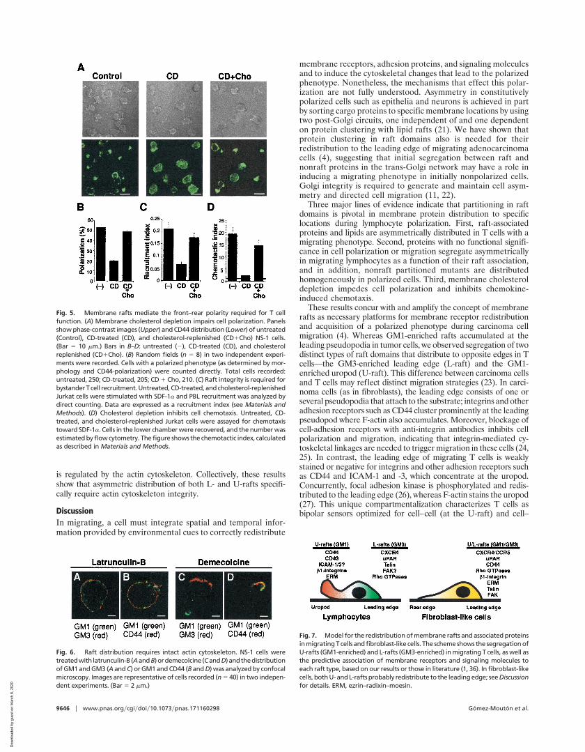
Fig. 7. Model for the redistribution of membrane rafts and associated proteinsin migrating T cells and fibroblast-like cells. The scheme shows the segregation ofU-rafts (GM1-enriched) and L-rafts (GM3-enriched) in migrating T cells, as well asthe predictive association of membrane receptors and signaling molecules toeach raft type, based on our results or those in literature (1, 36). In fibroblast-likecells, both U- and L-rafts probably redistribute to the leading edge; see Discussionfor details. ERM, ezrin–radixin–moesin.
Fig. 5. Membrane rafts mediate the front–rear polarity required for T cellfunction. (A) Membrane cholesterol depletion impairs cell polarization. Panelsshow phase-contrast images (Upper) and CD44 distribution (Lower) of untreated(Control), CD-treated (CD), and cholesterol-replenished (CD1Cho) NS-1 cells.(Bar 5 10 mm.) Bars in B–D: untreated (2), CD-treated (CD), and cholesterolreplenished (CD1Cho). (B) Random fields (n 5 8) in two independent experi-ments were recorded. Cells with a polarized phenotype (as determined by mor-phology and CD44-polarization) were counted directly. Total cells recorded:untreated, 250; CD-treated, 205; CD 1 Cho, 210. (C) Raft integrity is required forbystander T cell recruitment. Untreated, CD-treated, and cholesterol-replenishedJurkat cells were stimulated with SDF-1a and PBL recruitment was analyzed bydirect counting. Data are expressed as a recruitment index (see Materials andMethods). (D) Cholesterol depletion inhibits cell chemotaxis. Untreated, CD-treated, and cholesterol-replenished Jurkat cells were assayed for chemotaxistoward SDF-1a. Cells in the lower chamber were recovered, and the number wasestimated by flow cytometry. The figure shows the chemotactic index, calculatedas described in Materials and Methods.
Fig. 6. Raft distribution requires intact actin cytoskeleton. NS-1 cells weretreatedwith latrunculin-B (AandB)ordemecolcine (CandD)andthedistributionof GM1 and GM3 (A and C) or GM1 and CD44 (B and D) was analyzed by confocalmicroscopy. Images are representative of cells recorded (n 5 40) in two indepen-dent experiments. (Bar 5 2 mm.)
9646 u www.pnas.orgycgiydoiy10.1073ypnas.171160298 Gomez-Mouton et al.
Dow
nloa
ded
by g
uest
on
Mar
ch 8
, 202
0

matrix (at the L-raft) interactions (28). Thus, the T cell uropodmorphologically and functionally resembles the leading pseudo-podia of fibroblast-like cells including GM1-enriched raft accumu-lation at this cell pole. Accordingly, ezrin–radixin–moesin proteinsthat link F-actin to the cytoplasmic tails of adhesion moleculespartition in GM1-enriched rafts (29) and redistribute to the leadingedge in fibroblasts (30) but redistribute to the uropod in lympho-cytes (31).
There is also correspondence between leading-edge-redistrib-uted molecules in fibroblast-like cells and T cells. Raft-associatedproteins such as chemokine receptors and uPAR redistribute to theleading edge in migrating fibroblast-like cells (4, 32) and lympho-cytes (1, 33). Different raft platforms are used—GM1 rafts infibroblast-like cells and GM3 rafts in T cells. Two distinguishableGM3- and GM1-enriched raft subfractions also are reported innonlymphoid cells (34, 35): the GM3 rafts are enriched in specificproteins such as FAK, Src family kinases, and the small RhoGTPases (36), all of which concentrate at the leading edge ofmigrating T cells (1). GM1- and GM3-enriched rafts may redis-tribute to the same cell pole in fibroblast-like cells (37). Known L-and U-raft markers are outlined in Fig. 7.
Asymmetric protein redistribution during polarization dependson raft association rather than on their functional significance in cellmotility or signaling. A prominent role is reported for cytoskeletalinteractions in CD43, CD44, and ICAM-3 redistribution to theuropod (1). VSVG3-GFP and HA are unlikely to associate withcytoskeleton or interact with other T cell membrane proteins; thus,their asymmetric redistribution may be caused by the coalescenceof laterally diffusing rafts in specific cell locations such as theuropod. These results suggest that membrane receptor partitioningto rafts may occur upstream of their linkage to cytoskeleton andfunction as a mechanism for uropod redistribution. Nonetheless, anintact actin (but not tubulin) cytoskeleton is needed for L- and
U-raft redistribution, concurring with other reports (38). Becauseezrin–radixin–moesin proteins partition to rafts, raft association ofCD43, CD44, and other adhesion receptors may favor their linkageto the cytoskeleton for correct redistribution.
The physiological implications of raft coalescence for acquisitionof front–rear polarity and cell chemotaxis are evident, as it mayprovide a mechanism for the integration of distinct signalingpathways. That chemoattractant receptors partition in these do-mains suggests that asymmetrical raft redistribution amplifies che-motactic signal detection. The amplification may occur at thereceptor level or downstream through local activation of chemo-tactic intermediates such as Rho GTPases and generation ofphosphatidylinositol 4,5-bisphosphate, found in raft domains and atthe leading edge of neutrophils (39). There is functional evidencethat raft clustering may activate local signal transduction; indeed,compartmentalization of cell receptors and signaling moleculesbetween raft and nonraft membrane domains is required forefficient antigen-mediated T cell activation (40).
In addition to the essential role of rafts as organizational prin-ciples in T cell receptor signaling and immunological synapsestabilization, we show that membrane rafts may also be a mecha-nism for local activation of signaling pathways involved in the cellasymmetries required for T cell migration.
We thank Drs. P. Labrador and J. Stein for critical reading of themanuscript, Drs. P. Keller and P. Scheiffele for gifts of VSVG and HAconstructs, respectively, and for valuable discussion, and C. Mark foreditorial assistance. This work was supported by grants from the SpanishSecretarıa de Estado de Polıtica Cientıfica y TecnologicayEuropean Union,the Comunidad Autonoma de Madrid, and the Pharmacia Corporation.The Department of Immunology and Oncology was founded and issupported by the Spanish National Research Council and the PharmaciaCorporation.
1. Sanchez-Madrid, F. & del Pozo, M. (1999) EMBO J. 18, 501–511.2. Manes, S., Mira, E., Gomez-Mouton, C., Lacalle, R. & Martınez-A., C. (2000)
IUBMB Life 49, 89–96.3. Seveau, S., Keller, H., Maxfield, F., Piller, F. & Halbwachs-Mecarelli, L. (2000)
Blood 95, 2462–2470.4. Manes, S., Mira, E., Gomez-Mouton, C., Lacalle, R., Keller, P., Labrador, J.
& Martinez-A., C. (1999) EMBO J. 18, 6211–6220.5. Harder, T. & Simons, K. (1999) Eur. J. Immunol. 29, 556–562.6. Moran, M. & Miceli, M. (1998) Immunity 9, 787–796.7. Rozelle, A., Machesky, L., Yamamoto, M., Driessens, M., Insall, R., Roth, M.,
Luby-Phelps, K., Marriott, G., Hall, A. & Yin, H. (2000) Curr. Biol. 10, 311–320.8. Scheiffele, P., Roth, M. & Simons, K. (1997) EMBO J. 16, 5501–5508.9. Toomre, D., Keller, P., White, J., Olivo, J. C. & Simons, K. (1999) J. Cell Sci.
112, 21–33.10. Keller, P., Toomre, D., Dıaz, E., White, J. & Simons, K. (2001) Nat. Cell Biol.
3, 140–149.11. Mira, E., Lacalle, R., Gonzalez, M., Gomez-Mouton, C., Abad, J., Bernad, A.,
Martınez-A., C. & Manes, S. (2001) EMBO Rep. 2, 151–156.12. Campanero, M., Sanchez-Mateos, P., del Pozo, M. & Sanchez-Madrid, F.
(1994) J. Cell Biol. 127, 867–878.13. Pelletier, A., van der Laan, L., Hildbrand, P., Siani, M., Thompson, D., Dawson,
P., Torbett, B. & Salomon, D. (2000) Blood 96, 2682–2690.14. Hug, P., Lin, H., Korte, T., Xiao, X., Dimitrov, D., Wang, J., Puri, A. &
Blumenthal, R. (2000) J. Virol. 74, 6377–6385.15. Mellado, M., Rodrıguez-Frade, J. M., Manes, S. & Martınez-A., C. (2001)
Annu. Rev. Immunol. 19, 397–421.16. Kniep, B., Cinek, T., Angelisova, P. & Horejsi, V. (1994) Biochem. Biophys. Res.
Commun. 203, 1069–1075.17. van der Goot, F. G. & Harder, T. (2001) Semin. Immunol. 13, 89–97.18. Roper, K., Corbeil, D. & Huttner, W. (2000) Nat. Cell Biol. 2, 582–592.19. Harder, T., Scheiffele, P., Verkade, P. & Simons, K. (1998) J. Cell Biol. 141,
929–942.20. Simons, M., Keller, P., De Strooper, B., Beyreuther, K., Dotti, C. & Simons,
K. (1998) Proc. Natl. Acad. Sci. USA 95, 6460–6464.21. Simons, K. & Toomre, D. (2000) Nat. Rev. Mol. Cell Biol. 1, 31–39.
22. Bershadsky, A. & Futerman, A. (1994) Proc. Natl. Acad. Sci. USA 91, 5686–5689.23. Friedl, P., Brocker, E. & Zanker, K. (1998) Cell Adhes. Commun. 6, 225–236.24. Huttenlocher, A., Ginsberg, M. & Horwitz, A. (1996) J. Cell Biol. 134,
1551–1562.25. Maaser, K., Wolf, K., Klein, C., Niggemann, B., Zanker, K., Brocker, E. &
Friedl, P. (1999) Mol. Biol. Cell 10, 3067–3079.26. Entschladen, F., Niggemann, B., Zanker, K. & Friedl, P. (1997) J. Immunol.
159, 3203–3210.27. Sullivan, J. & Mandell, G. (1983) Cell Motil. 3, 31–46.28. Serrador, J., Nieto, M. & Sanchez-Madrid, F. (1999) Trends Cell Biol. 9,
228–232.29. Michaely, P., Mineo, C., Ying, Y. & Anderson, R. (1999) J. Biol. Chem. 274,
21430–21436.30. Amieva, M. & Furthmayr, H. (1995) Exp. Cell Res. 219, 180–196.31. Serrador, J., Alonso-Lebrero, J., del Pozo, M., Furthmayr, H., Schwartz-Albiez,
R., Calvo, J., Lozano, F. & Sanchez-Madrid, F. (1997) J. Cell Biol. 138,1409–1423.
32. Manes, S., del Real, G., Lacalle, R., Lucas, P., Gomez-Mouton, C., Sanchez-Palomino, S., Delgado, R., Alcamı, J., Mira, E. & Martınez-A., C. (2000)EMBO Rep. 1, 190–196.
33. Estreicher, A., Muhlhauser, J., Carpentier, J., Orci, L. & Vassalli, J. (1990)J. Cell Biol. 111, 783–792.
34. Iwabuchi, K., Handa, K. & Hakomori, S. (1998) J. Biol. Chem. 273, 33766–33773.
35. Chigorno, V., Palestini, P., Sciannamblo, M., Dolo, V., Pavan, A., Tettamanti,G. & Sonnino, S. (2000) Eur. J. Biochem. 267, 4187–4197.
36. Yamamura, S., Handa, K. & Hakomori, S. (1997) Biochem. Biophys. Res.Commun. 236, 218–222.
37. Manes, S., Lacalle, R., Gomez-Mouton, C., del Real, G., Mira, E. & Mar-tınez-A., C. (2001) Semin. Immunol. 13, 143–157.
38. Foger, N., Marhaba, R. & Zoller, M. (2001) J. Cell Sci. 114, 1169–1178.39. Servant, G., Weiner, O. D., Herzmark, P., Balla, T., Sedat, J. W. & Bourne,
H. R. (2000) Science 287, 1037–1040.40. Janes, P., Ley, S., Magee, A. & Kabouridis, P. (2000) Semin. Immunol. 12,
23–34.
Gomez-Mouton et al. PNAS u August 14, 2001 u vol. 98 u no. 17 u 9647
CELL
BIO
LOG
Y
Dow
nloa
ded
by g
uest
on
Mar
ch 8
, 202
0