seokha yoo, youngwon kim, sun-kyung park, sang-hwan ji
TRANSCRIPT
KS
RA
INTRODUCTION
Recently, the use of ultrasonography has become popu-
lar in operating rooms. The lumbar neuraxial block was
traditionally performed using a surface landmark-guided
technique. However, ultrasound (US)-guided technique
has been more frequently used for neuraxial block. This ar-
ticle reviews the sonoanatomy of the lumber spine,
US-guided techniques for neuraxial block, and current evi-
dence for the clinical usefulness of US-guided lumbar
neuraxial block.
GROSS ANATOMY OF THE LUMBAR VERTEBRAE
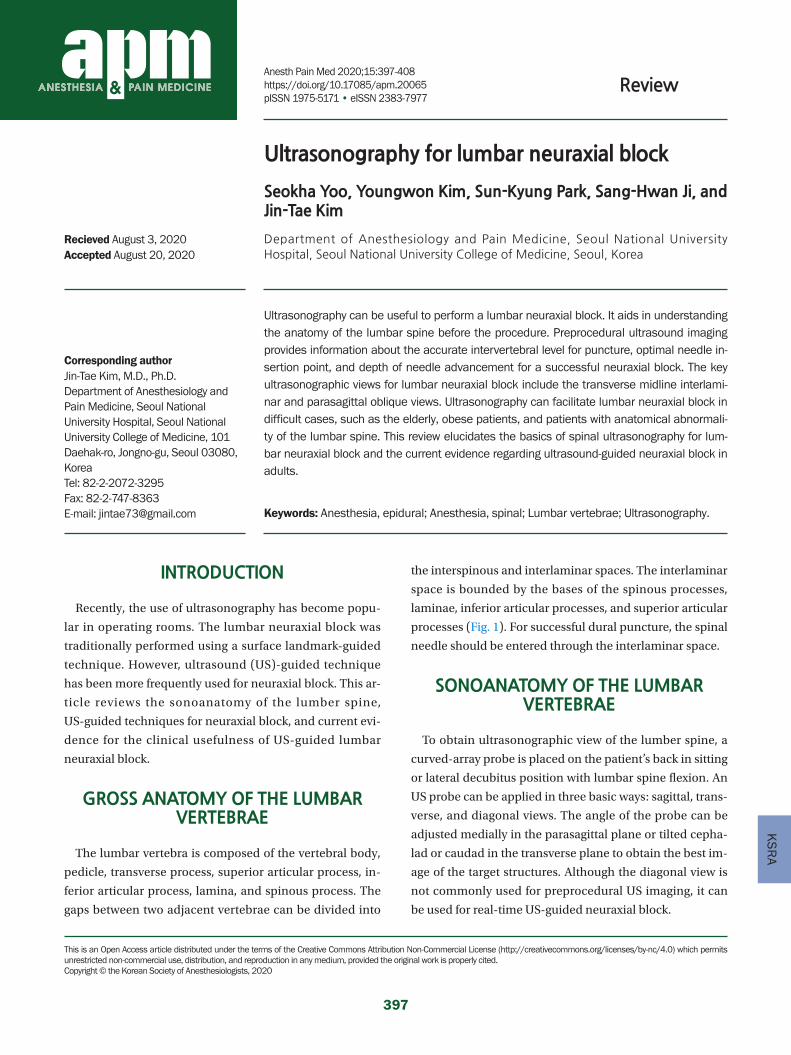
The lumbar vertebra is composed of the vertebral body,
pedicle, transverse process, superior articular process, in-
ferior articular process, lamina, and spinous process. The
gaps between two adjacent vertebrae can be divided into
Ultrasonography for lumbar neuraxial block
Seokha Yoo, Youngwon Kim, Sun-Kyung Park, Sang-Hwan Ji, and Jin-Tae Kim
Department of Anesthesiology and Pain Medicine, Seoul National University
Hospital, Seoul National University College of Medicine, Seoul, KoreaRecieved August 3, 2020Accepted August 20, 2020
Corresponding author Jin-Tae Kim, M.D., Ph.D. Department of Anesthesiology and Pain Medicine, Seoul National University Hospital, Seoul National University College of Medicine, 101 Daehak-ro, Jongno-gu, Seoul 03080, Korea Tel: 82-2-2072-3295Fax: 82-2-747-8363E-mail: [email protected]
Ultrasonography can be useful to perform a lumbar neuraxial block. It aids in understanding the anatomy of the lumbar spine before the procedure. Preprocedural ultrasound imaging provides information about the accurate intervertebral level for puncture, optimal needle in-sertion point, and depth of needle advancement for a successful neuraxial block. The key ultrasonographic views for lumbar neuraxial block include the transverse midline interlami-nar and parasagittal oblique views. Ultrasonography can facilitate lumbar neuraxial block in difficult cases, such as the elderly, obese patients, and patients with anatomical abnormali-ty of the lumbar spine. This review elucidates the basics of spinal ultrasonography for lum-bar neuraxial block and the current evidence regarding ultrasound-guided neuraxial block in adults.
Keywords: Anesthesia, epidural; Anesthesia, spinal; Lumbar vertebrae; Ultrasonography.
ReviewAnesth Pain Med 2020;15:397-408https://doi.org/10.17085/apm.20065pISSN 1975-5171 • eISSN 2383-7977
the interspinous and interlaminar spaces. The interlaminar
space is bounded by the bases of the spinous processes,
laminae, inferior articular processes, and superior articular
processes (Fig. 1). For successful dural puncture, the spinal
needle should be entered through the interlaminar space.
SONOANATOMY OF THE LUMBAR VERTEBRAE
To obtain ultrasonographic view of the lumber spine, a
curved-array probe is placed on the patient’s back in sitting
or lateral decubitus position with lumbar spine flexion. An
US probe can be applied in three basic ways: sagittal, trans-
verse, and diagonal views. The angle of the probe can be
adjusted medially in the parasagittal plane or tilted cepha-
lad or caudad in the transverse plane to obtain the best im-
age of the target structures. Although the diagonal view is
not commonly used for preprocedural US imaging, it can
be used for real-time US-guided neuraxial block.
This is an Open Access article distributed under the terms of the Creative Commons Attribution Non-Commercial License (http://creativecommons.org/licenses/by-nc/4.0) which permits unrestricted non-commercial use, distribution, and reproduction in any medium, provided the original work is properly cited.Copyright © the Korean Society of Anesthesiologists, 2020
397
SAGITTAL VIEWS OF THE LUMBAR SPINE
There are five basic sagittal plane views of the lumbar
spine according to the probe location and direction. By mov-
ing the probe from a lateral position to the midline of the
neuraxis, sagittal transverse process, sagittal articular pro-
cess, sagittal lamina, and sagittal spinous process views can
be obtained (Fig. 2A–D). From the probe position having the
sagittal articular process view or sagittal lamina view, the
parasagittal oblique view can be obtained by tilting the
probe medially towards the midline (Fig. 2E). The parasagit-
tal oblique view can be used for the determination of opti-
mal intervertebral level for puncture by identifying the inter-
vertebral level at which the posterior complex (ligamentum
flavum–dura complex) and the anterior complex (the poste-
rior longitudinal ligament, posterior surface of the vertebral
body, and intervertebral disc) are visualized most clearly. It
is also useful to select the intervertebral level at which the
interlaminar height is the largest.
TRANSVERSE VIEWS OF THE LUMBAR SPINE
There are two basic transverse views for lumbar neuraxi-
al block: transverse spinous process view and transverse
interlaminar view. The transverse spinous process view is
used to determine the midline composed of connecting
spinous process tips (Fig. 3A). The transverse interlaminar
view can be obtained by sliding the probe in a cephalad or
caudad direction from the transverse spinous process view
(Fig. 3B). Slight cephalad or caudad tilt in the transverse
interlaminar view may be needed to obtain the image
showing the dural sac located between the anterior and
posterior complexes (Fig. 3C).
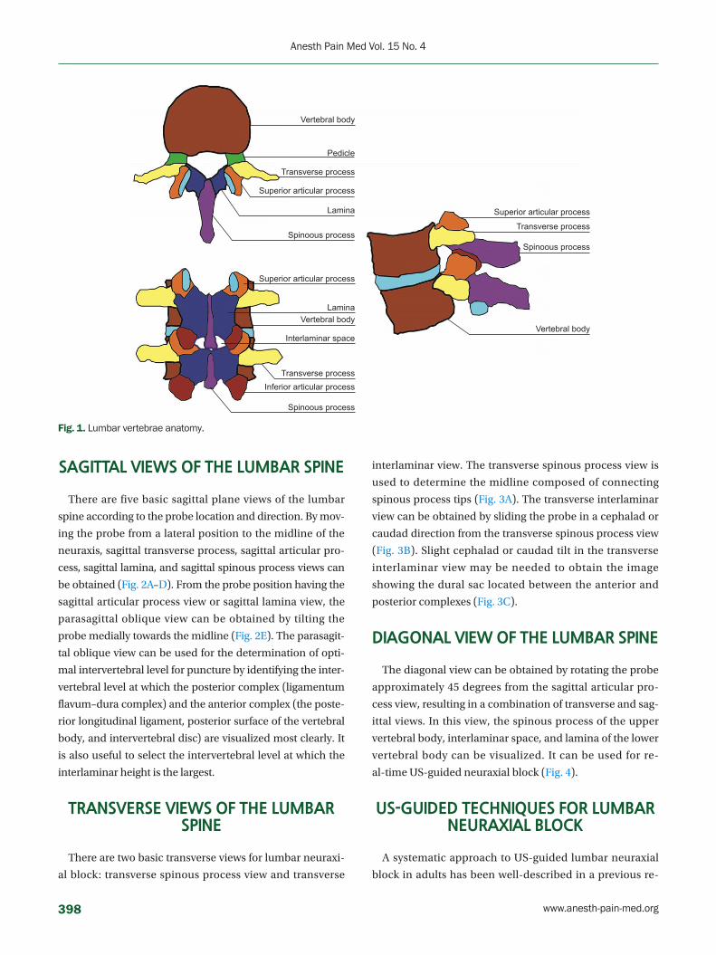
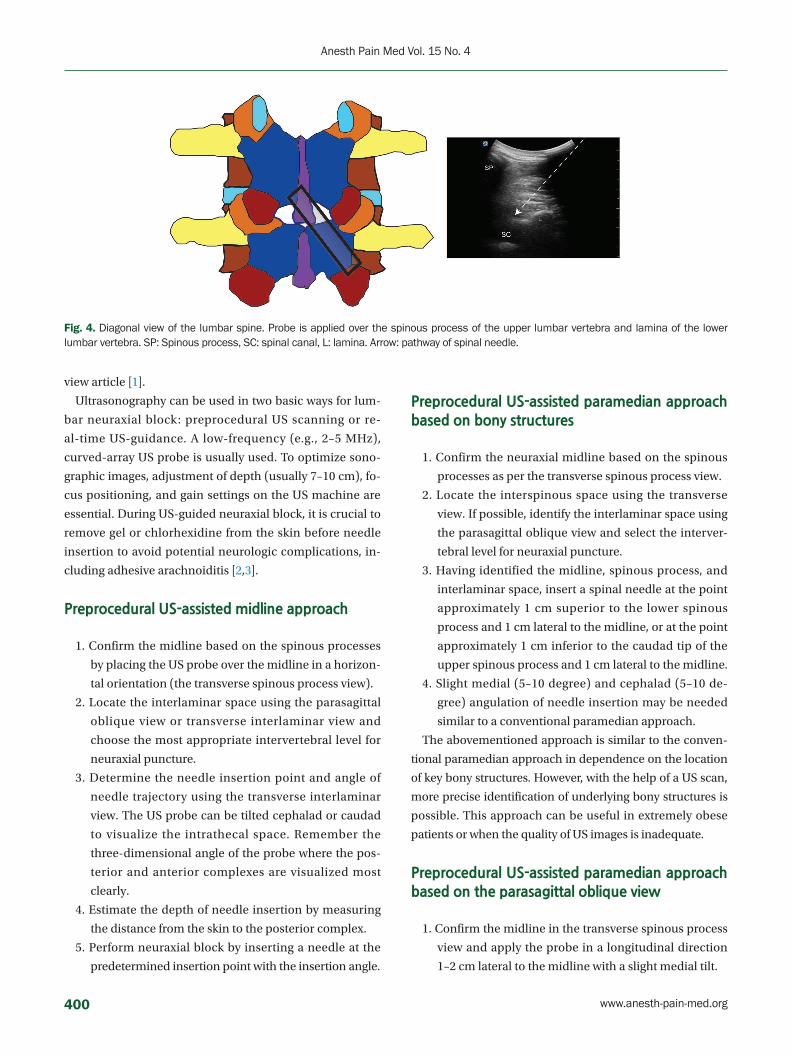
DIAGONAL VIEW OF THE LUMBAR SPINE
The diagonal view can be obtained by rotating the probe
approximately 45 degrees from the sagittal articular pro-
cess view, resulting in a combination of transverse and sag-
ittal views. In this view, the spinous process of the upper
vertebral body, interlaminar space, and lamina of the lower
vertebral body can be visualized. It can be used for re-
al-time US-guided neuraxial block (Fig. 4).
US-GUIDED TECHNIQUES FOR LUMBAR NEURAXIAL BLOCK
A systematic approach to US-guided lumbar neuraxial
block in adults has been well-described in a previous re-
Vertebral body
Pedicle
Transverse process
Superior articular process
Superior articular process
Transverse process
Spinoous process
Vertebral body
Lamina
Spinoous process
Superior articular process
LaminaVertebral body
Interlaminar space
Transverse processInferior articular process
Spinoous process
Fig. 1. Lumbar vertebrae anatomy.
398 www.anesth-pain-med.org
Anesth Pain Med Vol. 15 No. 4

KS
RA
B
D
A
C
E
A B C D E
Fig. 2. Sagittal views of the lumbar spine. (A) Sagittal transverse process view, (B) sagittal articular process view, (C) sagittal lamina view, (D) sagittal spinous process view, (E) parasagittal oblique view. TP: transverse process, AP: articular process, L: lamina, SP: spinous process, PC: posterior complex, AC: anterior complex, SC: spinal canal (intrathecal space).
B
A
C
A
B
C
Fig. 3. Transverse views of the lumbar spine. (A) Transverse spinous process view, (B) transverse interspinous process view, (C) tilted transverse interspinous process view. SP: spinous process, AP: articular process, L: lamina, PC: posterior complex, AC: anterior complex, SC: spinal canal (intrathecal space).
www.anesth-pain-med.org 399
Ultrasound and lumbar neuraxial block
view article [1].
Ultrasonography can be used in two basic ways for lum-
bar neuraxial block: preprocedural US scanning or re-
al-time US-guidance. A low-frequency (e.g., 2–5 MHz),
curved-array US probe is usually used. To optimize sono-
graphic images, adjustment of depth (usually 7–10 cm), fo-
cus positioning, and gain settings on the US machine are
essential. During US-guided neuraxial block, it is crucial to
remove gel or chlorhexidine from the skin before needle
insertion to avoid potential neurologic complications, in-
cluding adhesive arachnoiditis [2,3].
Preprocedural US-assisted midline approach
1. Confirm the midline based on the spinous processes
by placing the US probe over the midline in a horizon-
tal orientation (the transverse spinous process view).
2. Locate the interlaminar space using the parasagittal
oblique view or transverse interlaminar view and
choose the most appropriate intervertebral level for
neuraxial puncture.
3. Determine the needle insertion point and angle of
needle trajectory using the transverse interlaminar
view. The US probe can be tilted cephalad or caudad
to visualize the intrathecal space. Remember the
three-dimensional angle of the probe where the pos-
terior and anterior complexes are visualized most
clearly.
4. Estimate the depth of needle insertion by measuring
the distance from the skin to the posterior complex.
5. Perform neuraxial block by inserting a needle at the
predetermined insertion point with the insertion angle.
Preprocedural US-assisted paramedian approach based on bony structures
1. Confirm the neuraxial midline based on the spinous
processes as per the transverse spinous process view.
2. Locate the interspinous space using the transverse
view. If possible, identify the interlaminar space using
the parasagittal oblique view and select the interver-
tebral level for neuraxial puncture.
3. Having identified the midline, spinous process, and
interlaminar space, insert a spinal needle at the point
approximately 1 cm superior to the lower spinous
process and 1 cm lateral to the midline, or at the point
approximately 1 cm inferior to the caudad tip of the
upper spinous process and 1 cm lateral to the midline.
4. Slight medial (5–10 degree) and cephalad (5–10 de-
gree) angulation of needle insertion may be needed
similar to a conventional paramedian approach.
The abovementioned approach is similar to the conven-
tional paramedian approach in dependence on the location
of key bony structures. However, with the help of a US scan,
more precise identification of underlying bony structures is
possible. This approach can be useful in extremely obese
patients or when the quality of US images is inadequate.
Preprocedural US-assisted paramedian approach based on the parasagittal oblique view
1. Confirm the midline in the transverse spinous process
view and apply the probe in a longitudinal direction
1–2 cm lateral to the midline with a slight medial tilt.
Fig. 4. Diagonal view of the lumbar spine. Probe is applied over the spinous process of the upper lumbar vertebra and lamina of the lower lumbar vertebra. SP: Spinous process, SC: spinal canal, L: lamina. Arrow: pathway of spinal needle.
400 www.anesth-pain-med.org
Anesth Pain Med Vol. 15 No. 4
KS
RA
2. Identify the interlaminar space in the parasagittal
oblique view and select the intervertebral level that
provides the largest interlaminar space.
3. Determine the medial angle of the sagittal plane pro-
viding the clearest image of the interlaminar space.
Slight cephalad or caudad angulation of the probe
may be necessary in some cases.
4. Estimate the depth of needle insertion by measuring
the distance from the skin to the posterior complex.
5. Insert a needle at the designated insertion point with
the designated angle.
Paramedian approach based on the parasagittal oblique
view has potential advantages over the midline approach
using the transverse interlaminar view because the
parasagittal oblique view provides better visibility of the
interlaminar space than the transverse interlaminar view,
especially in the elderly. When the US beam reaches the
spinal canal in the parasagittal oblique view, the needle
can also reach the canal through the same pathway. When
using US-assisted paramedian approach, cephalad or cau-
dad needle angulation may not be required. This approach
can be the most direct way to the intrathecal or epidural
space through the interlaminar space considering only
medial angulation.
Real-time US-guided neuraxial block
Real-time US-guided neuraxial block is a feasible and
promising technique that can result in successful neuraxial
anesthesia in difficult cases [4,5]. However, it is tricky to
perform because of the large size of the probe, small gauge
of the needle, and relatively deep target structure. There
are several methods to perform real-time US-guided
neuraxial block, including sagittal, transverse, and diago-
nal in-plane approaches.
Real-time US-guided spinal anesthesia using in-plane
approach based on the parasagittal oblique view can in-
crease first-attempt success rate compared to the land-
mark-guided paramedian approach technique [6]. Needle
approach from the non-dependent side may lead to dry tap
due to gravity, even if the needle tip is placed in the intra-
thecal space. A prospective observational study showed
that real-time US-guided spinal anesthesia using in-plane
approach based on the diagonal view was successfully per-
formed in 97 out of 100 consecutive patients within three
median needle passes [7]. Probe application site can be
slightly moved to secure the room for puncture site and
needle manipulation during the transverse in-plane para-
median approach [8]. Electromagnetic needle tracking sys-
tem can also be used for real-time US-guided spinal anes-
thesia [9].
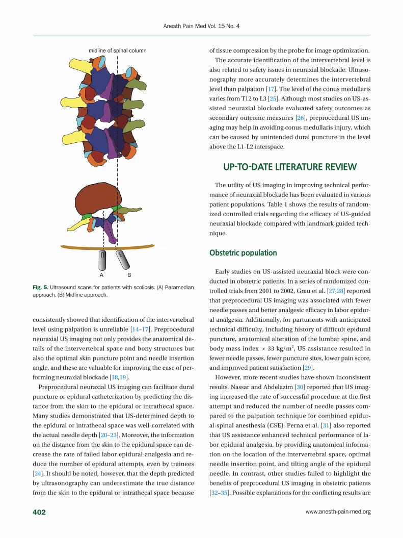
US-guided neuraxial block in patients with scoliosis
Preprocedural US assistance may have potential benefits
in neuraxial block for patients with scoliosis. Systematic al-
gorithms to guide neuraxial techniques in patients with
scoliosis have been described previously [10,11]. Several
earlier publications have demonstrated that the use of ul-
trasonography is useful for spinal anesthesia in patients
with scoliosis [5,12,13]. The lateral curvature of the scoliot-
ic spine can be confirmed by marking out all spinous pro-
cess tips using ultrasonography. Simple spinal radiographs
or computed tomography are also helpful. In addition to
the lateral curvature, rotational change of the vertebral
body should be considered when performing neuraxial
block in these patients. During the paramedian approach,
it is easier to insert a needle on the convex side of the ver-
tebral column after confirming the spinous process consid-
ering the needle insertion angle. For example, in the para-
median approach, if the rotation of the vertebral body is
approximately 15 degrees in a patient with scoliosis, the
needle insertion site is on the convex side of the spinous
processes, therefore, the angle of needle trajectory would
be perpendicular to the skin towards the interlaminar
space (Fig. 5A). On the other hand, when using the midline
approach through the interspinous space in a patient with
scoliosis, the angle of needle insertion would be 15 degrees
off the sagittal plane towards the convex side (Fig. 5B).
USEFULNESS OF US-GUIDED NEURAXIAL BLOCK
US imaging can provide important clinical information
for a successful neuraxial block. Ultrasonography aids in
identification of the accurate puncture level by providing
information, such as the widest inter-laminar space, depth
to the dura from the skin, and accurate spinal level.
To achieve successful neuraxial blockade, accurate iden-
tification of the intervertebral spaces is crucial. US imaging
is also useful in localizing the intervertebral spaces and
identifying lumbar vertebral level. Although many anesthe-
siologists used to identify the vertebral level by palpation
when performing neuraxial blockade, previous studies
www.anesth-pain-med.org 401
Ultrasound and lumbar neuraxial block
consistently showed that identification of the intervertebral
level using palpation is unreliable [14–17]. Preprocedural
neuraxial US imaging not only provides the anatomical de-
tails of the intervertebral space and bony structures but
also the optimal skin puncture point and needle insertion
angle, and these are valuable for improving the ease of per-
forming neuraxial blockade [18,19].
Preprocedural neuraxial US imaging can facilitate dural
puncture or epidural catheterization by predicting the dis-
tance from the skin to the epidural or intrathecal space.
Many studies demonstrated that US-determined depth to
the epidural or intrathecal space was well-correlated with
the actual needle depth [20–23]. Moreover, the information
on the distance from the skin to the epidural space can de-
crease the rate of failed labor epidural analgesia and re-
duce the number of epidural attempts, even by trainees
[24]. It should be noted, however, that the depth predicted
by ultrasonography can underestimate the true distance
from the skin to the epidural or intrathecal space because
of tissue compression by the probe for image optimization.
The accurate identification of the intervertebral level is
also related to safety issues in neuraxial blockade. Ultraso-
nography more accurately determines the intervertebral
level than palpation [17]. The level of the conus medullaris
varies from T12 to L3 [25]. Although most studies on US-as-
sisted neuraxial blockade evaluated safety outcomes as
secondary outcome measures [26], preprocedural US im-
aging may help in avoiding conus medullaris injury, which
can be caused by unintended dural puncture in the level
above the L1-L2 interspace.
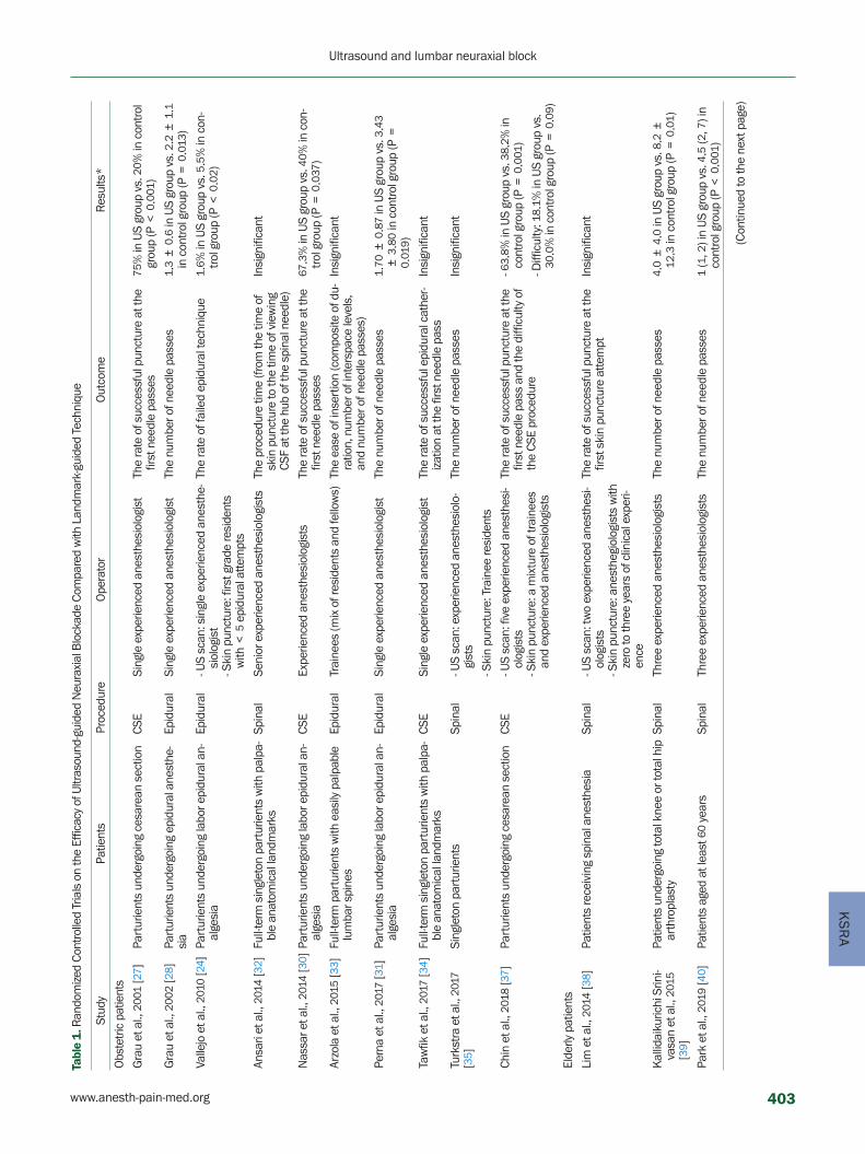
UP-TO-DATE LITERATURE REVIEW
The utility of US imaging in improving technical perfor-
mance of neuraxial blockade has been evaluated in various
patient populations. Table 1 shows the results of random-
ized controlled trials regarding the efficacy of US-guided
neuraxial blockade compared with landmark-guided tech-
nique.
Obstetric population
Early studies on US-assisted neuraxial block were con-
ducted in obstetric patients. In a series of randomized con-
trolled trials from 2001 to 2002, Grau et al. [27,28] reported
that preprocedural US imaging was associated with fewer
needle passes and better analgesic efficacy in labor epidur-
al analgesia. Additionally, for parturients with anticipated
technical difficulty, including history of difficult epidural
puncture, anatomical alteration of the lumbar spine, and
body mass index > 33 kg/m2, US assistance resulted in
fewer needle passes, fewer puncture sites, lower pain score,
and improved patient satisfaction [29].
However, more recent studies have shown inconsistent
results. Nassar and Abdelazim [30] reported that US imag-
ing increased the rate of successful procedure at the first
attempt and reduced the number of needle passes com-
pared to the palpation technique for combined epidur-
al-spinal anesthesia (CSE). Perna et al. [31] also reported
that US assistance enhanced technical performance of la-
bor epidural analgesia, by providing anatomical informa-
tion on the location of the intervertebral space, optimal
needle insertion point, and tilting angle of the epidural
needle. In contrast, other studies failed to highlight the
benefits of preprocedural US imaging in obstetric patients
[32–35]. Possible explanations for the conflicting results are
Fig. 5. Ultrasound scans for patients with scoliosis. (A) Paramedian approach. (B) Midline approach.
midline of spinal column
A B
402 www.anesth-pain-med.org
Anesth Pain Med Vol. 15 No. 4
KS
RA
Tabl
e 1
. Ran
dom
ized
Cont
rolle
d Tr
ials
on
the
Effic
acy
of U
ltras
ound
-gui
ded
Neu
raxi
al B
lock
ade
Com
pare
d w
ith L
andm
ark-
guid
ed T
echn
ique
Stud
yPa
tient
sPr
oced
ure
Ope
rato
rO
utco
me
Res
ults
*
Obs
tetr
ic p
atie
nts
Gra
u et
al.,
200
1 [2
7 ]Pa
rtur
ient
s un
derg
oing
ces
area
n se
ctio
nCS
ESi
ngle
exp
erie
nced
ane
sthe
siol
ogis
tTh
e ra
te o
f suc
cess
ful p
unct
ure
at th
e fir
st n
eedl
e pa
sses
75%
in U
S gr
oup
vs. 2
0 % in
con
trol
gr
oup
(P <
0.0
01)
Gra
u et
al.,
200
2 [2
8 ]Pa
rtur
ient
s un
derg
oing
epi
dura
l ane
sthe
-si
aEp
idur
alSi
ngle
exp
erie
nced
ane
sthe
siol
ogis
tTh
e nu
mbe
r of n
eedl
e pa
sses
1 .3
± 0
.6 in
US
grou
p vs
. 2.2
± 1
.1
in c
ontr
ol g
roup
(P =
0.0
13)
Valle
jo e
t al.,
201
0 [2
4 ]Pa
rtur
ient
s un
derg
oing
labo
r epi
dura
l an-
alge
sia
Epid
ural
- US
scan
: sin
gle
expe
rienc
ed a
nest
he-
siol
ogis
t- S
kin
punc
ture
: firs
t gra
de re
side
nts
with
< 5
epi
dura
l atte
mpt
s
The
rate
of f
aile
d ep
idur
al te
chni
que
1 .6 %
in U
S gr
oup
vs. 5
.5%
in c
on-
trol
gro
up (P
< 0
.02 )
Ansa
ri et
al.,
201
4 [3
2 ]Fu
ll-te
rm s
ingl
eton
par
turie
nts
with
pal
pa-
ble
anat
omic
al la
ndm
arks
Spin
alSe
nior
exp
erie
nced
ane
sthe
siol
ogis
tsTh
e pr
oced
ure
time
(from
the
time
of
skin
pun
ctur
e to
the
time
of v
iew
ing
CSF
at th
e hu
b of
the
spin
al n
eedl
e)
Insi
gnifi
cant
Nas
sar e
t al.,
201
4 [3
0 ]Pa
rtur
ient
s un
derg
oing
labo
r epi
dura
l an-
alge
sia
CSE
Expe
rienc
ed a
nest
hesi
olog
ists
The
rate
of s
ucce
ssfu
l pun
ctur
e at
the
first
nee
dle
pass
es67
.3%
in U
S gr
oup
vs. 4
0 % in
con
-tr
ol g
roup
(P =
0.0
37)
Arzo
la e
t al.,
201
5 [3
3 ]Fu
ll-te
rm p
artu
rient
s w
ith e
asily
pal
pabl
e lu
mba
r spi
nes
Epid
ural
Trai
nees
(mix
of r
esid
ents
and
fello
ws)
The
ease
of i
nser
tion
(com
posi
te o
f du-
ratio
n, n
umbe
r of i
nter
spac
e le
vels
, an
d nu
mbe
r of n
eedl
e pa
sses
)
Insi
gnifi
cant
Pern
a et
al.,
201
7 [3
1 ]Pa
rtur
ient
s un
derg
oing
labo
r epi
dura
l an-
alge
sia
Epid
ural
Sing
le e
xper
ienc
ed a
nest
hesi
olog
ist
The
num
ber o
f nee
dle
pass
es1 .
70 ±
0.8
7 in
US
grou
p vs
. 3.4
3 ±
3.8
0 in
con
trol
gro
up (P
=
0 .01
9 )
Taw
fik e
t al.,
201
7 [3
4 ]Fu
ll-te
rm s
ingl
eton
par
turie
nts
with
pal
pa-
ble
anat
omic
al la
ndm
arks
CSE
Sing
le e
xper
ienc
ed a
nest
hesi
olog
ist
The
rate
of s
ucce
ssfu
l epi
dura
l cat
her-
izat
ion
at th
e fir
st n
eedl
e pa
ssIn
sign
ifica
nt
Turk
stra
et a
l., 2
017
[35 ]
Sing
leto
n pa
rtur
ient
sSp
inal
- US
scan
: exp
erie
nced
ane
sthe
siol
o-gi
sts
The
num
ber o
f nee
dle
pass
esIn
sign
ifica
nt
- Ski
n pu
nctu
re: T
rain
ee re
side
nts
Chin
et a
l., 2
018
[37 ]
Part
urie
nts
unde
rgoi
ng c
esar
ean
sect
ion
CSE
- US
scan
: fiv
e ex
perie
nced
ane
sthe
si-
olog
ists
- Ski
n pu
nctu
re: a
mix
ture
of t
rain
ees
and
expe
rienc
ed a
nest
hesi
olog
ists
The
rate
of s
ucce
ssfu
l pun
ctur
e at
the
first
nee
dle
pass
and
the
diffi
culty
of
the
CSE
proc
edur
e
- 63 .
8 % in
US
grou
p vs
. 38 .
2 % in
co
ntro
l gro
up (P
= 0
.001
)
- Diff
icul
ty: 1
8 .1 %
in U
S gr
oup
vs.
30.0
% in
con
trol
gro
up (P
= 0
.09 )
Elde
rly p
atie
nts
Lim
et a
l., 2
014
[38 ]
Patie
nts
rece
ivin
g sp
inal
ane
sthe
sia
Spin
al- U
S sc
an: t
wo
expe
rienc
ed a
nest
hesi
-ol
ogis
ts- S
kin
punc
ture
: ane
sthe
giol
ogis
ts w
ith
zero
to th
ree
year
s of
clin
ical
exp
eri-
ence
The
rate
of s
ucce
ssfu
l pun
ctur
e at
the
first
ski
n pu
nctu
re a
ttem
ptIn
sign
ifica
nt
Kal
lidai
kuric
hi S
rini-
vasa
n et
al.,
201
5 [3
9 ]
Patie
nts
unde
rgoi
ng to
tal k
nee
or to
tal h
ip
arth
ropl
asty
Spin
alTh
ree
expe
rienc
ed a
nest
hesi
olog
ists
The
num
ber o
f nee
dle
pass
es4 .
0 ±
4.0
in U
S gr
oup
vs. 8
.2 ±
12
.3 in
con
trol
gro
up (P
= 0
.01 )
Park
et a
l., 2
019
[40 ]
Patie
nts
aged
at l
east
60
year
sSp
inal
Thre
e ex
perie
nced
ane
sthe
siol
ogis
tsTh
e nu
mbe
r of n
eedl
e pa
sses
1 (1
, 2) i
n U
S gr
oup
vs. 4
.5 (2
, 7) i
n co
ntro
l gro
up (P
< 0
.001
)
(Con
tinue
d to
the
next
pag
e)
www.anesth-pain-med.org 403
Ultrasound and lumbar neuraxial block
Patie
nts
with
diff
icul
t sp
inal
ana
tom
y
Gra
u et
al.,
200
1 [2
9 ]Pa
rtur
ient
s w
ith p
resu
med
diff
icul
t pun
c-tu
re (h
isto
ry o
f diff
icul
t epi
dura
l ane
sthe
-is
a; a
nato
mic
al a
ltera
tion
of th
e lu
mba
r sp
ine;
BM
I >
33
kg/m
2 )
Epid
ural
Sing
le e
xper
ienc
ed a
nest
hesi
olog
ist
The
num
ber o
f nee
dle
pass
es1 .
5 ±
0.9
in U
S gr
oup
vs. 2
.6 ±
1.4
in
con
trol
gro
up (P
< 0
.001
)
Chin
et a
l., 2
011
[12 ]
Patie
nts
who
had
diff
icul
t sur
face
ana
-to
mic
land
mar
ks (p
oorly
pal
pabl
e or
im-
palp
able
spi
nous
pro
cess
es a
nd B
MI
>
35 k
g/m
2 ; mod
erat
e to
sev
ere
lum
bar
scol
iosi
s; p
revi
ous
lum
bar s
pina
l sur
-ge
ry)
Spin
alTw
o ex
perie
nced
ane
sthe
siol
ogis
tsTh
e ra
te o
f suc
cess
ful p
unct
ure
at th
e fir
st s
kin
punc
ture
atte
mpt
65%
in U
S gr
oup
vs. 3
2 % in
con
trol
gr
oup
(P <
0.0
01)
Wan
g et
al.,
201
2 [4
3 ]Si
ngle
ton
part
urie
nts
with
BM
I ≥
30
kg/
m2
CSE
Sing
le e
xper
ienc
ed a
nest
hesi
olog
ist
The
rate
of s
ucce
ssfu
l pun
ctur
e at
the
first
ski
n pu
nctu
re a
ttem
pt10
0 % in
US
grou
p vs
. 70 %
in c
on-
trol
gro
up (P
= 0
.004
)
Ekin
ci e
t al.,
201
7 [4
4 ]Si
ngle
ton
part
urie
nts
with
impa
lpab
le
lum
bar s
pino
us p
roce
sses
Spin
alTw
o ex
perie
nced
ane
sthe
siol
ogis
tsTh
e nu
mbe
r of s
kin
punc
ture
s an
d th
e pr
oced
ure
time
- Num
ber o
f ski
n pu
nctu
res:
1.1
9 ±
0.4
7 in
US
grou
p vs
. 1.8
4 ±
0 .
85 in
con
trol
gro
up (P
< 0
.001
)
- Pro
cedu
re ti
me:
242
.34
± 6
3 .17
in
US
grou
p vs
. 204
.59
± 1
13.2
1 in
con
trol
gro
up (P
= 0
.105
)
Park
et a
l., 2
020
[13 ]
Patie
nts
who
had
lum
bar s
colio
sis
or h
is-
tory
of l
umba
r spi
ne s
urge
ry in
volv
ing
L2-
L5 v
erte
brae
Spin
alTh
ree
expe
rienc
ed a
nest
hesi
olog
ists
The
num
ber o
f nee
dle
pass
es1 .
5 (1
, 3) i
n U
S gr
oup
vs. 6
(2, 9
.3)
in c
ontr
ol g
roup
(P <
0.0
01)
Rea
l-tim
e U
S-gu
ided
te
chni
que
Gra
u et
al.,
200
4 [4
5 ]Pa
rtur
ient
s un
derg
oing
ces
area
n se
ctio
nCS
ESi
ngle
exp
erie
nced
ane
sthe
siol
ogis
tTh
e nu
mbe
r of n
eedl
e pa
sses
Sign
ifica
nt re
duct
ion
of n
eedl
e pa
sses
in re
al-ti
me
US
grou
p an
d pr
e-pr
oced
ural
US
grou
p, c
om-
pare
d to
con
trol
gro
up
Chon
g et
al.,
201
7 [6
]Pa
tient
s un
derg
oing
low
er li
mb
surg
erie
sSp
inal
Not
app
licab
leTh
e ra
te o
f suc
cess
ful p
unct
ure
at th
e fir
st s
kin
punc
ture
atte
mpt
87%
in re
al-ti
me
grou
p vs
. 43 %
in
palp
atio
n gr
oup
Elsh
arka
wy
et a
l., 2
017
[46 ]
Patie
nts
unde
rgoi
ng to
tal k
nee
or to
tal h
ip
arth
ropl
asty
with
diff
icul
t spi
nal a
nato
my
(age
≥ 5
5 ; B
MI
> 3
0 kg
/m2 ; s
colio
sis
with
30 -
degr
ee c
urva
ture
; im
palp
able
sp
inou
s pr
oces
ses)
Spin
alFi
ve e
xper
ienc
ed a
nest
hesi
olog
ists
The
num
ber o
f ski
n pu
nctu
res
Insi
gnifi
cant
BM
I: bo
dy m
ass
inde
x, C
SE: c
ombi
ned
spin
al-e
pidu
ral a
nest
hesi
a, U
S: u
ltras
ound
, CSF
: cer
ebro
spin
al fl
uid.
*Va
lues
are
pre
sent
ed a
s m
ean
± SD
or m
edia
n (1
Q, 3
Q).
Stud
yPa
tient
sPr
oced
ure
Ope
rato
rO
utco
me
Res
ults
*
Tabl
e 1
. Con
tinue
d
404 www.anesth-pain-med.org
Anesth Pain Med Vol. 15 No. 4
KS
RA
the characteristics of the study subjects and proceduralists.
All these studies evaluated the utility of ultrasonography in
parturients with palpable anatomical landmarks. In this
population, the benefit of US imaging may be underesti-
mated because neuraxial blockade is usually not compli-
cated in lean patients or those who had normal vertebral
anatomy. Regarding the proceduralists, experienced anes-
thesiologists performed the US scan and neuraxial block-
ade in two studies [32,34], while skin puncture was per-
formed by trainees after ultrasonographic examination by
experts in another study [35]. The guidance from a study
investigator during skin puncture or suboptimal needle
handling by the trainees may have led to the negative re-
sults [36]. However, in a recent large study conducted in
women undergoing cesarean section with CSE, the authors
found that US assistance improved technical performance
in patients with easily palpable landmarks, but not in those
with impalpable surface landmarks, and that the experi-
ence of proceduralists did not influence the first-pass suc-
cess rate of CSE procedure [37]. Further studies are still
needed to clarify which populations benefit the most
through US assistance.
Elderly patients
The efficacy of US-assisted neuraxial blockade is more
evident in elderly patients. In contrast to using the midline
approach in obstetric patients, the paramedian approach
was used in studies evaluating the utility of ultrasonogra-
phy in the elderly. Lim et al. [38] compared the rate of suc-
cessful dural puncture at the first attempt in patients re-
ceiving spinal anesthesia with or without preprocedural US
imaging. Although the first-attempt success rate was not
significantly different, shorter time was required to per-
form the procedure with US-assisted spinal anesthesia and
patients were more satisfied compared to the manual pal-
pation technique. Other studies showed consistent results
that the number of needle passes and skin punctures were
significantly decreased when using US-assisted spinal an-
esthesia, compared to the midline approach [39] or para-
median approach [40]. In general, neuraxial blockade is
more difficult in an older population than in relatively
younger obstetric patients, possibly due to degenerative
changes of the lumbar spine, such as the calcified interspi-
nous ligament and limited lumbar flexion [39]. These find-
ings supported that preprocedural US imaging may be
more beneficial in patients with difficult anatomy, as
shown in a recent meta-analysis [41]. Scanning both sides
and all spinal levels before selecting a puncture site for
US-guided spinal anesthesia is recommended. The L5-S1
intervertebral level is a good option for neuraxial anesthe-
sia in the elderly [42].
Patients with difficult anatomy (obesity, scoliosis, or history of spine surgery)
Several studies have evaluated whether US assistance
improves technical performance of neuraxial blockade in
patients with difficult anatomy, including moderate to se-
vere obesity, lumbar scoliosis, ankylosing spondylitis, or
history of lumbar spine surgery. Chin et al. [12] compared
the first-attempt success rate of spinal anesthesia with or
without US assistance in this population and found that
preprocedural US imaging facilitates the performance of
spinal anesthesia. Similar results were shown in obstetric
patients with difficult anatomical landmarks. Wang et al.
[43] reported that US scanning performed by single experi-
enced anesthesiologist before neuraxial blockade signifi-
cantly enhanced the first-attempt success rate. Another
study published by Ekinci et al. [44] demonstrated that the
number of skin punctures was significantly decreased
when using preprocedural US imaging, but total procedure
time was comparable with the conventional spinal anes-
thesia technique. Our recent study conducted in patients
with documented lumbar scoliosis or those with history of
previous spinal surgery also showed similar results that the
number of needle passes and puncture attempts were sig-
nificantly lower in the US group than in the control group,
but total procedure time was not significantly different be-
tween the two groups [13]. Despite of US scanning time,
difficulties in identifying the midline or intervertebral
space in patients with abnormal vertebral anatomy would
increase the procedural time in conventional palpation
technique, resulting in no difference in the overall proce-
dure time. Considering the reduced number of needle ma-
nipulations and better patient satisfaction, US neuraxial
imaging should be accompanied in patients who are ex-
pected to have difficult neuraxial blockade.
Real-time US-guided technique
There are limited studies assessing the benefits of re-
al-time US guidance technique. Grau et al. [45] compared
real-time US-guided CSE procedure using the parasagittal
www.anesth-pain-med.org 405
Ultrasound and lumbar neuraxial block
oblique view with preprocedural US scanning and conven-
tional landmark palpation technique and found that both
US-guided techniques significantly reduced the number of
needle passes. The advantage of real-time US guidance
was also reported in a recent study by Chong et al. [6]. They
found that first-attempt success rate was significantly high-
er when using real-time US-guided spinal anesthesia with
the parasagittal oblique view, compared to the palpa-
tion-based paramedian approach [6]. However, another
study on the efficacy of real-time US-guided spinal anes-
thesia in patients with difficult spinal anatomy showed no
advantage of real-time technique over conventional land-
mark technique [46]. Various approaches, including trans-
verse [8,47] and diagonal in-plane approaches [7], have
been investigated for real-time US-guided neuraxial block.
Despite some results showing the advantages of real-time
US guidance, there are still technical challenges to be ad-
dressed, such as visualization of a small-gauge needle
around the deep target structures.
CONCLUSION
For better clinical practice, it is recommended to apply
US guidance for neuraxial blockade. US-guided neuraxial
block can facilitate successful access to the intrathecal or
epidural space in patients with difficult spinal anatomy, as
well as in those with easily palpable anatomical landmarks.
Anesthesiologists who routinely perform lumbar neuraxial
block should be familiar with the sonoanatomy of the lum-
bar vertebrae and US-guided techniques to improve tech-
nical performance and safety.
CONFLICTS OF INTEREST
No potential conflict of interest relevant to this article
was reported.
AUTHOR CONTRIBUTIONS
Conceptualization: Jin-Tae Kim. Data curation: Seokha
Yoo, Youngwon Kim, Sun-Kyung Park, Sang-Hwan Ji. Meth-
odology: Jin-Tae Kim. Project administration: Jin-Tae Kim.
Writing-original draft: Seokha Yoo, Jin-Tae Kim. Writing-re-
view & editing: Seokha Yoo, Youngwon Kim, Sun-Kyung
Park, Sang-Hwan Ji, Jin-Tae Kim. Investigation: Seokha Yoo,
Youngwon Kim, Sun-Kyung Park, Sang-Hwan Ji.
ORCID
Seokha Yoo, https://orcid.org/0000-0003-4679-6027
Youngwon Kim, https://orcid.org/0000-0002-1071-5494
Sun-Kyung Park, https://orcid.org/0000-0002-4670-253X
Sang-Hwan Ji, https://orcid.org/0000-0001-6736-4464
Jin-Tae Kim, https://orcid.org/0000-0002-3738-0081
REFERENCES
1. Chin KJ, Karmakar MK, Peng P. Ultrasonography of the adult
thoracic and lumbar spine for central neuraxial blockade. An-
esthesiology 2011; 114: 1459-85.
2. Pintaric TS, Hadzic A, Strbenc M, Podpecan O, Podbregar M,
Cvetko E. Inflammatory response after injection of aqueous gel
into subarachnoid space in piglets. Reg Anesth Pain Med 2013;
38: 100-5.
3. Killeen T, Kamat A, Walsh D, Parker A, Aliashkevich A. Severe
adhesive arachnoiditis resulting in progressive paraplegia fol-
lowing obstetric spinal anaesthesia: a case report and review.
Anaesthesia 2012; 67: 1386-94.
4. Tran D, Kamani AA, Al-Attas E, Lessoway VA, Massey S, Roh-
ling RN. Single-operator real-time ultrasound-guidance to aim
and insert a lumbar epidural needle. Can J Anaesth 2010; 57:
313-21.
5. Chin KJ, Chan VW, Ramlogan R, Perlas A. Real-time ultra-
sound-guided spinal anesthesia in patients with a challenging
spinal anatomy: two case reports. Acta Anaesthesiol Scand
2010; 54: 252-5.
6. Chong SE, Mohd Nikman A, Saedah A, Wan Mohd Nazaruddin
WH, Kueh YC, Lim JA, et al. Real-time ultrasound-guided para-
median spinal anaesthesia: evaluation of the efficacy and the
success rate of single needle pass. Br J Anaesth 2017; 118: 799-
801.
7. Conroy PH, Luyet C, McCartney CJ, McHardy PG. Real-time ul-
trasound-guided spinal anaesthesia: a prospective observa-
tional study of a new approach. Anesthesiol Res Pract 2013;
2013: 525818.
8. Liu Y, Qian W, Ke XJ, Mei W. Real-time ultrasound-guided spi-
nal anesthesia using a new paramedian transverse approach.
Curr Med Sci 2018; 38: 910-3.
9. Niazi AU, Chin KJ, Jin R, Chan VW. Real-time ultrasound-guid-
ed spinal anesthesia using the SonixGPS ultrasound guidance
system: a feasibility study. Acta Anaesthesiol Scand 2014; 58:
875-81.
10. Bowens C, Dobie KH, Devin CJ, Corey JM. An approach to
neuraxial anaesthesia for the severely scoliotic spine. Br J An-
406 www.anesth-pain-med.org
Anesth Pain Med Vol. 15 No. 4
KS
RA
aesth 2013; 111: 807-11.
11. Ko JY, Leffert LR. Clinical implications of neuraxial anesthesia
in the parturient with scoliosis. Anesth Analg 2009; 109: 1930-
4.
12. Chin KJ, Perlas A, Chan V, Brown-Shreves D, Koshkin A, Vaish-
nav V. Ultrasound imaging facilitates spinal anesthesia in
adults with difficult surface anatomic landmarks. Anesthesiol-
ogy 2011; 115: 94-101.
13. Park SK, Bae J, Yoo S, Kim WH, Lim YJ, Bahk JH, et al. Ultra-
sound-assisted versus landmark-guided spinal anesthesia in
patients with abnormal spinal anatomy: a randomized con-
trolled trial. Anesth Analg 2020; 130: 787-95.
14. Whitty R, Moore M, Macarthur A. Identification of the lumbar
interspinous spaces: palpation versus ultrasound. Anesth An-
alg 2008; 106: 538-40.
15. Chin KJ, Perlas A, Singh M, Arzola C, Prasad A, Chan V, et al. An
ultrasound-assisted approach facilitates spinal anesthesia for
total joint arthroplasty. Can J Anaesth 2009; 56: 643-50.
16. Watson MJ, Evans S, Thorp JM. Could ultrasonography be used
by an anaesthetist to identify a specified lumbar interspace be-
fore spinal anaesthesia? Br J Anaesth 2003; 90: 509-11.
17. Furness G, Reilly MP, Kuchi S. An evaluation of ultrasound im-
aging for identification of lumbar intervertebral level. Anaes-
thesia 2002; 57: 277-80.
18. Balki M. Locating the epidural space in obstetric patients-ultra-
sound a useful tool: continuing professional development. Can
J Anaesth 2010; 57: 1111-26.
19. Chin KJ, Ramlogan R, Arzola C, Singh M, Chan V. The utility of
ultrasound imaging in predicting ease of performance of spinal
anesthesia in an orthopedic patient population. Reg Anesth
Pain Med 2013; 38: 34-8.
20. Gnaho A, Nguyen V, Villevielle T, Frota M, Marret E, Gentili ME.
Assessing the depth of the subarachnoid space by ultrasound.
Rev Bras Anestesiol 2012; 62: 520-30.
21. Helayel PE, da Conceição DB, Meurer G, Swarovsky C, de Ol-
iveira Filho GR. Evaluating the depth of the epidural space with
the use of ultrasound. Rev Bras Anestesiol 2010; 60: 376-82.
22. Balki M, Lee Y, Halpern S, Carvalho JC. Ultrasound imaging of
the lumbar spine in the transverse plane: the correlation be-
tween estimated and actual depth to the epidural space in
obese parturients. Anesth Analg 2009; 108: 1876-81.
23. Arzola C, Davies S, Rofaeel A, Carvalho JC. Ultrasound using
the transverse approach to the lumbar spine provides reliable
landmarks for labor epidurals. Anesth Analg 2007; 104: 1188-
92.
24. Vallejo MC, Phelps AL, Singh S, Orebaugh SL, Sah N. Ultra-
sound decreases the failed labor epidural rate in resident train-
ees. Int J Obstet Anesth 2010; 19: 373-8.
25. Kim JT, Bahk JH, Sung J. Influence of age and sex on the posi-
tion of the conus medullaris and Tuffier's line in adults. Anes-
thesiology 2003; 99: 1359-63.
26. Neal JM, Brull R, Horn JL, Liu SS, McCartney CJ, Perlas A, et al.
The second American Society of Regional Anesthesia and Pain
Medicine evidence-based medicine assessment of ultra-
sound-guided regional anesthesia: executive summary. Reg
Anesth Pain Med 2016; 41: 181-94.
27. Grau T, Leipold RW, Conradi R, Martin E, Motsch J. Ultrasound
imaging facilitates localization of the epidural space during
combined spinal and epidural anesthesia. Reg Anesth Pain
Med 2001; 26: 64-7.
28. Grau T, Leipold RW, Conradi R, Martin E, Motsch J. Efficacy of
ultrasound imaging in obstetric epidural anesthesia. J Clin
Anesth 2002; 14: 169-75.
29. Grau T, Leipold RW, Conradi R, Martin E. Ultrasound control
for presumed difficult epidural puncture. Acta Anaesthesiol
Scand 2001; 45: 766-71.
30. Nassar M, Abdelazim IA. Pre-puncture ultrasound guided epi-
dural insertion before vaginal delivery. J Clin Monit Comput
2015; 29: 573-7.
31. Perna P, Gioia A, Ragazzi R, Volta CA, Innamorato M. Can
pre-procedure neuroaxial ultrasound improve the identifica-
tion of the potential epidural space when compared with ana-
tomical landmarks? A prospective randomized study. Minerva
Anestesiol 2017; 83: 41-9.
32. Ansari T, Yousef A, El Gamassy A, Fayez M. Ultrasound-guided
spinal anaesthesia in obstetrics: is there an advantage over the
landmark technique in patients with easily palpable spines?
Int J Obstet Anesth 2014; 23: 213-6.
33. Arzola C, Mikhael R, Margarido C, Carvalho JC. Spinal ultra-
sound versus palpation for epidural catheter insertion in la-
bour: a randomised controlled trial. Eur J Anaesthesiol 2015;
32: 499-505.
34. Tawfik MM, Atallah MM, Elkharboutly WS, Allakkany NS, Ab-
delkhalek M. Does preprocedural ultrasound increase the first-
pass success rate of epidural catheterization before cesarean
delivery? A randomized controlled trial. Anesth Analg 2017;
124: 851-6.
35. Turkstra TP, Marmai KL, Armstrong KP, Kumar K, Singh SI. Pre-
procedural ultrasound assessment does not improve trainee
performance of spinal anesthesia for obstetrical patients: a
randomized controlled trial. J Clin Anesth 2017; 37: 21-4.
36. Chin KJ. Recent developments in ultrasound imaging for
neuraxial blockade. Curr Opin Anaesthesiol 2018; 31: 608-13.
37. Chin A, Crooke B, Heywood L, Brijball R, Pelecanos AM, Abey-
www.anesth-pain-med.org 407
Ultrasound and lumbar neuraxial block
pala W. A randomised controlled trial comparing needle
movements during combined spinal-epidural anaesthesia
with and without ultrasound assistance. Anaesthesia 2018; 73:
466-73.
38. Lim YC, Choo CY, Tan KT. A randomised controlled trial of ul-
trasound-assisted spinal anaesthesia. Anaesth Intensive Care
2014; 42: 191-8.
39. Kallidaikurichi Srinivasan K, Iohom G, Loughnane F, Lee PJ.
Conventional landmark-guided midline versus preprocedure
ultrasound-guided paramedian techniques in spinal anesthe-
sia. Anesth Analg 2015; 121: 1089-96.
40. Park SK, Yoo S, Kim WH, Lim YJ, Bahk JH, Kim JT. Ultra-
sound-assisted vs. landmark-guided paramedian spinal anaes-
thesia in the elderly: a randomised controlled trial. Eur J An-
aesthesiol 2019; 36: 763-71.
41. Jiang L, Zhang F, Wei N, Lv J, Chen W, Dai Z. Could preproce-
dural ultrasound increase the first-pass success rate of neurax-
ial anesthesia in obstetrics? A systematic review and me-
ta-analysis of randomized controlled trials. J Anesth 2020; 34:
434-44.
42. Bae J, Park SK, Yoo S, Lim YJ, Kim JT. Influence of age, laterality,
patient position, and spinal level on the interlamina space for
spinal puncture. Reg Anesth Pain Med 2020; 45: 27-31.
43. Wang Q, Yin C, Wang TL. Ultrasound facilitates identification of
combined spinal-epidural puncture in obese parturients. Chin
Med J (Engl) 2012; 125: 3840-3.
44. Ekinci M, Alici HA, Ahiskalioglu A, Ince I, Aksoy M, Celik EC, et
al. The use of ultrasound in planned cesarean delivery under
spinal anesthesia for patients having nonprominent anatomic
landmarks. J Clin Anesth 2017; 37: 82-5.
45. Grau T, Leipold RW, Fatehi S, Martin E, Motsch J. Real-time ul-
trasonic observation of combined spinal-epidural anaesthesia.
Eur J Anaesthesiol 2004; 21: 25-31.
46. Elsharkawy H, Maheshwari A, Babazade R, Perlas A, Zaky S,
Mounir-Soliman L. Real-time ultrasound-guided spinal anes-
thesia in patients with predicted difficult anatomy. Minerva
Anestesiol 2017; 83: 465-73.
47. Elsharkawy H, Saasouh W, Babazade R, Soliman LM, Horn JL,
Zaky S. Real-time ultrasound-guided lumbar epidural with
transverse interlaminar view: evaluation of an in-plane tech-
nique. Pain Med 2019; 20: 1750-5.
408 www.anesth-pain-med.org
Anesth Pain Med Vol. 15 No. 4