september 19-21, 2018
TRANSCRIPT

Organized by Media Partner Funded by
28th Annual Conference of the German Society for Cytometry
September19-21, 2018
––––––Friedrich Schiller University
Carl-Zeiß-Straße 3 Jena // Germany
www.dgfz.org

Program
WEDNESDAY, SEPTEMBER 19TH, 2018
9:00-12:00 Institute of Immunology // Presentation and Demonstration of Flow Cytometers 10:00-12:00 Center for Applied Research // Raman Microspectroscopy – a new Tool for Cultivation-free Pathogen Identification 12:00-13:00 Foyer // Welcome & Registration 13:00-14:30 Lecture Hall 3 // Nanotechnology Session 14:30-15:30 Lecture Hall 3 // Product Slam
15:30-16:30 FOYER // COFFEE BREAK & INDUSTRY SESSION
16:30-18:00 Lecture Hall 3 // Cutting Edge Session 18:00-19:00 Lecture Hall 3 // Keynote – Michael Bauer 19:00-22:00 Foyer // Welcome Reception 20:00-21:00 Seminar Room 131 // Scientific Publication with Special Aspects for Cytometry Part A 20:00-22:00 Seminar Room 113 // Core Facility Networking Event
THURSDAY, SEPTEMBER 20TH, 2018
9:00-10:30 Lecture Hall 3 // NEW! Core Facility Session: Reproducibilty in Flow Cytometry
10:30-11:00 FOYER // COFFEE BREAK & POSTER SESSION
11:00-12:30 Lecture Hall 3 // Hungarian Guest Session: Hungary 12:30-13:30 Lecture Hall 3 // Poster Pitch & Poster Session
13:30-14:30 FOYER // LUNCH & POSTER SESSION
14:30-16:00 Lecture Hall 3 // Klaus-Goerttler Session 16:00-17:00 Foyer // Poster Session 17:00-18:00 Lecture Hall 3 // Guest Lecture 18:00-19:30 Lecture Hall 3 // Members Assembly
19:30-23:00 ZEISS PLANETARIUM // SOCIAL EVENT & CONFERENCE DINNER
FRIDAY, SEPTEMBER 21ST, 2018
9:30-11:00 Lecture Hall 3 // Microbiology Session
11:00-11:30 FOYER // COFFEE BREAK
11:30-13:00 Lecture Hall 3 // Microscopy Session
13:00-14:00 FOYER // FAREWELL & SNACKS
Life in Focus
Dear Cytometrists,
welcome to the annu-al DGfZ meeting 2018!
The meeting will cover various aspects of cytometry, in scientific sessions such as on
nanotechnology, microscopy or microbiologi-cal applications, in a newly established core facility session, on posters, and in the indus-trial exhibition (Thanks to the sponsors for making this meeting possible!). Technology innovations will be presented in the Cutting Edge session as well as in the company product slam. This years European Guest Session will introduce our Hungarian partner organization, The Cell Analysis Section of the Hungarian Biophysical Society.
Jena has a unique tradition of fruitful interactions between technology and science
aimed at bringing ”Life in Focus“, as demon-strated in the work of people like Carl Zeiss and Ernst Abbe, Matthias Schleiden and Hans Knöll. This special spirit reflects the motivation of our society, and shall therefore also guide our meeting.
At this point I would like to thank all funding bodies and industrial partners for their generous support without which this meeting would not have been possible. I am looking forward to three days of scientific exchange in fascinating presentations and interesting discussions with colleagues, to meet old and to make new friends – let’s enjoy the various aspects of the exciting world of cytometry here in Jena!
Wolfgang Fritzsche – President DGfZ
_3_2

_5_4
Table of Contents5 General Information 6 Program 10 Jena – City of Light // Friedrich Schiller University – Thinking without Limits 11 Map Jena 12 Campus Map Jena 13 General Information DGfZ // Council 14 Sponsors & Funders 15 Floorplan Exhibition Hall 16 Core Facility Networking Event 17 Tutorials 18 Session 1: Nanotechnology 22 Session 2: Product Slam 24 Session 3: Cutting Edge Session 30 Keynote 32 Core Facility Networking Event 34 Session 4: NEW! Core Facility Session: Reproducibility in Flow Cytometry 38 Session 5: European Guest Session: Hungary 42 Poster Pitch & Poster Session 58 Session 6: Klaus-Goerttler-Session 62 Guest Lecture 64 Members Assembly 66 Session 7: Microbiology Session 72 Session 8: Microscopy Session 76 Address Book
General Information
VENUE ADDRESS
Friedrich Schiller University // Carl-Zeiss-Straße 3 // 07743 Jena // Germany
CONFERENCE CHAIR
(APL) Prof. Wolfgang Fritzsche //[email protected]
CONFERENCE ORGANIZERS
Marc Skupch // [email protected] Siegesmund // [email protected] Deckert // [email protected]
OPENING HOURS
Wednesday, September 19 // 12:00 – 22:30Thursday, September 20 // 8:30 – 19:30Friday, September 21 // 9:00 – 14:00
WIFI-PASSWORD
Information and assistance provided at the registration desk

_7_6
Program
WEDNESDAY, 19 / SEP / 2018
9:00-12:00 Institute of Immunology // Tutorial ”Presentation and Demonstration of Flow Cytometers“ // Nico Andreas // Institute of Immunology / Jena, Germany
10:00-12:00 Center for Applied Research // Tutorial ”Raman Microspectroscopy – a New
Tool for Cultivation-free Pathogen Identification“ // Petra Rösch // Institute for Physical Chemistry / Jena, Germany
12:00-13:00 FOYER // WELCOME & REGISTRATION
13:00-14:30 Lecture Hall 3 // Nanotechnology Session // Chair: Wolfgang Fritzsche, Chair: Ulrike Taylor 13:00-13:15 Opening // Wolfgang Fritzsche // Leibniz Institute of Photonic
Technology / Germany 13:15-14:00 Molecular Imaging of Cancer Cells with Ultra-small 5 nm Gold
Nanoparticles // Konstantin Sokolov // M. D. Anderson Cancer Center / United States of America
14:00-14:30 How to Re-activate Antibiotic Efficacy in Biofilm-associated
Lung Infections by Nanoparticles // Julia Ernst // Friedrich Schiller University Jena / Germany
14:30-15:30 Lecture Hall 3 // Product Slam // Chair: Frank Schildberg, Chair: Elmar Endl 15:30-16:30 FOYER // COFFEE BREAK & INDUSTRY SESSION
16:30-18:00 Lecture Hall 3 // Cutting Edge Session // Chair: Hyun-Dong Chang 16:30-17:00 Light Sheet for the Masses // Emmanuel Reynaud // UCD
Centre for Biomedical Engineering / Ireland 17:00-17:20 Luminescence Lifetime Eencoding in Flow Cytometry – First
Steps and Assessment // Daniel Kage // Bundesanstalt für Materialforschung und -prüfung (BAM)
17:20-17:40 Advanced Imaging Flow Cytometry // Andreas Kleiber //
Leibniz Institute of Photonic Technology / Germany 17:40-18:00 Opto Biolabs – Combining Optogenetics with Flow Cytometry
// Kathrin Brenker1,2 // 1: Opto Biolabs, Germany, 2: Albert-Ludwigs-Universität Freiburg
18:00-19:00 Lecture Hall 3 // Keynote // Sepsis – Changing the Paradigm // Michael Bauer // Jena University Hospital, Germany // Chair: Thomas Kamradt
19:00-22:00 FOYER // WELCOME RECEPTION AT INDUSTRY EXHIBITION
20:00-21:00 Seminar Room 131 // Tutorial ”Scientific Publication with Special Aspects
for Cytometry Part A“ // Attila Tárnok // University of Leipzig / Germany 20:00-22:00 Seminar Room 113 // Core Facility Networking Event // Chair: Desiree
Kunkel, Chair: Steffen Schmitt 20:00-20:15 Users Sorting on High Speed Sorters in a Shared Ressource
Environment: Effects of a Well Balanced Sorter Training // Claudia Dumrese // University of Zurich / Switzerland
20:15-20:30 Open IRIS – an Open Source Resource Management Tool
Implemented at the University of Mainz // Stefanie Bürger // IMB / Germany
20:30-20:45 Messenger Apps Facilitate Group Communication: Introduction
to Slack // Christian Kukat // Max Planck Institute for Biology of Ageing / Germany
THURSDAY, 20 / SEP / 2018
9:00-10:30 Lecture Hall 3 // NEW! Core Facility Session: Reproducibility in Flow Cytometry // Chair: Desiree Kunkel, Chair: Frank Schildberg 9:00-9:30 Validation and Standardization of Flow Cytometry Based
Immune Monitoring in Clinical Trials // Mathias Streitz // Institute for Medical Immunology, Charité / Germany
9:30-10:00 A Novel Method for Flow Cytometer Characterization by
Determination of Detector Background, Signal-to-noise, and Dynamic Range // Claudia Giesecke // Deutsches Rheuma-Forschungszentrum Berlin / Germany
10:00-10:15 Minimal Requirements for the Presentation of Flow Cytometry
Data // Steffen Schmitt // German Cancer Research Center (DKFZ) / Germany
10:15-10:30 cytometry.de – the Communication Platform of the DGfZ //
Elmar Endl // University Bonn / Germany
10:30-11:00 FOYER // COFFEE BREAK & POSTER SESSION

_9_8
11:00-12:30 Lecture Hall 3 // Hungarian Guest Session: Hungary // Chair: Gergely Toldi, Chair: Wolfgang Fritzsche 11:00-11:30 Immunophenotyping of Treatment Naive Patients with
Systemic Autoimmune Diseases by Single Cell Mass Cytometry // Gábor Szebeni // Hungarian Academy of Sciences / Hungary
11:30-12:00 Targeting the HER2 Oncoprotein: Lessons Learned from
Quantitative Cytometry // György Veréb // University of Debrecen / Hungary
12:00-12:30 The Effect of Microenvironmental Factors on the
Development of Myeloma Cells // Gábor Barna // Semmelweis University, Hungary / Hungary
12:30-13:30 Lecture Hall 3 // Poster Pitch & Poster Session
13:30-14:30 FOYER // LUNCH & POSTER SESSION
14:30-16:00 Lecture Hall 3 // Klaus-Goerttler-Session // Chair: Julia Reinhardt, Chair: Wolfgang Fritzsche 14:30-15:00 Deformability Cytometry and 1D Fluorescence Imaging in
Real-time // Philipp Rosendahl // TU Dresden / Germany 15:00-15:20 NKT Cells Promote Alternative Cross-priming in two Distinct
Phases // Christoph Heuser // Technical University of Munich / Germany
15:20-15:40 Alternative Splicing: An Alternative Level of NLRP3
Inflammasome Regulation // Florian Hoss // University of Bonn / Germany
15:40-16:00 Metabolic Regulation of ILC Mediated Barrier Protection and
Pathology // Christoph Wilhelm // University Hospital Bonn / Germany
16:00-17:00 Foyer // Poster Session 17:00-18:00 Lecture Hall 3 // Guest Lecture – Early Precursors of Fluorescence Cytometry:
Foundation of Cell Theory and Fluorescence Microscopy // Timo Mappes // Deutsches Optisches Museum / Germany // Chair: Wolfgang Fritzsche
18:00-19:30 Lecture Hall 3 // Members Assembly 19:30-22:30 Zeiss Planetarium // Social Event & Conference Dinner
FRIDAY, 21 / SEP / 2018
9:30-11:00 Lecture Hall 3 // Microbiology Session // Chair: Christin Koch, Chair: Stephan Schmid 9:30-10:00 Molecular Diagnostic in the Age of Multiresistan Gram-
negative Bacteria // Oliwia Makarewicz // University Hospital Jena / Germany
10:00-10:20 Raman Spectroscopic Cytometry for the Detection and
Characterization of Bacterial Infection // Astrid Tannert // Leibniz IPHT / Germany
10:20-10:40 Who am I and if Yes, How many? A new Way of
Phytoplankton Species Identification by Combination of Image Cytometry and Deep Learning // Susanne Dunker // Helmholtz-Centre for Environmental Research – UFZ / Germany
10:40-11:00 Microfluidic System for Single-cell Analysis in Picoliter-sized
Batch Bioreactors // Eugen Kaganovitch // Forschungszentrum Jülich GmbH / Germany
11:00-11:30 FOYER // COFFEE BREAK
11:30-13:00 Lecture Hall 3 // Microscopy Session // Chair: Anja Hauser, Chair: Raluca Niesner
11:30-12:00 histoCAT: Analysis of Cell Phenotypes and Interactions in Multiplex Image Cytometry Data // Denis Schapiro // University of Zurich / Switzerland
12:00-12:30 Super-resolution Microscopy: Challenges and Potentials in
Biomedical Research // Christian Eggeling // Leibniz Institute of Photonic Technology / Germany
12:30-12:45 Histo Cytometry using Multi Epitope Ligand Cartography
(MELC) // Ralf Köhler // Immunodynamics, German Rheumatism Research Center, Berlin / Germany
12:45-13:00 Lecture Hall 3 // uC2 – An Open-Source Optical Toolbox for
Multi-Modal Imaging in the Incubator // Benedict Diederich // Leibniz IPHT / Germany
13:00-14:00 FOYER // FAREWELL & SNACKS

200 m
_11_10
Map Jena
Jena – City of Light
Jena is one of the most popular university and college cities in Central Germany and has an outstanding reputation as high-tech center. It is the second largest city in Thuringia and attractively situated in the picturesque landscape of the Saale Valley. The city surprises visitors with gratifyingly short distances, an almost Mediterranean flair and a contemporary and open-minded atmosphere. Jena’s nickname “City of Light“ stands for its many scientific institutions, world-renowned high-technology companies
and young start-up businesses in the field of optics and photonics. Essentially, the name “City of Light” is inspired by Jena’s bright minds. Among researchers, Jena is one of the best-known places in Germany. One quarter of the 108.000 inhabitants are students and 4500 scientist work at the universities and research institutes. For many entrepreneurs, the city with one of the highest number of patent applications provides the perfect environment to realize their ideas.
Friedrich Schiller University – Thinking without Limits
The FSU Jena still maintains the sense of innovation which characterized its ear-ly period, and today it views itself as a forward-looking hub for new research fields and emerging generations of scientists. As a result, the University traditionally thinks and operates on an interdisciplinary and international basis. Over 18,000 students currently study at the University in Jena, and a large proportion of these students have an international background. Moreover,
Jena’s character has been fundamentally shaped by the embedding of the University within the city’s historically rich and diverse cultural landscape, as well as the emergence of the region as a hotbed for innovative and economically successful high-tech compa-nies. As an active initiator in the fields of science, culture, and business, the University is committed to promoting the development of the city and region as a whole.
Campus
Planetarium
Instituteof Immu
nology
ZAF
Bahnhof

_13_12
General Information DGfZ
The Society of Cytometry (Gesellschaft fuer Zytometrie, GZ) was founded in 1989 in Heidelberg (Germany) by the Foundation Council represented by Cess Cornelisse, Georg Feichter, Wolfgang Goehde, Klaus Goerttler, Holger Hoehn, Andreas Radbruch, Peter Schwarzmann, and Günter Valet. An association was born dedicated to provide an interdisciplinary platform for interested scientists basically in the field of flow and image cytometry. Founding members were scientists whose personal scientific development was and is still closely interlinked with the development of cytometric technologies in Europe.
Council EXECUTIVE BOARD
President // (APL) Prof. Wolfgang Fritzsche // Leibniz Institute of Photonic Technology (Leibniz IPHT), Nanobiophotonics Department Vice President // Prof. Anja Hauser // Deutsches Rheuma-Forschungszentrum (DRFZ) Berlin, Immundynamik Secretary // Dr. Thomas Kroneis // Medical University Graz, Center for Molecular Medicine Institute of Cell Biology, Histology & Embryology Treasurer // Christian Plinski // Leibniz-Institut für Nutztierbiologie (FBN)
ADVISORY BOARD
Dr. Wolfgang Beisker // GSF – Institut für Toxikologie, Labor Durchflußzytometrie Dr. Christin Koch // Helmholtz Centre for Environmental Research – UFZ, Environmental Microbiology Dr. Desiree Kunkel // Center for Regenerative Therapies Campus Virchow-Klinikum Dr. Henrik Mei // Deutsches Rheuma-Forschungszentrum (DRFZ) Berlin Dr. Raghav Palankar // Institute of Immunology and Transfusion Medicine, University Medicine Greifswald Dr. Frank A. Schildberg // Klinik und Poliklinik für Orthopädie und Unfallchirurgie der Uniklinik Bonn Dr. Stephan Schmid // Uniklinikum Regensburg, Klinik für Innere Medizin 1 Dr. Frank Schmidt // University of Greifswald Functional Genomics Dr. Gergely Toldy // Birmingham Women’s Hospital, Birmingham, UK
Campus Map Jena

�
����������
����������
������
����������
Bio-photonics.w
orldLounge
�
��
� �
����
��
��
����
��
����
����
Coffee Bar
Lecture Hall
CateringCatering
PosterExhibition
BD
Fraunhofer
BioLegend
Bio-RadA
HF
APE
Beckman
Merck
Cytek
IPHT
Thermo
FisherQ
Instruments
Miltenyi
FluidigmAgilent
OLS
Rest-room
s
_15_14
Excellent Sponsors
Funded by
Excellent Plus Sponsors Floorplan Exhibition Hall

_17_16
Tutorials
It is a good tradition of the Annual Conference of the German Society for Cytometry to offer preconference tutorials. The tutorials are a great opportunity to gain deeper insights and practical skills with a special focus on cytometry related content. Therefore the tutorials are recommended especially, but not solely for students. The tutorials are free of charge for registered participants of the conference.
Tutorial 1
9:00-12:00 Presentation and Demonstration of Flow Cytometers // Nico Andreas // University Hospital Jena / Institute of Immunology
Tutorial 2
10:00-12:00 Raman Microspectroscopy – a new Tool for Cultivation-free Pathogen Identification // Petra Rösch // Center for Applied Research Jena
Tutorial 3
20:00-21:00 Scientific Publication with Special Aspects for Cytometry Part A // Attila Tárnok // Seminar Room 131 / Friedrich Schiller University
2nd G
erm
an M
ass
Cyt
omet
ry U
ser
Foru
m
Janu
ary
24-2
5, 2
019
Ber
lin
Save
th
e da
te
January 24-25
Berlin
2019
Pro
gram
Expe
rt T
alks
Pos
ter
Sess
ion
Dat
a A
naly
sis
Wor
ksho
pP
roje
ct D
iscu
ssio
nG
et T
oget
her
Spea
kers
Ber
tram
Ben
gsch
, Ger
man
yEl
ena
Hsi
eh, U
SAA
ndre
w D
uckw
orth
, UK
Sofie
van
Gas
sen,
Bel
gium
Car
a W
ogsl
and,
Nor
way
Org
aniz
ing
Com
mit
tee
Hen
rik
Mei
, Axe
l Sch
ulz,
Ja
cque
line
Hir
sche
r, U
te H
offm
ann
Ger
man
et S
teer
ing
Com
mit
tee
Ezio
Bon
ifaci
o, D
resd
en
Man
fred
Hön
ig, U
lm
Dés
irée
Kun
kel,
Ber
lin
Hen
rik
Mei
, Ber
linM
arie
-Lau
re Y
aspo
, Ber
lin
Con
tact
mas
scyt
omet
ry@
drfz
.de
Reg
istr
atio
nw
ww
.drf
z.de
/cyt
of-f
orum
-201
9
2nd German Mass Cytometry User Forum
January 24-25, 2019Berlin
Save the date
Janu
ary
24-
25
Ber
lin
2019
ProgramExpert TalksPoster SessionData Analysis WorkshopProject DiscussionGet Together
SpeakersBertram Bengsch, GermanyElena Hsieh, USAAndrew Duckworth, UKSofie van Gassen, BelgiumCara Wogsland, Norway
Organizing CommitteeHenrik Mei, Axel Schulz, Jacqueline Hirscher, Ute Hoffmann
Germanet Steering CommitteeEzio Bonifacio, Dresden Manfred Hönig, Ulm Désirée Kunkel, Berlin Henrik Mei, BerlinMarie-Laure Yaspo, Berlin
Registrationwww.drfz.de/cytof-forum-2019
2nd G
erman M
ass Cytom
etry User Forum
January 24-25, 2019B
erlin
Save the date
January 24-25
Berlin
2019
Program
Expert TalksP
oster SessionD
ata Analysis W
orkshopP
roject Discussion
Get Together
SpeakersB
ertram B
engsch, Germ
anyElena H
sieh, USA
Andrew
Duckw
orth, UK
Sofie van Gassen, B
elgiumC
ara Wogsland, N
orway
Organizing C
omm
itteeH
enrik Mei, A
xel Schulz, Jacqueline H
irscher, Ute H
offmann
Germ
anet Steering Com
mittee
Ezio Bonifacio, D
resden M
anfred Hönig, U
lm
Désirée K
unkel, Berlin
Henrik M
ei, Berlin
Marie-Laure Yaspo, B
erlin
Contact
masscytom
Registration
ww
w.drfz.de/cytof-forum
-2019
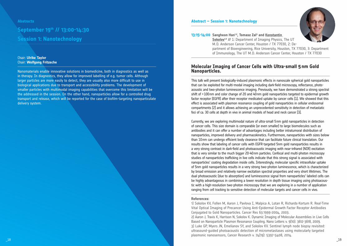
Abstract – Session 1: Nanotechnology
13:15-14:00 Sangheon Han1,2, Tomasz Zal3 and Konstantin Sokolov1,2 // 1: Department of Imaging Physics, The UT M. D. Anderson Cancer Center, Houston / TX 77030, 2: De-partment of Bioengineering, Rice University, Houston, TX 77030, 3: Department of Immunology, The UT M. D. Anderson Cancer Center, Houston / TX 77030
Molecular Imaging of Cancer Cells with Ultra-small 5 nm Gold Nanoparticles. This talk will present biologically-induced plasmonic effects in nanoscale spherical gold nanoparticles that can be exploited for multi-modal imaging including dark-field microscopy, reflectance, photo-acoustic and two-photon luminescence imaging. Previously, we have demonstrated a strong spectral shift of > 100 nm and color change of 20 and 40 nm gold nanoparticles targeted to epidermal growth factor receptor (EGFR) after their receptor medicated uptake by cancer cells [1]. We showed that this effect is associated with plasmon resonance coupling of gold nanoparticles in cellular endosomal compartments [2] and it allows achieving an unprecedented sensitivity in detection of metastatic foci of ca. 30 cells at depth in vivo in animal models of head and neck cancer [3].
Currently, we are exploring multimodal nature of ultra-small 5 nm gold nanoparticles in detection of cancer cells. This size domain is comparable (or even smaller) to large biomolecules such as antibodies and it can offer a number of advantages including better intratumoral distribution of nanoparticles, improved delivery and pharmacokinetics. Furthermore, nanoparticles with sizes below than 10 nm can undergo efficient body clearance that can facilitate future clinical translation. Our results show that labeling of cancer cells with EGFR-targeted 5nm gold nanoparticles results in a very strong contrast in dark-field and photoacoustic imaging with near-infrared (NIR) excitation that is very similar to the much bigger 20-40 nm particles. Confocal and multi-photon microscopy studies of nanoparticles trafficking in live cells indicate that this strong signal is associated with nanoparticles’ coating degradation inside cells. Interestingly, molecular specific intracellular uptake of 5nm gold nanoparticles results in a very strong two-photon luminescence, which is characterized by broad emission and relatively narrow excitation spectral properties and very short lifetimes. The dual photoacoustic (due to absorption) and luminescence signal from nanoparticles’ labeled cells can be highly advantageous in combining a lower resolution in depth tissue imaging using photoacous-tic with a high-resolution two-photon microscopy that we are exploring in a number of application ranging from cell tracking to sensitive detection of molecular targets and cancer cells in vivo. References:1) Sokolov KV, Follen M, Aaron J, Pavlova I, Malpica A, Lotan R, RichardsKortum R. RealTime Vital Optical Imaging of Precancer Using AntiEpidermal Growth Factor Receptor Antibodies Conjugated to Gold Nanoparticles. Cancer Res 63:19992004, 2003.2) Aaron J, Travis K, Harrison N, Sokolov K. Dynamic Imaging of Molecular Assemblies in Live Cells Based on Nanoparticle Plasmon Resonance Coupling. Nano Letters v. 9(10): 36123618, 2009.3) Luke GP, Myers JN, Emelianov SY, and Sokolov KV. Sentinel lymph node biopsy revisited: ultrasoundguided photoacoustic detection of micrometastases using molecularly targeted plasmonic nanosensors, Cancer Research v. 74(19): 53975408, 2014.
Abstracts
September 19th // 13:00-14:30
Session 1: Nanotechnology
Chair: Ulrike TaylorChair: Wolfgang Fritzsche Nanomaterials enable innovative solutions in biomedicine, both in diagnostics as well as in therapy. In diagnostics, they allow for improved labelling of e.g. tumor cells. Although larger particles are more easily to detect, they are usually also more difficult to use in biological applications due to transport and accessibility problems. The development of smaller particles with multimodal imaging capabilities that overcome this limitation will be the addressed in the session. On the other hand, nanoparticles allow for a controlled drug transport and release, which will be reported for the case of biofilm-targeting nanoparticulate delivery system.
_19_18

_21_20
deionized water. Physiological NaCl was identified as the most suitable inhalation solution due to minor alteration of the particle characteristics and was therefore chosen for the further nebulization experiments. Laser diffraction (HELOS, Sympatec, Germany) was used to analyze the aerodynamic characteristics of nebulized formulations over 10 min to determine the mean median aerosol diameter (MMAD), the geometric standard deviation (GSD) and the fine particle fraction (FPF). All nebulized formulations demonstrated appropriate output and aerodynamic characteristics for peripheral lung delivery. The MMAD increased only slightly due to the incorporation of NPs in aerosols, e.g. for the vibrating mesh nebulizer, the MMAD was found to be 5.4 µm for the Tb-NP200 formulation compared to 4.3 µm measured in the NP-free set-up. Despite a large output rate of the vibrating-mesh nebulizer, the air-jet tech-nology was beneficial for deep lung delivery indicated by the larger amount of aerosolized particles smaller than 5 µm (85% FPF) and an MMAD between 2.1 µm and 2.3 µm.
In conclusion, we demonstrated that PLGA and PEG-PLGA based NPs displayed excellent properties as biocompatible, mucus-penetrating delivery systems for antibiotics with im-proved deposition and bacterial killing of biofilm-embedded and mucus covered pathogens. The nebulization of NPs, especially via air-jet technique, offers a highly suitable approach to deliver differently sized NPs efficient to the deep lungs. Acknowledgments:The authors thank the German Research Foundation (DFG) for financial support (FI 899/41, FI 899/42, PL 320/31, PL 320/32). References:1.) M. KlingerStrobel, Expert Opin Drug Deliv, 2015, 12, 135174.2.) J.C. Sung, Trends Biotechnol, 2007, 25, 563570.3.) J. Ernst, Eur J Pharm Biopharm, 2018, 113, 120129.
14:00-14:30 Julia Ernsta, Mareike Klinger-Strobelb, Kathrin Arnolda, Jana Thamma, Oliwia Makarewiczb, Mathias Pletzb, Dagmar Fischera,c // a: Friedrich Schiller University, Institute of Pharmacy, Pharmaceutical Technology and Biopharmacy, Lessingstraße 8, Jena / Germany, b: Jena University Hospital, Institute for Infectious Diseases and Infections Control, Am Klinikum 1, Jena / Germany, c: Friedrich Schiller University Jena, Jena Center for Soft Matter (JCSM), Jena / Germany
How to Re-activate Antibiotic Efficacy in Biofilm-associated Lung Infections by Nanoparticles Bacteria, such as Pseudomonas aeruginosa and Burkholderia cepacia, are a major cause of chronic lung infections in cystic fibrosis (CF) patients. The ability of the bacteria to form biofilms and the presence of a thick and stagnant mucus in the airways of CF patients lead to antibiotic therapy failure and request innovative antibiotic delivery systems to improve the antibiotics´ effectiveness in the CF environment [1]. Biodegradable nanoparticles (NP) as carriers for antimicrobials are promising to break through the mucus and biofilm barrier but are demanding in pulmonal delivery because of particle aggregation and exhalation [2]. In the present study, differently sized nanoparticles made of fluorescently labeled poly(lactic-co glycolic acid) (PLGA) and poly(ethylene glycol)-grafted PLGA (PEG-PLGA) were compared and evaluated as lung applicable carriers to re-activate the efficacy of the antibiotic tobramycin (Tb) in biofilms of P. aeruginosa and B. cepacia.
NPs loaded with Tb were prepared by a double-emulsion evaporation method [3] and exhibited mean hydrodynamic diameters of 200 nm (Tb-NP200) and 900 nm (Tb-NP900), respectively, with narrow size distributions measured by dynamic light scattering (Zetasizer Nano ZS, Malvern Instruments, Herrenberg, Germany) and confirmed by scanning electron microscopy. Tb incorporation increased the zeta potentials to about -10 mV compared to drug-free particles of about 30 mV. The drug content was 1.7 µg Tb per mg NP analyzed by HPLC. To determine the in vitro mucus permeation of NPs, artificial mucus (AM) was filled in ThinCerts™ layered by fluorescently labeled NPs and placed in simulated lung fluid. By measuring the fluorescence intensity in the upper compartment over time, a size and surface potential dependent permeation was found as Tb-NP200 permeated faster compared to Tb-NP900 and Tb-loaded NPs faster than the corresponding drug-free NPs. In vitro biocompati-bility studies showed that no cytotoxicity in A-549 cells were observed for all particle types up to a concentration of 1 mg/mL and 24 h incubation time. For biofilm experiments, bacteria were cultivated in AM-containing chamber slides to allow the formation of a biofilm close to those of CF patients or in a microfluidic device to imitate the physiological shear flow in the body. The excellent penetration abilities of Tb-loaded particles through AM and biofilms and the remarkable antimicrobial efficacy in comparison to the free drug was confirmed by confocal laser scanning microscopy of live/dead stained biofilms.
Nebulization experiments were performed by air-jet (PARI boy, LC SPRINT, PARI, Germa-ny) or vibrating-mesh technology (IH 50, Beurer, Germany). First, the re-suspension of the NPs in therapeutically relevant inhalation solution (0.9% NaCl, 3% NaCl and 7% NaCl) was tested and resulted in slightly increased hydrodynamic diameters compared to the NPs in

_23_22
Abstracts
September 19th // 14:30-15:30
Session 2: Product Slam
Chair: Elmar EndlChair: Frank Schildberg Industrial partners of the conference will get the chance to present their newest innovationsand products within three minutes in this latest event. This type of appetizer is a fantasticmarketing opportunity supplying talking points for later conversations. As bonus, there willbe a prize for the best performance – chosen by vote of the audience.
Exhibitor Presenter Agilent Technologies Jesper Hojlund Becton Dickinson Uwe Speck BioLegend Jana Sarkander Bio-Rad Knut Petkau Cytek Tarek Dubois Fluidigm Andrius Serva Fraunhofer IPA Stefan Scheuermann Merck Millipore Peter Rhein Miltenyi Christoph Vess OLS Peter Engel

_25_24
Abstracts
October 4th // 16:30-18:00
Session 3: Cutting Edge Session
Chair: Hyung-Dong Chang Cutting-edge research not only evolves from newly invented and optimized technologies and strategies, but may arise from combining well-known technologies in an new and optimized way. The combinations of flow cytometry and Next Generation Sequencing or microscopy and mass spectrometry are just recent examples. New technologies not only facilitate or allow high-throughput of cellular and biochemical analyses but also can lead to the discovery of novel biological phenomena and processes. This session will introduce some of these novel developments and showcase the high dynamics of the field of cytometry.
Abstract – Session 3: Cutting Edge Session
16:30-17:00 Emmanuel Reynaud // UCD Centre for Biomedical Engineering / Dublin / Ireland
Light Sheet for the Masses More than a hundred years ago in Jena a physicist and a chemist combining their efforts created the first light sheet for imaging. Their efforts won them the Nobel prize in 1925 but their idea of uncoupling illumination and detection was lost.But with better laser illumination and electronic devices, this basic principle has become a revolution in biological imaging since its revival in 1993. It is now available for histology, pathology, developmental biology, plant biology, cell biology as well as cytometry.So far more than 80 acronyms described at least one type of light sheet microscope, but is it that amazing? The talk will walk you through history and the jungle of variations around the same theme and how this technology will affect biological imaging as well as cytometryin the coming decade. Biography:Dr. Emmanuel G. Reynaud, MSc, BSc, PhD, is a Lecturer in Integrative Biology at the UCD School of Biomolecular & Biomedical Science. Prior to joining UCD, he was a Researcher and Postdoctoral Fellow at the European Molecular Biology Laboratory, where his research focused on development of new imaging methods (e.g. Light Sheet Microscopy) and optical micromanipulations in Cell Biology (e.g. laser nanosurgery), in the laboratory of Prof. Ernst H.K Stelzer and Dr Rainer Pepperkok. He is also the cofounder of the Light Sheet Microscopy community alongside Dr Pavel Tomancak. He also built and coordinated for 2.5 years a unique imaging platform during the circumnavigation of the Earth as part of the Tara Oceans (20092012). He has been awarded a Knight of Palmes academiques for his educational works.

_27_26
References:1.) P. K. Chattopadhyay and M. Roederer, Methods, 2012, 57, 2518.2.) V. V. Tuchin: Advanced Optical Flow Cytometry: Methods and Disease Diagnoses. 2011, WileyVCH, 1st Ed.3.) K. Hoffmann, T. Behnke, D. Drescher, J. Kneipp and U. ReschGenger, ACS Nano, 2013, 7, 667484.4.) K. Hoffmann, T. Behnke, M. Grabolle and U. ReschGenger, Anal Bioanal Chem, 2014, 406, 331522.5.) S. Schmutz, M. Valente, A. Cumano and S. Novault, PLoS One, 2016, 11, e0159961.6.) L. Bene and L. Damjanovich, Cytometry A, 2015, 87, 1013.7.) J. A. Steinkamp, T. M. Yoshida and J. C. Martin, Review of Scientific Instruments, 1993, 64, 34403450.8.) C. Deka, L. A. Sklar and J. A. Steinkamp, Cytometry, 1994, 17, 94101.9.) A. V. Gohar, R. Cao, P. Jenkins, W. Li, J. P. Houston and K. D. Houston, Biomed Opt Express, 2013, 4, 1390400.10.) J. F. Keij and J. A. Steinkamp, Cytometry, 1998, 33, 318323.11.) J. A. Steinkamp and J. F. Keij, Review of Scientific Instruments, 1999, 70, 46824688.12.) Y. Lu, J. Zhao, R. Zhang, Y. Liu, D. Liu, E. M. Goldys, X. Yang, P. Xi, A. Sunna, J. Lu, Y. Shi, R. C. Leif, Y. Huo, J. Shen, J. A. Piper, J. P. Robinson and D. Jin, Nature Photonics, 2013, 8, 3236.
17:20-17:40 Andreas Kleiber // Leibniz Institute of Photonic Technology / Jena / Germany
Advanced Imaging Flow Cytometry Imaging flow cytometry (IFC) is a hybrid technology which extends conventional flow cytom-etry with additional high resolution morphological information. The objective of our work is to develop a microfluidic system for conventional and tomographic IFC.
Both, conventional and tomographic IFC is realized by advanced 3D hydrodynamic focusing which automatically aligns all particles as a sheet at a controllable z-position. Tomographic imaging Flow Cytometry extends conventional imaging flow cytometry for the image based measurement of 3D-geometrical features of cells. The required multidirectional views are generated by rotating all cells while passing the imaging window of the developed microflu-idic chip. Rotation is implemented by guiding them at a shear flow position of the parabolic velocity profile. All cells pass the detection chamber as a two dimensional sheet under controlled rotation where each cell is imaged multiple times.
Experimental results show a strong focusing quality even under flow velocities below 1 mm/s. For the tomographic IFC, white blood cells with fluorescent stained nuclei are been recorded in parallel for the bright field and the fluorescence channel. Different subtypes of white blood cells can be distinguished by the shape of its nucleus. The experiments show that the multidirectional imaging enhances the identification of these subtypes compare to a single 2D view. Ongoing experiments are focusing on a label free classification of a mixed population of eight allergic pollen types using a convolutional neuronal network (CNN). The
17:00-17:20 D. Kagea,d, K. Hoffmanna, M. Wittkampb, J. Ameskampb, W. Göhdeb, T. Thielec, U. Schedlerc, U. Resch-Gengera // a: Bundesanstalt für Materialfor-schung und -prüfung (BAM) / Berlin / Germany, b: Quantum Analysis GmbH / Münster / Germany, c: PolyAn GmbH / Berlin / Germany, d: Institut für Physik, Humboldt-Universität zu Berlin / Berlin / Germany
Luminescence Lifetime Encoding in Flow Cytometry – First Steps and Assessment Numerous analytical techniques in biomedical research rely on multiparametric analyses [1,2]. This often implies encoding or labeling by means of easily distinguishable properties like dif-ferent luminescence features. Optical encoding is frequently combined with high-throughput optical-spectroscopic methods such as flow cytometry, one of the most widespread tech-niques in the life sciences [3,4]. Typically, luminescence encoding schemes rely on spectral (color) and/or intensity codes which can be prone to spectral crosstalk limiting the number of distinguishable codes [5]. A promising alternative is to exploit the characteristic parameter luminescence lifetime for species identification and encoding [6]. This has been suggested for flow cytometry already several decades ago [7]. However, up to now there are only few re-ports on the challenges of lifetime measurements within the short interaction times in flow cytometry. Moreover, most reports focus on scanning techniques, specialized applications without a general assessment, and on measurements in the frequency domain [8-12].
Here, we present first results from flow cytometry measurements in the time-domain using a custom-designed instrument with luminescence lifetime analysis capability. In this respect, major challenges of time-resolved flow cytometry and requirements imposed on temporal resolution and discrimination are discussed that originate from the short interaction time per code and the resulting weak signal-to-background ratio. To that end, we studied a set of lifetime-encoded polymer microbeads loaded with luminophores with varying luminescence decay kinetics. The experimental results were supplemented by numerical simulations of the decay kinetics and underlying photon statistics.
Our results show that lifetime encoding in flow cytometry may serve two current trends [2]: simplified instrument setups for cost-effective high-throughput methods on the one hand, and highly multiplexed analyses by introducing the additional encoding parameter lumines-cence lifetime on the other hand. Acknowledgments:We acknowledge financial support from the Federal Ministry of Education and Research 13N13357 (Forschungsförderung Photonik, VDI Technologiezentrum GmbH). We would like to thank the Laboratory of NanoBioengineering, National Research Nuclear University (Moscow Engineering Physics Institute, Russian Federation) for providing encoded polymer microbeads.

_29_28
whole process requires a high effort in data-processing containing algorithms for object detection, particle tracking and mapping (multi-channel applications) and a CNN-model for the particle classification.
In our work we report on a microfluidic system and method for tomographic imaging flow cytometry, where the angular velocity of a rotating cell is controlled by its z-position in the parabolic velocity profile of a carrier fluid. We also show the need of advanced data-process-ing tools for image analysis. Acknowledgments:We acknowledge the microsystem group and the cleanroom staff at the IPHT for the devel-opment and realization of the microfluidic units. The funding from WaterChip (EU Era-NET-DLR 01DQ16009A) is gratefully acknowledged.
17:40-18:00 Kathrin Brenker // Opto Biolabs / Freiburg / Germany
Opto Biolabs – Combining Optogenetics with Flow Cytometry Optogenetic tools allow isolated, functional investigations of almost any signaling molecule within complex signaling pathways. A major obstacle is the controlled delivery of light to the cell sample and hence the most popular tools for optogenetic studies are microscopy-based cell analyses and in vitro experiments. The flow cytometer has major advantages over a mi-croscope, including the ability to rapidly measure thousands of cells at single cell resolution. However, it is not yet widely used in optogenetics.
Opto Biolabs is an EXIST-funded SpinOff from the Univeristy of Freiburg and builds custom-ized illumination devices for optogenetic flow cytometry. Opto Biolabs’ pxONE illuminates cells at specific wavelengths, light intensities and temperatures during flow cytometric measurements. To demonstrate the utility of the pxONE, we characterized the photoswitch-ing kinetics of Dronpa proteins and performed calcium flux experiments. This protocol can be adapted to almost all optically controlled substances and substantially expands the set of possible experiments. More importantly, it will greatly simplify the discovery and develop-ment of new optogenetic tools and accelerate the screening for novel drugs.

_31_30
Abstracts
September 19th // 18:00-19:00
Keynote – Michael Bauer
Chair: Thomas Kamradt
Michael Bauer is professor and chair for Anesthesiology and Critical Care Medicine as well as spokesman of the Center for Sepsis Control and Care (CSCC) at the Jena University Hospital. He worked as a post-doc at Johns Hopkins University addressing molecular mechanisms of organ failure. He serves in the board of directors in research programs by the German Research Foundation (DFG) and the Federal Ministry of Education and Research (BMBF), such as “PolyTarget” or “InfectoGnostics” and was part of the task force to redefine sepsis (“Sepsis-3”).
Abstract – Keynote
18:00-19:00 Michael Bauer // Jena University Hospital / Germany
Sepsis – Changing the Paradigm Sepsis is the most common preventable cause of death in hospitals. The adoption of the new sepsis definition last year shifted the interest from systemic inflammation (SIRS) to organ dysfunction as the clinical hallmark of an inappropriate host response. Based on a better understanding of the molecular mechanisms, the focus of the new definition is no longer the inflammatory response, but rather the impairment of organ function which results not exclusively from inflammation but also from e.g. metabolic dysfunction. The paradigm thus moves away from the infection and the systemic inflammatory response, and toward that which makes sepsis so dangerous in terms of both disease dynamics and outcome: organ dysfunction. This change of perspective requires novel diagnostic tools to enable early recognition of infected patients with an increased risk of developing sepsis in clinical routine, even outside of the intensive care unit. The new definition also promotes development of new treatment strategies with improved ability to treat sepsis causally. Diagnostic uncertainty is the main driver for delays in therapy, the mis- and overuse of antibiotics, and the failure to identify patients who might benefit from adjunctive therapies. There is a need for new sepsis biomarkers that can aid in therapeutic decision making and add information about screening, diagnosis, risk stratification, and monitoring of the response to therapy. The most promising novel approaches for diagnosis of infection and the ensuing host response consist of light-based (photonic) tools as well as on transcriptomic, proteomic, or metabolic profiling. Novel approaches to sepsis diagnostics promise to transform sepsis from a single syndrome into a group of distinct ‘endophenotypes’ and help in the development of better diagnostic tools and effective adjunctive sepsis therapies.

_33_32
Abstract – Core Facility Networking Event
20:00-20:15 Claudia Dumrese // University of Zurich / Switzerland
Users Sorting on High Speed Sorters in a Shared Ressource Environment: Effects of a Well Balanced Sorter Training The Cytometry Faciltiy of University Zurich provides dedicated sorter training on high speed cell sorters to it`s user base for 9 years already. The effects on sorter performance, as well as user behavior in a highly supportive shared ressource environment are presented along with a training schedule and usage rules as an example for successful operator free system.
20:15-20:30 Stefanie Bürger // Institute of Molecular Biology, Mainz / Germany
Open IRIS – an Open Source Resource Management Tool Implemented at the University of Mainz Open IRIS is free non-commercial resource management tool that was developed at the Friedrich Miescher Institute in Basel. This presentation should give you an overview about the major functions of the software and what it takes to implement Open IRIS as a cam-pus-wide booking system as we did at the University of Mainz.
20:30-20:45 Christian Kukat // Max Planck Institute for Biology of Ageing, Cologne / Germany
Messenger Apps Facilitate GroupCommunication: Introduction to Slack Slack is a workplace messaging app and team collaboration tool which is used widely in science labs (alternatives are Rocket.Chat and Mattermost). We present how we use it in our core facility and how it has improved our team communication, compared to using email.
Abstracts
September 19th // 20:00-22:00
Core Facility Networking Event
Chair: Desiree KunkelChair: Steffen Schmitt
This event should be an opportunity to meet and share your experiences and challenges working in a core facility. We will have three short presentations and hopefully lots of ideas to discuss afterwards in an informal atmosphere among colleagues.
We hope to spend a wonderful evening with you at the DGfZ meeting 2018 in Jena.
_35_34
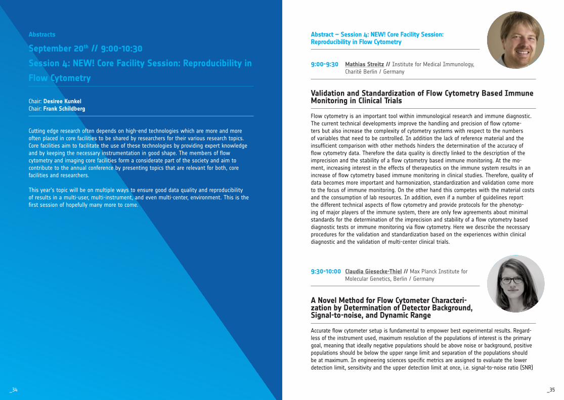
Abstract – Session 4: NEW! Core Facility Session: Reproducibility in Flow Cytometry
9:00-9:30 Mathias Streitz // Institute for Medical Immunology, Charité Berlin / Germany
Validation and Standardization of Flow Cytometry Based Immune Monitoring in Clinical Trials Flow cytometry is an important tool within immunological research and immune diagnostic. The current technical developments improve the handling and precision of flow cytome-ters but also increase the complexity of cytometry systems with respect to the numbers of variables that need to be controlled. In addition the lack of reference material and the insufficient comparison with other methods hinders the determination of the accuracy of flow cytometry data. Therefore the data quality is directly linked to the description of the imprecision and the stability of a flow cytometry based immune monitoring. At the mo-ment, increasing interest in the effects of therapeutics on the immune system results in an increase of flow cytometry based immune monitoring in clinical studies. Therefore, quality of data becomes more important and harmonization, standardization and validation come more to the focus of immune monitoring. On the other hand this competes with the material costs and the consumption of lab resources. In addition, even if a number of guidelines report the different technical aspects of flow cytometry and provide protocols for the phenotyp-ing of major players of the immune system, there are only few agreements about minimal standards for the determination of the imprecision and stability of a flow cytometry based diagnostic tests or immune monitoring via flow cytometry. Here we describe the necessary procedures for the validation and standardization based on the experiences within clinical diagnostic and the validation of multi-center clinical trials.
9:30-10:00 Claudia Giesecke-Thiel // Max Planck Institute for Molecular Genetics, Berlin / Germany
A Novel Method for Flow Cytometer Characteri-zation by Determination of Detector Background, Signal-to-noise, and Dynamic Range Accurate flow cytometer setup is fundamental to empower best experimental results. Regard-less of the instrument used, maximum resolution of the populations of interest is the primary goal, meaning that ideally negative populations should be above noise or background, positive populations should be below the upper range limit and separation of the populations should be at maximum. In engineering sciences specific metrics are assigned to evaluate the lower detection limit, sensitivity and the upper detection limit at once, i.e. signal-to-noise ratio (SNR)
Abstracts
September 20th // 9:00-10:30
Session 4: NEW! Core Facility Session: Reproducibility in
Flow Cytometry
Chair: Desiree KunkelChair: Frank Schildberg
Cutting edge research often depends on highend technologies which are more and more often placed in core facilities to be shared by researchers for their various research topics. Core facilities aim to facilitate the use of these technologies by providing expert knowledge and by keeping the necessary instrumentation in good shape. The members of flow cytometry and imaging core facilities form a considerate part of the society and aim to contribute to the annual conference by presenting topics that are relevant for both, core facilities and researchers.
This year’s topic will be on multiple ways to ensure good data quality and reproducibility of results in a multiuser, multiinstrument, and even multicenter, environment. This is the first session of hopefully many more to come.

_37_36
and the dynamic range (DNR). Recent introduction of the quantiFlash®, a pulsed precision LED light source, now enables a new way of determination of these performance metrics for flow cytometry, independent of sample or bead preparation and instrumental factors not related to signal intensity. We used the quantiFlash® to characterize an instrument’s response to a sta-ble input light signal over the entire PMT gain range. As a consequence, we propose a method to determine a flow cytometer’s SNR and DNR. This allows the selection of a voltage to opti-mize the signals delivered by the PMTs with respect to the background consisting of scattered laser light and electronic noise. Both contributions to the background vary depending on PMT voltage and are now ascertainable and distinguishable with the here proposed method. Such knowledge further allows for separation of technical and biological background which can help with experiment design. In conclusion, we introduce a new practical method for instrument sensitivity characterization, show how the optimal PMT voltage can be defined with respect to the SNR and DNR and discuss practical implications.
10:00-10:15 Steffen Schmitt // German Cancer Research Center (DKFZ) , Heidelberg / Germany
Minimal Requirements for the Presentation of Flow Cytometry Data As a growing number of publications is indicating, reproducibility of experimental data is widely discussed in many biomedical and preclinical research fields. The correct interpretation of flow cytometric data - a relative measure of cellular properties - is strongly dependent on the context in which they have been generated. Therefore, minimal requirements for present-ing this type of data have been published a while ago. These requirements could serve as a kind of standard, helping to improve the quality of flow cytometric data and will be briefly introduced during this session.
10:15-10:30 Elmar Endl // University Bonn / Germany
cytometry.de – the Communication Platform of the DGfZ
cytometry.de has been developed by a group of core facility managers to serve as a commu-nication platform of the german society for cytometry (DGfZ). This communication platform and the connected task force have been officially integrated into the society at the member assembly 2017. The idea is to give core facility staff and researchers working in flow cytom-etry the opportunity to present themselves and to implement tools to communicate in a easy and personal way.
The platform along with its history will be introduced and the features that have been added so far will be discussed.

_39_38
Abstract – Session 5: European Guest Session: Hungary
11:00-11:30 Gábor Szebeni // Hungarian Academy of Sciences, Budapest / Hungary
Immunophenotyping of Treatment Naive Patients with Systemic Autoimmune Diseases by Single Cell Mass Cytometry
INRODUCTION. Epidemiological data highlights the rising incidence of rheumatoid arthritis (RA), systemic lupus erythematosus (SLE) and systemic sclerosis (SSC) in the developed world over the last decades. Currently available treatments are palliative reducing the symp-toms and supporting patient’s wealth. Therefore, studies are much needed to deeply reveal cellular features responsible for pathologies or to identify early diagnostic and prognostic markers associated with RA, SLE and SSC, respectively.
METHODS. We focus on the multiparametric and functional characterization of RA, SLE and SSC using single cell mass cytometry. Treatment-naive patients suffering either in RA, SLE or SSC who had not received non-steroidal anti-inflammatory drugs, disease modifying an-ti-rheumatic drugs and glucocorticoids until the time of blood sampling are enrolled aligned with age and gender matched healthy individuals. Healthy controls have a negative history of rheumatic symptoms, negative status upon detailed physical and laboratory examination. Human subjects are informed and handled in line with the Declaration of Helsinki and rele-vant Directives of the EU. PBMCs are purified by Ficoll density gradient centrifugation. CD4+ T-cells are purified by magnetic bead separation for whole transcriptome analysis. PBMCs are labeled by antibodies to detect markers at single cell resolution by mass cytometry. In mass cytometry detection is based on antibodies which are labeled by stable metal isotopes. Thus, autofluorescence and spectral overlapping are eliminated. Markers identified by the tran-scriptome analysis associated with either RA, SLE or SSC are screened for validation by mass cytometry at protein level.
RESULTS. After gating out the calibration beads and cellular doublets (191Ir+/193Ir+ het-erogeneous) live cells (195Pt cisplatin negative) are determined. Using 26 antibodies in one single tube the following sub-populations have been defined within single living CD45+ cells: B-cells, monocytes, CD16+ myeloid dendritic cells, NK-cells, CD4Tcm, CD4Tem, CD4Tnaive, CD4Th1, CD4Th2, CD4Th0, CD8Tcm, CD8Tem, CD8Tnaive, CD8Tc1, CD8Tc2, CD8Tc0. In order to characterize the phenotype of the above mentioned cell types the following markers have been investigated on each subtype: CD5, CD7, CD9, CD28, CD49d, CD161, CXCR3, CD25, CD69, CD27, CD57, CD127. Stochastic neighbor embedding (viSNE) analysis dissects the different immunophenotype of RA, SSC and SLE samples based on the multidimensional cellular relat-edness of 26 marker expression at single cell resolution in each disease.
CONCLUSION. Mass cytometry deeply reveals cellular heterogeneity on the basis of highly multiplex phenotypical and functional characterization in RA, SLE and SSC.
Abstracts
September 20th // 11:00-12:30
Session 5: Hungarian Guest Session: Hungary
Chair: Gergely ToldiChair: Wolfgang Fritzsche
The Cell Analysis Section of the Hungarian Biophysical Society was founded as an associated society of ISAC in 1995. The section recruits its members from among scientists working in the field of flow and imaging cytometry including those who develop and apply such techniques. A significant result of the scientific organizational activity of the section is to have organized the Hungarian Cell Analysis Conference several times. The popularity of the event is signified by the fact that more than 300 scientists regularly register for the conference. The Cell Analysis Section organized a lecture and practical course “European Summer School: Frontiers in Cell and Immune Technology” in cooperation with the German Cytometry Society (DGfZ) in 2003. The Cell Analysis Section played a key role in organizing the 26th conference of ISAC in Budapest between May 1721, 2008. The topics of high quality plenary and parallel lectures revolved around the key concept of the event “Cytometry in the Age of Systems Biology”. Almost 1100 scientists from 28 countries from around the world participated in the 5day event.
In this session three representatives will highlight cutting edge cytometry research in Hungary. Topics will include translational quantitative cytometry, immunophenotyping using mass cytometry and haematooncology research.

_41_40
11:30-12:00 György Veréb // University of Debrecen / Hungary
Targeting the HER2 Oncoprotein: Lessons Learned from Quantitative Cytometry
Breast cancers, and malignant tumors of several other organs (gastric, ovary, brain) often overexpress HER2 (neu, ErbB2), a receptor tyrosine kinase of the epidermal growth factor receptor family. This is often correlated with rapid clinical progression, resistance to radio or chemotherapy, and, overall, bad prognosis. Since HER2 is not, or weakly expressed in healthy human tissues, it can be considered an ideal target of antitumor therapy. In coherency with this, the first humanized antibody against solid tumors was trastuzumab (Herceptin), targeting HER2. Although trastuzumab treatment of HER2 positive tumors has shown con-siderable success, resistance occurs in a significant fraction of patients, both from the start of treatment and evolving during treatment. Earlier we have shown that an important cause of this resistance is the massive extracellular matrix evolved by tumors. Considering that the antibody can mainly act by down-regulating HER2 or by recruiting antibody dependent cellu-lar cytotoxicity (ADCC), various biochemical and immunological approaches can be proposed for decreasing or obviating the resistance against trastuzumab treatment.
Since HSP-90 serves to retain HER2 in a well-folded, but inactive conformation, the geldanamycin derivative 17-AAG can be used to enhance HER2 dimerization, consequential down-regulation, and thus to decrease proliferation of trastuzumab resistant breast cancer cells. However, this comes at a price, since HER2 activation is also increased transiently, and side effects in the clinic can be significant.
Recently, the myxobacterial antibiotic archazolid, a potent inhibitor of the lysosomal V-ATPase has been produced synthetically, which enabled widespread testing. It appears that archazolid interferes with the regular recirculation of HER2, results in its retention in phagolysosomes and autophagosomes, decreases HER2 phosphorylation, and inhibits the growth of xenograft tumors. Yet, its bioavailability needs to be improved to make it a viable option for adjuvant therapy.
Trastuzumab can also be combined with other HER2-targeted antibodies, such as pertuzum-ab, which increases the resistance-free period in tumor-bearing mice. The beneficial, additive effect of the combination is related to the fact that maximal approved clinical doses of either antibody alone do not saturate ADCC. Thus, it is recommended that both in the adjuvant and neoadjuvant setting the two antibodies are applied in combination from the start.
Finally, it is possible to build a chimeric antigen receptor (CAR) containing trastuzumab scFv for MHC non-restricted target recognition, and important motifs of the T cell receptor and co-signaling molecules. This, when transduced into naive T cells, enables the destruction of HER2 positive tumors by the CAR T cells that perform active reconnaissance, even when the therapeutic antibody cannot anymore penetrate the extracellular matrix with passive diffusion.
12:00-12:30 Gábor Barna, Ágnes Czeti, Gábor Szalóki, Ágnes Márk, Csilla Kriston, András Matolcsy // Semmelweis University, Budapest / Hungary
The Effect of Microenvironmental Factors on the Development of Myeloma Cells
1st Department of Pathology and Experimental Cancer Research, Semmelweis University, Budapest, Hungary
Multiple myeloma (MM) is an age-dependent monoclonal tumor of bone marrow plasma cells (PCs). It is the second most common hematopoietic malignancy. Introduction of autologous stem cell transplantation, proteasome inhibition and immunomodulatory agents improved the treatment of myeloma over the past two decades, but despite the overall impact of these therapies, MM remains fatal and only a small fraction of patients can be cured. The interac-tion between MM cells and the bone marrow microenvironment promotes drug resistance and cancer cells survival. In our research we studied how myeloma cells connect to its micro-environment and how the antigen expression pattern of MM cells changes during therapies. We focused on markers which mediates interactions between plasma cells and the microen-vironment, like CD27, CD28, CD29, CD81, CD56 or intracellular markers, like VS38c, which can be useful to identify MM cells during therapies in case loss of CD38 and CD138.
We have analysed 160 samples from patients with multiple myeloma and 14 samples from healthy individuals with normal PCs by flow cytometry. Cytogenetical abnormalities such as t (4;14), del17p, 1 q amplification, 1 p deletion, and t(11;14) were verified by FISH analysis.
Our results showed that the expression of CD29, CD27 and CD81 is lower in MM cells com-pared to normal PCs, MM patients with active CD29 showed better response to treatment. We found correlation between chromosome 11 hyperdiploidity and the decrease of CD27 expression. We detected that VS38c expression is heterogenous in MM cells. Its expression showed positive correlation with the expression of Dicer protein and the level of VS38c de-creased in patients with relapsed MM. Correlation between CD28 and VS38c expression also could be detected.
Our results suggest that the alterations in stromal-cell and MM-cell interactions facilitating cell survival and the development of a more aggressive and resistant phenotype of MM. Monitoring of prognostic markers during the therapy can be the key to follow these pheno-type changes and raise the possibility to personalize the therapy better to achieve longer remission and increase the chance of recovery.

_43_42
Abstract – Poster Pitch & Poster Session
P01 Carmen Aanei // University Hospital of Saint-Etienne / France
Database-Guided Flow-Cytometry: Simplification and Reproducibility in Flow Cytometry
Multiparameter immunophenotyping by flow cytometry is indispensable for the management of hematological malignancies. Despite the significant benefits provide by this technique that fulfils the requirements for high speed and broad applicability for diagnosis and follow-up of wide range of lymphoid disorders, there are still drawbacks for myeloid disorders. The difficulty is related to lack of consensus for panels of antibodies with high specificity and sensitivity for diagnostic assessment and prognostic stratification. Alongside, standardization of sample preparation, instrument setup protocols, fluorescence compensations and data interpretation across institutions are needed.
The aim of this study is to evaluate the potential of an immunophenotypic myeloid database for identification of surrogate markers for genetic aberrations frequently observed during AML diagnosis and which could guide the clinical protocols. In addition, evaluation of myeloid cell maturation abnormalities against an immunophenotypic myeloid database allow identifi-cation of the most relevant phenotypic abnormalities related to dysplastic changes in BM myeloid cells.
The database is an archive of cell-surface antigen expression profiles representative for normal and malignant hematopoiesis. The database of normal hematopoiesis was construct-ed with normal bone marrows from healthy donors of allogeneic bone marrow transplant or individuals with no evidence of a hematopoietic disease. The data were acquired on five different centers from France under the standardized conditions set by France Flow Clinical Cytometry Group. Immunostaining was performed after erythrocyte lysis using the 8-color antibody panel according to the EuroFlow consortium’s guideline (1). As already referred, the strategy of data analysis consist in separate analysis of the CD34+/- CD117+ immature committed precursors and of more mature myeloid cells (2-4). Data from distinct subtypes of acute myeloid leukemia (AML) were included in a database allowing to directly comparing antigen expression of malignant cells with those of their closest normal counterpart. Further-more, the comparison of patterns of expression was performed in bulk bone marrows of the three cytogenetic subgroups of AML: AML t(8;21), AML t(15;17) and AML inv(16)/t(16;16). The Compass database was constructed in InfinicytTM software. In addition, the Maturation databases were built to help evaluation of phenotypic abnormalities in cases with myelodys-plastic syndromes (MDS) or suspicions of MDS.
The Myeloid Database is a comprehensive visualization interface that can make it useful as a daily procedure for pathologists and cancer researchers for AML/MDS assessment. The reduc-tion of investigator subjectivity in data analysis is an important advantage of this method.
Abstracts
September 20th // 11:00-12:30
Poster Pitch & Poster Session

_45_44
Inclusion of an increased number of data from healthy donors in the database improves the robustness of the analysis. Undoubtedly information stored in database will have an impact on the diagnosis, prognosis, and treatment of myeloid malignancies in the future. Acknowledgments:The antibodies used in this study were provided by BD Biosciences. The authors are thankful for the clinician hematologists for their interest and involvement in this study and for the patients and healthy donors for their agreement to participate in this study. References:1.) JJM van Dongen et al., EuroFlow antibody panels for standardized n-dimensional flow cy-tometric immunophenotyping of normal, reactive and malignant leukocytes. Leukemia (2012) 1908 – 1975.2.) S Matarraz et al., The immunophenotype of different immature, myeloid and B-cell lineage-committed CD34+ hematopoietic cells allows discrimination between normal/reactive and myelodysplastic syndrome precursors. Leukemia (2008) 22, 1175–1183.3.) Paula Laranjeira et al., Immunophenotypic Characterization of Normal Bone Marrow Stem Cells. In: Ingrid Schmid, editor. Flow Cytometry - Recent Perspectives: InTech (2012), p. 457-477.4.) van Lochem EG et al., Immunophenotypic differentiation patterns of normal hematopoie-sis in human bone marrow: reference patterns for age-related changes and disease-induced shifts. Cytometry B Clin Cytom (2004) 60, 1-13.
P02 Anna Bajnok // Semmelweis University, Budapest / Hungary
Distinct Cytokine Patterns May Regulate the Severity of Neonatal Asphyxia – an Observational Study
Background: Neuroinflammation and a systemic inflammatory reaction are important features of perinatal asphyxia. Neuroinflammation may have dual aspects being a hindrance, but also a significant help in the recovery of the CNS. We aimed to assess intracellular cytokine levels of T-lymphocytes and plasma cytokine levels in moderate and severe asphyxia in order to identify players of the inflammatory response that may influence patient outcome.
The effect of cytokines is primarily exerted at a microenvironmental level, but we gener-ally measure the level of cytokines at the macroenvironmental level, from the serum. The relationship between cellular cytokine production and serum cytokine levels is undefined, cytokines in the serum come from various different sources and show a less stable kinetic in time. The advantage of analysing cytokine levels intracellularly by flow cytometry is that this methodology opens up the opportunity for precise characterization of the function and cytokine production of each cell type in a physiological, which could be of great value in identifying key cellular players of various inflammatory conditions.
Methods: We analyzed data of 28 term neonates requiring moderate systemic hypothermia in a single-centre observational study. Neonates were divided into a moderate (n = 17) and a severe (n = 11) group based on neuroradiological and aEEG characteristics. Blood samples were collected between 3-6 h of life, at 24 h, 72 h, 1 week and 1 month of life. Whole blood samples were stimulated for 6 h, then intracellular cytokine levels were determined using flow cytometry. Cytokine plasma levels were measured using Bioplex immunoassays.
Results: The prevalence and extravasation of IL-1b+ CD4 cells was higher in severe than in moderate asphyxia at 6 h. Based on ROC analysis, the assessment of the prevalence of CD4+ IL-1β+ and CD4+ IL-1β+ CD49d+ cells at 6 h appears to be able to predict severity of the insult at an early stage in asphyxia. Intracellular levels of TNF-α in CD4 cells were increased at all time points compared to 6 h in both groups. At 1 mo, intracellular levels of TNF-α were higher in the severe group. Plasma IL-6 levels were higher at 1 wk in the severe group and decreased by mo in the moderate group. Intracellular levels of IL-6 peaked at 24 h in both groups. Intracellular TGF-β levels were increased from 24 h onwards in the moderate group.
Conclusions: IL-1β and IL-6 appear to play a key role in the early events of the inflammatory response, while TNF-α seems to be responsible for prolonged neuroinflammation, potentially contributing to a worse outcome. TGF-β has a compensatory role in decreasing inflammation. These results provide novel data on the role of distinct cytokines in shaping the inflammato-ry response in perinatal asphyxia and influencing patient outcome.
P03 Sabine Baumgart // Deutsches Rheuma-Forschungszentrum Berlin / Germany
Reference Sample Quality Control in Mass Cytometry: Is it Necessary for Clinical Applications?
OBJECTIVE: Mass cytometry (MC) has become an established technology for cell diversity studies and in-depth immune cell profiling of patient samples. Several techniques have been implemented to secure the quality of mass cytometry data such as cell sample banking, standardized instrument setup protocols, signal normalization according to bead standards, sample barcoding, antibody cocktail freezing, signal spill-over correction, and algorithm-based data analysis. However, large sample numbers e.g. from patient cohorts require sample processing and measurement cycles on multiple days, illustrating the need to consider and monitor day-to-day variation in the data. To this end, the inclusion of a standardized refer-ence sample into each sample processing/measurement cycle has been suggested. Based on including such a reference sample into our mass cytometry workflow, we have investigated day-to-day data variation of 19 assay / measurement cycles performed across 3 months. METHOD: A 43-parameter mass cytometry immunophenotyping study was performed on 190 fixed and cryopreserved patient and control whole blood samples. Reference sample aliquots were processed once from a single control donor. For one sample processing/measurement

_47_46
cycle, 10 study samples were Pd-barcoded and pooled, and one aliquot of the reference sam-ple labeled with a separate barcode (mDOTA-103Rh) was added. All antibody staining, wash-ing, fixation etc. steps were carried out on the sample pool ensuring identical conditions for study and reference samples. Data were acquired on a Helios mass cytometer connected to a supersampler for approx. 3 hours per pool. After normalization and debarcoding, reference sample data were clustered using the FlowSOM algorithm and manual gating was performed by using FlowJo.
RESULTS: First, signals elicited by normalization beads were analyzed as a reference. As expected, the inter-pool coefficients of variation (CV) of the mean signal intensities of the bead elements (Ce, Eu, Ho, Lu) after data normalization were low (0.5 – 1.2 %), while CV of signals resulting from staining reference sample cells with metal-conjugated cell lineage-spe-cific antibodies ranged between 15 % for 196Pt channel (CD14, monocyte marker) and 62 % for 175 Lu channel (CD7, NK and T cell marker). This documents that despite employing consistent sample banking, standardized instrument setup and signal correction according to bead standards, some day-to-day variation of data remained that could not be explained by individual sample properties and was not fully eliminated by bead-based data normalization. Despite this signal variation, both manual gating and FlowSOM clustering delivered consis-tent results, indicating that signal variability had no major impact on cell subset identifica-tion. Coherently, CV of frequencies of major manually gated leukocyte subsets or FlowSOM cluster among total CD45+ leukocytes showed low variability in the reference sample across the 19 days. Here, the lowest inter-pool CV was detected for neutrophils, the most abundant cell type in leukocytes (11 %). Frequencies of B cells making up about only 2 % of leuko-cytes, showed a CV of 59 %.
Moreover, the intra-pool analyses of post-normalization reference sample data showed a time –dependent decrease of mean signal intensity in several mass channels to varying degree, not apparently related to a specific isotope or a defined mass range, revealing that bead-based data normalization does not fully eliminate time-dependent signal drift observed for cellular signals.
SUMMARY & CONCLUSION: Analyzing reference sample data from multi-day measurements helps monitoring the technical day-to-day variability in mass cytometry data, which can be considered when analyzing and interpreting study data. Reference sample data can be used to back up the robustness of mass cytometric studies, and to disclose any artifacts possibly caused during sample processing or data acquisition. The use of reference samples will be spe-cifically useful for monitoring assay and data consistency in multi-center studies. In addition, an error analysis for the entire process of data acquisition to estimate/calculate accuracy and precision of our mass cytometry approach can be performed based on reference sample data.
We conclude that the inclusion of reference samples in multi-day mass cytometry studies is suitable to assess the robustness of the data obtained, and should be considered a valuable additional quality control in mass cytometry.
P04 Sabine Baumgart // Deutsches Rheuma-Forschungszentrum Berlin / Germany High-dimensional Immune Cell Profiling in Patients with Active Inflammatory Bowel Disease
OBJECTIVE: Crohn’s disease (CD) is a chronic inflammatory disease affecting the entire gastrointestinal tract, whereby inflammation often occurs in patches and spreads into deeper layers of the intestinal tissue. Unlike CD, in ulcerative colitis (UC) the innermost mucosal lining of the colon or rectum becomes chronically inflamed. The hallmark of both active CD and UC is the infiltration of the intestinal mucosa by innate and adaptive immune cells. To characterize CD and UC – related immune cell abnormalities, we analyzed peripheral blood leukocytes from active CD and UC patients and compared them to a group of sex- and age-matched healthy controls (HC) by mass cytometry.
METHOD: We applied a 43-parameter mass cytometric immune phenotyping panel estab-lished to be compatible with “Smart Tube” whole blood fixation system. In total, blood sam-ples from 27 IBD patients stratified according to the disease activity scores (Harvey-Brad-shaw Index, HBI, for CD and MAYO score for UC), into 11 active CD (HBI > = 5), as well as 16 active UC patients (MAYO > = 3), and 30 HC were analyzed. Samples were barcoded with Pd isotopes, processed and analyzed on a mass cytometer (Helios) equipped with a super sampler. Normalized and manually debarcoded data were analyzed using a bioinformatic pipeline based on the FlowSOM algorithm. Global data were randomly subsampled to 30,000 cells per donor and clustered altogether by FlowSOM yielding a total of 40 cell population clusters. Frequencies of clusters were statistically analyzed for differences between CD, UC and HC using an unpaired T test.
RESULTS: As expected, FlowSOM clustering yielded clusters indicative of known T and B cell subsets, monocytes, dendritic cells etc. Both, CD and UC patients showed significantly aber-rant leukocyte composition compared to controls. In both diseases, frequencies of periph-eral blood NK cell, B cell, eosinophil, and TCRgd + CD4-CD8-T cell clusters were significantly decreased in comparison to controls. While frequencies of CD4+ central memory T cells with a CD161+ phenotype were increased only in active CD patients, frequencies of monocytes and regulatory T cells showed opposite deviations from controls in CD and UC, with increased frequencies in CD and decreased frequencies in UC patients. Within the T cell compartment of active UC patients, frequencies of CD4 and CD8 naïve T cells were lower in comparison to the control group.
SUMMARY & CONCLUSION: Our analysis reveals multiple shared and non-shared immunophe-notypic abberations of patients with active CD and UC. While shared abnormalities may point towards immune cell subset regulation as a result of general gut inflammation, disease-specific differences observed may reflect rather specific immunopathogenic pathways active in CD and UC. In conclusion, our study demonstrates the power of mass cytometry paired with whole blood preservation of patient blood samples to provide a comprehensive overview of changes in global immune cell profiles in patients suffering from inflammatory bowel diseases.

_49_48
P05 Lisa Budzinski // German Rheumatism Research Centre Berlin / Germany
Osmium-labeled Microspheres for Bead-based Assays in Mass Cytometry
Polystyrene microspheres are broadly applied in flow cytometry for instrument setup and monitoring instrument stability, for assessing fluorescent spillover and in various cytometric assays e. g. for absolute quantification of cellular receptors and multi-analyte profiling. The implementation of bead-based assays in mass cytometry for the same purposes is strongly desired but hampered by the lack of functionalized beads associated with sufficient amounts of heavy metal allowing for the unequivocal detection of these by the mass cytometer.
We here introduce osmium tetroxide labeling for polystyrene microspheres as a simple, quick, and universal approach to produce various kinds of functionalized beads applicable in mass cytometry. Osmium detection does not interfere with any existing reagents routinely used in mass cytometric assays, and osmium labeling of various commercially available antibody capture beads resulted in stably and uniformly labeled beads, while retaining their antibody-capture functionality. Osmium-labeled antibody capture beads retained both their functionality and osmium signal for at least two weeks when stored at -80 °C.
We show that osmium-labeled antibody capture beads can be employed for uncompromised characterization of metal-antibody conjugates, and for signal spillover assessment in complex mass cytometric data sets resulting from e.g. minor isotopic impurities of metal labels. Furthermore, osmium-labeled beads facilitate the absolute quantification of cell-surface receptors in mass cytometry.
Osmium labeling of polystyrene beads permits robust implementation of beads-based assays in mass cytometry, broadening the applicability of mass cytometry in biomedical and basic research, and increasing the quality and quantity of information retrievable in mass cytometric studies.
P06 Hans-Jörg Bühring // University Clinic of Tübingen / Germany
Analysis of Myeloid Cell Populations in Human Blood and Bone Marrow by Multicolor Flow Cytometry
Clear identification of specific cell populations by flow cytometry is important to define the immune status of patients. The objective of this study was to develop a multi-parametric flow cytometry panel for prospective immune-profiling of high risk multiple myeloma patients in a phase II multicenter trial. We performed 15-color immunofluorescence flow cytometry on freshly isolated human blood and bone marrow samples. We defined a multicolor flow
panel that identifies major leukocyte populations. These include (1) three monocyte subsets (CD33+SSClow): classical monocytes (CD14+CD16-), intermediate monocytes (CD14+CD16+) and non-classical monocytes (CD14-CD16+); (2) CD11c+ dendritic cells (CD14−CD11c+CD1c+H-LA-DR+), (3) plasmacytoid dendritic cells (CD14−CD11c−HLA-DR+CD123+), and (4) three gran-ulocyte subsets (CD33+SSChigh): neutrophils (CD16-CD15+), eosinophils (CD16+CD15+), and granulocyte precursors (CD15intCD16-). Additionally, the expression of the checkpoint-markers Tim-3, PD-1 and PD-L1 were analyzed for each immune population. In conclusion, we de-fined a multi-parametric flow cytometry panel in human blood and bone marrow that allows identification of multiple immune cell types at once. This study has important implications for defining myeloid cells in high risk multiple myeloma patients.
P07 Zishu Liu // Helmholtz Centre for Environmental Research, Leipzig / Germany
Microbial Flow Cytometry – a High Dynamic Resolution Method for Monitoring Ecological Stabilities of Microbial Communities.
Microbial communities drive many processes which affect human well-being directly as the human microbiome or indirectly as in natural environments or in biotechnological applications. Their complexity and their dynamics are difficult to monitor, and currently, sequence-based approaches are limited with respect to the temporal resolution. However, in order to eventually control microbial community dynamics, monitoring schemes of high tem-poral resolution are required. Flow cytometry provides single-cell based data in the required temporal resolution and we here use such data to compute ecological stability properties of microbial communities, such as resistance, resilience, displacement speed and elasticity. For resilience, we additionally introduce an on-line methodology which is suitable for continuous community monitoring.
Due to the workflow’s immanent ability to support high temporal sample densities, even in generation-time level, such monitoring tools will allow for a fast, continuous, and cost effective screening of ecological stable behaviour of microbes. The proposed workflow was tested on a long-term continuous reactor experiment employing both an artificial and a natural microbial community which were exposed to identical pulse disturbances. 16S rRNA amplicon sequencing of sorted cells was used to verify the findings. The computed stability properties uncovered superior stabilities of the natural community, and demonstrated the global applicability of the protocol to microbial communities in various environments, like the diverse localities of the human body or in surface water and in bioreactors, and will contrib-ute to the development of managing schemes to manipulate microbial community structures and performances.

_51_50
P08 Henrik Mei // German Rheumatism Research Centre Berlin / Germany
Immune Cell Profiling of Rheumatoid Arthritis Patients undergoing Helminth Therapy by Mass Cytometry
Based on the hygiene hypothesis, the treatment of rheumatoid arthritis (RA) by immune modulation induced by infection with eggs of the intestinal helminth Trichuris suis (TSO) promises amelioration of the disease, associated with fewer side effects as compared to currently approved therapies. In order to gain insight in the mechanism of action of TSO therapy and to facilitate precision medicine in the treatment of RA, we studied the composi-tion and activation state of PBMC of RA patients undergoing targeted and timely controlled TSO treatment in a placebo-controlled trial by mass cytometry.
We developed a mass cytometric antibody panel comprising 44 markers for complex immune profiling focused on T and B cell subsets and their activation status. A high degree of assay standardization was achieved by novel beta-2-microglobulin-based live-cell barcoding in conjunction with a new preservation method for metal-labeled antibody cocktails.
The comparison of 31 age- and gender-matched healthy controls with 36 RA patients prior to TSO therapy revealed numerous RA-related phenotypical disturbances in the T cell, B cell and monocyte lineages. RA patients showed diminished frequencies of MAIT cells, significantly lower frequencies of IgG- and IgM-memory B cells and plasmablasts, while a subset of CD14hi CD16lo monocytes was increased. Interestingly, we identified significantly lower expression of the inflammatory chemokine receptor CXCR3 in RA patients’ T, B and NK cell subsets. After treatment with TSO, RA patients exhibited a transient increase of CD23+ naive B cells and γδ T cells. Ongoing work aims at further delineating trajectories of immune cell subsets over the treatment course and correlating these with the serological response to TSO and the clinical improvement of RA in the patients.
This study demonstrates the utility of massively high-parametric immune profiling by mass cytometry in chronic inflammatory conditions to identify immune cell aberrations for further consideration in immunopathogenesis research and therapy.
P09 Raghavendra Palankar // University Medicine Greifswald / Germany
2-Photon Polymerization and 3D Printing of Deformable Microstructures for Quantifying Platelet Cytoskeletal Biomechanics
Circulating platelets are essential players in haemostasis and thrombosis, as they “survey” the integrity of the vascular system. Upon vascular injury, platelets rapidly adhere to the
exposed extracellular matrix (ECM). After adhesion, they spread by forming protrusions like lamellipodia and filopodia and form a haemostatic plug to seal the wound. This process is associated with generation of contractile forces through the coordination of physicochemi-cal interactions between platelet cytoskeletal components (e.g. actin, myosin and tubulin) and surface receptors (e. g. glycoprotein Ib-V-IX complex, integrins α2β1 and αIIbβ3) that recognize the exposed ECM. However, under in vivo physiological conditions and in blood flow platelets encounter a complex and dynamically changing biophysical microenvironment. So far, the biomechanics of force generation by platelet filopodia and lamellipodia and the role of cytoskeletal components in optimally sensing extracellular biophysical environment to accom-plish haemostasis are poorly understood. Recent advances in microfabrication engineering have enabled the development of elastic vascular tissue-mimetic niches with a microfluidic environment for platelet function analysis.
Here we present a rapid prototyping of 3D printed deformable microstructures using two-photon lithography to assess biomechanics during fundamental biological events during platelet adhesion, spreading and activation using novel surface topographies.
P10 Vikram Srinivasa Raghavan // Indian Institute of Science, Bangalore / India
Validating Effectiveness of HIV-ART Using Superparamagnetic Iron Oxide Nanoparticles (SPIONs)-conjugated CD4 Cell Count
Antiretroviral therapy (ART) for HIV infected patients includes combination of antiretroviral (ARV) drugs to suppress the HIV virus and stop the progression of HIV disease. Patients un-der ART medication are monitored to ensure treatment efficacy. Viral load and CD4cell counts are being performed to determine the prognosis and for monitoring the response to therapy 1,2. CD4 cell counts are measured at the time of diagnosis and every 3-6 months. Current-ly, expensive and bulky flow cytometers are used for monitoring the ART efficacy. There is need for a novel, simple and inexpensive technique to test for counting CD4 cells in resource limited settings in developing and underdeveloped countries. We developed a technique to quantify CD4 cells in whole-blood; using magnetic nanoparticles and widely used automat-ed hematology analyzer. Magnetic field assisted cell separation is an interesting technique, in which magnetic particles are bound over target cells and separated using an external magnet. Targeted binding of nanoparticle to particular type of cell is achieved by conjugating antibodies to nanoparticles 3,4.
Magnetic assisted cell sorting technique is utilized for this purpose using Superparamagnetic Iron Oxide Nanoparticles (SPIONs). The nanoparticles were synthesized using conventional co-precipitation; the particle size was controlled by tuning the molar ratio of Fe2+:Fe3+ ions and the magnetic properties were studied. Among the different size of SPIONs, 20 nm size ones were found most appropriate according to their magnetophoretic force from theoretical calculation. These SPIONs were surface functionalized to covalently link antibodies specific to

_53_52
CD4 cells. The CD4-tagged SPIONs (CD4-SPIONs) had effective magnetization of 60 emu / g and average hydrodynamic diameter of 100 nm.
Here, we demonstrate an indirect method of counting CD4 cells in blood using CD4-SPIONs and Automated Hematology Analyzer. To test the concept of magnetic separation of cells; we initially used pure isolated white blood cells (WBCs) and mixed with CD4-SPIONs to separate CD4 cells from the total WBCs. The performance of cell separation is highly efficient, but isolation of WBCs is a tedious task. Hence, we experimented with whole blood and diluted blood. In whole blood, the separation efficiency was very low as the interference due to very high cells concentration. Different dilution ratios were experimented and at 1:26 ratio of whole blood and saline; optimum cell separation was achieved. Fig. 1 shows the separated CD4 cells count from total blood cells. The concentration of CD4-SPIONs, incubation time, separation time were optimized with respect to the dilution ratio of 1:26. The volume of SPI-ONs-CD4 reagent required for optimum separation is between 60-80 microliters for a reaction Fig. 2. All experiments were cross validated with conventional standard method and plotted with reference value as shown in Fig. 2. This technique is validated by counting the CD4 cells in whole blood with both commercial diagnostic flow cytometer and the CD4-SPIONs analyz-er. The test results between the two systems are in good agreement.
Further, for more accurate and direct count of target cells, a system with imaging capability and cell sorting in flow need to be developed. This can be achieved by integration of micro-fluidics and microscopy system. Currently, we are performing the above Off-chip experiments in an On-chip microfluidic device (MFD) which we have customized for this application. This MFD will be incorporated with an inexpensive portable imaging flow cytometry (IFC) system for absolute CD4 counts with simple image processing algorithms.
P11 Denise Steinmetz // Friedrich Schiller University, Jena / Germany
Application of Flow Cytometry Analysis for Studying Toxicological Effects of Nanoparticles on Human Caco-2 Cells
Flow cytometry is a fast, reliable, and low-cost high-throughput screening tool for study-ing the toxicological properties of hazardous substances on human cells. Nanotoxicological research has also been able to benefit from the advantages of this technique in order to estimate potential risks of engineered nanomaterials. However, in addition to the advantages of toxicological analysis by flow cytometry, unintentional interactions of nanoparticles with the used measurement systems have been discovered, leading to partial false-positive or false-negative results.
In our recent work, we have therefore studied induction of apoptosis and influence on the cell cycle in human intestinal cells (Caco-2) after exposure with three metal oxide nanopar-ticles: titanium dioxide (TiO
2), copper oxide (CuO), and zinc oxide (ZnO). Shortly, cells were
grown for 48 h and treated with nanoparticle concentrations from 1-100 µg / ml for 24 h. After harvesting and washing the cells, they were stained with Annexin-FITC and propidium iodide (PI) for analysis of apoptosis and a PI staining solution (0.1 % Triton X-100, 100 µg / ml DNase-free RNase, 10 µg / ml PI) for the detection of cell cycle phases. Additionally, the interaction of the used nanoparticles (100 µg / ml in double distilled water) with the assay components Annexin-FITC and PI was investigated in a cell-free solution.
While our results show that TiO2 did not influence the induction of apoptosis, an increase
of apoptotic and dead cells at 100 μg / ml was measured for CuO (8 % apoptotic cells; 38 % dead cells) and ZnO (26 % apoptotic cells, 50 % dead cells). The cell cycle analysis showed that the proportion of cells in the G1 phase increased for TiO
2 and ZnO and decreased in the
G2 phase. No influence of nanoparticles with PI was observed, while an unspecific binding of Annexin-FITC with TiO
2 and ZnO led to an increase of FITC signal in the cell-free system.
As a conclusion, toxicological effects of nanoparticles on cells can be detected by flow cytom-etry, whereby the non-specific influence of these small particles on the test system is not negligible and must be excluded in preliminary experiments.
P12 Marie Urbicht // Deutsches Rheuma-Forschungszentrum Berlin / Germany
Assessing Subset-specific Leukocyte Signaling Aberrations in Chronic Inflammatory Diseases: Paving the Way for Precision Medicine in Chronic Inflammation
Objective: Aberrant signaling in leukocytes contributes to the establishment and maintenance of chronic inflammation and autoimmunity. While the activity of signaling cascades and their abnormalities are known to be cell subset-specific, we lack a systems-level understanding of which immune cells signal aberrantly in different chronic inflammatory conditions and through which pathways.
Method: A new sample collection workflow involving whole blood fixation and cryopreser-vation was established to arrest the ex vivo or stimulation-induced activation of leukocytes within 30 minutes after blood collection, providing access to cellular activation states that are otherwise subject to time- or environment-dependent degradation. Mass cytometry, a multi-plex analysis tool offering more than 50 cytometric channels, is applied to these samples for simultaneous in depth-phenotyping of peripheral blood leukocyte subsets and interrogation of cell type-specific intracellular signaling on the levels of abundance and / or phosphorylation status of intracellular signal transducers.
Results: In a proof-of-concept study, the workflow was evaluated by monitoring pro-tein phosphorylation in ex vivo-stimulated whole blood samples from healthy donors in a time-dependent manner. Short-term kinetics for responses to IFN-α, TNF-α, IL-6 and

_55_54
PMA / ionomycin were recorded by simultaneously measuring the activation of intracellular signal transducers like STAT1,3,5, Syk / Zap70, PLCγ2 or MAP kinases in neutrophils, mono-cytes, B- and T-lymphocytes. These data were used to determine stimulus-specific reference response signatures in a cell-type specific manner. Preliminary data from a small set of patient samples suggests that aberrant pathway activation between diseased and healthy individuals can already be detected in ex vivo samples, without performing re-stimulation.
Summary and Outlook: By transferring this approach to patient blood samples and combining it with computational data analysis, this study aims to identify cellular activation patterns that are either commonly or specifically dysregulated in different chronic inflammatory con-ditions. It can be expected that these data will provide valuable new insights into disease pathogenesis and single cell-based signaling processes targeted by a new class of small molecule therapeutics in the field of chronic inflammatory diseases. Altogether, this informa-tion will contribute to a better understanding and transfer of therapeutic concepts between different disciplines and provide a new basis for precision medicine to stratify individual patients with respect to the best-fitting therapy option. References:1.) Baumgart et al., OMIP-034: Comprehensive immune phenotyping of human peripheral leu-kocytes by mass cytometry for monitoring immunomodulatory therapies. Cytometry A (2017)2.) Mei et al., Platinum-conjugated antibodies for application in mass cytometry. Cytometry A. (2016)
P13 Rico Bongaarts // Union Biometrica, Geel / Belgium
COPASTM Vision Flow Cytometer Sorts and Captures Images
Union Biometrica has developed instrumentation for large particle flow cytometry that can capture images in flow. Adding imaging capability to flow cytometry greatly enhances the phe-notyping of samples by providing morphological and spatial information of the sample constit-uents not collected by conventional flow cytometers. Traditional measurements of size, optical density, and fluorescence, as well as Profiler data, are also collected, and these measurements are used for making sorting/dispensing decisions. The collected images and flow cytometry measurements are synchronized so that objects dispensed to wells of multiwell plates can be traced back to their corresponding image. Our COPASTM technology platform is designed for large particles making it ideally suitable for large single cells, cell clusters, and small model organisms. The COPASTM Vision instrument is based on this platform and is ideally suited for samples made up of particles of varying sizes and shapes. Our data from the COPAS Vision shows proof-of-principle support for increased level of phenotyping of these types of samples, including the small model organism C. elegans and marine meiofauna samples.
P14 Manuela Herber // Miltenyi Biotec, Bergisch Gladbach / Germany
Optimized 7-Color REAfinity™ Antibody Cocktail for Immunophenotyping
Introduction: Immunophenotyping by multicolor flow cytometry is a powerful diagnostic tool to assess the status of a patient’s immune system, e. g. for immune monitoring and comple-mentary research in clinical trials or the detection of minimal residual disease after treatment for leukemia. Other possible applications include the quality control of cellular therapy products or preclinical research, e. g. mouse xenograft models. Having reliable, consistently performing antibodies and dyes is crucial to all the aforementioned applications. Here we present an im-proved ready-to-use seven color immunophenotyping cocktail, made up solely of our Miltenyi Biotec REAfinity™ antibodies, for the differentiation of nine leukocyte subsets. REAfinity™ are recombinant antibodies of human IgG1 isotype with a mutated region within the Fc portion, which prevents Fcγ receptor binding. Furthermore, they are characterized by high antigen specificity and consistent quality compared to conventional hybridoma-derived monoclonal an-tibodies. Here we show their superior performance in a multicolor flow cytometry application.
Methods: Whole blood of healthy human donors was stained with a cocktail containing anti-bodies recognizing the following antigens: CD3, CD4, CD8, CD14, CD16, CD19, CD45, and CD56. The cocktail either consisted of hybridoma-derived or REAfinity™ antibodies. Optimization of the REAfinity™ cocktail allowed for dead cell exclusion using 7-AAD. After red blood cell lysis stained samples were acquired on the MACSQuant Analyzer 10. This instrument supports the enumeration of the identified cell subsets via volumetric cell count determination. For further validation of the REAfinity™ cocktail additional cell materials were used, e. g. peripheral blood mononuclear cells (PBMCs), leukapheresis or cultured chimeric antigen receptor (CAR) T cells.
Results: Both REAfinity™ and hybridoma-derived cocktails enabled the identification and enumeration of B cells, CD4+ and CD8+ T cells, NK cells, NKT cells, monocytes, neutrophils and eosinophils. Labeling with the REAfinity™ cocktail resulted in improved separation of the target populations when compared to the hybridoma-derived cocktail. For whole blood, this was in large part due to reduced unspecific background caused by Fc receptor binding on granulocytes when using the REAfinity™ cocktail. For the other cell materials tested, the cocktail showed consistent and reproducible results.
Conclusion: Results presented here demonstrate improved performance of the new REAfinity™ immunophenotyping kit in comparison with hybridoma-derived antibodies, due to high antigen specificity and lower unspecific background. Together with an upcoming new Express Mode (automated gating and analysis tool), the kit allows for rapid and highly repro-ducible immunophenotyping of various cell materials.

_57_56
P15 Bodo Kohring // Kohring Consulting, Spenge / Germany
The Domain of Image Based, Fluorescence Analysers in the Generation and Monitoring of CAR-T Cell Therapy
There is a new therapeutic concept, based on a creation of modified CAR-T (chimeric antigen receptor T-lymphocyte) cells, dispensed to patients by a one-time intravenous infusion. As the CAR-T cells are the essence of this novel medicine, the monitoring of their status over the whole production process, as well as the monitoring of patient’s blood samples, is an indispensable part of the quality assurance of the therapy concept.
We will present innovative, image based methods, using a fluorescent and bright field cell culture analyser. The Countstar Fluorescence (FL) is an ideal system to combine not only cell concentrations, and viabilities measurements, but additionally documents various cell-spe-cific characteristics of CAR-T cells, and their stability during the consecutive steps of a QbD designed manufacturing process. We were able to detect, and to quantify changes of cells during their cultivation process.The Countstar FL demonstrated to be fast, accurate, and sensitive analyser.
P16 Peter Rhein // Merck Chemicals GmbH, Darmstadt / Germany
Advanced Applications with CCD Based Flow Cytometry
Beside our traditional CCD based imaging flow cytometers (ImageStream® and FlowSight®), Merck has launched this year the new CellStream® benchtop flow cytometry system, a high-ly-customizable and compact flow cytometer that is the first to use a camera for detection. Its unique optics system and design provides researchers with unparalleled sensitivity and flexibility when analyzing cells and submicron particles. Within the CellStream® system, the Amnis® time delay integration (TDI) and camera technology rapidly captures low resolution cell images and transforms them into high-throughput intensity data. Researchers acquire the intensity data they are accustomed to from traditional flow cytometers, but with greater fluorescence sensitivity. Here we will take a deeper look inside the instrument design, sys-tem performance and software of this exciting new flow cytometer.
P17 Andrius Serva // Fluidigm GmbH, South San Francisco / USA
Maximizing Human Immune Monitoring Studies with Mass Cytometry
Immune monitoring is an essential method for quantifying changes in immune cell popula-tions in chronic inflammation, infectious disease, autoimmune disease and cancer studies. The extreme heterogeneity of immune cells demands a high-parameter approach to more fully and efficiently quantify the immune response in health and disease. Mass cytometry is an ideal solution, enabling the simultaneous detection of over 40 phenotypic and func-tional markers in a single tube of sample. We report development of a 29-marker panel for mass cytometry based on the Human ImmunoPhenotyping Consortium (HIPC) consensus panel [Maecker et al. Nature Reviews Immunology (2012)], expanded to allow identification of additional leukocyte subsets, particularly T cells. Automated data analysis with Verity Software House GemStone™ software has been developed specifically for data collected with the panel. Extensive panel testing for repeatability, reproducibility and agreement of full versus partial panel population identification was performed. Repeatability was tested with a single PBMC sample stained by a single technician in two technical replicates and acquired in triplicate on two Helios™ mass cytometers. SDs for percent of parent were 1 % or less for 16 identified populations. Reproducibility was tested by determining the variability in measurements of five PBMC lots stained by five technicians and collected on two Helios in-struments. CVs on mean percent of 13 populations were under 15 % for all but three of 130 measurements. Lastly, R2 values for agreement of percent parent populations using the full 29-marker panel compared to a 10-marker panel for T cell populations were 0.94 or higher. We conclude that this panel kit can provide consistent immune population identification and enumeration for any given lot of PBMC.

_59_58
Abstract – Session 6: Klaus-Goerttler-Session
14:30-15:00 Philipp Rosendahl // TU Dresden / Germany
Laureat-Lecture:
Deformability Cytometry and 1D Fluorescence Imaging in Real-time
During the last decades, tools for rapid characterization of large cell quantities have become indispensable not only for basic research but also clinical diagnostics. The gold standard for cell characterization is flow cytometry. Its success is closely tied to the availability of fluo-rescent labels. But what if there is no molecular marker known for the cells of interest? Or if the label changes cell function? Or cells shall be used for transplantation? As an attractive alternative, deformability cytometry exploits cell mechanics as a sensitive, inherent, label-free functional marker, but lacks the specificity provided by a fluorescent signal. Here we present real-time fluorescence and deformability cytometry (RT-FDC), the ideal, combined system. It facilitates fluorescence detection as in conventional flow cytometry, extended by 1D analysis of spatial information encoded in the fluorescence pulse shape, and adds bright field imaging for mechanical phenotyping of single cells — all in real-time at rates of 100 cells/s. We show utility of RT-FDC for the most common fluorescent labels: Fluorescent surface markers (CD34) are used to separate human hematopoietic stem and progenitor cells (HSPCs) from an unpurified apheresis sample as harvested for bone marrow transplantations. Membrane permeant dyes identify reticulocytes by their ribonucleic acid (RNA) content in a blood sam-ple. And endogenously expressed fluorescent proteins (FUCCI) reveal cell cycle phases in an unsynchronized sample of retinal pigment epithelial cells (RPE1). In addition, we can now also directly correlate mechanical characteristics with fluorescence intensity and localization for each single cell to improve correct classification. In future, this combined approach could establish mechanical phenotyping as equivalent to fluorescent labeling, or even identify subpopulations invisible to molecular labels.
15:00-15:20 Christoph Heuser // Technical University of Munich / Germany
NKT Cells Promote Alternative Cross-priming in two Distinct Phases
Cross-priming is essential for the generation of antiviral and antitumor immunity from cytotoxic T cells (CTL). For successful CTL cross-priming, dendritic cells need to be licensed by CD40L signaling delivered by T helper cells or by Natural Killer T (NKT) cells. In classical,
Abstracts
September 20th // 14:30-16:00
Session 6: Klaus-Goerttler-Session
Chair: Julia ReinhardtChair: Wolfgang Fritzsche
Since 1996 the DGfZ awards a younger scientist with the so called KlausGoerttlerPrize,which goes along with prize money of 1000 €. The award is named after Prof. Klaus Goerttler, he was a pathologist and trendsetting member of the foundation board of our society.
The Prize is dedicated to award a younger scientist for a scientific work out of the wide fieldof Cytometry which appears outstanding both in scientific quality and innovation as well aspresentation and layout. The scientific work reflects a (almost) finished scientific graduation(Diploma, Bachelo, Ph.D. or an equivalent work) and was selected out of several submissionsby the council board of the society.

_61_60
i. e. T helper-cell mediated, cross-priming, it is believed that a brief licensing interaction can result in a licensed state of dendritic cells that wanes over time. In consequence, dendritic cells, T helper cells, and CTL may hypothetically interact sequentially. The in vivo dynamics of alternative (i.e. NKT-cell mediated) cross-priming are unknown. Using a novel approach to conditionally deplete NKT cells in vivo, we compared the sequence of cellular interactions in classical and alternative cross-priming. Strikingly, in classical cross-priming, CTL expansion was blunted when they were not present simultaneously with T helper cells early after immunization. In alternative cross-priming, NKT cells activated DCs during this time, but their simultaneous presence with CTL only had a weak effect on the CTL response. NKT cells provided productive CD40L signaling later, when they had relocated to the splenic white pulp. Depletion of NKT cells after the initial activation of DCs, therefore, shortened the CTL expansion phase and compromised their differentiation into effector phenotype CTL. In contrast, depletion of T helper cells at this time did not alter the quantity or quality of the CTL response.
These data suggest that NKT cells unfold their effect on the primary expansion of CTL in two phases: They activate DCs in the marginal zone upon which both cell types relocate to the white pulp. There, NKT cells shape the CTL response with the help of CD40L, ensuring optimal CTL generation. T helper cells, on the contrary, provide CD40L signaling early after immunization and lose their influence on the CTL response thereafter. These results provide novel insights into the dynamic three-cell interactions during the cross-priming of CTL.
15:20-15:40 Florian Hoss // University of Bonn / Germany
Alternative Splicing: An Alternative Level of NLRP3 Inflammasome Regulation
Some cytosolic pattern recognition receptors (e. g. NLRP3) can recruit multi-protein signaling platforms, termed inflammasomes, which recognize diverse danger signals derived from either pathogens or sterile cell damage.
Inflammasomes are powerful inducers of caspase-1-dependent pyroptosis and maturation of IL-1 β and IL-18. These cytokines are potent pro-inflammatory mediators implicated in numerous infectious, metabolic, and autoimmune diseases. Therefore, activation of the inflammasome is tightly regulated.
During inflammasome activation, all ASC molecules of a cell are recruited into a single cluster, resulting in a dramatic molecular redistribution. Using flow cytometry, this relocalization event can be monitored by pulse shape analysis.
Intriguingly, we observed that the percentage of NLRP3-activatable human primary cells is strongly reduced in comparison to murine cells. In the following, we could show, that human
but not murine, NLRP3 mRNA is subject to alternative splicing. The major alternative NLRP3 isoform lacks the first two canonical leucine-rich repeats, is not activated by commonly used NLRP3 stimuli and does not induce inflammasome formation. Consequently, cells expressing the NLRP3 splice variant are protected from pyroptosis. We could link this loss of function of the alternative splice variant to the inability to interact with NEK7. Furthermore, we could show differential expression of the different isoforms on single cell level, potentially render-ing some sub-fractions of a cell pool inert to activation and pyroptosis.
We propose that alternative splicing represents an additional, not yet described level of NLRP3 regulation and it is very likely that similar regulatory mechanisms are conserved across most NLR proteins.
15:40-16:00 Christoph Wilhelm // University Hospital Bonn / Germany
Metabolic Regulation of ILC Mediated Barrier Protection and Pathology
Innate lymphoid cells (ILC) are a group of immune cells, which control immunity, inflammation and homeostasis in tissues like the lung, gut and the skin. While the primary function of ILC is the guarding of the tissue barrier, chronic activation may result in chronic inflammation. Recently, we could show that type 2 innate lymphoid cells (ILC2) rely on fatty acid oxidation (FAO) during helminth infection to maintain their protective function. How-ever, whether both protective and pathogenic ILC responses underlie a similar metabolic control is completely unknown. Here we dissected the metabolic control of type 2 innate lymphoid cells (ILC2) with the aim to reveal metabolic programs dictating protection or immune pathology.

_63_62
Abstract – Guest Lecture
17:00-18:00 Timo Mappes // Deutsches Optisches Museum / Germany
Early Precursors of Fluorescence Cytometry: Foundation of Cell Theory and Fluorescence Microscopy
Fluorescence based cytometry is eventually based on (1) understanding of cells, and (2) the principle of fluorescence. The foundation of both topics is closely connected with the site of Jena.
The cell theory was formulated in 1839 by Theodor Schwann (1810-1882) and Matthias Jakob Schleiden (1804-1881). Schleiden performed the major part of his research in Jena, closely collaborating with the microscope makers of his time. The developments in optics enabled him to microscopically see and characterize the cellular structures and eventually to postulate: (A) All living organisms are composed of one or more cells, and (B) the cell is the most basic unit of life.
Pushing the resolution in microscopy has been a major driver of development since estab-lishing the Abbe equation: d = λ / 2 NA. Reducing the wavelength into the deep Ultraviolet and thus improving the resolution, August Köhler (1866-1948) and Moritz von Rohr (1868-1940) designed dedicated optics. In 1903 the team presented a microscope’s beam path designed entirely for 275 nm, enabling wide-field immersion microscope-photography with magnifi-cations up to 2500 x. While doing so they were the first to observe autofluorescence in the microscopic sample.
Abstracts
September 20th // 17:00-18:00
Guest Lecture – Timo Mappes
Chair: Wolfgang Fritzsche
After completing his doctorate (DrIng), Mappes began his career in 2006 in Karlsruhe as a member of the concept team to merge the University of Karlsruhe with the Karlsruhe Research Center to form the Karlsruhe Institute of Technology (KIT). After completing his postdoc, the scientist became the head of an independent junior research group and spokesman of the Young Investigator Network, and gives lectures there to this day. In 2012 he joined ZEISS; his most recent role was at Carl Zeiss Vision International GmbH, where he headed the departments Technology and Innovation, and Information Technology – an R&D team spread across four continents. His duties as Director of the German Optical Museum and Professor for the History of Physics at the University of Jena include forging close scientific networks between the museum, research and teaching.

Conference Dinner (Access with conference badge only)
19:30-23:00 Zeiss Planetarium // Am Planetarium 5 / 07743 Jena
Conference Dinner & Award Ceremony
Enjoy the captivating movement of the celestial body come rain or shine. The Zeiss- Planetarium was the fourth big planetarium to open worldwide. Since March 1926 people are able to study the stars on a projection screen of nearly 800 m2. After the destructions during WW II, Jena’s planetarium remained as the oldest one in the world. In 1984 / 85 extensive reconstructions led to many advancements. The installation of computer-operated technology and the spatial expansion led to a new orientation in program offers and visitor service.
Join us for the DGfZ Networking & Get-Together at the Zeiss Planetarium on Thursday eve-ning. There will be a buffet, drinks and samples of various Planetarium shows. Furthermore the Klaus Goerttler prize will be awarded, as well as the poster prize and the prize for the best product slam presentation. How to get there: The Planetarium is within walking distance from the city center. From the conference venue: 9 min walk (700 m)
Abstracts
September 20th // 18:00-19:30
Members Assembly
_65_64

_67_66
Abstract – Session 7: Microbiology Session
9:30-10:00 Oliwia Makarewicz // University Hospital Jena / Germany
Molecular Diagnostic in the Age of Multiresistant Gram-negative Bacteria
Currently, the spread of multiresistant Gram-negative bacteria (MRGN) producing extend-ed-spectrum β-lactamases (ESBL) and carbapenemases is a global thread to public health. Bloodstream infections with MRGN are associated with increased mortality, which is primarily due to delayed appropriate treatment resulting in clinical failure. As most sep-sis guidelines, the German guideline suggest an empiric treatment with a β-lactam with antipseudomonal activity (i. e. piperacillin/tazobactam, ceftazidim, cefepim or a carbapenem) in an optional combination with an aminoglycoside or a fluoroquinolone, which does not cover ESBL-producers except for carbapenems (last resort antibiotics). With up to 72 hours to result, the standard blood culture-based routine diagnostic is highly sensitive for detection of bloodstream infection, but takes often too long for critically ill or septic patients.
In attempts to improve and accelerate diagnostic procedures, numerous molecular methods have been developed. Commercially available molecular approaches focus on species identi-fication and cover only a limited number of resistance genes and are therefore of minor use for clinical decisions regarding antibiotic treatment. Whereas a simple PCR based approach maybe suitable for multi-drug resistant Gram-positives with ‘monogenetic’ resistance, most of these tests fail to accurately detect MRGN. This is explained by the highly genetically and phenotypically diverse superfamily of β-lactamases containing more than 1500 variants subdivided in at least four purely related classes mediating resistance to penicillins, mono-bactams, cephalosporins (ESBL) and/or carbapenemases. The resistance genes are usually encoded on highly mobile multi-resistance plasmids driving a fast inter- and intra-species spread of those in Gram-negatives. Moreover, other factors such as reduced influx and increased efflux, or changes in expression levels, influence the phenotypic resistance profiles, but these factors are not covered by molecular tests. Thus, an one-fits-all assay remains unlikely to be developed covering the species, all β-lactamases and other resistance mecha-nisms. Therefore, we need to better understand the dynamics of resistance genes in MRGN to find appropriate targets for molecular diagnostics. In the meantime, blood-culture based diagnostic remains the gold standard.
Abstracts
September 21st // 9:30-11:00
Session 7: Microbiology Session
Chair: Christin KochChair: Stephan Schmid
Microscopy and culture based approaches are still the standard for microbial routine diagnostics in clinical settings but also for the analysis of drinking water quality. In this session we present alternative methods for microbial monitoring based on Raman spectroscopy of individual cells as well as image cytometry. These versatile technologies can be applied without the need of any external labelling or staining procedure making sample preparation very easy and enabling fast sample analysis, thus, reducing diagnosis times dramatically. Further, successful examples will be given how the current challenges of the respective data analysis and identification of specific species can be solved.

_69_68
10:00-10:20 Astrid Tannert // Leibniz IPHT Jena / Germany
Raman Spectroscopic Cytometry for the Detection and Characterization of Bacterial Infection
Infectious diseases are one of the leading causes for deaths worldwide. In order to efficiently treat an infection, physicians need to know which pathogen is causing the infection and –in case of a bacterial infection – the pathogen’s antibiotic susceptibility. Established microbiolog-ical methods used in routine clinical diagnostics are mainly based on cultivation to detect the bacteria and to generate enough biological material for subsequent analysis, such as antibiotic susceptibility testing. Thus, they need at least a full day to provide the result. Faster methods are urgently needed to administer tailored antibiotic therapy already early on.
In this presentation, a new Raman spectroscopy-based approach is presented which holds the potential to dramatically reduce diagnosis times. By means of Raman spectroscopy (individual) cells can be characterized without the need of any external label making sample preparation very easy. The inelastic scattered light provides highly specific information of the overall molecular composition of the investigated cells yielding a so-called “spectroscopic fingerprint”. This spectroscopic fingerprint in combination with multivariate statistical data analysis can be used to characterize immune cells from the host, but also to characterize the pathogen causing the infection.
Here, we will focus on the characterization of the bacterial pathogen and present how Raman spectroscopy can be used to distinguish different pathogens. This will exemplarily be shown for bacteria from patient’s urine samples. Furthermore, Raman spectroscopy can be applied to differentiate the bacteria’s response to antibiotic treatment. From this response valuable information on the pathogen’s antibiotic susceptibility can be extracted. This can be done in a qualitative manner to classify the bacteria as sensitive or resistant; as well as in a quanti-tative manner yielding the antibiotic’s minimal inhibitory concentration (MIC). Acknowledgements:Financial support by the BMBF via the Integrated Research and Treatment Center “Center for Sepsis Control and Care” (CSCC, FKZ 01EO1502) and via the Research Campus InfectoGnos-tics (FKZ 13GW0096F) is highly acknowledged. Furthermore, the project was supported by the Free State of Thuringia (FKZ 2015 FGI 0011 and 2016 FGI 0010) with cofinancing from the European Union within the European Regional Development Fund (EFRE). The work is supported by the COST Action “Raman-based applications for clinical diagnostics — Raman-4Clinics” (BM 1401) and the DFG via the Jena Biophotonic and Imaging Laboratory (JBIL).
10:20-10:40 Susanne Dunker1,2, David Boho3, Jana Wäldchen4, Patrick Mäder3 // 1: Helm-holtz-Centre for Environmental Research – UFZ / Germany, 2: German Centre for integrative Biodiversity Research – iDiv / Germany, 3: Technical University Ilmenau / Germany, 4: Max-Planck-Institute for Biogeochemistry
Who am I and if Yes, How any? A new Way of Phytoplankton Species Identification by Combination of Image Cytometry and Deep Learning
Phytoplankton species composition is directly linked to water quality. Especially drinking water reservoirs, bathing and ballast water need to be regularly monitored for harmful species. Microscopic investigations are the common standard of phytoplankton monitor-ing, guaranteeing high quality of analyses. However, by using microscopy, there are severe limitations like taxonomic expert requirement, difficulties in providing standardized taxonomic knowledge and time consumption (limiting the samples which can be processed in total). Image cytometry allows to collect thousands of images per minute. This technique allows to separate first phytoplankton functional groups based on fluorescence images and subse-quently using brightfield images for species identification. Images of reference species can ideally be used for training of a deep learning network. Deep learning is an advanced form of machine learning, avoiding subjective feature selection. A training and validation of a dataset with nine phytoplankton species at three different life cycle stages (young, well developed and senescent cells) revealed a high accuracy (97 %) of correct species identification and life cycle assignment. The innovative combination of high throughput image cytometry and deep learning is therefore suggested as a promising tool to monitor water quality in future.

_71_70
10:40-11:00 Eugen Kaganovitch1, Bastian Wollenhaupt1, Philipp Popp2, Thorsten Mascher2, Dietrich Kohlheyer1,3 // 1: Forschungszentrum Jülich GmbH / Germany, 2: Institut für Mikrobiologie – TU Dresden / Germany, 3: Aachener Verfahrenstechnik (AVT) – RWTH Aachen / Germany
Microfluidic System for Single-cell Analysis in Picoliter-sized Batch Bioreactors
In this contribution we demonstrate a polydimethylsiloxane (PDMS)-based device for the cultivation and analysis of bacterial cells under batch conditions. Microfluidic cultivation de-vices, in combination with time-lapse microscopy, provide simple but capable tools to study biotechnologically relevant parameters like cell growth and productivity of individual cells [1]. In comparison to other cell analysis tools, these devices facilitate a high level of environmen-tal control combined with time-resolved observation of living cells.
So far, most microfluidic cultivation systems feature chambers for cell growth, which are con-nected to continuous medium perfusion, thereby facilitating a continuous nutrient supply and removal of side products [2], leading to continuous cell growth. However, most conventional analyses and cultivations are performed in batch mode. Results obtained from continuously perfused systems can therefore hardly be transferred to applications conducted in batch mode.
We present a device featuring six channels, each containing 49 picoliter-sized bioreactors that can be reversibly isolated from continuous medium supply (Fig. 1). Therefore the cultiva-tion medium is removed by a continuous flow of pressurized air, preserving a fixed volume of cultivation medium inside the bioreactors.
We show the application of our device for the analysis of survival strategies of Bacillus subtilis in case of nutrient deficiency. Therefore, the cells were cultivated in batch mode in our device until stationary phase. The live-cell imaging revealed a heterogeneous production of a toxin which is assumed to be responsible for cannibalism within the cell population during nutrient limitations (Fig. 2). The physiological relevance and the consequences of the heterogeneous toxin production are still under investigation. We were also able to observe cell growth and spore formation when the majority of the population died, indicating that Bacillus cells exhibit a range of physiological states during the stationary phase in order to increase the chances for the population to survive adverse environmental conditions.
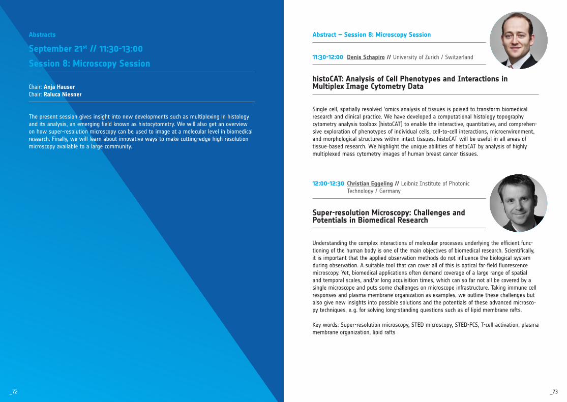
_73_72
Abstract – Session 8: Microscopy Session
11:30-12:00 Denis Schapiro // University of Zurich / Switzerland
histoCAT: Analysis of Cell Phenotypes and Interactions in Multiplex Image Cytometry Data
Single-cell, spatially resolved ‘omics analysis of tissues is poised to transform biomedical research and clinical practice. We have developed a computational histology topography cytometry analysis toolbox (histoCAT) to enable the interactive, quantitative, and comprehen-sive exploration of phenotypes of individual cells, cell-to-cell interactions, microenvironment, and morphological structures within intact tissues. histoCAT will be useful in all areas of tissue-based research. We highlight the unique abilities of histoCAT by analysis of highly multiplexed mass cytometry images of human breast cancer tissues.
12:00-12:30 Christian Eggeling // Leibniz Institute of Photonic Technology / Germany
Super-resolution Microscopy: Challenges and Potentials in Biomedical Research
Understanding the complex interactions of molecular processes underlying the efficient func-tioning of the human body is one of the main objectives of biomedical research. Scientifically, it is important that the applied observation methods do not influence the biological system during observation. A suitable tool that can cover all of this is optical far-field fluorescence microscopy. Yet, biomedical applications often demand coverage of a large range of spatial and temporal scales, and/or long acquisition times, which can so far not all be covered by a single microscope and puts some challenges on microscope infrastructure. Taking immune cell responses and plasma membrane organization as examples, we outline these challenges but also give new insights into possible solutions and the potentials of these advanced microsco-py techniques, e. g. for solving long-standing questions such as of lipid membrane rafts.
Key words: Super-resolution microscopy, STED microscopy, STED-FCS, T-cell activation, plasma membrane organization, lipid rafts
Abstracts
September 21st // 11:30-13:00
Session 8: Microscopy Session
Chair: Anja HauserChair: Raluca Niesner
The present session gives insight into new developments such as multiplexing in histology and its analysis, an emerging field known as histocytometry. We will also get an overview on how superresolution microscopy can be used to image at a molecular level in biomedical research. Finally, we will learn about innovative ways to make cuttingedge high resolution microscopy available to a large community.
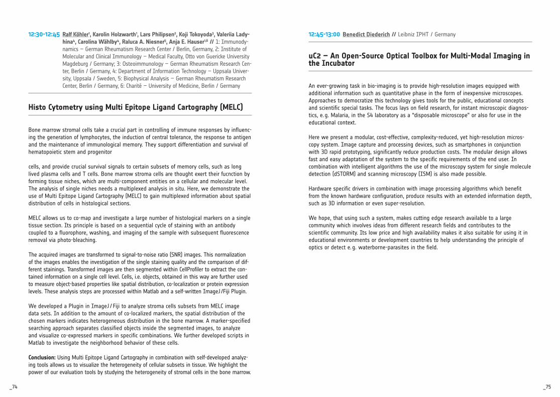
_75_74
12:30-12:45 Ralf Köhler1, Karolin Holzwarth1, Lars Philipsen2, Koji Tokoyoda3, Valeriia Lady-hina4, Carolina Wählby4, Raluca A. Niesner5, Anja E. Hauser1,6 // 1: Immunody-namics – German Rheumatism Research Center / Berlin, Germany, 2: Institute of Molecular and Clinical Immunology – Medical Faculty, Otto von Guericke University Magdeburg / Germany; 3: Osteoimmunology – German Rheumatism Research Cen-ter, Berlin / Germany, 4: Department of Information Technology – Uppsala Univer-sity, Uppsala / Sweden, 5: Biophysical Analysis – German Rheumatism Research Center, Berlin / Germany, 6: Charité – University of Medicine, Berlin / Germany
Histo Cytometry using Multi Epitope Ligand Cartography (MELC)
Bone marrow stromal cells take a crucial part in controlling of immune responses by influenc-ing the generation of lymphocytes, the induction of central tolerance, the response to antigen and the maintenance of immunological memory. They support differentiation and survival of hematopoietic stem and progenitor
cells, and provide crucial survival signals to certain subsets of memory cells, such as long lived plasma cells and T cells. Bone marrow stroma cells are thought exert their fucnction by forming tissue niches, which are multi-component entities on a cellular and molecular level. The analysis of single niches needs a multiplexed analysis in situ. Here, we demonstrate the use of Multi Epitope Ligand Cartography (MELC) to gain multiplexed information about spatial distribution of cells in histological sections.
MELC allows us to co-map and investigate a large number of histological markers on a single tissue section. Its principle is based on a sequential cycle of staining with an antibody coupled to a fluorophore, washing, and imaging of the sample with subsequent fluorescence removal via photo-bleaching.
The acquired images are transformed to signal-to-noise ratio (SNR) images. This normalization of the images enables the investigation of the single staining quality and the comparison of dif-ferent stainings. Transformed images are then segmented within CellProfiler to extract the con-tained information on a single cell level. Cells, i.e. objects, obtained in this way are further used to measure object-based properties like spatial distribution, co-localization or protein expression levels. These analysis steps are processed within Matlab and a self-written ImageJ /Fiji Plugin.
We developed a Plugin in ImageJ / Fiji to analyze stroma cells subsets from MELC image data sets. In addition to the amount of co-localized markers, the spatial distribution of the chosen markers indicates heterogeneous distribution in the bone marrow. A marker-specified searching approach separates classified objects inside the segmented images, to analyze and visualize co-expressed markers in specific combinations. We further developed scripts in Matlab to investigate the neighborhood behavior of these cells.
Conclusion: Using Multi Epitope Ligand Cartography in combination with self-developed analyz-ing tools allows us to visualize the heterogeneity of cellular subsets in tissue. We highlight the power of our evaluation tools by studying the heterogeneity of stromal cells in the bone marrow.
12:45-13:00 Benedict Diederich // Leibniz IPHT / Germany
uC2 – An Open-Source Optical Toolbox for Multi-Modal Imaging in the Incubator
An ever-growing task in bio-imaging is to provide high-resolution images equipped with additional information such as quantitative phase in the form of inexpensive microscopes. Approaches to democratize this technology gives tools for the public, educational concepts and scientific special tasks. The focus lays on field research, for instant microscopic diagnos-tics, e. g. Malaria, in the S4 laboratory as a “disposable microscope” or also for use in the educational context.
Here we present a modular, cost-effective, complexity-reduced, yet high-resolution micros-copy system. Image capture and processing devices, such as smartphones in conjunction with 3D rapid prototyping, significantly reduce production costs. The modular design allows fast and easy adaptation of the system to the specific requirements of the end user. In combination with intelligent algorithms the use of the microscopy system for single molecule detection (dSTORM) and scanning microscopy (ISM) is also made possible.
Hardware specific drivers in combination with image processing algorithms which benefit from the known hardware configuration, produce results with an extended information depth, such as 3D information or even super-resolution.
We hope, that using such a system, makes cutting edge research available to a large community which involves ideas from different research fields and contributes to the scientific community. Its low price and high availability makes it also suitable for using it in educational environments or development countries to help understanding the principle of optics or detect e. g. waterborne-parasites in the field.

_77_76
Address Book
Aanei, Carmen-Mariana // [email protected] // CHU de SaintEtienne Altmaier, Julia // lenzj@unimainz.de // Universitätsmedizin Mainz Andreas, Nico // [email protected]jena.de // University Hospital Jena Barna, Gábor // [email protected]univ.hu // Semmelweis University, Hungary Barths, Jochen // [email protected] // University of Strasbourg Bauer, Michael // [email protected]jena.de // Jena University Hospital Baumgart, Sabine // [email protected] // DRFZ Beisker, Wolfgang // wbeisker@iotaes.de // IOTA / ES Bender, Tanja // [email protected] // Becton Dickinson GmbH Bergmann, Wendy // [email protected]rostock.de // University Hospital Rostock Bergmann, Tobia // [email protected] // Beckman Coulter GmbH Bieber, Kristin // [email protected]tuebingen.de // Uniklinikum Tübingen Bodinek, Jan // jan.bodinek@uniklinikfreiburg.de // Uniklinik Freiburg Boede, Sally // sally.boede@unijena.de // FriedrichSchillerUniversität Jena Bongaarts, Rico // [email protected] // Union Biometrica Braun, Michael // [email protected] // Beckman Coulter GmbH Brede, Christian // [email protected] // Miltenyi Biotec Brenker, Kathrin // [email protected] // Opto Biolabs Budzinski, Lisa // [email protected] // German Rheumatism Research Centre (DRFZ), Berlin Bühring, Hans-Jörg // hansjoerg.buehring@unituebingen.de // University of Tübingen Bürger, Stefanie // s.buerger@imbmainz.de // IMB Caldwell, Randolph // randolph.caldwell@helmholtzmuenchen.de // Helmholtz Zentrum München – Deutsches Forschungszentrum für Gesundheit und Umwelt
Chang, Hyun-Dong // [email protected] // DRFZ Berlin Cheetham, Mark // markc@propellabs.com // Propel Labs Europe Deckert, Felicitas // felicitas.deckert@leibnizipht.de // Leibniz Institute of Photonic Technology Diederich, Benedict // benedict.diederich@leibnizipht.de // LeibnizIPHT Dolf, Andreas // andreas.dolf@unibonn.de // Uni Bonn Dumrese, Claudia // [email protected] // University of Zurich Dunker, Susanne // [email protected] // HelmholtzCentre for Environmental Research – UFZ; German Centre for integrative Biodiversity Research – iDiv Eggeling, Christian // christian.eggeling@leibnizipht.de // Leibniz Institute of Photonic Technology Endl, Elmar // eendl@unibonn.de // University Bonn Ernst, Julia // julia.ernst@unijena.de // Friedrich Schiller University Jena Ferreira-Gomes, Marta // marta.fgomes@unijena.de // Friedrich Schiller University Jena Folz-Donahue, Kat // [email protected] // Max Planck Institute for Biology of Ageing Freytag, Daniela // [email protected] // BD Fritzsche, Wolfgang // wolfgang.fritzsche@leibnizipht.de // Leibniz IPHT Jena Gaetjen, Marcel // [email protected] // BD Life Sciences Gawanbacht-Ramhormose, Ali // Ali.Gawanbacht[email protected] // BD Life Sciences Geisdorf, Linda // [email protected] // CyTUM UNIKAT Geist, Sören // [email protected] // Alere Technologies GmbH Giesecke-Thiel, Claudia // [email protected] // Deutsches Rheuma Forschungszentrum Berlin Gil-Pulido, Jesus // [email protected] // Institute of Molecular Biology Göttinger, Christoph // c.goettlinger@unikoeln.de // Universität zu Köln

_79_78
Gross, Stefanie // stefanie.gross@ukerlangen.de // Universitätsklinikum Erlangen Guck, Jochen // jochen.guck@tudresden.de // TU Dresden Hauser, Anja // [email protected] // Charité – Universitätsmedizin Berlin Herber, Manuela // [email protected] // Miltenyi Biotec GmbH Heuser, Christoph // [email protected] // Technical University of Munich Hoffmeister, Marc // [email protected] // Miltenyi Biotec Hojlund, Jesper // [email protected] // Agilent Horst-Hassinger, Ulla // ulla.horst[email protected] // Agilent Hoss, Florian // florian.hoss@unibonn.de // University of Bonn Hübschmann, Thomas // [email protected] // Helmholtz Centre for Environmental Research – UFZ Jansen, Stephan // [email protected] // Agilent Technologies AG Kaganovitch, Eugen // e.kaganovitch@fzjuelich.de // Forschungszentrum Jülich GmbH Kage, Daniel // [email protected] // Bundesanstalt für Materialforschung und prüfung (BAM) Kaiser, Toralf // [email protected] // DRFZ Kamradt, Thomas // [email protected]jena.de // Universitätsklinikum Jena Kärner, Nadine // [email protected] // Beckman Coulter GmbH Killig, Monica // [email protected] // Miltenyi Biotec Kleiber, Andreas // andreas.kleiber@leibnizipht.de // Leibniz Institute of Photonic Technology Klein, Markus // [email protected] // Merck Life Sciences Koch, Christin // [email protected] // Symrise AG Köhler, Ralf // [email protected] // German Rheumatism Research Center, Berlin Kohring, Bodo // kohringconsult@tonline.de // Kohring consulting Krause, Alexander // [email protected] // AHF analysentechnik AG
Kukat, Christian // [email protected] // Max Planck Institute for Biology of Ageing Kunkel, Desiree // [email protected] // BCRT, Berlin Lennartz, Klaus // klaus.lennartz@unidue.de // Universitätsklinikum Essen Liu, Zishu // [email protected] // Helmholtz Centre for Environmental Research GmbH UFZ Lösche, Andreas // [email protected]leipzig.de // University of Leipzig Ludwig, Carsten // [email protected] // Merck Life Sciences Makarewicz, Oliwia // [email protected]jena.de // University Hospital Jena Mappes, Timo // timo.mappes@deutschesoptischesmuseum.de // Deutsches Optisches Museum Mei, Henrik // [email protected] // DRFZ Berlin Müller, Susann // [email protected] // UFZ Neicke, Thomas // thomas_neicke@apeberlin.de // APE GmbH Niesner, Raluca // [email protected] // DRFZ, Berlin Palankar, Raghavendra // palankarr@unigreifswald.de // University Medicine Greifswald Pelz-Ackermann, Oliver // oliver.pelz[email protected] // BD Life Sciences Petkau, Knut // [email protected] // BioRad Laboratories GmbH Raba, Katharina // [email protected]duesseldorf.de // Universitätsklinikum Düsseldorf Rahn, Hans-Peter // hprahn@mdcberlin.de // MDC Reinhardt, Julia // jreinhar@unibonn.de // University of Bonn Reis, Christian // [email protected] // Fraunhofer IPA Respa, Annedore // [email protected] // Essen Bioscience Ltd Reynaud, Emmanuel // [email protected] // UCD Centre for Biomedical Engineering Rhein, Peter // [email protected] // Merck Chemicals GmbH Richter, Lisa // [email protected]muenchen.de // LudwigMaximiliansUniversität

_81_80
Rose, Angelika // [email protected] // Miltenyi Biotec Rosendahl, Philipp // philipp.rosendahl@tudresden.de // TU Dresden Rothenburger, Linda // linda.rothenburger@leibnizfli.de // Leibniz Institute on Aging Sarkander, Jana // [email protected] // BioLegend Schapiro, Denis // [email protected] // University of Zurich Schattenberg, Florian // [email protected] // Helmholtz Zentrum für Umweltforschung – UFZ Leipzig Schenk zu Schweinsberg, Alexander // [email protected] // QInstruments Scheuermann, Stefan // [email protected] // Fraunhofer IPA Schildberg, Frank // [email protected] // Harvard Medical School Schmid, Stephan // [email protected] // Uniklinikum Regensburg Schmitt, Steffen // steffen.schmitt@dkfzheidelberg.de // German Cancer Research Center (DKFZ) Schubert, Katrin // Katrin.Schubert@leibnizfli.de // Leibniz Institute on Aging Schumacher, Anna-Lena // [email protected] // Max Planck Institute for Biology of Ageing Schütze, Kristian // schuetzk@unimainz.de // Unimedizin Mainz Serva, Andrius // [email protected] // Fluidigm GmbH Siegesmund, Daniel // daniel.siegesmund@leibnizipht.de // LeibnizIPHT Simmat, Olaf // [email protected] // QInstruments Skupch, Marc // Marc.Skupch@leibnizipht.de // LeibnizInstitut für Photonische Technologien Sokolov, Konstantin // [email protected] // M. D. Anderson Cancer Center Sommerauer, Michael // [email protected] // AHF analysentechnik AG Speck, Uwe // [email protected] // BD Life Sciences Srinivasa, Raghavan Vikram // [email protected] // Indian Institute of Scinece
Stehling, Martin // martin.stehling@mpimuenster.mpg.de // Max Planck Institute for Molecular Biomedicine Steinmetz, Denise // denise.steinmetz@unijena.de // Friedrich Schiller University Jena Stöhr, Susanne // [email protected] // BD Streitz, Mathias // [email protected] // Institute for Medical Immunology, Charité Szebeni, Gábor // [email protected] // Hungarian Academy of Sciences Tannert, Astrid // astrid.tannert@leibnizipht.de // LeibnizIPHT Jena Tárnok, Attila // [email protected]leipzig.de // University of Leipzig Taylor, Ulrike // [email protected] // FriedrichLoefflerInstitut, Institut für Nutztiergenetik Thalheim, Christine // [email protected] // Fluidigm Toldi, Gergely // [email protected] // Birmingham Women’s and Children’s Hospital Urbicht, Marie // [email protected] // Deutsches RheumaForschungszentrum Berlin Veréb, György // [email protected] // University of Debrecen Vess, Christoph // [email protected] // Miltenyi Biotec Vollbrandt, Tillman // [email protected] // Universität zu Lübeck von Volkmann, Konrad // konrad_vonvolkmann@apeberlin.de // APE GmbH Wang, Haoran // [email protected] // Leibniz IPHT Warth, Sarah // sarah.warth@uniulm.de // Universität Ulm Weiß, Erwin // erwin.weiss@tudresden.de // TU Dresden / Center for Molecular and Cellular Bioengineering (CMCB) Wich, Melissa // melissa.wich@unijena.de // FriedrichSchillerUniversität Jena Wilhelm, Christoph // cwilhelm@unibonn.de // University Hospital Bonn Wollenberg, Ivonne // ivonne_wollenberg@biorad.com // BioRad Laboratories Würch, Andreas // [email protected] // MPI for IE Freiburg

_83_82
SIGN UP NOW.WWW.BIOPHOTONICS.WORLD
TheDigitalNews andNetworkingPlatform.
EVENTSPECIALOn Wed Sep 19th andThu Sep 20th from 3:30 pm we serve our specialty GinPhotonics.
Join us for fun, networking and table soccer at our Biophotonics.World Lounge.
Explore. Connect. Collaborate.

Visions
in Cytometry29th Annual Conference of the German Society for Cytometry
Organized by
Topics:
Nanotechnology
Cutting Edge
Microbiology
Microscopy
Klaus-Goerttler-Prize
Core-Manager Meeting
Product Slam
Poster Session
Industry Exhibition
September 25-27, 2019Berlin
Contact
Anja [email protected]