serious hazards of transfusion hazards of transfusion shot affiliated to the royal college of...
TRANSCRIPT
SERIOUS HAZARDS OF TRANSFUSION SHOT
Affiliated to the Royal College of Pathologists
British Blood Transfusion Society, British Society for Haematology
Faculty of Public Health Medicine, Institute of Biomedical Science
NHS Confederation, Health Protection Agency Centre for Infections
Royal College of Anaesthetists, Royal College of Nursing
Royal College of Obstetricians and Gynaecologists
Royal College of Paediatrics and Child Health
Royal College of Physicians, Royal College of Surgeons,
The four UK Blood Services
Annual Report2005

SERIOUS HAZARDS OF TRANSFUSION
ANNUAL REPORT
2005
Affiliated to the Royal College of Pathologists
British Blood Transfusion Society, British Society for Haematology
Faculty of Public Health Medicine, Institute of Biomedical Science
NHS Confederation, Health Protection Agency Centre for Infections
Royal College of Anaesthetists, Royal College of Nursing
Royal College of Obstetricians and Gynaecologists
Royal College of Paediatrics and Child Health
Royal College of Physicians, Royal College of Surgeons, The four UK Blood Services

by The Serious Hazards of Transfusion Steering Group
National Co-ordinators: Dr Dorothy Stainsby
Ms Katy Davison (HPA Centre for Infections)
Scheme Manager: Mrs Hilary Jones (SHOT office)
Data Collection Specialist: Mrs Aysha Boncinelli (SHOT Office)
Standing Working Group
Stainsby D* Jones H* Cohen H* Asher D*
Atterbury C* Boncinelli A* Brant L * Chaffe W
Chapman C E* Davison K Gerrard R Gray A
Knowles S* Milkins C* Norfolk D R* Taylor C
* Writing Group members
on behalf of the SHOT Steering Group
Requests for further information should be addressed to:
Non-infectious hazards
SHOT Office
Manchester Blood Centre
Plymouth Grove
Manchester
M13 9LL
Tel: 0161 251 4208Fax: 0161 251 4395
Email: [email protected]
Website: http://www.shot-uk.org
Enquiries: [email protected]
ISBN 0 9532 789 8 0
SHOT Annual Report 2005
Published 30th November 2006
page 2
Infectious hazards
Ms. Katy Davison
Clinical Scientist
HPA Centre for Infections
61 Colindale Avenue
London NW9 5EQ
Tel: 020 8200 6868
Fax: 020 8200 7868
Email: [email protected]

Page
1. KEYNOTE MESSAGE 6
2. SUMMARY OF FINDINGS AND RECOMMENDATIONS 8
3. PROGRESS REPORT ON PREVIOUS RECOMMENDATIONS 13
4. OVERVIEW OF RESULTS 2005 16
5. INCORRECT BLOOD COMPONENT TRANSFUSED 20
6. NEAR MISS EVENTS 40
7. ACUTE TRANSFUSION REACTIONS 45
8. DELAYED TRANSFUSION REACTIONS 54
9. TRANSFUSION-RELATED ACUTE LUNG INJURY 63
10. POST-TRANSFUSION PURPURA 73
11. TRANSFUSION-ASSOCIATED GRAFT-VERSUS-HOST DISEASE 75
12. TRANSFUSION TRANSMITTED INFECTIONS 77
13. ACKNOWLEDGEMENTS 81
14. REFERENCES 82
SHOT Annual Report 2005
Contents
page 3
ACE Angiotensin-converting enzyme
AHG Antihuman globulin
AHTR Acute haemolytic transfusion reaction
ALG Antilymphocyte globulin
ALI Acute lung injury
APTT Activated partial thromboplastin time
ARDS Acute respiratory distress syndrome
ATR Acute transfusion reaction
BBTS ASM British Blood Transfusion Society Annual Scientific Meeting
BCSH British Committee for Standards in Haematology
BMS Biomedical scientist
BSE Bovine spongiform encephalopathy
BSMS Blood Stocks Management Scheme
CCU Coronary care unit
CEO Chief Executive Officer
CfH Connecting for Health
CMO Chief Medical Officer
CMV Cytomegalovirus
CXR Chest x-ray
DAT Direct antiglobulin test
DH Department of Health
DHTR Delayed haemolytic transfusion reaction
DIC Disseminated intravascular coagulation
DNA Deoxyribonucleic acid
DTR Delayed transfusion reaction
EDTA Ethylenediaminetetraacetic acid
ELISA Enzyme-linked immunosorbent assay
EU European Union
FBC Full blood count
FFP Fresh frozen plasma
GI Gastrointestinal
GP General practitioner
HAV Hepatitis A virus
HBc Hepatitis B core
HBsAg Hepatitis B surface antigen
HBV Hepatitis B virus
HCV Hepatitis C virus
HDN Haemolytic disease of the newborn
HDU High dependency unit
HELPP Haemolysis, elevated liver enzyme levels and a low platelet count
HHV-8 Human herpes virus
HIV Human immunodeficiency virus
HLA Human leucocyte antigen
HPA Human platelet antigens
HPA Health Protection Agency
SHOT Annual Report 2005
Glossary of Terms
page 4

HSC Health Service circular
HTC Hospital transfusion committee
HTLV Human T-cell leukaemia virus
HTT Hospital transfusion team
IAT Indirect antiglobulin test
IBCT Incorrect blood component transfused
ICU Intensive care unit
Ig Immunoglobulin
IV Intravenous
JPAC Joint Professional Advisory Committee
LISS Low-ionic-strength-solution
MHRA Medicines and Healthcare products Regulatory Agency
MLA Medical laboratory assistant
NAITP Neonatal alloimmune thrombocytopenic purpura
NBS National Blood Service
NBTC National Blood Transfusion Committee (England)
NHL Non Hodgkins Lymphoma
NHS National Health Service
NIBSC National Institute for Biological Standards and Control
NPSA National Patient Safety Agency
ODA Operating department assistant
PAD Preoperative autologous donation
PBSC Peripheral blood stem cell
PCR Polymerase chain reaction
PFGE Pulsed-field gel electrophoresis
PNH Paroxysmal nocturnal haemoglobinuria
PSM Platelet suspension medium
PTP Post-transfusion purpura
RAADP Routine antenatal anti-D prophylaxis
RAST Radioallergosorbent test
RBRP Right blood to right patient
RCI Red cell immunology
RDW Red cell distribution width
RNA Ribonucleic acid
SABRE Serious adverse blood reactions and events
SAC Standing advisory committee
SACTTI Standing advisory committee, transfusion transmitted infection
SpR Specialist Registrar
TACO Transfusion associated circulatory overload
TA-GvHD Transfusion associated – Graft versus host disease
TRALI Transfusion related acute lung injury
TTI Transfusion transmitted infection
TTP Thrombotic thrombocytopenic purpura
UKBTS UK blood transfusion services
vCJD Variant Creutzfeldt Jakob disease
SHOT Annual Report 2005
page 5
Glossary of Terms

Improvements in patient safety
There are some encouraging messages to be found in this year’s SHOT report. The number of ABO incompatible transfusionshas fallen from 19 in 2004 to 10 in 2005, an all-time low and a 54% reduction since 2001/2002. The reasons for this arecomplex, but the vitally important contribution of transfusion practitioners, as recommended by SHOT 1 and HSC 2002/009‘Better Blood Transfusion’ 2 to patient safety must be acknowledged and appreciated. The recent National Comparative Auditof Transfusion3 indicated that 75% of the 270 responding hospitals in the UK have a transfusion practitioner in post, thusthere remains scope for further improvement.
The collaborative project between SHOT, the Chief Medical Officer’s National Blood Transfusion Committee (NBTC) and theNational Patient Safety Agency (NPSA), aimed at reducing blood administration errors, promises to consolidate thisimprovement by ensuring the competency of all staff involved in blood transfusion. The NPSA/SHOT/NBTC Safer PracticeNotice ‘Right patient, right blood’ 4 also requires Trusts to risk assess their transfusion process and ensure that the final patientidentification check is carried out next to the patient, by matching the blood pack with the patient’s wristband or personalidentifier. These must be worn by every patient.5 A further important outcome of this project has been the bringing togetherof the IT Working Groups of the NPSA and NBTC, the Connecting for Health (CfH) ‘Do Once and Share’ (DOAS) BloodTransfusion project, SHOT and the British Committee for Standards in Haematology (BCSH) Transfusion Taskforce in acollaborative project to develop a standard specification for electronic tracking of the transfusion process, based on theprocess map developed by DOAS. This initiative represents the start of a structured national approach to the use of IT in bloodtransfusion as recommended in previous SHOT reports.
This year’s report also documents the reduction in cases of immune-mediated Transfusion Related Acute Lung Injury (TRALI)following implementation by the UK Blood Services of a policy of using male donors, as far as possible, for fresh frozen plasma(FFP) and the plasma contribution to platelet pools. Since its inception, SHOT has called for a national over-arching body withresponsibility for prioritising blood safety initiatives. This is still awaited, but in its absence the blood services have taken noteof SHOT findings and worked towards reducing preventable risks, notably TRALI and bacterial contamination of platelets.
SHOT welcomes the ongoing commitment of the Chief Medical Officers to transfusion safety and looks forward to theforthcoming ‘Better Blood Transfusion 3’ initiative.
Reporting to SHOT
This year, reports of events, reactions and near misses were received from 69% of hospitals. Comparison with data on red cellissues from the Blood Stocks Management Scheme6 (BSMS) over a 3-year period 2003-2005 enables benchmarking of SHOTreporting, which will be fed back to individual hospitals over the next few months. Opportunities exist for further explorationof data sharing, in collaboration with hospitals.
Open reporting without fear has been a cornerstone of SHOT since its inception and must continue to be preserved andencouraged. This philosophy, strongly promoted by the NPSA as an ‘open and fair culture’, does not absolve individuals fromprofessional responsibility and accountability, but emphasises the need for healthcare professionals and organisations toexplore the underlying reasons for errors within a supportive environment, with the ultimate aim of improving patient careand safety.7 This approach must be extended into the new organisations that are evolving in the NHS. A punitive blame cultureis destructive and counterproductive.
Keynote Messages 2005 SHOT Annual Report 2005
1 Keynote Messages
page 6

Laboratory initiative
The focus of SHOT in recent years has been on improving the safety of blood administration at the bedside. However, errorsin hospital transfusion laboratories occurred in 37% of incorrect blood component transfused (IBCT) cases reported in 2005.The SHOT plenary session at the British Blood Transfusion Society (BBTS) Annual Scientific Meeting (ASM) in December 2005highlighted some of the underlying reasons for laboratory errors and identified the need for a national initiative to supportimprovements in practice. Additionally the Blood Safety and Quality Regulations8 require hospital transfusion laboratories toimplement quality systems and demonstrate continuous quality improvement. SHOT is collaborating in an initiative, led by theInstitute of Biomedical Science (IBMS) and involving other relevant professional bodies, aimed at improving standards inlaboratories, and hence reducing errors. The current move by the IBMS to develop Consultant Biomedical Scientist (BMS) postsis an important step forward in the move to enhance transfusion practice.
As a first step, a stakeholder workshop is planned for 7th March 2007.
Near miss
SHOT recognises the importance of information from near miss events. Internal reporting and investigation of near misses isa requirement of HSC 2002/009 ‘Better Blood Transfusion’,2 but, although the number of these events received by SHOT hasincreased year-on-year, only 55% of hospitals reported them this year. Following a survey of near miss events and a workshopon 21st November 2006 we will be ‘re-launching’ near miss reporting in 2007, with the intention of improving the quality ofinformation gained from these events.
Priorities for the future
The UK Blood Safety and Quality Regulations8, implemented on 8th November 2005, have had a major impact on hospitaltransfusion laboratories and also on SHOT. Hospitals are now reporting electronically, using the Serious Adverse BloodReactions and Events (SABRE) system developed by the Medicines and Healthcare Products Regulatory Agency (MHRA), andthe monitoring of reports received suggests that the transition has been successful. Compliance with the new regulationspresents challenges for hospital laboratories, but also opportunities to drive improvements.
Discussions are ongoing between SHOT, MHRA, the UK Blood Services, National Blood Transfusion Committees and theDepartment of Health, to clarify the respective roles and responsibilities of SHOT and MHRA. It is essential that a structure isestablished that enables UK haemovigilance to flourish and the UK’s international recognition in this field to be preserved.
Keynote Messages 2005 SHOT Annual Report 2005
page 7
Dr Hannah Cohen MD FRCP FRCPath Dr Dorothy Stainsby FRCP FRCPath
Chair, SHOT Steering Group SHOT National Medical Co-ordinator

Summary of Findings and Recommendations SHOT Annual Report 2005
2 Summary of Findings and Recommendations
page 8
2
Participation
Two hundred and seventy-nine of 403 eligible hospitals in the UK submitted at least one appropriate report, or near miss,giving an overall participation rate of 69%.
Of the 124 non-reporting hospitals, 1 is a high red cell user by the BSMS criteria (i.e. >11,000 units p.a.) and 7 are moderateusers (6,000 to 11,000 units) although 2 of these are bordering on the high category. Sixty-seven hospitals are low users(<6,000 units p.a.). No data were available for the remaining 49 non-reporting hospitals most of which receive their stocksvia other hospitals. Feedback on participation will be provided to individual hospitals.
Total events reported
The 2005 report includes data from 609 cases, including 3 transfusion transmitted infection (TTI) reports received from theNBS/Health Protection Agency Centre for Infections Surveillance (NBS/HPA CIS). A further report of probable variantCreutzfeldt Jakob disease (vCJD) transmission has also been included.
Figure 1
Breakdown of reports received in 2005 (n=609)
Transfusion related mortality
There were 5 transfusion related deaths. In 1 case involving an ABO incompatible red cell transfusion (case 1, chapter 5 of thefull report) there was certain and conclusive evidence that death was related to transfusion (imputability 3). In another, causedby an anaphylactic reaction to FFP (case 10, chapter 7), the evidence was clearly in favour (imputability 2). In 2 patients, deathwas possibly due to TRALI (cases 7 and 13 in the TRALI tables available on the SHOT website www.shot-uk.org) and 1 patient(case 17, chapter 5) died possibly related to overtransfusion (all 3 cases, imputability 1).
Incorrect blood component transfused (“wrong blood”) incidents
Four hundred and eighty-five reports were analysed, of which 481 (99%) were ‘no-harm’ events in which the patient sufferedminor or no morbidity. These reports were analysed in 7 sub-groups, summarised in Table 1.

Table 1
Types of events
In each sub-group, the contribution of errors in clinical areas and in laboratories was assessed.
In 50/87 (57%) ‘wrong blood’ cases, in which 115 separate errors were identified, the pre-transfusion checking procedurewas carried out incorrectly or omitted altogether.
Hospital transfusion laboratory errors occurred in 179/485 (37%) of all cases.
There were 169 IBCT reports in which an incorrect blood component was transfused due to a bedside administration errorand the time of transfusion was known. Thirty-seven percent of these took place between 2000 hours and 0800 hours. Thesedata were compared with an observational study on the time and location of blood transfusion carried out in 28 hospitals inthe Northern and Yorkshire regions in September 2005 (H Tinegate, C Thompson, unpublished data), which found that28.5% of red cell units were transfused between 2000 hours and 0800 hours, indicating that blood transfusions outside ofcore hours are inherently less safe.
Near miss events
SHOT defines ‘near miss’ as any error which, if undetected, could result in the determination of a wrong blood group, or issue,collection or administration of an incorrect, inappropriate or unsuitable component, but which was recognised beforetransfusion took place. 1358 near miss incidents were reported during 2005, an increase of 26% compared to 2004. A further204 reports were reported as error logs from 3 hospitals.
As in previous years, patient mis-identification at the blood sampling stage resulting in ‘wrong blood in tube’ was the mostfrequently reported event, accounting for 574/1358 (42.2%) of reports.
Transfusion related acute lung injury (TRALI)
Twenty-three case reports of suspected TRALI were analysed in 2005, of which 6 were considered highly likely or probable(imputability 2-3) and the diagnosis was supported by the finding of a relevant antibody in the donor. In none of these 6 caseswas FFP the implicated component. Three were related to platelets (1 pool and 2 apheresis), 2 to red cells and 1 tocryoprecipitate. In all 6 cases the donor of the implicated component was female.
Type of event Number (%)
‘Wrong blood’ events where a patient received a blood component intended for a different patient or of an incorrect group.
87 (18%)
22 (4.5%)
2 (0.5%)
141 (29%)
67 (14%)
79 (16%)
87 (18%)
485
Other pre-transfusion testing errors – including incorrect D groups,missed allo-antibodies and missed serological incompatibility.
Blood of the incorrect group given to recipients of ABO mismatchedPBSC or bone marrow transplant.
Failure to provide blood of appropriate specification or that did not meet the patient’s special requirements.
Inappropriate or unnecessary transfusions.
‘Unsafe’ transfusion where there were handling or storage errors.
Events relating to administration of anti-D immunoglobulin.
Total
Summary of Findings and Recommendations SHOT Annual Report 2005
page 9

Other immune reactions
There were 68 analysable reports of acute transfusion reactions (ATR) of which 5 were haemolytic, 25 anaphylactic, 28 severeallergic, 7 hypotensive or unclassifiable and 3 febrile. Twenty-three reactions were due to red cells, 24 to FFP and 19 toplatelets. It is of particular concern that, in 8/24 (33%) of the adverse reactions to FFP, including one fatality and 2 cases ofserious morbidity, there did not appear to be a clear clinical indication for FFP use.
Twenty-eight delayed haemolytic transfusion reactions (DHTR) were analysed. Six patients were asymptomatic and 22 hadevidence of increased red cell destruction but without renal impairment.
There were 2 reports of post-transfusion purpura (PTP) and no reported case of transfusion-associated graft-versus-hostdisease (TA-GvHD).
There were no reports of reactions associated with reinfusion of autologous blood.
Transfusion transmitted infections
Forty-six reports of suspected transfusion transmitted infections were made from blood centres throughout the UK (41 inEngland and Wales and 5 in Scotland) to the NBS/HPA Centre for Infection Surveillance during 2005. Three reports, 1 ofhepatitis B (HBV) transmission and 2 cases of bacterial contamination of platelets, were confirmed as TTIs. In addition therewere 2 reports of predicted hepatitis A (HAV) transmission. A further report was received from the Health Protection Agencyof a clinical diagnosis of vCJD in a blood transfusion recipient.
Summary of Findings and Recommendations SHOT Annual Report 2005
page 10
SHOT RECOMMENDATIONS 2005
Formulation of these recommendations has included consultation with stakeholders, in response to feedback last yearfrom the National Blood Transfusion Committee. This consultation process will be further developed in the future and it ishoped that it will strengthen support for the recommendations and ensure implementation.
Specific recommendations relevant to individual chapters and learning points from IBCT events can be found in the mainreport and on the website, and will also be included in educational material aimed at specific professional groups, which willbe developed and distributed during the year.
Recommendations made in previous reports remain active, and progress against these is summarised in Chapter 3. Inparticular, SHOT continues to support a nationally co-ordinated initiative to evaluate information technology, and theestablishment of an over-arching body to prioritise transfusion safety initiatives.
The ultimate responsibility for implementation of SHOT recommendations in hospitals lies with their respective ChiefExecutive Officers (CEOs). However, the day-to-day responsibility is likely to be delegated to the consultant haematologist withresponsibility for transfusion, together with the Hospital Transfusion Committee (HTC) and Hospital Transfusion Team (HTT).The introduction of transfusion practitioners and multidisciplinary HTTs with adequate staffing and support underpins hospital transfusion safety. It is essential that transfusion safety is maintained as hospitals reprofile clinical services in response tofinancial and other organisational pressures.
1 ‘Right patient – right blood’: This joint initiative between the NPSA, SHOT and the NBTC aims to reduce the riskof ABO incompatible transfusions by improving the safety of blood administration. Hospitals must act on the SaferPractice Notice ‘Right patient right blood’4 within the required timescale. A crucial element of this initiative, alsorequired by the Blood Safety and Quality Regulations 2005,8 is competency-based training, which must beimplemented for all staff involved in the blood transfusion process. It is essential that hospital CEOs recognise that thisis a necessary and ongoing process and will add to the workload of the HTT.
Action: Hospital CEOs.
2 Appropriate use of blood components: Considerable progress has been made in limiting unnecessary transfusionof red cells, but the use of FFP and platelets continues to rise. Acute transfusion reactions are more frequent followingtransfusion of plasma-rich components than red cells and this year in 8/24 (33%) of severe allergic or anaphylacticreactions to FFP, including one fatality and 2 cases of serious morbidity, there did not appear to be a clear clinicalindication for FFP use.
Current national BCSH guidelines on the appropriate use of FFP and platelets9,10 should be incorporated into localprotocols that are readily available in relevant clinical and laboratory areas, included in induction and update trainingand subject to clinical audit.
Action: Consultant haematologists with responsibility for transfusion together with the HTC and HTT.
3 Better laboratory practice: Hospital laboratory errors occurred in 37% of all IBCT reports. An initiative aimed atimproving practice in hospital transfusion laboratories is under way, led by the IBMS. In the meantime, local qualityimprovements must be supported and resources provided to underpin the development of quality systems. It isessential that the quality and responsiveness of hospital transfusion laboratories is maintained as Pathology Services inEngland face major reorganisation following the Carter Report,11 with the possible development of independentPathology Trusts and diversification of providers of pathology services.
Action: Hospital CEOs.
4 Avoid blood transfusions outside of core hours: Available data indicate that blood administration and pre-transfusion testing outside of core hours are less safe and should be avoided unless clinically essential. Hospitalsplanning to move to ‘24/7’ working must employ adequate numbers of appropriately skilled clinical and laboratorystaff to ensure transfusion safety. It may be useful to audit the occurrence of patient safety incidents in hospitals duringdifferent time periods.
Action: Hospital CEOs, consultant haematologists with responsibility for transfusion together withHTCs and HTTs.
Summary of Findings and Recommendations SHOT Annual Report 2005
page 11
5 Investigation of serious transfusion reactions: All serious transfusion reactions must be fully investigated. Anupdate of BCSH guidelines is in progress.
Action: Consultant haematologists with responsibility for transfusion should implement current bestpractice.12,13
6 Communication of complex transfusion requirements: Failure to communicate special transfusion requirementsis an important contributory factor in many cases of IBCT. Effective mechanisms must be developed for communicationof information on complex transfusion requirements (e.g. for patients requiring irradiated components, those withallo-antibodies, stem-cell transplant recipients). The involvement of pharmacists in ‘flagging’ prescription of purineanalogues is helpful in ensuring provision of irradiated components. Patient awareness and empowerment14 should beencouraged. Organisations should work together to implement and where necessary develop appropriate tools (e.g.documentation for patients transferred between hospitals, patient held booklets, standard antibody cards withaccompanying advice).
Action: UK National and Regional Blood Transfusion Committees to facilitate and coordinate, HospitalCEOs to implement.
7 Increase safety of routine antenatal anti-D prophylaxis: Reports of errors relating to anti-D immunoglobulin(Ig) administration are increasing, and 2 cases were reported in 2005 in which misinterpretation of the antenatalantibody investigation resulted in severe haemolytic disease of the fetus. Implementation of routine antenatal anti-Dprophylaxis15 must be supported by education of primary care clinicians and hospital laboratory staff. Currentlegislation16 surrounding the issue and prescription of anti-D Ig requires clarification and is a potential source of systemerror. National guidelines17 on antenatal testing must be incorporated into agreed local policies and subject to clinicalaudit.
Action: Royal Colleges of Midwives, General Practitioners, Obstetricians and Gynaecologists, Consultanthaematologists, HTCs and HTTs.
8 Further measures by the blood services to reduce TRALI and bacterial contamination: Measures implementedthus far appear to have reduced the risks of TRALI and bacterial contamination of platelets. Further measures requireevaluation including the implications for availability and efficacy of blood components as well as cost-effectiveness.
Action: UK Blood Services, Department of Health (DH) advisory mechanisms.
Recommendations for future developments
1 Blood transfusion outside the hospital setting: Against the background of a trend towards provision of carecloser to the patient, there is a need for a standard of practice to be developed for transfusion in the communitysetting, including provision for appropriate management and reporting of adverse events.
Action: UK National Blood Transfusion Committees to facilitate and co-ordinate.
2 Need for clinical studies: There is a paucity of good quality randomised studies from which to develop evidence-based transfusion practice. Well designed clinical studies should be undertaken to answer some of the questions thatarise in clinical practice, including the optimal methods of patient identification, systems organisation and appropriateblood product support in different clinical settings. This will require action from clinical researchers, statistical &analytical support and assistance from funding bodies.
Action: UK National Blood Transfusion Committees, UK Blood Services, Funding bodies e.g. HealthTechnology Assessment and National Institute for Healthcare Research.
3 Future development of haemovigilance: The implementation of the Blood Safety and Quality Regulations8
provides a unique opportunity to develop and re-enforce haemovigilance in the UK. It is essential that a structure isestablished that enables UK haemovigilance to flourish and to maintain its international recognition.
Action: DH, UK Blood Services, UK National Blood Transfusion Committees.
Summary of Findings and Recommendations SHOT Annual Report 2005
page 12

Year first
madeRecommendation Target Progress
2004 The RTC structure provides a potential forumfor debate and sharing of problems andsolutions in a supportive environment withexpert clinical input. SHOT reportableincidents should be a standing agenda itemfor regional BMS forums and SPOT meetings.The RTCs should support translation ofguidelines into local practice
RTCs and user groups NBS Hospital Liaison teamsfocussed support on RTCs in 2005. RTCs setting up working groups in2006
2004 Further national initiatives are needed to driveforward blood safety issues in hospitaltransfusion laboratories
NBTCs, with relevantprofessional bodies
2003 Hospital transfusion laboratory staffing mustbe sufficient for safe transfusion practice
Trust CEOs See above
2003 BCSH guidelines on transfusion of neonates and children should be implemented
RCPCH, RCN, staff in paediatric units and transfusion laboratories
SHOT ‘Lessons for paediatric staff’in preparation. NBS Paediatricconference planned for Feb 2007
2003 The NBTCs and counterparts should take a pro-active lead in driving forward blood safetyissues in hospitals
NBTCs NBS Regional Hospital TransfusionTeams (Consultant, Nurse and Scientist) active in each regionParallel initiatives in Scotland, Walesand NI Educational tools developed
2002 HTTs must be established and supported Trust CEOs Survey in 2004 (M Murphy and CHowell) showed 70% of Trusts had HTT but only 30% weresupported
2002 Blood transfusion must be in the curriculum for student nurses, medical undergraduates and newly qualified doctors
GMC, PMETB, Undergraduate Deans, NMC
An education subgroup of theNBTC has been established. Thisgroup is linking with Deans of Medical Schools and Universities toget transfusion included in their curricula. SNBTS trainingpackage www.learnbloodtransfusion.org.ukendorsed in Scotland, Wales and NI
Recommendations made in previous years remain pertinent, and the following table indicates what progress has been made.The earlier reports did not indicate responsibility for implementation, - for some recommendations this has been addedretrospectively.
There is currently no effective mechanism for monitoring implementation of recommendations in hospitals.
Progress report on previous recommendations SHOT Annual Report 2005
page 13
3 Progress report on previous recommendations
Identified as a keyrecommendation in 2005. Launchof an initiative in 2007 led by IBMSaimed at improving laboratorypractice

Progress report on previous recommendations SHOT Annual Report 2005
page 14
Year first
madeRecommendation Target Progress
2002 Blood transfusion should be in the curriculum of specialist trainees, especially anaesthetists and critical care nurses
Medical Royal Colleges, Universities
See above
2002 Blood transfusion should only be prescribed by authorised clinicians
Endorsed by CMO Annual Report2003
2002 Mechanisms must be put in place for appropriate and timely communication of information regarding special requirements
NBTCs, Trust CEOs Card now available for patientsrequiring irradiated components. Further work needed. Carriedforward as key recommendationfor 2005
2002 Resources must be made available in Trusts to ensure that appropriate and effective remedial action is taken following transfusion errors
SHAs, PCTs, Trust CEOs through HTCs and risk management structures
No mechanisms for monitoring
2002 SHOT recommendations must be on the clinical governance agenda
Trust CEOs No mechanisms for monitoring
2001 An open learning and improvement culture must continue to be developed in which SHOT reporting is a key element
Trust CEOs Philosophy supported by NPSA.SHOT has developed a training tool for root cause analysis
2001 An ongoing programme of education and training for all staff involved in transfusion
NBTCs and network, Trust CEOs, NPSA/NBTC/SHOT initiative
Mandated by NPSA SPN ‘RightPatient, Right Blood’. Also a requirement of CNST standards.Educational tool www.learnbloodtransfusion.org.ukdeveloped by SNBTS
2001 Appropriate use of blood components must be strenuously promoted and evaluated. This must include monitoring for serious adverse effects of alternatives to transfusion
NBTC, Trusts CEOs Successive BBT initiatives promotethis. NBS Appropriate Use Group and patients clinical team active.Red cell usage has fallen by >15%since 2000. The NationalComparative Audit programme isauditing platelet use and use of blood in primary elective unilateralTHR in 2006.
2001 Transfusion practitioners should be appointed in all trusts
Trust CEOs Requirement of BBT2. Nowappointed in 75% of hospitals(National Comparative Audit organisational audit 2005)
2001 More transfusion medical consultant time is needed in hospital trusts
Requirement of BBT2, but national shortage of consultanthaematologists
2001 Existing procedures should be re-examined for flaws that could lead to systems errors
BCSH guidelines on BloodAdministration currently underreview.

Progress report on previous recommendations SHOT Annual Report 2005
page 15
Year first
madeRecommendation Target Progress
2000 Basic epidemiological research is needed into the timing and location of transfusions in the hospital setting
Where and when study undertaken2005 (H. Tinegate andC. Thompson)
1999 All institutions where blood is transfused must actively participate in SHOT
Trust CEOs Requirement of BBT and CNST.Murphy and Howell survey indicated that 99% of responding hospitals(95% of NHS Trusts) participate.69% reported events or near misses in 2005
1999 Education in blood transfusion must be included in the curriculum for all clinical staff involved in prescribing and administering blood. All staff involved in the transfusion chain in hospitals must receive appropriate training which must be documented. Effectiveness of training should be assessed by competency assessment.
See above
1998 IT as an aid to transfusion safety should be assessed and developed at national level.
NBTC IT WG, NPSA/NBTC/SHOT initiative, CfH
Co-ordination now achievedbetween NBTC, NPSA, CfH.National standard specification under development.Implementation is dependant oncentral funding through CfH or byindividual Trusts
1997 There is a need for a national body with relevant expertise and resource to advise government on priorities for improvements in transfusion safety.
DH MSBTO currently under review byDH. Outcome awaited

This year’s report analyses data collected between 1st January 2005 and 31st December 2005 inclusive.
Number of hospitals
The number of hospitals eligible to participate this year was 403. Two hundred and seventy-nine submitted at least oneappropriate report, either a full incident or a near miss, making the overall participation rate 69% which is a slight increaseon the 67% rate last year. If those hospitals which only reported near miss events are excluded, the participation rate falls to54%. A breakdown of the types of incidents reported by hospitals is shown in Figure 2.
Figure 2 Breakdown of hospital reporting 2004 and 2005
Non-reporting hospitals
One hundred and twenty-four hospitals submitted no appropriate reports. Of these, 1 hospital is a high red cell consumer bythe BSMS definition (i.e. >11,000 units p.a.) and 7 are moderate consumers (6,000 to 11,000 units) although 2 of themoderate consumers are bordering on the high category. Sixty-seven hospitals are low consumers (<6,000 units p.a.). No datawere available for the remaining 49 non-reporting hospitals most of which receive their stocks via other hospitals.
The number of full incidents reported by each hospital varies considerably (minimum 1, maximum 16). This is showngraphically in figure 3.
Overview of Results 2005 SHOT Annual Report 2005
4 Overview of results 2005
page 16
200
180
160
140
120
100
80
60
40
20
0
5260
80
29
138
190
135124
No reports
2004
Near Miss only Full incidents only Both
2005

Table 2
Summary of completed questionnaires received.
Numbers of components issued
Component issues are shown in table 3. There are currently no comprehensive data available on the numbers of transfusionscarried out in the UK against which to measure the numbers of transfusion incidents. However figures from BSMS reveal thatoverall wastage of red cells in hospitals is only 2% making issue figures a useful proxy for blood usage.
Table 3
Total issues of blood components from the Transfusion Services of the UK in 2004/2005
Overview of Results 2005 SHOT Annual Report 2005
page 17
Questionnairesincluded in analysis 485 68 28 2 0 23 3 609
IBCT ATR DTR PTP TA-GvHD TRALI TTI Totals
Figure 3
Numbers of incidents reported by individual hospitals
81
50
34
24
11
2
74
2 21 1
90
80
70
60
50
40
30
20
10
0x1 x2 x3 x4 x5 x6 x7
Incidents
Nu
mb
er of h
osp
itals
x8 x10 x13 x15 x16
Red Cells 2,428,934
Platelets 258,528
Fresh frozen plasma 313,019
Cryoprecipitate 102,719
TOTAL 3,103,200

Cumulative data 1996 - 2005
Figure 4
Numbers of incidents included in the analyses (n=3239)
Overview of Results 2005 SHOT Annual Report 2005
page 18
185
52 13
%2317Figure 5
Comparison of report types reported 1996 - 2005

Overview of Results 2005 SHOT Annual Report 2005
page 19
* Major morbidity is classified as the presence of one or more of the following:
• Intensive care admission and/or ventilation
• Dialysis and/or renal impairment
• Major haemorrhage from transfusion-induced coagulopathy
• Intravascular haemolysis
• Potential risk of D sensitisation in a female of child-bearing potential
** Excludes 7 cases from 1998/99 which were not classified
Table 4
Cumulative mortality / morbidity table 1996 - 2005
Death definitelyattributed to transfusion (imputability 3)
46 7 2 6 1 13 8 9
Death probablyattributed to transfusion(imputability 2)
13 3 4 1 0 0 5 0
Death possiblyattributed to transfusion(imputability 1)
46 12 7 1 1 0 25 0
Sub total 1 105 22 13 8 2 13 38 9
Sub total 2 3112 2284 319 275 44 0 147 43
Total** 3232 2317 335 284 46 13 185 52
Major morbidity*probably or definitelyattributed to transfusionreaction (imputability 2/3)
296 94 13 29 13 0 110 37
Minor or no morbidityas a result of transfusionreaction
2816 2190 306 246 31 0 37 6
Outcome unknown 15 11 3 1 0 0 0 0
Total IBCT ATR DTR PTP TA-GvHD TRALI TTI

Incorrect Blood Component Transfused SHOT Annual Report 2005
page 20
Definition
All reported episodes where a patient was transfused with a blood component or plasma product which did not meet theappropriate requirements or which was intended for another patient.
524 completed IBCT questionnaires were received.
Thirty-nine reports were withdrawn by the analysts. Thirteen of 39 were ‘right blood to right patient’ incidents, in which thepatient received the intended component despite a serious breach of protocol. These are discussed at the end of this section.A further 26 did not meet the criteria for IBCT, one of these was transferred to the Acute Transfusion Reaction chapter of thereport.
This chapter describes the findings from 485 analysed cases, a 9.3% increase from 2004.
Total numbers of IBCT reports continue to increase, with no sign of a plateau. However the number of ‘wrong blood’ events,in which there is a risk of a potentially fatal haemolytic transfusion reaction, is the same as in 2004, whilst the number of ABOincompatible red cell transfusions has fallen to 10 (c.f. 19 in 2004).
In figure 6 the blue bars represent the total number of IBCT reports and the black line is the number of ABO incompatible redcell transfusions.
Figure 6
ABO incompatible red cell transfusions
5. Incorrect Blood Component Transfused
10
500
450
400
350
300
250
200
150
100
50
0
1996/97 1997/98 1998/99 1999/00 2000/01 2001/02 2003 2004
IBCT cases Reported
ABOIncompatiblered cell transfusion of the IBCT cases reported
63
1336 26 34
1722 26
19
110
136
200190
346 348
439
2005
485

Patients
273 Females
207 Males
5 No data available
Ages ranged from 1 day to 96 years.
Forty-five reports (9%) related to patients under 18 years of whom 25 (5%) were infants under 12 months.
Mortality and morbidity
1 death was due to an ABO incompatible transfusion (Case 1, imputability 3)1 patient died, possibly related to overtransfusion (Case 17, imputability 1)1 patient suffered major morbidity due to overtransfusion (Case 18, imputability 2)1 recipient of an ABO incompatible haemopoietic stem cell transplant received blood of the incorrect group and suffered asevere acute haemolytic reaction
In addition, 2 cases were reported in which misinterpretation of the antibody investigation at antenatal booking resulted insevere haemolytic disease of the fetus, resulting in an intrauterine death in one case and severe morbidity requiring exchangetransfusion in another. These cases (Cases 21 and 22) are described in section 7 below.
Analysis of cases
IBCT case reports can be analysed in a number of different ways. This year they are divided into 7 sub-groups, as follows
Incorrect Blood Component Transfused SHOT Annual Report 2005
page 21
Type of event Number (%)
Total 485
‘Wrong blood’ events where a patient received a blood component intended for a differentpatient or of an incorrect group.
87 (18%)
22 (4.5%)
2 (0.5%)
141 (29%)
67 (14%)
79 (16%)
87 (18%)
Other pre-transfusion testing errors - including incorrect D groups, missed allo-antibodies andmissed serological incompatibility.
Blood of the incorrect group given to recipients of ABO mismatched PBSC or bone marrowtransplant.
Failure to provide blood of appropriate specification or that did not meet the patient’s specialrequirements.
Inappropriate or unnecessary transfusions.
‘Unsafe’ transfusions where there were handling or storage errors.
Events relating to administration of anti-D immunoglobulin.
In each sub-group, an attempt has been made to assess the contribution of errors in clinical areas and in laboratories. Becauseof the increasing emphasis on the importance of good laboratory practice, hospital transfusion laboratory errors, whichoccurred in 179/485 (37%) of all cases, are summarised in Table 12 at the end of the chapter.
Time and location of transfusion
Previous SHOT Annual Reports have analysed the time and location of transfusion errors, but it has not been possible to drawconclusions from these findings because of lack of denominator data. In September 2005, an observational study on the timeand location of blood transfusion was carried out in 28 hospitals in the Northern and Yorkshire regions (H Tinegate, CThompson, unpublished data).

Incorrect Blood Component Transfused SHOT Annual Report 2005
page 22
The fate of all red cell units issued during a 7 day period (n=3118) was recorded, and compared to 169 SHOT reports in whichan incorrect blood component was transfused due to an administration error and the time and location was known.
The study found that 888/3118 (28.5%) of red cell units were transfused between 2000 hours and 0800 hours, whereas63/169 (37%) of blood administration errors took place during this period (p=<0.03). These data support the recommendationthat blood should not be transfused at night unless clinically essential.
Transfusions on in-patient wards were associated with excess risk (57.5% of red cells transfused vs 72.2% of errors,p=<0.001), whereas transfusions on day units (12.7% of red cells transfused vs 4.1% of errors, p=<0.001) and intensive careunit (ICU) / high dependency unit (HDU) (13.0% of transfusions vs 5.9% of errors, p=<0.001) were relatively safer.
1 ‘Wrong blood’ events (n=87)
These patients, who received a blood component intended for a different patient or of an incorrect group, could potentiallyhave been at risk of life-threatening haemolytic transfusion reactions.
• Ten patients received ABO incompatible red cell transfusions, 2 of which were also D incompatible.
• Nine patients received ABO incompatible FFP or cryoprecipitate (group O components given to patients of other groups).
• Two patients received ABO incompatible platelets (group O to patients of other groups in error)
• Eight D negative patients inadvertently received D positive cellular components, - none of these was a female of child-bearing age.
• One patient with anti-c received group O rr red cells.
• The remaining 57 patients received components that were fortuitously compatible.
Case 1 - Fatal ABO incompatible transfusion
A 69 year old male with a ruptured abdominal aortic aneurysm was taken to theatre after midnight. The patient’s wristbandwas removed for insertion of an arterial line. A sample had been sent previously to the laboratory for a blood group andantibody screen - the group was recorded as O D positive. Six units of red cells were requested and crossmatched - all weretransfused during the operation. A further 4 units were crossmatched and delivered to the satellite blood refrigerator in thetheatre suite. When the patient began bleeding again, a nurse was sent for the next 4 units, but instead collected 4 group AD positive units crossmatched for another patient. A staff nurse and a healthcare assistant checked the blood against thecompatibility slip. One unit of blood was administered by a consultant anaesthetist and 1 by a specialist registrar (SpR) withouta patient identity check. The error was noticed as the 3rd unit was about to be given. The patient suffered an acute haemolytictransfusion reaction, was admitted to ICU for dialysis and ventilation but died 2 days later.
Causes of ‘wrong blood’ events
Errors occurred at all critical points in the transfusion chain, i.e. patient sampling, laboratory pre-transfusion testing, collectionof blood from storage site and administration at the bedside. The site of the primary error, which led to the mistransfusion, isshown in Table 5. This table also illustrates those cases where the primary error could have been detected at a later stage inthe chain, but was not. The most common scenario was that the wrong unit of blood was delivered to the clinical area andstaff carrying out the pre-transfusion checking procedure failed to detect the error.

Incorrect Blood Component Transfused SHOT Annual Report 2005
page 23
Table 5
Site of the primary error that led to mistransfusion
Site of Primary Error No of cases (%)
CLINICAL (patient sampling) 4 (4.5%)
Also laboratory error 1
CLINICAL (wrong blood delivered to clinical area) 23 (26.4%)
Also failure of bedside check 23
CLINICAL (blood administered to wrong patient) 23 (26.4%)
LABORATORY 37 (42.5%)
Also failure of bedside check 4
Total cases 87
Total errors 115
Sample errors
Four cases were reported in which the sample for pre-transfusion testing was taken from the wrong patient or labelled withanother patient’s details. One resulted in an ABO incompatible transfusion. Most such errors can be detected in the transfusionlaboratory and are near misses.
Case 2 - Beware patients with the same name!
Fred Bloggs and Joe Bloggs were on the same ward. Neither was previously known to the laboratory. Fred required blood fora revision hip arthroplasty, but the sample was taken from Joe, labelled with Fred’s details and grouped as AB D positive. Fredreceived four units of AB D positive blood; the error was detected when a repeat sample was found to be group O D positive.He suffered no ill effects.
Case 3 - Vigilance needed in the laboratory
A sample for pre-transfusion testing was taken from the wrong patient. The patient’s previous record was held on a legacysystem but the laboratory staff did not look it up. The blood provided was ABO compatible.
Laboratory errors
In 37/87 (42.5%) of ‘wrong blood’ reports the originating error occurred in the hospital transfusion laboratory. In one furthercase (3 above) an error in sampling that could have been picked up by the laboratory was missed due to a previous groupbeing on a legacy system. Twenty-seven errors involved testing and 10 were errors in component selection and labelling.
In 22 cases there was an error in ABO typing. In 9 the wrong sample was selected for testing, in 10 cases there weretranscription/recording errors, in 2 cases interpretation errors and in one case the reason for the incorrect result could not beascertained. Where the wrong sample was selected, manual tests were being performed in 5 cases, in 1 case the wrong samplewas labelled before being loaded onto an analyser and in 3 cases the error was unclear. Transcription errors occurred duringmanual testing in 6 cases and in 4 cases where automation was used without an interface connection. Fifteen of these 22errors occurred ‘out of hours’ and 6 occurred during routine working hours, whilst in 1 the time of error was not given. Twelveof the cases were classified as urgent, 6 as routine and in 4 cases the urgency of the test was not given. Three of these 22patients received ABO incompatible red cell transfusions, and 6 incompatible FFP.
In 9 cases there were component selection errors. Three of these were group O FFP or cryoprecipitate provided for patients ofgroup A or B, one of which was a group B D negative infant who also received D positive platelets. A further 4 D positivecomponents (red cells in 3 cases and platelets in 1) were supplied in error to D negative patients. One report was of issue ofplatelets to the wrong patient and in another case two human leucocyte antigen (HLA) matched platelets that arrived in thelaboratory at the same time were switched and issued to the wrong patients.
Learning points
• Basic training for biomedical scientists must reiterate careful sample identification at the point of test.
• Robust systems must be in place for recording results of both manual and automated tests if electronic interfaces are notin place.
• Competency based training for laboratory staff must include those working out of hours.
• A laboratory quality system, as required by the Blood Safety and Quality Regulations, must include internal incidentreporting mechanisms and appropriate, documented, corrective actions.
Learning points
• A final patient identification check must always be carried out before transfusion using the identity band or formally riskassessed alternative attached to the patient.
• Safety systems must be supported by training and education of all staff involved in transfusion, to ensure that correctprocedures are followed.
• Routine pre-transfusion testing should not be done outside of core hours unless there are adequate numbers ofappropriately skilled staff.
• Administration of blood should only take place at night when clinically essential.
• Procedures must be in place for collection of blood from refrigerators and must include the requirement to check againstthe patient’s minimum identification dataset.
Incorrect Blood Component Transfused SHOT Annual Report 2005
page 24
The remaining 6 cases include 3 incorrect D types (one where the wrong sample was tested, one recording error and onehistoric error that could not be investigated), 2 crossmatching errors (incorrect sample used in both cases) and a labelling error.
It is of interest to note that 12 of the 27 laboratory ‘testing’ errors occurred because the wrong sample was selected for test.
22 of these 37 errors (59%) occurred outside of core hours.
Collection and administration errors
In 23 cases the wrong blood was collected from the refrigerator and delivered to the clinical area, and the error was notdetected when the blood was administered to the patient. In a further 23 cases, the correct blood was delivered to the clinicalarea but was given to the wrong patient. Four cases were reported in which a laboratory error might have been detected atthe bedside but was not. Thus there were 50 cases where the pre-transfusion checking procedure was carried out incorrectlyor omitted altogether.
Seven of these errors resulted in ABO incompatible transfusions - it is notable that in 6 of these the ‘checking’ of the bloodwas done using the compatibility form, whilst in the seventh a theatre prescription was used as the patient wristband wasinaccessible.
Case 4 - Transfusion errors may affect more than one patient.
‘Emergency O D negative’ blood was requested for a patient bleeding in theatre. A nurse collected 2 units that were group OD negative but crossmatched and labelled for 2 other patients. One unit was blood of a very rare phenotype - the intendedrecipient’s planned surgery had to be postponed whilst the blood service screened further units.
Case 5 - Safety systems are only effective if correct procedures are followed.
A nurse was asked to set up a transfusion for Jill Archer. She found that Jill was not wearing a special ‘red label’ transfusionwristband, and the laboratory informed her that no blood had been requested. She informed a doctor who promised to ‘sortit out’. Meanwhile blood was delivered to the ward for Kathy Perks, together with some spare ‘red label’ numbers. The nurseassumed that the blood was for Jill Archer, she attached the labels to a wristband and transfused the blood. A second doctordiscovered the error when reviewing Jill Archer and finding that blood had not been prescribed. The blood was compatible.
2 Other pre-transfusion testing errors - incorrect D groups, missed alloantibodies andmissed incompatibilities (n=22)
Three of these 22 cases involved neonates and 12/22 occurred ‘out of hours’.Cases where antigen negative blood should have been selected for a patient with a known antibody, but was not, are includedin the ‘Special requirements not met’ section.
The 22 errors can be split into procedural errors i.e. incorrect test/component selection (15 cases of which 3 were neonates)and testing errors i.e. the correct tests were performed but incorrect results were obtained (7 cases).
10/22 errors (6/7 testing errors and 4 procedural errors) involved the antibody screen. In 3 of these cases, all neonatal patients,the antibody screen was either not performed when it should have been or maternal results were not looked up. In 3 casesa positive antibody screen was ‘missed’ due to software problems in automated systems, in 1 case a weakly positive resultwas modified to negative on an automated system, in 1 case a weak antibody was missed by a manual technique andinvestigation of the case revealed a pipetting problem, and in 1 case the BMS forgot to read the antibody screen. In one caseoutdated screening cells were used.
The seventh testing error was a missed anti-A1.
A further 5 procedural errors included 1 case where a repeat antibody identification panel should have been set up and wasnot, 2 cases where crossmatch compatible blood was issued without selection of appropriate antigen negative units, a casewhere electronic issue was used when an indirect antiglobulin test (IAT) crossmatch should have been performed and a casein which the laboratory failed to look for a masked allo-antibody in a patient with a positive direct antiglobulin test (DAT),resulting in a haemolytic transfusion reaction.
In 5 cases, laboratories failed to request fresh samples for pre-transfusion testing from recently transfused patients,contravening BCSH guidelines and running the risk of missing a recently developed allo-antibody.
In 1 case a BMS thought that ‘high risk’ patients need not be tested pre-transfusion and entered negative results for acrossmatch that had not been performed.
A number of cases of laboratory errors appear to show chaotic practices either because laboratories are too busy or becauseof ‘poor housekeeping’.
Case 6
A patient had undergone emergency plastic surgery and was found to have a post-operative Hb 6.5g/dL. Four units of bloodwere requested. There was no historical transfusion record. The on-call BMS carried out a group and antibody screen, andissued 4 units red cells as compatible by immediate spin cross-match, but failed to read the antibody screen. This was onlynoticed to be positive when an antibody screen on another patient was read. Anti-E was identified by panel. The BMS phonedthe ward to halt the transfusion - the patient had received <50 mL with no adverse sequelae.
Case 7
A crossmatch request was received via A & E for a patient with a fractured neck of femur. The laboratory staff processed thesample on the IBG analyser; results showed a positive antibody screen. As the blood was not required immediately, a decisionwas made to perform an antibody panel during the next routine day. Later that afternoon, during the on-call period, a requestwas made for 2 units of blood. A panel was then performed and the results suggested an antibody, but the results were notconsistent with those of the antibody screen. This was later found to be due to the fact that the screening cells had beenchanged in the last 24 hours but the result sheet had not been changed to the new batch - unknown to the on-call memberof staff. Six units of blood were put up for crossmatch of which 2 were compatible. In view of the disparity between paneland screen, these 2 units were issued as crossmatch compatible with the appropriate documentation and were not screenedfor the presence of the suspected antigen.
Incorrect Blood Component Transfused SHOT Annual Report 2005
page 25

3 Blood of wrong group given to recipients of ABO mismatched haemopoetic stem celltransplants (n=2)
Recipients of ABO mismatched stem cell transplants require the utmost care in provision of blood components duringengraftment.18 Two cases were reported in 2005 in which patients were given blood components of an incorrect ABO group.
In 1 case the laboratory was not informed that the patient had received a transplant, and only suspected this when discrepantABO grouping results started to develop. In the second case the laboratory staff did not adhere to the protocol and selectedblood of the incorrect group, then compounded the error by incorrectly performing the crossmatch and failing to detectincompatibility. The patient suffered an acute haemolytic transfusion reaction.
4 Failure to provide components of appropriate specification or that did not meetspecial requirements (n=141)
There was a similar number of cases in this category to last year (143).
These cases are summarised in Table 6.
In this subgroup of cases, errors occurred at all points in the transfusion process and all types of hospital, including specialistcentres with a high throughput of patients with special transfusion requirements.
Selection of unsuitable components by laboratory staff is common and, if the wrong product is issued, failures in the clinicalprocess often lead to mistransfusion. The majority of selection errors are made by regular, experienced staff during normalworking hours, although on-call staff who do not routinely work in the laboratory may be less likely to consult the historicaltransfusion record - this finding has clear implications for training and regular reinforcement /audit of standard operatingprocedures.
Table 6
Special requirements not met
Incorrect Blood Component Transfused SHOT Annual Report 2005
page 26
Special requirement No of cases
Irradiated components 89
CMV negative components 6
Irradiated and CMV negative 16
Antigen negative red cells for patient with known antibody 20
Antigen negative and Irradiated 1
HPA1a/5b negative platelets for NAITP 2
Neonatal red cell transfusion, exchange transfusion 4
Viral inactivated non-UK FFP for a child 1
HLA matched platelets 1
Pre-deposited autologous red cells 1
Total 141
Irradiated components
As in previous years, failure to provide irradiated components formed the large majority of cases in this category. One hundredand six patients (c.f. 84 in 2004) were placed at risk of TA-GvHD although no actual cases of TA-GvHD were reported in 2005.There were 58 males and 48 females with a mean age of 49.5 years (range 6 days to 95 years). Between them, these patientsreceived 204 units of red cells and 28 platelet transfusions. The clinical indications for irradiation in these patients are shownin Table 7.

Table 7
Indication for irradiated products
The site of the primary error, which led to the failure to provide irradiated components, is shown in Table 8. This table alsoillustrates those cases where further significant errors in the transfusion chain occurred and contributed to the transfusionincident (e.g. the primary error may have occurred in the laboratory, but clinical errors in requesting, prescription or bedsidechecking allowed the component to be transfused).
Table 8
Site of the primary error that led to the failure to provide irradiated components
Incorrect Blood Component Transfused SHOT Annual Report 2005
page 27
Indication for irradiated components No of cases
Purine analogue therapy 44
Stem cell transplantation 29
Hodgkin’s Disease 17
Di George syndrome 5
Other T-cell immunodeficiency 2
Severe aplastic anaemia/ALG 3
Neonate, previous in utero transfusion 1
Miscellaneous 5
Total 106
Site of Primary Error No of cases (%)
LABORATORY 30 (28%)
(also clinical error) 27
CLINICAL 70 (66%)
(also laboratory error) 9
ADMINISTRATIVE OR I.T. ERROR 3
(also laboratory or clinical error) 2
PHARMACY 1
(also laboratory or clinical error) 1
BLOOD SERVICE 2
(also laboratory or clinical error) 2
Total cases 106
Although laboratory errors are a common cause of failure to administer irradiated products, in almost every case a concomitantclinical error removed an opportunity to prevent the mistransfusion. Sixteen of the 30 cases would have been prevented by acorrectly performed final bedside check against the accurately completed prescription sheet. Laboratory errors equally involvedfailure to check (or correctly interpret) the historical record (16 cases) or failure to notice or action the requirement for irradiatedproducts indicated on the request form (14 cases). Twenty-eight of the 30 laboratory errors (93%) in this category were madeby regular transfusion BMS staff during normal working hours.
Incorrect Blood Component Transfused SHOT Annual Report 2005
page 28
Clinicians continue to be unaware of the indications for irradiated products in their patients (especially Hodgkin’s disease), failto communicate with the laboratory and make errors in requesting and prescribing. Better communication between clinicalteams ‘sharing care’ for patients is essential. Thirty-seven per cent of the patients who had received purine analogues and16% of cases involving stem cell transplantation had been treated at another hospital but no record of their specialtransfusion requirement had been communicated to the local hospital or transfusion laboratory.
This report includes 5 cases of babies or children with Di George syndrome undergoing surgery for congenital heart disease.In 4 cases the clinical team failed to indicate the diagnosis or order irradiated products and 1 case was due to laboratory error.
Errors primarily caused by administrative or IT problems included a patient with duplicate hospital numbers (the IrradiatedProducts flag was only recorded under one of the numbers), an episode caused by implementing a new laboratory computersystem which didn’t automatically transfer warning ‘flags’ and a case where a new hospital Patient Administration System ledto failure to locate the correct historical record on the laboratory computer (case 8 below). One patient had several volumesof hospital notes but the Irradiated Products sticker was only on one of them.
One of the two Blood Centre errors involved emergency issue of non-irradiated red cells to a hospital that routinely uses onlyirradiated cellular blood products. The non-irradiated red cells were ‘missed’ by both the hospital transfusion laboratory andthe clinical area. In the other case, clinical urgency did not allow time to irradiate the red cells before issue.
Twenty-six of the 106 cases (24%) could have been prevented by a properly performed bedside check against the accuratelycompleted prescription. Twenty-one cases (20%) could have been prevented if all hospital laboratories could access a commondatabase.
Many hospitals have a system where the pharmacy informs the transfusion laboratory of all patients prescribed purineanalogues. However, in two cases, the information downloads were only carried out monthly and a patient was transfusedwith irradiated products in the interval between prescription and notification of the transfusion laboratory.
Case 8
A new Patient Administration System generates dates of birth in US format (month/day/year). The laboratory computer cannotsearch by unique indicators (hospital or NHS number) and historical records are located by entering date of birth, first nameand surname. Using the date of birth format from the request form failed to locate the correct patient record that containedan Irradiated Products flag.
Cytomegalovirus (CMV) negative components
Sixteen of the 22 cases of failure to transfuse CMV negative components (73%) were also associated with failure to issueirradiated components. Affected patients ranged from 1 day to 82 years of age (mean 35 years). None of these cases wasreported to result in CMV transmission.
Ten cases were primarily caused by laboratory errors, 11 were clinical errors and one case resulted from failure of the BloodCentre to issue CMV negative red cells in an extreme emergency. Analysis shows that the root causes of failure were muchthe same as for irradiated products. Only one case involved the emergency issue of blood components. Regular staff, workingduring normal hours, made 90% of the laboratory errors. Two cases involved specialist clinical teams being unaware of thelocal policy, based on consensus advice,19 for the provision of CMV negative products in patients with HIV infection.
Seven of the cases (32%) could have been prevented by a properly performed bedside check against the accurate prescription.
Antigen negative red cells for patient with known antibody
There were 21 reports in this category, in patients ranging from 2 months to 89 years of age. Nineteen of these cases (90%)were due to laboratory error. Eight involved failure to consult the historical record on the laboratory computer (6 wereperpetrated by on-call staff who do not work routinely in the transfusion laboratory and 2 of these were locum staff). In 4cases laboratory staff, because of incorrect interpretation of results or ‘human error’, selected an incorrect component. Twocases were communication errors. In one case staff failed to communicate information between shifts and, in the other,incompatible computer systems in two laboratories in the same Trust were unable to transfer the historical record betweensites. Transcription errors in manually transferring historical data to a new laboratory computer system led to two reports fromthe same hospital (Cases 9 & 10).

Two cases were due to clinical errors. In one case the ward medical staff failed to contact the laboratory even though thepatient produced an antibody warning card from another hospital. In the second case, there was a failure to inform thelaboratory when the donor for an allogeneic blood stem cell transplant was changed (Case 11).
Cases 9 and 10
A hospital commissioned a new laboratory computer system. Unfortunately, it was not possible to transfer data electronicallyfrom the old system to the new. Manual transcription of the historical record led to two errors. In the first case, a patient withanti-c and anti-E was transcribed as anti-C and anti-E. The second case was altered from anti-Lu(a) and anti-E to anti-Lu(a)and anti-e. This led to the transfusion of 9 units of red cells of the inappropriate groups, but with no significant clinicalsequelae.
Case 11
A D positive patient underwent haemopoietic stem cell transplantation from a D positive donor. Unfortunately, the graft failedand the patient underwent a second transplant, this time from a D negative donor. The laboratory was not informed of thesecond transplant and continued to supply D positive red cells. The immunosuppressed patient received a total of 79 Dpositive red cell transfusions over a 5 month period without any adverse reactions or becoming sensitised to the D antigen.
Neonatal transfusions
There were 6 incidents involving neonatal transfusions of red cells or platelets outwith the above categories.
In two cases of neonatal alloimmune thrombocytopenia (NAITP) there was a failure to issue HPA1a/5b negative platelets. Onecase was a combination of poor clinical communication and failure to consult the historical record (the baby had already hadintrauterine platelet transfusion). In the second case, HLA-matched platelets for another patient arrived in the same urgentdelivery from the blood centre as the HPA1a/5b negative platelets for the baby. There was poor communication between theclinical team and the laboratory and ‘HLA-matched platelets’ were written on the baby’s prescription chart. The HLA-matchedplatelets were issued and transfused to the baby with no adverse clinical sequelae.
Two neonates needing urgent red cell transfusion were given the emergency O D negative blood intended for adult patientsrather than the emergency paediatric pack. In both cases the clinical staff (Special Care Baby Unit and Obstetric OperatingTheatre) were unaware of the location of the emergency paediatric blood or the special requirements of their patient.
Failure to issue viral-inactivated non-UK FFP for a child less than 16 years
In 2005 there was only one reported case, compared to 9 in 2004.
Preoperative autologous donation of red cells (PAD)
One case was reported to SHOT in 2005.
Case 12
A 64-year-old woman was scheduled for primary total hip replacement. The orthopaedic surgeon arranged for 2 units ofautologous red cells to be collected in the hospital prior to surgery. The laboratory procedure was not to ‘reserve’ theautologous units on the patient’s laboratory computer record until a request for blood was received. When the clinical teamrequested blood, the on-call biomedical scientist (who did not work regularly in the transfusion laboratory) crossmatched andissued 2 units of allogeneic blood, which were transfused to the patient.
As well as highlighting problems with internal laboratory procedures, communication and clinical checking, this case wellillustrates that PAD does not protect patients against the most common serious hazard of transfusion, i.e. transfusion of anincorrect component. The patient’s preoperative Hb was only 10.8g/dL after the donations, increasing her risk of needingperioperative transfusion and exposing her to transfusion hazards. Preoperative optimisation of Hb, strict transfusion triggersand use of cell salvage, where appropriate, obviates the need for transfusion in most such cases. ‘Routine’ use of PAD is nolonger supported by the National Blood Transfusion Committee for England and North Wales or the English National BloodService.
Incorrect Blood Component Transfused SHOT Annual Report 2005
page 29

Incorrect Blood Component Transfused SHOT Annual Report 2005
page 30
Learning points
• Hospital and laboratory IT systems should use compatible patient ID parameters to ensure that correct historicaltransfusion records are accessed rapidly and efficiently. Laboratory IT systems should be updated with special requirementsand data should be transferred electronically to new systems. Systems should, if possible, be routinely updated with newrules, e.g. methylene blue non-UK FFP for patients under 16.
• Several laboratory errors were caused by failure to notice the Special Requirements box on the transfusion request form.The format of transfusion request forms should be reviewed to ensure this section is appropriately prominent. Electronicrequesting systems should ensure completion of this section is mandatory. Laboratories should also insist on appropriateclinical details on request forms - ‘anaemia’ or ‘pre-op’ is not sufficient.
• Clinicians have a responsibility to be aware of the special transfusion needs of their patients and to ensure that localsystems for notifying the laboratory are followed. Hospitals should consider implementing a system of informing thelaboratory as soon as the requirement for irradiated components is identified. In the case of purine analogue therapy,routine notification of the transfusion laboratory by the hospital pharmacy is an effective safety measure, although datashould be transferred at frequent intervals to prevent patients receiving non-irradiated products in the ‘window period’.Where patients have several volumes of hospital notes, each should be ‘flagged’ with the special transfusion requirements.
• Blood request forms must be accurately completed and transfusion prescriptions must indicate special requirements.
• The final bedside check is the last barrier to mistransfusion and appears to fail in 20 to 40% of cases - research into waysof improving its effectiveness and evaluation of new technologies to improve the process is essential.
• Communication, both between clinicians in specialist treatment centres and local hospitals, and between clinical teamswithin hospitals, must be improved. Data on special transfusion requirements should be communicated betweentransfusion laboratories in hospitals that routinely ‘share’ patients.
• Greater emphasis should be placed on involving patients in ensuring their special transfusion requirements are met. Simplyissuing ‘Irradiated Component’ cards to patients appears to have been of limited benefit. The introduction of a patientheld booklet (analogous to the commonly used anticoagulant booklet), together with targeted education, should beconsidered for patients following stem cell transplantation and purine analogue therapy and would be a suitable area forclinical research and pilot studies.
5 Inappropriate or unnecessary transfusions (n=67)
Reports of these cases, in which patients received blood components unnecessarily, have increased from 56 in 2004. Theunderlying causes are shown in table 9. SHOT does not currently accept reports of non-compliance with guidelines onappropriate use. Such cases are difficult to assess retrospectively by a third party, and appropriate use of blood is best evaluatedby well constructed prospective clinical audit such as the National Blood Service/Royal College of Physicians NationalComparative Audit.
However 7 cases are included in which patients were grossly overtransfused, contributing to the death of one patient andmajor morbidity in another. We plan in future years to include a category of transfusion associated circulatory overload (TACO)and have included these cases in anticipation of this development.

Table 9
Site/stage of primary error leading to inappropriate transfusion
Incorrect Blood Component Transfused SHOT Annual Report 2005
page 31
Primary error Number
Total cases 67
Total errors 95
Unsuitable sample for FBC, e.g. from ‘drip arm or from wrong patient (CLINICAL)
Also laboratory error
Also clinical (request) error
Analytical error (HAEMATOLOGY LABORATORY)
Also clinical (request) error
Near-patient testing error (CLINICAL)
FBC misinterpreted or wrongly transcribed resulting in request error (CLINICAL)
Wrong component/product selected (TRANSFUSION LABORATORY)
Wrong component collected from hospital transfusion laboratory (CLINICAL)
Also failure of pre-transfusion check against prescription
Overtransfusion due to clinical misjudgement (CLINICAL)
27
6
5
10
2
5
5
4
9
15
7
The most frequent underlying cause in this sub-category was faulty blood sampling; from a ‘drip arm’ in 11 cases, settled in asyringe in 3, haemolysed in 1, insufficient in 1. In a further case a sample was taken from a Hickman line, apparently using thecorrect technique, but was dilute. Two cases resulted from a full blood count (FBC) sample taken from the wrong patient. Inthe remaining 8 cases the cause of the sample error was not found. In 3 cases the haematology laboratory issued a provisionalreport and requested a repeat sample, but instead the patient was transfused. In 6 cases the haematology laboratory failed toinvestigate a large discrepancy between the current and recent result, subsequently found in 4 cases to be due to clots in thesample.
Case 13 - Faulty blood sample and lack of communication results in unnecessary transfusion.
Samples for full blood count and biochemistry were taken from a patient using a syringe, because of difficulties with venousaccess. The biochemistry laboratory reported that the sample was haemolysed and requested a repeat. The haematologylaboratory were not alerted to the potential problem and did not notice the haemolysis. They processed the sample and issuedan erroneous report, as a result of which the patient was transfused with 2 units of blood.
Case 14 - Does the clinical picture fit the laboratory report?
A female patient was admitted as an emergency with an intra-uterine death. The full blood count results and coagulationscreen suggested a diagnosis of disseminated intravascular coagulation but there were no clinical signs of this complication.The ward queried the results with the laboratory and were reassured that they were genuine. Two units of red cells and 4 unitsof cryoprecipitate were transfused. The sample was subsequently discovered to contain clots.
A further 15 cases resulted from analytical errors, 5 of which were near-patient testing, including 2 haemoglobin results fromblood gas analysers.
Case 15 - Haemoglobin result from blood gas analyser cannot be relied upon.
A collapsed patient was admitted to a coronary care unit. A haemoglobin estimation on a blood gas analyser gave a result of2g/dL. A sample was sent to the laboratory and in the meantime 2 units of uncrossmatched group O D negative blood weretransfused. The haemoglobin result from the laboratory was 10.7g/dL. The patient suffered no ill effects as a result of thetransfusion.

Seven reported cases of overtransfusion illustrated the difficulty of evaluating acutely bleeding patients and the importanceof clinical and laboratory monitoring.
In 6 of these cases, blood loss was over-estimated and too much blood was given, contributing to one death (case 17) andone case of major morbidity (case 18). The pitfalls of blood administration to infants are illustrated by case 19.
Case 17 - wrong diagnosis leads to inappropriate transfusion.
A 62 year old female patient was admitted in a collapsed state with abdominal distension and thought to have a rupturedabdominal aortic aneurysm. A Hb result on a blood gas analyser was 15g/dL. Notwithstanding, the patient was transfusedwith 3 units of ‘emergency O D negative’ blood; the post-transfusion Hb was 18.6g/dL. She developed cardiac failure andsubsequently died. The presumptive diagnosis of ruptured abdominal aortic aneurysm was not confirmed and the cause ofdeath was uncertain.
Case 18 - importance of regular monitoring in acute bleeding.
A patient with gastro-intestinal bleeding was admitted with a Hb of 6.3g/dL. Four units of blood were prescribed. Duringtransfusion of the third unit the patient was noted to be pale and was continuing to bleed. A further 6 units of blood weregiven without any interim monitoring. Following transfusion of all 10 units, the patient had a Hb of 19.6g/dL and haddeveloped severe circulatory overload.
Case 19 - Transfusion to infants needs careful monitoring.
During a surgical procedure on a 3 month old infant, the anaesthetist was administering blood via a 3-way tap. He ‘lost count’of the volume of blood transfused and the post-operative haemoglobin level was 20g/dL. The infant was venesected andsurvived without ill-effect.
6 ‘Unsafe’ transfusions (n=79)
Seventy-nine patients (c.f. 54 last year) received potentially ‘unsafe’ transfusions - details are given in Table 10. Although thesecases of handling errors are relatively low risk, the increase in reporting reflects improved vigilance and awareness of theimportance of maintaining integrity of the ‘cold chain’ in hospital, and of adherence to national guidelines (BCSH andHandbook of Transfusion Medicine)20,12 on blood component handling and administration. These cases have not beenanalysed according to laboratory or clinical responsibility, as in many cases responsibilities for satellite refrigerators were notclearly assigned.
There was no resulting mortality or serious morbidity.
Incorrect Blood Component Transfused SHOT Annual Report 2005
page 32
In 4 cases, a decision to transfuse was based on a laboratory report that was either misunderstood (in one case a white cellcount was mistaken by a junior doctor for a haemoglobin and in one case the red cell distribution width (RDW) was taken tobe the platelet count) or wrongly transcribed (in one a mother’s FBC result was written in her infant’s notes).
Thirteen cases were reported in which there was apparent confusion over which blood component had been recommendedand/or prescribed, reflecting a lack of knowledge of the indications, and in some cases the appearance, of components, anda lack of rigour in prescribing and administering blood.
Case 16 - Be careful how you delegate!
A haematology SpR requested platelets from the transfusion laboratory for his patient, and instructed the house officer to‘write them up’. The house officer asked a nurse how to prescribe platelets and was advised to write ‘2 bags FFP over 30mins’. A different nurse, on seeing the prescription, telephoned the laboratory to request FFP, which was provided andtransfused. The SpR discovered the error on finding the labelled platelets still on the agitator next morning
Learning points
• All staff undertaking phlebotomy must understand the importance of correct patient identification and correct samplingtechnique, and must be assessed as competent.
• Blood should only be prescribed by a doctor who has undergone training in blood transfusion and has been assessed ascompetent.
• Diagnostic laboratories must carry out checks to identify large changes in parameters (‘delta checks’) and shouldcommunicate discrepancies to other laboratories.
• Near patient testing must be subject to the same standards of validation and quality assurance as the diagnostic laboratory.

Incorrect Blood Component Transfused SHOT Annual Report 2005
page 33
Type of error Number
Total errors 79
Blood out of temperature control
Blood component given was past its expiry or suitability date
Blood components transfused over an excessive time period
Other
431
242
93
34
Table 10
1 Blood out of temperature control (n=43)
In 3 of these cases the transfusion was also prolonged.13 cases related to the same incident, in which there was failure of a satellite refrigerator in a clinical area, the refrigerator was takenout of use and was clearly marked, but ward staff continued to remove blood from the main blood issue refrigerator and store it inthe failed refrigerator. In another incident, a satellite refrigerator in a theatre suite failed, the alarm sounded, and theatre staff re-set the temperature in orderto silence the alarm. In 2 cases an electronic tracking system was over-ridden.In 2 cases blood was stored in a ward drug refrigerator, and in 1 case in a satellite blood refrigerator that was not yet commissioned.Five reports related to the same incident, in which a cold room failure occurred following a planned electrical shut-down at a weekend.The laboratory was unattended and the remote alarm did not sound. Two cases related to failure of a hospital transfusion laboratory refrigerator and also of the alarm.Two cases related to units of blood that were left in a satellite refrigerator during cleaning by a medical laboratory assistant. Thetemperature in the refrigerator was later noted to be outside acceptable levels during and for several hours after cleaning. In one case blood was out of temperature control during transit with a patient between hospitals. The laboratory at the receivinghospital accepted it into stock and subsequently issued it.In 1 case FFP had been out of temperature control in transit from the blood centre. The receiving hospital was not informed until 17days later, by which time 3 patients had been transfused.In one case thawed FFP was held on a ward at room temperature for over 4 hours prior to transfusion.In 12 cases red cells were in uncontrolled conditions in clinical areas for >30 minutes, before either being transfused over a period of>4 hours or returned to stock and subsequently re-issued and transfused.
2 Blood given past its expiry or suitability date (n=24)
In 2 cases the blood was also out of temperature control prior to transfusion, in one of which the transfusion was prolonged.In 8 cases there was a failure of stock control by the laboratory, followed in all 8 cases by failure to note the expiry date when theblood was given. One of these was a neonate in extremis, - O D negative blood was taken from the emergency stock and transfused,2 days past expiry.In 5 cases blood components were issued close to expiry, and clearly labelled, but transfusion was delayed. In one of these cases anelectronic system was by-passed.In 9 cases the ‘crossmatch expiry’ was marked on the compatibility label but was not adhered to by clinical staff giving the blood. Oneof these units was also past its expiry date.
3 Blood components transfused over an excessive time period. (n=9)
UK guidance12 recommends that: • From starting the infusion of red cells (puncturing the pack with the infusion set) to completion, infusion of the pack should
take a maximum of 4 hours• Platelets should be infused over not more than 30 minutes• Infusion of FFP should be completed within 4 hours
4 Others. (n=3)
In one a gelatinous precipitate was present in solvent detergent FFP (SD-FFP), and in another an unsuitable giving set was used.Case 20 showed a lack of understanding by nursing and laboratory staff of correct procedures for handling blood components.

Case 20
Two units of FFP were requested and thawed, but were not labelled. They were collected from the laboratory by a porter anddelivered to ICU. Two nurses ‘checked’ the FFP and set up the first unit. The BMS then discovered the labels on the bench andrecalled the FFP. The first unit was taken down, a spigot was inserted and it was returned to the laboratory. The BMS attachedthe labels and returned the FFP to the ICU where the transfusion was re-commenced.
7 Adverse events relating to anti-D immunoglobulin (Ig) (n=87)
Eighty-seven events were related to anti-D immunoglobulin administration (c.f. 67 in 2004) and are summarised in table 11 below.
Table 11
Primary errors in cases involving anti-D Ig administration
Incorrect Blood Component Transfused SHOT Annual Report 2005
page 34
Learning points
• The need for every satellite refrigerator should be carefully risk assessed and reviewed regularly. Clear protocolsestablishing the responsibilities of the laboratory and nursing staff must be implemented.
• Transfusion laboratory stock control procedures should ensure that expired units are cleared from issue locations.
• Nurses giving blood must be familiar with current guidelines on the handling of blood components.
• Competency training for ward staff must reiterate the requirement for red cells to ONLY be stored in monitored bloodrefrigerators and must highlight the differences between a blood refrigerator and a normal ward refrigerator.
• Pre-transfusion checking procedures must include checking of the expiry date of the component and noting any end-datefor suitability provided by the laboratory.
Type of event Number
Total cases
Total errors
Omission or late administration of anti-D IgClinical error in 20 (7/15 in community) Laboratory error in 7 (2 also clinical errors)
Anti-D Ig given to D positive patient All clinical errors (7/23 in community, 2 also laboratory error)
27
23
Anti-D Ig given to patient with immune anti-D Clinical error in 4 (2/4 in community) Laboratory error in 3 (1 also clinical error)
7
Anti-D Ig given to patient with weak D antigen 1
All laboratory errors6
Anti-D Ig given to mother of D negative infant Laboratory error in 4 (1 also clinical error)Clinical error in 3
7
Anti-D Ig given to wrong patientAll clinical errors in hospitals
6
Expired anti-D Ig givenAll clinical errors (8/9 in community)
9
Other2 (1 in laboratory, 1 clinical error) 2
87
93
1 These events should probably be regarded as limitations of available technology and not errors.
2 One patient given 10 x correct dose issued by laboratory, 1 given IV preparation because of incorrect ward protocol.
For the first time, cases were reported in which misinterpretation of the antibody investigation at booking resulted in severehaemolytic disease of the fetus, resulting in an intrauterine death in one case (case 21) and severe morbidity requiringexchange transfusion in another (case 22). In a further case (case 23) no routine antenatal serology was done. This case hasnot been included in the numerical analysis as it does not fulfil the criteria for IBCT.
Case 21
Anti-D was detected at booking and a repeat sample requested by the laboratory. This was not sent; the GP interpreted theresults as normal, and entered the patient on the routine antenatal anti-D prophylaxis (RAADP) programme. The referencelaboratory did quantitation on the 28 week sample and found the anti-D level to be 141iu/mL. They alerted the Fetal MedicineUnit who attempted to contact the GP, but in the meantime the patient was admitted with an intrauterine death.
Case 22
Anti-D was detected at booking but assumed by the laboratory to be due to prophylactic anti-D Ig given to coveramniocentesis. No quantification or follow-up was carried out. In fact the patient had been found to have immune anti-D in1994, but the laboratory computer records prior to 1995 were not accessible and the clinical staff had not looked up thenotes of the previous pregnancy. No quantification was done during pregnancy - at delivery the infant had severe haemolyticdisease of the newborn (HDN) and required exchange transfusion.
Case 23 (not included in numbers)
A patient delivered an infant with severe HDN. No samples had been taken during pregnancy. A historic group O D negativewas recorded in the notes.
Incorrect Blood Component Transfused SHOT Annual Report 2005
page 35
Learning points
• Training and competency assessment of BMSs in antenatal serology testing and the indications for issue of anti-D Ig mustbe comprehensive.
• There is an urgent need for education of primary care staff in the basic principles of antenatal serology and current relevantguidelines.

page 36
Incorrect Blood Component Transfused SHOT Annual Report 2005
Table 12
Summary of blood transfusion laboratory errors - all cases (where known)
‘Wrong Blood’ -ABO group
22 9 10 2
‘Wrong Blood’ -Others
15 3 1 9
72
4
1 1
1
ABO mismatchedtransplant
1
72
1
SpecialRequirementsNot Met
InappropriateTransfusion
Anti-D Errors
Unsafe Tx
Other Pre-tx TestingErrors
Total errors
4
23
20
22 15 7
20
1 3 13 1 5
TotalErrors
WrongSample
Transcription Interpretation ComponentSelection
Errors
Labelling ProceduralErrors
IncorrectProtocol
Testing Notknown
179 13 14 2 85 1 49 1 12 2
‘Right blood to right patient’ (RBRP) (n=67)
As in previous years, we have given reporters the opportunity to report incidents where the right blood was transfused to theright patient despite one or more errors which should have led to the unit being rejected. These incidents do not fit thedefinition for IBCT but are, nevertheless, instructive. They are not included in the overall numbers of IBCT.
The 67 cases are summarised in table 13.
Table 13Right blood to right patient episodes
Elements which were wrong on blood packs, documentation, identity bands etc. Number of incidents
DOB alone or with other elements
Name alone or with other elements
Hospital or NHS number
Transposed labels on 2 units
Units unlabelled
Hospital transfusion lab records not signed on collection
22
16
11
7
3
3
1
1
1
1
1
Miscellaneous:2 labels on 1 unit
Address only
Platelets issued retrospectively
DOB missing completely
1 unit given without prescription

Regardless of what the error was or where it was made or by whom, the vast majority of these transfusions (90%) shouldhave been prevented by one or more checking procedures.
Table 14 shows where the error(s) should have been picked up but were not or were ignored.
Table 14
The checking procedure(s) which failed to identify the error(s)
Incorrect Blood Component Transfused SHOT Annual Report 2005
page 37
In IBCT cases, except in very unusual circumstances, if there was a clinical decision to transfuse despite the component beingin some way unsuitable, the incident would not be included in the analysis. However, in the case of ‘right blood to rightpatient’, clinical decisions to transfuse are often taken because the clinician is unable to see the potential for error and suchdecisions are made in routine situations as often as in emergencies.
Checking procedure Number of incidents
Sampling and bedside checking
Laboratory + bedside checking
Collection
Collection + bedside checking
Blood centre + laboratory + bedside checking
Bedside checking
Clinical decision to proceed
Laboratory + collection + bedside checking
Laboratory
40
5
4
3
2
2
1
1
1

RBRP case 48
A sample from a premature baby was taken and labelled as ‘Baby Girl’ instead of ‘Baby Boy’. This was noticed by the doctorwho took the decision to proceed despite the non-urgent situation. Mis-labelling in the case of neonates is particularlyhazardous until the child has been given a full name.
Incidents in which patients are transfused with units labelled with completely the wrong details often involve such a grossfailure of the checking procedure that it is difficult to imagine how this can have happened. On the other hand, ‘right bloodto right patient’ incidents illustrate how vital it is to carry out the bedside check in minute detail. It is quite possible that 2patients on the same ward may have almost identical details which may perhaps differ only by a variation in the spelling ofone of their names. Small differences in spelling may be easy to miss but still have the potential for disaster.
RBRP case 60
A misspelt surname on a request form from a hospital to a blood service laboratory went unnoticed by the Blood Centre, whoissued a report form and unit labels with the same misspelling. This error was not picked up by the hospital laboratory, norat collection or at the bedside. The error was eventually noticed at the bedside check for a second unit.
Incorrect Blood Component Transfused SHOT Annual Report 2005
page 38
Learning points
• Clinicians should be aware of the potential dangers of transfusing patients when the checking process has highlighted adiscrepancy in available information. If the situation is not an emergency it must be standard policy not to transfuse andto begin the process again.
• Staff carrying out the bedside check must check all details in minute detail since a discrepancy in only one letter or digitis potentially dangerous.
Incorrect Blood Component Transfused SHOT Annual Report 2005
page 39
COMMENTARY
Notable findings this year were
• There has been a further encouraging reduction in ABO incompatible transfusions, but patient mis-identification continuesto cause ‘wrong blood’ events. The NPSA Safer Practice Notices on wristbands5 and ‘Right patient-right blood’4 arewelcome initiatives.
• Comparison of SHOT reports with denominator data indicates an excess of errors at night.
• Hospital transfusion laboratory errors occurred in 37% of all IBCT cases, an increase from previous years.
• Reports of failure to provide blood of the correct specification for the patient are increasing.
• The reported infant mortality and morbidity due to haemolytic disease of the newborn as a result of misinterpretation ofantenatal serology is of major concern.
• In 6 of the 7 reports of ABO incompatible transfusion due to administration error, the pre-transfusion check was carriedout away from the bedside using the compatibility form.
RECOMMENDATIONS
The first four recommendations relating to these findings are incorporated in the main recommendations and appear in theSummary. They are repeated here for completeness.
• Avoid blood transfusions outside of core hours: Blood administration and pre-transfusion testing outside of corehours have been shown to be less safe and should be avoided unless clinically essential. Hospitals planning to move to24/7 working must ensure that adequate numbers of appropriately skilled clinical and laboratory staff are available toensure transfusion safety. It may be useful to audit the occurrence of patient safety incidents in hospitals during differenttime periods.
Action: Hospital CEOs, consultant haematologists with responsibility for transfusion together with HTCsand HTTs.
• Better laboratory practice: An initiative aimed at improving practice in hospital transfusion laboratories is under way,led by the professional bodies. In the meantime, local quality improvements must be supported and resources provided tounderpin the development of quality systems. It is essential that the quality and responsiveness of hospital transfusionlaboratories is maintained as Pathology Services in England face major reorganisation following the Carter Report,11 withthe possible development of independent Pathology Trusts and diversification of providers of pathology services.
Action: Hospital CEOs.
• Communication of complex transfusion requirements: Effective mechanisms must be developed forcommunication of information on complex transfusion requirements (e.g. for patients requiring irradiated components,those with allo-antibodies, stem-cell transplant recipients). Patient awareness and empowerment should be encouraged.Organisations should work together to implement and where necessary develop appropriate tools (e.g. documentationfor patients transferred between hospitals, patient held booklets, standard antibody cards with accompanying advice).
Action: UK National and Regional Blood Transfusion Committees to facilitate and co-ordinate, Hospital CEOs to implement.
• Improve safety of routine antenatal anti-D prophylaxis: Implementation of routine antenatal anti-D prophylaxis15
must be supported by education of primary care clinicians and hospital laboratory staff. Current legislation16 does notpermit issue of anti-D Ig from the laboratory without a clinical request. National guidelines17 on antenatal testing must beincorporated into agreed local policies and subject to clinical audit.
Action: Royal Colleges of Midwives, General Practitioners, Obstetricians and Gynaecologists, Consultanthaematologists, HTCs and HTTs.
• Hospital transfusion teams should review their system for blood issue and consider whether the compatibility form can bewithdrawn.
Action: Consultant haematologists with responsibility for transfusion.
Definition
Any error which, if undetected, could result in the determination of a wrong blood group, or issue, collection or administrationof an incorrect, inappropriate or unsuitable component, but which was recognised before transfusion took place.
During 2005, 1358 appropriate near miss incidents were reported to SHOT. This is an increase of 26% on the 1076 reportedin 2004.
In addition to the incidents submitted on the near miss questionnaires, 3 hospitals sent ‘bulk’ reports. These figures werecollected over a 3 (2 hospitals) and 5 (1 hospital) month period and totalled 204. Of these, 27 reports were not SHOTreportable as they involved unlabelled or no sample being received by the laboratory. As no specific details were provided forthe ‘bulk’ reports, these incidents are not included in the totals.
One incident was written off as the reporter failed to return the completed questionnaire within 6 months and 95 incidentswere withdrawn from the analysis. Of the 95 incidents withdrawn, 22 were recategorised as incorrect blood componenttransfused (including 4 involving anti D administration) and 6 were right blood to right patient events. In 10 incidents thereporter did not have sufficient information available in order to complete the questionnaire and 13 incidents were reportedtwice. Forty-four incidents were withdrawn as they did not fulfil the criteria for near miss.
The categories and numbers of incidents reported during 2005 are shown in figure 7.
Figure 7
Categories and proportions of near miss events (n=1358)
Near Miss Events SHOT Annual Report 2005
page 40
6 Near Miss Events
Category 1: Sample errors (806 cases)
Sample errors were again the most frequently reported near miss events, comprising 59.3% of all incidents. There were328/1358 cases (24.1% of errors) where the sample was taken from the intended patient but labelled with another patient’sdetails and in 245/1358 (18.0% of errors) cases the sample was taken from the wrong patient but labelled with the intendedpatient’s details. Errors in the ‘Other’ category at the sampling stage accounted for 233/1358 (17.2% of errors). The majority of these casesinvolved samples which were not fully labelled or had one or more identifiers which belonged to another patient.
Category 2: Request errors (145 cases)
Approximately 10% (145/1358) of incidents reported were errors at the request stage. There were 48 (3.5% of errors) casesof components requested for the wrong patient and 45 (3.3% of errors) cases where special requirements were not specifiedor were specified incorrectly. The majority of request errors were prevented from going on to be full incidents by the vigilanceof the laboratory staff.
Category 3: Laboratory sample handling and/or testing errors (87 cases)
This category comprised approximately 6% (87/1358) of errors reported. There were 24 (1.8% of errors) cases which involvedmistakes in transcription and 21 cases where an erroneous result was obtained or a result was misinterpreted. More than 50%of laboratory sample handling and/or testing errors were detected by the laboratory staff either by comparison with thepatient’s historical record or another laboratory check which was performed before releasing the component.
Category 4: Laboratory component selection, handling, storage and issue errors (111 cases)
Approximately 8% of errors occurred in this category. There were 43 incidents involving an avoidable failure by the laboratoryto provide for the patient’s special requirements. These errors occurred due to laboratory staff failing to act on details in thepatient’s historical record or on the request form. Forty- seven percent of errors which fell into this category were detected bythe ward staff whilst performing the bedside check.
Category 5: Component collection, transportation, ward handling and administration errors (209 cases)
These errors comprised approximately 15% of cases. The majority of the errors occurred at the component handling orstorage stage, 130 of the 209 cases. In 54 of these cases the components were not kept in a temperature controlledenvironment and in 14 cases the components were stored inappropriately, for example platelets being refrigerated. Thirty-fivecases involved components which were stored in ward refrigerators. In 2 of these cases, the components were later returnedto stock in the transfusion laboratory and in 10 cases the components were available for transfusion after expiry. There were39 cases where components were collected for the wrong patient, porters were involved in 19 of these.
Table 15 shows the originating errors and at what stage of the transfusion process the error occurred.
Near Miss Events SHOT Annual Report 2005
page 41

Table 15
Originating errors (n=1358)
Near Miss Events SHOT Annual Report 2005
page 42
Originating error No. of errors % of errors
Sample error - 806 (59.3%)
Sample taken from wrong patient but labelled as per intended patient
Sample taken from intended patient but labelled as per another patient
Other
Request error - 145 (10.7%)
Wrong component requested
Special requirements incorrectly specified which were not previously known to the lab
Product requested for wrong patient
Other
Lab sample handling &/or testing errors - 87 (6.4%)
Incorrect patient details used
Erroneous result obtained
Result interpretation error
Transcription error
Other
Lab component selection, handling, storage & issue errors - 111 (8.2%)
Avoidable failure by the laboratory to provide for the patient’s special needs
Incorrect selection of component e.g. expired or wrong type of unit
Incorrect labelling of component
Incorrect storage of component
Component issued for wrong patient
Other
Component collection, transportation, ward handling & administration errors - 209 (15.4%)
Incorrect transportation of component
Component collected for wrong patient
Incorrect handling/storage of component
Error in identification of correct patient at administration
Other
245 18.0
328 24.1
233 17.2
19 1.4
45 3.3
48 3.5
33 2.4
5 0.4
11 0.8
10 0.7
24 1.8
37 2.7
43 3.2
1.8
2.0
0.3
0.3
0.6
25
27
4
4
8
15 1.1
39 2.9
130 9.6
3 0.2
22 1.6
Staff involved in near miss incidents
One thousand two hundred and sixty reports gave information about who was involved in the error, 96 reporters were unableto identify staff involved and 2 reporters gave no response to this question.
The distribution of the staff involved is shown in table 16.
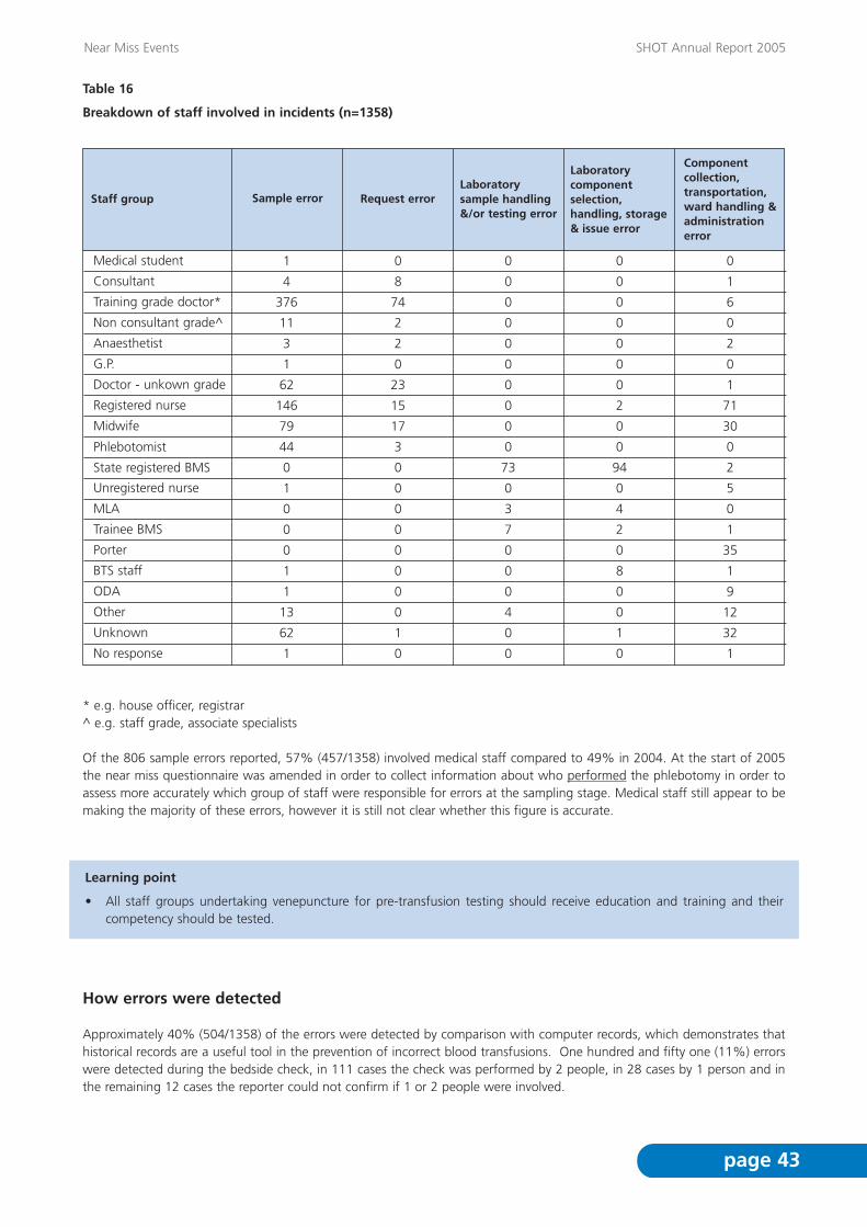
Near Miss Events SHOT Annual Report 2005
page 43
* e.g. house officer, registrar^ e.g. staff grade, associate specialists
Of the 806 sample errors reported, 57% (457/1358) involved medical staff compared to 49% in 2004. At the start of 2005the near miss questionnaire was amended in order to collect information about who performed the phlebotomy in order toassess more accurately which group of staff were responsible for errors at the sampling stage. Medical staff still appear to bemaking the majority of these errors, however it is still not clear whether this figure is accurate.
Learning point
• All staff groups undertaking venepuncture for pre-transfusion testing should receive education and training and theircompetency should be tested.
How errors were detected
Approximately 40% (504/1358) of the errors were detected by comparison with computer records, which demonstrates thathistorical records are a useful tool in the prevention of incorrect blood transfusions. One hundred and fifty one (11%) errorswere detected during the bedside check, in 111 cases the check was performed by 2 people, in 28 cases by 1 person and inthe remaining 12 cases the reporter could not confirm if 1 or 2 people were involved.
Table 16
Breakdown of staff involved in incidents (n=1358)
Staff group Sample error Request errorLaboratory sample handling&/or testing error
Laboratorycomponentselection, handling, storage & issue error
Componentcollection,transportation,ward handling &administration error
Medical student
Consultant
Training grade doctor*
Non consultant grade^
Anaesthetist
G.P.
Doctor - unkown grade
Registered nurse
Midwife
Phlebotomist
State registered BMS
Unregistered nurse
MLA
Trainee BMS
Porter
BTS staff
ODA
Other
Unknown
No response
1 0 0 0
0
0
0
0
0
0
2
0
0
94
0
4
2
0
8
0
0
1
0
0
1
6
0
2
0
1
71
30
0
2
5
0
1
35
1
9
12
32
1
0
0
0
0
0
0
0
0
0
73
0
3
7
0
0
0
4
0
0
8
74
2
2
0
23
15
17
3
0
0
0
0
0
0
0
0
1
0
4
376
11
3
1
62
146
79
44
0
1
0
0
0
1
1
13
62
1
Near Miss Events SHOT Annual Report 2005
page 44
The future of near miss
The number of near miss reports received increased again in 2005, and participation in the scheme has risen to 55% (223/403eligible hospitals). In order to gain a clearer picture of the number and types of near miss events that are occurring, thecollection of individual near miss events was suspended at the start of 2006. The reporting of near miss events to MHRA underthe terms of the EU Directive remains unchanged. During 2006 a 6 month survey (June - December) will collect summaryinformation relating to the number of events at each of the following stages of the transfusion process; sampling, request,laboratory testing and issue and collection and administration.
COMMENTARY
• Errors at the sampling stage continue to comprise over 50% of the near miss incidents reported. As in 2004, the datasuggests that medical staff were involved in 57% of these errors and again highlights the need for inclusion of educationin blood safety in the medical curriculum at undergraduate and postgraduate levels.
• Approximately 10% of errors reported occurred at the request stage. The majority of these were detected by the vigilanceof the laboratory staff.
• Laboratory staff failed to provide for the patients special requirements in 39% of the cases in the laboratory componentselection, handling, storage and issue errors category.
• There were 209 cases which fell in to the component collection, transportation, ward handling and administration errorscategory, of these 130 (62%) involved components which were inappropriately handled or stored.
• Since near miss reporting began 5 years ago, the number of reports submitted have increased by 200%. However, only55% of hospitals are regularly participating in the scheme.
RECOMMENDATIONS
• Training and education in blood sampling, including positive patient ID, should be included in the curriculum for all staffinvolved in venepuncture.
Action: Chief Medical Officers (CMOs) NBTC and counterparts, Undergraduate Deans of Schools of Nursingand Medicine.
• All staff involved in the pre-transfusion sampling, testing and issue of blood must be deemed competent having undergoneappropriate training, which must be documented.
Action: Trust CEOs through risk management structures.
• Ward staff at all levels must be trained in appropriate storage of blood components once they have been collected fromthe transfusion laboratory.
Action: Ward managers, HTTs.

Death
Outcome Imputability level Case number Component Reaction type Comment
2 10 FFP Anaphylactic/anaphylactoid
Obstructivejaundice. Vit K not given
Major morbidity 2 6 Red cells Anaphylactic/anaphylactoid
Cardiac arrest,severely atopicrecipient
2 8 Red cells Unclassifiable Hypoxia requiringventilation
2 11 FFP Anaphylactic/anaphylactoid
Cardiac arrest
2 12 FFP Anaphylactic/anaphylactoid
Myocardial infarct
2 13 Platelets Unclassifiable Respiratory arrest
1 9 Red cells ALI Multiple factors
2 14 Granulocytes fromfamily member
Anaphylactic/anaphylactoid
Ventriculartachycardia
Acute Transfusion Reactions SHOT Annual Report 2005
page 45
7 Acute Transfusion Reactions
Definition
Acute transfusion reactions are defined in this report as those occurring at any time up to 24 hours following a transfusionof blood or components, excluding cases of acute reactions due to incorrect component being transfused as these are coveredin Section 4
Eighty-nine completed questionnaires were analysed. Twenty-one were withdrawn by the analyst; 15 of these were reports ofnon-haemolytic febrile reactions, one of which occurred following the re-infusion of red cells salvaged post-operatively, in 5the symptoms were due to the underlying pathology and the final report withdrawn was of an unavoidable incompatibletransfusion that did not lead to a reaction.
This chapter highlights the main findings from 68 completed questionnaires.
Gender (68 reports)
Males 35 Females 33
Age (68 reports)
Age range 6 months to 84 yearsMedian 56 years
Deaths and major morbidity
There was 1 death in this group, likely to be related to transfusion (imputability level 2) and 7 reports of major morbidity; 6likely to be related (imputability level 2) and 1 possibly related (imputability level 1) to transfusion.
Table 17
Deaths and major morbidity
Components implicated (68 reports)
Red cells 23Platelets 20 (8 apheresis units and 12 from pooled buffy coats)
Granulocytes 1Fresh frozen plasma (FFP) 22FFP - solvent-detergent 1Red cells and FFP 1

Reactions in which red cells were implicated
There were 23 cases, with 3 instances of major morbidity; 2 likely to be due to the transfusion and 1 possibly due to thetransfusion. 17 reactions occurred during the transfusion, 3 within 2 hours, 1 within 7 hours and 1 within 24 hours ofcompleting the transfusion. In the final case the precise timing was unclear.
The following reactions were seen:
Table 18
Reactions in which red cells were implicated
Acute Transfusion Reactions SHOT Annual Report 2005
page 46
+ anaphylactic/anaphylactoid (hypotension with 1 or more of: rash, dyspnoea, angioedema)++ allergic (1 or more of: rash, dyspnoea or angioedema without hypotension)
Haemolytic Reactions
In 3 of the 5 cases, a reference laboratory was involved in either providing antigen matched units or the subsequentinvestigation of the reaction.
Case 1
A case of a 69 year old lady with a transfusion-dependent myeloproliferative disorder and multiple red cell alloantibodies (anti-K+Kpa+S+C+Fya+Kna/McCa) who had experienced a delayed transfusion reaction to a previous transfusion (see case 13 in theDelayed Transfusion Reaction Chapter). She developed an anti-M over this period but the red cells causing the acute reactionwere M-negative.
Case 2
A 65 year old male with gastrointestinal (GI) bleeding had been transfused with red cells within the previous 14 days. A sampleless than 48 hours old was used to crossmatch 2 further units. The patient developed a fever and back pain at some stagewithin the 24 hours following the transfusion. This was not documented contemporaneously, and the nursing staff wereunaware of a reaction having occurred. There was however no post-transfusion increment in haemoglobin and brown plasmawas noted in the sample sent to the laboratory 36 hours later. The pre-transfusion antibody screen was negative using plasma(DiaMed). Post-transfusion, anti-Jka was found in the plasma and eluted from the red cells. Retrospective testing of a pre-transfusion serum sample showed weak reactions with no demonstrable specificity.
Case 3
A 33 year old female, with unexplained red cell aplasia, required monthly red cell transfusions and she had been previouslynoted to pass dark red urine in the 24 hours following transfusion. On this occasion a poor haemoglobin increment andhyperbilirubinaemia were also noted. The DAT was positive (C3d only), her antibody screen was consistently negative usingDiaMed ID and there was no evidence of paroxysmal nocturnal haemoglobinuria (PNH). The reference laboratory found anauto anti-c reacting by DiaMed enzyme IAT.
Subsequent transfusions of R1R1 cells have been given with no adverse effects.
Case 4
A 72 year old lady with disseminated carcinoma of the ovary and a previous carcinoma of the breast was transfused in acommunity hospital with 2 units of red cells. Within 2 hours of completing the second unit, she became febrile and passeddark urine. She was known to have been transfused in 1999 but there was no record of subsequent transfusions. Her pre-transfusion antibody screen using Immucor Capture-R was negative, as were 4 previous antibody screens dating back to 1999.The units had been issued electronically.
Reaction type Number of cases
Haemolytic
Anaphylactic+
Allergic++
5
5
8
Unclassifiable
Febrile
2
3 (of 14 initially reported)

15 x x x x x
x x x
x
x x
x x x x
x x nk
x x x
x x nk
x x x x
16
17
7
18
19
20
21
Case No. Fever Rigors Hypertension Rash Dyspnoea Hypoxia Angioedema
The laboratory was only informed of the reaction 10 days later and the post-transfusion sample revealed an anti-K reactingweakly in DiaMed IAT. Further investigation confirmed that the first unit given was K positive.
Case 5
A 45 year old male with HIV, anaemia and a positive DAT (IgG and C3d), being treated with antiretroviral therapy wastransfused with 2 units of red cells. The patient was not known to have autoimmune haemolysis (rare in this conditionalthough a positive DAT is common). After receiving 100mL of the second unit, he developed rigors, back pain, restlessnessand hypotension. He was noted to pass dark urine and his bilirubin subsequently rose to 51µmol/l. The unit was confirmedto be ABO identical and the antibody screen was negative pre- and post-transfusion. Further retesting of the pre-transfusionsample including a repeat crossmatch confirmed the negative findings, but samples were not referred to a referencelaboratory. He has not received further red cells in this hospital and there is no further follow-up.
Anaphylactic/anaphylactoid reactions
Five anaphylactic/anaphylactoid reactions were reported. There was one case of major morbidity (case 6) and a second patientalready on the intensive therapy unit (ITU), required further inotropic support following initial resuscitation.
Case 6
A 37 year old female was known to have severe atopic eczema, asthma and to be allergic to peanuts. As a toddler, shedeveloped urticaria following ingestion of peanut butter but by the age of 16 years had obstructive laryngeal oedema oninhaling peanut powder. She was not allergic to latex and had no previous transfusion history.
Following a caesarean section for haemolysis, elevated liver enzymes, and a low platelet count (HELPP) syndrome and a post-partum haemorrhage, she was transfused with 2 units of red cells when she returned to the ward. The first unit wastransfused uneventfully. The second unit was commenced after an interval of 2 hours and within the first 5 minutes she haddifficulty breathing, collapsed and had a cardiac arrest from which she was successfully resuscitated.
Serial mast cell tryptases showed an elevation to 60.1ug/l 2 hours after the arrest with a fall to normal levels 24 hours later,in keeping with mast cell degranulation. Subsequent investigation of the patient revealed a normal IgA level, a total serumIgE level >5000u (NR 5-120) and strongly positive IgE radioallergosorbent test (RAST) to nuts, eggs and wheat.
Given the temporal relationship of her reaction to the second unit of red cells, the question was raised as to whether thisdonor had eaten any of the allergens to which she was known to be allergic. However both donors’ sera were tested forpeanut protein with negative findings. An alternative explanation of the patient having inadvertently ingested a knownallergen was thought less likely given her immediate post-operative state.
Of the 4 remaining cases, 3 were not investigated and the fourth was not IgA deficient.
Allergic Reactions
There were 8 allergic reactions reported with features as noted below:
Table 19
Clinical features of allergic reactions to red cells
Acute Transfusion Reactions SHOT Annual Report 2005
page 47
(nk = not known)

Acute Transfusion Reactions SHOT Annual Report 2005
page 48
In 4 of the cases, fever and dyspnoea (without recorded wheeziness) were the only manifestations, with reduced oxygensaturation, when measured. HLA antibodies were recorded in 2 but not tested for in the remaining 2.
In 5 out of the 8 cases IgA levels were measured, with one IgA deficiency but no anti-IgA.
Case 7
A 22 year old male climber had a combined tibia/fibula fracture and required a post-operative transfusion. After 50mL, hebecame febrile with rigors, wheezy, hypertensive and vomited. His white cell count was normal at 7.2 x 10⁹/l before thetransfusion, but fell to 1.6 x 10⁹/l immediately following with only 0.02 x 10⁹/l neutrophils. He responded promptly tohydrocortisone, antihistamine and a bronchodilator. The neutrophil count returned to normal within 5 hours. Cultures of thepatient and the red cells were negative, a mast cell tryptase was not performed.
The patient’s serum did not contain granulocyte specific antibodies and the transient nature of the neutropenia suggests thatmargination could have occurred. The chest x-ray (CXR) showed no abnormality.
Febrile reactions
Of the 17 febrile reactions reported this year, 3 are included in the report. Two were the result of red cell alloantibodies notdetectable in the patients’ pre-transfusion antibody screen, and the electronic issue and transfusion of incompatible units. Ananti-Wra caused fever and chills following the transfusion of 140mL red cells, and an anti-Bga caused fever towards the end ofthe transfusion. In neither case were features of haemolysis mentioned.
A third febrile reaction in a 74 year old male resulted in an overnight admission for observations since the reaction provokedchest pain and restlessness.
Of these 17 febrile reactions, sufficiently severe to stop the transfusion, there are only 9 reports of the units being cultured.
Unclassifiable reactions
Case 8
An 83 year old male with a lymphoplasmacytoid lymphoma and an IgM paraprotein had already received 4 units of red cellswithin the last 48 hours. During transfusion of the fifth unit he became febrile, hypertensive and dyspnoeic. The transfusionwas stopped for immediate investigation of red cell incompatibility and then recommenced. The symptoms returned, worsethan previously and, since his dyspnoea and wheeziness did not immediately respond to piriton, hydrocortisone andsalbutamol,(oxygen saturation 82% on 100% O2) he required intubation. He recovered within 3 hours. Investigations for TRALIwere negative and although the features could be in keeping with a severe allergic reaction, a component ofhyperviscosity/fluid overload cannot be excluded.
Case 9
A 32 year old male with sickle cell disease was admitted with an evolving acute chest syndrome and over the following 48hours he was transfused with 3 units of red cells followed by an exchange transfusion of 6 units. His chest symptoms improved.On the third day he was given a further 2 units of red cells without incident. The transfusion finished at 2340 hours and hisobservations remained stable until 0600 hours when he had a grand mal fit. He was subsequently noted to be hypoxic andtachypnoeic and required intubation. He then had a cardiac arrest from which he was successfully resuscitated. The CXRshowed bilateral pulmonary infiltrates consistent with acute lung injury (ALI).
TRALI was suspected by the intensivists but the last 2 units and the patient’s serum tested negative for HLA and granulocyteantibodies. Volume overload was considered in view of his known renal impairment. However in addition to the red cells, otherfactors including his recent chest syndrome and likely hypoxia during a grand mal fit, could have contributed to thedevelopment of ALI.
Reactions in which FFP was implicated
There were 24 reports in this group (one in conjunction with red cells), with one death likely to be due to the transfusion and2 instances of major morbidity also likely to be due to the transfusion. Eighteen occurred during the transfusion and 6 within2 hours of the transfusion.
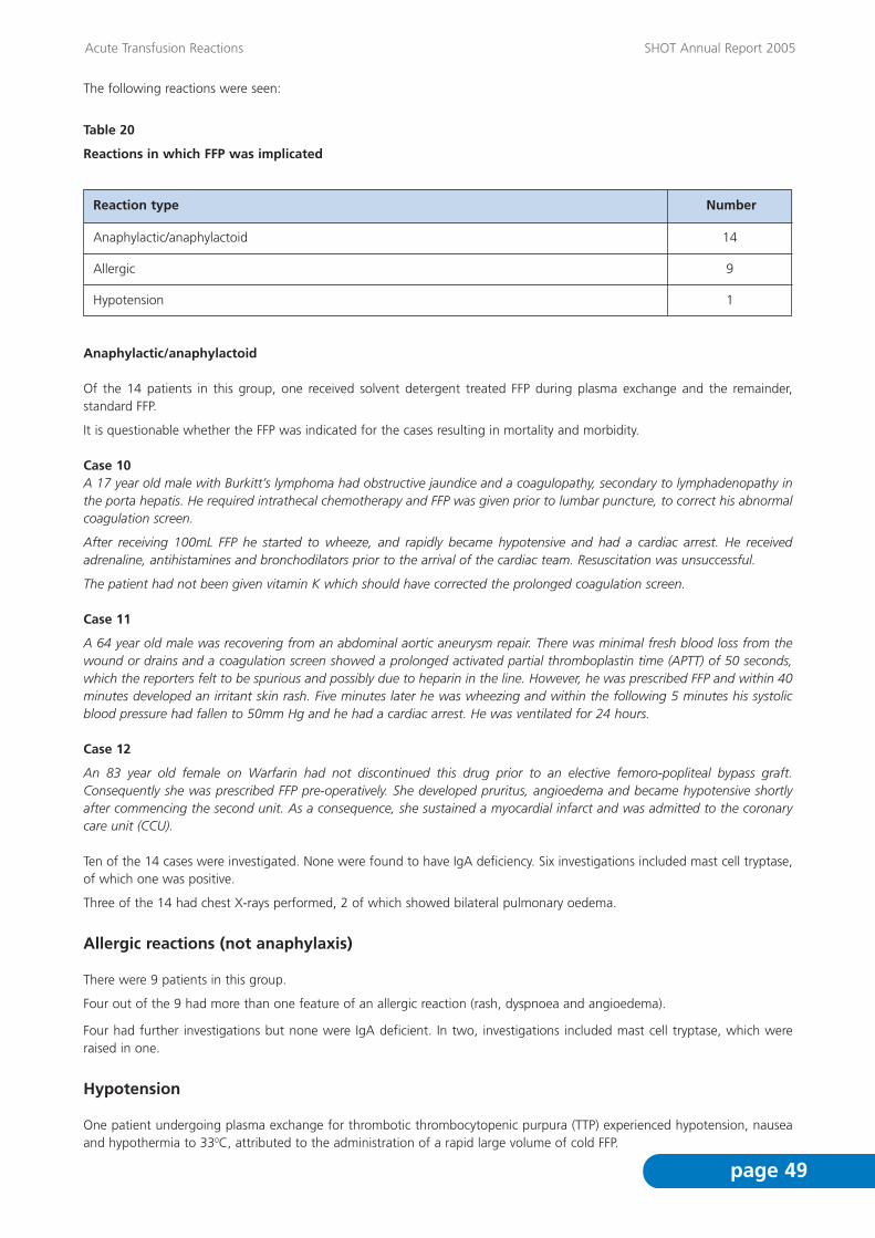
Acute Transfusion Reactions SHOT Annual Report 2005
page 49
The following reactions were seen:
Table 20
Reactions in which FFP was implicated
Anaphylactic/anaphylactoid
Of the 14 patients in this group, one received solvent detergent treated FFP during plasma exchange and the remainder,standard FFP.
It is questionable whether the FFP was indicated for the cases resulting in mortality and morbidity.
Case 10A 17 year old male with Burkitt’s lymphoma had obstructive jaundice and a coagulopathy, secondary to lymphadenopathy inthe porta hepatis. He required intrathecal chemotherapy and FFP was given prior to lumbar puncture, to correct his abnormalcoagulation screen.
After receiving 100mL FFP he started to wheeze, and rapidly became hypotensive and had a cardiac arrest. He receivedadrenaline, antihistamines and bronchodilators prior to the arrival of the cardiac team. Resuscitation was unsuccessful.
The patient had not been given vitamin K which should have corrected the prolonged coagulation screen.
Case 11
A 64 year old male was recovering from an abdominal aortic aneurysm repair. There was minimal fresh blood loss from thewound or drains and a coagulation screen showed a prolonged activated partial thromboplastin time (APTT) of 50 seconds,which the reporters felt to be spurious and possibly due to heparin in the line. However, he was prescribed FFP and within 40minutes developed an irritant skin rash. Five minutes later he was wheezing and within the following 5 minutes his systolicblood pressure had fallen to 50mm Hg and he had a cardiac arrest. He was ventilated for 24 hours.
Case 12
An 83 year old female on Warfarin had not discontinued this drug prior to an elective femoro-popliteal bypass graft.Consequently she was prescribed FFP pre-operatively. She developed pruritus, angioedema and became hypotensive shortlyafter commencing the second unit. As a consequence, she sustained a myocardial infarct and was admitted to the coronarycare unit (CCU).
Ten of the 14 cases were investigated. None were found to have IgA deficiency. Six investigations included mast cell tryptase,of which one was positive.
Three of the 14 had chest X-rays performed, 2 of which showed bilateral pulmonary oedema.
Allergic reactions (not anaphylaxis)
There were 9 patients in this group.
Four out of the 9 had more than one feature of an allergic reaction (rash, dyspnoea and angioedema).
Four had further investigations but none were IgA deficient. In two, investigations included mast cell tryptase, which wereraised in one.
Hypotension
One patient undergoing plasma exchange for thrombotic thrombocytopenic purpura (TTP) experienced hypotension, nauseaand hypothermia to 330C, attributed to the administration of a rapid large volume of cold FFP.
Reaction type Number
Anaphylactic/anaphylactoid
Allergic
Hypotension
14
9
1

Acute Transfusion Reactions SHOT Annual Report 2005
page 50
Inappropriate use of FFP
Coagulation results are not available for all cases satisfying the definition of massive transfusion, but these have been includedas clinically indicated.
Category
Clinically indicated TTP - 2DIC with haemorrhage - 1Massive transfusion - 4Liver disease with intervention - 2Pre-operative correction coagulopathy - 1Post-operative bleed with raised INR - 4
14
Possibly indicated In obstructive jaundice, without trial ofVitamin K prior to lumbar puncture - 1Postoperative aneurysm repair, prolongedAPTT, ? spurious - 1
2
Not indicated Liver disease with no bleeding - 1Postoperative, no bleeding - 3Carcinoma colon, no bleeding - 1Warfarin reversal prior to elective surgery orfor minor haemorrhage - 3
8
No. of patients Indication given
Reactions in which platelets were implicated
There were 20 reactions to platelets, of which 17 occurred during the transfusion, 2 within 2 hours and one within 7 hoursfollowing the transfusion. One patient suffered major morbidity, likely to be as a result of the transfusion.
Table 21
Reactions in which platelets were implicated
Reaction type Number of cases
Anaphylactic/anaphylactoid
Allergic
Hypotension
5
11
3
Unclassifiable 1
Anaphylactic/anaphylactoid reactions
Five patients suffered anaphylactic/anaphylactoid reactions, 3 following the transfusion of pooled buffy coat derived plateletsand 2 following apheresis platelets.
One patient initially developed a skin rash and mild dyspnoea. The transfusion was temporarily stopped whilst an antihistamineand hydrocortisone were given. On restarting the transfusion the patient rapidly developed an anaphylactoid reaction.
Three of the 5 who were receiving platelets in a day ward setting required admission.
All were multi-transfused recipients. Three were investigated for HLA and human platelet antigen (HPA) antibodies withnegative findings. Three of the 5 are still platelet dependent and receiving platelets in platelet suspension medium (PSM) withno adverse reactions.

Acute Transfusion Reactions SHOT Annual Report 2005
page 51
Allergic reactions
Eleven patients suffered allergic reactions.
Seven out of the 11 had more than one feature of an allergic reaction (rash, dyspnoea and angioedema).
Five were investigated, 2 for IgA deficiency and 2 for HLA antibodies, with negative findings. Two of the 5 were also testedfor mast cell tryptase, one of which was increased.
Hypotensive reactions
Three patients, who were not receiving angiotensin-converting enzyme (ACE) inhibitors, had hypotension alone.
Unclassifiable
Case 13A 24 year old female with high grade Non Hodgkins Lymphoma (NHL), was platelet and red cell dependent post allograft.Within 5 minutes of the transfusion of apheresis platelets, she started to sweat profusely, complained of ‘tightness’ in herchest, developed a tachycardia and her BP rose to 170/110. On examination she had reduced air entry with crackles and anoxygen saturation of 75% and subsequently had a respiratory arrest.The patient was an out-patient who had received no other parenteral fluids that day, making volume overload very unlikely.Cultures of the patient and the platelet concentrate were negative.A CXR performed following the arrest showed ‘ground glass’ changes, rather than bilateral pulmonary oedema. She recoveredon ITU but was subsequently found to have a large intracerebral bleed, which may have been precipitated by the reaction.
Reaction in which granulocytes were implicated
There was one case of an anaphylactic/anaphylactoid reaction with major morbidity.
Case 14
A 32 year old male with aplastic anaemia received granulocytes from a family member. During the transfusion, his bloodpressure dropped to unrecordable levels and he lost consciousness. He was successfully resuscitated with hydrocortisone,piriton and adrenaline and subsequently required amiodarone for adrenaline induced ventricular tachycardia.He was admitted to HDU.
Response times
The majority of patients were seen as soon as possible by a doctor, and a Consultant Haematologist was also consulted forreactions involving red cells or platelets.
Table 22
Time taken for patient to be reviewed by a doctor
* includes case in which both FFP and red cells were transfusedincludes case involving granulocyte concentrate
Response times Red Cells (23) FFP (24) * Platelets (21)
< 15 minutes
< 30 minutes
< 60 minutes
> 60 minutes
Unknown
Total
Involvement of Haematologist
12
3
2
2
4
23
19
20
1
3
24
10
15
3
1
2
21
15

Acute Transfusion Reactions SHOT Annual Report 2005
page 52
Changes made to procedures
Two hospitals have reinforced the need to avoid elective transfusions during night shifts as a result of reactions beinginadequately managed at this time.
Reporting of acute transfusion reactions
All but 1 acute transfusion reactions were reported to the hospital laboratory and the majority were also reviewed at theHospital Transfusion Committee. Three of the 5 haemolytic reactions involved the Transfusion Centre and other reactions werereported to them when samples were referred for investigation.
Table 23
Reporting of reactions to the Hospital Transfusion Committee, Hospital Laboratory and the local TransfusionCentre
* includes case in which both FFP and red cells were transfusedincludes case involving granulocyte concentrate
COMMENTARY
• Case 1 illustrates the difficulties posed by patients with complex antibodies requiring repeat transfusions.
• There is still significant inappropriate prescription of FFP.
• There has been an increase in the number of allergic or anaphylactic/anaphylactoid reactions reported due to red cells.
• In only 9/17 patients reported to have significant febrile reactions to red cells were bacterial cultures performed.
• The reactions cause by red cell alloantibodies (anti-Wra and anti-Bga), not detectable using screening cells meeting BCSHrecommendations, did not result in haemolysis.
• Transfusion reactions occurring at night or in a community setting may not be managed as promptly.
Reported to Red Cells (23) FFP (24) * Platelets (21)
Hospital TransfusionCommittee
Hospital laboratory
Transfusion centre
20
23
18
16
24
7
16
20
12
Acute Transfusion Reactions SHOT Annual Report 2005
page 53
RECOMMENDATIONS
• Pre-transfusion testing on patients who have been recently transfused and require further transfusion should be carried outin accordance with BCSH Guidelines21 relating to the timing of pre-transfusion samples.
Action: Hospital transfusion laboratories.
• BCSH Guidelines22 on the management of anticoagulation in the peri-operative period and on the management ofexcessive anticoagulation should be followed. Excessive anticoagulation with minor haemorrhage should be treated bystopping the drug and if necessary with intravenous vitamin K.
• BCSH guidelines9 for the use of FFP should be followed. Its use for the correction of abnormal coagulation results in theabsence of bleeding is not justified.
Action: Consultant haematologists with responsibility for transfusion should ensure that BCSH guidelinesare incorporated into local protocols.
• All serious transfusion reactions must be fully investigated. Bacterial cultures must be taken in a febrile reaction, when therise in temperature exceeds 1.50C or the reaction is otherwise sufficiently severe to merit discontinuing transfusion. Anupdate of BCSH guidelines is in progress.
Action: Consultant haematologists with responsibility for transfusion should implement current bestpractice.12,13
• Blood should not be transfused outside of core hours unless clinically essential.
Action: Hospital CEOs, consultant haematologists with responsibility for transfusion together with HTCsand HTTs.
• Against the background of a trend towards provision of care closer to the patient, there is a need for a standard of practiceto be developed for transfusion in the community setting, including provision for appropriate management and reportingof adverse reactions and events.
Action: UK National Blood Transfusion Committees to facilitate and co-ordinate.

Delayed Transfusion Reactions SHOT Annual Report 2005
page 54
Definition
Delayed transfusion reactions are defined as those occurring more than 24 hours following a transfusion of blood or bloodcomponents. In practice, these are usually delayed haemolytic reactions due to the development of red cell alloantibodies.Simple serological reactions (antibody development without a positive DAT or evidence of haemolysis) are excluded.
Twenty-nine delayed transfusion reaction (DTR) questionnaires were received, one of which was transferred to the AcuteTransfusion Reaction chapter.
This chapter describes the main findings from 28 completed questionnaires.
Patients
11 males and 17 females
Ages ranged from 8 to 89
Definition of severity of reaction/ clinical sequelae
Symptoms and signs are divided into 4 categories as follows:
• Group 1 Asymptomatic (with positive DAT only)• Group 2 Falling haemoglobin ( Hb)/positive DAT/spherocytes (2 of these parameters)• Group 3 Hb + jaundice positive DAT spherocytes• Group 4 As group 3 + renal impairment
Severity
One patient required ICU admission and subsequently died, but this was thought to be unrelated to the transfusion(Imputability 0).
6 patients were asymptomatic with a positive DAT and antibody development only (Group 1).
22 patients had evidence of increased red cell destruction but without renal impairment:
• In 7 cases the only sign was a fall in haemoglobin, with or without a positive DAT and spherocytes (Group 2)• In 15 cases there was a fall in haemoglobin and a raised plasma bilirubin, although clinically detectable jaundice was only
reported in 5 cases (Group 3). One of these patients died (case 9 - see vignette), with no evidence that the death wasrelated to the transfusion and no detectable alloantibody.
Sequelae
6 patients were already on ICU, 5 required admission to the ward from outpatients or day care, and at least 5 required furthertransfusion. Only one was reported as posing a ‘risk to life’ (case 17).
8 Delayed Transfusion Reactions
+_+_

Delayed Transfusion Reactions SHOT Annual Report 2005
page 55
Figure 8
Time relationship to transfusion
8
7
6
5
4
3
2
1
01 3 4 5 6 7 8 10 11
No of days
No
of
case
s
12 14 15 16 20 25 30
Median = 7 days
Range = 1 to 30 days
Figure 8 shows the interval in days between the implicated transfusion and signs or symptoms of a DHTR. The intervals givenare necessarily those when the signs or symptoms were first noted; in asymptomatic cases this relates to the number of daysthat elapsed before a repeat sample happened to be tested.
There were 3 cases where symptoms were noted within 72 hours of transfusion: in the first (case 2) no earlier recenttransfusion was reported and repeat testing of the pre-transfusion sample was not undertaken; in the second (case 12 - seevignette) another transfusion had been given 12 days earlier; in the third (case 27), another 3 transfusions had been given inthe preceding 16 days, the most recent of which was 5 days before the DHTR. In both of these latter cases the earliertransfusions were more likely to have been implicated in the DHTR than the reported transfusion.

Delayed Transfusion Reactions SHOT Annual Report 2005
page 56
Serological findings
Kidd antibodies were the most commonly implicated, in 11/28 (39%) of cases, either singly or in conjunction with otherspecificities. Two patients had no detectable antibodies and are described as vignettes. Table 24 shows the specificity of newantibodies in plasma and eluate, and the number of days post transfusion. Tables 25 and 26 show new specificities by bloodgroup system and severity, respectively.
Table 24
Case number
New antibody (ies) in plasma
Antibodies in Eluate CommentsNo. dayspost tx
1 No eluate performed 7
2 No eluate performed 3
Anti-Jkb
Anti-Jka Pre-existing anti-E+K. No recordof recent transfusion
3 No eluate performed 10Anti-Jka Already in ICU
4 Anti-E 30Anti-K + E
5 Non-specific reactions 25None in plasma Further transfusion required
6 No eluate performed 7Anti-Fya
7 No eluate performed 15Anti-Jka + c Required admission
8 No eluate performed 14Anti-S Required admission
9 No eluate performed 4None Sharp rise in bilirubin. DATnegative. Died unrelated totransfusion
10 Anti-Jka 5None in plasma
11 Anti-E + Jka 5Anti-E + Jka
12 Anti-S 1-2Anti-S Also transfusion 12 dayspreviously
13 No eluate performed 7Anti-M Required admission. History ofanti-C+Fya+S+K+Kpa+Kna+McCa.DAT negative.
14 Eluate negative 11Anti-Lua + Jkb Required admission
Already in ICU. Antibody screenweakly positive on retrospectivetesting.
Already in ICU. Pre-existinganti-K+E. Anti-M previouslyidentified elsewhere.
15 Anti-S 20Anti-S
16 No eluate performed 7Anti-Fya + E(E enzyme only)
17 No eluate performed 6M+cold agglutinins
Required admission and furthertransfusion
18 Anti-E 16Anti-c + E
19 Eluate negative 10Anti-C
Already in ICU20 Anti-Fya 11Anti-Fya + E(E enzyme only)
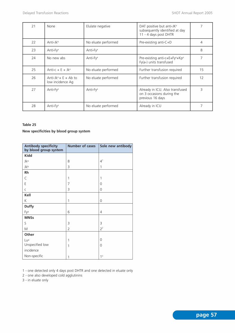
Delayed Transfusion Reactions SHOT Annual Report 2005
page 57
Table 25
New specificities by blood group system
1 - one detected only 4 days post DHTR and one detected in eluate only2 - one also developed cold agglutinins3 - in eluate only
Antibody specificity by blood group system
Kidd
Jka
Jkb
Rh
C
E
c
Kell
K
Duffy
Fya
MNSs
S
M
Other
Lua
Unspecified low
incidence
Non-specific
Number of cases
8
3
1
7
3
1
6
3
2
1
1
1
Sole new antibody
41
1
1
0
0
0
4
3
22
0
0
13
21 Elulate negative 7None
Anti-Jkb
DAT positive but anti-JKa
subsequently identified at day11 - 4 days post DHTR
Pre-existing anti-C+D22 No eluate performed 4
Anti-Fya23 Anti-Fya 8
No new abs Pre-existing anti-c+E+Fya+Kpa
Fy(a-) units transfused24 Anti-Fya 7
Anti-c + E + Jka Further transfusion required25 No eluate performed 15
Anti-Jka + E + Ab tolow incidence Ag
Further transfusion required26 No eluate performed 12
Anti-Fya Already in ICU. Also transfusedon 3 occasions during theprevious 16 days
27 Anti-Fya 3
Anti-Fya Already in ICU 28 No eluate performed 7
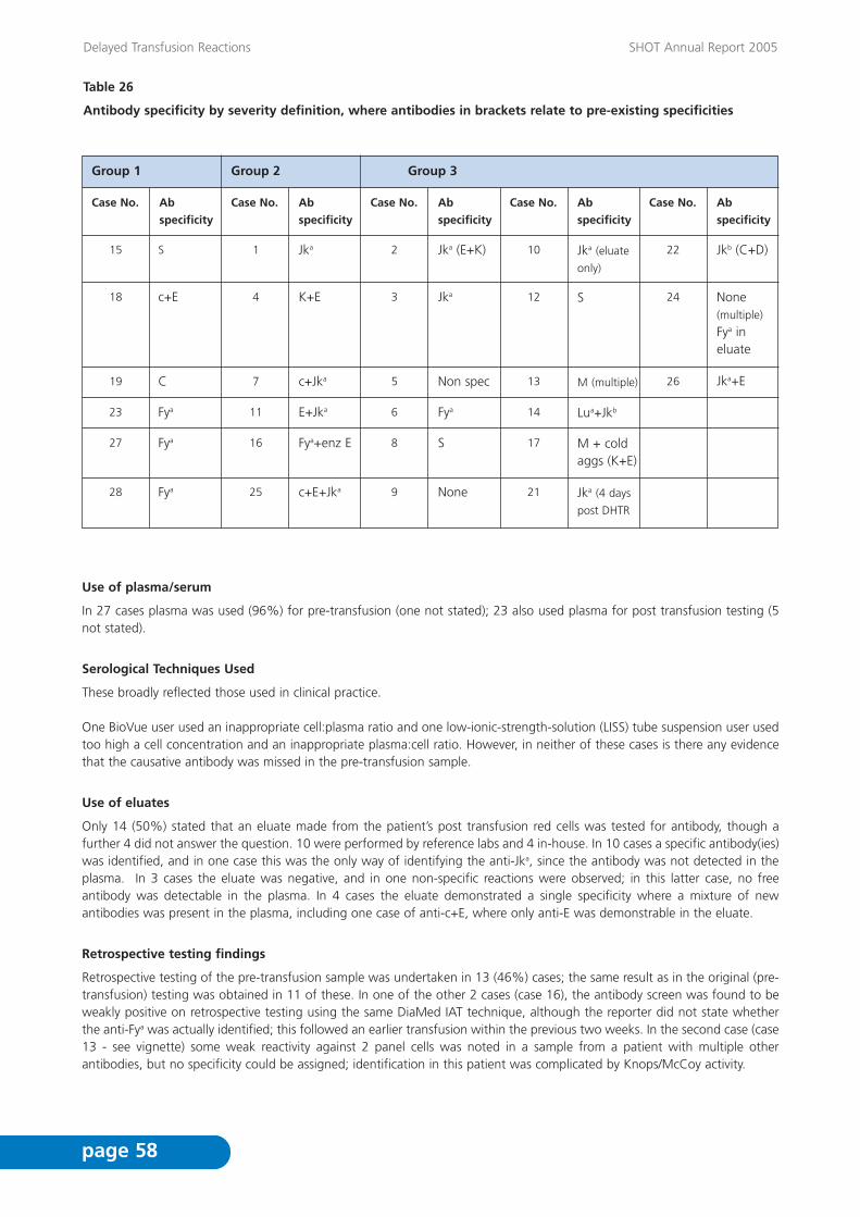
Delayed Transfusion Reactions SHOT Annual Report 2005
page 58
Table 26
Antibody specificity by severity definition, where antibodies in brackets relate to pre-existing specificities
Use of plasma/serum
In 27 cases plasma was used (96%) for pre-transfusion (one not stated); 23 also used plasma for post transfusion testing (5not stated).
Serological Techniques Used
These broadly reflected those used in clinical practice.
One BioVue user used an inappropriate cell:plasma ratio and one low-ionic-strength-solution (LISS) tube suspension user usedtoo high a cell concentration and an inappropriate plasma:cell ratio. However, in neither of these cases is there any evidencethat the causative antibody was missed in the pre-transfusion sample.
Use of eluates
Only 14 (50%) stated that an eluate made from the patient’s post transfusion red cells was tested for antibody, though afurther 4 did not answer the question. 10 were performed by reference labs and 4 in-house. In 10 cases a specific antibody(ies)was identified, and in one case this was the only way of identifying the anti-Jka, since the antibody was not detected in theplasma. In 3 cases the eluate was negative, and in one non-specific reactions were observed; in this latter case, no freeantibody was detectable in the plasma. In 4 cases the eluate demonstrated a single specificity where a mixture of newantibodies was present in the plasma, including one case of anti-c+E, where only anti-E was demonstrable in the eluate.
Retrospective testing findings
Retrospective testing of the pre-transfusion sample was undertaken in 13 (46%) cases; the same result as in the original (pre-transfusion) testing was obtained in 11 of these. In one of the other 2 cases (case 16), the antibody screen was found to beweakly positive on retrospective testing using the same DiaMed IAT technique, although the reporter did not state whetherthe anti-Fya was actually identified; this followed an earlier transfusion within the previous two weeks. In the second case (case13 - see vignette) some weak reactivity against 2 panel cells was noted in a sample from a patient with multiple otherantibodies, but no specificity could be assigned; identification in this patient was complicated by Knops/McCoy activity.
Group 1 Group 2 Group 3
Case No.
15
Ab
specificity
Case No.
S 1
Ab
specificity
Case No. Ab
specificity
Case No. Ab
specificity
Case No. Ab
specificity
Jka 2 Jka (E+K) 22 Jkb (C+D)10 Jka (eluate
only)
18 c+E 4 K+E 3 Jka 24 None(multiple)
Fya ineluate
12 S
19 C 7 c+Jka 5 Non spec 26 Jka+E13 M (multiple)
23 Fya 11 E+Jka 6 Fya 14 Lua+Jkb
27 Fya 16 Fya+enz E 8 S 17 M + coldaggs (K+E)
28 Fya 25 c+E+Jka 9 None 21 Jka (4 days
post DHTR
Delayed Transfusion Reactions SHOT Annual Report 2005
page 59
Clinical management and review
15 (54%) of cases were referred to the Blood Centre Reference Laboratory and 25 (89%) to the HTC. Twelve of these werereported to both.
Vignettes
No antibodies detected - no clear evidence of a DHTR (imputability 1)
Case 5
A 34 year old female patient with post-operative bleeding required 3 units of red cells. 26 days later her DAT was stronglypositive (IgG) and spherocytes were noted on her blood film. Samples were referred to the blood service red cell referencelaboratory, but no antibodies were detected in the plasma, and an eluate made from the patient’s red cells showed nospecificity. The Hb dropped by 3g/dL between day 25 and day 27, with a small rise in bilirubin, but no signs of bleeding. Sherequired a further transfusion, which was followed by a similar drop in Hb over the next 48 hours. During this time she alsobecame pyrexial, and Candida was detected in the blood cultures. The patient was subsequently transfused with R1R1 K- redcells, although the presence of transfused red cells made her phenotype impossible to determine; her Hb then remained stable.
Case 9
An 84 year old female patient received 12 units of red cells over an 8 day period for rectal bleeding. 3 days after the mostrecent transfusion, she became jaundiced and was suffering from acute respiratory distress syndrome (ARDS); her bilirubin rosefrom 20 to 127 µmol/L and reached a peak of 231 2 days later. The Hb also fell, but it is unclear whether this was due tobleeding or the transfusion or both. She received a further 2 units 9 days later and once again her bilirubin rose from 167 to291µmol/L. A DAT was not performed until 8 days later when it was negative. The patient was subsequently admitted to ICUand died, but this was thought to be unrelated to the transfusion.
Difficult to classify
Case 10A 75 year old female patient received 3 units of red cells for an inferior mesenteric artery bleed. A group and save sampletested 3 days previously gave a positive antibody screen and a positive DAT (both IgG and complement coating). The samplewas referred to the blood service red cell reference laboratory 2 days later, where anti-Jka was eluted from the patient’s redcells, although no plasma antibody was detected. It is not clear why non-phenotyped blood was issued based on an empiricalcrossmatch. The patient did not get the expected increment in Hb and had a raised bilirubin 5 days after transfusion; however,the reporter indicated that these signs were indistinguishable from those due to multiple disease factors.
This case is difficult to classify: the anti-Jka presumably developed as a result of a transfusion given 14 to 28 days previouslyand was clearly present in the pre-transfusion sample. Whether the blood was transfused without Jka typing, due to a lack ofcommunication or a misunderstanding is not clear. It is also unclear which symptoms were due to a transfusion reaction andwhich were due to disease. This could therefore equally be classified as an acute transfusion reaction or an IBCT.
Case 12
A 7 year old female patient with sickle cell disease was admitted with a painful crisis and a Hb of 6.6g/dL. The antibody screenwas negative and she was transfused with a unit of red cells. 12 days later she was re-admitted, again with painful crisis. HerHb was initially 8.5g/dL but on repeat was found to be only 4.5g/dL. The antibody screen was negative, and although the DATwas weakly positive (due to both IgG and complement coating), this was not investigated until 3 days later because it was aFriday night. She again received one unit of red cells. Over the following 2 days, dark urine was noted and laboratory testsindicated that she had no increment in Hb and a raised bilirubin; a DHTR was suspected. Anti-S was identified post transfusionand was eluted from the patient’s red cells. Both red cell units were found to be S positive. The patient recovered quickly.
This case was reported as a DHTR occurring 1-2 days post the second transfusion. However, the timing and the pre-transfusionDAT, suggest that this is more likely to be a combination of a DHTR due to the transfusion given 12 days previously and anacute haemolytic transfusion reaction (AHTR) following the 2nd transfusion of S positive blood. The picture is also complicatedby the sickle cell crisis.

Delayed Transfusion Reactions SHOT Annual Report 2005
page 60
More unusual/interesting cases
Case 13
A 69 year old woman with a myeloproliferative disorder and a history of multiple red cell transfusions, was known to haveanti-K+Kpa+S+C+Fya+Kna/McCa, although only the anti-K and -Kpa had been recently detectable. On this occasion the pre-transfusion sample showed further reactions and the sample was sent to the reference laboratory for further investigation andcrossmatching. The reference laboratory found anti-Kna and McCa, but no convincing evidence of new antibodies andprovided 3 units of crossmatched blood, 2 of which were transfused 4 days later. Seven days post transfusion the patientdeveloped left upper quadrant pain, progressive splenomegaly and jaundice, and her Hb dropped. The reference laboratorywas sent a post-transfusion sample on which it made an immediate verbal report of anti-M; both transfused units weresubsequently confirmed as M positive. Five days later another transfusion was required and M negative blood was supplied.After 200mL of the first unit, the patient developed fever and rigors, and passed dark urine, requiring admission from the dayward. Spherocytes and hyperbilirubinaemia were later noted. The DAT was negative following both transfusions and noeluates were performed. Bacterial contamination of the unit was excluded and the cause of the acute reaction was notestablished. The acute reaction has also been reported in the ATR chapter (case 1).
This is an unusual case; the anti-M was reported to have been weak (2+) and the DAT was negative, so would be an unusualcause of a DHTR; however there appears to be no obvious alternative cause. Anti-M has been excluded as a cause of the AHTRsince the latter units transfused were M negative.
Case 2
A 72 year old female patient was bleeding following over-anticoagulation with Warfarin, and required a 3 unit red celltransfusion The pretransfusion antibody screen was positive and anti-E+K was identified in the plasma using a BioVue IAT and2-stage enzyme technique; crossmatch compatible, antigen negative blood was transfused. 3 days later the patient passeddark urine and became jaundiced. Laboratory investigations demonstrated a raised bilirubin, a falling Hb and spherocytes.Anti-Jka in addition to anti-E and -K was identified in the plasma, and the DAT was positive, due to both IgG and complementcoating. No eluate was performed and the pre-transfusion sample was no longer available for testing.
Although 3 days is a shorter interval than is classically expected for a DHTR, this patient had no recent history of transfusionand there was no indication of anti-Jka in the pre-transfusion sample, even using a 2-stage enzyme technique.
Case 17
A 16 year old male patient required a transfusion in ICU following emergency surgery. Anti-E+K were identified in his plasmaand one unit of antigen negative, crossmatch compatible blood was transfused. 6 days later he passed dark urine and becamejaundiced. Laboratory investigations demonstrated a raised bilirubin and a falling Hb; the DAT was positive due to both IgGand complement coating and anti-M plus a non-specific cold autoagglutinin were detected in the plasma and confirmed by areference laboratory. An eluate was not performed. Retrospective testing of the pre-transfusion plasma using the sameDiaMed IAT technique confirmed that only anti-E and anti-K were detectable pre-transfusion. Anti-M had apparently beenidentified by a different hospital 2 months previously, but this information was not available until after the patient had beentransfused.
Delayed Transfusion Reactions SHOT Annual Report 2005
page 61
Case 24
An 84 year old male patient with myelodysplasia and a history of transfusion (> 3 months previously) required several units ofred cells over a 2 day period, following a total hip replacement. The patient was known to have anti-c+E+Fya+Kpa, andcrossmatch compatible antigen negative blood was transfused. 7 days later it was noted that the patient had increasedreticulocytes and a raised bilirubin. The reference laboratory confirmed that the DAT was positive due to IgG coating, but thatno new antibodies were detectable. Anti-Fya was identified in an eluate made from the patient’s red cells, despite all thetransfused units apparently being confirmed as Fy(a-).
COMMENTARY
• According to the literature, DHTRs caused by anti-M are rare. Anti-M was found in two cases of DHTR this year, with clinicalsigns of red cell destruction in both cases; i.e. dark urine, falling Hb and raised bilirubin 6 to 7 days post transfusion. Bothof these cases had complex serology, with pre-existing alloantibodies, and in one case newly developed cold agglutinins.Unfortunately in neither case was extensive investigation undertaken to prove whether or not anti-M was responsible forthe red cell destruction. It is particularly difficult to draw any conclusions in case 17, since there were no details availableregarding the patient’s underlying diagnosis, the DAT was positive for complement as well as IgG and anti-M does not bindcomplement, and the transfused unit was not typed for M. There have been 5 cases in previous SHOT reports where anti-M has been identified as a new specificity; in 4 of these, additional specificities were also identified, but there is no recordof eluates being performed.
• In all cases (where an answer was given) plasma rather than serum was used for both pre and post transfusioninvestigations. It is known that weak complement binding antibodies, e.g. some examples of anti-Kidd, may be missedwhen using plasma, unless more sensitive techniques are used, e.g. enzyme IAT.
• In 3 cases DHTRs were reported to have occurred within 72 hours of the implicated transfusion. However, in 2 of these,earlier transfusions were more likely to have been implicated in the reaction.
• Only 50% of investigations included testing an eluate made from the patient’s red cells. Where a mixture of antibodies ispresent, an eluate may help to distinguish which specificity(ies) is more likely to be implicated in a haemolytic reaction.Furthermore, the implicated antibody may only be present in an eluate, as in case 10. Identification of all specificitiespresent is essential if further haemolytic reactions are to be prevented.
• As in previous years, communication problems have contributed to DHTRs, where information about previously knownantibodies has not been available at the time of a subsequent transfusion.

Delayed Transfusion Reactions SHOT Annual Report 2005
page 62
RECOMMENDATIONS
• All cases of suspected AHTR and DHTR should be appropriately investigated, and ideally referred to a reference laboratory.Referring hospitals should make it clear to reference laboratories that they are investigating a DHTR to ensure that timely,appropriate tests are undertaken. Clinical details should be completed on the request forms and the donation numbers ofthe units transfused should be included, so that their phenotype can be determined. Reference laboratories should ensurethat investigation of DHTRs includes testing an eluate made from the patient’s red cells when the DAT is positive.
Action: Hospital blood transfusion laboratories and reference laboratories
• Inconclusive antibody screens should be investigated prior to transfusion and results confirmed with a reference laboratoryif necessary.
Action: Hospital blood transfusion laboratories
• Investigation of a suspected DHTR should include retesting of the pre-transfusion sample (where still available) by differentor more sensitive techniques. Consideration should also be given to requesting clotted samples for investigation ofsuspected DHTRs and using polyspecific antihuman globulin (AHG). These actions may involve referral to a referencecentre.
Action: Hospital blood transfusion laboratories.
• Hospitals and reference laboratories should be encouraged to publish case reports of DHTRs, after appropriateinvestigations have been undertaken, and where the implicated antibody is not recognised as a common cause of suchreactions.
Action: Hospital blood transfusion laboratories and reference laboratories.
• In line with recommendations made in the BCSH Guidelines,21 consideration should be given to issuing antibody cards orsimilar information to all patients with clinically significant red cell antibodies. These should be accompanied by patientinformation leaflets, explaining the significance of the antibody and impressing that the card should be shown in the eventof a hospital admission or being crossmatched for surgery. Laboratories should be informed when patients carryingantibody cards are admitted.
Action: The CMO’s NBTC and its counterparts in Scotland, Wales, and Northern Ireland.
• There is a need for a review, co-ordinated by a professional national body, of how long specimens should be kept post-transfusion. The review needs to consider the relative risks and benefits of storing specimens beyond the time that theyare suitable for use in further crossmatching tests.
Action: BBTS and BCSH.

Transfusion Related Acute Lung Injury SHOT Annual Report 2005
page 63
9 Transfusion Related Acute Lung Injury
Definition
Transfusion-related acute lung injury was defined as acute dyspnoea with hypoxia and bilateral pulmonary infiltrates occurringduring or in the 24 hours after transfusion, with no other apparent cause.
Twenty-six case reports of suspected TRALI were received in this reporting year. Of these, 3 were subsequently withdrawn bythe reporters for a variety of reasons.
Twenty-three cases were analysed. Of these, 6 patients died, 16 suffered short-term major morbidity and one had long termmorbidity. Of the 6 patients who died, death was possibly due to TRALI in 2 cases (imputability 1) and unlikely to be relatedin 4 cases (imputability 0). Figure 9.
Figure 9
Summary of cases reported
The 23 reports received in 2005 related to transfusions which occurred between December 2003 and October 2005 inclusive.Two reports related to transfusions in 2003, 10 related to 2004 and 11 to 2005.
Completed Reports analysed01.01.2005 to 31.12.2005
26
Analysedfor TRALI
23
Highly Likely
3Probable
3Possible
3Unlikely
14
Withdrawn byreporters
3

Transfusion Related Acute Lung Injury SHOT Annual Report 2005
page 64
Assessment of TRALI reports
TRALI can be a difficult diagnosis because there is no single test for this condition and it is easily confused with other causesof acute lung injury and cardiogenic pulmonary oedema. If the clinical picture occurs in a previously fit patient and relevantleucocyte antibodies are found, the diagnosis is straightforward. Often however, it occurs in patients who have other riskfactors for the development of ALI or ARDS. When TRALI is suspected, a detailed assessment of the clinical event is requiredtogether with investigation of the patient and donors. Early discussion with the Blood Service is recommended and bloodsamples (ethylenediaminetetraacetic acid (EDTA) and clotted) from the patient should be sent promptly to a Blood ServiceReference laboratory. Clinical factors which were taken into consideration in the assessment of reported cases included: pre-existing cardiac, pulmonary or other disease e.g. leukaemia with pulmonary leucostasis; time between transfusion andrespiratory deterioration; radiological features; possibility of infection; other risk factors for ALI/ARDS; evidence of circulatoryoverload and/or impairment of cardiac function and time to respiratory recovery with supportive treatment.
Results of TRALI investigations may not be definitive. Because of the frequency of leucocyte antibodies in the donorpopulation, donor antibodies would also be found in many uneventful transfusions if they were similarly investigated. In anNBS study of 1166 female donors, HLA antibodies were found in 14.5% (personal communication, Dr S. MacLennan).
Two Transfusion Medicine Specialists who also reviewed cases last year have assessed the likelihood of TRALI in each case. Allcases reported to the NBS have also been initially assessed by 2 Intensive Care Specialists. Reports were graded on the basisof clinical features and available laboratory results. Complete results of relevant serological investigation were not available in7 cases.
As in previous years, cases were divided into four groups: ‘Highly likely’ where there was a convincing clinical picture andpositive serology; ‘Probable’ where there was either a less convincing history and positive serology or a good history and lessconvincing or absent serology; ‘Possible’ where either the clinical picture or serology was compatible with TRALI, but othercauses could not be excluded; and ‘Unlikely’ where the picture and serology were not supportive of the diagnosis.
Website tables
Summarised information is presented in this Chapter. Data extracted from individual TRALI questionnaires and laboratoryresults for each case have been tabulated and are available on the SHOT website www.shot-uk.org
TRALI Table 1 Patient and component detailsTRALI Table 2 Clinical and radiological features of cases reported as TRALI TRALI Table 3 Treatment, investigation results and likelihood of case being TRALI
Patient characteristics
Patient characteristics are tabulated in TRALI Table 1 on the SHOT website www.shot-uk.org
Age and sex
An analysis of cases by age and sex is shown in Figure 10. Cases of suspected TRALI were reported in patients aged from 3 to70 years. Four patients were under 18 years of age. More patients were female (14) than male (9). Analysis of 186 suspectedTRALI cases reported to SHOT from 1996 to the present shows a slight excess in female patients (100 cases, 53.8%) comparedwith males (84 cases, 45.2%), in 2 cases the sex of the patient is not recorded (1%).

Transfusion Related Acute Lung Injury SHOT Annual Report 2005
page 65
41 to 50 51 to 6031 to 40
age category (years)
nu
mb
er o
f ca
ses
0 to 10
Number female
Number male
8
7
6
5
4
3
2
1
011 to 20 21 to 30 61 to 70 70>
Reason for transfusion
Reports have been analysed according to the reason for transfusion (Figure 11). The most frequent underlying reasons fortransfusion were haematological and medical (7 cases, 30% each); surgical procedures accounted for five (22%) cases.Obstetrics and Gynaecology specialities contributed 2 and 1 cases respectively. One report concerned the use of FFP forWarfarin reversal in a patient with a GI bleed; details of coagulation investigations were not provided.
Figure 11
TRALI reports by indication for transfusion
Figure 10
TRALI reports analysed according to age and sex

Transfusion Related Acute Lung Injury SHOT Annual Report 2005
page 66
Clinical features
Clinical presentation
Details of all reported cases are tabulated in TRALI Table 2 on the SHOT website www.shot-uk.org.
All suspected cases were reported to have been hypoxic and 19 (83%) patients were treated in Intensive Care Units. Sixteenpatients received mechanical ventilation of whom 4 were already on ventilators at the time of the incident. The clinicaldescriptions of reactions suspected to be TRALI were very similar for the 6 patients in whom the diagnosis was subsequentlysupported by serological results and for the 17 cases without such immune evidence. Fever was reported in 8 of 19 patients,for whom this was recorded, with similar numbers in the group with serological support (3 of 6, 50%) and in those without(5 of 9, 56%). Hypotension was reported as part of the reaction in 14 of 21 cases. This feature was reported in 3 of 6 patientswith supporting serological evidence and 11 of 15 patients without. Signs of heart failure were recorded in 1 of 22 cases andin this case TRALI investigations were negative.
Patient outcomes
Details of all reported cases are tabulated in TRALI Table 3 on the SHOT website www.shot-uk.org.
Six patients died but the majority of patients (16) made a full recovery from the episode. One patient suffered long-termmorbidity with residual cerebral and pulmonary damage following probable TRALI. Of those 6 patients who died, 3 wereassessed as unlikely to be TRALI, 1 as possible, 1 as probable and 1 as highly likely. Death was considered possibly related toTRALI in 2 cases (imputability 1) and unlikely to be associated with TRALI in 4 cases (imputability 0).
The overall survival of all reported cases was 74%; in the subgroup of 6 cases with proven leucocyte incompatibility survivalwas 83% and in the 10 patients where incompatibility was excluded survival was 70%. The case with persisting cerebral andpulmonary morbidity had proven leucocyte incompatibility.
Donors
All donors in whom relevant antibodies were identified were female. In general, untransfused males were excluded frominvestigation. Transfused males were routinely investigated but none was identified with relevant antibodies. The UK hasexcluded individuals transfused after January 1st 1980 from donating since April 2004.
Laboratory results
Details of all reported cases are tabulated in Table 2 on the SHOT website www.shot-uk.org.
All cases were referred to the local Blood Centre for investigation and 20 of 23 cases were subsequently investigated atReference Laboratories. Complete TRALI investigation results were achieved in 16 cases and partial investigation results for 4cases. Laboratory investigations were not undertaken in 3 cases; 1 of these had involved massive transfusion with multipleARDS risk factors and the reason for not investigating the other 2 cases was not clear.
Donor antibodies
Relevant donor leucocyte antibodies (i.e. donor HLA or granulocyte antibody corresponding with patient antigen) were foundduring investigation of 6 of 16 (37.5%) complete case investigations this year. This compares with relevant antibody identifiedin 12 of 17 complete investigations in 2004 (70%) and 21 of 30 in 2003 (70%). The reduction in the proportion of cases withrelevant antibodies this year is likely to be linked to the reduction in antibody positive FFP cases. It is also possible that morecases are being reported which are not TRALI. This year all 6 cases with relevant donor antibodies involved anti-HLAcorresponding with patient HLA. One case involved HLA Class I antibody alone, 2 cases involved HLA Class II alone and in 3cases both HLA Class I and II antibodies corresponded with patient HLA. No granulocyte antibody was identified with provenspecificity for patient antigen. In 1 case an IgM only granulocyte antibody was identified but confirmation of reactivity withpatient cells was not possible. In another case, HNA 2a specific antibody was identified but fresh cells were not available fromthe patient for HNA 2 phenotyping because she had died; HLA antibody with A2 specificity antibody had also been identifiedin another donor for this HLA A2 positive patient. In a further case, a female donor was found to have HNA 1a specificantibody but the recipient was negative for this antigen. In 10 cases relevant antibodies were excluded and 7 investigationswere incomplete.

Transfusion Related Acute Lung Injury SHOT Annual Report 2005
page 67
12
10
8
6
4
2
0HLA Class I HLA Class II HLA Class I
and IIGranulocyte None
relevantIncomplete
Nu
mb
er o
f ca
ses
Relevent antibody specificity
Patient antibodies
Leucocyte antibodies were found in 7 patients, all were HLA antibodies. In 2 of these cases a match was found with donorantigen and in 5 cases it was not established whether the antibody had donor specificity. Investigations for patient HLA orgranulocyte antibodies were negative in 11 cases and five patients were not tested. It is considered unlikely that leucocyteantibodies in patients have relevance to TRALI pathogenesis in the context of component leucodepletion.
Components
Details of all implicated components are tabulated in TRALI Table 1 on the SHOT website www.shot-uk.org
Platelets were the most commonly implicated component with proven relevant leucocyte antibodies (3 cases; 1 apheresis and2 buffy coat derived. The apheresis donor was female and in the latter 2 cases the implicated donors were also both femaleand each contributed both plasma and buffy coats to the platelet pools. The other 3 cases with serological support were dueto red cells in optimal additive solution (2) and cryoprecipitate (1). The donor in one of the cases associated with red cells inoptimal additive solution had antibodies with specificity for HLA Class I antigens (A2 and B13) and also HLA Class II antigens(DR 4 and DR 53) all of which were expressed by the patient.
FFP alone or in combination with other components was suspected to have caused TRALI in 9 reported cases but none of thesecases had proven donor white cell antibody with specificity for patient antigen. Seven of these cases were classed as unlikelyto be TRALI, 1 as possible and 1 as probable. All the cases assessed as unlikely had no serological support for an immune basisfor TRALI and all had other risk factors which could have caused their respiratory deterioration. The case assessed as possibleTRALI had massive blood loss following infectious mononucleosis with splenic rupture, massive transfusion of blood and otherfluids and a three-hour operation before respiratory deterioration. More than 60 donors contributed transfused components.Due to the scale and the presence of other risk factors for pulmonary damage the donors were not investigated. In the casewhich was assessed as probable the history was consistent with TRALI and a female FFP donor was found to have HLA ClassI antibodies with 91% panel reactivity and HLA Class II antibodies specific for DR15 and 16 but a patient sample was notavailable for testing for concordance (see full case report below).
Analysis of the results of investigation for donor white cell antibodies is shown in Figure 12.
Figure 12
Analysis of results of donor antibody investigations
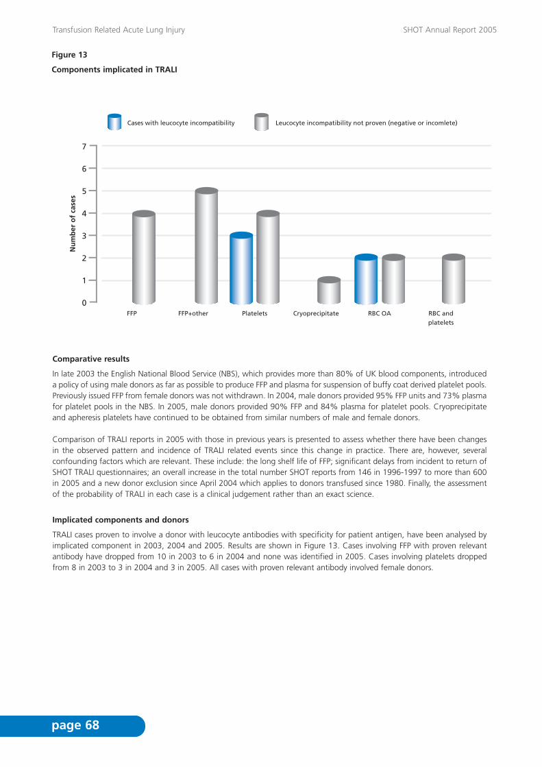
Transfusion Related Acute Lung Injury SHOT Annual Report 2005
page 68
Figure 13
Components implicated in TRALI
7
6
5
4
3
2
1
0FFP FFP+other Platelets Cryoprecipitate RBC OA
Cases with leucocyte incompatibility Leucocyte incompatibility not proven (negative or incomlete)
RBC and platelets
Nu
mb
er o
f ca
ses
Comparative results
In late 2003 the English National Blood Service (NBS), which provides more than 80% of UK blood components, introduceda policy of using male donors as far as possible to produce FFP and plasma for suspension of buffy coat derived platelet pools.Previously issued FFP from female donors was not withdrawn. In 2004, male donors provided 95% FFP units and 73% plasmafor platelet pools in the NBS. In 2005, male donors provided 90% FFP and 84% plasma for platelet pools. Cryoprecipitateand apheresis platelets have continued to be obtained from similar numbers of male and female donors.
Comparison of TRALI reports in 2005 with those in previous years is presented to assess whether there have been changesin the observed pattern and incidence of TRALI related events since this change in practice. There are, however, severalconfounding factors which are relevant. These include: the long shelf life of FFP; significant delays from incident to return ofSHOT TRALI questionnaires; an overall increase in the total number SHOT reports from 146 in 1996-1997 to more than 600in 2005 and a new donor exclusion since April 2004 which applies to donors transfused since 1980. Finally, the assessmentof the probability of TRALI in each case is a clinical judgement rather than an exact science.
Implicated components and donors
TRALI cases proven to involve a donor with leucocyte antibodies with specificity for patient antigen, have been analysed byimplicated component in 2003, 2004 and 2005. Results are shown in Figure 13. Cases involving FFP with proven relevantantibody have dropped from 10 in 2003 to 6 in 2004 and none was identified in 2005. Cases involving platelets droppedfrom 8 in 2003 to 3 in 2004 and 3 in 2005. All cases with proven relevant antibody involved female donors.

Transfusion Related Acute Lung Injury SHOT Annual Report 2005
page 69
8
7
6
5
4
3
2
1
0FFP FFP+other Platelets Cryo RBC
2003 2004 2005
Deaths
The number of reported deaths at least possibly due to TRALI each year from 1996 to 2005 inclusive are shown on Figure 15.The total numbers of reports of suspected TRALI are also shown. Reports of suspected TRALI peaked in 2003 followed by adrop in 2004 and 2005. Reported deaths, at least possibly attributable to TRALI, showed a gradual increase from 2 in 1996/97to 7 in 2003 followed by a decrease to 3 in 2004 and 2 in 2005.
The numbers of deaths over the past 3 years in cases classed as highly likely or probable TRALI were 4 of 22 cases in 2003(cause of death possibly due to TRALI in 2 and likely to be related in 2). In 2004, 2 deaths occurred in 13 highly likely orprobable TRALI cases of which 1 was likely to be related to TRALI and 1 was possibly related. In 2005 there has been 1 deathin 6 patients in this group, this death was unrelated to TRALI.
Figure 14
Cases of TRALI with relevant donor antibody analysed by implicated component and by year 2003-2005

Transfusion Related Acute Lung Injury SHOT Annual Report 2005
page 70
Figure 15
Deaths at least possibly due to TRALI and number of TRALI reports analysed by year 1996-2005
Year *15 months in 01/02
No
. rep
ort
s
96/97 97/98 98/99 99/00 00/01 01/02* 2003 2004 2005
Suspected TRALI reports
Number of deaths
40
35
30
25
20
15
10
5
0
Case histories
The case numbers used here correspond with those used on the web-based tables.
Case 5
A 17-year-old female was admitted as an emergency in December 2003 for treatment of a ruptured ectopic pregnancy withintra-abdominal bleeding. She had been previously fit and well. Her preoperative haemoglobin was 8.0g/dL and she wastransfused with two units of red cells in optimal additive solution in theatre. The estimated intra-abdominal blood loss was800 mLs. Within two hours of transfusion there was ‘a dramatic deterioration in lung compliance and oxygenation. This wasaccompanied by frothy secretion in the endotracheal tube’. The reaction was not accompanied by fever, rigors, hypotensionor hypercapnia and there were no clinical signs of heart failure. She was admitted to ICU and mechanical ventilation wasrequired for the next 18 hours following which she made a full recovery. CXR following respiratory deterioration showed‘Bilateral perihilar and upper lobe air space shadowing’. Donor investigation identified that the female donor of one of thered cell units had HLA Class II antibody specific for DR1. The patient was DR1 positive. It was concluded that the case washighly likely to have been TRALI.
Case 19
A 40-year-old female underwent a radical hysterectomy for cervical cancer in June 2005. She had a postoperative bleed andwas transfused with 4 units of red cells to replace blood loss and 4 units of FFP to correct coagulation abnormality. Almostimmediately after completing transfusion of the final unit of FFP she became dyspnoeic and her oxygen saturation droppedto 60% on 100% oxygen. She required intubation, transfer to ICU and mechanical ventilation for 24 hours. Her chest x-rayshowed ‘pulmonary oedema’ but there were no clinical signs of heart failure and diuretic treatment was not given. Thereporter was ‘certain’ that her symptoms and signs were related to transfusion. Her clinical condition ‘reversed relativelyquickly’ and she made a complete physical recovery. Investigation of the FFP donors revealed that 3 were untransfused maleswho were excluded from further testing. The donor of the final FFP unit was an untransfused female donor, who had beenpregnant twice, 9 and 14 years previously. She was found to have HLA Class I antibodies with 91% panel reactive activityand also HLA Class II antibodies. Unfortunately, a sample from the patient was not provided at the time of the reaction andshe has declined to provide one subsequently. This case was assessed as probable TRALI because of the clinical features ofthe reaction together with donor HLA antibody results which were likely to correlate with patient HLA Class I antigen. In theabsence of a patient sample, this could not be proven.

Transfusion Related Acute Lung Injury SHOT Annual Report 2005
page 71
Case 2
A 29-year-old female had a history of multiple hospital admissions with atonic bowel and several previous laparotomies. Shewas admitted in December 2003 with small bowel obstruction which required further laparotomy to free adhesions andjejunostomy. She was admitted to ICU post operatively due to inadequate respiratory effort with ‘asthma’ and wasmechanically ventilated. On ICU there was a rapid drop in haemoglobin and she was transfused with 4 units of FFP transfusedover an hour and a quarter to correct abnormal clotting related to ‘impaired liver function’ and 4 units of red cells over 8hours. During the final red cell unit she became hypoxic with tachycardia and her CXR showed ‘pulmonary oedema’. Fluidbalance for the 9 hours before hypoxia was more than 600 mL positive and the reporters indicated possible fluid overload.The patient remained on a ventilator for 9 days and died subsequently. The reporter indicated that this death appeared to bedue to transfusion but another cause could not be excluded. Donor investigation revealed no significant antibody, one femaleFFP donor had very weak anti HLA Class I and II antibodies which were considered to be of doubtful significance. The patienthad HLA Class I antibodies with 95% panel reactivity and also had HLA Class II antibody specific for DR II but all the transfusedcomponents were leucocyte depleted so these antibodies were considered unlikely to be of relevance.
This case was assessed as unlikely to be TRALI because of possible circulatory overload and doubtful serology.
COMMENTARY
• TRALI cases assessed as highly likely/probable dropped from 22 in 2003 to 13 in 2004 and to 6 in 2005.
Although numbers have decreased, TRALI remains a serious consequence of transfusion with six probable or highly likelycases this year.
• Two deaths were at least possibly related to TRALI; this is the lowest mortality since 1996.
• All cases were correctly referred to Blood Centres but occasionally the opportunity to send a patient sample was missed.In 4 reported cases investigation was incomplete due to missing donor or patient samples. Three reported cases were notinvestigated.
• Female donors were implicated in all cases in which a relevant leucocyte antibody was found (6). All such cases involvedantibodies with HLA specificities. Granulocyte-specific antibodies were not proven to be implicated in any case.
• Platelets were the most commonly implicated component with proven relevant leucocyte antibodies this year (3 cases: 2apheresis and 1 buffy coat derived). The number of cases of TRALI due to transfusion of platelets with donor leucocyteantibody proven to correspond with patient antigen decreased from 8 in 2003 to 4 in 2004 and 4 in 2005.
• No case of TRALI due to transfusion of FFP from a donor with leucocyte antibody proven to correspond with patientantigen was found this year. This contrasts with 8 such cases reported in 2003 and 6 in 2004, all from female FFP donors.It is probable that one case this year was caused by anti HLA in FFP from a female donor but patient testing was notpossible to confirm this.
• It appears likely that the reductions observed in the number of TRALI reports, deaths and cases firmly implicating FFP andplatelets in 2004 and 2005 relate to the policies of preferentially obtaining FFP and plasma for platelet pooling from maledonors since late 2003 and the exclusion, since April 2004, of donors transfused since 1980.
• Two cases with proven serological support concerned transfusion of red cells in optimal additive solution, while 1 caseinvolved cryoprecipitate. This suggests that as little as 30 mL of plasma can trigger a TRALI reaction.

Transfusion Related Acute Lung Injury SHOT Annual Report 2005
page 72
RECOMMENDATIONS
• Hospital staff should continue to be aware of TRALI and report possible cases to the local Blood Centre to facilitateinvestigation. Detailed clinical information is needed to allow accurate assessment of these cases. Blood samples (clottedand EDTA) from affected patients should be sent for laboratory investigation early. Continued education of all relevant staffabout this condition is encouraged.
Action: HTTs.
• Cases should be evaluated early by the consultant(s) involved and there should be early liaison with the local Blood Centre.There should be a team approach including the haematologist and chest physician and/or ICU consultant.
Action: Clinical users of blood and consultant haematologists with responsibility for transfusion.
• Serological investigation of suspected TRALI cases must include tests for antibodies to HLA Class I, HLA Class II andgranulocyte specific antigens.
Action: Reference laboratories.
• UK Blood Services must continue to keep female contributions to FFP and plasma for platelet pools to an absoluteminimum.
Action: UK Blood Services.
• Further TRALI risk reduction might be achieved by preferential recruitment of male apheresis platelet donors, screeningfemale platelet donors for leucocyte antibodies or by replacing part of the supernatant plasma with additive solution.
Action: UK Blood Services.

Post-Transfusion Purpura SHOT Annual Report 2005
page 73
Definition
Post-transfusion purpura was defined as thrombocytopenia arising 5-12 days following transfusion of red cells associated withthe presence in the patient of antibodies directed against the HPA (Human Platelet Antigen) systems.
Three cases were reported as possible PTP of which two fulfilled the SHOT definition. Both of these are described below.
Case histories
Case 1
A 70 year old female was transfused uneventfully with 10 units of red cells, 3 platelet pools and 8 units of FFP during andafter aortic and mitral valve replacement and coronary artery bypass graft. Her pre-operative platelet count was 208 x 109 /land post-operative platelet count was 59 x 109 /l. Eight days after transfusion she developed a purpuric rash, bruising and aminor haemorrhage. Her platelet count at this stage was 3 x 10⁹ /l and she did not respond to random donor platelettransfusion.
Investigation revealed that the patient had HPA-1a antibodies and her platelet genotype was HPA-1a negative. Enzyme-linkedimmunosorbent assay (ELISA) investigation for heparin/platelet factor 4 antibodies was negative. These results are consistentwith a diagnosis of PTP due to anti-HPA-1a. She was treated with intravenous immunoglobulin and subsequently with HPA-1a negative platelets. Her thrombocytopenia was complicated by haemorrhagic pleural effusion.
She had had two pregnancies, the most recent being more than twenty years previously and had no history of neonatalalloimmune thrombocytopenia in either baby. She had not been transfused before this hospital admission. Platelet recoveryto >50 x 109 /l occurred within 9 days and the patient made a full clinical recovery. The clinical picture and laboratory findingssupported a diagnosis of PTP due to anti-HPA-1a.
Case 2
An 81 year old female underwent a right hemicolectomy for a tumour in the ascending colon. She was transfuseduneventfully with 7 units of red cells pre and peri-operatively. Post-operatively she was given 2 units of FFP because ofprolonged clotting times and one unit of platelets because her platelet count was 38 x 109 /l . Subsequent routine post-operative blood counts showed a platelet count of 99 x 109 /l on the 10th post-operative day and 5 x 10⁹ /l on the 14th. Shehad a past history of an episode of thrombocytopenia in 1984 which responded to steroids. On that occasion, her plateletcount was 13 x 109 /l and bone marrow aspirate indicated peripheral consumption. She later developed autoimmunehepatitis. The reporter initially thought ITP was the most likely reason for the recent episode reported to SHOT but felt PTPwas possible given the recent history of transfusion. Investigation revealed pan-reactive platelet specific antibodies with arelative HPA-1a specificity. The patient’s platelet genotype was HPA-1a negative. The laboratory report concluded that herresults were consistent with post transfusion purpura. She had had a single pregnancy more than 20 years before and hadbeen transfused more than 20 years previously. She was treated with intravenous immunoglobulin and steroids and herplatelet count recovered to >50 x 109 /l in 4 days. This patient died subsequently of causes unrelated to thrombocytopenia.This case has been included as PTP due to anti-HPA-1a.
10 Post-Transfusion Purpura

Transfusion Associated-Graft versus Host Disease SHOT Annual Report 2005
page 74
COMMENTARY
• Two cases of confirmed PTP were reported this year. The graph (Figure 16) shows the number of cases of confirmedPTP reported to SHOT each year since 1996. The drop in numbers of cases of PTP since the introduction of universalleucodepletion in 1999 has been maintained.
Figure 16
Number of cases of confirmed PTP reported to SHOT each year
• As well as a drop in the number of reported cases of PTP since the introduction of leucodepletion; there has also been achange in the transfusion profile before development of PTP. Before April 2000 all 29 patients, for whom components wererecorded, had received red cell transfusion without platelet transfusion before PTP developed. After April 2000, 5 of 8patients had received both RBC and platelet transfusion and 3 had received RBC alone.
• Both confirmed cases were female; this is consistent with previous years. Overall, including this year, there have been 43cases of confirmed PTP reported since 1996. Of these, 42 were female and one was male. A history of previous pregnancywas reported in each female case. The male patient had a history of previous transfusion before the implicated transfusion.
• The only relevant antibody identified this year was anti-HPA-1a. This has been the most commonly implicated plateletantibody; it has been identified in 36 of 43 (83%) cases of antibody-proven PTP reported to SHOT since 1996.
RECOMMENDATIONS
• Clinicians need to maintain awareness of this rare but treatable complication of transfusion.
• When PTP is suspected there should be referral to a platelet reference laboratory for relevant investigation.
Action: Clinical users of blood and consultant haematologists with responsibility for transfusion.
12
10
8
6
4
2
0
11
9
10
5
3
2
1
0
2
1996-1997 1997-1998 1998-1999 1999-2000 2000-2001 2001-2002 2003 2004 2005
Nu
mb
er o
f ca
ses
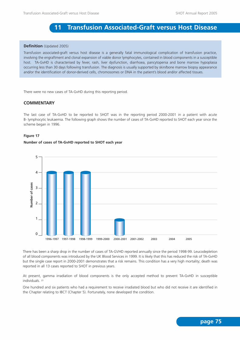
Transfusion Associated-Graft versus Host Disease SHOT Annual Report 2005
page 75
5
4
3
2
1
01996-1997 1997-1998 1998-1999 1999-2000 2000-2001 2001-2002 2003 2004 2005
Nu
mb
er o
f ca
ses
Definition (Updated 2005)
Transfusion associated-graft versus host disease is a generally fatal immunological complication of transfusion practice,involving the engraftment and clonal expansion of viable donor lymphocytes, contained in blood components in a susceptiblehost. TA-GvHD is characterised by fever, rash, liver dysfunction, diarrhoea, pancytopenia and bone marrow hypoplasiaoccurring less than 30 days following transfusion. The diagnosis is usually supported by skin/bone marrow biopsy appearanceand/or the identification of donor-derived cells, chromosomes or DNA in the patient’s blood and/or affected tissues.
There were no new cases of TA-GvHD during this reporting period.
COMMENTARY
The last case of TA-GvHD to be reported to SHOT was in the reporting period 2000-2001 in a patient with acute B- lymphocytic leukaemia. The following graph shows the number of cases of TA-GvHD reported to SHOT each year since thescheme began in 1996.
Figure 17
Number of cases of TA-GvHD reported to SHOT each year
There has been a sharp drop in the number of cases of TA-GVHD reported annually since the period 1998-99. Leucodepletionof all blood components was introduced by the UK Blood Services in 1999. It is likely that this has reduced the risk of TA-GvHDbut the single case report in 2000-2001 demonstrates that a risk remains. This condition has a very high mortality; death wasreported in all 13 cases reported to SHOT in previous years.
At present, gamma irradiation of blood components is the only accepted method to prevent TA-GvHD in susceptibleindividuals. 23
One hundred and six patients who had a requirement to receive irradiated blood but who did not receive it are identified inthe Chapter relating to IBCT (Chapter 5). Fortunately, none developed the condition.
11 Transfusion Associated-Graft versus Host Disease

Transfusion Associated-Graft versus Host Disease SHOT Annual Report 2005
page 76
RECOMMENDATIONS
• Gamma irradiation of blood components for those at risk of TA-GvHD remains essential. BCSH Blood Transfusion Task ForceGuidelines24 define groups requiring this prophylaxis.
Action: Consultant haematologists with responsibility for transfusion should ensure that BCSH guidelinesare incorporated into local protocols.
• Awareness of the potential for this condition must be maintained by all involved in the transfusion process.
• Good communication is required in all cases but particularly when patient care is shared between different hospitals.Hospitals must have clear protocols to ensure accurate information relating to this risk is communicated in a timely manner.Provision of the BCSH/NBS patient card and leaflet are also recommended.
Action: UK National and Regional Blood Transfusion Committees to facilitate and co-ordinate, HospitalCEOs to implement.
• New chemo or immuno-therapeutic regimens must be evaluated for their potential to predispose individuals to TA-GvHD.Regular update of guidelines is required to include up to date recommendations relating to drugs and protocols withpotent immunosuppressive effects.
Action: BCSH.
Transfusion Transmitted Infections SHOT Annual Report 2005
page 77
Definition
A report was classified as a transfusion transmitted infection if, following investigation: -
• The recipient had evidence of infection post-transfusion, and there was no evidence of infection prior to transfusion andno evidence of an alternative source of infection
And, either
• At least one component received by the infected recipient was donated by a donor who had evidence of the sametransmissible infection,
Or
• At least one component received by the infected recipient was shown to contain the agent of infection
Reports of suspected transfusion transmitted infections
Forty-six reports of suspected transfusion transmitted infections were made from blood centres throughout the UK (41 inEngland and Wales and 5 in Scotland) to the NBS/HPA Centre for Infection Surveillance during 2005. Three reports (1 HBVand 2 bacteria) were determined to be TTIs according to the above definition. Two reports were of predicted HAVtransmissions. 40 cases were concluded as not transfusion transmitted infections (14 HBV, 6 hepatitis C (HCV), 1 dual HBVand HCV, 1 HTLV, 4 HIV and 14 bacteria). One case (HCV) is pending complete investigation. A further report was receivedfrom the Health Protection Agency of a clinical diagnosis of vCJD in a blood transfusion recipient. All UK blood centrescontributed to the scheme.
Case report of transfusion transmitted hepatitis B
A previously hepatitis B surface antigen (HBsAg) negative donor was found to be positive after routine testing in August 2005.An archive sample of a donation made four months earlier was retested and found to be weakly positive for HBVdeoxyribonucleic acid (DNA), but negative for all other HBV markers. The plasma from this donation had been discardedhowever the red cells had been transfused to a female recipient, aged 55 years following a gastro-intestinal haemorrhage.This recipient was tested and found to have evidence of a recently acquired hepatitis B infection: anti-HBV core IgM, anti-HBVcore (HBc) and anti-HBV e-antigen (anti-HBe) positive and HBsAg and HBV DNA negative. The investigation concluded thatthe HBV infection in the recipient was due to an HBV infectious donation in the early acute phase of infection.
Case report of transfusion transmitted Enterobacter cloacae
One recipient (63 year old female) received four platelet units following a cerebral bleed due to chronic severe resistant ITP inMarch 2005. The first 3 units (two apheresis and one pooled platelets) were transfused without problems, however aftertransfusion of the fourth unit (3 day old pooled platelets) the recipient developed rigors, temperature, tachycardia andwheeze. Enterobacter cloacae was cultured from the patient and from the pooled platelet unit. All 4 donors were investigated:Enterobacter cloacae was not cultured from arm swabs from any of the donors. Blood and urine cultures from all four donorswere also negative. The patient made a full recovery within 12 hours following treatment. The probable source of therecipient’s reaction was concluded to be a unit of platelets contaminated with Enterobacter cloacae: no source ofcontamination was identified.
Case report of transfusion transmitted Staphylococcus epidermidis
One recipient (36 year old male) developed pyrexia and rigor following transfusion of a pooled platelet unit. Cultures fromthe implicated unit and the patient grew penicillin resistant Staphylococcus epidermidis. Molecular types of isolates from thepack and patient were indistinguishable using pulsed-field gel electrophoresis (PFGE). Four donors were recalled for armswabbing and, although a number of staphylococci were grown, none of the donors was colonised with the identical strainof S.epidermidis found to be contaminating the pack. Skin flora can vary from day-to-day and there was a considerable gapbetween donation and swabbing, so a donor could not be ruled out as the possible source of contamination. Three relatedred cell units were recalled; all were free from contamination. The fourth red cell unit was transfused with no report of anyadverse events. This case was concluded to be bacterial contamination with S.epidermidis of a pooled platelet unit, assumedbut not proven to originate from the skin of one of the donors.
12 Transfusion Transmitted Infections

Transfusion Transmitted Infections SHOT Annual Report 2005
page 78
Reports of further incidents
Hepatitis A
1. In December 2005 the blood services were notified of a confirmed acute hepatitis A infection in a regular blood donor, whodeveloped symptoms eight days following donation. The archive was tested by polymerase chain reaction (PCR) and found tobe positive. The red cell unit was discarded, but the platelets had been used in a pooled platelet unit (suspended in plasmafrom another donor) and transfused to a female recipient 2 days after donation. Upon notification of the infection in thedonor, the recipient was given passive and active immunisation, as per recommended guidelines25 and tested for HAV. Tracesof HAV antibodies were found, which may have been passive transfer from previous blood transfusions. A blood sample 3months later from the recipient was HAV IgM positive, at a low level and there was a mild elevation in liver function tests. Theconclusion of the investigation was that although transmission from a donor with confirmed hepatitis A was predicted, promptimmunisation appears to have prevented transmission or reduced the impact to sub-clinical levels with no sequelae.
2. In July 2005 a donor notified a blood centre that he had been clinically diagnosed with hepatitis A, 3 weeks after donation.The red cell component of this donation was discarded. The recipient of the platelets died of other causes, prior to testing forHAV. The FFP was transfused to a patient with alcoholic liver failure. When tested this recipient had evidence of immunity toHAV (HAV IgM negative, total HAV positive). This patient subsequently died. There was insufficient sample from the donor’sindex archive to test by PCR. A previous archive sample from the donor was HAV IgG negative. The donor has not respondedto requests for additional samples and to date no serology for the donor has been seen by the blood services. The donor’srecord is flagged for HAV testing at their next attendance. This case is concluded to be a predicted potential transmission ofhepatitis A, where the diagnosis in the donor was not confirmed and no transmission to any recipient was detected due todeath or probable immunity.
vCJD
In early 2006 a further case of vCJD associated with a blood transfusion was reported. In 1997 a patient received a unit ofnon-leucodepleted red blood cells. The donor developed symptoms of vCJD about 20 months after donation and subsequentlydied. The recipient developed symptoms in 2005 and a clinical diagnosis of vCJD was made in early 2006. The recipient is amethionine homozygote at codon 129 of the prion protein gene. As the recipient is a UK resident, dietary exposure to bovinespongiform encephalopathy (BSE) cannot be excluded. The recipient was alive at the time of preparation of this report. (Formore information on variant CJD see http://www.cjd.ed.ac.uk/).
Reports from previous years
The case reported as pending in 2004 (HHV-8) is now nearing completion; all donors have been recalled and have providedblood samples for HHV-8 testing. Results will be reported when available.
Cumulative bacterial data
Since 1995, 31 cases of transfusion transmitted bacterial infection have been reported, of which 6 recipients died (Figure 18).The majority of these cases relate to platelet units (9 apheresis and 18 pooled). In 2004 there was a further incident involvingcontamination of a pooled platelet pack with staphylococcus epidermidis, which did not meet the TTI definition becausetransmission to the recipient was not confirmed, but it would seem likely (not included in figure 18).

Transfusion Transmitted Infections SHOT Annual Report 2005
page 79
Figure 18
Confirmed bacterial transfusion transmitted infections, by year of transfusion and type of unit transfused(Scotland included from 10/1998)
Further cumulative data is available at http://www.hpa.org.uk/infections/topics_az/BIBD/menu.htm.
COMMENTARY
• For a case to be reported to this surveillance system, an infection must first be diagnosed, transfusion suspected as themeans of acquisition/transmission, and this suspicion has to be communicated to the blood service. Under-ascertainmentcan be the result if omissions occur at any of these stages. Therefore surveillance of TTIs tends to be biased towardsascertainment of acute cases that are clinically apparent or investigation of newly acquired infection in returning blooddonors. However, each year some infections among individuals who have received blood transfusion(s) in the past arereported to the blood services and investigated. All cases reported to the blood services have been reported to SHOT.
• Each year the number of reports received is small and fluctuations are to be expected. This year’s findings are consistentwith the current very low estimated risk of HIV, HCV and HBV infectious donations entering the UK blood supply.
• Notification from blood donors of infections that developed post donation enabled the blood service to identify two caseswhere transmission of HAV was predicted. Prompt action with passive and active immunisation to one recipient appearsto have either prevented transmission or reduced the impact to sub-clinical levels. This emphasises the importance of blooddonors notifying the blood service of infections detected after donation.
• The report of a third case of vCJD infection in a recipient of non-leucodepleted red blood cells provides further evidencethat vCJD may be transmitted through blood transfusion. In all three cases the possibility that the recipient acquiredinfection through dietary exposure to BSE could not be ruled out. For the first reported case it was estimated that thechance of observing a case of vCJD in a recipient in the absence of TTI was about 1 in 15,000 to 1 in 30,000.26 A numberof precautions are in place to reduce the risk of transmission through blood transfusion. These include leucodepletion ofblood components and the exclusion of candidate blood donors who have received transfusions since January 1st 1980from donating blood.
8
7
6
5
4
3
2
1
01995
Red cells
platelets
1996 1997 1998 1999 2000 2001 2002 2003 2004 2005

Transfusion Transmitted Infections SHOT Annual Report 2005
page 80
• Although 16 cases suspected to be due to bacteria were reported and investigated during 2005, only two cases wereconfirmed. It is important that hospitals notify the blood services as soon as bacterial infection in a recipient is suspectedand return the pack for complete investigation. The small number of confirmed bacterial cases seen in the past two yearsis encouraging and suggests that actions to reduce bacterial contamination, such as sample diversion pouches andenhanced donor arm cleansing, are effective.
• The Standing Advisory Committees (SAC) of the Joint UKBTS/NIBSC Professional Advisory Committee (JPAC) makerecommendations to the Guidelines for the Blood Transfusion Services in UK in relation to the prevention of transfusion-transmitted infections. For example, the SAC Transfusion Transmitted Infection (SACTTI) regularly reviews the residual riskof transfusion transmitted HCV, HIV and HBV infections to assess any need for additional testing methods, such as HIVRNA testing, HBV DNA or anti-HBc. The SAC Care and Selection of Donors ensures donor deferral criteria are optimal interms of exclusion of donors with behaviour that may put them at increased risk of contracting transfusion transmissibleinfections.
RECOMMENDATIONS
• Hospitals should continue to report and investigate all possible incidents of post-transfusion infection appropriately andadequately.
Action: HTTs.
• Donors should be reminded to report any infections developing post donation to their local blood centre.
• UK Blood Service collection teams should ensure donor selection guidelines are adhered to at all times in order to preventtransmission of blood borne infections.
Action: UK Blood Services.
• Efforts to prevent bacterial contamination of blood components should continue. These include:
• Continuation of diversion of the first 20-30 mL of the donation (likely to contain any organisms entering the collectionneedle from the venepuncture site)
• Careful attention to adequate cleansing of donors’ arms
• Adherence to BCSH guidelines (1999)20 with regard to the visual inspection of blood components immediately prior totransfusion, to check for any irregular appearance.
Action: UK Blood Services, hospital transfusion laboratories, staff undertaking pre-transfusion bedsidechecking.
• Hospitals should consult the blood service about the investigation of transfusion reactions suspected to be due to bacteria.Attention should be paid to the sampling and storage of implicated units or their residues and packs returned to the bloodservice for testing.
Action: HTTs.
Acknowledgements SHOT Annual Report 2005
page 81
The Steering Group would like to take this opportunity to thank the following individuals and organisations for theircontributions without which the publication of this ninth annual SHOT report would not have been possible.
The Blood Transfusion Services of the United Kingdom for funding and support
~~~~~~~~~~
The Royal College of Pathologists for their support and use of facilities
~~~~~~~~~~
Karen Carter - Marketing Services, NHSBT, for help and advice with the production of the report
~~~~~~~~~~
Kirk Beard - NHSBT, for the provision of data relating to the issue of blood components from theTransfusion Services of the UK
~~~~~~~~~~
Rob Hick & Judith Chapman- Blood Stocks Management Scheme, for the provision of data relating tothe issue of blood components to hospitals in the UK
~~~~~~~~~~
Marcela Contreras, NHSBT, Neil Soni, Imperial College Medical School and Cliff Morgan, RoyalBrompton Hospital for expert review of the TRALI cases
~~~~~~~~~~
Hazel Tinegate, Claire Thompson and colleagues in the Northern and Yorkshire regions forundertaking the “Where and When” study
~~~~~~~~~~
Clinical and Scientific staff, in hospitals and reference laboratories, who have contributed to theclinical and laboratory investigation of cases.
~~~~~~~~~~
Paul Ashford and Ceri Frampton - WBS, for maintenance of the SHOT website
~~~~~~~~~~
MHRA and Assad Afzal of Charles McKenzie Consulting for provision of the SABRE system andongoing technical support
~~~~~~~~~~
Angela Allen, Personal Assistant to Dorothy Stainsby
~~~~~~~~~~
Christine Smith, Personal Assistant to Hannah Cohen
All hospitals who have participated in SHOT reporting
Without your support, SHOT would not be possible
13 Acknowledgements

1 Asher D, Atterbury C L J, Chapman C et al, Serious Hazards of Transfusion Annual Report 2000/01, ISBN 0 9532 789 4 8,9 April, 2002
2 Health Service Circular, Better Blood Transfusion - Appropriate Use of Blood, HSC 2002/009 04 July 2002
3 National Comparative Audit of Blood Transfusionhttp://www.blood.co.uk/HOSPITALS/safe_use/clinical_audit/National_Comparative/index.asp
4 NPSA. Safer Practice Notice 14: Right patient, right blood. October 2006A link to this document will be available on the SHOT website http://www.shot-uk.org as soon as possible
5 NPSA SPN on Safer Patient Identification 22nd November, 2005http://www.npsa.nhs.uk/site/media/documents/1440_Safer_Patient_Identification_SPN.pdf
6 Blood Stocks Management Schemehttp://www.bloodstocks.co.uk/
7 The NHS Confederation / NPSA, Creating a virtuous circle, ISBN 185947 089 0, 2003
8 The Blood Safety and Quality Regulations 2005, ISBN 0110990412
9 British Committee for Standards in Haematology Blood Transfusion Task Force. Guidelines for the use of fresh frozenplasma, cryoprecipitate and cryosupernatant: British Journal of Haematology 2004: 126, 11-28www.bcshguidelines.com
10 British Committee for Standards in Haematology Blood Transfusion Task Force. Guidelines For The Use Of PlateletTransfusions. British Journal of Haematology 2003: 122, 10-23 www.bcshguidelines.com
11 Report of the Review of Pathology Services in England http://www.dh.gov.uk/PublicationsAndStatistics/Publications/PublicationsPolicyAndGuidance/PublicationsPolicyAndGuidanceArticle/fs/en?CONTENT_ID=4137606&chk=J4t5ZX
12 The Handbook of Transfusion Medicine 4th editionhttp://www.transfusionguidelines.org.uk/index.asp?Publication=H1&Section=9&pageid=8
13 SHOT Minimum standards for investigation of transfusion related adverse reactionshttp://www.shotuk.org/standards4investigation.htm
14 Please ask, NPSAhttp://www.npsa.nhs.uk/pleaseask/
15 Pregnancy - routine anti-D prophylaxis for rhesus negative womenhttp://www.nice.org.uk/page.aspx?o=TA41
16 Statutory Instrument 2003 No. 696, The Prescription Only Medicines (Human Use) Amendment Order 2003http://www.opsi.gov.uk/si/si2003/20030696.htm
17 British Committee for Standards in Haematology Blood Transfusion Task Force. Guidelines for blood grouping andantibody testing in pregnancy.http://www.bcshguidelines.com/pdf/pregnancy_070606.pdf
References SHOT Annual Report 2005
page 82
14 References

References SHOT Annual Report 2005
page 83
18 Practical Transfusion Medicine 2nd Ed., 2005, Eds Murphy M, Pamphilon DH, Chapter 9, page 130
19 Laupacis A, Brown J, Costello B et al, Prevention of post-transfusion CMV in the era of universal WBC reduction: aconsensus statement. Transfusion 2001; 41: 560-9
20 British Committee for Standards in Haematology Blood Transfusion Task Force. The administration of blood and bloodcomponents and the management of the transfused patient. Transfusion Medicine 1999, 9, 227-238www.bcshguidelines.com
21 British Committee for Standards in Haematology Blood Transfusion Task Force. Guidelines for compatibility procedures inblood transfusion laboratories. Transfusion Medicine, 2004, 14, 59-73www.bcshguidelines.com
22 Baglin, T. P., Keeling, D. M. & Watson, H. G. (2006) Guidelines on oral anticoagulation (Warfarin): third edition - 2005update. British Journal of Haematology 132 (3), 277-285.www.bcshguidelines.com
23 Nollet, Kenneth E. & Holland, Paul V. (2003)Toward a coalition against transfusion-associated GvHD. Transfusion 43 (12), 1655-1657.
24 British Committee for Standards in Haematology Blood Transfusion Task Force. Guidelines for gamma irradiation of bloodcomponents for the prevention of transfusion-associated graft-versus-host disease. Transfusion Medicine 1996; 6, 261-271 www.bcshguidelines.com
25 Crowcroft NS, Walsh B, Davison KL et al. Guidelines for the control of hepatitis A virus infection. Communicable Diseaseand Public Health, 2001. Vol 4(3);213-227.
26 Llewelyn CA, Hewitt PE, Knight RSG, et al. Possible transmission of variant Creutzfeldt-Jakob disease by blood transfusion.Lancet, 2004. 363; 417-421