serious hazards of transfusion - iakh.de · change and evolution in healthcare demand greater...
TRANSCRIPT

SERIOUS HAZARDS OF TRANSFUSION
ANNUAL REPORT
2003
Affiliated to the Royal College of Pathologists
British Blood Transfusion Society, British Society for Haematology
Faculty of Public Health Medicine, Institute of Biomedical Science
Institute of Healthcare Management, NHS Confederation
Health Protection Agency Communicable Disease Surveillance Centre
Royal College of Anaesthetists, Royal College of Nursing
Royal College of Obstetricians and Gynaecologists
Royal College of Paediatrics and Child Health
Royal College of Physicians, Royal College of Surgeons, UK Transfusion Services

by The Serious Hazards of Transfusion Steering Group
National co-ordinators: Dr Dorothy Stainsby
Ms Katy Davison (Health Protection agency CDSC)
Scheme Manager: Mrs Hilary Jones (SHOT office)
Data Collection Specialist: Mrs Aysha Boncinelli (SHOT Office)
Writing Group
Stainsby D Cohen H
Jones H Knowles S
Milkins C Chapman C
Gibson B Davison K
Norfolk D R Taylor C
Revill J Asher D
Atterbury CLJ Gray A
on behalf of the SHOT Steering Group
Requests for further information should be addressed to:
Non-infectious hazards
SHOT Office
Manchester Blood Centre
Plymouth Grove
Manchester
M13 9LL
Tel: 0161 251 4208Fax: 0161 251 4395
Email: [email protected]
Website:http//www.shot-uk.org
ISBN 0 9532 789 6 4
SHOT Annual Report 2003
Published 5th July 2004
page 2
Infectious hazards
Ms. Katy Davison
Clinical Scientist
Health Protection agency CDSC
61 Colindale Avenue
London NW9 5EQ
Tel: 020 8200 6868
Fax: 020 8200 7868
Email: [email protected]

Page
1. KEYNOTE MESSAGE – CHANGE AND EVOLUTION IN HEALTHCARE DEMAND GREATER HAEMOVIGILANCE 6
2. MAIN FINDINGS AND RECOMMENDATIONS 8
3. CUMULATIVE DATA 1996 - 2003 16
4. INCORRECT BLOOD COMPONENT TRANSFUSED 17
5. "NEAR MISS" EVENTS 35
6. ACUTE TRANSFUSION REACTION 39
7. DELAYED TRANSFUSION REACTION 47
8. TRANSFUSION-RELATED ACUTE LUNG INJURY 57
9. POST-TRANSFUSION PURPURA 65
10. TRANSFUSION-ASSOCIATED GRAFT VERSUS HOST DISEASE 67
11. TRANSFUSION-TRANSMITTED INFECTIONS 69
12. SHOT EVENTS REPORTED IN PATIENTS LESS THAN 18 YEARS OF AGE 78
13. ADVERSE EVENTS RELATING TO AUTOLOGOUS TRANSFUSION 85
14. ACKNOWLEDGEMENTS 86
15. REFERENCES 87
SHOT Annual Report 2003
Contents
page 3

ALI Acute Lung Injury
ALT Alanine Aminotransferase
ARDS Acute respiratory distress syndrome
ATR Acute transfusion reaction
BBTS British Blood Transfusion Society
BCSH British Committee for Standards in Haematology
BMS Biomedical scientist
CABG Coronary artery bypass graft
CAT Column agglutination technology
CDSC Communicable Disease Surveillance Centre
CEO Chief Executive Officer
CMO Chief Medical Officer
CMV Cytomegalovirus
CNST Clinical Negligence Scheme for Trusts
CVS Chorionic villus sampling
CXR Chest X-Ray
DAT Direct antiglobulin test
DHTR Delayed haemolytic transfusion reaction
DNA Deoxyribonucleic acid
DOH Department of Health
DTR Delayed transfusion reaction
EC European Commission
ECMO Extracorporeal membrane oxygenation
EU European Union
FFP Fresh frozen plasma
HAV Hepatitis A virus
HBc Hepatitis B core
HBs Ag Hepatitis B surface antigen
HBV Hepatitis B virus
HCV Hepatitis C virus
HDN Haemolytic disease of the newborn
HDU High Dependency Unit
HIV Human immunodeficiency virus
HLA Human leucocyte antigen
HNA Human neutrophil antigen
HPA Human platelet antigen
HPA CDSC Health Protection Agency Communicable Disease Surveillance Centre
HTC Hospital Transfusion Committee
HTLV Human T-cell leukaemia virus
HTT Hospital Transfusion Team
IAT Indirect antiglobulin test
SHOT Annual Report 2003
Glossary of Terms
page 4

IBCT Incorrect blood component transfused
ICU Intensive care unit
INR International normalised ratio
Ig Immunoglobulin
IUT Intrauterine transfusion
LISS Low ionic-strength saline
MB Methylene blue
MHRA Medicines and Healthcare Products Regulatory Authority
MSBT Microbiological Safety of Blood and Tissues for Transplantation
NBS National Blood Service
NBS/HPA National Blood Service/Health Protection Agency
NBTC National Blood Transfusion Committee (England)
NEQAS National External Quality Assurance Scheme
NISS Normal ionic-strength saline
NPSA National Patient Safety Agency
PCR Polymerase chain reaction
PCT Primary Care Trust
PTI Post-transfusion infection
PTP Post-transfusion purpura
PTR Post-transfusion reaction
RNA Ribonucleic acid
RTC Regional Transfusion Committee
SHA Strategic Health Authority
SHO Senior House Officer
SPOT Specialist practitioners of transfusion
TA-GVHD Transfusion-associated graft-versus-host disease
TRALI Transfusion-related acute lung injury
TTI Transfusion-transmitted infection
TTP Thrombotic thrombocytopenia purpura
vCJD Variant Creutzfeldt-Jakob disease
VSD Ventricular septal defect
SHOT Annual Report 2003
page 5
Glossary of Terms

CHANGE AND EVOLUTION IN HEALTHCARE DEMAND GREATER HAEMOVIGILANCE
With this 7th annual report, SHOT provides an increasingly authoritative analysis of serious transfusion hazards in the UK andan evidence base for blood safety initiatives, policies and guidelines. This report is condensed to make it more user-friendlywith detailed data available on the SHOT website www.shot-uk.org. Recommendations are targeted at specific groups orbodies to maximise their effectiveness.
Current figures re-emphasise the importance of errors in the transfusion process. In 2003, 358 reports were received ofincorrect or inappropriate blood component transfused (IBCT), a further increase of 25% over the previous 12 months. Errorscontinue to occur at all stages of the transfusion process; misidentification of patients, blood samples and blood componentsbeing the main source of errors. It is likely that the increased number of reports is related to the establishment, asrecommended in ‘Better Blood Transfusion’ HSC 2002/009,1 of hospital transfusion teams (HTTs) and Specialist Practitionersof Transfusion (SPOTs), who drive greater vigilance, and hence reporting of errors which may have previously goneunrecognized.
Eighty-five per cent of hospitals reported that they participated in SHOT in 2003. However, only 47% reported adverse events,possibly reflecting a lack of HTTs and SPOTs in these Trusts. Individual hospitals can make a positive contribution to transfusionsafety by active participation in SHOT, so that the picture of transfusion risks is as complete and accurate as possible. Thoroughand systematic investigation of all adverse events is essential, requiring clinical input from the consultant haematologist withresponsibility for blood transfusion, who should sign off each case report. Evidence of active participation is likely to berequired in the future as part of external reviews of clinical governance arrangements, e.g. by the Health Care Commission.
On a more encouraging note, despite the increased number of errors reported, there is an overall downward trend in thenumber of ABO incompatible transfusions (Figure 3). This suggests that we may be starting to see the improvement in theproportion of ‘serious’ events compared to overall events, which characterises a developing safety culture. Tools for analysisof errors have been developed by the National Patient Safety Agency (NPSA)2 who have rolled out a programme of trainingand support. Examples of how such tools can be effective in establishing the root cause of incidents can be found on theSHOT website.
Clinical audit is a useful tool with which to monitor the implementation of policies and processes. The recent NationalComparative Audit of Transfusion,3 carried out jointly by the National Blood Service (NBS) and the Royal College of Physicians,identified variation and poor practice in the administration of blood. In 2001, the Scottish National Blood Transfusion Service,in collaboration with NHS hospitals in Scotland, undertook a 3-year study of a transfusion-practitioner-led clinical effectivenessprogramme, including audit and education. Observable improvements in transfusion practices were demonstrated at theintervention sites. A motivated and enthusiastic transfusion practitioner, supported by a local transfusion team, was able toraise awareness of blood transfusion throughout the hospital.4 Audit and feedback at local level can also be effective inimproving practice and, together with education and training, is a key role of the SPOT. HTTs and SPOTs must now beestablished in all trusts. The Chief Medical Officer’s (CMO’s) National Blood Transfusion Committee (NBTC) in England and itscounterparts in Scotland, Wales and Northern Ireland were constituted with a remit to promote implementation of BetterBlood Transfusion. SHOT looks to these bodies to take a proactive lead role in driving forward blood safety issues in hospitals.
Eight of the 34 articles of the new European Union (EU) Directive on Blood Transfusion5 are directly applicable to hospital bloodbanks; this becomes UK law in February 2005 and mandates notification of adverse events/reactions. The Directive requirespositive traceability of all blood components from donor to patient, placing great demands on the quality and integration ofhospital IT systems. The UK Government must designate a competent authority to regulate Blood Establishments by February2005, and must also indicate how it will demonstrate compliance by hospital blood banks and for haemovigilance. A nationalguideline on the specification and performance of blood bank computer systems, developed by the British Committee forStandards in Haematology (BCSH), will be welcome.
Keynote Message SHOT Annual Report 2003
1 Keynote Message
page 6

Safety in the hospital transfusion laboratory is compromised by difficulties in recruitment and retention of suitably qualifiedstaff and increasing demands on them as a result of moves towards 24/7 hospital activity. Recruitment to new roles, such asdoctors’ assistants, may attract experienced biomedical scientists (BMS) away from the bench. The initiatives outlined in‘Making the Change’6 which include the development of National Occupational Standards for Healthcare Scientists, flexiblecareer pathways, improved status through higher specialist training and a stronger regulatory framework through the HealthProfessions Council, provide a much needed opportunity to increase and consolidate the workforce and must beimplemented.
We are pleased that the IT Working Group of the CMO’s NBTC in England has put the needs of the transfusion communityfirmly on the agenda of bodies, such as the Design Authority, responsible for setting standards and priorities in the NHS ITstrategy. The current SHOT annual report again highlights that failures of communication of special requirements or lack ofavailability of previous transfusion history contribute to ‘wrong blood’ events. It is crucial that NHS IT systems have the abilityto use unique patient-specific identifiers, such as the NHS Number, so that essential information is available to all who needit. Systems for positive patient ID have considerable potential to improve patient safety in blood transfusion as well as otherareas such as drug administration. The development of integrated systems across disciplines will have important economic aswell as patient-safety benefits. SHOT encourages these initiatives and emphasises the need for standards and establishingminimum specifications across the NHS. SHOT data will be an important source of information for the evaluation of ITsolutions.
During the past 12 months, important blood safety initiatives have been introduced by the blood services. Precautions toreduce the incidence of bacterial contamination of platelets are being implemented and in England fresh frozen plasma (FFP)is now made almost exclusively from male donors in order to reduce the risk of transfusion related acute lung injury (TRALI).Other blood services await the impact of the latter precaution with great interest, but it should be remembered that increasingawareness of this condition and hence increased reporting may mask a reduction in incidence. Once again, SHOT data will beof importance in the evaluation of the impact of these new blood safety measures.
This year’s report includes the first documented case of possible transfusion transmitted variant Creutzfeld-Jakob Disease(vCJD). It is highly unlikely that this case would have been identified without the transfusion epidemiology surveillance schemeundertaken jointly by the blood services and the vCJD Surveillance Unit.7 In the UK, precautions in place to reduce the risk ofprion transmission, include leucodepletion of all blood components, the use of viral-inactivated FFP obtained outside the UKfor children born after 1 January 1996, importation of plasma for fractionation and the exclusion of donors who have receiveda blood transfusion in the UK after 1980. This latter precaution, implemented in April 2004, is expected to result in a 3–5%reduction in availability of donated blood. There is therefore an increased imperative towards blood conservation, requiring aco-ordinated approach led at national level to maximise effectiveness. It is crucial that there is also a parallel initiative tomonitor for adverse effects of alternatives to transfusion, which will necessitate appropriate co-ordination between SHOT,Medicines and Healthcare Products Regulatory Authority (MHRA) and other experts in the field.
Since its first report, SHOT has recommended the establishment of an overarching body to evaluate and prioritise blood safetyinitiatives. It is hoped that the forthcoming review of the Department of Health Advisory Committee on the MicrobiologicalSafety of Blood and Tissues for Transplantation will extend its remit and membership, and empower it to take on this role.
As always, we would like to thank hospital staff who participate in SHOT and ensure its continuing success.
Dr Hannah Cohen MD FRCP FRCPath Dr Dorothy Stainsby FRCP FRCPath
Chair, SHOT Steering Group SHOT National Medical Co-ordinator.
Keynote Message SHOT Annual Report 2003
page 7

SUMMARY OF MAIN FINDINGS
Participation
In 2003, 351/415 (85%) hospitals returned cards stating that they participated in the SHOT scheme. This is an 8% dropcompared with 2001-2002, due to the fact that the SHOT office has lacked manpower resource to pursue non-respondersand cannot do so in future. Evidence of participation is required for the Clinical Negligence Scheme for Trusts (CNST) and maybe required for other accreditation schemes. Reporting of adverse reactions to blood transfusion will become mandatory withimplementation of the EU Directive.5 It is the responsibility of hospitals to ensure that they return the card in good time inorder to obtain the required receipt.
Hospital transfusion teams should note that:
• Participation cards will be mailed in January/February each year. They should contact the SHOT office if they have not received the card by mid-February.
• The card should be completed and returned as soon as possible and strictly by the deadline.
• They should receive a receipt within 4 weeks and should contact the SHOT office if not.
• The SHOT office retains records of reports and next year will contact hospitals if there is a discrepancy between our recordsand the number of reports stated on the return.
195/415 hospitals stated that they reported incidents; the level of active participation is therefore unchanged at 47%. A totalof 480 initial reports were received in 2003, and 449 questionnaires analysed plus 8 TTI reports not on standard SHOTquestionnaires.
Incorrect blood component transfused ("wrong blood") incidents
In 2003, 358 reports were received; a 25% increase over the previous equivalent 12 month period and 75% of all reportsreceived this year. It is likely that the establishment in hospitals of transfusion teams and SPOTS is resulting in greater vigilanceand hence reporting of previously unrecognised errors. Although the total number of errors reported continues to increase,there is an overall downward trend in the number of ABO incompatible transfusions reported, suggesting that we may bestarting to see the improvement in the proportion of ‘serious’ events compared to overall events which characterises adeveloping safety culture.
Again this year multiple errors have contributed to ‘wrong blood’ events with a total of 588 errors occurring in 348 analysedcases and multiple errors in 52%.
The distribution of errors in IBCT cases is remarkably consistent, with approximately 70% of errors occurring in clinical areas(30% prescription, sampling, request and 40% collection and administration) and 30% in laboratories. Again this year thecommonest error (156/588) was failure of the pre-transfusion bedside checking procedure.
A detailed breakdown of types of error can be found on the SHOT website.
Outcomes of errors this year were 33 ABO incompatible transfusions, of which 4 were also RhD incompatible (26 incompatiblered cell transfusions and 7 incidents of incompatible FFP, cryoprecipitate or platelets). There were 22 cases of unintended RhDincompatible transfusions and 22 cases where other red cell antigen incompatible transfusions were given. There was onepossible transfusion related death but no definite or probable cases. Sixteen patients suffered major morbidity.
"Near-miss" events
906 near miss reports were received this year from 163 hospitals
The picture of "near miss" reporting is consistent, showing sample errors to be by far the most numerous (50% in 2000/2001,59% in 2001/2002 and 60% in 2003). Common sources of error were;
• Patient mis-identification.
• Spurious haematology results due to dilute samples from "drip arms" and samples in syringes dispensed into tubes without mixing.
• Point-of-care (POC) testing variations.
• Labelling of samples remote from the patient and pre-labelling of specimen tubes.
Main Findings and Recommendations SHOT Annual Report 2003
2 Main Findings and Recommendations
page 8

• Addressograph labels used wrongly or placed in wrong patients’ notes.
Anecdotal evidence from reporting hospitals suggests that "near miss" events are frequent but under-reported anddiscussions are underway concerning how better to collect this data in the future.
Immune complications
Thirty-six case reports of suspected TRALI were analysed in 2003. Nine patients died; 1 death was considered to be definitelydue to transfusion, 7 possibly due to transfusion and 1 unrelated. Twenty-two patients suffered short term major morbidityand five minor morbidity.
Plasma rich components were implicated in 20 of 21 cases with proven leucocyte incompatibility between donor and patient.
There were 39 analysable reports of acute transfusion reactions (ATR) accounting for 1 probable transfusion related death and2 cases of major morbidity.
Twenty-five delayed haemolytic transfusion reactions (DHTR) were analysed, a marked reduction from last year’s 47 reports. Itis unlikely that the incidence of these reactions has fallen, and there is a concern DHTRs are under-recognised.
Again this year there were no reports of transfusion-associated graft-versus-host disease (TA-GVHD), and only 1 reportmeeting the SHOT definition of post-transfusion purpura (PTP).
Transfusion-transmitted infections
In 2003, 38 reports of possible transfusion-transmitted infections (TTIs) were made to the surveillance scheme. After theinvestigation had been completed, eight reports were classified as probable TTIs (2 Hepatitis B virus (HBV), 1 HumanImmunodeficiency virus (HIV), 1 Hepatitis A virus (HAV), 1 malaria and 3 bacterial contaminations). One of the cases ofbacterial contamination died. Twenty-four reports were found not to be related to transfusion and 3 had an undeterminedsource. Full investigations on 2 cases are still pending. The UK’s National CJD Surveillance Unit and the NBS reported the firstpossible case of transfusion transmitted vCJD, identified in 2003 following the death of a transfusion recipient.7
SHOT in patients under 18 years of age
59 case reports in 2003 (13%) involved patients less than 18 years of age, 53 of which were reports of IBCT.
Twenty-eight of 348 (8%) of analysed IBCT incidents occurred in patients less than 12 months of age. The percentage of redcells transfused to this group has been reported to be 1.2%8 suggesting a disproportionately high incidence of transfusionerrors in this age group (see chapter 4 IBCT). 20/28 of these (71%) involved infants in their first month of life, reflecting thepattern of transfusion in the paediatric population.
It is clear from some of the reported errors that there is lack of awareness amongst laboratory, nursing and medical staff ofthe special needs of paediatric recipients of blood and blood components.
Nine paediatric patients suffered morbidity or potential morbidity but recovered including 5 who developed intra-vascularhaemolysis and 2 RhD negative females who received RhD positive red cells.
Autologous transfusion
SHOT has received no reports this year of adverse reactions relating to autologous pre-deposit donation. There have beenthree reports of adverse events relating to the re-infusion of autologous blood, two of these are included in the IBCT chapterand one was an ATR following re-infusion of salvaged red cells.
With increasing emphasis on blood conservation techniques it is important that adverse events are reported and documented,so that the relative risks of alternatives to allogeneic blood transfusion can be assessed.
Main Findings and Recommendations SHOT Annual Report 2003
page 9

GENERAL RECOMMENDATIONS
1. Participation in SHOT must be active
In every hospital, every serious transfusion adverse event must be identified, fully investigated and reported to SHOT.Thorough and systematic investigation of all errors, immunological reactions and post-transfusion infections, with appropriate involvement of the local blood centre, is essential, and requires clinical input from the consultanthaematologist with responsibility for blood transfusion, who should sign off each case report.
Action: Trust Chief Executive Officers (CEOs) through Hospital Transfusion Committees (HTCs) and riskmanagement structures, consultant haematologists, hospital staff involved in the blood transfusion process
2. An open learning and improvement culture must continue to be developed in which SHOT reporting is a keyelement
Development of a culture in which there is an appreciation of the potential for error and an emphasis on learning from adverse events is key to participation in SHOT. Fear of criticism or disciplinary action and uncertainty about the consequences of reporting blood transfusion errors leads to underreporting. This results in lost opportunities to learn fromerrors and improve practice.
The evolution of a more open approach to reporting has other potential implications for SHOT reporters; for example, itwould in future be possible for SHOT to provide comparative data to regional transfusion committees and user groupswho request it. SHOT will continue to protect the confidentiality of individuals and hospitals, nevertheless there may bebenefits to be gained by a more open approach, particularly when bidding for resources or evaluating the efficacy ofvarious intervention strategies.
Action: Trust CEOs through risk management structures, staff involved in the blood transfusion process
3. Resources must be made available in Trusts to ensure that appropriate and effective remedial action is takenfollowing transfusion errors
Action: Strategic Health Authorities (SHAs), Primary Care Trusts (PCTs), Trust CEOs through HTCs and riskmanagement structures
4. Hospital transfusion teams must be established and supported
As recommended in HSC 2002/009,1 hospitals involved in blood transfusion must establish and support a HTT to drivegreater vigilance, and hence reporting of errors which may have previously gone unrecognised. The HTT requires clinicalleadership ideally from a consultant haematologist, with dedicated sessions, supported by a SPOT (nurse, BMS or medicalprofessional) and the blood bank manager. Adequate administrative resources must be made available.
Action: Trust CEOs through HTCs
5. Hospital blood bank laboratory staffing must be sufficient for safe transfusion practice
Hospitals must ensure that blood transfusion laboratories have adequate numbers of appropriately trained BMSs to coverthe 24-hour working day, including a core of permanent blood transfusion laboratory staff. National standards should beestablished for manpower appropriate to the level of workload and this should be subject to inspection.
The initiatives outlined in ‘Making the Change’,6 which include the development of National Occupational Standards forHealthcare Scientists, flexible career pathways, improved status through higher specialist training and a stronger regulatoryframework through the Health Professions Council, provide an opportunity to increase and consolidate the workforce andmust be implemented.
Action: Trust CEOs, clinical directors of pathology, professional and accrediting bodies
6. Education and training is of key importance for safe and effective blood transfusion practice
i) Blood transfusion must be included in the curriculum for student nurses, and medical undergraduates. This teaching must include all aspects of blood transfusion safety and should not be confined to basic blood groupserology.
Action: Deans of Schools of Nursing and Medical Schools
ii) Blood transfusion should also be included in the curriculum of specialist trainees, particularly anaesthetists andcritical care nurses who are usually at the ‘front line’ of transfusion of vulnerable patients, e.g. unconscious patients
Main Findings and Recommendations SHOT Annual Report 2003
page 10

in theatres and intensive care units (ICUs) and patients requiring massive blood transfusion.
Action: Medical Royal Colleges, Universities
iii) The disproportionate number of errors in paediatric patients reflects lack of knowledge by clinical and laboratorystaff of their transfusion requirements. A BCSH guideline on blood transfusion in neonates and older children whichdetails special requirements for these patients has recently been published9 and should be implemented.
Action: Staff in paediatric units and transfusion laboratories
iv) An ongoing programme of education and training in blood transfusion is essential for hospital staff, includingconsultants and BMSs, involved in the transfusion process. A web based ‘tool kit’ for education and training in bloodtransfusion developed in Scotland is available.10 Implementation of the administration tool throughout the UK willrequire additional resource.
Action: Local, regional and national transfusion committee network
v) This year there were several instances in which inappropriate remedial action was taken following errors, both within the laboratory and clinical areas. The regional transfusion committee structure, facilitated by the bloodservices, provides a potential forum for debate and sharing of problems and solutions in a supportive environmentwith expert clinical input. SHOT reportable incidents should be a standing agenda item for regional BMS forums andSPOT meetings. An important role of the Regional Transfusion Committee (RTC) is to support translation ofguidelines into local practice.
Action: RTCs and user groups
7. Mechanisms must be put in place for appropriate and timely communication of information regarding specialtransfusion requirements
Poor communication is an important cause of adverse events. In the longer term, IT offers robust solutions, but interimarrangements are required and must be locally implemented and audited.
Action: Trust CEOs through HTCs, HTTs
8. Appropriate use of blood components must be strenuously promoted and alternatives to transfusionevaluated. The latter must include monitoring for serious adverse effects.
Appropriate use of blood is an integral part of any blood safety strategy and should be monitored by regular audit. Guidance on the use of blood components is available on www.transfusionguidelines.org.uk and as the Handbook ofTransfusion Medicine11, and is revised in accordance with current BCSH guidelines. Attention is drawn to the recent BCSHguidelines on the use of FFP, cryoprecipitate and cryosupernatant.12 Continued efforts are needed to ensure that practitioners and patients have ready access to up-to-date, simple, consistent and user-friendly information on bestpractice.
As a consequence of the first documented case of probable transfusion transmitted vCJD, included in this year's reportthere is an increased imperative towards appropriate use of blood together with a nationally led and appropriately resourced evaluation of alternatives to allogeneic blood transfusion. It is crucial that there is a parallel initiative on monitoring for serious adverse effects of alternatives to transfusion so that the relative risks of pharmacological alternatives to blood and autologous transfusion are compared with those of allogeneic transfusion.
Action: Department of Health (DOH), CMO's NBTC, Trust CEOs through HTCs, clinicians administering blood transfusion, hospital transfusion teams
9. Electronic aids to transfusion safety should be assessed and developed at national level
As noted in previous SHOT Reports, we believe that information technology has great potential to improve transfusionsafety.
i) Blood transfusion must be included on the agenda of the Design Authority and other bodies responsible for setting standards and priorities in the NHS IT strategy
During the last 12 months the IT Working Group of the CMO’s NBTC in England has succeeded in getting the needsof the transfusion community firmly on the agenda of those bodies, such as the Design Authority, responsible for setting standards and priorities in the NHS IT strategy. It is crucial that NHS IT systems have the ability to use uniquepatient-specific identifiers, such as the NHS Number, so that blood banks can share information (eg the presence ofalloantibodies or the need for irradiated or cytomegalovirus (CMV) negative blood products) between hospitals.
Main Findings and Recommendations SHOT Annual Report 2003
page 11

ii) National standards and specifications should be developed for blood bank laboratory computer systems
There is a clear need to develop national standards and specifications for blood bank laboratory computer systemsto ensure the range of functions, flexibility and connectivity essential for safe modern practice. Blood banks are moving rapidly into areas such as electronic selection of compatible blood, automated testing and computer-controlled issue from blood refrigerators, all of which demand absolutely secure patient/specimen ID, informationtransfer and decision algorithms. The new EU Directive on Blood Transfusion,5 with its requirement for the traceability of all blood products from donor to patient, also places great demands on the quality and integration ofhospital IT systems. The BCSH guidelines on blood bank computer systems are under review and detailed guidanceon the specification and performance of blood bank computer systems will be welcome.
iii) Bar-code technology for positive patient identification, and to control and monitor access to hospitalblood refrigerators, should undergo continued development and evaluation co-ordinated at national level
At the clinical interface, there are now at least two effective and well-validated commercial electronic systems basedon bar-code technology available to control and monitor access to hospital blood refrigerators. Extension ofelectronic control to the bedside (sampling and blood administration) continues to be the subject of field studies asthe technology becomes more reliable and user friendly. As noted in previous SHOT Reports, we believe that thesesystems for positive patient ID have considerable potential to improve safety of blood transfusion as well as in areassuch as drug administration. The development of integrated systems across disciplines will have important economicas well as patient-safety benefits to hospitals. SHOT encourages these initiatives and, once again, emphasises theneed for setting standards and minimum specifications across the NHS.
Action: CMO’s NBTC, Design Authority, British Blood Transfusion Society (BBTS), BCSH, NPSA, enthusiastsin the field
10. The CMO’s NBTC in England and its counterparts in Scotland, Wales and Northern Ireland should take a proactive lead in driving forward blood safety issues in hospitals
These bodies were constituted with a remit to promote implementation of Better Blood Transfusion. SHOT looks to thesebodies to take a proactive lead role in driving forward blood safety issues in hospitals.
Action: CMO's NBTC in England and its counterparts in Scotland, Wales and Northern Ireland
11. There is a need for a national body, with relevant expertise and resource, to advise government on prioritiesfor improvements in transfusion safety
Each SHOT report contains specific recommendations. However SHOT has no authority over implementation and cannotmonitor compliance. Decision-making pathways are needed to enable data from SHOT to influence blood safety policyand prioritisation of resource allocation for the development, evaluation and implementation of improvements intransfusion safety.
Action: DOH
SPECIFIC RECOMMENDATIONS
Incorrect Blood Component Transfused
• Hospital risk management committees must ensure that all staff undertaking venepuncture for blood sampling must havereceived the necessary training and have their practical competency formally assessed and recorded. Blood samples shouldbe taken from a free flowing venepuncture site, and the tube should be filled to capacity and inverted gently several timesto adequately mix the sample and any anticoagulant. The person taking the sample must complete the tube label beforeleaving the patient, checking the details are correct with the patient and against the ID wristband or equivalent.
Action: Hospital risk management committees
• Hospital risk management procedures should include ‘drills’ for high risk situations such as massive transfusion, involvingall relevant staff.
Action: Hospital risk management committees
• Prevention of TA-GVHD in patients receiving purine analogues is the responsibility of prescribers, but can and must be supported by the pharmaceutical industry and pharmacists and by suppliers of laboratory IT systems. All patients should
Main Findings and Recommendations SHOT Annual Report 2003
page 12

receive an information card and leaflet and haematologists must ensure that there is an effective system of flagging special transfusion requirements in the laboratory. Referrals for shared care must include timelycommunication of all relevant information.
Action: Clinicians prescribing purine analogues and administering blood transfusion; hospital transfusion teams; pharmacists, pharmaceutical industry; suppliers of laboratory IT systems
• Hospital blood bank laboratory staffing must be sufficient for safe transfusion practice. Hospitals must ensure that bloodtransfusion laboratories have adequate numbers of appropriately trained biomedical scientists to cover the 24-hour working day, including a core of permanent blood transfusion laboratory staff. Standards should be established for manpower appropriate to the level of workload and this should be subject to inspection.
Action: Clinical directors of pathology, professional and accrediting bodies
• Paediatric units undertaking transfusion must ensure that staff are educated in the special transfusion requirements of children. Laboratory IT systems should be regularly updated to support implementation of new guidelines.
Action: Paediatricians and laboratory staff responsible for transfusion of paediatric patients; HTCs
• The most important contribution which could now be made to the safety of blood transfusion would be an initiative to improve the safety of the bedside pretransfusion checking procedure. This will require investment in education and audit,and also in evaluation and implementation of suitable information technology. The CMO’s NBTC has the necessary remitto take this forward.
Action: CMOs NBTC through regional and hospital transfusion committees: hospital transfusion teams
Immunological reactions
Acute transfusion reactions
• The guideline dealing with the investigation and management of acute transfusion reactions is awaited and emphasisshould be placed upon the need for identifying underlying causes that will impact upon the choice of future componenttherapy.
Action: BCSH
• There is continued evidence of inappropriate clinical use of FFP, despite the availability of recently published BCSH guidelines on appropriate use12 and existing recommendations for the management of warfarin reversal.13 Further localaudits and educational programmes should be encouraged through the Transfusion Committee network.
Action: Regional and hospital transfusion committees
• The recent BCSH transfusion guideline for neonates and older children9 states that group A recipients should receive group A platelets, but accepts that group O can be transfused as a second alternative provided that these componentsare lacking high titre anti-A or anti-B. However Transfusion Service testing for high titre anti-A/B cannot confidently exclude all dangerous donations and the Services should be encouraged to ensure that sufficient group A platelets arealways available for these patients.
Action: UK Transfusion Services
Delayed transfusion reactions
• Investigation of a suspected DHTR should include retesting of the pre-transfusion sample (where still available) by differentor more sensitive techniques. This may involve referral to a reference centre.
Action: Hospital transfusion laboratories
• Automated systems or changes to indirect antiglobulin test (IAT) technology should be validated using a range of weak antibodies to ensure appropriate sensitivity.
Action: Hospital transfusion laboratories
• Consideration should be given to issuing antibody cards to all patients with clinically significant red cell antibodies. Theseshould be accompanied by patient information leaflets, explaining the significance of the antibody and impressing that the card should be shown in the event of a hospital admission or being crossmatched for surgery. Laboratories should beinformed when patients carrying antibody cards are admitted.
Action: Hospital transfusion laboratories supported by UK Transfusion Services
Main Findings and Recommendations SHOT Annual Report 2003
page 13

• There is a need for a review, co-ordinated by a professional national body, of how long specimens should be kept post-transfusion. The review needs to consider the relative risks and benefits of storing specimens beyond the time that theyare suitable for use in further crossmatching tests.
Action: BCSH and BBTS
Post-transfusion purpura
• Clinicians need to maintain awareness of this rare but treatable complication of transfusion.
• When PTP is suspected there should be urgent referral to a platelet reference laboratory for relevant investigation.
Transfusion-related acute lung injury
• Every effort must be made to avoid unnecessary transfusion of plasma rich blood components including FFP and platelets.
Action: Clinicians administering blood transfusion
• FFP continues to be associated with risks of reactions including TRALI and should only be used when clinically indicated inaccordance with BCSH guidelines12. Guidelines for the management of high international normalised ratios (INRs) due towarfarin therapy should also be followed.13
Action: Clinicians administering blood transfusion
• Transfusion of whole blood should be discouraged.
Action: HTTs
• Hospital staff should continue to be aware of TRALI and report possible cases to the local Blood Centre to facilitate investigation. Continued education of all relevant staff about this condition is encouraged.
Action: HTTs, clinicians administering blood transfusion
• Cases should be evaluated early by the consultant(s) involved and there should be early liaison with the local Blood Centre.A team approach including the haematologist and chest physician and/or ICU consultant will definitely be helpful.
Action: Clinicians administering blood transfusion, chest physicians and ICU consultants
• Serological investigation of suspected TRALI cases must include tests for antibodies to human leucocyte antigen (HLA) Class II, HLA Class I and granulocyte specific antigens.
Action: Reference laboratories
• The NBS TRALI risk reduction project has led to the implementation of procedural changes such as using plasma from maledonors only for FFP. UK Transfusion Services should continue with implementation of such initiatives; attention is also beingpaid to the plasma contribution to platelet pools and reducing the risk of TRALI posed by female apheresis donors.
Action: UK Transfusion Services
Transfusion-associated graft-versus-host disease
• Gamma irradiation of blood components for those at risk of GVHD remains essential. BCSH Blood Transfusion Task ForceGuidelines define groups requiring this prophylaxis.
• Awareness of the potential for this condition must be maintained by all involved in the transfusion process.
• Good communication is required in all cases but particularly when patient care is shared between different hospitals. Hospitals must have clear protocols to ensure accurate information relating to this risk is communicated in a timely manner. Provision of the BCSH/NBS patient card and leaflet are also recommended.
• New chemo or immuno-therapeutic regimens must be evaluated for their potential to predispose individuals to TA-GVHD.Regular update of guidelines is required to include up to date recommendations relating to drugs and protocols withpotent immunosuppressive effects.
Action: Clinicians prescribing purine analogues and administering blood transfusion, HTTs, pharmacists, pharmaceutical industry, suppliers of laboratory IT systems
Main Findings and Recommendations SHOT Annual Report 2003
page 14

Transfusion-transmitted infections
• Transfusion-transmitted bacterial infection remains an avoidable cause of death and major morbidity and merits increasedefforts to prevent bacterial contamination of blood components. These include:
- Continuation of diversion of the first 20-30 mL of the donation (likely to contain any organisms entering thecollection needle from the venepuncture site)
- Careful attention to adequate cleansing of donors’ arms
- Adherence to BCSH guidelines (1999)14 with regard to the visual inspection of blood components for any irregular appearance immediately prior to transfusion.
• UK Blood Services should continue to review and implement options available to minimise the risk of bacterial contamination of platelets.
• Hospitals should consult guidelines and the blood service about the investigation of transfusion reactions suspected to bedue to bacteria. Attention should be paid to the sampling and storage of implicated units.
• Hospitals should continue to report and investigate all possible incidents of post-transfusion infection appropriately and adequately.
Patients less than 18 years of age
• Laboratory, nursing and medical staff should all be aware of the special consideration of component selection and/or requirement for product manipulation for neonatal and paediatric transfusion. Specific education of these staff inpaediatric transfusion practice is crucial.
Action: HTTs
• The wearing and checking of wrist or ankle namebands is essential in the paediatric age group, who may not be able toidentify themselves verbally, and may be the last opportunity to identify an error arising earlier in the transfusion chain.
Action: Staff of paediatric units
• The recent BCSH transfusion guideline for neonates and older children9 states that group A recipients should receive group A platelets, but accepts that group O can be transfused as a second alternative provided that these componentsare lacking high titre anti-A. However, Transfusion Service testing for high titre anti-A cannot confidently exclude all hightitre anti A donors and they should be encouraged to ensure that Group A platelets are always available for these patients.
Action: UK Transfusion Services
• The importance of good and accurate communication at every level in transfusion practice must be emphasised to preventunnecessary error.
Action: All involved in the transfusion process
Autologous transfusion
• All adverse events associated with autologous donation and re-infusion should be reported to SHOT so that the relativerisks of alternatives to allogeneic transfusion can be assessed.
Action: HTTs
Main Findings and Recommendations SHOT Annual Report 2003
page 15

Cumulative Data 1996 - 2003 SHOT Annual Report 2003
page 16
3 Cumulative Data 1996 - 2003
2000
1500
1000
500
250
045
150
13
1451
44
228 253
7
IBCT66.2%
TTI2.1%
TRALI6.8%
TA-GVHD0.6%
PTP2%
DTR10.4%
ATR11.5%
Unclassified0.3%
2000
1500
1000
500
250
045
139
13
1393
44
213 233
7
IBCT66.7%
TTI2.2%
TRALI6.7%
TA-GVHD0.6%
PTP2.1%
DTR10.2%
ATR11.2%
Unclassified0.3%
This year, in keeping with the major chapters, most of the cumulative data is available on the SHOT website. The only figurespresented here this year are the numbers of initial report forms and questionnaires received.
Initial report forms received: 2191 Questionnaires analysed: 2087
Figure 1
Initial reports by incident 1996/97 – 2003 (n=2191)
Figure 2
Questionnaires by incident 1996/97 – 2003 (n= 2087)

Definition
This section describes all reported episodes where a patient was transfused with a blood component or plasma product which did not meet the appropriate requirements or which was intended for another patient.
In the 12 month period Jan to Dec 2003, 358 new initial reports were received. This is a 25% increase over the previousequivalent 12 month period and IBCT reports comprised 75% of all reports received this year. It is likely that the establishmentin hospitals of transfusion teams and SPOTS is resulting in greater vigilance and hence reporting of errors which may havepreviously gone unrecognised. Although the total number of adverse events reported continues to increase, there is an overalldownward trend in the number of ABO incompatible transfusions reported (Fig 3), suggesting that we may be starting to seethe improvement in the proportion of ‘serious’ events compared to overall events which characterises a developing safetyculture.15
Figure 3
ABO incompatible transfusions since 1996
This chapter analyses 348 completed questionnaires, including 22 which were outstanding from the previous year. Completedquestionnaires are outstanding on 32 initial reports and will be analysed next year. In addition, 61 reports were withdrawn asnot meeting the criteria for IBCT and 1 has been "written off" due to failure to submit a completed questionnaire within anappropriate timescale.
All names in the vignettes are fictitious.
Analysis of reported errors
Analysis of the gender, age of recipients and blood components implicated in the incident can be found on the SHOT website.
The IBCT questionnaire requests much detail regarding the circumstances of events and adverse outcomes and thisinformation is used to analyse each case individually and draw conclusions regarding the distribution and types of errors.Much of the raw data obtained from the questionnaires is available on the website. This chapter seeks to highlight andillustrate some of the important issues identified from reported incidents.
Incorrect Blood Component Transfused SHOT Annual Report 2003
4 Incorrect Blood Component Transfused
page 17
Nu
mb
er o
f re
po
rts
400
350
300
250
200
150
100
50
0
50
45
40
35
30
25
20
15
10
5
0
1996/97 1997/98 1998/99 1999/00 2000/01 2001/02 2002/03
Reports
Incomp
Nu
mb
er o
f in
com
pat
ible
tra
nsf
usi
on
s

Errors occur in the transfusion of patients of all ages and in the administration of all types of components. It is notable thatthis year 28/348 (8%) of IBCT incidents related to patients under the age of 12 months. In a recent study on the epidemiologyof blood transfusion8 the proportion of red cells transfused to this age group was 1.2%, so it would appear that there is adisproportionately high incidence of errors involving these patients. This finding is discussed in more detail in Chapter 12.
Outcomes
Of the 348 fully analysed cases there were 33 ABO incompatible transfusions of which 4 were also RhD incompatible, (26incompatible red cell transfusions and 7 incidents in which patients received incompatible FFP, cryoprecipitate or platelets). Inaddition there were 22 cases of unintended RhD incompatible transfusions and 22 cases where other red cell antigenincompatible transfusions were given.
Mortality and morbidity
This year there were no definite or probable transfusion related deaths due to incorrect blood component transfused and onlyone possible case. The outcome of all IBCT cases is summarised in Table 1 below.
Major morbidity is defined as one or more of:
• Intensive care admission and/or ventilation
• Dialysis and/or renal impairment
• Major haemorrhage from transfusion-induced coagulopathy
• Intravascular haemolysis
• Potential risk of RhD sensitisation in a female of child-bearing potential
Table 1
Outcome of cases of incorrect blood component transfused (n=348)
Incorrect Blood Component Transfused SHOT Annual Report 2003
page 18
Category
ABOincompatibility
RhD incompatible
ABO/RhDcompatible
Other red cellincompatibility
Inappropriatetransfusion
Special requirements notmet
Anti-D
Other
Total
Survived /no illeffects
19
18
45
20
32
103
24
54
315
Majormorbidity
8
4
0
1
2
1
0
0
16
Died unrelatedtotransfusion
5
0
4
1
2
3
0
1
16
Died possiblyrelated totransfusion
1
0
0
0
0
0
0
0
1
Diedprobablyrelated totransfusion
0
0
0
0
0
0
0
0
0
Died definitelyrelated totransfusion
0
0
0
0
0
0
0
0
0
Outcomeunknown
0
0
0
0
0
0
0
0
0
TOTAL
33
22
49
22
36
107
24
55
348

Multiple errors
Again this year multiple errors have in many cases contributed to ‘wrong blood’ events as shown in Fig 4. There were a totalof 588 errors occurring in 348 analysed cases, with multiple errors in 52%. In some cases these have been separate butcontributory errors, but in the majority a primary error has occurred which subsequent checks have failed to detect. The finalopportunity to detect an earlier error is the bedside check, which, as last year, was the most common site of failure.
Events due to single errors are fewer than last year but are of particular concern, as these, such as patient misidentificationat the sampling stage or transposition of samples in the laboratory, indicate critical stages in a process where errors cannotsubsequently be detected. Review of systems is required if such errors are to be prevented in the future.
Figure 4
Multiple errors continue to contribute to many "wrong blood" transfusions
Distribution of errors
Figure 5 shows the distribution, according to the main reporting categories, of a total of 588 errors from the analysis of 348completed reports. A more detailed analysis of the distribution of total errors is available on the SHOT website.
Figure 5
Distribution of total errors according to the main reporting categories
Incorrect Blood Component Transfused SHOT Annual Report 2003
page 19
2
7
38
135
166
5 errors
4 errors
3 errors
2 errors
1 error
0 20 40 60 80 100 120 140 160 180
250
200
150
100
50
0
183
161
10
232
2
Collection,administration
40%
Hospital BloodBank31%
*Other = errors in software made by IT department
Prescription,sampling,request27%
Other*0%
BloodCentre
2%

The distribution of errors in IBCT cases is remarkably similar to last year, with approximately 67% of errors occurring in clinicalareas and 31% in laboratories. This year there has been a slight increase in the proportion of laboratory errors (28% last year)and a reduction in the proportion of collection and administration errors (43% last year). A detailed breakdown of types oferror can be found on the SHOT website.
Site of transfusion
Three hundred and thirty-three reports gave information regarding the site of transfusion. In the absence of denominator dataon blood transfusion activity, it can only be noted that clinical areas where there is a high ratio of nursing staff to patients areby no means exempt from errors. In all of these environments, factors likely to contribute to errors include inability of thepatient to confirm their identity, identification wristband hidden or removed for venous access, and staff under stress becauseof clinical urgency.
Figure 6
Site of transfusion when error occurred in a clinical area
*Other = 3 x GP surgery, 1 x Community, 1 x Ambulance
Errors in prescription, requesting of blood components and patient sampling n=161
This year there were 161 errors in 154 cases at the prescription, request, and sampling stage. In 44% of cases the primaryerror occurred at this stage of the process. Approximately half of all errors at this stage (88/161) related to failure to indicatespecial transfusion requirements. This section also includes errors in blood sampling and labelling, potentially leading to ABOincompatible transfusion. SHOT analysis of ‘near-misses’ and clinical audit of samples rejected by laboratories16 have alsohighlighted the importance of errors at this stage.
Samples from wrong patient
On 10 reported occasions, the sample used for pre-transfusion testing had been taken from the wrong patient, and, becausethe patient had not been previously grouped, the error could not be detected. One of these errors was not detected until 6months later when the laboratory received a further sample. Five patients received ABO incompatible blood as a result ofsample errors, 4/5 suffered major morbidity; one patient (case 1) was already terminally ill and died 3 weeks later.
Case 1 Misguided teamwork leads to incompatible transfusion
A terminally ill patient was admitted as an emergency through A & E with a haemoglobin level of 74g/L. A nurse offered totake a sample for group and crossmatch, but took the sample from the wrong patient and handed it unlabelled to the SeniorHouse Officer (SHO), who labelled it away from the bedside. The patient, who was group O, received 4 units of group A redcells. Over the next 4 days his haemoglobin fell to 40g/L and he became jaundiced. He died 3 weeks later from metastaticmalignancy.
Incorrect Blood Component Transfused SHOT Annual Report 2003
page 20
250
200
150
100
50
0
37
217
35 3417
5
Ward (62.9%)
Theatre,includingrecovery(10.7%)
Out Patients/Day Unit(10.1%)
ICU(9.9%) A & E
(4.9%) Other*(1.4%)

Case 2 Reluctance to take a further sample puts patient at risk
An on-call BMS received an urgent telephone request for 2 units of red cells, and was informed that there was a samplealready in the laboratory. In this small hospital the on-call BMS was also responsible for phlebotomy out-of-hours and wasreluctant to take a further sample from the patient. He was unable to find a transfusion sample, but instead located a fullblood count sample taken the previous day and labelled with the patient’s details. In contravention of laboratory procedureshe used this sample for pre-transfusion testing; crossmatching and issuing 2 units of group A blood. An acute haemolytictransfusion reaction occurred after the first 30ml of blood were transfused. A further sample was taken from the patient andthe correct group was found to be O. The sample had been taken from the wrong patient. The wrongly transfused patientsurvived the episode. The BMS was dismissed.
Wrongly labelled samples
In 2 cases the sample was taken from the correct patient but labelled with the wrong details, resulting in mis-transfusion.
Case 3 Benefit of historical record lost by incorrect labelling
This patient had been previously found to have auto-antibodies and a reference laboratory had recommended R1R1 blood.The wrong surname was written on both the sample and request form and an emergency admission number was giveninstead of a hospital number. The only correct reference points were the first name and the date of birth. The laboratory wastherefore unable to find any previous record of the patient that would have alerted them to provide phenotyped blood. Thepatient suffered no ill effects.
Case 4 Confusion on SCBU results in wrong transfusion
Two infants on a neonatal unit, Baby Bloggs and Baby Soap, had the same date of birth. Baby Bloggs required transfusion.The SHO labelled the request form and the sample with Baby Soap’s details and verbally requested blood for Baby Soap. MrsSoap’s sample was used for pretransfusion testing. Group O RhD negative blood was selected. The baby suffered no ill effects.
See also Case 10, Chapter 12.
Learning points
• Misidentification of patient samples can result in potentially fatal ABO incompatible transfusion; there may be no meansof detecting the error further down the chain.
• Discrepancies of labelling may result in duplication of patient records and loss of valuable information.
Inappropriate transfusions due to sample errors, analytical errors, communication failures and prescription errors
In 29 cases this year, patients were unnecessarily transfused or over-transfused as a result of errors in blood sampling ortesting, mis-communication or mis-documentation of haematology results. Eleven of these were dilute samples taken from‘drip arms’ or allowed to settle in syringes. In two cases the laboratory suggested the possibility of a dilute sample but clinicalstaff nevertheless proceeded with transfusion.
There were six errors by haematology laboratories; two of which were wrong haemoglobin determinations, one a wrongfibrinogen estimation leading to unnecessary transfusion of cryoprecipitate and three were spuriously low platelet counts dueto clots or clumping as a result of which patients received platelet transfusions. Again this year, a haemoglobin level in onecase was wrongly determined from a blood gas analyser; four other wrong haemoglobin results were unexplained.
In 7 cases, haematology results were wrongly documented or misinterpreted; in three of these the white cell count was takento be the haemoglobin level.
One patient with sickle cell disease was transfused on the basis of wrong clinical advice from a specialist nurse at anotherhospital. In one case FFP was requested for and transfused to the wrong patient.
Incorrect Blood Component Transfused SHOT Annual Report 2003
page 21

Three paediatric patients were over-transfused because of wrongly calculated prescriptions; these cases are discussed inchapter 12. One of these was also a laboratory error as adult red cell units were selected for an 18 month old child.
One adult patient (case 8) suffered major morbidity and required venesection as a result of overtransfusion.
Case 5 Laboratories should not accept unsuitable samples
An elderly male was admitted for investigation of chronic diarrhoea. A blood sample was sent to the haematology laboratoryfor a full blood count; the laboratory reported the result but queried whether the sample was dilute and requested a repeat.No repeat sample was sent; 6 units of blood were crossmatched and transfused. Post transfusion the patient waspolycythaemic, but suffered no clinical ill effects.
Case 6 Poor sampling technique starts a series of errors
A post-operative blood sample was sent from a patient following repair of a fractured neck of femur. The haematologylaboratory reported a Hb of 39g/L. The ward sent a nurse to the blood bank with instructions to collect uncrossmatched‘emergency O negative’ blood. Because of an earlier refrigerator breakdown, the hospital blood stock was kept in the samerefrigerator as blood for issue; the nurse removed 2 units of group O RhD negative blood from stock instead of taking blooddesignated and labelled for emergency issue. It was later realised that the low Hb level was incorrect due to poor samplingtechnique and the patient had not required the transfusion.
Case 7 Do the results match the clinical picture?
A male patient (age not given) was transfused with 8 units of red cells on the basis of a Hb of 23g/L. The patient was notbleeding and his clinical condition is not recorded. Post transfusion his Hb level was 188g/L. The cause of the spurious Hbresult could not be determined. The patient survived with no ill effects.
Case 8 Post-transfusion increment should be monitored
A female adult patient of small stature was admitted with haematuria and Hb estimation was 63g/L. Four units of red cellswere transfused, following which the Hb was 166g/L. A doctor failed to note the post transfusion Hb and prescribed a further4 unit transfusion. Following this the Hb was 205g/L and the patient was noted to be hypertensive. Venesection was carriedout daily for 3 days. The patient survived.
Case 9 Wrong Hb leads to unnecessary surgery
A young woman was admitted as an emergency with acute abdominal pain. A full blood count was done and a low Hb (levelnot given) was noted. As a result, a provisional diagnosis of ruptured ectopic pregnancy was made; the patient was transfusedwith 2 units of blood and a laparotomy was carried out. When no evidence of bleeding was found, a repeat sample was sentfor full blood count and found to be normal. It was then realised that the first sample had been taken from the ‘drip arm’.
Learning points
• Procedures for blood sampling must state that samples must not be taken from a ‘drip arm’.
• A decision to transfuse must take account of clinical findings as well as laboratory results.
• Unexpected laboratory results should be reviewed and confirmed by a repeat sample. Haematology laboratories should not issue unvalidated results.
• Robust procedures must be in place in all clinical areas for recording telephoned results.
• Blood gas analysers are not suitable for haemoglobin estimation.
Incorrect Blood Component Transfused SHOT Annual Report 2003
page 22

Failure to meet special requirements (107 cases). Better communication is urgently needed
One hundred and seven patients received blood components that did not meet special requirements, accounting for 31% ofIBCT cases. The majority (88/107) involved errors at the request stage, though in 14 of these cases the requester was notaware of the special requirement as the patient’s care was shared with another hospital who had not communicated thenecessary information. In 39 cases the laboratory failed to select the appropriate component – these are further discussed inthe section on laboratory errors.
Of these 107 events, 81 involved a patient at risk of TA-GVHD, for whom there was a failure to provide irradiated components.Fortunately there was no case of TA-GVHD and it is likely that leucodepletion of cellular components offers some protection,though gamma-irradiation remains the only reliable means of preventing this universally fatal complication.17 The commonestindication for irradiation (46/81) was treatment with a purine analogue.
Other ‘special requirements’ which were not met included CMV negative components (19, including 10 which should alsohave been irradiated), methylene blue (MB) FFP for children (6), components suitable for neonates (4), antigen negative bloodfor patients with known antibodies (2), components for patients post-ABO mismatched marrow transplant (2) and K-negativeblood for young females where this is hospital policy (3).
One patient who had predeposited autologous blood for elective surgery was transfused with allogeneic blood because ofmultiple communication failures.
Case 10 Lack of awareness of guidelines puts patient at risk
A 66 year old male patient received fludarabine for chronic lymphatic leukaemia. The ward staff were unaware of theindication for irradiated blood components and so the laboratory was not informed. Over a 5 month period the patientreceived 13 units of unirradiated red cells.
Case 11 Failure of communication in shared care
A 14 year old male was admitted for an open lung biopsy following which he bled and required transfusion. He had previouslyreceived a stem cell transplant in another hospital in the same Trust, but there was no facility to link the two transfusionlaboratory computer systems and the requester was not aware of the previous history. Non-irradiated red cells were given.
Case 12 No notice taken of an informed patient
An elderly male patient was admitted to hospital A with an ischaemic foot. He informed the ward staff that he requiredregular transfusion with ‘special blood’ at hospital B. The ward confirmed with the transfusion laboratory at hospital B thathe had an anti-ANWJ but this information was not passed on to the laboratory at hospital A who were undertaking pre-transfusion testing. The antibody screen was negative and 3 units of red cells were issued electronically and transfused. Thepatient had a rise in temperature and a raised bilirubin, and died 8 days later from bronchopneumonia.
Case 13 Lack of effective IT ‘flagging’
A young woman received an out-of-hours transfusion for iron deficiency anaemia. The clinical indication for urgency is notapparent from the report. The hospital had a policy of providing K-negative blood for women of child-bearing age, but theon-call BMS selected 4 units of red cells, one of which was K-positive. There was no ‘flag’ on the laboratory IT system to alertthe BMS to the requirement for K-negative blood. The patient later became pregnant and on routine antenatal screening wasfound to have developed anti-K. Fortunately her partner was K-negative.
Incorrect Blood Component Transfused SHOT Annual Report 2003
page 23

Learning points
• Robust systems are needed to ensure that patients at risk of TA-GVHD receive irradiated cellular components. The pharmaceutical industry and hospital pharmacists have important roles to play.
• There must be effective communication when patient care is shared between hospitals, to ensure that relevant informationis available to all concerned.
• There is a need for education regarding guidelines and policies on special transfusion requirements.
• Patients should, wherever possible, be educated and empowered regarding their special requirements and staff should take note of information from patients.
Hospital Transfusion Laboratory Errors n=183
There were a total of 183 errors in this category occurring in 155 case reports. These are a diverse and complex group ofproblems; a breakdown of types of errors is on the SHOT website.
In 122/348 (35%) of all IBCT cases the primary error occurred in the hospital transfusion laboratory.
There were 118 reports of laboratory errors in which information was provided on the time the error took place.
69/118 (58%) were during normal ‘core’ working hours
49/118 (41%) were outside of core hours, either on-call or on a shift system
Preliminary analysis of a workload survey undertaken by SHOT in 2004 indicates that only 21% of all transfusion laboratorywork is undertaken outside of ‘core hours’. Further scrutiny of these data is required but there appears to be evidence tosupport the impression gained from this and previous SHOT reports that errors are more likely to occur outside core hours.
Wrong ABO group determination – a major danger area and staff under pressure
These 17 cases, in which patients were put at risk of potentially fatal ABO haemolytic transfusion reactions, resulted fromselection of the wrong sample for testing (8 cases), interpretation or transcription errors (9 cases). As a result, 6 patientsreceived ABO incompatible red cells and one incompatible FFP. Two patients suffered major morbidity, both survived. Onepatient died from injuries.
Wrong sample for ABO grouping
In these 8 cases (including 2 ‘paired’ cases involving 4 patients) wrong ABO group determinations resulted from bloodgrouping using the wrong patient’s sample. Four patients received ABO incompatible transfusions; 2/4 suffered majormorbidity, but both survived.
A further sample transposition (involving 2 patients) was reported in which both patients were fortuitously Group O RhDpositive.
Cases 14 and 15
Bert Fry required an elective transfusion but blood was requested out of hours. Emma Carter was admitted as an emergencyfollowing a miscarriage and required urgent transfusion. Neither patient was known to the laboratory. The on-call BMSinadvertently transposed the two samples on the bench. Fortunately, because of stock levels, he allocated group O RhDnegative blood to Bert Fry, whom he had grouped as A RhD negative but was in fact B RhD positive. Emma Carter, whosecorrect group was A RhD negative, was grouped as B RhD positive and given 2 units of B RhD positive red cells. She suffereda haemolytic transfusion reaction and required anti-D immunoglobulin (Ig) and exchange transfusion.
Cases 16 and 17
Two patients required elective transfusion following cardiac surgery. The BMS labelled gel cards for both patients buttransposed the samples and added the wrong patient serum and cells to the reagents. As one patient was group A RhDpositive and one group B RhD negative, both received an ABO incompatible transfusion which in one case was also Rhincompatible. In addition, the patient who was B RhD negative also had an anti-E but did not receive antigen negative redcells. Although a whole unit of incompatible blood was transfused to both patients, neither suffered serious morbidity. Thelaboratory has since changed its procedures.
Incorrect Blood Component Transfused SHOT Annual Report 2003
page 24
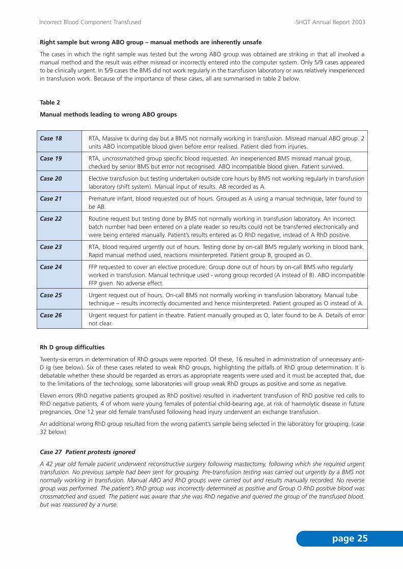
Right sample but wrong ABO group – manual methods are inherently unsafe
The cases in which the right sample was tested but the wrong ABO group was obtained are striking in that all involved amanual method and the result was either misread or incorrectly entered into the computer system. Only 5/9 cases appearedto be clinically urgent. In 5/9 cases the BMS did not work regularly in the transfusion laboratory or was relatively inexperiencedin transfusion work. Because of the importance of these cases, all are summarised in table 2 below.
Table 2
Manual methods leading to wrong ABO groups
Case 18 RTA, Massive tx during day but a BMS not normally working in transfusion. Misread manual ABO group. 2units ABO incompatible blood given before error realised. Patient died from injuries.
Case 19 RTA, uncrossmatched group specific blood requested. An inexperienced BMS misread manual group, checked by senior BMS but error not recognised. ABO incompatible blood given. Patient survived.
Case 20 Elective transfusion but testing undertaken outside core hours by BMS not working regularly in transfusionlaboratory (shift system). Manual input of results. AB recorded as A.
Case 21 Premature infant, blood requested out of hours. Grouped as A using a manual technique, later found to be AB.
Case 22 Routine request but testing done by BMS not normally working in transfusion laboratory. An incorrect batch number had been entered on a plate reader so results could not be transferred electronically and were being entered manually. Patient’s results entered as O RhD negative, instead of A RhD positive.
Case 23 RTA, blood required urgently out of hours. Testing done by on-call BMS regularly working in blood bank.Rapid manual method used, reactions misinterpreted. Patient group B, grouped as O.
Case 24 FFP requested to cover an elective procedure. Group done out of hours by on-call BMS who regularly worked in transfusion. Manual technique used - wrong group recorded (A instead of B). ABO incompatibleFFP given. No adverse effect.
Case 25 Urgent request out of hours. On-call BMS not normally working in transfusion laboratory. Manual tube technique – results incorrectly documented and hence misinterpreted. Patient grouped as O instead of A.
Case 26 Urgent request for patient in theatre. Patient manually grouped as O, later found to be A. Details of errornot clear.
Rh D group difficulties
Twenty-six errors in determination of RhD groups were reported. Of these, 16 resulted in administration of unnecessary anti-D Ig (see below). Six of these cases related to weak RhD groups, highlighting the pitfalls of RhD group determination. It isdebatable whether these should be regarded as errors as appropriate reagents were used and it must be accepted that, dueto the limitations of the technology, some laboratories will group weak RhD groups as positive and some as negative.
Eleven errors (RhD negative patients grouped as RhD positive) resulted in inadvertent transfusion of RhD positive red cells toRhD negative patients, 4 of whom were young females of potential child-bearing age, at risk of haemolytic disease in futurepregnancies. One 12 year old female transfused following head injury underwent an exchange transfusion.
An additional wrong RhD group resulted from the wrong patient’s sample being selected in the laboratory for grouping. (case32 below)
Case 27 Patient protests ignored
A 42 year old female patient underwent reconstructive surgery following mastectomy, following which she required urgenttransfusion. No previous sample had been sent for grouping. Pre-transfusion testing was carried out urgently by a BMS notnormally working in transfusion. Manual ABO and RhD groups were carried out and results manually recorded. No reversegroup was performed. The patient’s RhD group was incorrectly determined as positive and Group O RhD positive blood wascrossmatched and issued. The patient was aware that she was RhD negative and queried the group of the transfused blood,but was reassured by a nurse.
Incorrect Blood Component Transfused SHOT Annual Report 2003
page 25

Case 28 Wrong RhD group on cord sample from direct antiglobulin test (DAT) positive infant
Following delivery by a RhD negative woman, a cord sample was sent to the laboratory for RhD typing. The infant was DATpositive and the BMS had difficulty in interpreting the cord RhD group. To ‘be safe’ he issued anti-D Ig. There was no clinicalurgency and the results could have been reviewed be a more senior member of staff the following day. The infant was in factRhD negative.
Case 29 Historic RhD group unavailable because of numbering discrepancy
A male patient was admitted through A&E with gastrointestinal bleeding. An on-call BMS undertook blood grouping butgrouped the patient as RhD positive, when in fact he was RhD negative. The patient had been grouped previously but becausehe was allocated an A&E number the historical group was not available.
The proposed corrective action by the laboratory did not appear to include changing the patient ‘look-up’ procedures.
Antibody screen or ID errors and crossmatch errors – experienced support is needed to resolve complex cases
Fifteen cases were reported in which there were technical or clerical errors in antibody screening, identification or crossmatch.As a result 11 patients received incompatible red cells of whom 2 suffered haemolytic reactions. Six cases involved multipleerrors. Again it is of note that 7/15 of these instances occurred out of core hours and 5/15 were urgent transfusions. Four ofthe cases involved a BMS not normally working in the transfusion laboratory. Seven cases were missed antibodies, in anotherthree full pretransfusion testing was wrongly omitted. In 2 instances, blood was labelled as compatible and placed in an issuelocation pending resolution of serological problems - not surprisingly the blood was collected and transfused. Two BMSs weredisciplined as a result of errors.
An additional 2 cases were reported in which pre-transfusion testing was carried out using a sample which had been storedfor longer than recommended by guidelines.
Case 30 A complex serological problem out of hours
Blood was requested out of hours for a ‘routine’ transfusion. The on-call BMS did not normally work in the transfusionlaboratory. The antibody screen was positive, however the BMS wrongly interpreted it as due to ‘non-specific IAT antibodies’despite a negative IAT control. He crossmatched and issued apparently compatible blood. The following day furtherinvestigations showed an anti-S and anti-Kpa. Two of the transfused units were S positive. The patient had a poorhaemoglobin increment. The BMS was suspended from on-call duties.
In a similar case the BMS contacted the ward and agreed to delay the transfusion until the next day so that the positiveantibody screen could be resolved. However he crossmatched and labelled the blood and placed it in an issue location – itwas collected and transfused.
There are lessons to be learned from all the individual cases reported under these headings, - short vignettes can be foundon the SHOT website.
Special requirements not met or inappropriate blood component selected – many preventable by better IT
Laboratory errors in 39 cases related to failure to meet special requirements. Of these, 22/39 patients were put at risk of TA-GVHD by failure to provide irradiated components. One patient’s stem cell harvest was delayed as a consequence.
In 29/39 cases, the failure could have been prevented by an effective ‘flag’ on the laboratory IT system. One laboratory hadlost its ‘flags’ when a new IT system was installed, resulting in 2 cases of failure to irradiate, and computer breakdown causeda further 3. In 9 cases laboratory staff had not updated the computer system. In one case the BMS ignored a computer ‘flag’and in 5 the wrong component was selected by the BMS.
An additional seven cases were reported in which the patient’s previous transfusion history was unavailable resulting in failureto provide suitable antigen negative blood. One laboratory had a paper record system; in the other 5, use of a different patientnumber meant that the historic record was not retrieved. Two of these patients suffered haemolytic transfusion reactions dueto anti-Fya .
Incorrect Blood Component Transfused SHOT Annual Report 2003
page 26

These cases highlight the need for laboratory IT systems to be able to search on a limited dataset and to provide effective‘flags’ for patients with special transfusion requirements. There must be robust back-up systems in place for computer down-time, both planned and unplanned.
Case 31 Previous history not available from paper record system
A patient with thalassaemia major required elective transfusion. The laboratory was not computerised and no previous recordswere found in the paper filing system. Antibody screen was negative and the transfusion was uneventful. It was subsequentlydiscovered that the patient had anti-K, undetectable for the past 3 years. When next investigated a strong anti-K was found.
Eight additional cases were reported in which laboratory staff selected components which were unsuitable for a variety ofother reasons; 3/8 were for paediatric patients; 5/8 patients suffered some morbidity.
Telephone requests must be fully documented
Two almost identical reports were received in which telephone requests were incompletely documented in the laboratory,resulting in the wrong patient’s sample being selected for pre-transfusion testing. In neither case was the transfusion urgent.In both cases there were 2 patients on the same ward with similar names, and both the laboratory and the ward failed tocheck the full details. Fortunately in both cases the blood given was ABO compatible. One of these cases is described in detailbelow.
Case 32 Don’t rely on name alone for identification of samples or patients
Edith Watt and Edith Watts were on the same orthopaedic ward. Neither had been grouped previously. Their serum sampleswere adjacent in the laboratory storage rack. The laboratory received a telephoned request for blood for Edith Watt who wasgoing to theatre for repair of her fractured neck of femur. The request was not documented in the laboratory. The BMS pickedout Edith Watts’ sample and used it to crossmatch blood which he labelled with Edith Watts’ name, date of birth and hospitalnumber. The nurse collecting blood for Edith Watt did not notice the discrepancy, nor did the 2 nurses who administered theblood. Edith Watt was not wearing a wristband and her notes were not available. She was group O RhD negative and receivedblood which was group O RhD positive.
In the following remarkable case no fewer than 5 individuals failed to spot the discrepancy between patient details on thepack and on the patient wristband.
Case 33 ‘Same name’ coincidences must be expected
A patient was admitted to St Elsewhere’s with a ruptured abdominal aortic aneurysm. The ward telephoned the laboratoryrequesting Group O uncrossmatched blood. The laboratory had previously received a sample for grouping from anotherpatient with exactly the same name, who was Group A. The BMS, recognising the name but without checking the date ofbirth or hospital number, issued 4 units of uncrossmatched group A blood labelled with the date of birth and hospital numberfrom the previous request. Two nurses gave the first unit without noticing the discrepancies. The patient was transferred toLarge University Hospital (LUH) with a nurse escort taking with her the remaining 3 units of blood. On arrival in A&E, the nursefrom St Elsewhere’s together with a nurse from LUH gave the second unit without checking against the wristband. The patientwas taken immediately to theatre where the anaesthetist gave the third unit, again without checking. Only when the fourthunit was given was the discrepancy noticed. The patient was found to be group O. He died on the operating table; theincompatible transfusion was not thought to have contributed to his death.
Stock control failures
In 27 cases failure of laboratory stock control procedures contributed to transfusion of a blood component which had expired(21 units of red cells and 1 FFP) or had been out of temperature control. IT systems are available to aid stock control andprevent issue of expired units from blood banks and satellite refrigerators.
Incorrect Blood Component Transfused SHOT Annual Report 2003
page 27

Learning points from laboratory errors
• Laboratory staff working out of hours and/or under pressure are prone to make errors and must be supported by robustprocedures and technology.
• Staff must not be required to work beyond their level of competence or experience.
• Procedures for rapid testing are more error prone than routine automated procedures and should only be used when thereis clinical urgency.
• Patient name coincidences will happen and systems must be in place to protect against the consequences.
• Verbal communications are a potential source of errors and must require the same points of identity as written requests.
• Transfusion laboratory stock control procedures should ensure that expired units are cleared from issue locations. Laboratory IT systems must not allow expired units to be issued.
• Transfusion laboratory IT systems should provide effective ‘flagging’ of special requirements and alert staff to select appropriate components.
Errors in the collection and administration of blood components (n=232)
There were 232 errors in collection and administration of blood components in 187 cases.
In 36% of reported cases the primary error occurred at this stage of the transfusion process.
Of the 176 cases in which the time of the transfusion was reported, 65/176 (37%) took place between 8pm and 8am. It isof note that of the 33/176 (19%) transfusions started between midnight and 8am, 16/33 were stated to be ‘routine’.
There were 156 instances of failure of the bedside checking procedure, which was again the most common error in thetransfusion process. In 21/156 cases, this was the primary error, resulting in wrong transfusions being given. In 10 of thesecases there were 2 patients being transfused simultaneously on the ward; four ‘paired’ reports were received.
In 45 cases a wrong blood component was collected from the hospital storage site and the error was not detected at thebedside. It is notable that 10 of these cases related to acutely bleeding patients undergoing urgent or massive transfusions incritical care situations (operating theatres, recovery suites, A & E departments, intensive care units or delivery suites).
In 135 cases, the bedside check failed to detect an error earlier in the transfusion chain; in 24 of these an expired unit wastransfused. System failures included checking blood against documentation away from the bedside, absence of identificationwristbands and in some cases complete omission of the bedside identification check.
Cases relating to children are also discussed in chapter 12.
In one case blood was delivered directly to A&E by the blood service and transfused without reference to the transfusionlaboratory. In another a label became detached from a platelet pack and was re-attached to the wrong unit.
Outcomes of collection and administration errors
Twelve patients received ABO incompatible transfusions. Two of these patients died from massive bleeding, the incompatibletransfusion was not thought to have contributed to their deaths. A further 2 patients suffered major morbidity as a result ofacute haemolytic transfusion reactions.
Incorrect Blood Component Transfused SHOT Annual Report 2003
page 28

Collection of incorrect component (n=45)
Table 3
Collection errors according to grade of staff involved and whether or not a formal check was made at this stage
*1 x Auxiliary nurse, 1 x Unqualified ODA, 1 x Anaesthetist, 1 x Ward clerk, 1 x SHO
The recent National Comparative Audit of Transfusion3 has demonstrated poor practice in the administration of blood at thebedside. The cases described here are a direct result of unsafe practices such as those identified in the audit, and initiativesto improve this area of practice are urgently needed.
Case 34 Two patients, same surname – a well known pitfall and 2 similar cases
Joe Soap, who was O RhD negative, required an urgent transfusion for bleeding oesophageal varices. There was anotherpatient (Fred Soap) with the same surname on the ward for whom blood had also been crossmatched. Fred Soap was O RhDpositive. Fred Soap’s compatibility form had been stuck in Joe Soap’s notes. An agency nurse was sent to the blood bank tocollect blood for Joe. She collected a unit of Fred’s blood which was then checked against Joe’s notes but with reference toFred’s compatibility form. No bedside check was carried out.
A nurse noticed the discrepant name when taking down the empty bag.
Case 35
David Archer and Tony Archer were patients on the same ward and blood was crossmatched for both. David was O RhDpositive and Tony was A RhD positive. The porter collecting blood for David removed a unit of Tony’s blood and Tony’scompatibility form. Nursing staff checked the details on the bag label against the (wrong) form, which was signed and placedin David’s notes. David was not wearing a wristband and no identity check was done. The transfusion was stopped when hedeveloped a fever, but only when the laboratory came to investigate the reaction was the ABO incompatibility realised. Hesurvived the event.
Case 36 No light shed on wrong blood
A patient (group B RhD positive) was undergoing endoscopy in theatre for acute upper gastro-intestinal bleeding. A unit ofgroup O RhD positive blood intended for another patient was collected from the theatre satellite refrigerator. Because thetheatre was in semi-darkness for the endoscopy, the anaesthetist was unable to read the label and administered the bloodwithout checking.
Case 37 Wrong blood given to patient in the intensive care unit (ICU) and delay in recognising an acute reaction
This case was particularly well investigated and a number of errors was identified. The patient, who was being treated in anintensive care unit for oesophageal carcinoma, received an ABO incompatible transfusion resulting in major morbidity but hesurvived. The transfusion was elective but was undertaken in the evening, pre-transfusion testing was done by an on-call BMS.A porter was sent to collect the blood, having been given only a surname (verbally) without any documentation. He collectedthe wrong unit from the blood bank refrigerator and did not correctly log the unit out. Two qualified nurses checked the unitagainst a compatibility form but did not check it at the bedside. Moreover the blood was held at room temperature for over30 minutes before the start of transfusion. The patient developed fever, chest pain and haemoglobinuria but observationswere not recorded at appropriate intervals and the transfusion reaction was not recognised until almost a whole unit hadbeen transfused.
Incorrect Blood Component Transfused SHOT Annual Report 2003
page 29
GRADE OF STAFF FORMAL ID CHECK
Yes No Unknown
Qualified nurse / midwife 5 6 5
Unqualified nurse / midwife 2 0 1
Porter 5 5 0
Locum / agency 1 2 0
Qualified ODA 0 3 0
Unknown 2 2 1
Other* 1 3 1

Failure of bedside checking procedure (n=156)
Table 4
Grades of staff involved in bedside incidents
Grade of Staff Number of cases
Qualified nurse & qualified nurse 98
Qualified nurse only 21
Qualified nurse & unqualified nurse 6
Qualified nurse & doctor 5
Qualified nurse & locum / agency 4
Doctor only 3
Doctor & doctor 2
Doctor & qualified ODA 2
Unqualified nurse & Unqualified nurse 1
Locum / agency only 1
No response 13
Cases 38 and 39 Why give non-urgent elective transfusion at night?
Two patients on an oncology unit required elective transfusions. Freda Fry was O RhD positive; Linda Snell was B RhD negative.The two units of blood were collected after midnight from the blood bank and checked on the ward away from the bedside.The units were inadvertently transposed and transfused to the wrong patients. Freda Fry suffered a severe acute haemolyticreaction after the first 50 ml of transfusion and required admission to ICU. She later recovered. Linda Snell’s transfusion wasstopped; she suffered no ill effects.
Cases 40 and 41 Patients in adjacent beds in A&E given one another’s blood
These 2 patients were in adjacent beds in an A&E department at night. Both required transfusion. One was O RhD positive,the other one A RhD positive. It is not known whether they were wearing wristbands. Two units of blood were inadvertentlytransposed and given to the wrong patients. A nurse noted the discrepancy and the transfusions were stopped, neitherpatient suffered any ill effect.
Learning points
• Name coincidences are a source of error here as at other stages of the transfusion process and must be guarded againstby full positive identification at every stage.
• In emergency situations safety must not be compromised for expediency.
• The final patient identity check must be done at the bedside against an identification wristband or equivalent attached tothe patient. No other form of checking is acceptable under any circumstances.
• Transfusion should not take place at night unless clinically indicated.
Transfusion of ‘unsafe’ units – incorrectly stored or expired
Forty-three reports were received of inappropriate handling of blood components. These included 21 units of red cellstransfused past their expiry date. All of these involved a stock control error by the laboratory (2 relating to satelliterefrigerators) plus failure to check the expiry date at the bedside. There were 12 instances of red cells out of temperaturecontrol in clinical areas. In 4 cases this involved blood which was being transferred between hospitals. Ten miscellaneoushandling errors included FFP stored at room temperature on a platelet agitator in a clinical area; platelets stored in a satelliterefrigerator; an irradiated neonatal unit returned to the laboratory for discard and taken by an SHO and FFP issued past itsexpiry date by a BMS.
Incorrect Blood Component Transfused SHOT Annual Report 2003
page 30

Learning points
• The pre-transfusion checking procedure must include a check of the expiry date of the component.
• Hospital transfusion teams must collaborate to ensure that procedures are in place for safe transfer of blood between hospitals.
Errors originating at the supplying blood centre
There were 10 errors by 7 blood centres. Errors included;
Failure of screening procedures to detect a strong anti-Fya in an apheresis platelet donor resulting in passive transfer ofantibody sufficient to cause a positive antibody screen.
Failure to detect a high titre haemagglutinin in red cells labelled as suitable for neonatal transfusion resulting in acceleratedred cell destruction in a group B infant.
Two instances in which a verbal report by a reference laboratory was inconsistent with the final report. In one case this resultedin failure to select antigen negative red cells; in another anti-D was inappropriately administered to a patient with a weak D.
Provision of RhD positive platelets when RhD negative had been requested for a RhD negative patient - this error was notdetected by the hospital laboratory or at the bedside.
An ad hoc delivery of Group O RhD negative blood directly to an A&E department instead of to the transfusion laboratory. Adoctor transfused the blood.
Failure to provide CMV negative components.
Errors in anti-D administration
SHOT has not actively encouraged reports of incorrect administration of anti-D Ig, but nevertheless each year reports of suchevents are received. This year there has been a reduction in the number of anti-D errors reported.
As with other IBCT reports, there are errors at all stages of the process, including patient identification, laboratory errors,incorrect serological reasoning in the laboratory and by clinical staff. The majority of errors are of commission and we receivefew reports of errors of omission, which are likely to have more serious clinical consequences.
This year 26 errors were reported in 24 cases, of which 16 related to incorrect or equivocal RhD grouping in laboratories.
Table 5Anti-D errors
Type of error Number
Misunderstanding of guidelines by midwife 3
Anti-D given to wrong patient 3
Late administration of anti-D Ig (>72hrs) 2
Patient already sensitised, misinterpreted by laboratory 2
RhD grouping error – cord sample 5 (2 due to reagent problem)
RhD grouping error – maternal sample 5
Weak RhD group (includes 1 reference laboratory) 6
Case 42
The wrong notes accompanied a patient to theatre for a caesarian section. Anti-D Ig was given on basis of historical groupin (wrong) notes without sending a confirmatory sample.
Incorrect Blood Component Transfused SHOT Annual Report 2003
page 31
Procedural review
Whilst recognition, reporting and investigation of incidents serves to identify flawed systems, full benefit can only be gainedfrom incident reporting if events are carefully analysed to determine root causes and contributory factors. Much has beenwritten on the subject of root cause analysis and a retrospective analysis of three cases reported last year is published on theSHOT website. HTTs and Transfusion Committees have a crucial role in ensuring that the loop is closed following an adverseevent and that opportunities are taken to use such incidents as learning opportunities.
In a number of cases the ‘corrective action’ consisted of the addition of checking steps to processes, thus making them morecomplex. Staff are more likely to follow correct procedures if they are simple and straightforward and opportunities should betaken to review and revalidate processes when errors have occurred.
Disappointingly, an element of blame was apparent in many of the accounts of events. One BMS was dismissed, two weredisciplined and others were removed from on-call rotas. Blame was also apportioned to some nurses, though not to medicalstaff who made errors. There still needs to be a shift of emphasis away from individual blame if incidents are to be fully sharedand lessons learned from them.
Incorrect Blood Component Transfused SHOT Annual Report 2003
page 32

COMMENTARY
• The number of inappropriate transfusion reports due to poor blood sampling techniques remains an area of concern. Inaddition to patient identification errors, these events may result from performing venepuncture on a limb into which an intravenous infusion is being given, inadequate filling of the sample tube or failing to mix the sample in the tube appropriately. These practices can cause spurious or incorrect laboratory results that can result in incorrect or inappropriaterequests for blood components. Patient identification errors at this stage are particularly hazardous as they may be undetectable.
• Errors are more likely to occur under exceptional circumstances, particularly in clinically urgent situations when staff are under pressure and there is a high level of stress. Drills and practices of emergency procedures are effective in other environments and can contribute to the development of a ‘safety culture’ in clinical areas and in laboratories.
• Patients continue to be put at risk due to failure to communicate special transfusion requirements; most commonly the need for irradiated components in patients prescribed purine analogues. The use of this group of drugs is increasing andgamma-irradiation remains the only proven method of preventing TA-GVHD.17
• There are numerous examples of laboratory staff working under pressure, either when blood has been requested urgently,or out of hours, or both. It is of concern that, among errors likely to result in incompatible transfusion, 8/9 ABO groupingerrors and 7/15 antibody identification or crossmatch errors were related to out-of-hours or urgent work. The increasingmoves towards a 24/7 level of hospital activity and the pressures of other NHS initiatives exacerbate these stresses.
• Administration of blood at the bedside remains an important risk area, as demonstrated by events reported to SHOT andby the findings of the National Comparative Audit3. A co-ordinated initiative is needed to address this area.
• A disproportionate number of incidents involving infants has been reported this year. These are further considered in chapter 12. There is a need for education of staff in paediatric units and laboratories, in special transfusion requirementsof children including volume calculation.
Incorrect Blood Component Transfused SHOT Annual Report 2003
page 33

RECOMMENDATIONS
• Hospital risk management committees must ensure that all staff undertaking venepuncture for blood sampling must havereceived the necessary training and have their practical competency formally assessed and recorded. Blood samples shouldbe taken from a free flowing venepuncture site, and the tube should be filled to capacity and inverted gently several timesto adequately mix the sample and any anticoagulant. The person taking the sample must complete the tube label beforeleaving the patient, checking the details are correct with the patient and against the ID wristband or equivalent.
Action: Hospital risk management committees
• Hospital risk management procedures should include ‘drills’ for high risk situations such as massive transfusion, involvingall relevant staff.
Action: Hospital risk management committees
• Prevention of TA-GVHD in patients receiving purine analogues is the responsibility of prescribers, but can and must be supported by the pharmaceutical industry and pharmacists and by suppliers of laboratory IT systems. All patients should receive an information card and leaflet and haematologists must ensure that there is an effective system of flagging specialtransfusion requirements in the laboratory. Referrals for shared care must include timely communication of all relevant information.
Action: Clinicians prescribing purine analogues and administering blood transfusion; HTTs; pharmacists, pharmaceutical industry; suppliers of laboratory IT systems
• Hospital blood bank laboratory staffing must be sufficient for safe transfusion practice. Hospitals must ensure that bloodtransfusion laboratories have adequate numbers of appropriately trained biomedical scientists to cover the 24-hour working day, including a core of permanent blood transfusion laboratory staff. Standards should be established for manpower appropriate to the level of workload and this should be subject to inspection.
Action: Clinical directors of pathology, professional and accrediting bodies
• Paediatric units undertaking transfusion must ensure that staff are educated in the special transfusion requirements of children. Laboratory IT systems should be regularly updated to support implementation of new guidelines.
Action: Paediatricians and laboratory staff responsible for transfusion of paediatric patients; HTTs
• The most important contribution which could now be made to the safety of blood transfusion would be an initiative to improve the safety of the bedside pretransfusion checking procedure. This will require investment in education and audit,and also in evaluation and implementation of suitable information technology. The CMO’s NBTC has the necessary remitto take this forward.
Action: CMO’s NBTC through regional and hospital transfusion committees: HTTs
Incorrect Blood Component Transfused SHOT Annual Report 2003
page 34

Definition :
Any error, which if undetected, could result in the determination of a wrong blood group, or issue, collection, or administration of an incorrect, inappropriate or unsuitable component but which was recognised before transfusion took place.
The history and future of "near miss"
Following small pilot schemes first launched in 1997/98 and repeated the following two years, SHOT invited all UK hospitalsto report "near miss" incidents beginning with the report year 2000/2001. The picture has been a consistent one showingerrors made during phlebotomy to be by far the most numerous (50% in 2000/2001, 59% in 2001/2002 and 60% in 2003).Anecdotal evidence from reporting hospitals suggests that "near miss" events are frequent and common but there isdemonstrable gross under-reporting. Last year, while overall reporting to SHOT was calculated to be 93%, "near miss" reportswere received from only 41% of participating hospitals. The most likely explanation for this is that the large number of eventswitnessed makes reporting of them difficult to sustain. Since the pattern of events appears to vary little from year to year itwould seem logical and advisable to move to a system of enabling hospitals to report their "near miss" events en masse ratherthan having to complete a single form for each event. We know that many hospitals report this way internally and we wouldlike to find methods of sharing this information without increasing greatly the workload of already over-stretched laboratorystaff. Many valuable lessons have been learned from the data we have collected so far but there seems little point in simplyre-iterating the same messages particularly since we are probably seeing only the tip of the iceberg. The chapter this year thenis considerably shorter than in previous years and aims to give an overview rather than an in-depth analysis of events we havereported on before.
For consistency, the categories and numbers of events reported this year are shown in figure 7.
Figure 7Categories and proportions of "near miss" events
Near Miss Events SHOT Annual Report 2003
5 Near Miss Events
page 35
600
500
400
300
200
100
0
100 9786
542
81
Sample errors60%
Component collection,transportation, ward
handling & administrationerrors11%
Laboratory componentselection, handling,
storage & issue errors11%
Laboratory samplehandling &/or testing
errors9% Request errors
9%

Category 1 : Sample errors (542 cases)
Once again sampling errors proved to be the most numerous comprising 60% of all "near miss" events reported. The majorityof these errors were picked up from historical records on laboratory computers or by the vigilance of laboratory staff incarrying out double checks. Several of these events involved errors in haematology or biochemistry results which, althoughnot strictly the responsibility of blood transfusion laboratories, would have resulted in unnecessary transfusions had they notbeen discovered in time. This is an important issue since there has been a large number of cases reported in the "IncorrectBlood Component Transfused" chapter which, unfortunately, were not detected in time. Many errors reported last year wereconsistent with those reported in earlier years. In particular the following types of error occur regularly and should be a priorityfor investigation and prevention :
• Samples in syringes, which are left unattended for a period and then dispensed into tubes without mixing.
• Dilute samples taken from "drip arms".
• Point-of-care (POC) testing variations.
• Labelling of samples remote from the patient.
• Pre-labelling of specimen tubes for RhD negative deliveries with subsequent confusion of tubes between mother and baby.
• Wrong addressograph labels used.
• Labels placed in wrong patients’ notes.
• Poor patient identification using only one or two identifiers.
Category 2 : Request errors (81 cases)
A frequently reported problem in this category was a failure to notify the transfusion laboratory of the need for CMV negativeand/or irradiated components. This is a commonly recurring theme both in "near miss" events and in full incidents and oftenindicates a need for improved communication between hospitals where patients are receiving shared care.
The majority of errors in this category were made by junior doctors, usually senior house officers, highlighting the need foreducation and training for medical staff requesting and prescribing blood components.
Category 3 : Laboratory sample handling / testing errors (86 cases)
A wide variety of errors was reported in this category consistent with previous years. It is interesting that many of theseincidents were said to have occurred because the staff were either "very busy" or "under pressure" indicating the need forTrusts to review staffing levels or shift patterns. Most of these laboratory errors were recognised by the staff themselvesretrospectively but not always before components had been issued.
Category 4 : Laboratory component selection, handling and storage errors (97 cases)
The majority of these reports related to poor housekeeping systems with evidence of poor practice in stock control of remoterefrigerators. In addition there were several cases involving the issue of units close to expiry which were to be used for theatrecover at a later date. There were also several cases in which a refrigerator alarm had failed but this was not recognised by thestaff.
Nine cases were reported in which the Blood Centre had provided the wrong specification of component to the hospital. Themost worrying of these was one in which granulocytes were provided which had not been irradiated. The hospital BMS wasunaware of the need for irradiation and issued the component to the ward. Fortunately the error was picked up at the pre-transfusion check.
Category 5 : Component issue, transportation and patient identification errors (100 cases)
Storage and transport problems accounted for 51% of errors in this category. Most of these involved storing units ininappropriate refrigerators, for example chemotherapy and drug refrigerators. Several cases were reported of blood being leftout of temperature control for extended periods including 1 unit which was left on top of a locker for 12 days. The collectionof an incorrect unit was another frequently reported error, this year comprising 31% of category 5 errors. Notably in 20 ofthese 31 cases the unit was collected by a porter and porters were also cited in 3 other category 5 incidents.
Near Miss Events SHOT Annual Report 2003
page 36

In one reported case the porter happened to meet the SPOT en route from the blood bank. He was found to have nodocumentation with him, had collected the wrong unit of blood and was planning to complete the collection slipretrospectively. In another case the porter collecting the blood had received no training and was unable to read English, andin a third, the person collecting the blood had forgotten her glasses!
Right blood to right patient
Each year there are a number of cases reported in which the patient receives entirely appropriate blood which was intendedfor them but which, nevertheless, had some element in the process which was wrong and which could, under differentcircumstances, have led to a serious transfusion error. It has always been difficult for SHOT to place these events appropriately.They do not constitute Incorrect Blood Transfused events since they are not covered by that definition. Nor can they really bedefined as "near miss" since that definition requires that no volume of blood should have been transfused. They are, however,fairly frequent and warrant some discussion. It has been decided, therefore, to include them in the numbers of "near miss"events as a separate category and to try to amend the definition accordingly before the next reporting year. There were 29such cases reported this year.
Problems in reporting
This year unforeseen technical problems with the SHOT database have meant that cases coming in to the office have not beenreviewed in a timely manner. This has produced the unfortunate effect of cases being reported as "near miss" which were,in fact, full IBCT incidents. However, by the time these cases were ready for review it was too late to obtain the necessaryinformation from hospitals which would have enabled us to convert them into IBCTs and report on them properly. These casesare, therefore, not counted in the numbers of either "near miss" events or IBCTs for this year. Reporters are asked to beparticularly careful about not reporting IBCTs as near misses and to contact the SHOT office at any time if they have any doubtabout a particular case. The one essential criterion to remember is that an event is not "near miss" by our definition if anyvolume at all has been transfused. The error must have been recognised before any blood component passed through thevenflon. The "near miss" definition does not allow for reporting of events where, for example, a patient received a smallvolume of red cells which were intended for another patient but which was, fortuitously, fully compatible and the patientsuffered no ill effects. These cases are defined as "Incorrect Blood Component Transfused" and must be reported on theappropriate form.
There were 6 such cases reported inappropriately as "near miss" which were, in fact, full IBCT incidents and these aresummarised below. At the end of each case the reporter’s rationale for reporting them as "near miss" is included and isintended to illustrate the sorts of false reasoning outlined above.
1. A patient on a coronary care unit was given 2 units of blood based on a Hb result of 98 g/L. Over the next 2 days there were widely differing Hb results for this patient ranging from 74 g/L to 138 g/L. A further 2 units were transfused in themeantime. It is thought that the discrepant results were due to dilute samples though this has not been fully established.
The case was reported as "near miss" because the patient suffered no ill effects
2. A patient due to go into theatre had Hb results in his notes which belonged to another patient. This result was used as an indicator for transfusion. Nursing and theatre staff questioned the need for the transfusion and discovered the error on investigation.
Reported as "near miss" because the transfusion was stopped after only a few mL had been transfused and the patient suffered no harm
3. A unit of blood was transfused to a patient 47 hours after the crossmatch had expired.
Case reported as "near miss" because the blood was crossmatched for the patient concerned
4. A transcription error in the laboratory led to incorrect patient details being used to group and crossmatch a unit of blood.
This was reported as "near miss" because the unit transfused was compatible
5. Prophylactic anti-D was issued by the laboratory and the ward informed that it was ready for collection. However the unitwas not collected until 5 days later despite daily reminders by the laboratory staff.
Reported as "near miss" because the anti-D was given eventually
Near Miss Events SHOT Annual Report 2003
page 37

6. Anti-D was not given to a pregnant woman who was RhD negative and who had chorionic villus sampling (CVS) in an antenatal clinic. The consultant in the clinic wrote her blood group in the CVS book as RhD positive based on a verbal report by the patient.
This was reported as "near miss" because the patient had been given anti-D in a previous pregnancy
"Near miss" reporting 2004 and 2005
Discussions are underway concerning how to collect "near miss" data in the future. We are aiming to develop new systemsin time for the start of reporting in 2005. Meanwhile we are continuing to accept reports on the current questionnaires andhospitals are encouraged to submit as many reports as possible. We know that some hospitals report their "near miss"incidents in bulk internally. Until the new systems are in place we would also encourage hospitals to phone the SHOT officefor advice if they would also like to report these cases to SHOT. We will endeavour to find ways of handling bulk reportsmanually until new systems have been developed.
Near Miss Events SHOT Annual Report 2003
page 38

Definition
Acute transfusion reactions are defined in this report as those occurring at any time up to 24 hours following a transfusionof blood or components, excluding cases of acute reactions due to incorrect component being transfused as these are coveredin Chapter 4
This category accounted for 9.3% of non-infectious hazards reported and 9.1% of all hazards.
There were 6 outstanding reports from the previous reporting year for which 5 questionnaires were eventually receivedalthough on review one of these was, in fact, an incorrect blood component transfused and is now transferred to that section.The remaining 4 are included in the analysis. Forty-four valid initial reports were received and 39 completed questionnaires(including 4 from the previous year). The 9 outstanding questionnaires will be included in next year’s analysis. An additional12 reports did not fit the definition of ATR and have been withdrawn by the analyst.
This chapter highlights the main findings from 39 completed questionnaires.
There were 5 deaths in this group; 1 probably related to the transfusion, 1 possibly related to the transfusion and 3 unrelatedto the transfusion. There were 2 instances of major morbidity and renal failure as a result of haemolytic transfusion reactions.
Gender (37 reports)
Male 21
Female 16
Age (38 reports)
Age range 3 months to 84 years
Median 66 years
Components implicated (39 reports)
Red cell - allogeneic 8
Red cell – post-operative salvage 1
Platelets 13 (8 apheresis units and 5 from pooled buffy coats)
Fresh frozen plasma 17 (all not viral inactivated)
Reactions in which red cells were implicated
There were 8 cases, with one death possibly related to the transfusion, and two cases of major morbidity. Six reactionsoccurred during the transfusion, 1 within 7 hours of completing the transfusion and 1 unknown. The following reactions wereseen:
Table 6Reactions in which red cells were implicated
Reaction type Number of cases
Haemolytic or incompatibility reaction 4
Anaphylactic/anaphylactoid+ 1
Allergic++ 1
Unknown/unclassifiable 2
+ anaphylactic/anaphylactoid (hypotension with 1 or more of: rash, dyspnoea, angioedema)
++ allergic (1 or more of: rash, dyspnoea or angioedema without hypotension)
Acute Transfusion Reactions SHOT Annual Report 2003
6 Acute Transfusion Reactions
page 39

Haemolytic or Incompatibility Reactions
In 4 cases, there were features of haemolysis
Case 1
A 75 year old male with no previous transfusion history had a coronary artery bypass graft (CABG) performed, during whichhe bled excessively and was transfused with 14 units of red cells, 13 units of FFP and one unit of platelets. Within 7 hours ofthe procedure, the patient developed a fever, rigors, and chest pain. He became hypotensive, was noted to pass dark urineand was found to have a strongly positive DAT(IgG), hyperbilirubinaemia, spherocytes, haemoglobinuria, disseminatedintravascular coagulation and deteriorating renal function that required dialysis.
The patient had two antibody screens pre-operatively. The first performed at the pre-assessment clinic was weakly positiveand a provisional specificity of an anti-E was given on the basis of a positive reaction with one R2R2 cell. The patient’s Rhphenotype was R2r, the DAT was negative and an auto-E was questioned. The second antibody screen performed onadmission was negative but he was crossmatched with E negative units. Post-operatively the patient’s plasma contained astrong anti-E ,reacting 5+ in IAT and anti-E was eluted from the red cells. These post-operative findings were confirmed bythe reference laboratory and an auto-anti-E was again suggested to be responsible.
There are very few cases of an autoantibody being stimulated by transfusion, and the rapid response over a few hours issurprising. However, the finding that anti-E could be eluted from the E negative units transfused would support the hypothesisthat this was an autoantibody.
Case 2
A 30 year old female was transfused 2 units of red cells on account of a post-partum haemorrhage and required 2 furtherunits a few days later. During the second transfusion episode, she became febrile, dyspnoeic and was noted to be passingdark urine. She was subsequently found to have a positive DAT (IgG only), haemoglobinuria and deteriorating renal functionand required dialysis and admission to ICU. Her pre and post-transfusion antibody screens were repeatedly negative using anautomated DiaMed technique. These samples were referred to the Reference Laboratory who detected an anti-Jkb and ananti-K in the pre-transfusion sample by papain IAT only, and confirmed that the implicated unit was Jkb positive.
The reporting hospital has since amended its protocol for investigating red cell transfusion reactions to include additionaltechniques.
Case 3
An example of a patient with an anti-Doa who as a result of two consecutive transfusions, experienced a delayed haemolytictransfusion reaction followed by an acute haemolytic transfusion reaction. The vignette is included in Chapter 7 (DelayedTransfusion Reactions).
Case 4
A 50 year old female with myelofibrosis required regular transfusion support. The patient had experienced a febrile reactionduring a transfusion of 3 units of red cells approximately 3 weeks earlier but a decision was taken to continue the transfusionwith chlorpheniramine cover. On the following occasion, the red cell transfusion was stopped after 50ml on account of rigorsand a tachycardia and following the transfusion the patient was noted to have a raised bilirubin. She had a negative IATantibody screen pre and post transfusion and the DAT was negative throughout. However when samples were referred to thelocal Reference Centre, the patient was found to have an anti-c+E together with a non-specific papain autoantibody in thepre-transfusion sample.
It is not known why these antibodies were missed and the reporting hospital has since amended its protocol for investigatingred cell transfusion reactions to include the use of enzymes and low ionic-strength saline (LISS) tube techniques.
Anaphylactic/anaphylactoid reactions
One patient who was IgA deficient developed an anaphylactic reaction due to anti-IgA.
Case 5
A 70 year old male had an anaemia of chronic disease and was undergoing debridement of an infected foot. On the firstoccasion, after 86ml of red cells had been transfused, the patient developed dyspnoea with tachycardia and profoundhypotension. He was successfully resuscitated and repeat testing of the pre and post transfusion sample revealed no red cellincompatibility and bacterial cultures were negative. Two weeks later, a further red cell transfusion was given withhydrocortisone and chlorpheniramine pre-medication. After one hour the patient developed severe rigors and the bloodpressure rose from 134/80 to 206/120.
Acute Transfusion Reactions SHOT Annual Report 2003
page 40

The transfusion was again aborted and the patient resuscitated with hydrocortisone and chlorpheniramine. At this stage thepatient was tested for IgA deficiency and found to be IgA deficient with an anti-IgA antibody. Subsequent transfusions ofwashed red cells have been tolerated.
A severe allergic reaction should have been suspected after the first transfusion.
Reactions of unknown aetiology
Case 6
An 80 year old female, with a non-Hodgkin’s lymphoma and previously untransfused, had a positive DAT(IgG only) but anegative red cell antibody screen (Ortho BioVue IAT). The patient was also reported to have an autoimmune haemolyticanaemia although no clinical details are available. Three units of red cells were issued electronically. During the third unit, thepatient developed a fever, breathlessness, hypotension and a reduced level of consciousness. The transfusion was stoppedand antibiotics were prescribed. The patient died two hours later. The patient had a negative antibody screen post transfusionand the units were compatible when retrospectively crossmatched.
No bacterial cultures were taken, a chest X-ray (CXR) was not performed and it is not clear whether any of the symptomswere related to the transfusion rather than underlying sepsis.
The reporting hospital now perform a serological crossmatch on patients with a positive DAT.
Case 7
A 20 year old male, who had undergone an orthotopic liver transplant 15 years earlier and who was known to have a warmautoimmune haemolytic anaemia had not required red cell support for several years. On the reported occasion, after anunspecified volume of red cells, the patient developed a fever, rigors, hypotension, nausea and vomiting. Pre-transfusiontesting revealed a positive DAT (IgG only),and an autoantibody reactive in the IAT with no apparent specificity. No testing wasundertaken to exclude underlying alloantibodies and no serological crossmatch was performed. The post transfusion sampleagain showed a strong autoantibody.
Subsequent red cell transfusions, given slowly, have been well tolerated.
Reaction to a unit of autologous salvaged blood
Case 8
An 84 year old male who had undergone a total knee replacement was transfused with unwashed red cells salvaged post-operatively. After 550ml had been given, the patient developed a fever, rigors and dyspnoea requiring bronchodilators andhigh dependency unit (HDU) admission. Bacterial cultures of both the unit and the patient were negative.
Filtered salvaged blood contains cytokines that may have been responsible for the reaction
Reactions in which FFP was implicated
There were 17 reports in this group, of which 15 occurred during the transfusion, and 2 within 2 hours. The followingreactions were seen:
Table 7
Reactions in which FFP was implicated
Reaction Type Number
Anaphylactic/anaphylactoid 8
Allergic 8
Unknown/unclassifiable 1
Acute Transfusion Reactions SHOT Annual Report 2003
page 41
Anaphylactic/anaphylactoid
There were 8 patients in this category, all of whom recovered from the reaction. Seven occurred during the transfusion, andwhere information is available, started after 50-200 mL FFP had been given. The eighth case was noted within 30 minutes ofcompleting the infusion.
Five cases had dyspnoea, of which one had a CXR performed (result unknown) and one had blood gases taken confirminghypoxia.
Four patients required adrenaline in addition to hydrocortisone and chlorpheniramine, of which 2 were also givenbronchodilators. A fifth patient was given hydrocortisone and chlorpheniramine and a sixth oxygen alone. It is not clear whatmedication was prescribed for the remaining 2 patients.
Five of the 8 patients were investigated for IgA deficiency, with negative findings.
Four out of 8 FFP transfusions can be justified, (included in table 8 below).
Case 9
A 68 year old man on warfarin was scheduled for a colonoscopy and biopsy and had been asked to discontinue hisanticoagulants three days before admission. On admission, the day before the intended procedure, his INR was 1.65 and hewas prescribed FFP.
Within 10 minutes of starting the transfusion, the patient had developed an urticarial rash. He was seen within a few minutesby which time he had become hypotensive, BP 58/33, wheezy, developed rigors and lost consciousness. He required two IMinjections of adrenaline, in addition to 200mg hydrocortisone, 20mg chlorpheniramine and crystalloids/colloids over 30minutes to become haemodynamically stable. He was also given nebulised salbutamol and oxygen. He had a normal IgA level.
Allergic reactions (not anaphylaxis)
There were 8 patients in this group, all of whom were also dyspnoeic. 6 had a rash which was accompanied by angioedemain 2 cases. The remaining 2 had rigors in addition to dyspnoea.
No CXRs were performed. 4 were investigated for IgA deficiency with negative findings and there were no investigationsperformed in the remaining 4 who had all been previously transfused.
3 out of the 8 FFP transfusions can be justified (included in table 8).
Case 10
A 43 year old male with Crohn’s disease, also on warfarin for a previous pulmonary embolus, was noted to have an infectedHickman line. He was scheduled for surgery 3 days later but was not informed to discontinue his warfarin. He was admitted2 days before surgery and was found to have an INR of 2.9 and was prescribed FFP.
At the end of the first unit, the patient developed a rash, a swollen tongue, and became dyspnoeic with an oxygen saturationof 88%. He responded to hydrocortisone, chlorpheniramine and nebulised salbutamol. His IgA level was normal.
Unclassifiable
Case 11
An 80 year old patient (gender not reported) was admitted with sepsis and disseminated intravascular coagulation. He/shewas transfused with one pool of platelets followed by FFP. During the second unit of FFP the patient became breathless buthad no other features of an allergic reaction. The unit was discontinued, the patient received hydrocortisone andchlorpheniramine and further FFP was transfused later at a slower rate without problems. Although a reaction to FFP couldhave accounted for the breathlessness, alternative explanations include fluid overload and the underlying sepsis.
Inappropriate use of FFP
Coagulation results are not available for some reports and these have been designated as possibly indicated. However, it isapparent that at least 5 of the 17 FFP transfusions were not indicated.
Acute Transfusion Reactions SHOT Annual Report 2003
page 42

Table 8
Indications for FFP transfusion
Reactions in which platelets were implicated
There were 13 reactions in this group, of which 11 occurred during the transfusion, 2 within 2 hours and 2 were recognisedup to 8 hours following the transfusion. One patient died as a result of an anaphylactoid reaction and there were 3 otherdeaths unrelated to the transfusion.
Table 9
Reactions in which platelets were implicated
Reaction Type Number of cases
Haemolytic 2
Anaphylactoid (see text above) 1
Allergic 9
Unknown 1
Haemolytic reactions
Case 12
A 31 year old male with acute lymphoblastic leukaemia had undergone an unrelated donor stem cell transplant. Both thedonor and the patient were genotyped as group A1. On day +8, the patient received platelets from a group O apheresis donorwho had tested negative for high titre anti-A/B on ten occasions. Although the patient was asymptomatic at the time of thetransfusion, within 24 hours there were features of haemolysis with a positive DAT, hyperbilirubinaemia, spherocytosis, afalling haemoglobin and deteriorating renal function. Dialysis was not required but the patient had delayed engraftment (>21days) with neutropenia dependent upon granulocyte-colony stimulating factor. He went on to become polymerase chainreaction (PCR) positive for CMV on day +25 and died of CMV pneumonitis on day+40.
Retrospective manual testing of the apheresis donor demonstrated an IgM anti-A titre of >1:1024 (saline agglutination) andan IgG anti-A titre of >1:8192 (LISS tube IAT). Although the patient died from CMV pneumonitis, he received CMVseronegative blood components. The haemolytic episode however could have contributed to the delayed engraftment.
Case 13
A 3 month old female infant, group A Rh D negative, was born with a ventricular septal defect (VSD) and cardiac atresia andwas being supported on extra-corporeal membrane oxygenation (ECMO). She had received large quantities of bloodcomponents over the previous 7 days and in the 24 hours leading up to the report had been given 7 aliquots of group O RhD negative neonatal platelets due to a shortage of A Rh D negative neonatal platelets at the Blood Service. Within 24 hourshaemolysis was noted with a free plasma haemoglobin of 316g/L. The DAT was positive and anti-A was eluted from the redcells. The baby subsequently died though not as a direct result of the haemolysis.
Acute Transfusion Reactions SHOT Annual Report 2003
page 43
Category Number patients Indication given
Clinically indicated 7 TTP – 1
Massive transfusion – 2
Liver disease with haemorrhage – 2
DIC with haemorrhage – 1
Warfarin reversal - 1*
(*INR >10, haematemesis, jaundice, abdominal pain ? for surgery)
Possibly indicated 5 Warfarin reversal, bleeding – 1
Perioperative bleed in the absence of massive transfusion – 4
Not indicated 5 Non-urgent warfarin reversal in absence of bleeding – 3
Liver disease in the absence of haemorrhage or intervention – 1 Enoxaparin (Clexane) reversal on account of bleeding at site of injections - 1

Anaphylactic/anaphylactoid reactions
Case 14
A 72 year old male with aplastic anaemia, known to have cardiac impairment, had been treated with antilymphocyte globulin(ALG) but was still red cell and platelet dependent, requiring 2 pools of the latter on a weekly basis. After receiving 250mLof an apheresis platelet concentrate he developed a rash which initially settled with hydrocortisone but he went on to becomehypotensive and dyspnoeic. The patient was resuscitated with antihistamine, hydrocortisone and adrenaline but sufferedcardiac arrest, developed irreversible ventricular fibrillation and died.
He had a normal level of IgA and culture of the platelet unit was negative.
Allergic reactions (not anaphylactic)
Nine cases of allergic reactions were reported in patients who had all been previously transfused. All patients made a fullrecovery following treatment with antihistamines and in some cases, hydrocortisone.
Seven of the 9 reactions were accompanied by fever and in 4 of these, the units were sent for bacterial culture with negativefindings.
Six were accompanied by transient dyspnoea and no chest X-rays were performed.
Two patients had no investigations performed.
Five patients were tested for HLA and/or human platelet antigen (HPA) antibodies and of these 2 were found to have HLAantibodies and 1 to have HPA antibodies. There is no information as to whether these patients were found to be refractoryto random platelets or whether the reaction occurred prior to the development of refractoriness.
Two patients were tested for IgA deficiency with negative findings and 1 patient was additionally tested for mast cell tryptaseand Am and Gm antibodies.
Unclassifiable
Case 15
A 72 year old male with end-stage chronic lymphocytic leukaemia required red cell and platelet support. Towards the end ofa transfusion of a unit of buffy coat derived platelets he developed a fever, chills, rigors and dyspnoea. A CXR was performedwhich showed upper lobe blood diversion and pulmonary oedema. He was treated with a diuretic, nebulised bronchodilators,oxygen and broad spectrum antibiotics. The platelet pool was not sent for culture but the red cells from the 4 donors werecultured with negative results. The patient’s discharge was postponed but he later recovered.
The CXR findings were more in favour of left ventricular failure than TRALI but this does not provide an explanation for thefever and rigors. Bacterial contamination of the platelet preparation has not been satisfactorily excluded.
Response times
The majority of patients were seen as soon as possible by a doctor but a haematologist was not always consulted in themanagement of a reaction and a minority of incidents involving platelets were brought to a haematologist’s attention.
Acute Transfusion Reactions SHOT Annual Report 2003
page 44

Table 10
Time taken for patient to be reviewed by a doctor
Changes made to procedures
Two laboratories that failed to detect red cell alloantibodies using their routine IAT technique now employ additionaltechniques for investigating haemolytic transfusion reactions.
The laboratory that employed electronic issue for a patient thought to have an autoimmune haemolytic anaemia has nowreverted to serologically crossmatching blood for these patients.
Reporting of acute transfusion reactions
Table 11
Reporting of reactions to the Hospital Transfusion Committee, Hospital Laboratory and the local TransfusionCentre
COMMENTARY
• The majority of ATRs (30/39) were due to FFP and platelets, as in previous years. 93% (26/28) allergic or anaphylactic/anaphylactoid reactions were due to these 2 components.
• It is apparent that FFP has a notable risk of causing an acute reaction and yet is often inappropriately prescribed, particularly when there is a non-urgent need for warfarin reversal.
• Two group A infants developed haemolytic transfusion reactions after receiving group O neonatal platelets, tested for hightitre anti-A/B.
• Only 5/8 patients experiencing an anaphylactic reaction to FFP were investigated for IgA deficiency.
• Two patients with serious reactions to platelets had no investigations whatsoever. Although platelet concentrates pose thehighest risk of bacterial contamination, cultures were only performed in 4/9 cases when the reaction was accompanied bya fever.
• Haematologists are not frequently involved in the management or investigations of suspected acute transfusion reactionswhich can lead to inappropriate diagnosis and treatment.
• Whilst there is no requirement to perform a DAT as part of routine pre-transfusion testing, samples from patients who arefound to have an autoimmune haemolytic anaemia should be referred to a reference laboratory to exclude underlying alloantibodies.
Acute Transfusion Reactions SHOT Annual Report 2003
page 45
Response Times Red cells (8) FFP (17) Platelets (13)
Stat 3 9 8
< 30 minutes 2 3 3
< 60 minutes 1 1
> 60 minutes
Not available 3 4 1
Late reaction
Total 8 17 13
Involvement of Haematologist 4 9 5
Reported to Red cells (8) FFP (17) Platelets (13)
Hospital Transfusion Committee 7 14 13
Hospital laboratory 7 17 10
Transfusion centre 5 8 11

RECOMMENDATIONS
• The BCSH guideline dealing with the investigation and management of acute transfusion reactions is awaited and emphasis should be placed upon the need for identifying underlying causes that will impact upon the choice of future component therapy.
Action: BCSH
• There is continued evidence of inappropriate clinical use of FFP, despite the availability of recently published BCSH guidelines12 on appropriate use and existing recommendations for the management of warfarin reversal13. Further local audits and educational programmes should be encouraged through the Transfusion Committee network.
Action: Regional and hospital transfusion committees
• The recent BCSH transfusion guideline for neonates and older children9 states that group A recipients should receive group A platelets, but accepts that group O can be transfused as a second alternative provided that these components are lacking high titre anti-A or anti-B. However Transfusion Service testing for high titre anti-A/B cannot confidently exclude all dangerous donations and the Services should be encouraged to ensure that sufficient group A platelets are always available for these patients.
Action: UK Transfusion Services
Acute Transfusion Reactions SHOT Annual Report 2003
page 46

Definition
Delayed transfusion reactions are defined in this report as those occurring more than 24 hours following a transfusion ofblood or blood components. In practice, these are usually delayed haemolytic reactions due to the development of red cellalloantibodies. Simple serological reactions (antibody development without a positive DAT or evidence of haemolysis) areexcluded.
This category accounted for 6.8% of non-infectious hazards reported and 6.7% of all hazards.
32 new initial reports were received and 2 were brought forward from the previous year. 2 additional reports were receivedwhich were not included in the analysis for this chapter. 9 reports received during the reporting period are still awaitingcompletion of a questionnaire and will be presented next year.
This chapter highlights the main findings from 25 completed questionnaires (23 from the current reporting year).
Age and sex
Age (25 reports)
Age range 31 -83 years
Median age 70 years
Sex (25 reports)
Males 8
Females 17
Interval between transfusion and symptoms
Figure 8 shows the interval in days between the implicated transfusion and signs or symptoms of a DHTR. The intervals givenare necessarily those when the signs or symptoms were first noted. However, it is possible that some extravascular haemolysiswas ongoing during or shortly after the transfusion in those cases where the causative antibody was detectable pre-transfusion, or when the reaction was clinically noted within 48 hours of the transfusion.
Figure 8
Interval between transfusion and symptoms
Median = 8 days
Range = 2 to 50 days
Delayed Transfusion Reactions SHOT Annual Report 2003
7 Delayed Transfusion Reactions
page 47
6
5
4
3
2
1
02 3 4 5 6 7 8 9 12 13 19 43 44 50 ns
Number of days post transfusion
No. cases

Reactions reported
There were 3 deaths in this group; none thought to be related to the transfusion. One patient required ICU admission, butdid not suffer any long term morbidity. The remaining patients suffered minor or no morbidity.
All reported reactions were probably caused by the administration of allogeneic red cells. In 2 cases the causative antibodywas detected pre-transfusion: in case 3, anti-M present in a mixture of antibodies was not initially detectable at 37oC, butcontributed to a severe DHTR 9 days later; in case 21 an unidentified antibody present in a mixture caused a reaction within2 days of the transfusion. In this latter case it is likely that there was ongoing extravascular haemolysis during or soon afterthe transfusion was started, and the same patient suffered an immediate haemolytic reaction following a subsequenttransfusion (see vignette).
In 3 cases, the causative antibody may have been detectable pre-transfusion, but there is insufficient evidence to be certain.In case 13 (see vignette) one specificity was retrospectively found in the pre-transfusion sample but detectable only with anenzyme technique, and a second enzyme-only antibody was retrospectively detected in a further sample taken 8 days later,and 5 days prior to signs of a DHTR. In the other 2 cases (case 8 and 25), anti-Kidd antibodies were probably present in thepre-transfusion samples, but insufficient testing was undertaken to be certain (see vignettes).
Thirty-nine new antibodies were identified post-transfusion in 23 patients. In addition to the 5 cases already described, 7 hadpre-existing antibodies: 6 received appropriately phenotyped blood and the 7th (with anti-Kpa) received crossmatch compatibleblood, but all developed further specificities as a result of the transfusion.
Six cases had no reported history of previous transfusion or pregnancy. In one of these (case 16), the reaction may have beendue to primary sensitisation, the antibody being detected 44 days post transfusion (grade 2/3). In the other 5 cases (2 male,3 female), the antibodies were found within 13 days of the transfusion, and it must therefore be assumed that these patientshad received previous transfusions or had previously been pregnant.
Two had a negative DAT: both had clear-cut haemolytic reactions (grade 3) with identifiable red cell antibodies.
Urgency of transfusion requirement
The transfusion was said to be routine in 15 patients and an emergency in 10. However two of the routine transfusions tookplace between 8pm and midnight, classified as during a shift system, although testing took place during routine hours; in onecase the pre-transfusion testing took place between midnight and 8am, classified as on-call, although the transfusion tookplace during routine hours, and in 1 case both the testing and the transfusion took place between midnight and 8am,classified as on-call.
New post transfusion antibodies
Table 12 shows the specificity of all new antibodies detected post-transfusion and table 13 antibodies in individual patients.
Delayed Transfusion Reactions SHOT Annual Report 2003
page 48

Table 12 – New specificities by blood group system
Delayed Transfusion Reactions SHOT Annual Report 2003
page 49
Antibody specificity by Number of cases Sole new antibodyblood group system
Kidd
Jka 10 7
Jkb 6 3
Rh
Cw 1 0
E 7* 2 (1 with anti-c)
c 3 1 (with anti-E)
e 1 0
Kell
K 2 0
Kpa 1 0
Duffy
Fya 2 2
MNSs
S 2 0
Other
P1 1 0
Ch 1 0
Kna 1 0
A1 1 0
* one retrospectively detected pre-transfusion but by enzyme only

Table 13 New post-transfusion antibodies in individual patients
Delayed Transfusion Reactions SHOT Annual Report 2003
page 50
Case number New antibody (ies) Comments
1 Jkb+S+Cw Pre-existing anti-Fya
2 Jkb Pre-existing anti-K
3 None Pre-existing anti-Fya+ c+E+S+M+Csa. Unit not typed for anti-M as not detectable at 37oC until post transfusion
4 E+Kpa+ non-spec enzyme antibody
5 c+E Known pre-existing anti-K but no identification panel performed pre-Tx
6 Jka Pre-existing anti-E+K
7 Jka + non-spec enzyme antibody
8 Jkb+ P1 Known pre-existing anti-E but no identification panel pre-Tx
9 Jka
10 Jka
11 c+E+Ch+K
12 Jka
13 Jka+K+E Anti-Jka by enzyme only, post tx. Anti-E and -Jka both detected retrospectively by enz only.
14 Jkb
15 Fya
16 E+Jka+?auto anti-c
17 Jkb
18 Jka Pre-existing anti-D+?C
19 e+A1 Pre-existing anti-K
20 Fya Pre-existing anti-Kpa
21 None Anti-c+E+Jkb+Cw+further unidentified antibody – eventually identified as anti-Doa
22 Jkb+S+Kna Positive screen pre-transfusion but ID panel negative
23 E
24 c+E+Jka+non-spec cold
25 Jka Known pre-existing anti-K + cold auto, but no identificationpanel performed pre-Tx

Severity of reaction/ clinical sequelae
Symptoms and signs could be divided into 4 categories as follows:
• Group 1 Asymptomatic (with positive DAT only)
• Group 2 Falling haemoglobin ( Hb)/positive DAT/spherocytes (2 of these parameters)
• Group 3 Hb + jaundice±positive DAT±spherocytes
• Group 4 As group 3 + renal impairment
Group 1
There were 7 patients in this group. One was already on ICU but all 7 survived with no sequelae. One was jaundiced and hada falling Hb, but it is likely that these signs could be attributed to the underlying clinical condition. One had a fever reportedduring transfusion, but there is no evidence that this was related to the antibody.
Group 2
There were 4 patients in this group. Two were already on ICU, but all 4 in this category survived with no sequelae. Althoughone of these patients was jaundiced and was reported to have dark urine (case 19), these findings could also be explained bythe underlying clinical condition.
Group 3
There were 11 patients in this group, of whom 7 survived with only short term symptoms and no risk to life, although 2 didrequire admission to the ward (cases 1 and 16). One of these (case 16) who gave no previous history of transfusion was re-admitted after 44 days, with an Hb of 44g/L, a raised bilirubin and no evidence of any remaining transfused cells; the picturewas complicated by his history of bleeds due to alcoholism.
Two patients died, with the cause unrelated to the transfusion.
One patient with sickle cell disease (case 3 – see vignette), suffered a life-threatening drop in Hb to 25g/L, but survived withno long term ill effects. One patient required ICU admission (case 22) but also survived with no long term ill effects.
Group 4
There were 3 patients in this group. One was already on ICU and died as the result of a ruptured abdominal aortic aneurysm.One (case 11) had to be re-admitted with haemoglobinuria. The third survived with no long term ill effects.
Table 14
Individual new antibodies grouped by severity of signs and symptoms
Delayed Transfusion Reactions SHOT Annual Report 2003
page 51
ª
ª
CaseNo.
4
7
8
9
12
23
25
Abspecificity
E+Kpa
Jka
Non specific+E+Jkb+P1Jka
Jka
E
Jka
CaseNo.
15
17
6
19
Abspecificity
Fya
Jkb
Jka
e+A1
CaseNo.
1
2
3
5
10
13
16
Abspecificity
Jkb+S+Cw
Jkb
None
c+E
Jka
Jka+K+E
E+Jka+?auto anti-c
CaseNo.
18
21
22
24
Abspecificity
Jka
None
Jkb+S+Kna
c+E+Jka+non-spec cold
CaseNo.
11
14
20
Abspecificity
c+E+K+Ch
Jkb
Fya
Group 1 Group 2 Group 3 Group 4

Analysis of serological information
Table 15 gives information on the techniques used for antibody screening in the 25 reported cases. An IAT crossmatch wasperformed in 18 cases (including all those where the antibody screen was positive), an immediate spin crossmatch in 1 case,and electronic issue in 6 cases.
Table 15
Techniques used for antibody screening and crossmatching
IAT screening technology Number of cases
CAT 23
NISS tube 1
Unknown to reporter* 1
* Performed by the Blood Centre
IAT Technology
Excluding the case where pre-transfusion testing was performed at the reference centre, 96% of cases were performed usingColumn Agglutination Technology (CAT). This reflects current practice in the UK where approximately 82% of antibodyscreens in non-reference laboratories are performed using CAT (data from UK National External Quality Assessment Scheme(UK NEQAS) questionnaire, February 2003).
Automation
At least 83% of the CAT users utilised automation (no data was available in 2 cases). In 2002, only 43% of UK laboratoriesutilised automation for antibody screening (data from UK NEQAS questionnaire, July 2002). The significance of this finding isas yet unknown, given the small number of cases.
Plasma/serum
Plasma was used in 22 cases (92%) and serum in 2 cases (1 not stated). This probably reflects current practice with dataobtained from a UK NEQAS questionnaire (Feb 2003) showing that approximately 86% of antibody screens undertaken in UKhospitals are performed using plasma rather than serum.
Screening cells
In eighteen cases the screening cells bore apparent homozygous expression of the relevant antigen. In 6 cases, although notstated, it is probable that they did, and in the last case no information was available as the pre-transfusion testing took placeat a different site.
The majority of reporting laboratories utilising a CAT technique, used screening cells that were validated for use by thesupplier. Of the 5 that used alternative screening cells, only 2 stated that they had validated the cell suspension in-house priorto use. There was no information available from 4 reporting laboratories.
Incubation time and details of techniques
Incubation times for antibody screening/identification and crossmatching by CAT varied from 10 to 30 minutes, but all werewithin recommended limits. All cell:serum ratios were correct for the column technology used and manufacturer’s instructionswere followed in all cases where information is available (21 of 23 cases). The serum:cell ratio used in the NISS tube case was2:1; this is lower than the recommended 4:118 (case 25 – see vignette).
Delayed Transfusion Reactions SHOT Annual Report 2003
page 52

Retrospective testing
In only 6 (25%) cases was the pre-transfusion sample retested, and the same result was obtained in 5 (83%) of them. In all6 cases a different individual repeated the testing, but in the 5 cases where the same result was obtained, the same techniqueswere used as for pre-transfusion testing. In the sixth case an enzyme technique was included in the retrospective testing andenzyme only antibodies were detected, one of which became reactive by IAT post transfusion (see vignette). In these 6 casesthe DTR was not recognised until 6-12 days post transfusion. In 4 cases where retrospective testing was not performed, theDTR was reported to have occurred 2-4 days post transfusion and samples for crossmatching were taken within 48 hours ofthe transfusion.
Direct Antiglobulin Test
The post transfusion DAT was reported to be negative in 2 cases. In 8 cases the coating was IgG only, in 2 cases C3d only andin 9 cases both (in addition, one had IgM coating).
Interval between drawing the crossmatch sample and transfusion
Table 16Interval between drawing the crossmatch sample and transfusion
Interval between sampling and transfusion No. cases
<48 hours 22
5 days 1
6 days 1
As far as it is possible to tell from the questionnaires, all of the samples were taken within the time limits recommended inthe BCSH guidelines. 19
Reporting to Blood Centres and Hospital Transfusion Committees
A total of 15/24 (63%) incidents were reported to the local Blood Centre and 21/24 (88%) to the HTC. No data was availablefor the remaining case. There seems to be a gradual upward trend in reporting cases of DTR to the local Blood Centre (46%,49% and 58% for the previous 3 years), whereas reporting to the HTC is more frequent but more erratic (79%, 85% and73% for the previous 3 years).
Case histories of some of the more informative or unusual cases are given below:
Case 3
A 37 year old female patient with sickle cell disease received an 8-unit emergency exchange transfusion following a stroke.The patient was known to have anti-Fya+c+E+S+M+Csa and crossmatched antigen negative blood was provided for all exceptthe anti-M, which at the time was not detectable at 37oC and was therefore thought to be of no clinical significance. Six dayslater she received a 3 unit top-up transfusion, crossmatched against a fresh sample, and again antigen negative for all exceptthe anti-M. Two days later she had dropped her Hb to 25gL, the DAT was positive and anti-M was identified both in theplasma at 37oC and in an eluate made from the patient’s red cells (tested by IAT). A further 3 units of M- blood weretransfused and produced a short-lived increase in Hb. The short-lived increase in Hb combined with an increase in the levelof HbS% and no reticulocytopenia, suggests destruction of both transfused cells and patient’s own cells. The haemolysis wastherefore likely to be the result of a combination of DHTR due to anti-M, and hyperhaemolysis (of patient’s own andtransfused cells).20
Case 13
An 83 male patient with MDS received a 2-unit top up transfusion on day 1; the antibody screen was negative. On day 9, anew sample was taken; again, the screen was negative and a further 2 units were crossmatched and transfused. On day 12,a new sample showed a weakly positive antibody screen by IAT, but the specificity was not clear; however anti-E and anti-Jka
were revealed by a 2-stage enzyme technique and the DAT was weakly positive. On day 13, a further sample was taken, andanti-E and anti-K were identifiable by IAT; anti-Jka was still only detectable by 2-stage enzyme. The patient’s Hb fell from 96gLto 80gL between day 10 and day 16, the bilirubin rose from normal levels to 53µmol/L by day 16 and spherocytes were notedon the blood film. A new sample, taken on day 16 and plasma aliquots from previous samples were referred to the NBS
Delayed Transfusion Reactions SHOT Annual Report 2003
page 53

reference laboratory. They confirmed anti-E+K by IAT and enzyme-only anti-Jka in the day 16 sample, enzyme-only anti-E andanti-Jka in the day 9 sample, and enzyme-only anti-E in the day 1 sample. One of the units transfused on day 1 was Jk(a+)and the other K+; one of the units transfused on day 9 was E+. It is not clear which of these antibodies contributed to theDHTR.
Case 21 (and case 3 from the ATR section)
A 68 year old female patient with myelodysplastic syndrome required a 2-unit top-up transfusion. The patient was known tohave anti-c+E+Jkb+Cw, but further reactions by IAT could not be identified, even by the reference centre. It was suspected thatthe unidentified antibody was HLA related, and R1R1 Jk(b-) red cells, weakly incompatible by IAT, were issued as suitable fortransfusion. Two days later the patient had chills, fever, jaundice and a falling Hb, and the DAT (not performed pre-transfusion)was positive with anti-IgG. The patient had a further red cell transfusion three weeks later with weakly incompatible R1R1Jk(b-) cells, which were selected and crossmatched by the Reference Laboratory, using an IAT and one stage papain technique;at this stage the antibody was suspected to be a ‘high-titre, low-avidity’ (HTLA) specificity (within the Knops blood groupsystem). However, she became restless, and developed a fever, chills and hypotension during the transfusion, which wascontinued at a slower rate. The International Blood Group Reference Laboratory confirmed the presence of an anti-Doa in thepost-transfusion sample four weeks later.
The frequency of Doa is 66% in northern European donors; therefore Do(a-) blood is not difficult to find. However, Dombrockantibodies are difficult to identify and usually occur in mixtures of antibodies. For this reason, their presence should beconsidered when unidentifiable reactions occur in the presence of other red cell alloantibodies.
Case 5
A 31 year old female patient with anaemia due to Crohn’s disease was routinely transfused as an outpatient with 3 units ofred cells. She was known to have anti-K, last investigated 6 weeks previously and as there was no history of transfusion, anidentification panel was not performed (in line with laboratory policy). The patient reported verbally that she had anti-E. Theblood was K-, E- and crossmatch compatible. Eight days later the patient presented with dark urine and jaundice; the DATwas positive and anti-c+E+K was identified in her plasma. A retrospective panel on the pre-transfusion sample confirmed thatonly anti-K was detectable (by both IAT and enzyme techniques). A subsequent enquiry from a neighbouring hospital revealeddetection of enzyme only anti-c+E in 1994.
Two similar cases are described where pretransfusion testing did not include antibody identification, despite known existingantibodies.
Case 8
A 35 year old male patient with alcoholic liver disease and bleeding varices required an emergency transfusion of severaldifferent blood components. The patient had a known anti-E and E negative units were crossmatched by a manual DiaMedtechnique and issued as compatible without an identification panel. Two days after transfusion, a further sample revealednon-specific reactions by IAT in addition to the anti-E; the DAT was positive with IgG, IgM and C3d reagents (not tested pre-transfusion). No retrospective testing was undertaken on the pre-transfusion sample as it was no longer available. The BloodService reference laboratory identified anti- E+Jkb+P1. The patient had raised levels of plasma bilirubin and alanineaminotransferase (ALT), and the Hb fell, all of which were consistent with his clinical condition. It is therefore not clearwhether or not the patient suffered from a haemolytic transfusion reaction, and the case has been classified as a category 1.
Case 25
Two units of blood were requested for a 71 year old female patient on-call for a non-urgent top-up transfusion, the reason for theanaemia not being recorded. The patient had been transfused 9 weeks earlier, when pre-transfusion testing revealed anti-K and anon-specific cold autoantibody, confirmed by a reference centre. The on-call BMS screened the plasma and crossmatched the unitsusing a normal ionic-strength saline (NISS) tube technique (2:1 serum to 3% cell ratio), rather than their routine automated BioVuetechnique, because the presence of the cold antibody interfered with the reactions obtained using BioVue. However, they did notuse an antibody identification panel. The two units were compatible and transfused and a third unit was requested. The reactionsto the third unit were recorded as ‘sticky’, but this was assumed to have been caused by the cold antibody and the unit wastransfused. The patient spiked a temperature during the transfusion of this 3rd unit, but no investigations were undertaken.Approximately 3 days later, a junior house officer queried the febrile episode as a transfusion reaction and another sample wastaken and sent to the reference laboratory for investigation. Anti-Jka was identified (in addition to anti-K and a non-specific coldantibody) in both the plasma and in an eluate made from the patient’s cells. The pretransfusion sample was still available, andantibody screening and crossmatching results gave the same reactions as they had done during pretransfusion testing. Anidentification panel was still not performed. The Jka antigen status of the transfused units was not recorded.
Delayed Transfusion Reactions SHOT Annual Report 2003
page 54

These two cases were difficult to categorise, since it is likely that the Kidd antibodies would have been detectable pre-transfusion, had the appropriate tests been performed. Had this been the case they would have been categorised as IBCT. Inaddition, there is no strong evidence of haemolysis in either case, which makes them serological rather than haemolytictransfusion reactions. Furthermore, the latter case was only detected because of belated investigation of a febrile reactionduring transfusion and isolated febrile reactions are not reportable to SHOT.
BCSH guidelines19 recommend that an identification panel be performed every time a new sample from a patient with apositive antibody screen is tested. The pre-transfusion sample was only kept for a very short time in case 8 (probably less than3 days), and in case 25, although still available, was not fully retested. It is particularly surprising that an identification panelwas not performed in case 25, since the patient had been recently transfused, the crossmatch was weakly incompatible andthe patient suffered a febrile reaction during the transfusion. BCSH guidelines18 also recommend a serum:cell ratio of 4:1 fora NISS tube technique to obtain maximum sensitivity.
COMMENTARY
• Kidd and/or c antibodies accounted for approximately 72% of all new antibodies, and were implicated in 83% of all patients in whom antibodies were found.
• 67% of patients developed more than one antibody. This highlights the importance of a thorough antibody investigationin patients who have already developed a red cell antibody as they are likely to be good responders.
• There were 3 cases (5, 8 and 25) where the patient had a known antibody, but an antibody identification panel was notperformed prior to transfusion,; in one of these cases there was a history of recent transfusion. This reflects non-compliance with BCSH guidelines,19 which state "If the patient is known to have a red cell alloantibody, the serum/plasmashould be checked on each occasion of testing to exclude the development of further alloantibodies".
• There is no evidence that laboratories are not following guidelines with respect to timing of samples in relation to the transfusion.
• Retrospective testing of the pre-transfusion sample was only undertaken in 25% of cases. There is a suggestion that somelaboratories are discarding the samples very soon after transfusion.
• It would appear that some laboratories are using screening cells that have not been validated for the IAT technology used.Data from UK NEQAS and the Welsh Assessment of Serological Proficiency Scheme (WASPS) exercises (99E7/00R9 and W10/03, respectively) has shown that a cell suspension lower than that recommended by the manufacturer can result inweak antibodies being missed.
RECOMMENDATIONS
• Investigation of a suspected DHTR should include retesting of the pre-transfusion sample (where still available) by differentor more sensitive techniques. This may involve referral to a reference centre.
Action: Hospital blood transfusion laboratories
• Automated systems or changes to IAT technology should be validated using a range of weak antibodies to ensure appropriate sensitivity.
Action: Hospital blood transfusion laboratories
• Consideration should be given to issuing antibody cards to all patients with clinically significant red cell antibodies. Theseshould be accompanied by patient information leaflets, explaining the significance of the antibody and impressing that the card should be shown in the event of a hospital admission or being crossmatched for surgery.19 Laboratories should be informed when patients carrying antibody cards are admitted.
Action: The CMO’s NBTC and its counterparts in Scotland, Wales, and Northern Ireland
Delayed Transfusion Reactions SHOT Annual Report 2003
page 55

• There is a need for a review, co-ordinated by a professional national body, of how long specimens should be kept post-transfusion. The review needs to consider the relative risks and benefits of storing specimens beyond the time that they are suitable for use in further crossmatching tests.
Action: BBTS and BCSH
Delayed Transfusion Reactions SHOT Annual Report 2003
page 56

Definition
Transfusion-related acute lung injury was defined as acute dyspnoea with hypoxia and bilateral pulmonary infiltrates occurringduring or in the 24 hours after transfusion, with no other apparent cause.
Forty-two case reports of suspected TRALI were received in this reporting year. Of these, 5 were subsequently withdrawn bythe reporters for a variety of reasons. This represents an increase in the number of reports compared with previous years. Inthe 12 months period from 01/10/2001 to 30/09/2002 there were 26 new reports of suspected TRALI and 15 new reports inthe corresponding period from 2000 to 2001.
Thirty-six cases were analysed. Of these, nine patients died, 22 suffered short term major morbidity and five suffered minormorbidity. Of the nine patients who died, the reporters considered death to be definitely due to transfusion in one case,possibly due to transfusion in seven cases and unrelated in one case. Of the 27 patients who survived, outcomes were goodwith only one patient recorded as having longer term morbidity. This patient was highly likely to have had TRALI. A summaryof cases is shown in Figure 9.
Figure 9
Summary of cases reported
Transfusion Related Acute Lung Injury SHOT Annual Report 2003
8 Transfusion Related Acute Lung Injury
page 57
Initial reports received 1.01.2003 to 31.12.2003
42
Analysed for TRALI
(after 5 withdrawn, 1 written off)
36
HighlyLikely
20
Probable
2
Possible
6
Unlikely
8

Assessment of TRALI reports
TRALI can be a difficult diagnosis to make as symptoms and signs may be indistinguishable from other causes of acute lunginjury and there is no single test for this condition. A high index of suspicion is required at the time and serologicalinvestigation of donors and patients after the event. If the symptoms and signs occur in a previously fit patient and relevantleucocyte antibodies are found, the diagnosis is straightforward. Often however, it occurs in patients who have other riskfactors for the development of Acute Lung Injury (ALI) or Acute Respiratory Distress Syndrome (ARDS).
Results of antibody investigations may not be definitive. Because of the frequency of leucocyte antibodies in the donorpopulation, donor antibodies would also be found in uneventful transfusions if they were similarly investigated. In a recentNBS study of 1166 female donors HLA antibodies were found in 14.5% (personal communication, Dr S. MacLennan). Thelikelihood of a case being TRALI was assessed by two medical specialists from the SHOT team (one a consultant in anaesthesiaand critical care, the other a consultant in transfusion medicine/immunology). Reports were graded on the basis of clinicalfeatures and available laboratory results. Complete results of relevant serological investigation were not available in six cases.
As in previous years, cases were divided into four groups: ‘Highly likely’ where there was a convincing clinical picture andpositive serology; ‘Probable’ where there was either a less convincing history and positive serology or a good history and lessconvincing or absent serology; ‘Possible’ where either the clinical picture or serology was compatible with TRALI, but othercauses could not be excluded; and ‘Unlikely’ where the picture and serology were not supportive of the diagnosis.
Website tables
Summarised information is presented in this Chapter. Data extracted from individual TRALI questionnaires and laboratoryresults for each case have been tabulated and are available on the SHOT website www.shot-uk.org.
TRALI Table 1 Patient and component details
TRALI Table 2 Treatment, investigation results and likelihood of case being TRALI
TRALI Table 3 Clinical and radiological features of cases reported as TRALI
Patient characteristics
Details of all reported cases are tabulated in TRALI Table 1 on the SHOT website.
Age and sex
An analysis of cases by age and sex is shown in Figure 10. Cases of TRALI were reported in all age groups. More patients werefemale (22) than male (14). Analysis of 145 TRAL1 cases reported by SHOT from 1996 to the present shows only a slight excessin females (77 cases, 53%) compared with males (68 cases, 47%).
Figure 10
TRALI cases by age and sex
Transfusion Related Acute Lung Injury SHOT Annual Report 2003
page 58
9
8
7
6
5
4
3
2
1
0
12
4
3 3
5
3
1 1 1 1 1 1
5
3
1
0 to 10 11 to 20 21 to 30 31 to 40 41 to 50 51 to 60 61 to 70 71 to 80 >80
age category (years)
Number female
Number male
nu
mb
er o
f ca
ses

Reason for transfusion
Reports have been analysed according to the reason for transfusion (Figure 11). The most frequent underlying reason fortransfusion was haematological disease (15 cases, 42 %); surgical procedures accounted for 10 (28%) cases. Medical andobstetrics and gynaecology specialities contributed five and four cases respectively. Two reports concerned the use of FFP forwarfarin reversal; details of clotting investigations and whether the patients were bleeding were not provided in these reports.There is insufficient clinical information reported to allow accurate assessment of the appropriateness of the abovetransfusions.
Figure 11
TRALI cases by indication for transfusion
Clinical features
Clinical presentation
Details of all reported cases are tabulated in TRALI Table 2 on the SHOT website.
All suspected cases were reported to have been hypoxic and 28 of 36 (78%) patients were treated in Intensive Care Units.Twenty two patients required assisted ventilation. The clinical descriptions of reactions suspected to be TRALI were very similarfor patients in whom the diagnosis was subsequently supported by serological results and for those without such supportingevidence. Fever was reported in 13 of 35 patients, in whom this was recorded, with similar numbers in the group withserological support (7 of 21, 33%) and in the group without this evidence (6 of 14, 43%) (Chi-Square, p=0.568). Hypotensionwas reported as part of the reaction in 23 cases. This feature was reported significantly more frequently in patients withsupporting serological evidence (17 of 21, 81%) than in those without (6 of 15, 40%) (Chi Square, p=0.012). Reporting ofthe presence or absence of heart failure was incomplete (10 reports) but there were six reports of clinical features suggestingheart failure including two who had subsequent serological support for a diagnosis of TRALI.
Patient outcomes
Details of all reported cases are tabulated in TRALI Table 3 on the SHOT website.
Nine patients died but the majority of patients (26) made a full recovery. One patient was reported to have recovered but withpersisting unspecified morbidity. Of those patients who died, the reporters considered death to be ‘definitely due totransfusion’ in one case, ‘possibly due to transfusion’ in seven cases and ‘unrelated to transfusion’ in one case. In two of thecases where the reporter considered that death was ‘possibly due to transfusion’ heart failure was present and investigationof all relevant donors in these two cases was negative for leucocyte antibodies.
Transfusion Related Acute Lung Injury SHOT Annual Report 2003
page 59
20
15
10
5
02
5 4
10
15
Haematological
Surgical
Obstetric/gynaecological
Medical
Warfarinreversal

Patient outcomes (death or recovery) have been analysed according to the results of serological investigations. Results weregrouped according to whether leucocyte incompatibility was present, absent or not completely tested and are shown in Figure12. Of the nine deaths: four were in cases with proven leucocyte incompatibility; incompatibility was excluded in two and theclinical picture was not typical; non-specific leucocyte antibodies were found in two but follow-up samples for crossmatchwere not available and in one case tests are still in progress. Incomplete results are more likely in those who have died becausestored patient samples are unsuitable for leucocyte crossmatch but may be suitable for typing.
Figure 12
Patient outcomes analysed by leucocyte compatibility
The overall survival in all cases was 75% and in the subgroup of 21 cases with proven leucocyte incompatibility survival was81%. The single case of persisting unspecified morbidity had proven leucocyte incompatibility.
Donors
All donors in whom relevant antibodies were identified were female. In general, untransfused males were excluded frominvestigation. Transfused males were investigated but none was identified with relevant antibodies.
Laboratory results
Details of all reported cases are tabulated in Table 2 on the SHOT website.
Each case was referred to the local Blood Centre for investigation and in all cases relevant donor and patient samples wereinvestigated at Reference Laboratories.
Donor antibodies
Relevant donor leucocyte antibodies were found during investigation of 21 of 36 cases. These were either specific antibodies(18 cases) corresponding with identified patient lymphocyte and/or granulocyte antigens or leucocyte antibodies without anidentified specificity but with positive donor/patient crossmatch results (three cases). Analysis of the results of investigation fordonor white cell antibodies is shown in Figure 13. Relevant HLA antibodies were identified in 16 cases. Of these, HLA Class Iantibody alone was identified in one case;
Transfusion Related Acute Lung Injury SHOT Annual Report 2003
page 60
18
16
14
12
10
8
6
4
2
0Proven incompatibility
Nu
mb
er o
f ca
ses
Compatible Testing incomplete
Died
Recovered

HLA Class II antibodies were implicated either alone or in combination with HLA Class I in 15 cases. Granulocyte specificantibodies were found less frequently (five cases). Of the remaining 15 cases, nine investigations gave negative results, twoinvestigations revealed antibody but it was not possible to identify whether this corresponded with patient antigen and fourinvestigations are still in progress.
Figure 13 Analysis of results of donor antibody investigations
Patient antibodies
Leucocyte antibodies were found in five patients. In four of these cases, donor leucocyte antibodies were also found whichcorresponded with patient antigen. In one case, non specific leucocyte antibodies (HLA and granulocyte) were found in thepatient and a non specific leucocyte antibody was found in one donor which was negative on crossmatch with patientleucocytes. A crossmatch between patient serum and donor cells was not performed. In no case of TRALI was a patientantibody identified as causative.
Components
Details of all implicated components are tabulated in TRALI Table 1 on the SHOT website.
The component most commonly implicated in 36 suspected cases of TRALI was FFP (14 cases) with three additional cases inwhich a combination of components including FFP was implicated (one implicated case per 27,000 FFP units issued). Plateletswere implicated in 10 cases (one implicated case per 25,000 platelet units issued), red cells not identified as whole blood infive cases (one implicated case per 529,000 red cell units issued), whole blood in two cases, a buffy coat in one case andcryoprecipitate in one case. Figure 14 shows an analysis of implicated components. Cases have been divided into two groups:one includes 21 cases in which there was proven donor/recipient leucocyte incompatibility and the other comprises 15 ‘other’cases in which incompatibility was not tested or not detected. All except one case in the subgroup with proven leucocyteincompatibility were assessed as ‘highly likely’ to be TRALI, the remaining case had multiple risk factors for ARDS and wasclassified as ‘probable’. It is notable that implicated components received by this subgroup with proven leukocyteincompatibility were FFP, platelets, whole blood, buffy coat and combinations including FFP. All of these components areplasma rich apart from the buffy coat which would have contained approximately 30mL plasma.
Whole blood was the implicated component in two reports; one of these was confirmed by serology, the other was not butthe clinical picture was very suggestive of TRALI. Both cases were adults, one was a man aged 52 who had a gastrointestinalhaemorrhage and was transfused with four units of whole blood, the other case was a 27 year old woman who wastransfused with four units of whole blood to treat post partum anaemia. Both patients recovered fully following intensive careincluding ventilation. Whole blood is infrequently issued to hospitals compared with other red cell components (red cells inoptimal additive solution and plasma reduced red cells). In 2003, 0.035% red blood cells were issued as whole blood fromthe NBS.
Transfusion Related Acute Lung Injury SHOT Annual Report 2003
page 61
12
10
8
6
4
2
0HLA Class I
1
11
4 4
1
9
6
HLA Class II HLA Class I and II
Granulocyte Granulocyte and
Lymphocyte
None relevent found
Inprogress or incomplete
Nu
mb
er o
f ca
ses
Relevent antibody specificity

None of the five suspected cases of TRALI associated with red cells not identified as whole blood was confirmed by serologicalinvestigation. In this group, one patient was transfused with red cells in optimal additive, three patients received red cells ofunreported type and one received plasma reduced red cells. Information regarding the type of red cell component was notrequested on the SHOT TRALI questionnaire.
Figure 14
Components implicated in TRALI
Case histories
The case numbers used here correspond with those used on the web based tables.
Case 5
A 78 year old man underwent a planned repair of an abdominal aortic aneurysm. He had a history of hypertension and hadmild renal impairment. During surgery he had an episode of myocardial ischaemia, a transient drop in oxygen saturation whenthe circulation was restored to the legs and prolonged bleeding. Post operatively he was admitted to the ICU and remainedon a ventilator. At this time he had mild pulmonary oedema. He was then given three units of FFP because of his postoperativebleeding and prolonged prothrombin time. During the third unit of FFP his oxygen saturation dropped suddenly with severeworsening of his pulmonary oedema. He also became febrile and hypotensive. The differential diagnosis at this time wasreperfusion injury or myocardial infarction. Central venous pressure was low and echo did not indicate left ventriculardysfunction or fluid overload and myocardial infarction was subsequently excluded. TRALI was later included in the differentialdiagnosis because of the combination of severe pulmonary oedema, low circulating blood volume and the temporalrelationship between the FFP transfusion and his deterioration. He remained on a ventilator requiring intensive support formultiorgan failure until he died eight days post-operatively. The Consultant in Anaesthesia and Intensive Care considered thatdeath occurred as a result of multi-organ failure resulting from severe pulmonary oedema. Serological investigations for TRALIrevealed that one donor had HLA Class II antibodies with specificity for HLA DR4 and DQ8. The patient was positive for bothantigens and it was concluded that this case was highly likely to be TRALI. The reporter of this case considered that death waspossibly due to transfusion but another cause could not be excluded.
Case 6
A 56 year old man underwent splenectomy for refractory immune thrombocytopenia purpura. Pre-operatively, he wasotherwise well and was transfused with a unit of pooled platelets as prophylaxis against bleeding. Respiratory problemsbecame apparent post-operatively in the recovery room. Following extubation he developed hypoxia and hypercapnianecessitating reintubation. Bilateral alveolar opacification was seen on chest X ray. He was admitted to the intensive care unitand was ventilated for four days. No evidence of cardiac dysfunction was found clinically or on echocardiography. Herecovered without long term morbidity.
Transfusion Related Acute Lung Injury SHOT Annual Report 2003
page 62
9
8
7
6
5
4
3
2
1
0FFP FFP+other Platelets Cryoprecipitate Whole blood Buffy coats
Cases with leucocyte incompatibility leucocyte incompatibility not proven
RBC plasma reduced or in OA solution
Nu
mb
er o
f ca
ses

Serological investigation revealed anti human neutrophil antigen (HNA) 1a (a granulocyte specific antibody) in a sample fromthe female donor who had contributed the plasma to the implicated platelet pool. The patient was found to be positive forHNA1a. which provided serological support for a diagnosis of TRALI and the case was assessed as highly likely to be TRALI.The patient also had HLA antibodies that were thought unlikely to be significant. Currently, all blood components areleucodepleted and it is unlikely that the number of residual leucocytes would be sufficient to cause TRALI mediated byleucocyte antibody of patient origin.
COMMENTARY
• TRALI remains a serious consequence of transfusion. There was one patient whose death was considered by the reporterto be ‘definitely due to transfusion’ and in seven cases death was considered ‘possibly due to transfusion’.
• The number of cases reported this year has increased again compared with previous years. It is not possible to determineif this is due to increased awareness of the condition or a true increase in case numbers.
• The cases illustrate that diagnosis of TRALI can be difficult. It is important to investigate suspected cases by referring themto the Blood Service laboratories. Serology is helpful but about 14.5% of female donors will have leucocyte antibodies and their presence in an implicated donor does not necessarily mean that they were interactive with the patient. Full laboratory investigation should include donor/patient leucocyte ‘crossmatch’ if multi-specific HLA or granulocyte specificdonor antibodies are found.
• Analysis of cases shows a preponderance of reports concerning patients with underlying haematological disease. Possiblereasons to explain this might include a high awareness of TRALI amongst haematologists; the use of platelets in the treatment of haematological disease; it is also possible that some haematological diseases or treatments might predisposeto this condition.
• Plasma rich components were implicated in 20 of 21 cases with proven leucocyte incompatibility between donor and patient.
• The only case of TRALI, confirmed by serology, in which the implicated component was red cells alone involved whole blood. Currently, information on the type of red cell component transfused is not routinely requested by SHOT. In future,it would be useful for SHOT to request and analyse the type of red cell components transfused (whole blood, plasma reduced or optimal additive) in cases of suspected TRALI.
• Following previous SHOT recommendations regarding investigation of TRALI, it is encouraging that in all cases there hadbeen liaison between hospitals and Blood Centres facilitating relevant investigations at reference laboratories.
• Donor HLA Class II antibodies were frequently implicated in cases of TRALI. They were detected in 15 of 21 cases of provenleucocyte incompatibility between donor and patient.
• Similar clinical features were observed in patients who had subsequent serological support for a diagnosis of TRALI and inthose who did not. Hypotension was found more often in those with serological support for the diagnosis than in thosewithout.
• A case of TRALI with serological supporting evidence occurred in a patient being treated for thrombotic thrombocytopeniapurpura. This had not been seen in previous SHOT reports.
• The UK Transfusion Services introduced a new donor exclusion in April 2004 which relates to vCJD. This applies to blooddonors and new apheresis donors who have been transfused since 1980. It is likely that this may reduce the risk of TRALI.
• A recent Consensus Conference on TRALI was held in March/April 2004 in Canada but the final Consensus Statement hasnot yet been issued. Recommendations from this conference should be considered in future policy making. Any agreed change in the definition of TRALI resulting from this conference will be adopted by the SHOT reporting scheme.
• The NBS has a current TRALI risk reduction project. The first action resulting from this work has been to make FFP from male donors only. Ninety percent success has been achieved with this initiative to date. All FFP imported for children bornafter January 1st 1996 will also be from male donors. Several years of SHOT monitoring will be needed to assess the impact of such policies.
Transfusion Related Acute Lung Injury SHOT Annual Report 2003
page 63

RECOMMENDATIONS
• Every effort must be made to avoid unnecessary transfusion of plasma rich blood components including FFP and platelets.
Action: Clinicians administering blood transfusion
• FFP continues to be associated with risks of reactions including TRALI and should only be used when clinically indicated inaccordance with BCSH guidelines.12 Guidelines for the management of high INRs due to warfarin therapy should also befollowed.13
Action: Clinicians administering blood transfusion
• Transfusion of whole blood should be discouraged.
Action: HTTs
• Hospital staff should continue to be aware of TRALI and report possible cases to the local Blood Centre to facilitate investigation. Continued education of all relevant staff about this condition is encouraged.
Action: HTTs; clinicians administering blood transfusion
• Cases should be evaluated early by the consultant(s) involved and there should be early liaison with the local Blood Centre.A team approach including the haematologist and chest physician and/or ICU consultant will definitely be helpful.
Action: Clinicians administering blood transfusion plus haematologists, chest physicians and ICU consultants
• Serological investigation of suspected TRALI cases must include tests for antibodies to HLA Class II, HLA Class I and granulocyte specific antigens.
Action: UK Transfusion Services
• The NBS TRALI risk reduction project has led to the implementation of procedural changes such as using plasma from maledonors only for FFP. UK Transfusion Services should continue with implementation of such initiatives; attention is also beingpaid to the plasma contribution to platelet pools and reducing the risk of TRALI posed by female apheresis donors.
Action: UK Transfusion Services
Transfusion Related Acute Lung Injury SHOT Annual Report 2003
page 64

Definition
Post-transfusion purpura was defined as thrombocytopenia arising 5-12 days following transfusion of red cells associated withthe presence in the patient of antibodies directed against the HPA (Human Platelet Antigen) systems.
Two cases were reported as possible PTP. One of these did not fulfil the definition for PTP and has been excluded. Details ofthe confirmed case are provided below.
Case history
A 65 year old female with chronic obstructive pulmonary disease developed pneumonia resulting in respiratory arrestfollowing which she required ventilation. Clostridium difficile gastroenteritis and methicillin resistant staphylococcus aureussepticaemia complicated her illness. She developed disseminated intravascular coagulation with a Hb of 86 g/L and plateletcount of 16 x 109/L following which she was transfused uneventfully with two units of apheresis platelets and five units ofred cells. Her platelet count recovered to 327 x 109/L. She developed further thrombocytopenia ten days after her transfusionswith purpura and minor haemorrhage. Her platelet count dropped to less than 10 x 109/L. She was treated with intravenousimmunoglobulin 25g /day and steroids. Her platelet count recovered to > 50 x 109/L in 13 days and to greater than 150 x109/L in 15 days. She made a full recovery, which was maintained after steroid withdrawal over five to six weeks
Investigation revealed anti HPA 1a and anti HLA antibodies. Her platelet genotype was HPA1a negative. These results areconsistent with a diagnosis of PTP due to anti HPA 1a. She had had three pregnancies and no history of neonatal alloimmunethrombocytopenia in any baby. The interval between her most recent pregnancy and the implicated transfusion was morethan 20 years. She had also been transfused previously, less than one year before the implicated transfusion. This was notcomplicated by any form of transfusion reaction.
Cumulative data on PTP
Figure 15 shows the number of cases of confirmed PTP reported to SHOT each year since 1996. This graph differs from asimilar graph in the last SHOT report because 5 suspected but unconfirmed cases of PTP from previous years have beenexcluded from analysis this year.
Figure 15
Number of cases of confirmed PTP reported to SHOT each year
Post-Transfusion Purpura SHOT Annual Report 2003
9 Post-Transfusion Purpura
page 65
12
10
8
6
4
2
01996-1997
11
910
5
32
1
1997-1998 1998-1999 1999-2000 2000-2001 2001-2002 2002-2003
Nu
mb
er o
f ca
ses

COMMENTARY
• A single case of confirmed PTP was reported this year. The drop in numbers of suspected cases of PTP since the introduction of universal leucodepletion in 1998 has been maintained.
• The case reported above was female; this is consistent with previous years. Overall, including this year, there have been 41 cases of confirmed PTP reported since 1996. Of these, 40 were female and one was male. A history of previous pregnancy was reported in each female case. The male patient had a history of previous transfusion before the implicatedtransfusion.
• The only relevant antibody identified this year was anti HPA 1a. This has been the most commonly implicated platelet antibody; it was identified in 34 of 41 (83%) cases of antibody–proven PTP reported to SHOT since 1996.
RECOMMENDATIONS
• Clinicians need to maintain awareness of this rare but treatable complication of transfusion.
• When PTP is suspected there should be urgent referral to a platelet reference laboratory for relevant investigation.
Post-Transfusion Purpura SHOT Annual Report 2003
page 66

Definition
Transfusion-associated graft-versus-host disease was defined as the development of the classical symptoms of fever, rash, liverdysfunction, diarrhoea and pancytopenia occurring 1-6 weeks following transfusion, without other apparent cause. Thediagnosis was usually supported by skin/bone marrow biopsy appearances and/or the presence of circulating donorlymphocytes.
There were no new cases of TA-GVHD during this reporting period.
Cumulative data
The last case of TA-GVHD to be reported to SHOT was in the reporting period 2000-2001 in a patient with acute B-lymphoblastic leukaemia. The following graph shows the number of cases of TA-GVHD reported to SHOT each year since thescheme began in 1996.
Figure 16
Number of cases of TA-GVHD reported to SHOT each year
COMMENTARY
• There has been a sharp drop in the number of cases of TA-GVHD reported annually since the period 1998-99. Leucodepletion of all blood components was introduced by the UK Blood Services in 1999. It is likely that this has reducedthe risk of GVHD but the single case report in 2000-2001 demonstrates that a risk remains. This condition has a very highmortality; death was reported in all 13 cases reported to SHOT in previous years.
• At present, gamma irradiation of blood components is the only accepted method to prevent TA-GVHD in susceptible individuals.17
• Eighty-one patients who had a requirement to receive irradiated blood but who did not receive it are identified in the Chapter relating to IBCT (chapter 4). Fortunately, none developed the condition.
Transfusion-Associated Graft-Versus-Host Disease SHOT Annual Report 2003
10 Transfusion-Associated Graft-Versus-Host Disease
page 67
5
4
3
2
1
01996-1997
4 4 4
0 0 0
1
1997-1998 1998-1999 1999-2000 2000-2001 2001-2002 2002-2003
Nu
mb
er o
f ca
ses

RECOMMENDATIONS
• Gamma irradiation of blood components for those at risk of GVHD remains essential. BCSH Blood Transfusion Task ForceGuidelines define groups requiring this prophylaxis. 21
• Awareness of the potential for this condition must be maintained by all involved in the transfusion process.
• Good communication is required in all cases but particularly when patient care is shared between different hospitals. Hospitals must have clear protocols to ensure accurate information relating to this risk is communicated in a timely manner. Provision of the BCSH/NBS patient card and leaflet, available from local blood centres in England, is also recommended.
• New chemo or immuno-therapeutic regimens must be evaluated for their potential to predispose individuals to TA-GVHD.Regular update of guidelines is required to include up to date recommendations relating to drugs and protocols with potent immunosuppressive effects.
Action: Clinicians prescribing purine analogues and administering blood transfusion; HTTss; pharmacists, pharmaceutical industry; suppliers of laboratory IT systems
Transfusion-Associated Graft-Versus-Host Disease SHOT Annual Report 2003
page 68

Definition of a Transfusion Transmitted Infection (TTI)
A report of an infection suspected to be due to transfusion was classified as a transfusion-transmitted infection if thefollowing criteria were met at the end of the investigation: -
• The recipient had evidence of infection post-transfusion, and there was no evidence of infection prior to transfusion andno evidence of an alternative source of infection
And, either
• At least one component received by the infected recipient was donated by a donor who had evidence of the same transmissible infection,
Or
• At least one component received by the infected recipient was shown to have been contaminated with the agent of infection
Summary
Since 1995, blood centres in England, Wales and Northern Ireland have reported possible transfusion transmitted infections,of which they have been informed, to the NBS/Health Protection Agency Communicable Disease Surveillance Centre (HPACDSC) Transfusion Transmitted Infection Surveillance scheme. A similar scheme has existed in Scotland since 1998; data fromthis are passed to the NBS/HPA scheme.
In 2003, 38 reports of possible transfusion transmitted infections in the UK were made to the surveillance scheme. After theinvestigation had been completed, 8 reports were classified as probable transfusion transmitted infections (2 HBV, 1 HIV, 1HAV, 1 malaria and 3 bacterial contaminations) one of whom died, 24 were found not to be related to transfusion and 3 hadan undetermined source. Full investigations on 2 cases are still pending. The UK’s National CJD Surveillance Unit and the NBSmade a report of the first possible case of transfusion transmitted vCJD, identified in 2003 following a death in a transfusionrecipient. Additionally, there were 38 reports of post transfusion reactions (PTRs) in England, Wales and Northern Irelandwhere the packs were returned for investigation to exclude bacterial contamination, although this was not thought the likelycause of the reaction. No evidence of contamination was found in these 38 cases.
The risk of acquiring an infection through blood transfusion in the UK remains very low. The low number of reports ofbacterial contaminations this year could be the result of improved donor arm cleaning and procedures to divert the first 20-30ml of each donation. However, transfusion transmitted bacterial infection remains an avoidable cause of death and majormorbidity and merits increased efforts to prevent bacterial contamination of components. Continued reporting andinvestigation of all possible incidents of transfusion transmitted infections is essential.
Introduction
In the investigation of incidents of infection suspected to be due to transfusion, markers of infection in the implicateddonation, or in subsequent samples from the donor(s) of the implicated donation(s), can confirm transfusion as the probablecause of infection, or identify the need to investigate other possible sources. The blood service must therefore be informedabout implicated transfusions so that investigations can be conducted to confirm or refute the suspicion that the implicatedtransfusion(s) may have been infectious. This is essential to prevent further transmissions by other components of the samedonation or future donations from chronically infected donors, or to reveal any systematic errors in laboratory testing, ortransfusion practices.
Infectious complications following transfusion differ from non-infectious complications in several ways that may affect theascertainment and investigation of incidents. The onset of symptoms related to a transfusion-transmitted viral infection mayoccur from several weeks to years after the date of the transfusion. Reports of incidents in a particular year can thereforeaccrue over subsequent years, and the number ascertained by the end of any period may not necessarily represent the numberof infections transmitted. The reporting of incidents involving acute infections that tend to be clinically apparent anddiagnosed within days after receipt of the infectious transfusion, such as bacteraemia, may be relatively complete, butincidents involving chronic viral infections may not. In addition, the occurrence of disease, or the observation of serologicalmarkers of infection in donors can lead to the ascertainment of TTIs through "lookback" - the tracing and testing of recipientsexposed to components collected from donors during potentially infectious periods. Recipients may be asymptomatic at thistime and only identified by this investigation.
Transfusion-Transmitted Infections SHOT Annual Report 2003
11 Transfusion-Transmitted Infections
page 69

A surveillance system to collect standardised information about infections suspected to have been transmitted by transfusionwas introduced in the UK (excluding Scotland) and the Republic of Ireland as a collaboration between the transfusion servicesand the HPA CDSC in October 1995. A similar collation of reports of incidents investigated by Scottish blood centres has beenin place in Scotland since October 1998. Data from the UK are included in this report.
Methods
Blood centres in England, Wales and Northern Ireland were asked to report possible incidents of infection due to transfusionof which they had been informed to the NBS/HPA CDSC Transfusion Transmitted Infection Surveillance, according to criterialisted in the Table 17. For each eligible incident, information about the recipient, the recipient’s infection, the transfusion(s)implicated, and details of the findings of the investigation were collected using a detailed proforma. (See www.shot-uk.orgfor a specimen proforma).
Classification of reports
After investigations were closed, reported incidents were classified as a TTI according to the definition given at the beginningof this chapter. Incidents in which the infection in the recipient was shown not to be due to transfusion, since all donationswere cleared or another source of infection was identified, were classified as not transfusion transmitted infections. Incidentswere classified as undetermined if the investigation was closed without being able to conclusively confirm or refute that bloodtransfusion was the source of the infection. If, during the investigation, another possible source of infection was identified orthe recipient was found to have a pre-existing infection, the incident was excluded.
Table 17
The inclusion and exclusion criteria for reporting eligible incidents of infection suspected to be due to transfusion to theNBS/HPA CDSC Transfusion Transmitted Infection Surveillance
Inclusion criteria:
An incident should be reported if receipt of the transfusion is confirmed, and either,
a) The infection in the recipient had been confirmed by detection of antibody, antigen, RNA/DNA or culture as appropriateand there was no evidence that the recipient was infected prior to transfusion
Or,
b) The recipient had acute clinical hepatitis of no known cause (including no evidence of acute hepatitis A virus (HAV), HBV, HCV, Epstein-Barr virus or CMV infection in post-transfusion samples to date).
Exclusion criteria:
An incident should NOT be reported if:
a) The incident involved HCV or HIV in recipients who had received transfusions in the UK prior to routine testing. [September 1991 for anti-HCV, October 1985 for anti-HIV] ±
b) The incident involved HTLV in a recipient identified through the HTLV National Lookback±±
c) The incident involved a transfusion outside UK
± The blood service is rarely able to conduct follow-up investigation of all untested donors implicated in post-transfusion HCVor HIV incidents, and these cases do not contribute to knowledge of the current infection transmission risks of bloodtransfusions. ±±Any post-transfusion HTLV infections identified through the HTLV National Lookback are excluded but will becollated, analysed and published elsewhere, as was done previously with HCV ‘lookback’.
Data received by 31/3/2004 about incidents of suspected transfusion-transmitted infections initially reported by blood centresbetween 1/1/2003 and 31/12/2003 are included in this report. Data received about incidents reported during the previousseven years of the surveillance system are included in the cumulative table and figure.
Blood centres in Scotland reported all incidents to the Microbiology Reference Unit of the Scottish Blood Transfusion Servicewhere they were investigated, and the details and conclusion of each case was then provided to the SHOT system.
Transfusion-Transmitted Infections SHOT Annual Report 2003
page 70

Results
Between 1/1/2003 and 31/12/2003, 38 reports were made to NBS/HPA CDSC Transfusion Transmitted Infection Surveillance:29 from blood centres in England, Wales and Northern Ireland and 8 from Scotland (Figure 17). All 12 of the blood centresin England, Wales and Northern Ireland, and all 5 centres in Scotland made reports. An additional report of a possibletransfusion transmitted vCJD was made by the UK’s National CJD Surveillance Unit and the National Blood Service.
Of the 29 reports from blood centres in England, Wales and Northern Ireland, six (22%) were classified as transfusion-transmitted infections: one due to HAV, one due to HBV, one due to HIV, one due to malaria and two due to bacterialcontaminations. For 20 (74%) reports (11 bacteraemia, 4 HBV infections, 3 HCV infections, 1 HIV infection, 1 CMV),investigation was complete and there was no evidence to implicate transfusion as the source of infection, these were classifiedas not transfusion transmitted infections. Two reports of incidents were classified as HCV infections of undetermined sourcedue to inconclusive investigation of the donation(s) implicated as the source of infection. A further HCV infection suspectedto be due to transfusion was still pending full investigation.
An additional 38 reports were made from blood centres in England, Wales and Northern Ireland of PTRs where bacterialcontamination was initially included in the differential diagnosis, but had no evidence of bacterial infection in either therecipient or the implicated component that could have caused the reaction.
In Scotland, blood centres made eight reports of infection suspected to be due to transfusion during 2003. Two reports (1HBV and 1 Escherichia coli) were classified as transfusion transmitted infection following complete investigations. For twoHBV, two HCV and one parvovirus infection investigations were complete and there was no evidence to implicate transfusionas the source of infection. One HCV infection suspected to be due to transfusion is still under investigation. Scottish casesreported since October 1998 have been included in the numbers of post-transfusion infections and transfusion-transmittedinfections shown in the tables and figures here since the 2000/01 SHOT Annual report.
Figure 17
Classification of reports made to the NBS/HPA CDSC Transfusion Transmitted Infection Surveillance from bloodcentres in the UK between 1/1/2003 and 31/12/2003.
Transfusion-Transmitted Infections SHOT Annual Report 2003
page 71
38 Possible transfusion transmitted infections
1 Possibletransfusiontransmitted
vCJD
8 Probable transfusion transmitted infections
24 Not-transfusiontransmitted infections
3 + 38Undetermined source
2 Pending fullinvestigation
36 investigation closed
38 Post-transfusion reactions(?bacteria) from England and
Wales

Details of transfusion-transmitted infections
A. Infections for which donation testing was mandatory
Hepatitis B virus
Two transfusion transmitted HBV infections were reported during 2003.
A previously Hepatitis B surface antigen (HbsAg) negative apheresis donor was found to be positive after routine testing inFebruary 2003. An archived sample of a donation made a month earlier was re-tested and found to be HBsAg, anti-HepatitisB core (HBc) and HBV DNA negative. A single recipient (two year old female) of this donation was traced, tested andmonitored. Six months following the transfusion of the implicated unit she was found to be HBsAg and HBV DNA positive.Sequencing information linked the infection in the donor to the recipient, thus the source of the recipient’s infection wasconfirmed to be probably due to an HBV infectious donation from a donor in the very early acute phase of infection.
A recipient (57 year old male) developed symptoms of acute hepatitis five months after receiving 2 units of red cells duringsurgery in 2002. The recipient was found to be HBsAg and anti-HBc IgM positive and the archived samples of both units weretested; one was found to be HBsAg, anti-HBc (total) and HBV DNA positive, the other was negative for all markers of HBVinfection. The recipient’s infection was concluded to be probably due to an HBV infectious unit of red cells from a first timedonor in the acute phase of infection with HBsAg at a low level that had not been detected by the HBsAg assay. The donorof the implicated unit was unaware of his infection and had donated again six months later (before investigation hadcommenced). By this time, the donor’s infection had resolved and so the unit tested negative at the routine HBsAg test andwas transfused. A single recipient of the second donation was traced and tested, but there was no evidence of transmission;the archive sample of this donation was found to be HBsAg and HBV DNA negative and anti-HBc (total) positive.
Hepatitis C virus
No transfusion transmitted HCV infections were reported during this year.
HIV
One transfusion transmitted HIV infection was detected by lookback in 2003. A previously anti-HIV negative donor was foundto be positive by routine testing of a donation in 2003. The archive of the previous anti-HIV negative donation made in 2002was retrieved for PCR testing and found to be HIV RNA positive. A single recipient (female, 45 years) was traced and tested15 months after transfusion of the red cells from the donation following post-operative bleeding and found to be anti-HIVand HIV RNA positive. The recipient reported having had an illness consistent with HIV seroconversion three weeks aftertransfusion, however had no other symptoms when identified as HIV positive. The probable source of the recipient’s infectionwas concluded to be an HIV infectious unit of red cells from a seroconverting donor. No source of the donor’s infection wasidentified.
A further incident of predicted HIV transmission from a seroconverting donor was identified in 2003. Here, an anti-HIVnegative donation (donated in October 2002) was found to be HIV RNA positive by retrospective PCR testing performedbecause the donor was anti-HIV positive at the time of the subsequent donation. Red cells from the seronegative unit hadbeen transfused to a recipient (a female aged over 80 years), who had received a single unit of red cells during surgery for afractured femur in 2002. The recipient died soon after surgery, and her HIV status was not determined.
In both of the above cases, the level of viraemia in the implicated donation was sufficient to have been detected by pooledPCR testing, although this is not currently part of routine testing in England and Wales.
HTLV
No transfusion transmitted HTLV infections were reported during this year.
B. Infections for which donation testing was not mandatory
Hepatitis A
One transfusion-transmitted HAV infection report was made in 2003. A repeat donor reported onset of jaundice 6 days aftermaking a donation in 2000. The donor was tested, found to be anti-HAV IgM positive and made a complete recovery. Thedonation had been processed: platelets were included in a pool; the plasma from the infected donor was not used and thered cells discarded. The recipient of the platelet pool (32 year old female) had received a bone marrow transplant 6 weeksprior to the transfusion and was known to be anti-HAV IgG and IgM negative at that time.
Transfusion-Transmitted Infections SHOT Annual Report 2003
page 72

Four weeks following transfusion, the recipient was found to be anti-HAV IgG positive but HAV IgM negative. The anti-HAVIgG reactivity was initially thought to be due to the high dose of Human Normal Immunoglobulin (HNIG) the recipient wasgiven following realisation she had been exposed to HAV. A further 4 weeks later the recipient was tested and found to beanti-HAV IgM positive, and subsequently developed symptoms of hepatitis A disease, but later recovered. The probable sourceof the recipient’s infection was concluded to be an HAV infectious unit of pooled platelets.
Malaria
One transfusion-transmitted malaria infection report was made in 2003. Low haemoglobin levels in a recipient (51 year oldmale) following transfusion over a period of three months for treatment of sickle cell disease prompted an investigation intothe probable cause. Review of a blood film from the recipient identified a low-level Plasmodium falciparum parasitaemiadespite a lack of travel outside the UK. The archived samples of the seven units of red cells received by the recipient wereretrieved and tested for malaria antibodies and the donors were contacted for any relevant travel history. One of the sevenunits had been tested prior to transfusion because the donor reported relevant travel history at the time of donation, and wasnegative, and was also found negative on re-testing. Five of the remaining six units were negative for malaria antibodies; allexcept one of these donors had no relevant travel history. The seventh donation tested positive for malaria antibodies: thedonor had lived in West Africa until the age of 21, although had not visited the area for seven years prior to the donation.The probable source of the recipient’s infection was concluded to be a P.falciparum infected unit of red cells. Although malariaantibody testing would have avoided this transmission, the donor did not qualify for testing under current guidelines.
Bacterial contamination
Three transfusion-transmitted bacterial contaminations were reported between 1/1/2003 and 31/12/2003. Two recipients hadmajor morbidity, and one died.
One recipient (42 year old male) developed rigors and hypotension following transfusion of a two-day old unit of apheresisplatelets for treatment of leukaemia in Scotland in 2003. The patient was resuscitated with intra-venous fluids and givenantibiotics but went on to develop a fever and symptoms of cardiac failure. The patient was transferred to intensive care formonitoring, but died 15 hours after the transfusion. E.coli was cultured from the recipient’s blood and the implicated plateletpack. Extensive investigation failed to reveal a source for the bacterial contamination, but since the venepuncture site of thedonor was not swabbed, the donor’s arm could not be excluded as a possible source. The probable source of the recipient’sinfection was concluded to be a unit of apheresis platelets contaminated with E.coli of unknown source.
One recipient (60 year old female) developed fever and diarrhoea following transfusion of a single unit of 4-day-old apheresisplatelets during treatment for acute myeloid leukaemia. Staphylococcus aureus of the identical strain was cultured from therecipient’s blood, the platelet pack and the venepuncture site of the donor. The recipient recovered after antibiotic treatmentand was discharged from hospital five days after the transfusion. The probable source of the recipient’s infection wasconcluded to be a unit of platelets contaminated with S. aureus from the donor’s arm.
One recipient (61 year old male) developed hypotension, breathlessness, fever and rigors following a transfusion of a 5-dayold unit of pooled platelets. Staphylococcus epidermidis was cultured from the patient, the pooled unit and (although notsame strain) the venepuncture site of one of the donors. The probable source of the recipient’s infection was concluded to bea unit of pooled platelets contaminated with S. epidermidis. The most likely source of the contamination was the donor’s armdespite the fact that the organism isolated was a different strain from that isolated from the patient and the platelet pack.
vCJD
The first possible case of transfusion transmitted vCJD was identified during 2003 by the UK’s National CJD Surveillance Unitand the NBS.7 The details of the case were passed to NBS/HPA CDSC Transfusion Transmitted Infection Surveillance and havebeen included here. In 2003, a case of vCJD was diagnosed after death in a transfusion recipient (aged 62 years).7 In 1996,this individual had received a unit of red cells from a donor who developed symptoms of vCJD 3 years later and died frompathologically confirmed vCJD in 2000. The implicated red cell unit was not leucodepleted and had been given to the recipientduring a transfusion of 5 units of red cells while undergoing surgery. The source of the recipient’s infection was concluded topossibly be a vCJD infectious unit of red cells. Due to the absence of further evidence that human prions can be transmittedby transfusion, and because (in this case) other possible sources such as dietary exposure to Bovine SpongiformEncephalopathy agent could not be excluded, the source of the recipient’s infection could not be confirmed as transfusion.
Transfusion-Transmitted Infections SHOT Annual Report 2003
page 73

Under-reporting
Each year, incidents of post-transfusion infection may be missed. However, the extent of under-reporting is unknown.Incidents ascertained by this surveillance system were diagnosed infections or reactions, suspected to be attributable totransfusion, communicated to the blood service and from there passed to the surveillance unit; failure at any one of thesesteps may result in under-reporting. The proportion of post-transfusion infections that are reported may vary between year asother factors such as testing performed on transfusion recipients, awareness of transfusion as a possible source of infection,reporting of information to blood centres and reporting of information from blood centres to the surveillance centre vary.
Previous year – 2001/2002
In the previous year’s SHOT Annual Report, of 34 reports made to NBS/HPA CDSC Transfusion Transmitted InfectionSurveillance Post-transfusion Infection Surveillance between 01/10/2002 to 31/12/2002, five (15%) of these weresubsequently classified as TTIs (see SHOT Annual Report 2001-02 for details). Two reports of investigations that were classifiedas pending full investigation in the 2001-02 SHOT Annual Report have been concluded not to be as a result of transfusion asall donations were cleared, and subsequently classified as not transfusion transmitted infections.
Cumulative data
The cumulative number of reports of PTI and PTR made to NBS/HPA CDSC Transfusion Transmitted Infection Surveillance byyear of transfusion since October 1995 is shown in Figure 18. The cumulative numbers of reports of TTIs made by year oftransfusion reported by the end of December 2003 are shown in Table 18.
Figure 18
Reports of possible transfusion transmitted infection in UK and post-transfusion reaction in England and Walesmade to NBS/HPA CDSC Transfusion Transmitted Infection Surveillance by year of report (Scotland included from 10/98)
Transfusion-Transmitted Infections SHOT Annual Report 2003
page 74
10/99-09/00 10/00-09/0110/98-9/9910/95-9/96
Post-transfusion reactions (?bacteria)
Not transfusion infections
Transfusion-transmitted infections
80
70
60
50
40
30
20
10
0
9
18
3 8
30
6
6
27
8
39
3
29
11
9
27
14
6
37
9
6
13
27
510/96-9/97 10/97-9/98 10/01-12/02 2003

Table 18
Cumulative total of reports of transfusion-transmitted infections made to NBS/HPA CDSC Transfusion TransmittedInfection Surveillance between 1/10/1995-31/12/2003 by date of transfusion. The number of incidents is shownwith the total number of identified infected recipients in brackets.
Cumulative data about bacterial transmissions
A summary of the species of bacteria and the type and age of the implicated components for the 29 transfusion-transmittedbacterial contaminations reported between 01/10/1995 and 31/12/2003 are shown in table 19.
Table 19Reports of transfusion-transmitted bacterial contaminations in UK made to NBS/HPA CDSC TransfusionTransmitted Infection Surveillance between 01/10/1995 and 31/12/2003 by species and component type and age(N=29).
a Infection was implicated in the death of a recipient.
Transfusion-Transmitted Infections SHOT Annual Report 2003
page 75
Year of transfusion Pre 1996 1996 1997 1998 1999 2000 2001 2002 2003 Total Deaths
Infection
HAV - 1(1) - - - 1 (1) - - 2 -
HBV 2(2) b 1(1) 1(1) 1(1) 2(3) 1(1) - 1(1) 1(1) 10 -
HCV - 1(1) 1(1) - - - - - 2 -
HIVc - 1(3) - - - - - 1(1) 2 -
Bacteria 1(1) 1(1) 3(3) 4(4)ax2 4(4)a 7(7)ax3 5(5) 1(1) 3(3) a 29 7
Malaria - - 1(1)a - - - - - 1(1) 2 1
HTLV I 2(2) - - - - - - - 2 -
Possible vCJD 1(1) 1 1
Total 5(5) b 6(8) 6(6)a 5(5)ax2 6(6)a 9(9) 5(5) 3(3) 5(5) 50 9
Platelets Red cells Age (in days) at use
1 2 3 4 5 NK ALL
All species 0 2 3 6 10 4 25 4
Bacillus cereus 3a 1 4
Coagulase negative Staphylococci 1 1 1 (23 days)
Enterobacter aerogenes 1a 1
Escherichia coli 1a 1a 1 3
group B Streptococcus 1 1 1 3
Morganella morganii 1 1
Serratia liquifaciens 1
Staphylococcus aureus 2 1a 3
Staphylococcus epidermidis 1a 2 6 9 1 (32 days)
Yersinia entercolitica 1a (33 days)
Notes: a Infection was implicated in the death of a recipient.b One household member who was caring for the recipient has been diagnosed with acute HBV.c One additional investigation failed to confirm or refute transfusion transmission of HIV infection during the early1990s. As the patient had received multiple transfusions, and had no other risk factors for infection, transfusionwith HIV infectious blood was concluded to be the probable, although unproven, source of infection.

Nine of the 25 contaminated platelet units were collected by apheresis from single donors, 15 were recovered from wholeblood donations (each from pooling of four donations) and for one the source of platelets was not specified. For 10 of thesecases, the donor’s arm was confirmed by subsequent testing to have been the probable source of the contamination. For someothers, investigation of donors’ arms was incomplete or inconclusive but the nature of the contaminating organism wassuggestive of a skin contaminant that was most likely to have been introduced to the pack at the time of collection. For 2cases, the donor’s blood was concluded to have been the source of the contamination (i.e. endogenous bacteria, socontamination of the pack not preventable by skin cleansing or diversion).
Cumulative data about Hepatitis B virus transmissions
Eight of the nine reports of transfusion-transmitted HBV infections in the UK made since October 1995 have been concludedto be probably due to infectious blood collected from donors with acute HBV infection, with only one (reported in the firstreporting year) due to infectious blood from a donor with later stage HBV infection. This is a change from that observed inearlier collations of transfusion-transmitted HBV. For example, between 1991 and 1997 only three of 14 transfusion-transmitted HBV infections reported to the Health Protection Agency were found to be due to donations from donors withacute infection, with the majority being due to donations from donors with chronic infection22. This change has implicationsfor possible options to further reduce the risk of transfusion-transmitted HBV infection. The value of anti-HBc screening fordetection of subliminal HBsAg at the end of carriage has declined, and the very sensitive HBsAg assays now in use lessen thepotential additional yield which would be gained from HBV DNA testing.
COMMENTARY
• A total of 76 reports of incidents involving infections suspected to be due to transfusion were made to the NBS/HPA CDSCTransfusion Transmitted Infection Surveillance in 2003, the highest number of reports since the scheme began in 1995.Eight (11%) of the reports were concluded to be probably due to transfusion of an infectious unit of blood; three due tobacteria, four due to viruses and one due to malaria. One report was of the first possible case of transfusion transmittedvCJD.
• Each year, the number of transfusion-transmitted infections reported is small and fluctuations are to be expected. Also, the reporting system is probably biased towards ascertainment of investigations of infections that cause rapid onset of acute disease such as bacteria; in the cumulative data bacteria have accounted for the majority (62%) of reported transmissions by transfusion and the majority (88%) of known deaths due to transfusion transmitted infections.
• For two of the three reports of transfusion transmitted bacterial infection, the probable source was confirmed to be the donor’s arm. This suggests that arm cleansing was inadequate to deal with the bacterial load. [despite improved cleansingprotocol]. It is of interest that the low number of cases of transfusion transmitted bacterial infection in 2003 has followedthe implementation of the strategy to divert the first 20-30 mL of the donation in 2002. The low number is unlikely to bedue to under-ascertainment as a high number of reports of investigations of suspected bacterial infection have been received suggesting enhanced awareness and reporting of possible bacterial transmissions. This observation requires confirmation from continuing surveillance in future years. Improved arm cleansing and methods for testing platelets for bacterial contamination are being considered to further reduce the risk of bacterial contamination.
• Many of the 38 reports of PTR incidents (in England, Wales and Northern Ireland) involved cases where bacterial contamination was not clinically felt the most likely diagnosis, but the packs were returned for culture for the sake of completeness. In 2 cases, bacteria were isolated from the recipient but there was no evidence to implicate transfusion asthe source of infection. In occasional cases full investigation was not possible because appropriate samples were not available. Conclusive investigation of a suspected bacteraemia in a transfusion recipient relies heavily on the collection andhandling of relevant samples at the hospital where the transfusion was performed. It is recommended that hospitals seekadvice from their local blood centre to ensure that cases are properly investigated.
• During 2003, reports were made of both transfusion transmitted HIV and HBV infections. The risk of an HIV or HBV infectious donation entering the blood supply still remains very low in the presence of the current routine testing protocolof blood donations; these include highly sensitive combined anti-HIV plus antigen assays and also HBsAg assays. The absence of any reports of transfusion transmitted HCV infections is consistent with the expected low risk of an HCV infectious donation entering the blood supply in the presence of anti-HCV and HCV RNA testing.
Transfusion-Transmitted Infections SHOT Annual Report 2003
page 76
• The ascertainment of infection in 3 donors (HIV, HBV, HAV) led to the tracing and testing of recipients exposed to components collected during their potentially infectious periods. Recipients were initially asymptomatic at this time andmay only have been identified by these investigations. This illustrates the importance of post-donation information andthe need to act on it.
• In 2003, the first possible incident of transfusion-transmitted vCJD was reported. Human prion disease may be transmissible via blood transfusion, but this report represents only a single case of vCJD in a transfusion recipient and further evidence is required before the source can be confirmed. Precautions are currently in place to reduce the risk of transmission through blood transfusion. These include leucodepletion of all blood components (since 1999), the use of virally inactivated FFP obtained outside the UK for vulnerable groups (children born after 1st January 1996), importationof plasma for fractionation (since 1998) and the exclusion of donors who have received a blood transfusion in the UK since1980 (implemented in April 2004).
• The Standing Advisory Committees (SAC) of the Joint UKBTS/NIBSC Executive Liaison Committee (JPAC) make recommendations to the Guidelines for the Blood Transfusion Services in UK in relation to the prevention of transfusion-transmitted infections. For example, SAC Transfusion Transmitted Infection (SACTTI) regularly reviews the residual risk oftransfusion transmitted HCV, HIV and HBV infections to assess the need for additional testing methods, such as HIV RNAtesting or anti-HBc. SAC Care and Selection of Donors ensures donor deferral criteria is optimal in terms of exclusion of donors with behaviour that may put them at high risk of infections.
RECOMMENDATIONS
• Transfusion-transmitted bacterial infection remains an avoidable cause of death and major morbidity and merits increasedefforts to prevent bacterial contamination of blood components. These include:
- Continuation of diversion of the first 20-30 mL of the donation (likely to contain any organisms entering the collection needle from the venepuncture site)
- Careful attention to adequate cleansing of donors’ arms
- Adherence to BCSH guidelines (1999)14 with regard to the visual inspection of blood components for any irregular appearance immediately prior to transfusion.
• UK Blood Services should continue to review and implement options available to minimise the risk of bacterial contamination of platelets.
• Hospitals should consult guidelines and the blood service about the investigation of transfusion reactions suspected to bedue to bacteria. Attention should be paid to the sampling and storage of implicated units.
• Hospitals should continue to report and investigate all possible incidents of post-transfusion infection appropriately andadequately.
Transfusion-Transmitted Infections SHOT Annual Report 2003
page 77

DEFINITION
This section describes all validated cases of serious transfusion complications reported to SHOT involving a patient less than18 years of age.
A total of 449 analysable reports were received by SHOT for the 12-month period January to December 2003. Of these 59involved patients less than 18 years of age (13%). During the period October 1996 to the end of December 2002 (6 yearsand 3 months) 141 validated cases were reported (8.65% of all analysable reports). The increase this year in the number ofreported adverse events is more likely to reflect a greater awareness by the paediatric community of blood transfusion safetyissues and the requirement to report errors to SHOT due to the establishment of multi-speciality Hospital Transfusion andClinical Risk Management Committees than a true increase in reportable events. This chapter deals with the findings from 59completed questionnaires. A further 4 reported cases were withdrawn; 1 because of an incomplete questionnaire, 1 becausethe criteria was not met and 2 because the transfusion was required for an obstetric cause and did not take place on apaediatric ward.
Last year the SHOT report acknowledged that in paediatric practice the transfusion of blood and blood components isconcentrated to a small number of high user specialities, primarily neonatal medicine, ECMO, haemato-oncology, intensivecare and cardiac surgery. These patient groups often have specific requirements for component selection and specification.Outwith high demand specialities the transfusion of blood is uncommon in children and this may lead to a lack of expertise.Both of these situations may be associated with an increased potential for error, which would be reflected in the IBCTcategory. Last year the analyses of reported cases for patients less than 18 years of age showed the distribution category ofadverse events to be roughly similar to that seen over all age groups with the exception of DHTR and PTP, both of which wereless common in children. For the year 2003, IBCT episodes were proportionally commoner in children, particularly infants,whilst DHTR, TA-GVHD, PTP and TTI events were not reported. One case in a 3 year-old initially reported as a DHTR was re-classified as IBCT following further investigation. Twenty-eight of 348 (8%) of IBCT incidents occurred in patients less than 12months of age. The percentage of red cells transfused to this group has been reported in an epidemiology study to be 1.2%8
suggesting a relatively higher incidence of IBCT errors in this age group (see chapter 4 IBCT).
AGE
Age was highly relevant. 29/59 (49%) of cases occurred in infants less than 1 year of age, of whom 20/29 (69%) were in their1st month of life. This reflects the pattern of transfusion in the paediatric population, in particular the relative high incidenceof transfusion in the neonate because of the complications of prematurity and congenital malformation. All but one reportedevents in infants less than 1 year of life were within the IBCT category, which in turn reflects the requirement for specialconsideration of blood component selection in the infant and neonate. The relative incidences of the two commonestreported events, IBCT and ATR, were similar across the paediatric age group after the first year of life. Confirmed TRALI eventswere not reported in the first year of life and PTP and TA-GVHD were not reported at all in patients less than 18 years of age(Table 20). There was 1 reported case of HBV transmission to a 2 year old but since the reporting system for TTI is different,the case is not reported here. See TTI chapter 11 for details.
Table 20
SHOT events reported in patients less than 18 years of age SHOT Annual Report 2003
12 Serious Hazards of Transfusion Events Reported in Patients Less Than 18 Years of Age
page 78
No of Cases Incident Type
Age IBCT ATR DTR TRALI TA-GVHD TTI
Birth - 1month 20 20
1 month - 1 year 9 8 1
1 -5 years 13 12 1
5 -10 years 3 2 1
>10 year 14 11 1 2
Total 59 53 3 3

OUTCOME
There were no deaths due to transfusion related events amongst the 59 reported cases, but there were 4 deaths fromunrelated causes.
Nine patients suffered significant morbidity or are at risk of future problems due to RhD sensitisation. These included 5 casesof haemolysis from ABO incompatible products and 2 RhD negative females who received RhD positive blood and whotherefore may be sensitised and have problems in future pregnancies (see IBCT Chapter 4). One 18 month old child wassignificantly over transfused and one neonate was exchange transfused with blood prepared for an intrauterine transfusion(IUT).
Table 21 Cases of transfusion related morbidity
SHOT events reported in patients less than 18 years of age SHOT Annual Report 2003
page 79
Case no.
1) 10mths
2) 4mth
3) 17yrs
4) 1d
5) 3yrs
6) 2yrs
7) 12yrs
8) 1d
9) 18mths
Component
SD FFP
Platelets and Cryo
RBC
RBC
RBC and Platelets
RBC
RBC
RBC
RBC
Group of component
Group O
Group O
Patient Group
Group A
Group A1 RBC;Group O platelets
B RhD pos
A RhD pos
-
-
Group of patient
Not tested
Group A
Not Stated
Group A
Group A2
B RhD neg
A RhD neg
-
-
Error
LaboratoryGroup O SD FFP issued.
Laboratory Large volumes of Group O plateletsand Cryo issued and transfused
LaboratoryRBC of patient rather than recipientgroup issued for ABO mismatchedBMT patient
LaboratoryGroup A RBC issued for Group Ainfant of Group O motherMild ABO HDN diagnosed retrospectively
Laboratory/Ward"Emergency" Group O plateletsadministeredGroup A1 RBCs transfusedAnti-A1 in donor platelets causedhaemolysis
Laboratory
Laboratory
Ward/collectionInfant previously had an IUT. Bloodfor IUT taken from satellite and usedfor exchange transfusion.
Request/administrationBlood prescribed in units rather thanmls.
Outcome
Haemolysis requiring transfusion
Probable haemolysis
Mild haemolysis
Severe haemolytic transfusion reaction. Required exchange transfusion with Group O RBCs
Haemolysis
Possible sensitisation
Possible RhD sensitisation.Required exchange transfusion and anti-D IgG
Major morbidity.Respiratory failure, hepatic failure and poor perfusion.
Over-transfusion requiring venesection and saline infusion

INCORRECT BLOOD COMPONENT TRANSFUSED (N=53)
There were 53 errors in this category in patients less than 18 years of age. Two additional teenage females have been excludedbecause they were treated for obstetric problems and would not have been managed on a paediatric ward and 1 case wasexcluded because of an incomplete questionnaire.
DISTRIBUTION OF ERRORS
Figure19 shows the distribution, according to the main reporting categories, of the 53 validated reports. Twenty-three of the53 reported errors of IBCT involved multiple errors (44%) and in a significant number more than two departments contributedto the error or failed to perform an appropriate check.
Figure 19
Distribution of errors
OUTCOME IN IBCT GROUP
• Forty patients suffered no sequelae as a result of receiving an incorrect blood product.• Nine patients suffered morbidity or potential morbidity but recovered and this group included 5 patients who developed
intra-vascular haemolysis and 2 RhD negative females who received RhD positive red cells.• Four patients died of their underlying condition.
DETAILS OF IBCT ERRORS
• The commonest error, 25 of 53 cases, involved the failure to request/issue blood/blood component of appropriate specification. These included
failure to request/issue irradiated blood/blood component (10 cases), failure to request/issue CMV negative blood/blood product (3 cases), failure to request/issue irradiated/CMV negative blood/blood products (4 cases), issue/administration of non-MB FFP to children born after 1996 (7 cases)wrong platelet product (1 case).
SHOT events reported in patients less than 18 years of age SHOT Annual Report 2003
page 80
Errors in Prescription, Requesting of Blood Components and Patient Sampling (9)
Hospital Transfusion Laboratory Errors (12)
Errors in Collection and Administration of Blood Components (10)
Errors Originating in the Supplying Blood Centre (1)
Combined Request/Lab/Admin (1)
Combined Req/Lab (6)
Combined Request/Admin (9)
Combined Lab/Admin (5)
0 2 4 6 8 10 12 14 16

Four of the seven cases of failure to provide MB FFP involved children greater than one year of age suggesting thatlaboratory/clinical staff were not familiar with the DOH guideline on FFP administration to children born after 1996. There isalso clearly a lack of awareness of the indications for CMV negative and irradiated blood and blood products in children.
• There were 9 cases that involved a patient receiving the wrong blood/blood product due to misidentification in sampling,requesting, collection or administration. Two of these involved telephone requests. Satellite refrigerators were also implicated in collection errors.
Case 10
Baby A and baby B were born on the same day. Baby A required transfusion, baby B did not. The SHO labelled the sampleand request form with baby B details and verbally requested blood for baby B. The laboratory used baby B mother’s samplefor pre-transfusion testing and selected Group O Rh D negative blood which was transfused to baby A.
Whilst this infant came to no harm, the misidentification of patients can lead to fatal ABO incompatible transfusions. Childrenand infants in particular are at risk from errors arising from the misidentification at the time of sampling and administrationbecause they cannot identify themselves verbally. The wearing and checking of wristbands are essential in the paediatric agegroup and may be the last opportunity to identify an error arising earlier in the transfusion chain.14
Case 11
Baby A in SCBU required a platelet transfusion. The SHO requested by telephone platelets for baby B and these were suppliedand transfused to baby B.
Case 12
A 2 month old infant whose mother had Von Willebrand's disease (vWD) was to receive platelets. The request from the SHOwas made by telephone. The laboratory recorded a request for and issued FFP which was transfused to the patient.
These two cases illustrate the need for care in identifying both the correct patient and the correct component. The risk oferror may be greater when a written request is not involved and requests are made by telephone.
Case 13
A 1-day old infant had had a previous IUT. Blood crossmatched for an IUT was taken from a satellite refrigerator and used forexchange transfusion. The infant suffered major morbidity including respiratory failure, poor perfusion and hepatic failure.
This case illustrates the need for staff involved with these patients to be aware of the specific requirements for blood forexchange transfusion and IUT, both of which are uncommon procedures.
• There were 3 reports in which the wrong component was transfused, but these involved the use of "adult" blood from satellite refrigerators in emergency situations for neonates.
• Two patients received the wrong group of product post-ABO mismatched bone marrow transplant for different reason. The wrong group of FFP was requested for one patient in a shared care setting because of failure of appropriatecommunication. These patients are at high risk of such errors and should carry patient held records or adequate communication should be guaranteed. The second error occurred in a 17-year old who was given RBC of his own bloodgroup rather than those of the donor. This highlights the need for an alert and check system in the laboratory for all patients with complex transfusion requirements and education of laboratory and medical staff.
• Three children were over-transfused due to a miscalculation of their transfusion requirements.
SHOT events reported in patients less than 18 years of age SHOT Annual Report 2003
page 81

Case 14
An 18-month old with a teratoma was to have a routine transfusion overnight. The SHO requested and prescribed "3 units"of blood believing that the laboratory would provide paedi-packs. The laboratory issued standard units. The infant wastransfused overnight but the next morning the day staff stopped the transfusion when they noted the child to be plethoric.The child’s haemoglobin was 216g/L and required venesection and a saline infusion.
This report raises a number of issues, the most important of which is that blood for children should be prescribed by volumeand not in units. Paedi-packs are not recommended for this child’s age group. Secondly, routine transfusion should not takeplace during the night when nursing staff levels are low. Thirdly, experienced staff might have realised that this volume oftransfusion was inappropriate for a child of this age.
• On 5 occasions blood/blood components were transfused which had expired or had been stored inappropriately.
Case 15
An 8-day old post-operative cardiac baby received an expired unit of blood. The expired unit had been returned to thelaboratory for discard but remained labelled. A doctor removed the expired blood from the laboratory and transfused it tothe infant.
Expired blood should never be transfused and in this particular case could have led to hyperkalaemia with significant sequelae.The checking procedure pre-transfusion should include the expiry date of the unit and this error should have been detectedat the bedside check.
• The number of serological laboratory errors was small (4 cases). A Group AB patient was grouped as a Group A and therefore had no consequence. The second error involved a failure to detect a maternal anti-c, which was of significancebecause her 2-day old pre-term infant was transfused Group O rr blood.
Case 16
A 2-year old Group B RhD negative girl received group O RhD positive RBC because of a Rh D grouping error in the laboratory.This has the potential for rhesus sensitisation and very serious consequences for pregnancies in the future.
Case 17
A 21-day old pre-term infant became DAT positive with spherocytes on his blood film after receiving blood labelled "high titrenegative for neonate" which was later found to have high titre IgG anti- A,B.
This serves to remind us that Transfusion Service testing for high titre anti A/B cannot confidently exclude all high titredonations.
• There were 3 reports of Group O platelets or cryoprecipitate given to Group A patients both of whom had subsequent haemolysis. At least one patient received large volumes of platelets. Group A individuals should receive groupspecific platelets but because of supply difficulties may receive group O platelets, provided that these components lack high titre anti–A.
Case 18
A 1 day old group A infant with undiagnosed mild ABO haemolytic disease of the newborn (HDN) - made retrospectively -was transfused group A red cells. The infant’s mother was group O and the infant suffered severe haemolysis and requiredexchange transfusion with group O blood.
Whilst a serious event, mild ABO HDN can be difficult to diagnose.
Case 19
A 3-year old male with neuroblastoma whose parents applied pressure to the ward staff to give platelets overnight to facilitateearly discharge. Emergency group O platelets were given at a dose of 25mL/kg instead of 15mL/kg. The child, who was groupA2, was also transfused with Group A 1 red cells and developed haemolysis. This was thought to be due to anti-A1 in thegroup O platelet donor.
SHOT events reported in patients less than 18 years of age SHOT Annual Report 2003
page 82

This case illustrates a number of points. Group O platelets were used instead of group specific platelets because of their readyavailability and parental/patient pressure took precedent over best practice. The increased dose of platelets may havecontributed to the haemolysis by providing a greater amount of anti-A1.
OUTCOME OF IBCT
Twenty-one episodes of IBCT involved emergency requests/transfusion, 30 were during routine hours and for 2 there wasinadequate information.
IMMUNOLOGICAL COMPLICATIONS
Acute Transfusion Reactions (N=3)
Age range: 4 months – 12 years.
Components transfused: Platelet concentrates in all 3cases
Aetiology of reaction – 2 were allergic reactions for which no cause was identified.
1 haemolytic reaction.
Case 20
A 4-month old group A infant was supported on ECMO following repair of a VSD. She received large volumes of group Oplatelets when group A platelets could not be supplied by the Blood Transfusion service. There was clinical and laboratoryevidence of haemolysis within 24 hours and anti- A was eluted from her red cells. Whilst this infant subsequently died thiswas not as a direct result of haemolysis.
No patient had sequelae directly related to the acute transfusion reaction.
Transfusion-Related Acute Lung Injury (N=3)
There were four cases of TRALI reported, one of which was not validated and three of which are reported in chapter 8.
COMMENTARY
• 29/59 (49%) of cases occurred in infants less than 1 year of age, with 20/29 of these (69%) involving infants in their firstmonth of life. This reflects the pattern of transfusion in the paediatric population, in particular the relative high incidenceof transfusion in the neonate because of the complications of prematurity and congenital malformations.
• Patients across the paediatric age spectrum often require special consideration of component selection or manipulation because of age and/or underlying diagnosis/intervention. 25 of the 53 cases of IBCT related to a failure to request or issuethe blood or blood components of the correct specification. There were 7 cases of failure to issue MB FFP to children withinthe required age group. It is clear from some of the reported errors that there is lack of awareness amongst laboratory, nursing and medical staff of the special needs of paediatric recipients of blood and blood components.
• Care must be taken in the selection of ABO compatible plasma products as there were 3 cases where group O platelets or FFP caused haemolysis in group A individuals.
• Errors arise due to failure to identify patients at the time of sampling and administration. Wristband checks are of paramount importance in younger children who are unable to identify themselves verbally.
• Delayed transfusion reactions are rare in children and TA-GVHD, PTP and TTI were not reported.
SHOT events reported in patients less than 18 years of age SHOT Annual Report 2003
page 83

RECOMMENDATIONS
• Laboratory, nursing and medical staff should all be aware of the special consideration of component selection and/or requirement for product manipulation for neonatal and paediatric transfusion. Specific education of these staff inpaediatric transfusion practice is crucial.
Action: HTTs
• The wearing and checking of wrist or ankle name bands is essential in the paediatric age group, who may not be able toidentify themselves verbally, and may be the last opportunity to identify an error arising earlier in the transfusion chain.
Action: Staff of paediatric units
• The recent BCSH transfusion guideline for neonates and older children9 states that group A recipients should receive group A platelets, but accepts that group O can be transfused as a second alternative provided that these components are lacking high titre anti-A. However, Transfusion Service testing for high titre anti-A cannot confidently exclude all hightitre anti A donors and they should be encouraged to ensure that Group A platelets are always available for these patients.
Action: UK Transfusion Services
• The importance of good and accurate communication at every level in transfusion practice must be emphasised to preventunnecessary error.
Action: All involved in the transfusion process
SHOT events reported in patients less than 18 years of age SHOT Annual Report 2003
page 84

SHOT has received no reports this year of adverse reactions relating to autologous pre-deposit donation. Although the use ofthis technique has fallen in recent years, previous studies have estimated that the incidence of associated adverse events isapproximately 2.6%23 suggesting that there is under-reporting to SHOT.
There have been three reports of adverse events relating to the re-infusion of autologous blood. Two of these are included inthe IBCT chapter and one as an ATR (case 8). The two IBCT cases are summarised below.
Case 1
A 40 year old woman was admitted for a renal transplant. Prior to operation she had donated 3 units of autologous blood,which was stored in the hospital blood bank. There was no documentation either in the patient’s notes or the laboratory toalert staff that autologous blood was available. When the laboratory received a request for 2 units of red cells, they providedallogeneic blood, one unit of which was transfused during the operation. Only when the expired autologous blood wasdiscovered in the blood bank stock refrigerator was the error recognised. The hospital has since instituted a system wherebyalerts are placed in the patient’s laboratory record and hospital notes when autologous blood is available.
This patient was exposed to risk in two ways; firstly her haemoglobin level was reduced by the removal of three units of blood,so that she was more likely to require peri-operative transfusion, and she was then unnecessarily exposed to allogeneic bloodbecause of poor communication.
Case 2
An unrelated volunteer bone marrow donor was receiving his predonated autologous blood post-operatively when nursingstaff noted that the in-line filter had become blocked. Closer inspection of the unit revealed the presence of multiple smallclots. These had not been noticed by the laboratory or by the nurses who checked the unit before administration. Thetransfusion was abandoned. There was no harm to the patient.
The reporter did not state whether the pre-deposited unit had been collected by the blood service or by the hospital. This casehighlights the importance of correct handling of autologous pre-deposited blood. The EU Directive on blood safety, which isdue to become law in February, requires that autologous blood is subject to the same regulations as homologous blood.
Case 3
See ATR chapter 6, case 8
Strategies for blood conservation include the use of blood salvage in appropriate circumstances. With increasing use of thesetechnologies it is important that adverse events are reported and documented, so that the relative risks of alternatives toallogeneic blood transfusion can be assessed. SHOT is therefore keen to receive reports of problems associated with allmodalities of autologous transfusion. Defects of blood salvage devices should also be reported to the MHRA.
Adverse Events Relating to Autologous Transfusion SHOT Annual Report 2003
page 85
13 Adverse Events Relating to Autologous Transfusion

Once again we are indebted to several individuals and organisations for their continuing help and support. The Steering Groupwould like to take this opportunity to thank them for their contributions without which the publication of this seventh annualSHOT report would not have been possible.
The Blood Transfusion Services of the United Kingdom for funding
The Royal College of Pathologists for their support and use of facilities
Steve Carroll and Richard Astbury - NBS, for IT Services
Financial Services - NBS
Karen Carter - Marketing Services, NBS, for help and advice with the production of the report
Kirk Beard – NBS, for the provision of data relating to the issue of bloodcomponents from the Transfusion Services of the UK
Rob Hick - Blood Stocks Management Scheme, for the provision of data relating tothe issue of blood components to individual hospitals in England and North Wales
Monty Mythen, Institute of Child Health, London, for reviewing the TRALI cases
Paul Ashford – WBS, for setting up and maintaining the SHOT website
Marie Jones, Personal Assistant to Hilary JonesJennifer Deans, Personal Assistant to Dorothy Stainsby
All hospitals who have participated in SHOT reporting
Without your support, SHOT would not be possible
Acknowledgements SHOT Annual Report 2003
14 Acknowledgements
page 86

1 Health Service Circular, Better Blood Transfusion – Appropriate Use of Blood, HSC 2002/009 04 July 2002
2 NPSA website www.npsa.nhs.uk
3 National Comparative Audit of Blood Transfusion www.blood.co.uk/hospitals/services/ClinAud/NatCompAud.htm
4 Gray, A, Buchanan, S, McClelland, DBL. (2003). Safe and effective transfusion in Scottish hospitals – the role of the
transfusion nurse specialist. Unpublished report for NHS Quality Improvement Scotland.
5 EU Directive 2002/98/EC, OJ L 33 8.2.2003, p30-40
6 Making the Change www.doh.gov.uk
7 Llewelyn, CA et al. Possible transmission of variant Creutzfeldt-Jakob disease by blood transfusion. Lancet 2004 363:
417-421
8 Wells, AW et al. Where does Blood go? Prospective observational study of red cell transfusion in north England. BMJ
2002 325; 803-4
9 British Committee for Standards in Haematology Blood Transfusion Task Force. Transfusion guidelines for neonates and
older children. British Journal of Haematology 2004;124 (4): 433-453
10 www.learnbloodtransfusion.org.uk
11 Handbook of Transfusion Medicine (Ed. DBL McClelland). Blood Transfusion Services of the UK, Third edition 2001,
ISBN 0 11 3224273, The Stationery Office
12 British Committee for Standards in Haematology Blood Transfusion Task Force. Guidelines for the use of fresh frozen
plasma, cryoprecipitate and cryosupernatant www.bcshguidelines.com
13 British Committee for Standards in Haematology Blood Transfusion Task Force, Guidelines on oral anticoagulation:
Third Edition. British Journal of Haematology 1998; 101: 374-387
14 British Committee for Standards in Haematology Blood Transfusion Task Force. The administration of blood and blood
components and the management of the transfused patient. Transfusion Medicine 1999, 9, 227-238
15 National Patient Safety Agency (2003). 7 Steps to Patient Safety
16 Murphy MF, Stearn BE, Dzik WH. Current performance of patient sample collection in the UK. Transfusion Medicine
2004 14, 113-123
17 Nollett KE and Holland PV. Toward a coalition against transfusion-associated GVHD. Transfusion 2003; 43:1655-1657
18 British Committee for Standards in Haematology Blood Transfusion Task Force. Guidelines for pre-transfusion
compatibility procedures in blood transfusion laboratories. Transfusion Medicine 1996, 6, 273-283
19 British Committee for Standards in Haematology Blood Transfusion Task Force. Guidelines for compatibility procedures
in blood transfusion laboratories. Transfusion Medicine 2004, 14, 59-73
20 Petz LD, Calhoun L, Shulman IA, Johnson C, Herron RM. The sickle cell haemolytic transfusion reaction syndrome.
Transfusion 1997; 37: 382-392
21 British Committee for Standards in Haematology Blood Transfusion Task Force. Guidelines on gamma irradiation of
blood components for the prevention of transfusion-associated graft-versus-host disease. Transfusion Medicine 1996,
6, 261-271
22 Soldan K, Ramsay M, Collins M. Acute hepatitis B infection associated with blood transfusion in England and Wales,
1991-7: review of database. BMJ 1999;318: 95
23 Howard MR, Chapman CE, Dunstan JA, Mitchell C, Lloyd HL. Regional Transfusion Centre pre-operative autologous
donation programme, the first 2 years. BMJ 1992; 305:1470-3
References SHOT Annual Report 2003
15 References
page 87
