serology of listeria monocytogenes i
TRANSCRIPT

SEROLOGY OF LISTERIA MONOCYTOGENES
I. CHARACTERISTICS OF THE SOLUBLE HEMOLYSIN'
KENNETH F. GIRARD2, ANTHONY J. SBARRA, AND WADI A. BARDAWILDepartment of Pathology and Medical Research, St. Margaret's Hospital, and Tufts University Medical
School, Boston, Massachusetts
Received for publication 29 August 1962
ABSTRACT
GIRARD, KENNETH F. (St. Margaret's Hospi-tal, Boston, Mass.), ANTHONY J. SBARRA, ANDWADI A. BARDAWIL. Serology of Listeria mono-cytogenes. I. Characteristics of the soluble hemoly-sin. J. Bacteriol. 85:349-355. 1963.-Our resultsclearly demonstrate that Listeria monocytogenes(strain 9-125) produces a soluble hemoly-sin. Such hemolysin is completely pre-cipitated out of sterile culture filtrates by60% saturation with ammonium sulfate at 5 C,and virtually all of the hemolytic activity residesin the so-called "euglobulin" fraction. Theprotein nature of this hemolysin is further indi-cated by its nondialyzable property, heat lability,susceptibility to proteolysis by trypsin, andantigenicity in the full sense. Paper electro-phoresis indicates that the isolated active fractionmigrates as a y-type globulin. A procedure fordetermining Listeria antihemolysin levels insera adapted from a method commonly employedfor quantitating antistreptolysin 0 is described.The relative value of anti-Listeria hemolysintitration as a possible serological aid in diagnosisremains to be determined and is presently underinvestigation.
There is a paucity of published information onthe hemolysin of Listeria monocytogenes. This israther evident in the succint appraisal given bySeeliger (1961) in his recent monograph: "thelysis of erythrocytes is effected by a filterablehemolysin which is also produced in blood freesubstrates and which attacks erythrocytes afterpassage through bacteria tight filters. As yet no
I Presented in part at the Annual Meeting of theAmerican Society for Microbiology, Kansas City,Mo., 8 May 1962.
2 Present address: Diagnostic Laboratories,Massachusetts State Department of Health,Jamaica Plain.
detailed reports exist on its chemical structure,its toxicity and antigenicity." Since such infor-mation, if available, might logically lead to im-provement of serological methods for detection ofListeria infections in both humans and animals,it was decided to investigate the matter. Difficul-ties involved in the use of the agglutination reac-tion for this purpose have been previously de-scribed (Girard and Gavin, 1957).
MATERIALS AND METHODSThe L. monocytogenes strain used in this work
is 9-125 (our number), originally isolated byPotel in Germany from a case of Listeria granulo-matosis infantiseptica (Potel, 1952). It wasreceived through the courtesy of E. G. D. Murrayin January 1955 along with 18 other strains, andis designated otherwise as Potel no. 3, 54 XXVIIIby Murray. It was one of several strains in the155 examined that was found to produce markedf3-hemolysins on blood agar plates. Although awide variety of media have been examined,both fluid and semisolid, Trypticase Soy broth(BBL) has been most frequently used for hemol-ysin production. However, Tryptose, TryptosePhosphate, and Todd-Hewitt broth (Difco)are equally suitable. A completely syntheticmedium, while affording good growth of L.monocytogenes 9-125, has so far yielded muchlower hemolysin values.
Trypticase Soy broth (TS broth) was dis-pensed in 100-ml portions into square-shaped,milk-dilution bottles of 160-ml capacity (Pyrex)with Bakelite screw caps and Vinylite liners,and sterilized by autoclaving. The standardinoculum used was 0.5 ml of a 6-hr culturegrown at 37 C in TS broth. Incubation at 20 Cwas most commonly used, although separatestudies were carried out at 37 and 6 to 8 C.After incubation, the crude hemolysin was
obtained by centrifuging the cultures in theoriginal screw-cap bottles at 480 X g for 30
349

GIRARD, SBARRA, AND BARDAWIL
min, followed by filtration through a 0.22-Apore-size Millipore filter membrane (type GS),using 5 to 10 psi positive air pressure. The filterapparatus used was a no. XX40-047-00 stainless-steel pressure filter holder (Millipore Filter Corp.,Watertown, Mass.). The only impediment toflow was the ifiter membrane; no prefilter orfilter-paper base was employed. The apparatusand the type and porosity of the filter are quiteimportant, since filtration through a 0.45-,upore-size Millipore filter membrane (type HA)frequently failed to sterilize culture supernatants.Filtration through Seitz, sintered-glass, andporcelain earth-type filters was usually accom-panied by marked losses of hemolytic activity.Because of the reduced flow rate through thefiner porosity filter membrane, a prior centrif-ugation at 10,000 rev/min for 30 min was em-ployed whenever large filtiate volumes wereneeded, as for chemical studies. The sterilefiltrates, which were always sterility-tested, wereplaced in 5-ml quantities in rubber-stopperedPyrex test tubes, quick-frozen in a carbon dioxideand alcohol bath, and stored at -70 C in a DryIce chest. As a preservative for hemolysin usedfor serological titrations, it was found thatchloramphenicol (10 tgg/ml) could be used with-out any adverse effects on hemolysin activity.These preparations, stored as above, maintainedtheir initial activity for periods of up to 6 months,after which a slight falling-off was noted.
Fortestingpurposes, a 3% suspension of washedhorse erythrocytes was used. The buffered saline(NaCl, 7.4 g; KH2PO4, 3.17 g; Na2HPO4, 1.81 g;water to 1 liter; and adjusted to pH 6.6 withNaOH, if necessary), as used for antistreptolysinO titrations, was employed routinely for&washingand for red blood cell (RBC) suspensions. To aportion of the buffered saline, 0.2% (w/v) sodiumhydrosulfite was added for use in titrating hemo-lytic activity. Such buffered saline with reducingagent (RA) added was made up daily in accordwith the anticipated work load.For routine titration of hemolysin activity,
the doubling dilution technique was employedin a total volume of 2.0 ml; i.e., to 0.5 ml ofplain buffered saline was added 0.5 ml of thehemolysin preparation to be assayed, and doub-ling dilutions were made in 0.5-ml volumes. Toeach of the hemolysin dilutions was added 1ml of buffered saline with added RA, followed by0.5 ml of 3% washed horse erythrocyte suspension(HoRBC). Incubation was carried out in a
water bath at 37 C for 45 min, followed bycentrifugation of all tubes at 2,000 rev/min for10 min. The final hemolysin in the first tube isconsidered 1:8 and the range usually coveredwas to 1:4,096. The use of the technique ofinterval doubling dilutions, starting with 1:10through 1:2,560, provided somewhat moreprecision in establishing end points, i.e., 1:8,1:10, 1:16, etc. For direct hemolytic titrations,the visually determined 100 and 50% hemolyticdoses (HDloo, HDso) were used for routine assays.In our experience, the RBC suspension needs tobe carefully controlled with respect to concen-tration and fragility of the cells in order toachieve reproducible values in the direct hemo-lytic titration, as described.
RESULTSTS broth cultures of L. monocytogenes incu-
bated at 20 C (stationary) show peak lysinproduction at 18 days, amounting usually to 320to 512 units [i.e., reciprocal of the greatesthemolysin dilution in the test described showing100% hemolysis (HDioo)]. At this temperature,hemolysin is slow to appear, being only about 8units at 4 days and 40 units at 10 days; but,once formed, it seems to hold up remarkablywell, being 128 units even at 44 days; The pHlevel changes from an initial 7.2 to 7.3 to 6.0 to6.2 after 24 hr and seems to remain at that levelthroughout the testing period. The addition ofCO2 to the cultures by bubbling or by providinga partial pressure atmosphere of 10, 20, or 30%did not enhance or accelerate hemolysin pro-duction. Peak values observed at 37 C werealways less than those obtained in parallelstudies at 20 C, although maximal values at37 C were customarily obtained in only 24 to 48hr. This is illustrated in Table 1, along with theobserved relationships to bacterial growth. At6 to 8 C, good growth is evident after 10 to 12days but no hemolysin is detectable. Thereafter,however, it accumulates rapidly, reaching a peakof 640 units at 32 days and descending slowlyafter that. Some detectable lysin is present evenafter 3 months at this temperature. On the otherhand, if the supernatant sterile filtrate containingactive hemolysin is stored at 6 to 8 C in stopperedglass containers, very little residual activityremains after 3 months. Thus, it would appearthat hemolysin continues to be produced inculture long after such culture has reached itsmaximal population density.
350 J. BACTERIOL.

SOLUBLE HEMOLYSIN OF L. MONOCYTOGENES
TABLE 1. Relation of growth* of Listeria monocyto-genes 9-125 to hemolysin production at 37 C
Icb-Hemolysin titer Opiation Hoo EDIs densityt Colony: count
hr
2 - 0.000 5 X 1044 - 8 0.002 7X 1057 16 128 0.040 4 X 1069 16 128 0.202 2 X 10815 16 128 0.623 8 X 10818 16 128 0.800 2 X 10821 32 256 0.780 6 X 10724 64 512 0.75048 128 1,024 0.600 4.7 X 10672 128 1,024 0.60096 64 512 0.580120 16 128 0.580 4 X 10⁶
* TS broth (5-ml amounts); standard inoculum,0.01 ml (for 6 hr at 37 C in TS broth cultures);after incubation period, cultures immediatelycentrifuged at 5 C at 3,000 rev/min for 30 min,and deposit resuspended to original volume inbuffered saline; all tests in triplicate.
t At 550 my with a Spectronic 20 (Bausch andLomb) colorimeter, 0.5-in. cuvettes, catalogue no.33-29-27.
$ Resuspended sedimented cells.
Effect of species origin of erythrocytes used fortesting. Parallel tests with samples of the samehemolysin batch were carried out with erythro-cyte suspensions (3%) from different animalspecies:namely, horse, sheep, rabbit, and human(type B). The differences in susceptibility of thefour RBC suspensions were not remarkable.There is seemingly about the same degree ofsusceptibility of rabbit as of horse RBC, althoughthe spread between the complete lytic dose andthe HD50 is somewhat broader with rabbit thanwith horse. Sheep and human (type B) seem tohave somewhat less and about equal suscepti-bility. This also correlates well with our observa-tions when using blood agar plates and studyingsurface f,-hemolysis of colonies.
Attempts to release hemolysin from L. mono-cytogenes by cellular disruption. From the data(Table 1), it is seen that a relatively suddenrelease of hemolysin seems to occur at 24 to 48hr which is, in turn, associated with a ratherconspicuous decrease in the amount of L. mono-cytogenes, as measured by optical densities of theresuspended sedimented cells. Since it appeared
that autolytic effects might account for thissudden release, we attempted to test this premiseby treating the resuspended L. monocytogenescell deposit from 21-hr cultures at 37 C in avariety of ways known to rupture bacterial cellwalls: i.e., sonic treatment, alternate rapid freez-ing and thawing, use of toluene overlay, additionof lytic concentration of penicillin, and shakingwith glass beads and alundum. The 21-hr culturewas selected because the number of Listeria ishighest, whereas the hemolysin activity is stillcomparatively low (Table 1). None of themethods employed, however, yielded anydemonstrable increase in hemolysin concentra-tion over and above that present in pretreatmentcontrols. Neither were we able to demonstrateany cell-bound hemolysin, as has been reportedfor group A streptococci (Ginsberg and Grosso-wicz, 1960).
Attempts to concentrate and purify L. mono-cytogenes hemolysin. Sterile, filtered (as described)hemolysin (150 ml) showing 320 units (HDloo)was treated with solid ammonium sulfate to 60%saturation at 5 C, and pH was adjusted to 6.6zt 0.2 with 15 N ammonium hydroxide. Afterstanding overnight at this temperature, theprecipitate was collected by centrifugation (5 C).Tests on the supernatant at this point showedno detectable hemolytic activity. The pre-cipitate was redissolved in buffered saline(pH 6.6) to 50 ml, and two additional pre-cipitations with 60% saturation were carriedout, allowing 15 hr at 5 C for each. Finally, theprecipitate was redissolved in buffered (pH 6.6)saline to 5 ml. A test at this point on this materialgave 4,096 units (HDloo). This was then subjectedto rotational dialysis overnight (15 hr) againstdeionized running water at 15 C. The followingmorning, both soluble and precipitated materialwas carefully removed from the dialyzing sacand centrifuged. A test of the supernatant"pseudoglobulin" fraction showed 16 units(HDloo); a test of the precipitated "euglobulin"fraction redissolved to the original volume (5 ml)with buffered (pH 6.6) saline yielded 3,200units (HDloo), or a tenfold increase over theoriginal, unprecipitated material. Although awide discrepancy is obvious in effective concen-tration recovery, nevertheless nearly all of theresidual hemolysin activity was found in thewater-insoluble, saline-soluble, "euglobulin" frac-tion. Since this fraction gave a total protein
VOL. 85, 1963 351
GIRARD, SBARRA, AND BARDAWIL
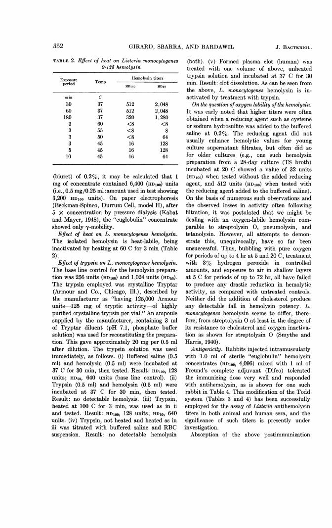
of heat on Listeria monocytogenes9-125 hemolysin
Exposure Te Hemolysin titersperiod Tmp His Hs
HDIloo HDro
mm Cmin c
30 37 512 2,04860 37 512 2,048180 37 320 1,280
3 60 <8 <83 55 <8 83 50 <8 643 45 16 1285 45 16 128
10 45 16 64
(biuret) of 0.2%, it may be calculated that 1mg of concentrate contained 6,400 (HDloo) units(i.e., 0.5 mg/0.25 ml:amount used in test showing3,200 HDloo units). On paper electrophoresis(Beckman-Spinco, Durrum Cell, model H), after5 X concentration by pressure dialysis (Kabatand Mayer, 1948), the "euglobulin" concentrateshowed only y-mobility.
Effect of heat on L. monocytogenes hemolysin.The isolated hemolysin is heat-labile, beinginactivated by heating at 60 C for 3 min (Table2).
Effect of trypsin on L. monocytogenes hemolysin.The base line control for the hemolysin prepara-
tion was 256 units (HDioo) and 1,024 units (HDbo).The trypsin employed was crystalline Tryptar(Armour and Co., Chicago, Ill.), described bythe manufacturer as "having 125,000 Armourunits-125 mg of tryptic activity-of highlypurified crystalline trypsin per vial." An ampoulesupplied by the manufacturer, containing 3 mlof Tryptar diluent (pH 7.1, phosphate buffersolution) was used for reconstituting the prepara-tion. This gave approximately 20 mg per 0.5 mlafter dilution. The trypsin solution was usedimmediately, as follows. (i) Buffered saline (0.5ml) and hemolysin (0.5 ml) were incubated at37 C for 30 min, then tested. Result: HDloo, 128units; HD5o, 640 units (base line control). (ii)Trypsin (0.5 ml) and hemolysin (0.5 ml) were
incubated at 37 C for 30 min, then tested.Result: no detectable hemolysis. (iii) Trypsin,heated at 100 C for 3 min, was used as in iiand tested. Result: HDloo, 128 units; HD50, 640units. (iv) Trypsin, not heated and heated as iniii was titrated with buffered saline and RBCsuspension. Result: no detectable hemolysin
(both). (v) Formed plasma clot (human) was
treated with one volume of above, unheatedtrypsin solution and incubated at 37 C for 30min. Result: clot dissolution. As can be seen fromthe above, L. monocytogenes hemolysin is in-activated by treatment with trypsin.On the question of oxygen lability of the hemolysin.
It was early noted that higher titers were oftenobtained when a reducing agent such as cysteineor sodium hydrosulfite was added to the bufferedsaline at 0.2%. The reducing agent did notusually enhance hemolytic values for youngculture supernatant filtrates, but often did so
for older cultures (e.g., one such hemolysinpreparation from a 28-day culture (TS broth)incubated at 20 C showed a value of 32 units(HDloo) when tested without the added reducingagent, and 512 units (HDloo) when tested withthe reducing agent added to the buffered saline).On the basis of numerous such observations andthe observed losses in activity often followingfiltration, it was postulated that we might bedealing with an oxygen-labile hemolysin com-
parable to streptolysin 0, pneumolysin, andtetanolysin. However, all attempts to demon-strate this, unequivocally, have so far beenunsuccessful. Thus, bubbling with pure oxygen
for periods of up to 4 hr at 5 and 20 C, treatmentwith 3% hydrogen peroxide in controlledamounts, and exposure to air in shallow layersat 5 C for periods of up to 72 hr, all have failedto produce any drastic reduction in hemolyticactivity, as compared with untreated controls.Neither did the addition of cholesterol produceany detectable fall in hemolysin potency. L.monocytogenes hemolysin seems to differ, there-fore, from streptolysin 0 at least in the degree ofits resistance to cholesterol and oxygen inactiva-tion as shown for streptolysin 0 (Smythe andHarris, 1940).
Antigenicity. Rabbits injected intramuscularlywith 1.0 ml of sterile "euglobulin" hemolysinconcentrates (HD,oo, 4,096) mixed with 1 ml ofFreund's complete adjuvant (Difco) toleratedthe immunizing dose very well and respondedwith antihemolysin, as is shown for one suchrabbit in Table 4. This modification of the Toddsystem (Tables 3 and 4) has been successfullyemployed for the assay of Listeria antihemolysintiters in both animal and human sera, and thesignificance of such titers is presently underinvestigation.
Absorption of the above postimmunization
TABLE 2. Effect
352 J. BACTERIOL.

SOLUBLE HEMOLYSIN OF L. MONOCYTOGENES
TABLE 3. Fine titration used for Listeriamonocytogenes 9-125 hemolysin
standardization*
Tubeno.
123456789
101112
Hemol-ysin
ml
0.20.150.1
Hemol-ysin
(diluted1:10)
ml
0.50.450.40.350.300.250.20.150.1
Amt ofhemolysin
ml
0.20.150.10.050.0450.0400.0350.0300.0250.020.0150.01
Bufferedsaline
ml
0.30.350.4
0.050.10.150.20.250.30.350.4
Lysist
1001001001001001001001001001009080
* Hemolysin batch 5-9-61, showing HD l oo valueof 160 units; 2 ml were found equivalent to 1HD1ss, or 0.5 ml contained 25 HDloo units. For 5HDloo/0.5 ml (standard hemolysin dose used fortitration of antihemolysin), original hemolysinwas diluted 1:5 with buffered saline with RAand used as shown in Table 4. All tubes contained1 ml of buffered saline with RA and 0.5 ml of 3%HoRBC.
t Incubated for 45 min at 37 C, then centrifugedand per cent lysis of the supernatant fluids wasread.
TABLE 4. Antigenicity of Listeria monocytogenes9-125 hemolysin: method employed for titration
of antihemolysin serum value*
Lysis readingsAntihemol-
Tube Serum Buffered ysinno. dilution saline (unit Preimmu- Postim-
value) nrizatuo mu-niain nization
ml ml % N1 0.8 0.2 12 trace 02 0.2 0.8 50 50 03 I 1.0 100 100 04 0.8 0.2 125 100 05 0.6 0.4 166 100 06 0.4 0.6 250 100 07 0.3 0.7 333 100 08 1.0 500 100 09 0.8 0.2 625 100 trace10 0.6 0.4 833 100 2011 0.4 0.6 1,250 100 5012 0.2 0.8 2,500 100 10013 0.1 0.9 5,000 100 100
* Serum considered to have 500 units of anti-lysin/ml. To each tube was added 0.5 ml of stand-ard hemolysin containing 5 HDsoo/0.5 ml; tubeswere agitated to mix and incubated for 15 min at37 C; 0.5 ml of 3% HoRBC (in buffered salinewith RA) was then added to each tube. Rabbitgiven a single intramuscular injection of 1.0 mlof L. monocytogenes 9-125 hemolysin concentrateshowing 3,200 IA (HDIoo), mixed with 1.0 ml ofFreund's (complete) adjuvant.
antihemolysin rabbit serum with dense suspen-sions of washed L. monocytogenes has not beenfound to reduce the anti-Listeria hemolysin valueof the serum. Also, Listeria antisera with highagglutinin titers, prepared by immunization ofrabbits with washed formalin-killed suspensionsof L. monocytogenes, have shown antihemolysinvalues comparable to those found in normalrabbit sera. Thus, the antigen responsible forantihemolysin stimulation does not appear toinvolve the surface antigenic mosaic of thebacterium itself.On the toxicity of Listeria hemolysin. Pre-
liminary tests on the toxicity of "euglobulin"hemolysin concentrates in animals (rabbits, mice,and guinea pigs) show that the hemolysin per seis virtually innocuous for these animals. Apossible clue to the mode of action of Listeriahemolysin is provided by the reaction observedon egg-yolk agar (Colbeck's EY Medium, Difco).Supernatant filtrates from two good hemolysin-producing strains also yielded good EY opacities,
whereas two nonhemolysin producers werenegative, as was the heat-inactivated hemolysincontrol. These findings seem to point to a phos-pholipinase or lipase, and are in accord with thesuggestion of Seeliger (1961) that hemolysin maybe a phospholipinase.
DISCUSSION
There is yet no clear evidence to support theinvolvement of an exotoxin in the pathogenesis ofListeria infection. Many investigators havetried to demonstrate such a factor(s) by usingfiltrates of known virulent Listeria culturesin a variety of animals and by different routes ofinoculation, without success (Murray, Webb, andSwann, 1926; Shaw and Turk, 1954; Burn, 1935;Jaeger and Myers, 1954). Chemical fractionsderived from the organism have been almostequally unrewarding. Although Stanley (1949)reported on a highly toxic polysaccharideprepared from L. monocytogenes, more recentlyPatocka, Schindler, and Mara (1959) were
VOL. 85, 1963 353

GIRARD, SBARRA, AND BARDAWIL
unable to demonstrate polysaccharide toxicity.On the other hand, these workers described theisolation of an infection-enhancing, heat-labileprotein substance from glycine lysates of washedListeria suspensions. Although devoid of toxicityper se, it has specific infection-potentiatingproperties and was said not to be anticomple-mentary. Later studies by Silverman, Elwell, andKautter (1961) on a protein fraction obtainedfrom L. monocytogenes suspensions by sonicdisintegration showed comparable mortality-enhancing activity, although the activity wasnonspecific in that the mortality quotient ofseveral unrelated pathogens was raised. Thesubstance was also said to be anticomplementaryand heat-stable. Thus it is not clear whether thesetwo agents are identical or are separate proteins.However, neither one would appear to bear anyidentity to the L. monocytogenes 9-125 hemolysinherein described, although the infection-poten-tiating property of hemolysin has not yet beenascertained. On the other hand, the "diffusiblesubstances" derived from a culture of L. mono-cytogenes as reported by Fraser (1962) may bearclose relationship to the hemolysin hereindescribed.Some authors have drawn attention to the
possible association of hemolysin production andvirulence (Hunter, Stahly, and Myers, 1950) andto type of growth (Seeliger, 1961). Mention hasalso been made by some workers (Burn, 1935;Stanley, 1948; Barrow and Pugh, 1958), and wehave observed on occasion, the simultaneousoccurrence of hemolytic and nonhemolyticcolonies within the same Listeria strain, and evenof different colonial variants within the hemolyticforms in a single strain (Girard, unpublisheddata). However, further work needs to be doneon the interrelationships that may exist beforeany inferences can be drawn. Certainly, ourpreliminary animal observations using highlyactive and purified hemolysin preparations do notlend support to a doctrine of hemolysin-correlatedvirulence. Njoku-Obi (personal communication)also reports that "no correlation of the titerscould be made with virulence." Although thiswould tend to rule out hemolysin as a possibleexotoxic factor in the conventional sense, itdoes not necessarily preclude its function as apossible accessory virulence factor in the patho-genesis of Listeria infection.
It is equally uncertain as to whether all freshlyisolated strains of L. monocytogenes produce
soluble hemolysin. In our studies of 155 labora-tory strains, nearly 30% failed to show anydetectable f3-hemolysis on primary plating withhorse, human, rabbit, and sheep blood agarplates (Girard, unpublished data). More recentwork on the same strains has shown that widevariations are encountered in soluble hemolysinproduction from strain to strain, some producingbarely detectable amounts. On the other hand,Seeliger (1961), in discussing plate hemolysis,stated that "reports on the complete loss of thisproperty (beta hemolysis) in some strains couldnot be verified at a critical re-investigation ...,"whereas Welshimer and Winglewish (1959)stated that "the hemolytic activity (beta he-molysis) may be extremely weak or absent."Obviously, the question is rather pertinent to thepossible potential diagnostic application of anti-Listeria hemolysin determinations.
ACKNOWLEDGMENTS
We are grateful to E. G. D. Murray of theUniversity of Western Ontario, London, Ontario,Canada, for kindly providing us with many of thestrains of Listeria used in our work. We are also in-debted to R. M. Bearns of the Department ofBacteriology and Immunology, McGill Uni-versity, and to Mrs. E. Mannis and C. DelGizzi, St. Margaret's Hospital, for technicalassistance.Supported by grant E-3236 from the National
Institute of Allergy and Infectious Diseases,U.S. Public Health Service.
LITERATURE CITED
BARROW, G. I., AND R. J. PUGH. 1958. Listeria(Erysipelothrix) monocytogenes meningitisin the newborn. J. Pathol. Bacteriol. 75:9-16.
BURN, C. G. 1935. Characteristics of a new speciesof the genus Listerella obtained from humansources. J. Bacteriol. 30:573-591.
FRASER, G. 1962. A plate method for the rapididentification of Listeria (erysipelothrix)monocytogenes. Vet. Record 74:50-51.
GINSBURG, I., AND N. GROSSOWICZ. 1960. Effect ofstreptococcal hemolysins on Ehrlich ascitestumour cells. J. Pathol. Bacteriol. 80:111-119.
GIRARD, K. F., AND W. F. GAVIN. 1957. Listeriosisin the newborn. J. Pathol. Bacteriol. 74:93-102.
HUNTER, M. C., G. L. STAHLY, AND W. G. MYERS.1950. Variations of Listeria monocytogenesproduced by beta particles from radiophos-phorus. Ohio J. Sci. 50:253-259.
354 J. BACTERIOL.

VOL. 85, 1963 SOLUBLE HEMOLYSIN
JAEGER, R. F., AND D. M. MYERS. 1954. Listeriamonocytogenes. A study of two strains isolatedfrom human sources. Can. J. Microbiol. 1:12-21.
KABAT, E. A., AND M. M. MAYER. 1948. Experi-mental immunochemistry, 1st ed. CharlesC Thomas, Publisher, Springfield, Ill.
MURRAY, E. G. D., R. H. WEBB, AND M. B. R.SWANN. 1926. A disease of rabbits charac-terized by a large mononuclear leucocytosis,caused by a hitherto undescribed bacillus:Bacterium monocytogenes (n. sp.). J. Pathol.Bacteriol. 29:407-439.
PATOCKA, F., J. SCHINDLER, AND M. MARA. 1959.Studies on the pathogenicity of Listeria mono-cytogenes. I. Protein substance isolated fromcells of Listeria monocytogenes enhancinglisteric infection. Zentr. Bakteriol. Parasit-enk. Abt. I Orig. 174:573-585.
POTEL, J. 1952. Zur granulomatosis infantiseptica.Zentr. Bakteriol. Parasitenk. Abt. I Orig.158:329-331.
SEELIGER, H. P. R. 1961. Listeriosis. HafnerPublishing Co., Inc., New York.
OF L. MONOGI-TOGENES 355
SHAW, C. S., AND D. C. TURK. 1954. A case ofmeningitis apparently caused by a Coryne-bacterium. J. Pathol. Bacteriol. 68:627-631.
SILVERMAN, S. J., L. ELWELL, AND D. A. KAUTTER.1961. A mortality enhancing extract isolatedfrom Listeria monocytogenes. J. Immunol.86:669-674.
SMYTHE, C. V., AND T. N. HARRIS. 1940. Someproperties of a hemolysin produced by groupA (beta) hemolytic streptococci. J. Immunol.38:283-300.
STANLEY, N. F. 1948. Listeria meningitis, a de-scription of a strain of Listeria monocytogenesand a report of a case. Med. J. Australia2:205-208.
STANLEY, N. F. 1949. Studies on Listeria mono-cytogenes. I. Isolation of a monocytosis-producing-agent (MPA). Australian J. Exptl.Biol. Med. Sci. 27:123-131.
WELSHIMER, H. J., AND N. G. WINGLEWISH. 1959.Listeriosis. Summary of seven cases of listeriameningitis. J. Am. Med. Assoc. 171:1319-1323.