silica-coated super paramagnetic iron oxide … · nanoparticles (spion) as biocompatible contrast...
TRANSCRIPT

Silica-coated super paramagnetic iron oxide nanoparticles (SPION) as biocompatible
contrast agent in biomedical photoacoustics Rudolf Alwi,1 Sergey Telenkov,1 Andreas Mandelis,1,* Timothy Leshuk,2 Frank Gu,2
Sulayman Oladepo,3 and Kirk Michaelian3 1Center for Advanced Diffusion-Wave Technologies (CADIFT), Department of Mechanical and Industrial
Engineering, University of Toronto, 5 King’s College Road, Toronto, ON, M5S 3G8, Canada 2Department of Chemical Engineering, University of Waterloo, 200 University Avenue West, Waterloo,
ON, N2L 3G1, Canada 3Natural Resources Canada, CanmetENERGY, 1 Oil Patch Drive, Devon, AB, T9G 1A8, Canada
Abstract: In this study, we report for the first time the use of silica-coated superparamagnetic iron oxide nanoparticles (SPION) as contrast agents in biomedical photoacoustic imaging. Using frequency-domain photoacoustic correlation (the photoacoustic radar), we investigated the effects of nanoparticle size, concentration and biological media (e.g. serum, sheep blood) on the photoacoustic response in turbid media. Maximum detection depth and the minimum measurable SPION concentration were determined experimentally. The nanoparticle-induced optical contrast ex vivo in dense muscular tissues (avian pectus and murine quadricept) was evaluated and the strong potential of silica-coated SPION as a possible photoacoustic contrast agents was demonstrated.
© 2012 Optical Society of America
OCIS codes: (170.5120) Photoacoustic imaging; (170.3880) Medical and biological imaging; (160.4236) Nanomaterials.
References
1. K. Homan, J. Shah, S. Gomez, H. Gensler, A. Karpiouk, L. Brannon-Peppas, and S. Emelianov, “Silver nanosystems for photoacoustic imaging and image-guided therapy,” J. Biomed. Opt. 15(2), 021316 (2010).
2. M. Pramanik, K. H. Song, M. Swierczewska, D. Green, B. Sitharaman, and L. V. Wang, “In vivo carbon nanotube-enhanced non-invasive photoacoustic mapping of the sentinel lymph node,” Phys. Med. Biol. 54(11), 3291–3301 (2009).
3. C. Ungureanu, A. Amelink, R. G. Rayavarapu, H. J. C. M. Sterenborg, S. Manohar, and T. G. C. van Leeuwen, “Differential pathlength spectroscopy for the quantitation of optical properties of gold nanoparticles,” ACS Nano 4(7), 4081–4089 (2010).
4. S. J. Yoon, S. Mallidi, J. M. Tam, J. O. Tam, A. Murthy, K. P. Johnston, K. V. Sokolov, and S. Y. Emelianov, “Utility of biodegradable plasmonic nanoclusters in photoacoustic imaging,” Opt. Lett. 35(22), 3751–3753 (2010).
5. J. Yao, K. Maslov, S. Hu, and L. V. Wang, “Evans blue dye-enhanced capillary-resolution photoacoustic microscopy in vivo,” J. Biomed. Opt. 14(5), 054049 (2009).
6. C. Kim, E. C. Cho, J. Chen, K. H. Song, L. Au, C. Favazza, Q. Zhang, C. M. Cobley, F. Gao, Y. Xia, and L. V. Wang, “In vivo molecular photoacoustic tomography of melanomas targeted by bioconjugated gold nanocages,” ACS Nano 4(8), 4559–4564 (2010).
7. A. H. Green, J. R. Norris, J. Wang, Z. Xie, H. F. Zhang, and P. J. La Riviere, “In vitro testing of a protease-sensitive contrast agent for optoacoustic imaging,” J. Biomed. Opt. 15(2), 021315 (2010).
8. L. M. Ricles, S. Y. Nam, K. Sokolov, S. Y. Emelianov, and L. J. Suggs, “Function of mesenchymal stem cells following loading of gold nanotracers,” Int. J. Nanomedicine 6, 407–416 (2011).
9. M. Pramanik, M. Swierczewska, D. Green, B. Sitharaman, and L. V. Wang, “Single-walled carbon nanotubes as a multimodal-thermoacoustic and photoacoustic-contrast agent,” J. Biomed. Opt. 14(3), 034018 (2009).
10. K. Wilson, K. Homan, and S. Emelianov, “Biomedical photoacoustics beyond thermal expansion using triggered nanodroplet vaporization for contrast-enhanced imaging,” Nat Commun 3, 618 (2012).
11. A. Agarwal, X. Shao, J. R. Rajian, H. Zhang, D. L. Chamberland, N. A. Kotov, and X. Wang, “Dual-mode imaging with radiolabeled gold nanorods,” J. Biomed. Opt. 16(5), 051307 (2011).
12. C. Jia, J. Xia, I. M. Pelivanov, C. H. Seo, X. Hu, Y. Jin, X. Gao, and M. O'Donnell, “Dynamic manipulation of magnetic contrast agents in photoacoustic imaging,” Proc. SPIE 7899, 78991R, 78991R–6 (2011).
#172516 - $15.00 USD Received 13 Jul 2012; revised 28 Aug 2012; accepted 11 Sep 2012; published 12 Sep 2012(C) 2012 OSA 1 October 2012 / Vol. 3, No. 10 / BIOMEDICAL OPTICS EXPRESS 2500

13. Y. S. Chen, W. Frey, S. Kim, P. Kruizinga, K. Homan, and S. Emelianov, “Silica-coated gold nanorods as photoacoustic signal nanoamplifiers,” Nano Lett. 11(2), 348–354 (2011).
14. A. Roggan, M. Friebel, K. Dörschel, A. Hahn, and G. Müller, “Optical properties of circulating human blood in the wavelength range 400-2500 nm,” J. Biomed. Opt. 4(1), 36–46 (1999).
15. A. A. Oraevsky, Photoacoustic Imaging and Spectroscopy, 1st ed. (Taylor & Francis, 2008), Chap. 20. 16. K. L. Kelly, E. Coronado, L. L. Zhao, and G. C. Schatz, “The optical properties of metal nanoparticles: the
influence of size, shape, and dielectric environment,” J. Phys. Chem. B 107(3), 668–677 (2003). 17. K. S. Lee and M. A. El-Sayed, “Gold and silver nanoparticles in sensing and imaging: sensitivity of plasmon
response to size, shape, and metal composition,” J. Phys. Chem. B 110(39), 19220–19225 (2006). 18. A. S. Thakor, J. Jokerst, C. Zavaleta, T. F. Massoud, and S. S. Gambhir, “Gold nanoparticles: a revival in
precious metal administration to patients,” Nano Lett. 11(10), 4029–4036 (2011). 19. L. Wang, Photoacoustic Imaging and Spectroscopy (CRC Press, 2009), Chap. 30. 20. J. B. Jackson and N. J. Halas, “Silver nanoshells: variations in morphologies and optical properties,” J. Phys.
Chem. B 105(14), 2743–2746 (2001). 21. V. K. Poon and A. Burd, “In vitro cytotoxity of silver: implication for clinical wound care,” Burns 30(2), 140–
147 (2004). 22. X. Chen, Nanoplatform-Based Molecular Imaging (John Willey & Sons, 2011), Chap. 12.2.2. 23. T. N. Erpelding, C. Kim, M. Pramanik, L. Jankovic, K. Maslov, Z. Guo, J. A. Margenthaler, M. D. Pashley, and
L. V. Wang, “Sentinel lymph nodes in the rat: noninvasive photoacoustic and US imaging with a clinical US system,” Radiology 256(1), 102–110 (2010).
24. C. Kim, K. H. Song, F. Gao, and L. V. Wang, “Sentinel lymph nodes and lymphatic vessels: noninvasive dual-modality in vivo mapping by using indocyanine green in rats--volumetric spectroscopic photoacoustic imaging and planar fluorescence imaging,” Radiology 255(2), 442–450 (2010).
25. N. Kosaka, M. Mitsunaga, M. R. Longmire, P. L. Choyke, and H. Kobayashi, “Near infrared fluorescence-guided real-time endoscopic detection of peritoneal ovarian cancer nodules using intravenously injected indocyanine green,” Int. J. Cancer 129(7), 1671–1677 (2011).
26. M. Qu, S. Mallidi, M. Mehrmohammadi, R. Truby, K. Homan, P. Joshi, Y. S. Chen, K. Sokolov, and S. Emelianov, “Magneto-photo-acoustic imaging,” Biomed. Opt. Express 2(2), 385–396 (2011).
27. Y. Jin, C. Jia, S. W. Huang, M. O’Donnell, and X. Gao, “Multifunctional nanoparticles as coupled contrast agents,” Nat Commun 1(4), 41 (2010).
28. R. Lawaczeck, M. Menzel, and H. Pietsch, “Superparamagnetic iron oxide nanoparticles: contrast media for magnetic resonance imaging,” Appl. Organomet. Chem. 18(10), 506–513 (2004).
29. A. S. Teja and P. Koh, “Synthesis, properties, and applications of magnetic iron oxide nanoparticles,” Prog. Cryst. Growth Charact. Mater. 55(1–2), 22–45 (2009).
30. S. Laurent, D. Forge, M. Port, A. Roch, C. Robic, L. Vander Elst, and R. N. Muller, “Magnetic iron oxide nanoparticles: synthesis, stabilization, vectorization, physicochemical characterizations, and biological applications,” Chem. Rev. 108(6), 2064–2110 (2008).
31. H. S. Choi, W. Liu, P. Misra, E. Tanaka, J. P. Zimmer, B. Itty Ipe, M. G. Bawendi, and J. V. Frangioni, “Renal clearance of quantum dots,” Nat. Biotechnol. 25(10), 1165–1170 (2007).
32. T. Neuberger, B. Schopf, H. Hofmann, M. Hofmann, and B. von Rechenberg, “Superparamagnetic nanoparticles for biomedical applications: possibilities and limitations of a new drug delivery system,” J. Magn. Magn. Mater. 293(1), 483–496 (2005).
33. L. Brannon-Peppas and J. O. Blanchette, “Nanoparticle and targeted systems for cancer therapy,” Adv. Drug Deliv. Rev. 56(11), 1649–1659 (2004).
34. R. Qiao, C. Yang, and M. Gao, “Superparamagnetic iron oxide nanoparticles: from preparations to in vivo MRI applications,” J. Mater. Chem. 19(35), 6274–6293 (2009).
35. J. E. Rosen, L. Chan, D.-B. Shieh, and F. X. Gu, “Iron oxide nanoparticles for targeted cancer imaging and diagnostics,” Nanomedicine 8(3), 275–290 (2012).
36. X. Chen, Nanoplatform-Based Molecular Imaging (Wiley, 2011), Chap. 23.3.1. 37. M. K. Yu, J. Park, Y. Y. Jeong, W. K. Moon, and S. Jon, “Integrin-targeting thermally cross-linked
superparamagnetic iron oxide nanoparticles for combined cancer imaging and drug delivery,” Nanotechnology 21(41), 415102 (2010).
38. A. Z. Wang, V. Bagalkot, C. C. Vasilliou, F. Gu, F. Alexis, L. Zhang, M. Shaikh, K. Yuet, M. J. Cima, R. Langer, P. W. Kantoff, N. H. Bander, S. Jon, and O. C. Farokhzad, “Superparamagnetic iron oxide nanoparticle-aptamer bioconjugates for combined prostate cancer imaging and therapy,” ChemMedChem 3(9), 1311–1315 (2008).
39. A. Kunzmann, B. Andersson, T. Thurnherr, H. Krug, A. Scheynius, and B. Fadeel, “Toxicology of engineered nanomaterials: focus on biocompatibility, biodistribution and biodegradation,” Biochim. Biophys. Acta 1810(3), 361–373 (2011).
40. Y. Xu, Y. Qin, S. Palchoudhury, and Y. Bao, “Water-soluble iron oxide nanoparticles with high stability and selective surface functionality,” Langmuir 27(14), 8990–8997 (2011).
41. N. R. Jana, C. Earhart, and J. Y. Ying, “Synthesis of water-soluble and functionalized nanoparticles by silica coating,” Chem. Mater. 19(21), 5074–5082 (2007).
42. A. Guerrero-Martínez, J. Pérez-Juste, and L. M. Liz-Marzán, “Recent progress on silica coating of nanoparticles and related nanomaterials,” Adv. Mater. (Deerfield Beach Fla.) 22(11), 1182–1195 (2010).
43. F. Šulek, M. Drofenik, M. Habulin, and Z. Knez, “Surface functionalization of silica-coated magnetic nanoparticles for covalent attachment of cholesterol oxidase,” J. Magn. Magn. Mater. 322(2), 179–185 (2010).
#172516 - $15.00 USD Received 13 Jul 2012; revised 28 Aug 2012; accepted 11 Sep 2012; published 12 Sep 2012(C) 2012 OSA 1 October 2012 / Vol. 3, No. 10 / BIOMEDICAL OPTICS EXPRESS 2501

44. S. Telenkov, A. Mandelis, B. Lashkari, and M. Forcht, “Frequency-domain photothermoacoustics: alternative imaging modality of biological tissues,” J. Appl. Phys. 105(10), 102029 (2009).
45. American National Standards Institute, “American National Standard for Safe Use of Lasers, “ANSI Z136.1–2007 (ANSI, 2007).
46. S. Telenkov and A. Mandelis, “Signal-to-noise analysis of biomedical photoacoustic measurements in time and frequency domains,” Rev. Sci. Instrum. 81(12), 124901 (2010).
47. S. Telenkov, R. Alwi, A. Mandelis, and A. Worthington, “Frequency-domain photoacoustic phased array probe for biomedical imaging applications,” Opt. Lett. 36(23), 4560–4562 (2011).
48. Y. S. Chen, W. Frey, S. Kim, K. Homan, P. Kruizinga, K. Sokolov, and S. Emelianov, “Enhanced thermal stability of silica-coated gold nanorods for photoacoustic imaging and image-guided therapy,” Opt. Express 18(9), 8867–8878 (2010).
49. X. Xu, M. Ge, C. Wang, and J. Z. Jiang, “High temperature stable monodisperse superparamagnetic core-shell iron-oxide@SnO2 nanoparticles,” Appl. Phys. Lett. 95(18), 183112 (2009).
50. H. S. Carslaw and J. C. Jaeger, Conduction of Heat in Solids, 2nd ed. (Clarendon, 1959), Chap. 9. 51. S. A. Telenkov and A. Mandelis, “Photothermoacoustic imaging of biological tissues: maximum depth
characterization comparison of time and frequency-domain measurements,” J. Biomed. Opt. 14(4), 044025 (2009).
52. H. Han, A. Johnson, J. Kaczor, M. Kaur, A. Paszczynski, and Y. Qiang, “Silica coated magnetic nanoparticles for separation of nuclear acidic waste,” J. Appl. Phys. 107(9), 09B520 (2010).
53. O. Baber, M. Jang, D. Barber, and K. Powers, “Amorphous silica coatings on magnetic nanoparticles enhance stability and reduce toxicity to in vitro BEAS-2B cells,” Inhal. Toxicol. 23(9), 532–543 (2011).
1. Introduction
Recently, there has been a significant increase in the use of contrast agents in photoacoustic imaging [1–13]. In medical imaging, a contrast agent is an organic or inorganic material administered into a patient, mainly via intravenous injection, to accentuate the image contrast of anatomical structures or fluids within the body. Although (oxy- and deoxy-) hemoglobin, the iron-containing and oxygen-transport protein in the red blood cells, absorbs in the near-infrared (near-IR) spectral region [14] and has become the primary endogenous contrast source for near-IR photoacoustic imaging, there is difficulty in detecting the presence of deep and small diameter blood vessels, e.g., tumor microvasculature network located a few centimeters beneath the skin. The main challenges are due to the high optical scattering of tissue and the optical absorption by subcutaneous blood vessels, particularly for early-stage tumor imaging when the angiogenesis has not fully developed and therefore its blood-pool contrast might not be sufficient enough to differentiate a malignant tumor from normal tissue. The use of nanoparticles with resonance absorption in the near infrared region is highly desirable because optical absorption by native tissue components is minimal in this range.
Among numerous potential photoacoustic contrast agents that have been investigated and reported, the current most popular choices are plasmonic gold nanoparticles [3,4,6,8,10,11,13]. A gold nanoparticle has a superior optical absorption profile attributed to its strong plasmon resonance peaks in the near-IR [15]. Furthermore, one can alter the shape and size of the plasmonic nanoparticles to maximize their optical absorption and scattering cross section and tune the absorption peak position [16,17], which is greatly beneficial for a variety of biomedical imaging applications. Nevertheless, the toxicity of gold nanoparticles has not been fully addressed. Thakor et al. [18] in their recent review article on the use of gold nanoparticles in a clinical setting stated that, inevitably, trade-offs will have to be made regarding some of their diagnostic and therapeutic properties vis-a-vis their associated toxicity profile.
Another commonly used metallic photoacoustic contrast agent is silver nanoparticles which also exhibit strong plasmonic resonance in the visible to near-IR spectrum [1,19]. In fact, silver exhibits higher light absorptivity than gold [20] and thus potentially produces a stronger photoacoustic response. However, due to their high reactivity, silver nanoparticles are considered to be more toxic than gold nanoparticles [21,22] for human use, which makes them less desirable.
Single-walled carbon nanotubes (SWCNT) [9] and dyes such as Evans blue [5], methylene blue [23] and indocyanine green (ICG) [7,24] have also been studied as photoacoustic contrast agents. Though SWCNT and dyes have great absorption in the NIR spectrum and there are wide selections of dyes to choose from for different wavelength absorption peaks, Kim et al.
#172516 - $15.00 USD Received 13 Jul 2012; revised 28 Aug 2012; accepted 11 Sep 2012; published 12 Sep 2012(C) 2012 OSA 1 October 2012 / Vol. 3, No. 10 / BIOMEDICAL OPTICS EXPRESS 2502

[6] mentioned that practically the safety of carbon nanotubes is doubted for human use, while the aforementioned dyes, ICG being the only Food-and-Drug-Administration (FDA) approved dye [25], suffer from short circulation time in vivo. Additionally, metallic (inorganic) nanoparticle photoacoustic contrast agents are thought to have better stability, tunable optical spectra (e.g. gold and silver), and less photo bleaching compared to their organic counterparts [11].
The concept of complementing photoacoustic imaging with other clinically-available imaging techniques (e.g. ultrasound, SPECT, MRI) does not only potentially give rise to new multimodality systems with an augmented diagnostic capability beyond the inherent limitations of individual components, but also to multimodal contrast agents. Recent efforts for such integration have motivated photoacoustic researchers to develop so-called hybrid contrast agents, such as perfluorocarbon-capsulated gold nanoparticles as a dual contrast agent for photoacoustic and ultrasound modalities [10], radioisotope (125I)-labeled gold nanorod as a dual contrast agent for photoacoustic and nuclear imaging (SPECT) [11], and gold-coated iron oxide (Fe3O4) nanoparticles for a new magnetomotive photoacoustic (mmPA) imaging [26,27]. The latter technique (mmPA) applies an external magnetic field across gold-coated magnetic (iron oxide; magnetite Fe3O4) nanoparticles during the photoacoustic data acquisition time, thereby yielding magnetically-induced movement of the nanoparticles which separates them from the non-magnetic static background. Eventually, by coherent motion processing of the data (or PA images) before and after the application of the external magnetic field, the motionless background signals can be suppressed thereby increasing the PA contrast specificity attributed to the highly photoabsorbing gold coating [12,26,27].
While bare iron oxide (Fe3O4) nanoparticles are not strong light absorbers in the near-IR, our present study demonstrates the potential use of silica-coated iron oxide (Fe3O4) nanoparticles as a PA contrast agent. Iron oxide nanoparticles display superparamagnetic property, which means there is no remnant magnetization once the external magnetic field is removed at room temperature if the core size of a single nanoparticle is sufficiently small and typically within 3-50 nm [28,29]. For this reason, apart from its unique magnetic profile, the term superparamagnetic is often used to emphasize the size of the iron oxide nanoparticle. From a biological standpoint, nanoparticles with diameter size 10-100 nm have been shown to be optimal for avoiding rapid clearance from systemic circulation due to removal by the organs of monoclear phagocytic systems (MPS) or by renal filtration [30–33]. Therefore, they exhibit the longest circulation time in the human body and allow the contrast agents to accumulate in the targeted area for an amount of time sufficient to carry out the imaging process.
Superparamagnetic iron oxide nanoparticles (SPION) have been widely used as FDA-approved contrast agents for magnetic resonance imaging (MRI) and are known to have an excellent safety profile [34]. Rosen et al. [35] provide a comprehensive review on the use of SPION, including their surface functionalization, for targeted cancer imaging and diagnosis. Furthermore, by incorporating targeting ligands, such as peptides, antibodies, and aptamers to the polymer surface of SPION, one can employ the nanoparticles as drug-delivery carriers or therapeutic agents [36–38].
In order to accomplish the task as a contrast agent, nanoparticles need to demonstrate key characteristics, such as great solubility in aqueous media and biocompability [39], modifiable surface chemistry for further functionalization that enables passive or active targeting to the diseased area [35], and enhanced cellular uptake and biodistribution [40]. The surface functionality (or coating) plays major role in the interaction between nanoparticles and biological systems. Jana et al. [41] have shown that silica coating is one of the most popular techniques for nanoparticle surface modification; it enhances colloidal stability with relatively easy regulation of the coating process [42] and silica coating is regarded as extremely biocompatible and accessible to versatile surface bioconjugation [43]. More importantly, with relevance to photoacoustic imaging, Chen et al. [13] recently showed that silica-coated gold nanorods could amplify photoacoustic signals 3-fold compared to gold nanorods without silica coating.
#172516 - $15.00 USD Received 13 Jul 2012; revised 28 Aug 2012; accepted 11 Sep 2012; published 12 Sep 2012(C) 2012 OSA 1 October 2012 / Vol. 3, No. 10 / BIOMEDICAL OPTICS EXPRESS 2503
In the present study, we report the use of silica-coated SPION as an alternative PA contrast agent for laser excitation in the near-IR spectral range. The motivation arises from the fact that SPION, widely-used contrast agents for MRI modality, are better-understood and have excellent safety record in clinical settings for human use compared to gold nanoparticles.
We conducted five types of experiments to evaluate the effectiveness of silica-coated SPION as a PA contrast agent: 1) we measured the maximum detectable depth of a known silica-coated SPION concentration inside a tissue mimicking Intralipid solution; 2) we investigated the effect of nanoparticle size variation on the generated PA response; 3) we examined whether the silica-coated SPION were optically stable in organic solvents (e.g. serum, blood); 4) we quantified the minimum resolvable concentration of silica-coated SPION at fixed depths inside a tissue mimicking Intralipid solution; and 5) we tested the detectability of silica-coated SPION inside chicken breast tissue and rat thigh muscle ex vivo.
2. Materials and experimental
2.1. Superparamagnetic iron oxide nanoparticles (SPION)
2.1.1. Materials
Iron (II) chloride (99%), iron (III) chloride (99%), tetramethylammonium hydroxide (TMAOH) (25% solution), tetraethyl orthosilicate (TEOS) (99%), and ethanol (99% ACS) were all purchased from Sigma-Aldrich, St. Louis, MO and used without further purification.
2.1.2. Silica-coated SPION synthesis
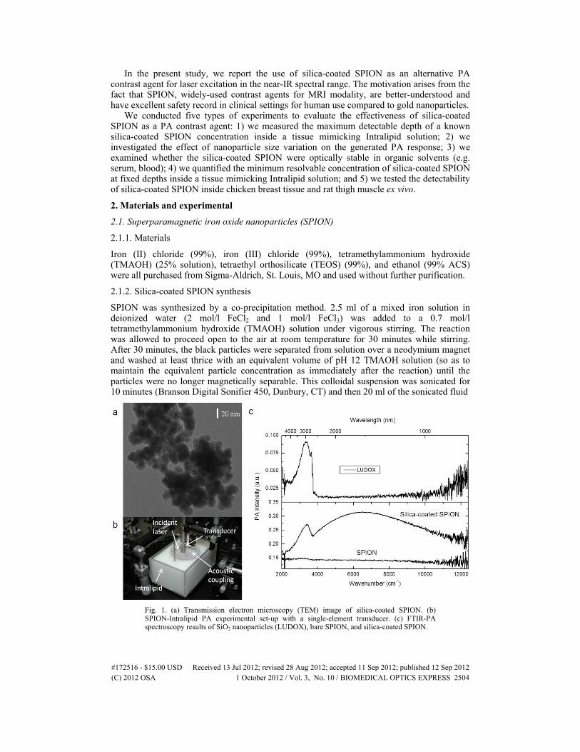
SPION was synthesized by a co-precipitation method. 2.5 ml of a mixed iron solution in deionized water (2 mol/l FeCl2 and 1 mol/l FeCl3) was added to a 0.7 mol/l tetramethylammonium hydroxide (TMAOH) solution under vigorous stirring. The reaction was allowed to proceed open to the air at room temperature for 30 minutes while stirring. After 30 minutes, the black particles were separated from solution over a neodymium magnet and washed at least thrice with an equivalent volume of pH 12 TMAOH solution (so as to maintain the equivalent particle concentration as immediately after the reaction) until the particles were no longer magnetically separable. This colloidal suspension was sonicated for 10 minutes (Branson Digital Sonifier 450, Danbury, CT) and then 20 ml of the sonicated fluid
Fig. 1. (a) Transmission electron microscopy (TEM) image of silica-coated SPION. (b) SPION-Intralipid PA experimental set-up with a single-element transducer. (c) FTIR-PA spectroscopy results of SiO2 nanoparticles (LUDOX), bare SPION, and silica-coated SPION.
#172516 - $15.00 USD Received 13 Jul 2012; revised 28 Aug 2012; accepted 11 Sep 2012; published 12 Sep 2012(C) 2012 OSA 1 October 2012 / Vol. 3, No. 10 / BIOMEDICAL OPTICS EXPRESS 2504

was mixed with 20 ml pH 12 TMAOH and 160 ml ethanol. 7 ml tetraethylorthosilicate (TEOS) was then added to this suspension while stirring and allowed to react at room temperature while stirring for ~18 hours. The silica-coated SPION was then magnetically recovered from solution, washed thrice with ethanol and thrice with deionized water by magnetic decantation, and sonicated into deionized water for 10 minutes before further use. Figure 1(a) shows a Transmission electron microscopy (TEM) image of an aggregated silica-coated SPION: The size of a single nanoparticle (iron oxide core plus silica coating) is less than 20 nm. The size of the particles and the thickness of the silica shell were obtained using measurements from multiple TEM images of the sample, and the sizing results were then averaged to obtain the values presented here. The silica shell is visible as a thin layer of less-electron-dense material surrounding the more-electron-dense material of the SPION cores [Fig. 1(a)].
2.2. Frequency-domain photoacoustic correlation imaging (photoacoustic radar)
Our spatially-resolved photoacoustic imaging modality utilizes millisecond-long and coded continuous wave (CW) optical excitation. The intensity of the laser output is frequency-modulated (chirped) to a specific pattern. The laser chirp-induced acoustic waves with a similar pattern are detected by an ultrasonic transducer and are processed through a cross-correlation algorithm in the frequency domain. At the signal processing stage, the received PA signal is correlated with a reference chirp used for laser source modulation. The cross-correlation results in energy compression of a long and low amplitude PA signal into a narrow correlation peak at a delay time corresponding to the location of the absorber. Further details of the frequency-domain photoacoustic correlation (FD-PA) system and instrumentation were described elsewhere [44].
For PA signal generation, a 1064-nm CW laser was used (IPG Photonics, Boston, MA) with 1-W incident power on the surface of tissue or Intralipid solution (Intralipid® 20%, Fresenius Kabi AB, Uppsala, Sweden) at an oblique angle (<20°) and 2 mm beam diameter. 1-5 MHz frequency-sweep was used as the periodic photoacoustic chirp wave generator. The SPION-Intralipid PA experimental set-up is pictured in Fig. 1(b). The duration of the modulation chirp driving the laser power was 1 ms and up to 1000 chirps were acquired and coherently averaged to increase signal-to-noise ratio (SNR).
The laser safety limit [45] defined as the maximum permissible exposure (MPE) can be exceeded when a large number of laser chirps irradiate the target tissue. The safety standard sets the maximum exposure level for λ = 1064 nm and exposure duration t = 10−7 – 10 s as EMPE = 5.5t0.25 (J/cm2) [46], where E stands for laser exposure. If I0 is defined as laser irradiance (W/cm2), then the maximum duration (tmax) of I0 to reach EMPE level can be obtained by tmax = (5.5/ I0)
4/3. In this study, given 1-W incident power and 2 mm beam diameter, the laser irradiance was ~32 W/cm2 and the maximum exposure duration for safety limit was ~0.1 s. In our experiments, up to 1000 chirps (1 ms duration for one chirp) were employed for the averaging purpose which could yield up to ~1 s laser exposure. Since the main objective of the present study was the demonstration of the silica-coated SPION as a potential contrast agent, strict adherence to the MPE limit was not always enforced. The number of chirps used in signal averaging can be optimized, thereby allowing one to improve the signal-to-noise ratio (SNR) and precision of the correlation measurements.
Using a peristaltic pump (PD 5206, Heidolph Instruments GmbH & Co.KG, Scwhabach, Germany), the nanoparticle solution was circulated inside a transparent polyolefin tube (not shown in Fig. 1(b)) with 1.7 mm inner diameter and 0.25 mm wall thickness. The SPION-filled tube was immersed in a light scattering Intralipid solution with a tissue-like effective optical attenuation coefficient (meff ~1.3 cm−1). A water container was used as an acoustic coupling medium from the tube to a single element 3.5-MHz focused (f = 25.4 mm, Panametrics v382, Olympus NDT Inc, Waltham, MA) or 1-MHz unfocused (Panametrics A303S) transducer. The bottom part of the container was sealed with a thin and transparent plastic film which became the water-Intralipid interface and corresponded to depth 0 mm for the Intralipid experiments. To ensure optimal detection of photoacoustic signals, the tube was
#172516 - $15.00 USD Received 13 Jul 2012; revised 28 Aug 2012; accepted 11 Sep 2012; published 12 Sep 2012(C) 2012 OSA 1 October 2012 / Vol. 3, No. 10 / BIOMEDICAL OPTICS EXPRESS 2505

positioned at the focal zone of the transducer (25.4 mm) for all the experiments. Therefore, in varying the depth of detection, the downward translation of the tube inside the Intralipid was also accompanied by a concomitant downward translation of the acoustic transducer.
To study SPION-induced PA contrast in real tissue, we imaged the presence of a silica-coated SPION-filled tube (8 nm core, 3 nm coating) inside a slab of ex vivo chicken pectoral muscle (~18 mm thickness). We used a phased array PA probe [47] consisting of a standard 64-element ultrasonic transducer array (Ultrasonix, Richmond, BC, CA) with central frequency 3.5 MHz, 80% bandwidth at −6 dB, and pitch 0.254 mm. 1-W laser irradiation, 2 mm beam diameter, 1 ms duration and 1-5 MHz sinusoidal chirp were employed.
Another ex vivo detection of nanoparticles inside a rat thigh was obtained using the single element 3.5-MHz focused transducer (Panametrics v382) and 0.44-W incident laser power. The beam diameter was ~2 mm and 1-5 MHz sinusoidal chirps of 1-ms duration were employed. By translating the laser beam and the transducer together along a particular axis, a 2-dimensional FD-PA image was created.
3. Results and discussion
3.1. SPION – Intralipid experiments
Near-IR FTIR-PA spectroscopy results [Fig. 1(c)] of silica nanoparticles (SiO2; LUDOX), bare SPION (8 nm Fe3O4 core), and silica-coated SPION (8 nm Fe3O4 core, 3 nm SiO2 coating), show approximately 1.6-fold increase in PA signal magnitude generated by silica-coated SPION compared to bare SPION spectra under 1064-nm wavelength excitation. The nanoparticles were in the dry state during the spectroscopy. Although silica absorbs poorly in the near-IR, an independent study by Chen et al. [13] observed that silica coating can increase the PA response of gold nanoparticles. Chen et al. [48] also showed that silica coating enhances the thermal stability of gold nanorods. On the other hand, Xu et al. [49], pointed out that superparamagnetic iron oxide nanoparticles have low thermal stability and limited high temperature applications. Furthermore, the optical absorption characteristics and/or the thermophysical properties of bare SPION might be altered due to laser exposure. Hence, one possible explanation for the increase of the thermoelastic response of the silica-coated SPION compared to the bare SPION might be due to enhanced optothermal properties. At this point, we have not yet attained a full understanding how the chemical interaction between the silica coating and the core iron oxide nanoparticles can affect the coated nanoparticles photothermally to yield the observed stronger PA response. Further investigation is required to examine any changes in the optical, thermophysical and thermoelastic responses and the thermal stability of SPION upon silica coating. The fact that the enhancement of the PA response is not uniform over the entire spectral range as shown in Fig. 1(c) indicates the change of the optical absorption properties of the nanoparticles across the scanned spectral region.
Unfortunately, we were not able to assess the effect of single silica coating thickness variation while keeping the size of the Fe3O4 core fixed, due to the uncontrollable agglomeration of nanoparticles with increased silica coating thickness. However, when using different sizes of silica-coated SPION, Fig. 2(a) shows that the strength of PA signals depends on the size of the nanoparticles and the silica coating thickness. Size matters in the process of optical-to-thermal and thermoelastic energy conversion. In the present case, we are dealing with a thermoelastic response generated from a group of nanoparticles scattered within a finite volume. Given the same density (~1.4 mg/mL) of the (assumed uniform) aqueous solution for two different sizes of silica-coated SPION, the smaller nanoparticles with larger surface area-volume ratio, when irradiated with constant optical flux leading to thermal energy conversion on the surface, will result in a larger thermal (or thermal-wave amplitude) response, typically increasing as 1/r [50], where r stands for radius of a spherical particle. The larger surface-to-volume ratio would thus contribute to an elevated cluster thermoelastic response. Therefore and typically, the smaller the absorber, the higher the temperature rise for constant optical flux
#172516 - $15.00 USD Received 13 Jul 2012; revised 28 Aug 2012; accepted 11 Sep 2012; published 12 Sep 2012(C) 2012 OSA 1 October 2012 / Vol. 3, No. 10 / BIOMEDICAL OPTICS EXPRESS 2506

Fig. 2. (a) PA responses of silica-coated SPION of different sizes. (b) Maximum depth detection determination of silica-coated SPION (8 nm core, 3 nm coating). (c) Effect of different solvents on the silica-coated SPION-generated PA signal. Minimum detectable concentration determination of the silica-coated SPION at depth 5 mm (d) and 10 mm (e) inside the Intralipid solution. The experiments were performed using 1-W 1064-nm laser beam incident on the surface of the Intralipid solution and 2 mm beam diameter. One milisecond durations of 1-5 MHz and 0.5-1.5 MHz sinusoidal chirps were employed for the 3.5-MHz focused (a-c) and the 1-MHz unfocused (b,d,e) transducers, respectively. H2O = water; PBS = Phosphate Buffer Saline; BSA = Bovine Serum Albumin.
and the stronger the thermoelastic (photoacoustic) response. Further investigation is needed to address the optimal size combination (iron oxide core and silica coating) issue quantitatively.
Using the empirically determined optimal size of silica-coated SPION (8 nm Fe3O4 core, 3 nm SiO2 coating) among the three different sizes shown in Fig. 2(a), we investigated the maximum detectable nanoparticle depth inside Intralipid solution (meff = 1.3 cm−1) with two different types of single element transducers: focused 3.5-MHz and unfocused 1-MHz. With the trade-off between axial resolution and attainable imaging depth, our previous study has
#172516 - $15.00 USD Received 13 Jul 2012; revised 28 Aug 2012; accepted 11 Sep 2012; published 12 Sep 2012(C) 2012 OSA 1 October 2012 / Vol. 3, No. 10 / BIOMEDICAL OPTICS EXPRESS 2507

shown that using a low frequency transducer could increase the imaging depth significantly [51]. Figure 2(b) demonstrates the potential of silica-coated SPION as PA contrast agents: the presence of nanoparticles was detectable up to ~9 mm and ~24 mm inside the turbid media using the single-element focused 3.5-MHz and the 1-MHz unfocused transducer, respectively.
Figure 2(c) shows that silica-coated SPION were optically stable in various solvents such as phosphate buffer saline (PBS) or bodily fluids (e.g. bovine serum albumin (BSA) and sheep blood). The hemoglobin in blood already acts as a PA contrast source (► line). Furthermore, a mixture of the silica-coated SPION and blood (~1.4 mg/ml) can increase the detectable penetration depth of the pure blood by more than ~1.5 times (▼ line). Independent studies have also shown the enhanced chemical [52,53] and thermal [48] stability of silica coatings.
We also investigated the minimum detectable concentration of silica-coated SPION (8 nm core, 3 nm coating) with water as the solvent at fixed depths of 5 mm and 10 mm inside the Intralipid solution, as were shown in Figs. 2(d) and 2(e) respectively. Before acquiring the PA signal from the SPION mixture, baseline reference PA signals [● lines; Fig. 2(d,e)] were obtained by filling the tube with water. The water-filled tube was slightly optically absorbing and generated a PA response above the noise level. Therefore, the PA amplitude of the water-filled tube was used as the reference and signal threshold. Using the 1-MHz unfocused transducer, our results indicated that at depth 5 mm and 10 mm, the nanoparticles were still detectable with concentration ~0.17 mg/ml and ~0.23 mg/ml, respectively. Since the water-filled tube reference was well above the noise level, these results suggest that deeper silica-coated SPION detection is likely without the use of a tube.
3.2. SPION – ex vivo animal experiments
Figure 3(a) shows that a tube containing the nanoparticles with ~1 mg/ml concentration and water as the solvent can be well imaged at depth ~9 mm inside chicken breast tissue ex vivo. The chicken pectoral tissue also absorbed light and therefore generated PA signals. There was PA signal generated at the top surface of the chicken breast when the latter was irradiated. In 18-mm thick chicken breast, photons reaching the bottom surface were able to generate a thermoelastic (PA) response from that depth. The PA contrast (or signal) from the bottom surface of the chicken breast appeared more elongated compared to the top surface due to the beam expansion as a result of light scattering.
Fig. 3. (a) FD-PA array image of a silica coated SPION-filled tube inside chicken breast tissue. (b) Two-dimensional FD-PA image of a rat thigh containing silica-coated SPION obtained using the 3.5-MHz focused transducer.
#172516 - $15.00 USD Received 13 Jul 2012; revised 28 Aug 2012; accepted 11 Sep 2012; published 12 Sep 2012(C) 2012 OSA 1 October 2012 / Vol. 3, No. 10 / BIOMEDICAL OPTICS EXPRESS 2508

Figure 3(b) shows another experiment in ex-vivo detection of silica-coated SPION (8 nm core, 3 nm coating) with ~3 mg/ml concentration inside a rat thigh. Approximately 1 ml of the nanoparticle solution in water was injected locally into the thigh and no tube was used. The SPION-induced PA contrast was well observed at depth ~7 mm. The main challenge with the local injection of the nanoparticles into the tissue without using a tube was to determine the location of the SPION; owing to partial nanoparticle solution ejection when the needle was pulled out, the nominal concentration of 3 mg cannot be considered accurate.
4. Conclusions
This study has demonstrated the potential of iron oxide nanoparticles with silica coating as a PA contrast agent under 1064- nm laser excitation. The fact that SPION is FDA-approved is a big advantage for its clinical translation in the future. Using PA Radar imaging with a modulated CW laser, we have assessed the maximum depth characterization of silica-coated SPION (8 nm core, 3 nm coating) up to 24 mm inside a tissue-like Intralipid solution. Furthermore, we also investigated the optical stability of the nanoparticles under various solvents, such as PBS, BSA, and sheep blood. The minimum detectable concentration of the silica-coated SPION at depths of 5 mm and 10 mm inside the Intralipid has been quantified to be ~0.17 and ~0.23 mg/ml, respectively. Finally, we have shown the detection of the presence of silica-coated SPION ex vivo in chicken breast and a rat thigh.
The specific concentrations of nanoparticles employed in the current work are likely above the safety threshold and may cause toxicity concerns should they be applied in vivo. However, the purpose of the current study is to demonstrate the potential of the unique core-shell structure of the SPION nanoparticles for application as photoacoustic contrast agents, that is, as a proof-of-concept demonstration of this formulation. The primary focus of the present work is on the unique photoacoustic properties of the silica-coated SPIONs we prepared, rather than on the biocompatibility of these particles. More detailed studies on the biological properties and the immunogenicity of these particles are currently ongoing in our laboratory. Integration of the nanoparticles into tumor model imaging in vivo will be also investigated.
Acknowledgments
This work was supported by the Natural Sciences and Engineering Research Council (NSERC) through Discovery and Strategic grants, by the Premier’s Discovery Award, Ministry of Research and Innovation (MRI), Ontario, the Canada Research Chairs Program and by the Canada Foundation for Innovation (CFI).
#172516 - $15.00 USD Received 13 Jul 2012; revised 28 Aug 2012; accepted 11 Sep 2012; published 12 Sep 2012(C) 2012 OSA 1 October 2012 / Vol. 3, No. 10 / BIOMEDICAL OPTICS EXPRESS 2509