sistem integumen
DESCRIPTION
sistem integumen manusia kedokteranTRANSCRIPT
dr. Erike Anggraini

Skin (cutaneous membrane)
Subcutaneous tissue below the skin
Accessory Structures◦ Sweat glands
◦ Sebaceous or oil glands
◦ Hair
◦ Nail

protection
maintenance of normal body temperature storage (of fat)
synthesis (of vitamin D)
excretion (of salts, water and wastes in sweat)
sensory perception
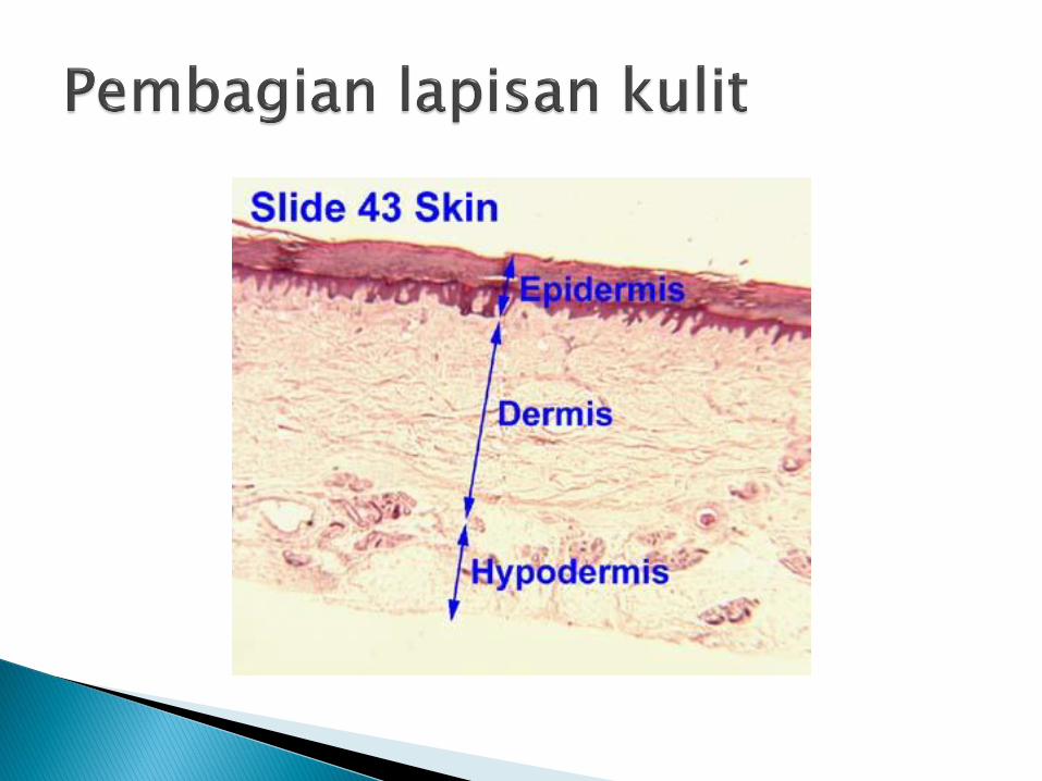
The skin is the largest organ of the body.
It consists of the epidermis and the dermis-hypodermis.
The epidermis is the outer cellular layer of stratified squamous epithelium, which is avascular and varies in thickness.
The dermis is a dense bed of vascular connective tissue.

mechanical and permeability barier
as a sensory and thermoregulatory organ.
initiate primary immune responses.
synthesis (of precusor vitamin D)
excretion (of salts, water and wastes in
sweat)
The epidermis is a layer of keratinized,
stratified squamous epithelium
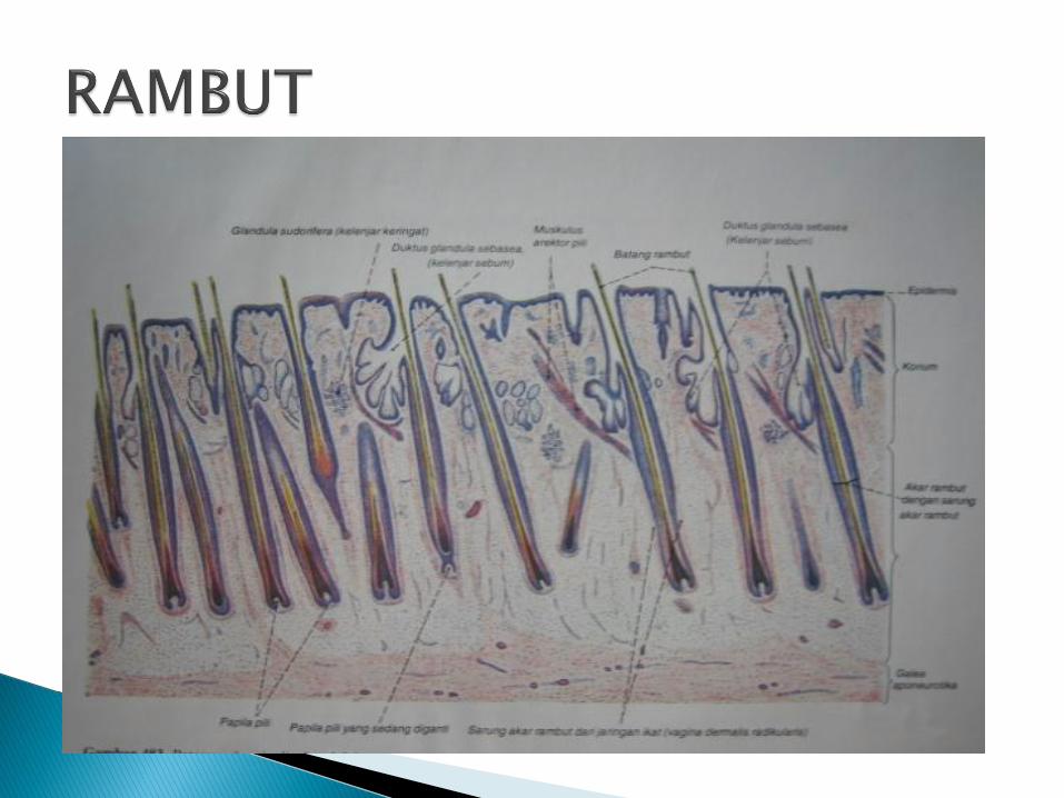
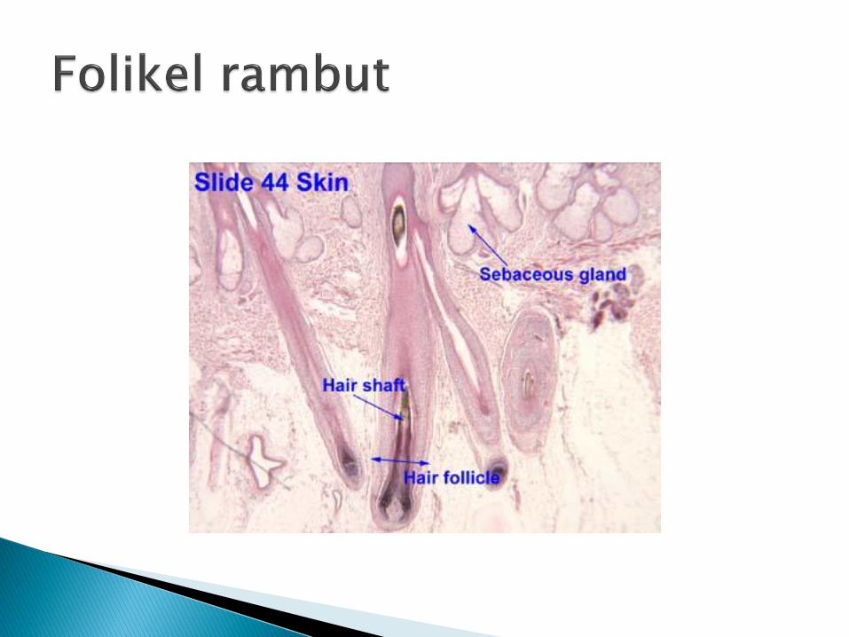
sends three appendages (hair follicles, sweat glands and sebaceous glands) into the underlying dermis
The three types of epidermal appendage extend into the dermis and, in some places, into the subcutaneous tissues.
Oxygen and nutrients diffuse from the underlying dermis

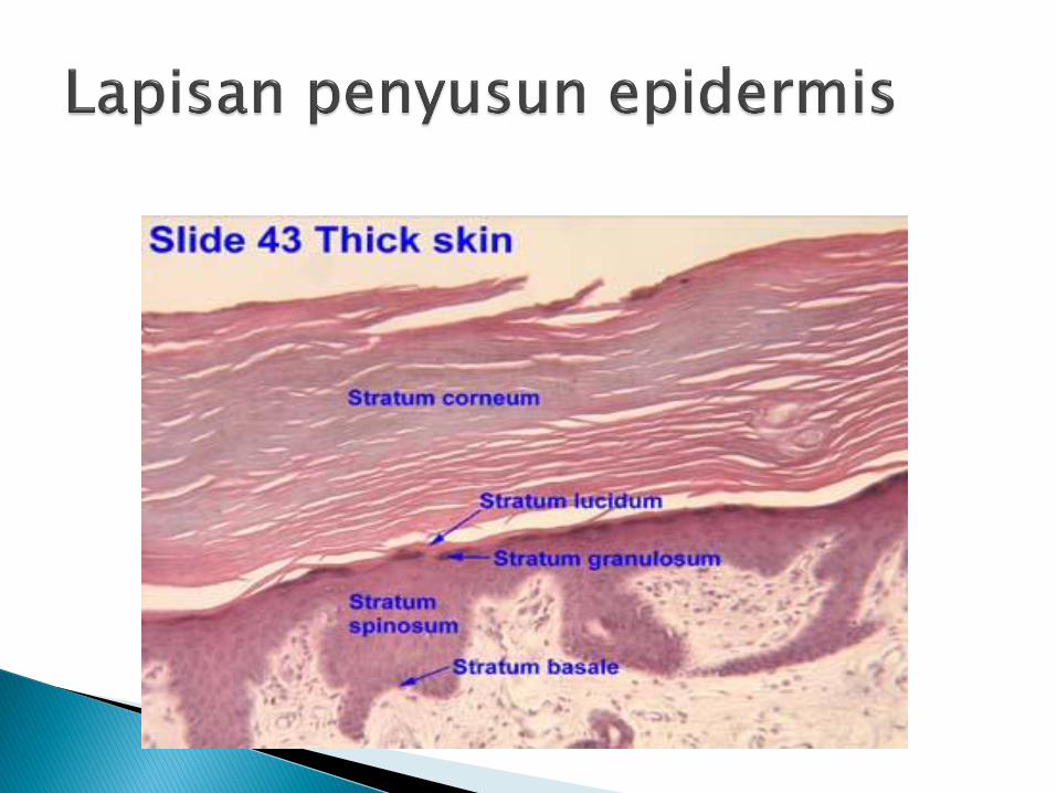
There are 5 layers (from outer most layer)◦ Stratum Corneum, S. Lucidum, S.granulosum, S.
spinosum, S. Basale
deepest layer (stratum germinativum, or
stratum basale)- rapidly dividing cells stem cells present
outermost layer- stratum corneum ,dead cells, keratinized cells
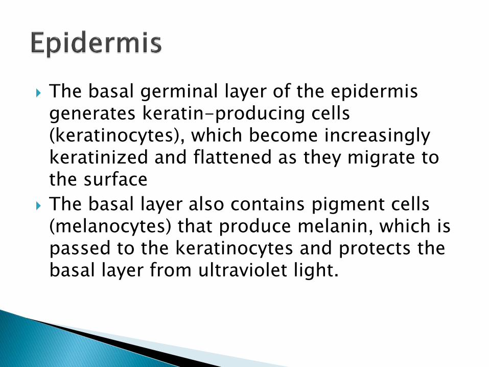
The basal germinal layer of the epidermis generates keratin-producing cells (keratinocytes), which become increasingly keratinized and flattened as they migrate to the surface
The basal layer also contains pigment cells (melanocytes) that produce melanin, which is passed to the keratinocytes and protects the basal layer from ultraviolet light.
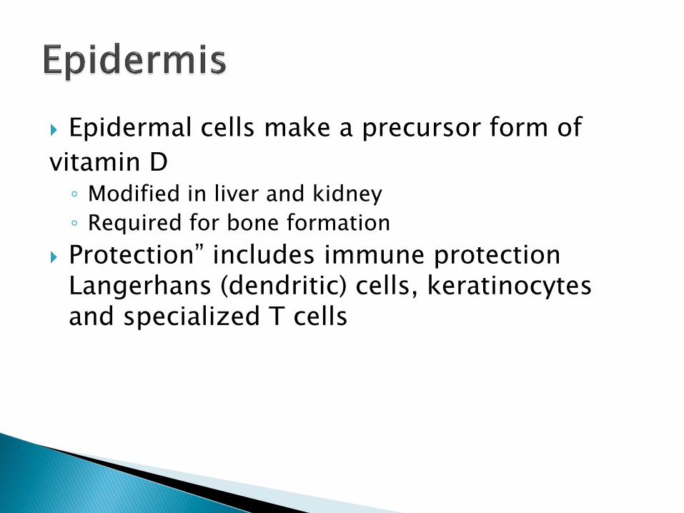
Epidermal cells make a precursor form of
vitamin D◦ Modified in liver and kidney
◦ Required for bone formation
Protection” includes immune protection Langerhans (dendritic) cells, keratinocytes and specialized T cells

The dermis is composed of collagen, elastic fibres and fat.
It supports blood vessels, lymphatics, nerves and the epidermal appendages.
The junction between the epidermis and the dermis is undulating where dermal papillae push up towards the epidermis.
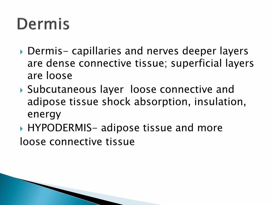
Dermis- capillaries and nerves deeper layers are dense connective tissue; superficial layers are loose
Subcutaneous layer loose connective and adipose tissue shock absorption, insulation, energy
HYPODERMIS- adipose tissue and more
loose connective tissue

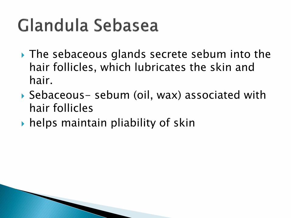
The sebaceous glands secrete sebum into the hair follicles, which lubricates the skin and hair.
Sebaceous- sebum (oil, wax) associated with hair follicles
helps maintain pliability of skin
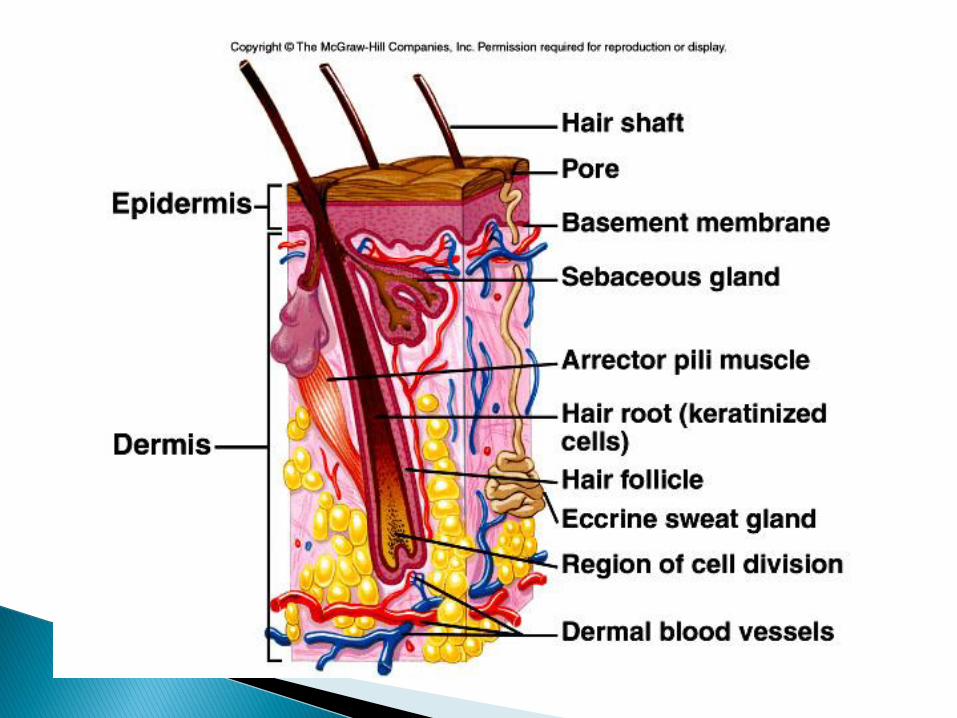
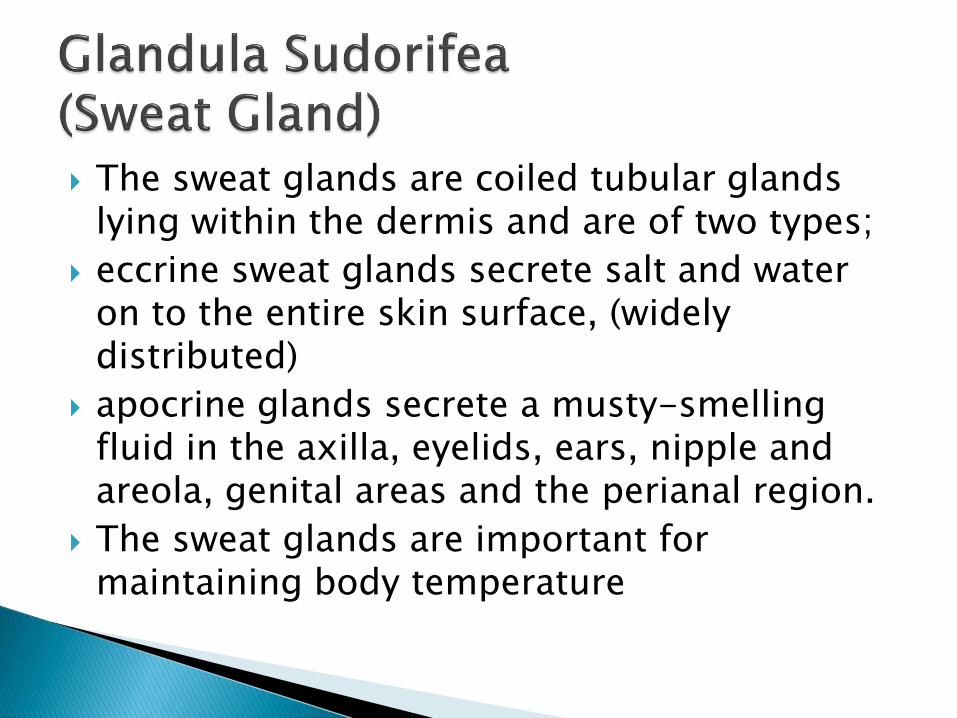
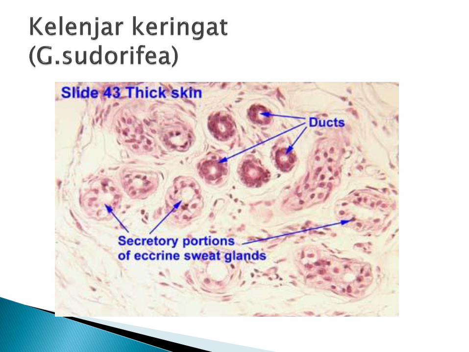
The sweat glands are coiled tubular glands lying within the dermis and are of two types;
eccrine sweat glands secrete salt and water on to the entire skin surface, (widely distributed)
apocrine glands secrete a musty-smelling fluid in the axilla, eyelids, ears, nipple and areola, genital areas and the perianal region.
The sweat glands are important for maintaining body temperature


Hair follicles produce hair, the colour of which is determined by melanocytes within the follicle
Melanocytes become less active with age. Gray hair is a mixture of pigmented and non-pigmented hairs.
Red hair results from a a modified type of melanin that contains iron.
The shape of the hair shaft determines texture.◦ Round shaft – straight hair
◦ Oval shaft – wavy hair, Flat shafts – curly or kinky hair

The nails are flat, horny structures composed of keratin.
They arise from a matrix of germinal cells, which can be seen as a white crescent (lunula) at the nail base.
If a nail is avulsed, a new nail grows from this matrix. If the matrix is destroyed, nail regeneration is impossible epidermal cells covering the nailbed thickens to form a keratinized protective layer.

dr. Erike Anggraini S
dr. Ahmad Azwar


PENGINDERAAN MANUSIA
1. PENGLIHATAN
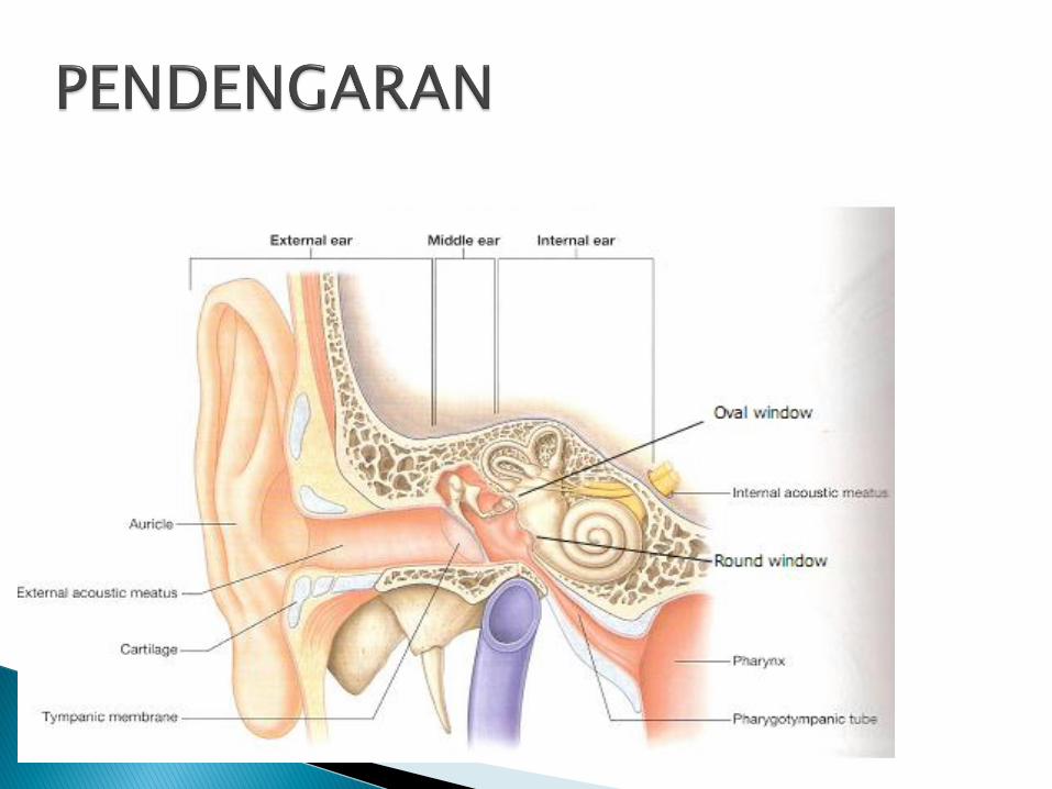
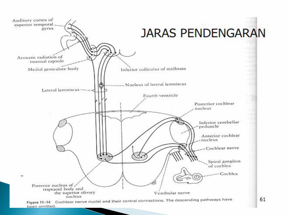
2. PENDENGARAN
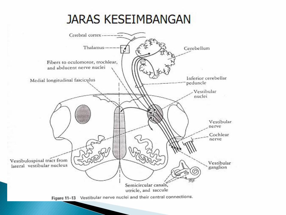
3. KESEIMBANGAN
4. PENGHIDUAN
5. PENGECAPAN
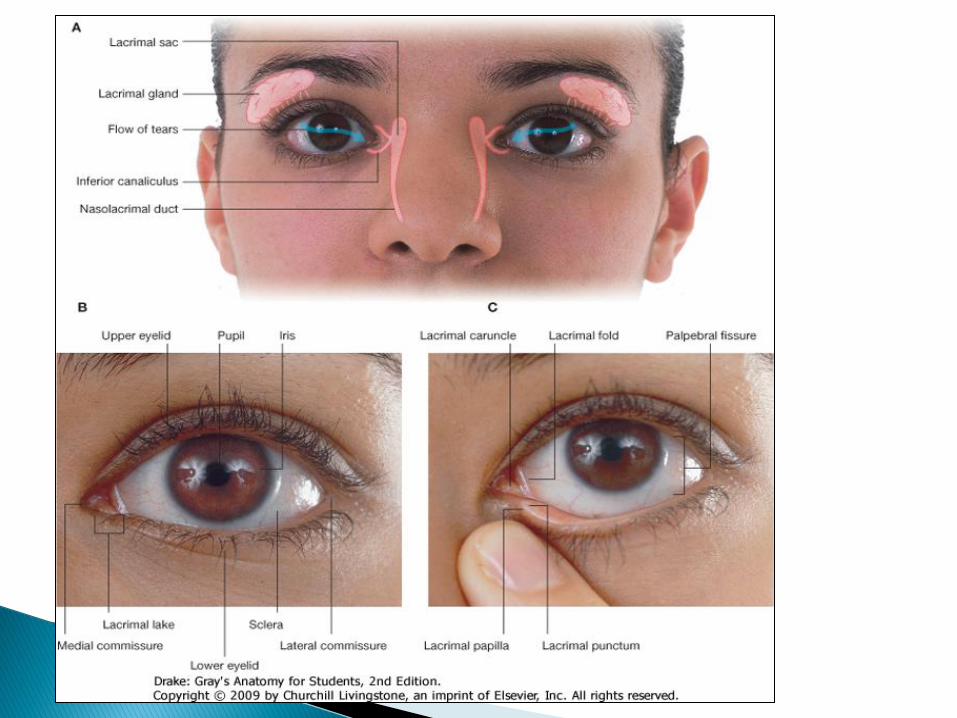
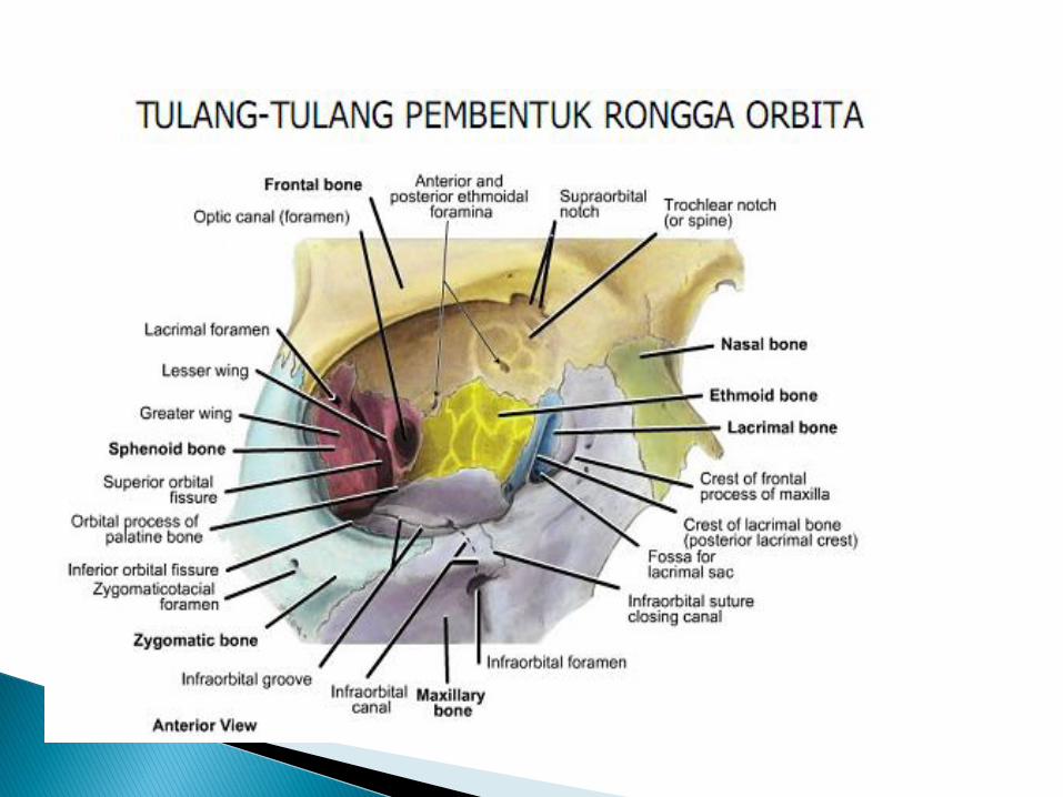


The sensory nerves are all branches of the trigeminal nerve [V]◦ the supra-orbital, supratrochlear, infratrochlear, and
lacrimal branches of the ophthalmic nerve [V1]; and◦ the infra-orbital branch of the maxillary nerve [V2]
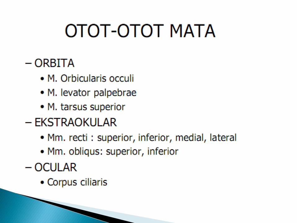
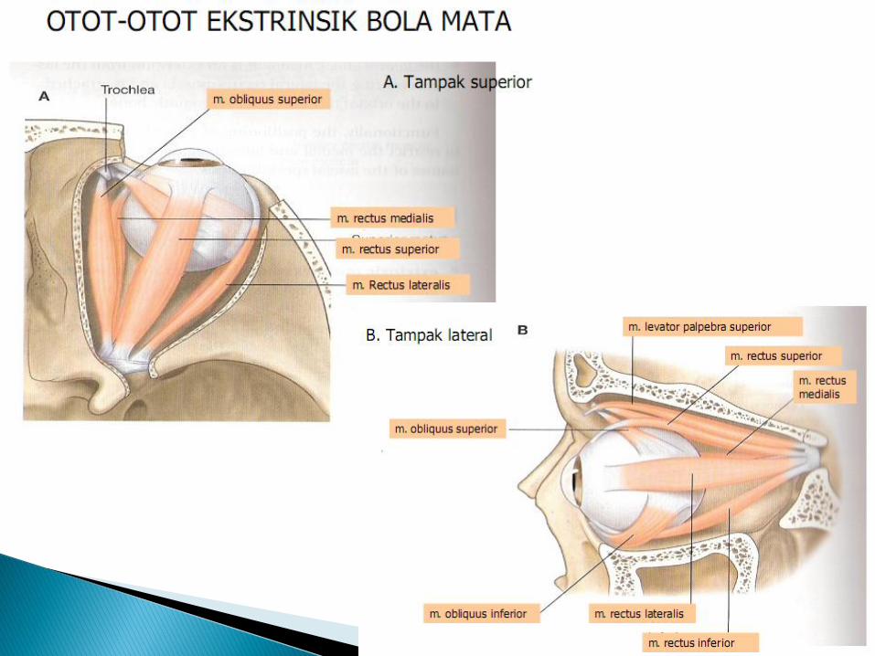
Motor innervation is from: ◦ the facial nerve [VII], the palpebral part
of the orbicularis oculi;◦ the oculomotor nerve [III], the levator palpebrae superioris;
◦ sympathetic fibers, the superior tarsal muscle.


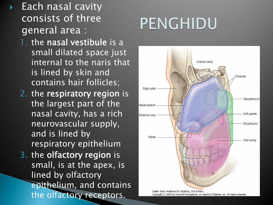
Each nasal cavity consists of three general area :1. the nasal vestibule is a
small dilated space just internal to the naris that is lined by skin and contains hair follicles;
2. the respiratory region is the largest part of the nasal cavity, has a rich neurovascular supply, and is lined by respiratory epithelium
3. the olfactory region is small, is at the apex, is lined by olfactory epithelium, and contains the olfactory receptors.


Tortora GJ, Derrickson BH. 2009. Principles of Anatomy and Physiology vol.1. Asia: John Wiley and Sons, Inc
Drake RL, Vogl AW, Mitchell AWM. 2010. Gray’s Anatomy . Canada : Churchill Livingstone
Moore L. 2010. Clinically Oriented Anatomy. USA :