skin wound healing is accelerated and scarless in the ... · quı´mica e imunologia, grupo de...
TRANSCRIPT

of November 29, 2018.This information is current as
MicrobiotaScarless in the Absence of Commensal Skin Wound Healing Is Accelerated and
Teixeira and Lucíola S. BarcelosS. Martins, Milene A. Rachid, Jacques R. Nicoli, Mauro M.Castro, Brígida G. A. Schirmer, Daniel Cisalpino, Flaviano Maria C. C. Canesso, Angélica T. Vieira, Tiago B. R.
http://www.jimmunol.org/content/193/10/5171doi: 10.4049/jimmunol.1400625October 2014;
2014; 193:5171-5180; Prepublished online 17J Immunol
MaterialSupplementary
5.DCSupplementalhttp://www.jimmunol.org/content/suppl/2014/10/17/jimmunol.140062
Referenceshttp://www.jimmunol.org/content/193/10/5171.full#ref-list-1
, 10 of which you can access for free at: cites 49 articlesThis article
average*
4 weeks from acceptance to publicationFast Publication! •
Every submission reviewed by practicing scientistsNo Triage! •
from submission to initial decisionRapid Reviews! 30 days* •
Submit online. ?The JIWhy
Subscriptionhttp://jimmunol.org/subscription
is online at: The Journal of ImmunologyInformation about subscribing to
Permissionshttp://www.aai.org/About/Publications/JI/copyright.htmlSubmit copyright permission requests at:
Email Alertshttp://jimmunol.org/alertsReceive free email-alerts when new articles cite this article. Sign up at:
Print ISSN: 0022-1767 Online ISSN: 1550-6606. Immunologists, Inc. All rights reserved.Copyright © 2014 by The American Association of1451 Rockville Pike, Suite 650, Rockville, MD 20852The American Association of Immunologists, Inc.,
is published twice each month byThe Journal of Immunology
by guest on Novem
ber 29, 2018http://w
ww
.jimm
unol.org/D
ownloaded from
by guest on N
ovember 29, 2018
http://ww
w.jim
munol.org/
Dow
nloaded from

The Journal of Immunology
Skin Wound Healing Is Accelerated and Scarless in theAbsence of Commensal Microbiota
Maria C. C. Canesso,* Angelica T. Vieira,† Tiago B. R. Castro,* Brıgida G. A. Schirmer,*
Daniel Cisalpino,† Flaviano S. Martins,† Milene A. Rachid,‡ Jacques R. Nicoli,†
Mauro M. Teixeira,x and Lucıola S. Barcelos*
The commensal microbiota has a high impact on health and disease bymodulating the development and homeostasis of host immune
system. Immune cells are involved in virtually every aspect of the wound repair process; however, the impact of commensal micro-
biota on skin wound healing is largely unknown. In this study, we evaluated the influence of commensal microbiota on tissue repair of
excisional skin wounds by using germ-free (GF) Swissmice. We observed that macroscopic wound closure rate is accelerated in the
absence of commensal microbiota. Accordantly, histologically assessed wound epithelization was accelerated in GF in comparison
with conventional (CV) Swiss mice. The wounds of GF mice presented a significant decrease in neutrophil accumulation and an
increase in mast cell and macrophage infiltration into wounds. Interestingly, alternatively activated healing macrophage-related
genes were highly expressed in the wound tissue of GF mice. Moreover, levels of the anti-inflammatory cytokine IL-10, the
angiogenic growth factor VEGF and angiogenesis were higher in the wound tissue of those mice. Conversely, scarring and levels
of the profibrogenic factor TGF-b1 were greatly reduced in GF mice wounded skin when compared with CV mice. Of note,
conventionalization of GF mice with CV microbiota restored wound closure rate, neutrophil and macrophage accumulation,
cytokine production, and scarring to the same extent as CV mice. Overall, our findings suggest that, in the absence of any contact
with microbiota, skin wound healing is accelerated and scarless, partially because of reduced accumulation of neutrophils,
increased accumulation of alternatively activated healing macrophages, and better angiogenesis at wound sites. The Journal
of Immunology, 2014, 193: 5171–5180.
The commensal microbiota consists of microorganisms thatare present on body surfaces covered by epithelial cellsexposed to the external environment such as skin and the
gastrointestinal tract (1). It has been shown that the impact of thecommensal microbiota on health and disease can be wide-rangingfrom protection against pathogenic microorganisms, absorption ofnutrients, and vitamin production to modulating the developmentand homeostasis of immune system (2–6).
Wound healing is a highly dynamic process that involves a com-plex sequence of cellular and biochemical events. In adult mammals,
after an early inflammatory stage characterized by infiltration of
neutrophils and macrophages, the formation of a fibroproliferative
tissue rich in immature collagen bundles and newly formed blood
vessels takes place and prompt re-epithelization occurs. Finally, the
maturation phase involves dermis collagen remodeling and scar
budding (7) as the usual outcome of tissue repair.Wound inflammatory leukocytes may interfere with virtually all
phases of tissue repair (8–12). On one hand, neutrophils (11) and
inflammatory classically activated macrophages (13) may impair
wound healing. On the other hand, macrophages may range from
a proinflammatory to an anti-inflammatory/angiogenic/healing
phenotype that serves to resolve inflammation and promote
healing (14).Our group has demonstrated that germ-free (GF) animals (which
have no commensal microbiota) display anti-inflammatory and
reduced hypernociception responses in models of ischemia and
reperfusion, systemic LPS, and carrageenan-induced pain (15–17).
Conversely, the chronic inflammatory response in murine models
of colitis, arthritis, and asthma may be exacerbated or prolonged
in GF animals (18). Animals free of commensal microbiota
therefore represent a valuable tool for understanding the intrinsic
mechanisms involved in tissue repair, especially when considered
the importance of a controlled inflammatory response after injury.
However, studies evaluating the influence of commensal micro-
biota on the healing of skin wounds are very scarce.The first published studies about the influence of commensal
microbiota on the healing emerged in the 1960s when histological
examination of oral wounds indicated no differences between GF
mice and their respective conventional (CV) controls animals re-
garding inflammatory response and collagen production (19). In
*Departamento de Fisiologia e Biofısica, Grupo de Imunofarmacologia, Instituto deCiencias Biologicas, Universidade Federal de Minas Gerais, 31270-901 Belo Horizonte,Minas Gerais, Brazil; †Departamento de Microbiologia, Grupo de Imunofarmacologia,Instituto de Ciencias Biologicas, Universidade Federal de Minas Gerais, 31270-901Belo Horizonte, Minas Gerais, Brazil; ‡Departamento de Patologia Geral, Grupo deImunofarmacologia, Instituto de Ciencias Biologicas, Universidade Federal de MinasGerais, 31270-901 Belo Horizonte, Minas Gerais, Brazil; and xDepartamento de Bio-quımica e Imunologia, Grupo de Imunofarmacologia, Instituto de Ciencias Biologicas,Universidade Federal de Minas Gerais, 31270-901 Belo Horizonte, Minas Gerais,Brazil
Received for publication March 10, 2014. Accepted for publication September 16,2014.
This work was supported by Conselho Nacional de Desenvolvimento Cientıfico eTecnologico, Fundacao do Amparo a Pesquisa do Estado de Minas Gerais, Coordenacaode Aperfeicoamento de Pessoal de Nıvel Superior, and Pro-Reitoria de Pesquisa daUniversidade Federal de Minas Gerais. M.C.C.C. holds a Coordenacao de Aperfei-coamento de Pessoal de Nıvel Superior master of science scholarship. F.S.M., J.R.N.,M.M.T., and L.S.B. hold Conselho Nacional de Desenvolvimento Cientıfico e Tec-nologico research fellowships.
Address correspondence and reprint requests to Dr. Lucıola S. Barcelos, Departa-mento de Fisiologia e Biofısica, Instituto de Ciencias Biologicas, Universidade Fed-eral de Minas Gerais, Avenida Antonio Carlos, 6627 – Pampulha, 31270-901 BeloHorizonte Minas Gerais, Brazil. E-mail address: [email protected]
The online version of this article contains supplemental material.
Abbreviations used in this article: CV, conventional; CVZ, conventionalized; GF, germ-free; KC, keratinocyte-derived chemokine; MPO, myeloperoxidase; NAG, N-acetyl-b-D-glucosaminidase.
Copyright� 2014 by The American Association of Immunologists, Inc. 0022-1767/14/$16.00
www.jimmunol.org/cgi/doi/10.4049/jimmunol.1400625
by guest on Novem
ber 29, 2018http://w
ww
.jimm
unol.org/D
ownloaded from
contrast, in a comparative study using GF and CV rats, a moreintense inflammatory reaction in skin wounds from the latteranimals was observed (20). Likewise, incisional wounds of CVmice exhibited higher tensile strength than in GF mice (21). Morerecently, Lai et al. (22) showed that the resident skin commensalmicrobiota was able to modulate local inflammatory responseafter injury.In this study, we evaluated the influence of commensalmicrobiota
on wound closure rate and tissue repair of excisional skin wounds byusing GF mice. Our main findings suggest that in the absence of anycontact with microbiota wound healing is accelerated and ischaracterized by reduced content of neutrophils, increased markersof macrophage polarization to an anti-inflammatory/wound healingphenotype, better angiogenesis stimulation into the wound bed, andreduced scar formation.
Materials and MethodsAnimal procedures
All procedures complied with the standards stated in the Guide for the Careand Use of Laboratory Animals and were conducted under conditionsapproved by the local animal ethics committee, Comissao de Etica no Usode Animais/Universidade Federal de Minas Gerais (protocol number 122/11). Eight- to 10-wk-old male and female GF Swiss were derived froma GF nucleus (Taconic Farms, Germantown, NY) and maintained inflexible plastic isolators (Standard Safety Equipment, Marietta, OH) usingclassical gnotobiology techniques (23). Conventional CV Swiss mice werederived from GF matrices and considered conventional only after sixgenerations in the conventional facility. The process of colonizing GF micewith microbiota from CV mice was performed as described previously(15). Briefly, fecal samples from CV mice were homogenized in saline(10%) and administered by oral gavage to GF mice. Three weeks later,these animals were conducted to excisional wound healing model, as de-scribed below. To assess whether there was adequate colonization of GFmice, fecal samples were cultured using a thioglycollate test (15). Allexperimental procedures in GF mice were conducted under aseptic con-ditions to avoid contamination of animals (16).
Wounding and measurement of wounds area
Mice were anesthetized and their dorsum shaved. Four excisional woundswere created on the dorsum of mice with the aid of a sterile 5-mm circularpunch, removing the entire thickness of the skin. After surgery, mice werecaged individually. The area of wounds was measured at the indicated timepoints with a digital caliper, and the results were expressed as percentageclosure relative to original size (12 [wound area]/[original wound area]3100) (24). Suggestive signs of local infection were not detected in thewounds area.
Quantification of neutrophil and macrophage tissueaccumulation
The extent of neutrophil accumulation in the wound tissue was measured byassaying myeloperoxidase (MPO) activity, as described previously (25, 26).Briefly, after euthanasia with an overdose of anesthetic, the wound andsurrounding skin area were removed and snap frozen in liquid nitrogen. Onthawing and processing, the tissue (100 mg tissue/1 ml buffer) was ho-mogenized in 0.02 mol/l NaPO4 buffer (pH 4.7) containing 0.015 mol/lNa-EDTA, centrifuged at 10,000 3 g for 10 min, and the pellet wassubjected to hypotonic lyses. After a further centrifugation, the pellet wasresuspended in 0.05 mol/l NaPO4 buffer (pH 5.4) containing 0.5% hex-adecyltrimethylammonium bromide and rehomogenized. Suspensionswere then subjected to three freeze-thaw cycles using liquid nitrogen andcentrifuged for 15 min at 10,000 3 g; supernatants were used for MPOassay. The assay was performed by measuring the change in OD at 450 nmusing 1.6 mM 3,39-5,59-tetramethylbenzidine (Sigma-Aldrich, St. Louis,MO) dissolved in DMSO (Merck, Rahway, NJ) and 0.003% H2O2 (v/v)dissolved in phosphate buffer (0.05 M Na3PO4 and 0.5% hexadecyl-trimethylammonium bromide [pH 5.4]). Results were expressed as therelative unit that denotes activity of MPO.
The extent of macrophages accumulation in the wound tissue wasmeasured by assaying N-acetyl-b-D-glucosaminidase (NAG) activity, asdescribed previously (26, 27). Briefly, the tissue (100 mg tissue/1 mlbuffer) was homogenized in 0.9% saline containing 0.1% Triton X-100(v/v) and centrifuged at 4˚C for 10 min at 3000 3 g; supernatants were
used for NAG assay. The assay was performed by measuring the change inOD at 405 nm using p-nitrophenyl-N-acetyl-b-D-glucosaminide (Sigma-Aldrich) and 0.2 M glycine buffer (pH 10.6). Results were expressed as therelative unit that denotes activity of NAG.
Total and differential blood cell count
Blood (5 ml) was collected from the tail vein of anesthetized mice anddiluted in Turk’s solution; total cell counts were performed in a modifiedNeubauer chamber. In addition, a blood smear was prepared and stainedwith Panotico Rapido (a Diff Quick–based stain, Laborclin, Brasil). Thepercentage of monocytes and neutrophils was determined using standardmorphological criteria (25).
Measurement of mRNA expression by real-time quantitativePCR
Animals were euthanized by an overdose of anesthetic. Wounds and sur-rounding skin were removed and perpendicularly cut into two halves. One-half was immediately frozen for molecular biology studies, whereas theother onewas fixed in formalin (4% in isotonic saline) and further processedfor histological analyses. To determine the expression of genes associatedwith inflammatory and alternative macrophage activation, total RNA wasobtained from wounds using TRIzol (Invitrogen, Carlsbad, CA), accordingto manufacturer’s instructions. Total RNA was reverse transcribed withSuperScript III (Invitrogen) as described by the manufacturer. Real-timequantitative PCR was performed on an ABI PRISM Step-One sequence-detection system (Applied Biosystems, Carlsbad, CA) using SYBR GreenPCR Master Mix (Applied Biosystems). The relative expression level ofgenes was determined by the 22DD cycle threshold (Ct) method, and data werenormalized by 18S ribosome subunit expression levels. All reactions werereplicated. Primers were generated for nos2 (forward, 59-AGC ACT TTGGGT GAC CAC CAG GA-39; reverse, 59-AGC TAA GTATTA GAG CGGCGG CA-39), IFN-g (forward, 59-ACA ATG AAC GCT ACA CAC TGCAT-39; reverse, 59-TGG CAG TAA CAG CCA GAA ACA-39), fizz1(forward, 59-ACC TTT CCT GAG ATT CTG CCC-39; reverse, 59-CAGTGG TCC AGT CAA CGA GTA AGC-39), dectin1 (forward, 59-GGAATC CTG TGC TTT GTG GTA GTA G-39; reverse, 59-GGA AGG CAAGAC TGA GAA AAA CCT C-39), mrc1 (forward, 59-TCT TTT ACGAGA AGT TGG GGT CAG-39; reverse, 59-ATC ATT CCG TTC ACCAGA GGG-39), arg1 (forward, 59-TGA CAT CAA CAC TCC CCT GACAAC-39; reverse, 59-GCC TTT TCT TCC TTC CCA GCA G-39), and 18S(forward, 59-CGT TCC ACC AAC TAA GAA CG-39; reverse, 59-CTCAAC ACG GGA AAC CTC AC-39).
Histological assessment
Samples were collected as described above. Five-micrometer paraffin-embedded sections were stained with H&E or toluidine blue and exam-ined under a light microscope (Olympus BX43 with a camera Olympus Q-color 5). Sections were also stained with Picro-Sirius for collagen quan-tification and analyzed under polarized light. Morphometric analyses wereperformed on digital images using ImageProPlus 7.0 Software.
The extent of epithelialization (distance that the neoepithelium hadextended from the margin of the wound as defined by the presence of hairfollicles in nonwounded skin; known as epithelial tongue) and scar tissueformation (area comprising from the epidermal–dermal junction down tothe panniculus carnosus and from the borders of the wound—outlinedusing the freeform outline tool in ImageProPlus to produce a pixel-basedmeasurement then converted to square micrometers) was determined onH&E-stained sections. The number of mast cells was determined bycounting cells in the entire area of wound tissue on toluidine-stainedsections. Organization and maturation of collagen bundles were assessedon Sirius Red–stained sections. All histomorphometric analyses wereperformed in a blind manner.
Measurement of cytokine/chemokine concentrations in thewounds
Animals were euthanized by an overdose of anesthetic and the wounds andsurrounding skin area were removed and snap frozen in liquid nitrogen. Onthawing and processing, the tissuewas homogenized in extraction solution (100mg tissue/1 ml), containing 0.4MNaCl, 0.05% Tween 20, 0.5%BSA, 0.1 mMPMSF, 0.1 mM benzethoniumchloride, 10 mM EDTA, and 20 KI aprotinin,using Ultra-Turrax. The suspension was then spun at 10,0003 g for 10 min at4˚C. The supernatant was used for ELISAs, which were performed using kitsfrom R&D Systems (Minneapolis, MN) for murine IL-10, VEGF, total TGF-b1, TNF-a, and CXCL1/keratinocyte-derived chemokine (KC), according tothe manufacturer’s instructions. All samples were assayed in duplicate. Thethreshold of sensitivity for each cytokine/chemokine was 7.5 pg/ml.
5172 SCARLESS WOUND HEALING IN GERM-FREE MICE
by guest on Novem
ber 29, 2018http://w
ww
.jimm
unol.org/D
ownloaded from
Statistical analysis
Analyses were performed using the GraphPad Prism 5.3 software. Resultsare presented as the mean 6 SEM. Comparisons between two groups werecarried out using Student t test for unpaired data. Three or more groupcomparisons were carried out using one-way ANOVA, followed byStudent-Newman-Keuls multiple comparisons test. Two-way ANOVAwasused for graph lines to verify the interaction between the independentvariables time and strain and was followed by Bonferroni posttest. A pvalue , 0.05 was considered significant.
ResultsWound closure is accelerated in germ-free mice
First, to evaluate whether the absence of commensal microbiotahad any impact on wound closure after an excisional skin injury, wefollowed the rate of macroscopic wound closure. We observed thatGF mice exhibited a significant increase in percentage of woundclosure after day 3 postwounding compared with CVanimals (p,0.001) (Fig. 1A).These macroscopic findings were confirmed by histological
assessment of epithelialization at day 7 postwounding. On H&E-stained paraffin sections, the length of the epithelial tongues (anindicative of the neoepithelium migration extent from the marginof the wound) was significantly increased in GF mice in com-parison with CV control mice (p , 0.001) (Fig. 1B). Thesefindings indicate that enhanced epithelialization contributed to theaccelerated wound closure of GF mice.
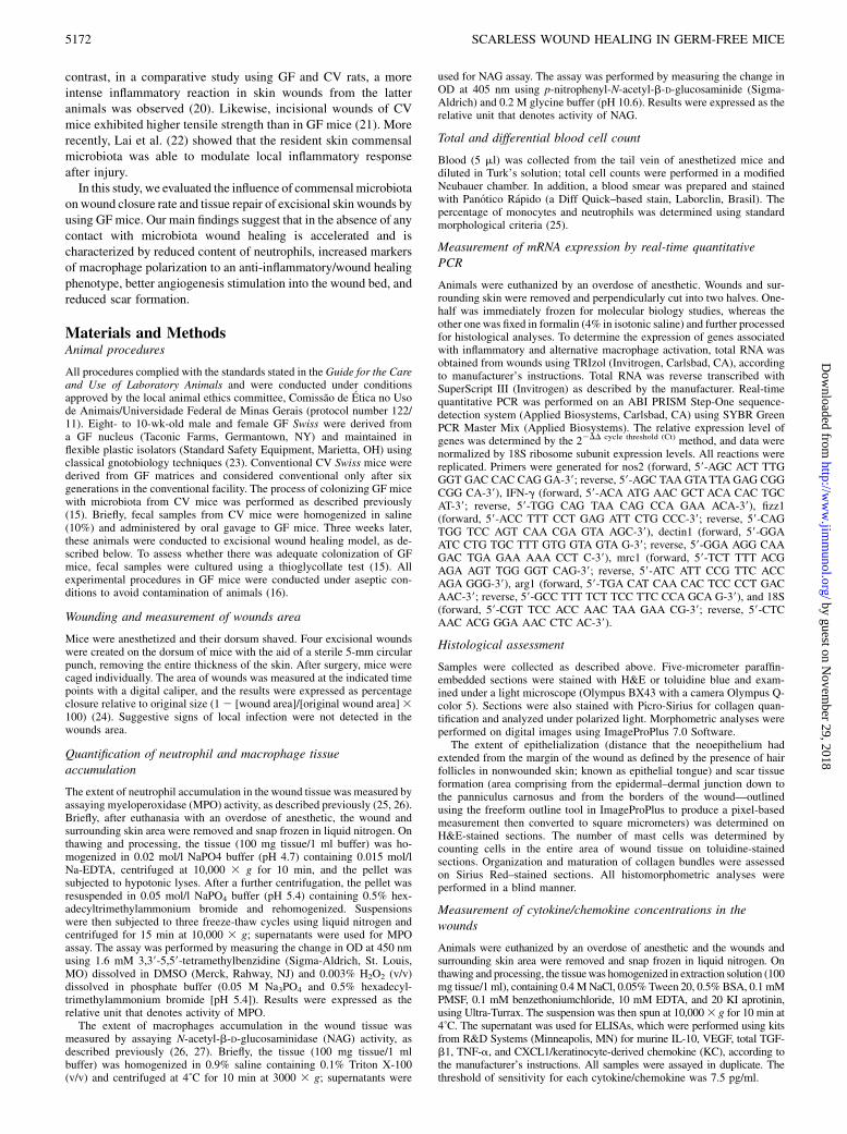
Sustained high levels of IL-10 and TNF-a in the early phase ofwound healing in GF mice
All steps of the healing process are controlled by a wide variety ofcytokines. We observed an increase in wound levels of theproinflammatory cytokine TNF-a after injury in both GF and CVgroups. However, although in CV mice TNF-a dropped to basallevels at day 1 postinjury, in GF mice it was kept at higher levelsat least till day 3 (p , 0.05) (Fig. 2A). Interestingly, levels of theanti-inflammatory cytokine IL-10 were higher in the nonwoundedskin of GF mice when compared with CV mice, remaining sig-nificantly high until day 3 after wounding (p , 0.05 for allgroups) (Fig. 2B). In wounds from CV mice, similar to TNF-akinetics, levels of IL-10 decreased to basal after its peak at day 1postinjury. Levels of CXCL1/KC, a chemoattractant for neu-trophils, did not show any difference between groups (Fig. 2C).These results suggest that GF mice are able to promptly respond toskin injury by increasing levels of inflammation mediators.
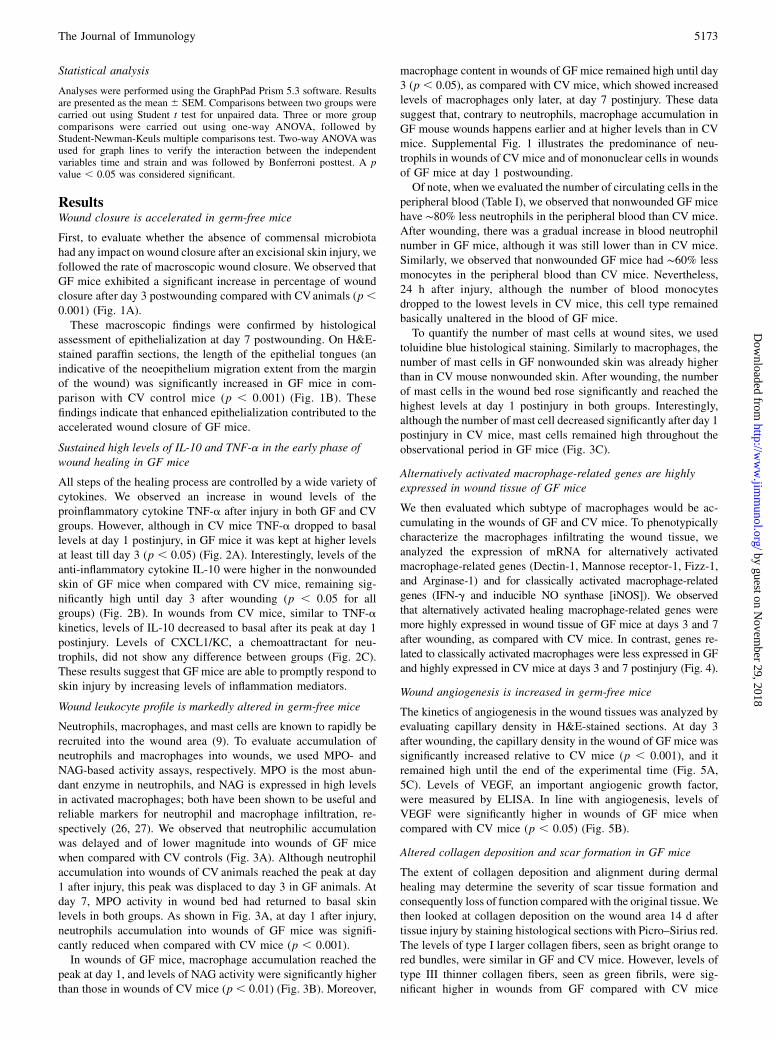
Wound leukocyte profile is markedly altered in germ-free mice
Neutrophils, macrophages, and mast cells are known to rapidly berecruited into the wound area (9). To evaluate accumulation ofneutrophils and macrophages into wounds, we used MPO- andNAG-based activity assays, respectively. MPO is the most abun-dant enzyme in neutrophils, and NAG is expressed in high levelsin activated macrophages; both have been shown to be useful andreliable markers for neutrophil and macrophage infiltration, re-spectively (26, 27). We observed that neutrophilic accumulationwas delayed and of lower magnitude into wounds of GF micewhen compared with CV controls (Fig. 3A). Although neutrophilaccumulation into wounds of CV animals reached the peak at day1 after injury, this peak was displaced to day 3 in GF animals. Atday 7, MPO activity in wound bed had returned to basal skinlevels in both groups. As shown in Fig. 3A, at day 1 after injury,neutrophils accumulation into wounds of GF mice was signifi-cantly reduced when compared with CV mice (p , 0.001).In wounds of GF mice, macrophage accumulation reached the
peak at day 1, and levels of NAG activity were significantly higherthan those in wounds of CV mice (p , 0.01) (Fig. 3B). Moreover,
macrophage content in wounds of GF mice remained high until day3 (p , 0.05), as compared with CV mice, which showed increasedlevels of macrophages only later, at day 7 postinjury. These datasuggest that, contrary to neutrophils, macrophage accumulation inGF mouse wounds happens earlier and at higher levels than in CVmice. Supplemental Fig. 1 illustrates the predominance of neu-trophils in wounds of CV mice and of mononuclear cells in woundsof GF mice at day 1 postwounding.Of note, when we evaluated the number of circulating cells in the
peripheral blood (Table I), we observed that nonwounded GF micehave ∼80% less neutrophils in the peripheral blood than CV mice.After wounding, there was a gradual increase in blood neutrophilnumber in GF mice, although it was still lower than in CV mice.Similarly, we observed that nonwounded GF mice had ∼60% lessmonocytes in the peripheral blood than CV mice. Nevertheless,24 h after injury, although the number of blood monocytesdropped to the lowest levels in CV mice, this cell type remainedbasically unaltered in the blood of GF mice.To quantify the number of mast cells at wound sites, we used
toluidine blue histological staining. Similarly to macrophages, thenumber of mast cells in GF nonwounded skin was already higherthan in CV mouse nonwounded skin. After wounding, the numberof mast cells in the wound bed rose significantly and reached thehighest levels at day 1 postinjury in both groups. Interestingly,although the number of mast cell decreased significantly after day 1postinjury in CV mice, mast cells remained high throughout theobservational period in GF mice (Fig. 3C).
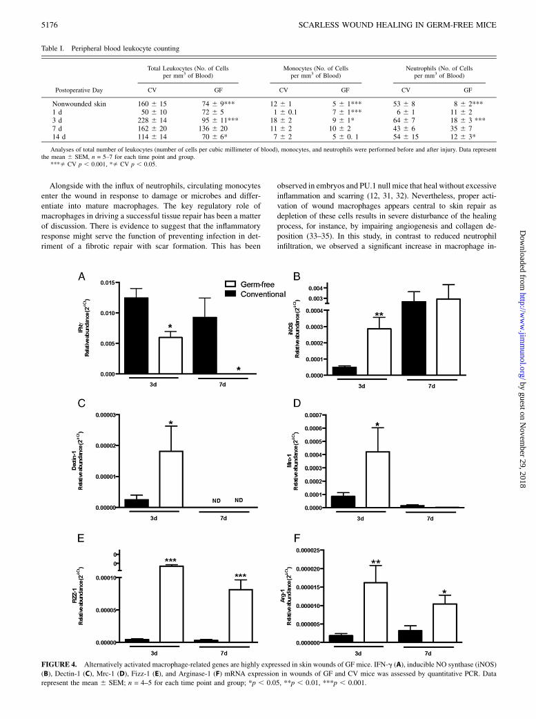
Alternatively activated macrophage-related genes are highlyexpressed in wound tissue of GF mice
We then evaluated which subtype of macrophages would be ac-cumulating in the wounds of GF and CV mice. To phenotypicallycharacterize the macrophages infiltrating the wound tissue, weanalyzed the expression of mRNA for alternatively activatedmacrophage-related genes (Dectin-1, Mannose receptor-1, Fizz-1,and Arginase-1) and for classically activated macrophage-relatedgenes (IFN-g and inducible NO synthase [iNOS]). We observedthat alternatively activated healing macrophage-related genes weremore highly expressed in wound tissue of GF mice at days 3 and 7after wounding, as compared with CV mice. In contrast, genes re-lated to classically activated macrophages were less expressed in GFand highly expressed in CV mice at days 3 and 7 postinjury (Fig. 4).
Wound angiogenesis is increased in germ-free mice
The kinetics of angiogenesis in the wound tissues was analyzed byevaluating capillary density in H&E-stained sections. At day 3after wounding, the capillary density in the wound of GF mice wassignificantly increased relative to CV mice (p , 0.001), and itremained high until the end of the experimental time (Fig. 5A,5C). Levels of VEGF, an important angiogenic growth factor,were measured by ELISA. In line with angiogenesis, levels ofVEGF were significantly higher in wounds of GF mice whencompared with CV mice (p , 0.05) (Fig. 5B).
Altered collagen deposition and scar formation in GF mice
The extent of collagen deposition and alignment during dermalhealing may determine the severity of scar tissue formation andconsequently loss of function compared with the original tissue.Wethen looked at collagen deposition on the wound area 14 d aftertissue injury by staining histological sections with Picro–Sirius red.The levels of type I larger collagen fibers, seen as bright orange tored bundles, were similar in GF and CV mice. However, levels oftype III thinner collagen fibers, seen as green fibrils, were sig-nificant higher in wounds from GF compared with CV mice
The Journal of Immunology 5173
by guest on Novem
ber 29, 2018http://w
ww
.jimm
unol.org/D
ownloaded from

(Fig. 6A, 6C, 6D). Levels of total TGF-b1, a profibrogenic cy-tokine, were significantly higher in wounds from CV mice whencompared with GF mice (p , 0.01 at day 3 and p , 0.05 at day 7)(Fig. 6B). Interestingly, scar tissue area was significantly larger inCV mice when compared with GF mice (Fig. 6E–G).
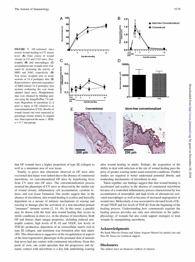
GF colonized mice restore wound healing to conventionalmouse level
To demonstrate that alterations observed in GF mice are due to theabsence of commensal microbiota, GF mice were colonized withfeces of CV mice for 21 d. After that, excisional wounds werecreated on conventionalized (CVZ) mice. Interestingly, we ob-served that CVZmice restore the phenotype of CVmice, as seen bythe wound closure rate, inflammatory cell accumulation and scartissue formation (Fig. 7). Wounding cytokines at day 3 after sur-gery were also unaltered (TNF-a 725 6 261 versus 975 6 218,IL-10 602 6 273 versus 855 6 188, and CXCL1 2913 6 314versus 3327 6 499, CV versus CVZ). These results suggest thatthe accelerated wound healing phenotype observed in the skin ofGF mice is in fact due to the absence of commensal microbiota.
DiscussionSurface tissues, such as skin and intestinal tract, are continuouslyexposed to a number of microorganisms, most of which areharmless or beneficial to the host. Although there is an increasingamount of literature systematically evaluating gut and skin mi-crobial communities diversity in healthy and disease (28) and alsoevaluating the role of gut microbiota on the development andmodulation of immune system (29), the influence of microbiota onskin wound healing is largely unknown. In this study, we disclosethe impact of microbiota on epithelization, inflammation, angio-genesis, and scar formation after excisional skin injury by a com-parative study in GF and CV mice. To the best of our knowledge,the present work provides the first direct evidence for innate/genetically encoded wound healing mechanisms in adult skinexcisional lesions in contrast with the ones that occur in the settingof body interactions with microorganisms.In adult mammals, wound healing is a highly dynamic process
that involves a complex and overlapping sequence of cellular andbiochemical events that range from an immediate response todamage of skin cells and invading microbial signals to inflam-matory and angiogenic responses and finally wound fibroplasia andscar formation (7). Damage or microbial signals activate patternrecognition receptors such as TLRs in leukocytes that then triggerantimicrobial defense and/or inflammatory signaling cascades(30). Interestingly, Lai et al. (22) showed that the resident skincommensal microbiota is able to modulate local inflammatoryresponses after injury in an epithelial-derived TLR dependentmanner. In the case of GF mice, the only cue to stimulate thehealing response just after skin injury are signals from the damageof skin structures once these animals are devoid of microbiota andthe whole process occurs in sterile conditions. In other words, theresponse to skin injury in GF animals is triggered by host-deriveddamage associated molecular patterns and activation of disturbedresident cells. Activation of inflammatory cells is consequently anintegral part of wound healing.
FIGURE 1. Wound closure is accelerated in GF mice. (A) Time-course
of wound closure in GF and CV mice. (B) Epithelial tongue length at day 7
postwounding. (C) Representative photomicrographies of H&E-stained
sections evidencing epithelial tongues (square). Results of wound closure
rate were expressed as percentage closure relative to original size (1 2[wound area]/[original wound area] 3 100). For epithelial tongue length,
measurement was considered the extension of the new epidermis from the
boundaries of the healthy one to the tip of migrating/proliferating kerati-
nocytes. Data represent the mean6 SEM, n = 7–9 mice for each time point
and group. ***p , 0.001. e, epithelium.
5174 SCARLESS WOUND HEALING IN GERM-FREE MICE
by guest on Novem
ber 29, 2018http://w
ww
.jimm
unol.org/D
ownloaded from
Neutrophils are one of the earliest immune cells recruited to thesite of injury. The major function of these cells is to protect the hostfrom infection by combating invadingmicroorganisms and clearingcellular debris. However, activated neutrophils secrete a batteryof bioactive substances, such as proteases and reactive oxygenintermediates, which in excess, can lead to tissue damage (8). Infact, Dovi et al. (11) demonstrated that neutropenia induced by anantineutrophil serum accelerated the rate of wound epithelialclosure without altering the overall quality of the dermal healingprocess in mice. In this study, besides accelerated epithelial clo-sure, we found reduced neutrophil content in 24-h wounds of GFmice, although CXCL1 levels were produced to the same extent asin CV mice in response to injury. Therefore, our data are con-
sistent with the idea that reduced neutrophil infiltration intowound site correlates with accelerated wound closure. Mecha-nistically, our results suggest that the delayed infiltration ofneutrophils after injury in GF mice is, at least partially, due to theneutropenia in these animals before injury and not due to reducedlocal production of neutrophil-related chemoattractants. Indeed,levels of CXCL1 were unaltered in GF mice as compared withtheir CV controls. However, we cannot exclude the possibilitythat neutrophils from GF mice were hyporesponsive to chemo-attractants (18).
FIGURE 2. Kinetics of local cytokine release during skin wound healing
in GF and CV mice. TNF-a (A), IL-10 (B), and CXCL1/KC (C) time-
course production profile into wounds of GF and CV mice. Levels of
cytokines were measured by sandwich ELISA. Data represent the mean 6SEM, n = 7–9 for each time point and group. *p , 0.05.
FIGURE 3. Kinetics of leukocyte accumulation into excisional skin
wounds in GF and CV mice. Neutrophils (A) and macrophages (B) ac-
cumulation into wounds were evaluated by measuring the activity of MPO
and NAG, respectively. (C) Mast cells were quantified by blinding counting
of toluidine blue–stained tissue sections. Data represent the mean 6 SEM;
n = 7–9 for each time point and group; *p , 0.05, **p , 0.01.
The Journal of Immunology 5175
by guest on Novem
ber 29, 2018http://w
ww
.jimm
unol.org/D
ownloaded from
Alongside with the influx of neutrophils, circulating monocytesenter the wound in response to damage or microbes and differ-entiate into mature macrophages. The key regulatory role ofmacrophages in driving a successful tissue repair has been a matterof discussion. There is evidence to suggest that the inflammatoryresponse might serve the function of preventing infection in det-riment of a fibrotic repair with scar formation. This has been
observed in embryos and PU.1 null mice that heal without excessiveinflammation and scarring (12, 31, 32). Nevertheless, proper acti-vation of wound macrophages appears central to skin repair asdepletion of these cells results in severe disturbance of the healingprocess, for instance, by impairing angiogenesis and collagen de-position (33–35). In this study, in contrast to reduced neutrophilinfiltration, we observed a significant increase in macrophage in-
Table I. Peripheral blood leukocyte counting
Postoperative Day
Total Leukocytes (No. of Cellsper mm3 of Blood)
Monocytes (No. of Cellsper mm3 of Blood)
Neutrophils (No. of Cellsper mm3 of Blood)
CV GF CV GF CV GF
Nonwounded skin 160 6 15 74 6 9*** 12 6 1 5 6 1*** 53 6 8 8 6 2***1 d 50 6 10 72 6 5 1 6 0.1 7 6 1*** 6 6 1 11 6 23 d 228 6 14 95 6 11*** 18 6 2 9 6 1* 64 6 7 18 6 3 ***7 d 162 6 20 136 6 20 11 6 2 10 6 2 43 6 6 35 6 714 d 114 6 14 70 6 6* 7 6 2 5 6 0. 1 54 6 15 12 6 3*
Analyses of total number of leukocytes (number of cells per cubic millimeter of blood), monocytes, and neutrophils were performed before and after injury. Data representthe mean 6 SEM, n = 5–7 for each time point and group.
***� CV p , 0.001, *� CV p , 0.05.
FIGURE 4. Alternatively activated macrophage-related genes are highly expressed in skin wounds of GF mice. IFN-g (A), inducible NO synthase (iNOS)
(B), Dectin-1 (C), Mrc-1 (D), Fizz-1 (E), and Arginase-1 (F) mRNA expression in wounds of GF and CV mice was assessed by quantitative PCR. Data
represent the mean 6 SEM; n = 4–5 for each time point and group; *p , 0.05, **p , 0.01, ***p , 0.001.
5176 SCARLESS WOUND HEALING IN GERM-FREE MICE
by guest on Novem
ber 29, 2018http://w
ww
.jimm
unol.org/D
ownloaded from

filtration into wounds of GF when compared with CV mice, sup-porting a beneficial role of wound macrophages for skin repair.One of the hallmarks of macrophages is their ability to become
activated in response to exogenous and endogenous “danger” signalswith the potential of enhancing inflammation. With the eventualelimination of the insult, macrophages contribute to the resolutionof the inflammatory response (36). In fact, macrophages are a di-verse and dynamic population of cells that can perform a widerange of critical functions in wounding healing. Activated macro-phages make up a spectrum of activation status varying froma classical inflammatory phenotype (M1) to a nonclassical or al-ternative phenotype (M2), also referred to as “repair macrophages,”that promote wound healing and angiogenesis (14, 37, 38). Inter-estingly, alternatively activated macrophage-related genes werehighly expressed in wound tissue of GF mice, suggesting the pre-dominant presence of this macrophage phenotype in the absence ofmicrobiota. In fact, the most important point when considering therole of monocytes/macrophages in skin wound healing in GF miceis possibly the predominance of cells with the M2 phenotype in thewounds of those animals. These cells are known to contribute to theresolution of the inflammatory process and to stimulate angiogen-esis as well as the production of type III collagen by fibroblasts. Asa consequence, we observed an increase in the kinetics of thehealing process and better quality of dermis remodeling, favoringa regenerative instead of a fibrotic repair process.Corroborating this idea, we found high levels of IL-10 in wounds
of GF animals. IL-10 is a regulatory cytokine with pivotal func-tions in the control of inflammation and immune-mediated tissuedamage. IL-10 may not only decrease the inflammatory response toinjury but also create an environment favorable to differentiationof regulatory M2 macrophages and regenerative wound healing(39). In fact, IL-10 may be responsible for the scarless repair ob-served in fetal skin (40). Globally, this could additionally explainthe accelerated wound healing without scarring and the predomi-nant expression of genes related to M2 macrophages phenotype inthe wounds of GF mice. Although IL-10 levels were increased andIL-10 can actually decrease inflammation, there were high levels ofthe proinflammatory cytokine TNF-a and much inflammation asseen by the increase of macrophages and mast cells in the woundbed. These data suggest a controlled inflammatory process in GFanimals that favored successful wound healing.Mast cells are able to release a variety of soluble mediators, but
their function is less understood when compared with other in-flammatory cells. Although traditionally viewed as effector cells ofallergic reaction and parasitic diseases, an important role for mastcells in tissue homeostasis and wound healing is now increasinglyrecognized (41–43). Of note, in this study, we found a high andsustained infiltration of mast cells into wounds of GF animals duringthe whole experimental period in contrast to a transient peak of mastcell infiltration 24 h after wounding in CV mice. On one hand, thisinflammatory cell type seems to play an important role in the pro-liferation phase where angiogenesis is essential for provision ofoxygen and nutrients to the nascent tissue. Similarly to M2 mac-rophages, mast cells release angiogenic growth factors, includingVEGF and metalloproteinases that prepare surrounding tissue forangiogenesis during skin repair (44). In fact, the rich content of M2macrophages and mast cells strongly suggests a connection withthe high levels of VEGF and high number of capillaries in thewounds of GF animals. On the other hand, mast cells can limitinflammatory skin reaction by producing IL-10 (45) that, in turn,can downregulate mast cell FcεRI IgE receptor expression sup-porting protection against skin allergy sensitization (46).In addition to inflammation and angiogenesis, both macrophages
and mast cells are also able to regulate fibroplasia at wound sites,
FIGURE 5. Wound angiogenesis is increased in GF mice. Density of
blood vessels (A) and VEGF levels in wounds (B) of GF and CV mice.
(C) Representative photomicrographies of H&E-stained 3 d postinjury
skin sections evidencing blood vessels (arrows). The density of blood
vessels was evaluated by blinding counting in H&E-staining sections and
is represented as number of vessels per square millimeter of granulation
tissue. Levels of VEGF were measured by sandwich ELISA. Data rep-
resent the mean 6 SEM; n = 7 for each time point and group; *p , 0.05,
**p , 0.01, ***p , 0.001.
The Journal of Immunology 5177
by guest on Novem
ber 29, 2018http://w
ww
.jimm
unol.org/D
ownloaded from
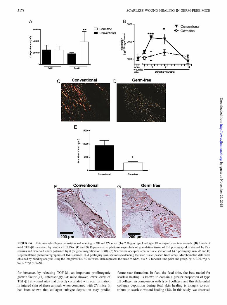
for instance, by releasing TGF-b1, an important profibrogenicgrowth factor (47). Interestingly, GF mice showed lower levels ofTGF-b1 at wound sites that directly correlated with scar formationin injured skin of these animals when compared with CV mice. Ithas been shown that collagen subtype deposition may predict
future scar formation. In fact, the fetal skin, the best model forscarless healing, is known to contain a greater proportion of typeIII collagen in comparison with type I collagen and this differentialcollagen deposition during fetal skin healing is thought to con-tribute to scarless wound healing (48). In this study, we observed
FIGURE 6. Skin wound collagen deposition and scarring in GF and CV mice. (A) Collagen type I and type III occupied area into wounds. (B) Levels of
total TGF-b1 evaluated by sandwich ELISA. (C and D) Representative photomicrographies of granulation tissue of 7 d postinjury skin stained by Pic-
rosirius and observed under polarized light (original magnification 340). (E) Scar tissue occupied area in tissue sections of 14 d postinjury skin. (F and G)
Representative photomicrographies of H&E-stained 14 d postinjury skin sections evidencing the scar tissue (dashed lined area). Morphometric data were
obtained by blinding analysis using the ImageProPlus 7.0 software. Data represent the mean6 SEM; n = 5–7 for each time point and group. *p, 0.05, **p,0.01, ***p , 0.001.
5178 SCARLESS WOUND HEALING IN GERM-FREE MICE
by guest on Novem
ber 29, 2018http://w
ww
.jimm
unol.org/D
ownloaded from
that GF wounds have a higher proportion of type III collagen aswell as a minimum area of scar tissue.Finally, to prove that alterations observed in GF mice after
excisional skin injury were indeed due to the absence of commensalmicrobiota, we conventionalized GF mice by transferring fecesfrom CV mice into GF mice. The conventionalization processrestored the phenotype of CV mice as observed by the similar rateof wound closure, inflammatory cell accumulation, cytokine re-lease, and scar tissue formation. Our results suggest that, in theabsence of microbiota, skin wound healing is scarless and basicallydependent on a mosaic of intrinsic mechanisms of sensing andreacting to damage plus the activation of a non-microbial-primed“immature” immune system (2, 14, 16). In this sense, a parallelmay be drawn with the fetal skin wound healing that occurs insterile conditions in utero (i.e., in the absence of microbiota). BothGF and fetuses share unique properties, including reduced neu-trophil content, high levels of IL-10 and VEGF, low levels ofTGF-b1 production, deposition of an extracellular matrix rich intype III collagen, and minimum scar formation after skin injury(49). This observation is suggestive of the recapitulation of aspectsof the fetal regenerative phenotype in the postnatal skin of animalsthat never had any contact with commensal microbiota. From thispoint of view, one could speculate that the progressive and dy-namic contact with microbiota is a key link underlining scarring
after wound healing in adults. Perhaps, the acquisition of theability to deal with infection at the site of wound healing pays theprice of greater scarring under usual nonsterile conditions. Furtherstudies are required to better understand potential fibrotic andnonhealing mechanisms of microbiota in skin.Taken together, our findings suggest that skin wound healing is
accelerated and scarless in the absence of commensal microbiotabecause of a controlled inflammatory process characterized by lowaccumulation of neutrophils and high levels of alternatively acti-vated macrophages as well as because of increased angiogenesis atwound sites. Molecularly, it was associated to elevated levels of IL-10 and VEGF and low levels of TGF-b1 from the beginning of thehealing process. Understanding how commensals regulate thehealing process provides not only new directions in the patho-physiology of wounds but also could support strategies to treatwounds by manipulating microbiota.
AcknowledgmentsWe thank Marcelo Gomes and Valner Augusto Mussel for animal care and
Ilma M. Souza for technical support.
DisclosuresThe authors have no financial conflicts of interest.
FIGURE 7. GF-colonized mice
restore wound healing to CV mouse
level. (A) Time course of wound
closure in CV and CVZ mice. Neu-
trophils (B) and macrophages (C)
accumulation into wounds were eval-
uated by measuring the activity of
MPO and NAG, respectively. (D)
Scar tissue occupied area in tissue
sections of 14 d postinjury skin. (E)
Representative photomicrographies
of H&E-stained 14 d postinjury skin
sections evidencing the scar tissue
(dashed lined area). Morphometric
data were obtained by blinding anal-
ysis using the ImageProPlus 7.0 soft-
ware. Reposition of microbiota 21 d
prior to injury in GF, referred to as
conventionalization (CVZ). Results of
wound closure rate were expressed as
percentage closure relative to original
size. Data represent the mean6 SEM;
n = 5–7 mice/group.
The Journal of Immunology 5179
by guest on Novem
ber 29, 2018http://w
ww
.jimm
unol.org/D
ownloaded from
References1. Tlaskalova-Hogenova, H., R. Stepankova, T. Hudcovic, L. Tuckova
B. Cukrowska, R. Lodinova-Zadnıkova, H. Kozakova, P. Rossmann, J. Bartova,D. Sokol, et al. 2004. Commensal bacteria (normal microflora), mucosal im-munity and chronic inflammatory and autoimmune diseases. Immunol. Lett. 93:97–108.
2. Fagundes, C. T., F. A. Amaral, A. T. Vieira, A. C. Soares, V. Pinho, J. R. Nicoli,L. Q. Vieira, M. M. Teixeira, and D. G. Souza. 2012. Transient TLR activationrestores inflammatory response and ability to control pulmonary bacterial in-fection in germfree mice. J. Immunol. 188: 1411–1420.
3. Macia, L., A. N. Thorburn, L. C. Binge, E. Marino, K. E. Rogers,K. M. Maslowski, A. T. Vieira, J. Kranich, and C. R. Mackay. 2012. Microbialinfluences on epithelial integrity and immune function as a basis for inflam-matory diseases. Immunol. Rev. 245: 164–176.
4. Vijay-Kumar, M., J. D. Aitken, F. A. Carvalho, T. C. Cullender, S. Mwangi,S. Srinivasan, S. V. Sitaraman, R. Knight, R. E. Ley, and A. T. Gewirtz. 2010.Metabolic syndrome and altered gut microbiota in mice lacking Toll-like re-ceptor 5. Science 328: 228–231.
5. Kelly, D., T. King, and R. Aminov. 2007. Importance of microbial coloni-zation of the gut in early life to the development of immunity. Mutat. Res. 622:58–69.
6. Smith, K., K. D. McCoy, and A. J. Macpherson. 2007. Use of axenic animals instudying the adaptation of mammals to their commensal intestinal microbiota.Semin. Immunol. 19: 59–69.
7. Shaw, T. J., and P. Martin. 2009. Wound repair at a glance. J. Cell Sci. 122:3209–3213.
8. Wilgus, T. A. 2008. Immune cells in the healing skin wound: influential playersat each stage of repair. Pharmacol. Res. 58: 112‑116.
9. Eming, S. A., T. Krieg, and J. M. Davidson. 2007. Inflammation in wound repair:molecular and cellular mechanisms. J. Invest. Dermatol. 127: 514–525.
10. Stramer, B. M., R. Mori, and P. Martin. 2007. The inflammation-fibrosis link? AJekyll and Hyde role for blood cells during wound repair. J. Invest. Dermatol.127: 1009–1017.
11. Dovi, J. V., L. K. He, and L. A. DiPietro. 2003. Accelerated wound closure inneutrophil-depleted mice. J. Leukoc. Biol. 73: 448–455.
12. Martin, P., D. D’Souza, J. Martin, R. Grose, L. Cooper, R. Maki, andS. R. McKercher. 2003. Wound healing in the PU.1 null mouse—tissue repair isnot dependent on inflammatory cells. Curr. Biol. 13: 1122‑1128.
13. Ploeger, D. T., N. A. Hosper, M. Schipper, J. A. Koerts, S. de Rond, andR. A. Bank. 2013. Cell plasticity in wound healing: paracrine factors of M1/M2polarized macrophages influence the phenotypical state of dermal fibroblasts.Cell Commun. Signal. 11: 29.
14. Ferrante, C. J., and S. J. Leibovich. 2012. Regulation of macrophage polarizationand wound healing. Adv. Wound Care 1: 10–16.
15. Amaral, F. A., D. Sachs, V. V. Costa, C. T. Fagundes, D. Cisalpino, T. M. Cunha,S. H. Ferreira, F. Q. Cunha, T. A. Silva, J. R. Nicoli, et al. 2008. Commensalmicrobiota is fundamental for the development of inflammatory pain. Proc. Natl.Acad. Sci. USA 105: 2193–2197.
16. Souza, D. G., C. T. Fagundes, F. A. Amaral, D. Cisalpino, L. P. Sousa,A. T. Vieira, V. Pinho, J. R. Nicoli, L. Q. Vieira, I. M. Fierro, and M. M. Teixeira.2007. The required role of endogenously produced lipoxin A4 and annexin-1 forthe production of IL-10 and inflammatory hyporesponsiveness in mice. J.Immunol. 179: 8533–8543.
17. Souza, D. G., A. T. Vieira, A. C. Soares, V. Pinho, J. R. Nicoli, L. Q. Vieira, andM. M. Teixeira. 2004. The essential role of the intestinal microbiota in facili-tating acute inflammatory responses. J. Immunol. 173: 4137–4146.
18. Maslowski, K. M., A. T. Vieira, A. Ng, J. Kranich, F. Sierro, D. Yu,H. C. Schilter, M. S. Rolph, F. Mackay, D. Artis, et al. 2009. Regulation ofinflammatory responses by gut microbiota and chemoattractant receptor GPR43.Nature 461: 1282–1286.
19. Gordon, H. A., and L. Pesti. 1971. The gnotobiotic animal as a tool in the studyof host microbial relationships. Bacteriol. Rev. 35: 390–429.
20. Donati, R. M., D. W. Frank, L. R. Stromberg, and M. M. McLaughlin. 1971. Theeffect of the germfree state on wound healing. J. Surg. Res. 11: 163–172.
21. Okada, M. 1994. The influence of intestinal flora on wound healing in mice.Surg. Today 24: 347–355.
22. Lai, Y., A. Di Nardo, T. Nakatsuji, A. Leichtle, Y. Yang, A. L. Cogen, Z. R. Wu,L. V. Hooper, R. R. Schmidt, S. von Aulock, et al. 2009. Commensal bacteriaregulate Toll-like receptor 3‑dependent inflammation after skin injury. Nat. Med.15: 1377–1382.
23. Pleasants, J. E. 1974. Letter: Legislation—the solution to a dilemma. J. OralSurg. 32: 166.
24. Maeda, S., M. Fujimoto, T. Matsushita, Y. Hamaguchi, K. Takehara, andM. Hasegawa. 2011. Inducible costimulator (ICOS) and ICOS ligand signalinghas pivotal roles in skin wound healing via cytokine production. Am. J. Pathol.179: 2360–2369.
25. Vieira, A. T., C. T. Fagundes, A. L. Alessandri, M. G. Castor, R. Guabiraba,V. O. Borges, K. D. Silveira, E. L. Vieira, J. L. Goncalves, T. A. Silva, et al.2009. Treatment with a novel chemokine-binding protein or eosinophil lineage-ablation protects mice from experimental colitis. Am. J. Pathol. 175: 2382–2391.
26. Barcelos, L. S., A. Talvani, A. S. Teixeira, L. Q. Vieira, G. D. Cassali,S. P. Andrade, and M. M. Teixeira. 2005. Impaired inflammatory angiogenesis,but not leukocyte influx, in mice lacking TNFR1. J. Leukoc. Biol. 78: 352–358.
27. Barcelos, L. S., A. Talvani, A. S. Teixeira, G. D. Cassali, S. P. Andrade, andM. M. Teixeira. 2004. Production and in vivo effects of chemokines CXCL1-3/KC and CCL2/JE in a model of inflammatory angiogenesis in mice. Inflamm.Res. 53: 576‑584.
28. Gallo, R. L., and L. V. Hooper. 2012. Epithelial antimicrobial defence of the skinand intestine. Nat. Rev. Immunol. 12: 503–516.
29. Maslowski, K. M., and C. R. Mackay. 2011. Diet, gut microbiota and immuneresponses. Nat. Immunol. 12: 5–9.
30. Muzio, M., N. Polentarutti, D. Bosisio, P. P. Manoj Kumar, and A. Mantovani.2000. Toll-like receptor family and signalling pathway. Biochem. Soc. Trans. 28:563–566.
31. Cowin, A. J., M. P. Brosnan, T. M. Holmes, and M. W. Ferguson. 1998. En-dogenous inflammatory response to dermal wound healing in the fetal and adultmouse. Dev. Dyn. 212: 385‑393.
32. Hopkinson-Woolley, J., D. Hughes, S. Gordon, and P. Martin. 1994. Macrophagerecruitment during limb development and wound healing in the embryonic andfoetal mouse. J. Cell Sci. 107: 1159–1167.
33. Leibovich, S. J., and R. Ross. 1975. The role of the macrophage in wound repair:a study with hydrocortisone and antimacrophage serum. Am. J. Pathol. 78: 71–100.
34. DiPietro, L. A., M. Burdick, Q. E. Low, S. L. Kunkel, and R. M. Strieter. 1998.MIP-1a as a critical macrophage chemoattractant in murine wound repair. J.Clin. Invest. 101: 1693–1698.
35. Goren, I., N. Allmann, N. Yogev, C. Sch€urmann, A. Linke, M. Holdener,A. Waisman, J. Pfeilschifter, and S. Frank. 2009. A transgenic mouse model ofinducible macrophage depletion: effects of diphtheria toxin-driven lysozyme M-specific cell lineage ablation on wound inflammatory, angiogenic, and contrac-tive processes. Am. J. Pathol. 175: 132–147.
36. Zhang, X., and D. M. Mosser. 2008. Macrophage activation by endogenousdanger signals. J. Pathol. 214: 161–178.
37. Mosser, D. M., and J. P. Edwards. 2008. Exploring the full spectrum of mac-rophage activation. Nat. Rev. Immunol. 8: 958–969.
38. Martinez, F. O., A. Sica, A. Mantovani, and M. Locati. 2008. Macrophageactivation and polarization. Front. Biosci. 13: 453‑461.
39. Peranteau, W. H., L. Zhang, N. Muvarak, A. T. Badillo, A. Radu, P. W. Zoltick,and K. W. Liechty. 2008. IL-10 overexpression decreases inflammatory media-tors and promotes regenerative healing in an adult model of scar formation. J.Invest. Dermatol. 128: 1852–1860.
40. Liechty, K. W., H. B. Kim, N. S. Adzick, and T. M. Crombleholme. 2000. Fetalwound repair results in scar formation in interleukin-10‑deficient mice ina syngeneic murine model of scarless fetal wound repair. J. Pediatr. Surg. 35:866‑872; discussion 872‑863.
41. Weller, K., K. Foitzik, R. Paus, W. Syska, and M. Maurer. 2006. Mast cells arerequired for normal healing of skin wounds in mice. FASEB J. 20: 2366‑2368.
42. Galli, S. J., and M. Tsai. 2008. Mast cells: versatile regulators of inflammation,tissue remodeling, host defense and homeostasis. J. Dermatol. Sci. 49: 7–19.
43. Oskeritzian, C. A. 2012. Mast cells and wound healing. Adv. Wound Care 1: 23–28.44. Ng, M. F. 2010. The role of mast cells in wound healing. Int. Wound J. 7: 55–61.45. Grimbaldeston, M. A., S. Nakae, J. Kalesnikoff, M. Tsai, and S. J. Galli. 2007.
Mast cell-derived interleukin 10 limits skin pathology in contact dermatitis andchronic irradiation with ultraviolet B. Nat. Immunol. 8: 1095–1104.
46. Kennedy Norton, S., B. Barnstein, J. Brenzovich, D. P. Bailey, M. Kashyap,K. Speiran, J. Ford, D. Conrad, S. Watowich, M. R. Moralle, et al. 2008. IL-10suppresses mast cell IgE receptor expression and signaling in vitro and in vivo. J.Immunol. 180: 2848–2854.
47. Braund, R., S. Hook, and N. J. Medlicott. 2007. The role of topical growthfactors in chronic wounds. Curr. Drug Deliv. 4: 195–204.
48. Merkel, J. R., B. R. DiPaolo, G. G. Hallock, and D. C. Rice. 1988. Type I andtype III collagen content of healing wounds in fetal and adult rats. Proc. Soc.Exp. Biol. Med. 187: 493‑497.
49. Lo, D. D., A. S. Zimmermann, A. Nauta, M. T. Longaker, and H. P. Lorenz.2012. Scarless fetal skin wound healing update. Birth Defects Res. C EmbryoToday 96: 237‑247.
5180 SCARLESS WOUND HEALING IN GERM-FREE MICE
by guest on Novem
ber 29, 2018http://w
ww
.jimm
unol.org/D
ownloaded from