sleep cine magnetic resonance imaging—a dynamic evaluation...
TRANSCRIPT

mo2“t
5ac
Operative Techniques in Otolaryngology (2012) 23, 19-24
Sleep cine magnetic resonance imaging—A dynamicevaluation of the airway
Sally R. Shott, MD
From the Department of Otolaryngology—Head and Neck Surgery, University of Cincinnati, Cincinnati Children’s
Medical Center, Cincinnati, Ohio.It has become more apparent in recent years that increasing numbers of children continue to haveobstructive sleep apnea despite previous removal of the tonsils and adenoids. This occurs not only inchildren in “at-risk” populations, such as those with Down syndrome and obesity, but also in otherwise“normal” children. Sleep cine magnetic resonance imaging (MRI) provides a high-resolution exami-nation of the airway during sleep without ionizing radiation exposure and allows for identification ofsite or sites of residual airway obstruction. This technique is particularly helpful since the diagnosticexamination performed in awake adults for sleep apnea is not often possible in the pediatric population.The technique of sleep cine MRI and examples of its use are presented. Sample patient scenarios areused to illustrate how it can assist with treatment planning. Anesthesia use during the cine MRI isdiscussed.© 2012 Elsevier Inc. All rights reserved.
KEYWORDS:Cine MRI;Obstructive sleepapnea;Dexmedetomidine
Although removal of the tonsils and adenoids (T&A) isthe most common initial surgical intervention for obstruc-tive sleep apnea (OSA) in children, studies have shown thatresidual airway obstruction after this surgery is not onlycommon in some populations of children such as in childrenwith Down syndrome (DS) and in obese children1-5 but also
ore frequent than previously believed in otherwise typical,r “normal,” children. Mitchell, in 2007, showed a 10 to0% incidence of persistent sleep apnea in a group of 79typical” children, ie, without major medical problems, af-er T&A.6 Tauman et al, using a much more strict definition
of “surgical cure,” showed complete normalization of allcomponents evaluated in a sleep study in only 25% of theirtest population of “typical” children.7 This compares to the% total success rate seen in the article by Shott et al, wheresimilarly strict definition of “cure” was used in a group of
hildren with DS.2 If “cure” is defined as more similar to thedefinitions used in the study by Mitchell, almost 50 to 70%
Address reprint requests and correspondence: Sally R. Shott, MD,Department of Otolaryngology—Head and Neck Surgery, University ofCincinnati, Cincinnati Children’s Medical Center, 3333 Burnet Avenue,Cincinnati, OH 45229.
E-mail address: [email protected].
1043-1810/$ -see front matter © 2012 Elsevier Inc. All rights reserved.doi:10.1016/j.otot.2011.06.005
of the children with DS in this study continued to have OSAafter T&A.
It is therefore becoming more common to be presentedwith a child who has persistent OSA despite previous T&Awhere further interventions may be needed, both surgicaland medical.
If there is persistent obstruction and OSA after T&A, thefirst step is to define the sites or sites of residual obstruction.In the surgical treatment of OSA in adults, many of thetreatment protocols follow pathways determined by the pa-tient’s degree of, or grade of, obstruction. The Muellermaneuver and the various grading systems used in adults,such as the Friedman Palate Position Grading system, theFujita, and/or the Mallampati scoring methods, are not eas-ily obtained in the pediatric population. Children simply arenot always able to fully cooperate and the oral cavity ex-amination is often challenging and rarely consistent.8 Inaddition, even if cooperation is possible, in their criticalreview of the techniques of airway evaluation, Stuck andMauer point out that the sites of obstruction detected inawake patients by the Mueller maneuver and these gradingsystems do not always correlate with sites of obstruction
during sleep.9 Because of the limitations implicit in exam-
ssMmt
dt
ifc
20 Operative Techniques in Otolaryngology, Vol 23, No 1, March 2012
ining the airways of young patients, other methods of as-sessment have been developed for evaluating and identify-ing the site or sites of obstruction that may persist afterT&A in the pediatric population and/or in patients who areeither more difficult to examine or are inconsistent in theirexamination.
At our institution, Cincinnati Children’s Hospital Medi-cal Center, we routinely use the sleep cine magnetic reso-nance imaging (MRI) to evaluate these more complexpatients with persistent OSA. Cine MRI provides a high-resolution examination of the dynamic airway withoutadded risk of ionizing radiation exposure. Images of theairway can be simultaneously gathered in different projec-tions without overlap of structures as is seen with fluoro-scopic studies. It is particularly helpful in evaluating chil-dren with multiple sites of obstruction. It can identify bothstatic and dynamic sites of obstruction and has been helpfulin identifying the sites or sites of obstruction in over 90% ofthe patients we have studied.5 The MRI images are obtainedwith mild sedation administered by an anesthesiologist. Thetype of anesthetic agent used is important in trying to mimicnatural sleep as closely as is possible.
Patients are imaged in a supine position with the neck ina neutral position from the level of the nasopharynx down tothe level of the cervical trachea. Midline sagittal cine, axialcine, and sagittal and axial fast spin-echo inversion recoveryT2-weighted images are done. Studies are performed on a1.5 T MRI Unit (Signa Excite HD; GE Healthcare, Milwau-kee, WI) with the patients in a supine position and in a headand neck vascular coil or a cervical spine coil. One hundredtwenty-eight consecutive images are done over approxi-mately 2 minutes during episodes of airway obstructionand/or oxygen desaturation, so each image representsroughly 1 second. The images, done within seconds of eachother, can then be presented in a “cine” or movie format. Itis important to avoid using positive pressure ventilation andartificial airways during the examinations.
Initially, cine MRI studies were done in adults10-13 andhowed distinct differences in the amount of airway ob-truction in awake versus sleeping examinations. CineRIs have been used to successfully diagnose vocal cordobility, tracheomalacia, and vascular compression of the
rachea in children.14 It has also been used in the evaluationof velopharyngeal insufficiency.15 Donnelly et al appliedsimilar techniques to the upper airway in children, firststudying children without sleep apnea who required MRIsfor other reasons. These studies showed that in childrenwithout OSA there is minimal motion in the airway, lessthan 5 mm of movement, at level of the nasopharynx, theposterior oropharynx, and the hypopharynx.16 However, inchildren with polysomnographically confirmed OSA, onsleep cine MRI evaluation, there is greater than 5 mm ofmovement at these 3 levels of the airway.5,17 Motion in theairway can be further classified as static patent, dynamicpatent, static collapsed, or dynamic collapsed.
Due to the increased brightness of lymphoid tissue com-pared to surrounding soft tissue and muscle on T2-weighted
images, the cine MRI also clearly delineates adenoid re- igrowth or presence as well as lingual tonsillar hypertrophyas contributing factors to airway obstruction (Figure 1).Adenoid enlargement is reported if residual adenoid tissueis greater than 12 mm in thickness and if there is intermit-tent obstruction of the posterior nasopharynx seen on thesagittal cine MRIs.5 Lingual tonsil hypertrophy has beenefined being thicker than 10 mm in diameter and abuttinghe posterior pharyngeal wall.18
The dynamic sagittal cine segments provide a good as-sessment for glossoptosis, with abnormal posterior motionof the tongue during sleep (Figure 2). Pharyngeal collapse isseen when the tongue, the posterior pharyngeal wall, and thevelum oppose each other, causing nasopharyngeal and oro-pharyngeal obstruction. Cine MRI axial views of the hypo-pharynx allows one to characterize the pattern of obstruc-tion in either an anterior to posterior direction or a morelateral wall movement and collapse. If there is both anteri-or–posterior and lateral wall collapse, a circumferential pat-tern of collapse is present (Figure 3).
Donnelly has published a well-organized “how I do it”article on how to do cine MRI sleep studies, includinganatomic descriptions, magnetic resonance techniques, im-age interpretations, and commonly seen findings.19
Because it is a dynamic examination, this type of eval-uation allows one to determine if the airway obstruction isdue to adenoid regrowth, lingual tonsil hypertrophy, mac-roglossia, both relative and true macroglossia, glossoptosis,or hypopharyngeal collapse. There may be an isolated levelof obstruction or there can be multiple sites of obstruction.By viewing the cine images more slowly, one can oftenassess primary and then secondary levels of obstruction.
The use of sleep cine MRIs has provided a better under-standing of the various site or sites of potential obstructionthat can occur (Figure 4).20 This radiologic study can assistn surgical planning. We have found that it is not uncommonor patients to have multiple sites of obstruction and thesean be seen concomitantly on the same noninvasive exam-
Figure 1 Due to increased brightness of lymphoid tissue com-pared to surrounding soft tissue and muscle on T2-weighted im-ages, the cine MRI also clearly delineates adenoid regrowth (A) orrecurrence as well as lingual tonsillar hypertrophy (L). Note theabsence of retroglossal space in this patient.
nation. Table 1 shows the incidence of various sites of

21Shott Sleep Cine MRI
obstruction in a group of 29 children with DS who contin-ued to have persistent OSA after T&A.21
Practical applications and use of sleep cineMRIs
At Cincinnati Children’s Hospital, we have a Sleep TeamMeeting once a month to review children with more com-plex OSA. Participants include members of the pulmonarydepartment who are board certified in sleep science and whoread the sleep studies, a nurse practitioner who works withthe children and their families with continuous positiveairway pressure (CPAP) therapy, members of the pediatricotolaryngology department, a dentist interested in the use ofdental appliances for sleep apnea, as well as members of theradiology department. There is also input from oral/maxil-lofacial surgeons and psychologists who work with behav-ior modification to improve CPAP compliance. For eachchild, we review the various symptoms associated with theirOSA, their previous medical and surgical history, their sleepstudies, and also their sleep cine MRI. All of these factorscome into play in the process of making recommendations
Figure 2 Glossoptosis, as seen on 2 consecutive, (A) midline,(B) sagittal, and (C) axial fast gradient-echo cine MRIs in severaldifferent children with OSA. Arrow points to glossoptosis with thecollapse of the retroglossal airway.
for treatment.
For example, patient A had undergone a T&A severalyears ago but presented to the office with her mother com-plaining of chronic open mouth breathing, restless sleep,and falling asleep at school. Her examination confirmedchronic mouth breathing. Nasopharyngoscopy was not tol-erated in the office and even lateral neck x-rays were diffi-cult because of the patient having a developmental delay. Asseen in the cine MRI sagittal view of Figure 5, this child hadevidence of adenoid regrowth and hypertrophy. The cinesagittal views also showed evidence of glossoptosis andmacroglossia. However, by slowing down the cine views ofthe MRI, it appeared that the nasal obstruction occurredprior to the base of tongue obstruction. Her sleep studyshowed mild residual OSA. Because of her chronic mouthbreathing, poor sleep, and falling asleep at school, it was feltthat the adenoid regrowth was the major source of obstruc-tion. Revision adenoidectomy was suggested with the un-derstanding that the base of tongue obstruction might alsohave to be addressed. Her postoperative sleep study wasmuch improved and her symptoms improved such that sheis no longer snoring and no longer falling asleep at school.Therefore, no other interventions were suggested.
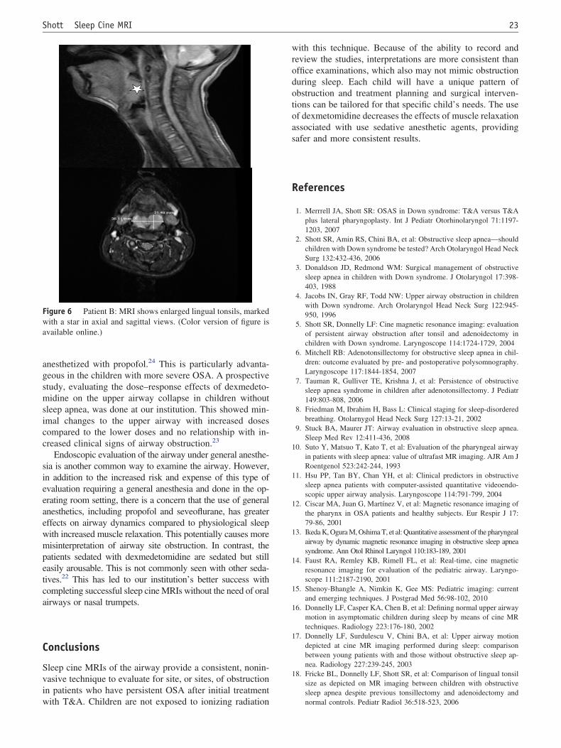
By contrast, patient B, a young man with DS, hadsevere OSA with significant hypoxemia and hypercarbiaon his sleep study, done after his T&A. Although hisparents reported improvement in his symptoms, he wasstill falling asleep at school and on the bus home. At-tempts using CPAP had failed. On the sagittal cine MRIviews, his sleep cine MRI showed evidence of relativemacroglossia, glossoptosis, and significant enlargementof his lingual tonsils (Figure 6). Axial cine views showedcircumferential collapse at the level of the hypopharynx.Due to these findings, a surgical approach was recom-mended, starting with a lingual tonsillectomy. The planwas then to perform a postoperative sleep study 3 monthsafter this surgery and consider further surgery on the baseof tongue, addressing the macroglossia and glossoptosis,if needed. As his postoperative sleep study after thelingual tonsillectomy was significantly improved, thesecond surgery was not necessary.
Figure 3 Axial views of the airway at the level of the hypo-pharynx with arrows showing circumferential airway collapse that
involves the lateral, anterior, and posterior walls.19
l
22 Operative Techniques in Otolaryngology, Vol 23, No 1, March 2012
Anesthesia
Although the ideal would be to do the sleep cine MRI undernatural sleep, this is not practical. In addition to difficultiesin achieving spontaneous sleep during the daytime hours,there is the problem with the very noisy environment of thegradient-echo sequences used to create the cine MRI im-ages. Even those who fall asleep would be awakened by thisnoise.
Due to the at-risk population requiring this study, we feelit is important to have a staff anesthesiologist present for thestudy to provide safe and effective anesthesia. Unfortu-nately, anesthesia agents cause increased airway collapsibil-ity and obstruction through a combination of both airwayrelaxant effects and respiratory depression. The pharyngealmuscle tone, which is decreased in natural sleep, is evenmore compromised in the face of anesthetic effects. Theideal anesthetic agent therefore should provide a state assimilar as possible to physiological sleep with spontaneousbreathing, without the need for artificial airway support viaoral or nasal airway adjuncts.
Sedatives and anesthetic drugs commonly used for ra-diographic studies include propofol, pentobarbital, benzo-diazepines, ketamine, and dexmedetomidine. Mahmoud et
Table 1 Causes of persistent obstructive sleep apneadespite previous T&A in children with Down syndrome asdepicted on static and dynamic cine magnetic resonanceimaging (MRI)21
Glossoptosis 63%Recurrent adenoids 63%Macroglossia 74%Enlarged lingual tonsils 30%Hypopharyngeal collapse 22%
Twenty-seven patients, mean age 9.9 years.
Figure 4 The various anatomic locations of upper airway obsine.)
al in their commentary on the anesthetic challenges of doingsleep MRIs reviewed the pros and cons of these variousdrugs. Propofol and barbituates can worsen upper airwayobstruction and cause respiratory depression and apnea.Benzodiazepines relax the pharyngeal muscles, causing ob-struction. Ketamine has been shown to have no effect onpharyngeal musculature in adults and has been used suc-cessfully in a few reported cases when combined withdexmedetomidine.22
At our institution, the anesthesiologists use dexmedeto-midine, an alpha-2 adrenergic agonist, as their drug ofchoice for the sleep MRIs.22 This drug works similarly toclonidine but with a higher sensitivity to the alpha-2 recep-tors. The sedative properties parallel natural non-rapid eyemovement sleep with minimal respiratory depression.23
There is also a more reliable response to this drug comparedto other anesthetic agents with fewer episodes of oxygendesaturation and airway obstruction compared to those
n in pediatric OSA.20 (Color version of figure is available on-
Figure 5 Patient A: Sagittal cine MRI with adenoid regrowth
tructio
(A � adenoids).
gsmsicc
ca
23Shott Sleep Cine MRI
anesthetized with propofol.24 This is particularly advanta-eous in the children with more severe OSA. A prospectivetudy, evaluating the dose–response effects of dexmedeto-idine on the upper airway collapse in children without
leep apnea, was done at our institution. This showed min-mal changes to the upper airway with increased dosesompared to the lower doses and no relationship with in-reased clinical signs of airway obstruction.23
Endoscopic evaluation of the airway under general anesthe-sia is another common way to examine the airway. However,in addition to the increased risk and expense of this type ofevaluation requiring a general anesthesia and done in the op-erating room setting, there is a concern that the use of generalanesthetics, including propofol and seveoflurane, has greatereffects on airway dynamics compared to physiological sleepwith increased muscle relaxation. This potentially causes moremisinterpretation of airway site obstruction. In contrast, thepatients sedated with dexmedetomidine are sedated but stilleasily arousable. This is not commonly seen with other seda-tives.22 This has led to our institution’s better success withompleting successful sleep cine MRIs without the need of oralirways or nasal trumpets.
Conclusions
Sleep cine MRIs of the airway provide a consistent, nonin-vasive technique to evaluate for site, or sites, of obstructionin patients who have persistent OSA after initial treatment
Figure 6 Patient B: MRI shows enlarged lingual tonsils, markedwith a star in axial and sagittal views. (Color version of figure isavailable online.)
with T&A. Children are not exposed to ionizing radiation
with this technique. Because of the ability to record andreview the studies, interpretations are more consistent thanoffice examinations, which also may not mimic obstructionduring sleep. Each child will have a unique pattern ofobstruction and treatment planning and surgical interven-tions can be tailored for that specific child’s needs. The useof dexmetomidine decreases the effects of muscle relaxationassociated with use sedative anesthetic agents, providingsafer and more consistent results.
References
1. Merrrell JA, Shott SR: OSAS in Down syndrome: T&A versus T&Aplus lateral pharyngoplasty. Int J Pediatr Otorhinolaryngol 71:1197-1203, 2007
2. Shott SR, Amin RS, Chini BA, et al: Obstructive sleep apnea—shouldchildren with Down syndrome be tested? Arch Otolaryngol Head NeckSurg 132:432-436, 2006
3. Donaldson JD, Redmond WM: Surgical management of obstructivesleep apnea in children with Down syndrome. J Otolaryngol 17:398-403, 1988
4. Jacobs IN, Gray RF, Todd NW: Upper airway obstruction in childrenwith Down syndrome. Arch Orolaryngol Head Neck Surg 122:945-950, 1996
5. Shott SR, Donnelly LF: Cine magnetic resonance imaging: evaluationof persistent airway obstruction after tonsil and adenoidectomy inchildren with Down syndrome. Laryngoscope 114:1724-1729, 2004
6. Mitchell RB: Adenotonsillectomy for obstructive sleep apnea in chil-dren: outcome evaluated by pre- and postoperative polysomnography.Laryngoscope 117:1844-1854, 2007
7. Tauman R, Gulliver TE, Krishna J, et al: Persistence of obstructivesleep apnea syndrome in children after adenotonsillectomy. J Pediatr149:803-808, 2006
8. Friedman M, Ibrahim H, Bass L: Clinical staging for sleep-disorderedbreathing. Otolarnygol Head Neck Surg 127:13-21, 2002
9. Stuck BA, Maurer JT: Airway evaluation in obstructive sleep apnea.Sleep Med Rev 12:411-436, 2008
10. Suto Y, Matsuo T, Kato T, et al: Evaluation of the pharyngeal airwayin patients with sleep apnea: value of ultrafast MR imaging. AJR Am JRoentgenol 523:242-244, 1993
11. Hsu PP, Tan BY, Chan YH, et al: Clinical predictors in obstructivesleep apnea patients with computer-assisted quantitative videoendo-scopic upper airway analysis. Laryngoscope 114:791-799, 2004
12. Ciscar MA, Juan G, Martínez V, et al: Magnetic resonance imaging ofthe pharynx in OSA patients and healthy subjects. Eur Respir J 17:79-86, 2001
13. Ikeda K, Ogura M, Oshima T, et al: Quantitative assessment of the pharyngealairway by dynamic magnetic resonance imaging in obstructive sleep apneasyndrome. Ann Otol Rhinol Laryngol 110:183-189, 2001
14. Faust RA, Remley KB, Rimell FL, et al: Real-time, cine magneticresonance imaging for evaluation of the pediatric airway. Laryngo-scope 111:2187-2190, 2001
15. Shenoy-Bhangle A, Nimkin K, Gee MS: Pediatric imaging: currentand emerging techniques. J Postgrad Med 56:98-102, 2010
16. Donnelly LF, Casper KA, Chen B, et al: Defining normal upper airwaymotion in asymptomatic children during sleep by means of cine MRtechniques. Radiology 223:176-180, 2002
17. Donnelly LF, Surdulescu V, Chini BA, et al: Upper airway motiondepicted at cine MR imaging performed during sleep: comparisonbetween young patients with and those without obstructive sleep ap-nea. Radiology 227:239-245, 2003
18. Fricke BL, Donnelly LF, Shott SR, et al: Comparison of lingual tonsilsize as depicted on MR imaging between children with obstructivesleep apnea despite previous tonsillectomy and adenoidectomy and
normal controls. Pediatr Radiol 36:518-523, 2006
24 Operative Techniques in Otolaryngology, Vol 23, No 1, March 2012
19. Donnelly LF: Obstructive sleep apnea in pediatric patients: evaluationwith cine MR sleep studies. Radiology 236:768-778, 2005
20. Shott SR, Richter GT: Oral and oropharyngeal obstruction, chapter8, in Haver KE, Brigger MT, Hardy SC, et al. (eds): PediatricAerodigestive Disorders. San Diego, CA, Plural Publishing, Inc,2009
21. Donnelly LR, Shott SR, LaRose CR, et al: Causes of persistent ob-structive sleep apnea in children with trisomy 21 as depicted on MR
cine studies. AJR Am J Roentgenol 183:173-181, 200422. Mahmoud M, Gunter J, Sadhasivam S: Ciné MRI airway studies inchildren with sleep apnea: optimal images and anesthetic challenges.Pediatr Radiol 39:1034-1037, 2009
23. Mahmoud M, Radhakrishman R, Gunter J, et al: Effect of increasingdepth of dexmedetomidine anesthesia on upper airway morphology inchildren. Paediatr Anaesth 20:506-515, 2010
24. Mahmoud M, Gunter J, Donnelly LF, et al: A Comparison of dexme-detomidine with propofol for magnetic resonance imaging sleep stud-
ies in children. Anesth Analg 109:745-753, 2009