smac mimetics in combination with trail selectively target...
TRANSCRIPT
1
Smac mimetics in combination with TRAIL
selectively target cancer stem cells in
nasopharyngeal carcinoma
Man-si Wu1, Guang-feng Wang2, Zhi-qiang Zhao3, Yi Liang1,
Heng-bang Wang2, Miao-yi Wu2, Ping Min2, Li-zhen Chen1,
Qi-sheng Feng1, Jin-xin Bei1, Yi-xin Zeng1 and Dajun Yang1,2
Authors’ Affiliations: 1 State Key Laboratory of Oncology in Southern China, Sun Yat-Sen University Cancer Center, Guangzhou, China; 2Ascentage Pharma Group Corp. Limited, Jiangsu, China; 3The First Affiliated Hospital, Sun Yat-Sen University, Guangzhou, China Running Title: Targeting CSCs in Nasopharyngeal Carcinoma Keywords: Cancer stem cells, Smac mimetic, TRAIL, Nasopharyngeal Carcinoma Abbreviation List: CSCs, Cancer Stem Cells; IAPs, Inhibitors of Apoptosis Protein Family; TRAIL, TNF-Related Apoptosis Inducing Ligand; ILP2, IAP-like Protein; ML-IAP, Melanoma IAP; XIAP, X-link IAP; cIAP1, Cellular IAP1; cIAP2, Cellular IAP2; NAIP, Neuronal Apoptotic Inhibitor Protein; BRUCE, BIR-containing Ubiquitin Conjugating Enzyme; Smac/DIABLO, Second mitochondria-derived activator of caspases /Direct IAP Binding Protein with Low Isoelectric Point; FTC, fumitremorgin C; RTV, relative tumor volume. Corresponding Authors: Yi-xin Zeng and Dajun Yang are both the corresponding authors. Yi-Xin Zeng, State Key Laboratory of Oncology in Southern China, Sun Yat-Sen University Cancer Center, 651 Dongfeng Road East, Guangzhou 510060, China. Phone: 86-20-8734-3333; Fax: 86-20-8734-3171; E-mail: [email protected] and Dajun Yang, State Key Laboratory of Oncology in Southern China, Sun Yat-Sen University Cancer Center, 651 Dongfeng Road East, Guangzhou 510060, China. Phone: 86-20-8734-3149; Fax: 86-20-8734-3171; E-mail: [email protected] No potential conflicts of interest were disclosed.
on July 5, 2019. © 2013 American Association for Cancer Research. mct.aacrjournals.org Downloaded from
Author manuscripts have been peer reviewed and accepted for publication but have not yet been edited. Author Manuscript Published OnlineFirst on May 22, 2013; DOI: 10.1158/1535-7163.MCT-13-0017
2
Abstract
Nasopharyngeal carcinoma is a common malignancy in Southern China. After radiotherapy
and chemotherapy, a considerable proportion of nasopharyngeal carcinoma patients suffered
tumor relapse and metastasis. Cancer stem cells (CSCs) have been demonstrated with the
resistance against therapies and thus considered as the initiator of recurrence and metastasis in
tumors, where the anti-apoptotic property of CSCs play important role. Smac/DIABLO is an
inverse regulator for the inhibitors of apoptosis protein family (IAPs), which have been involved in
the apoptosis. Here, the effects of Smac mimetics on the CSCs of nasopharyngeal carcinoma were
studied both in vitro and in vivo, using two clones of nasopharyngeal carcinoma cell line CNE2 as
models. We found that one of the clones, S18, had CSC-like properties and IAPs were
overexpressed. The combination of Smac mimetics and TNF-related apoptosis inducing ligand
(TRAIL) can reduce the percentage of SP cells and inhibit the colony- and sphere-forming abilities
of S18 cells, indicating their ability to attenuate the CSCs. Moreover, in a nasopharyngeal
carcinoma xenograft model, the administration of Smac mimetics in combination with TRAIL also
led to the elimination of nasopharyngeal carcinoma stem cells. Furthermore, the Smac mimetics
in combination with TRAIL induced the degradation of cIAP1 and XIAP and thus induced
apoptosis in vitro and in vivo. Taken together, our data show that Smac mimetics exerted an
antitumor effect on nasopharyngeal carcinoma cancer stem cells, and this combination
treatment should be considered as a promising strategy for the treatment of nasopharyngeal
carcinoma.
on July 5, 2019. © 2013 American Association for Cancer Research. mct.aacrjournals.org Downloaded from
Author manuscripts have been peer reviewed and accepted for publication but have not yet been edited. Author Manuscript Published OnlineFirst on May 22, 2013; DOI: 10.1158/1535-7163.MCT-13-0017
3
Introduction
Nasopharyngeal carcinoma is an endemic malignancy that occurs predominantly in
populations from southern China, Southeast Asia, North Africa, and the Arctic Circle( Eskimos and
other Arctic natives)(1). The application of chemotherapeutic drugs becomes more important in
the treatment for nasopharyngeal carcinoma in recent years, and cisplatin, 5-fluorouracil and
taxel are the most commonly used drugs (2, 3). However, some patients still suffered from failure
of treatment including relapse and metastasis, which thought to be originated from cancer stem
cells (3-9).
Cancer stem cells (CSCs) have the properties of self-renewal, differentiation and resistance
to chemotherapy or radiotherapy (7, 10). Although the CSCs represent a small proportion of the
tumor cells, they are key players in tumor initiation, recurrence and metastasis (8-12). Therefore,
CSCs has been considered as the important therapeutic target in anticancer treatments (6, 9,
13-16). Many compounds have been shown to selectively target CSCs in cancers, such as
salinomycin (6), T�RI inhibitors (9), lupeol (14), sulforaphane (15), thioridazine (16), and others.
As in other cancers, nasopharyngeal carcinoma contains a small fraction of tumor cells with
properties of CSCs. Studies have demonstrated that the Side population (SP) cells, identified by
having ability to pump out a fluorescent dye (Hoechst 33342), have certain characteristics of CSCs
similar with those in liver cancer and gastrointestinal system cancer, suggesting that SP
phenotype can be a marker of CSC for nasopharyngeal carcinoma (13, 17, 18). However, no
effective compound has been discovered to target nasopharyngeal carcinoma CSCs.
CSCs are thought as the key players in the resistance to chemo- or radio-therapy (11, 12),
while their ability to escape from the apoptosis pathway may render them the resistance
on July 5, 2019. © 2013 American Association for Cancer Research. mct.aacrjournals.org Downloaded from
Author manuscripts have been peer reviewed and accepted for publication but have not yet been edited. Author Manuscript Published OnlineFirst on May 22, 2013; DOI: 10.1158/1535-7163.MCT-13-0017
4
property to the therapies (19, 20). It has been shown that the inhibitors of apoptosis protein (IAP)
family members are important anti-apoptotic proteins to regulate the apoptosis processes
(19-21). Among the eight proteins in the family, namely survivin, IAP-like protein (ILP2),
melanoma IAP (ML-IAP), X-link IAP (XIAP), cellular IAP1 (cIAP1), cellular IAP2 (cIAP2), neuronal
apoptotic inhibitor protein (NAIP) and BIR-containing ubiquitin conjugating enzyme (BRUCE),
XIAP is the most well characterized one, and cIAP1 and cIAP2 are the two closest XIAP paralogs
(22, 23). Moreover, XIAP is the only IAP protein that binds directly to caspases, and inhibits their
activities, which result in promoting resistance to apoptosis in cancer cells (20, 24, 25). In
contrast, the cIAPs can bind to caspases without inhibiting their activities (23).
On the other hand, second mitochondria-derived activator of caspases /direct IAP binding
protein with low isoelectric point (Smac/DIABLO) is a negative regulator of IAP proteins and
released in response to apoptotic stimuli (26). By binding to XIAP and cIAPs, Smac can release the
inhibition of caspase or lead to the degradation of cIAPs, which turn the cells into apoptosis
process (26, 27). Therefore, regarding the potency of Smac to revert the anti-apoptosis of cancer
cells, several Smac mimetics have been designed and synthesized as antitumor drugs in recent
years (28-31). These compounds can induce cIAP1/2 degradation and prevent XIAP from binding
to caspases, which induce the apoptosis of tumor cells with little effect on normal cells (27, 32).
In addition, synergy effect has been reported for Smac mimetics and TNF-related
apoptosis-inducing ligand (TRAIL), which is a TNF family ligand with ability to induce apoptosis in
cancer cells (33-36).
Attempting to provide more effective treatment for nasopharyngeal carcinoma patients, we
evaluated two Smac mimetics AT-406 and SM-164, in combination with TRAIL (37, 38), for their
on July 5, 2019. © 2013 American Association for Cancer Research. mct.aacrjournals.org Downloaded from
Author manuscripts have been peer reviewed and accepted for publication but have not yet been edited. Author Manuscript Published OnlineFirst on May 22, 2013; DOI: 10.1158/1535-7163.MCT-13-0017
5
abilities to selectively target CSCs in nasopharyngeal carcinoma. Our study showed that IAP
proteins were overexpressed in nasopharyngeal carcinoma cancer stem cells and Smac mimetics
can selectively reduce CSCs both in vitro and in vivo. The results suggest that the Smac mimetics
AT-406 and SM-164 may be promising drugs for the effective treatment of nasopharyngeal
carcinoma.
Materials and Methods
Cell Culture
S18 and S26 cells, clones of the human nasopharyngeal carcinoma cell line CNE2, were
maintained in Dulbecco's modified Eagle's medium (DMEM, Invitrogen) supplemented with 10%
heat-inactivated fetal bovine serum (Invitrogen), 100 units/ml penicillin G and 100 �g/ml
streptomycin at 37°C in 5% CO2. These two clones of CNE2 were kind gifts from Dr. Chaonan Qian
(Sun Yat-sen University Cancer Center, China). All cell lines were passaged less than six month.
SP Detection
S18 and S26 cells were treated with the test compounds (negative control, 5 ng /ml TRAIL, 5
�M AT-406, 0.1 �M SM-164, 5 �M AT-406 + 5 ng/ml TRAIL, or 0.1 �M SM-164 + 0.1 ng/ml TRAIL)
for 48 hours, harvested and then resuspended in an ice-cold DMEM (supplemented with 2% fetal
bovine serum) at a density of 1×10⁶ cells/ml. Then, the cells were incubated at 37°C in 5% CO2 for
10 minutes. The DNA binding dye Hoechst 33342 (Sigma-Aldrich) was then added to the cells at a
final concentration of 5 �g/ml (as a negative control, cells were incubated with 10 �M
fumitremorgin C (FTC, an inhibitor of ABCG2 which could block the pumping out of Hoechst
on July 5, 2019. © 2013 American Association for Cancer Research. mct.aacrjournals.org Downloaded from
Author manuscripts have been peer reviewed and accepted for publication but have not yet been edited. Author Manuscript Published OnlineFirst on May 22, 2013; DOI: 10.1158/1535-7163.MCT-13-0017
6
33342 in CSCs, Sigma-Aldrich) for 5 minutes prior to the addition of the Hoeches dye), and the
cells were incubated at 37°C in 5% CO2 in the dark for 90 minutes and mixed every 15 minutes.
Then, the cells were washed twice with PBS, resuspended in PBS and kept at 4°C in the dark
before flow cytometric analysis (Experience Xtremes MoFlo XDP cell Sorter, Beckman Coulter).
RNA extraction, reverse transcription and quantitative real-time PCR
Total RNA of S18 and S26 cells were extracted using TRIzol reagent (Invitrogen) according to
the manufacturer’s instructions. cDNA was synthesized using Thermo Scientific Maxima First
cDNA Synthesis Kit (Thermo). Real-time PCR amplification was performed using Platinum SYBR
Green qPCR SuperMix-UDG with ROX (Invitrogen) on a Hard-Shell PCR Plates (Bio-Rad). Relative
quantification of each target gene was normalized by using an endogenous control (GAPDH).
Cell Viability Assay
Cell viability was measured using MTT assay. S18 and S26 cells were counted, plated in
triplicate at 2500 cells per well (200 �l) in 96-well plates, and allowed to grow overnight. For
individual groups, cisplatin, 5-fluorouracil, taxel, or TRAIL, AT-406, SM-164 was added to the wells
in a concentration gradient. For combination groups, negative control, 5 �M AT-406, 0.1 �M
SM-164 were mixed with a concentration gradient of TRAIL, and then added to the wells. Cell
viability was measured 48 hours later by adding MTT solution. The observation value was
detected at 490nm.
Colony Formation Assay
on July 5, 2019. © 2013 American Association for Cancer Research. mct.aacrjournals.org Downloaded from
Author manuscripts have been peer reviewed and accepted for publication but have not yet been edited. Author Manuscript Published OnlineFirst on May 22, 2013; DOI: 10.1158/1535-7163.MCT-13-0017
7
S18, S26 or treated S18 cells (treated with negative control, 5 ng/ml TRAIL, 5 �M AT-406, 0.1
�M SM-164, 5 �M AT-406 + 5 ng/ml TRAIL, or 0.1 �M SM-164 + 0.1 ng/ml TRAIL for 48 hours
prior) were counted, plated in triplicate at 100 cells per well in 6-well plates (Corning), and
cultured in DMEM (supplemented with 10% fetal bovine serum) for approximately 10 days. Then,
the cells were washed twice with PBS and fixed in methanol for approximately 10 minutes. After
two additional washes with PBS, the cells were dyed with crystal violet for 30 minutes. Then, the
crystal violet was washed out and the numbers of the colonies were counted.
Sphere Formation Assay
S18, S26 or treated S18 cells (treated with negative control, 5 ng/ml TRAIL, 5 �M AT-406, 0.1
�M SM-164, 5 �M AT-406 + 5 ng/ml TRAIL, or 0.1 �M SM-164 + 0.1 ng/ml TRAIL for 48 hours
prior) were counted, plated in triplicate at 300 cells per well in ultra-low attachment 6-well plates
(Corning), and cultured in DMEM/F12 medium (Invitrogen) with 20 ng/ml recombinant human
fibroblast growth factor-basic (amino acids 1-155) (Invitrogen), 20 ng/ml recombinant human
epidermal growth factor (Hu EGF) (Invitrogen) and B-27 supplement (Invitrogen) for
approximately 2 weeks. The spheres were counted under a light microscope.
Cell Apoptosis Detection
Drug-induced apoptosis was evaluated by Annexin V and Propidium Iodide (PI) staining using
an Annexin V-EGFP apoptosis detection kit (KeyGEN). Treated S18 and S26 cells (treated with
negative control, 5 ng/ml TRAIL, 5 �M AT-406, 0.1 �M SM-164, 5 �M AT-406 + 5 ng/ml TRAIL, or
0.1 �M SM-164 + 0.1 ng/ml TRAIL for 48 hours) were harvested, washed twice with PBS, and
on July 5, 2019. © 2013 American Association for Cancer Research. mct.aacrjournals.org Downloaded from
Author manuscripts have been peer reviewed and accepted for publication but have not yet been edited. Author Manuscript Published OnlineFirst on May 22, 2013; DOI: 10.1158/1535-7163.MCT-13-0017
8
resuspended in Binding Buffer (500 �l, 1-5×10⁵ cells). Annexin V-EGFR and PI (5 �l each) were
then added to the cells, and the mixture was incubated for 15 minutes in the dark at room
temperature. The stained cells were analyzed using a Cytomics FC500 flow cytometer (Beckman
Coulter).
Western Blot Analysis
Compound treated S18 and S26 cells or xenograft tumor tissues were lysed in lysis buffer on
ice, electrophoresed in a 10% Bis-Tris gel in MOPS running buffer and transferred to
polyvinylidene difluoride membranes. The membranes were then blocked in 5% milk for 1 hour
and subsequently incubated with various primary antibodies at 4°C overnight, followed by
incubation with secondary antibodies conjugated to horseradish peroxidase. The
chemiluminescence reagent was then added, and the signals were detected using a sheet of
photographic film.
Antibodies and drugs
The antibodies used for the western bloting were as follows: NAIP (#5782-1, Epitomics),
cIAP1 (#7065, Cell Signaling Technology), cIAP2 (#3130, Cell Signaling Technology), XIAP (#2042,
Cell Signaling Technology), survivin (#2808, Cell Signaling Technology), livin(#5471, Cell Signaling
Technology), PARP (#9542, Cell Signaling Technology), caspase 3 (3G2) (#9668, Cell Signaling
Technology), cleaved caspase 3 (Asp175) (5A1E) (#9664, Cell Signaling Technology), tubulin
(AT819, Beyotime), and actin (#60008-1-lg, Proteintech). TRAIL was provided by the Ascentage
Pharma Group Corp. Limited. in Shanghai, China.
on July 5, 2019. © 2013 American Association for Cancer Research. mct.aacrjournals.org Downloaded from
Author manuscripts have been peer reviewed and accepted for publication but have not yet been edited. Author Manuscript Published OnlineFirst on May 22, 2013; DOI: 10.1158/1535-7163.MCT-13-0017
9
Animal experiments
For the tumorgenesis assay, 4-week-old female athymic nude mice were obtained from the
Animal Experimental Center of the Guangdong Academy of Medical Sciences (Guangzhou, China)
and were given subcutaneous injections of 1×10³, 5×10³, 1×10⁴, 5×10⁴, 1×10⁵, or 5×10⁵ S18 or
S26 cells in their left or right axillary area. The mice were monitored twice per week for 5 weeks.
For the compound sensitivity assay, 4-week-old female athymic nude mice were obtained
from the Sino-British Sippr/BK Lab. Animal LET., Co. (Shanghai, China) and were subcutaneous
injected 1×10⁶ S18 or S26 cells in the right axillary area. When the xenograft tumors developed to
approximately 100 mm3, the mice were randomly divided into 6 groups (for each cell line) with
no differences in tumor size. Then, the mice were treated with AT-406 at 100mg/kg, po, qd,
1-5week x 3weeks, or SM-164 at 3mg/kg, iv, qd, 1-5week x 3weeks alone or in combination with
TRAIL at 10mg/kg, iv, qdx3weeks. Tumor volume and body weight were measured 2 times per
week. The T/C rate was also used to evaluate the tumor response to these compounds. T/C rate
was calculated using the ratio of the relative tumor volume (RTV) of the treated group (T) to the
RTV)of the control group (C). The RTV was calculated using the ratio of the average tumor volume
of the day n to the average tumor volume of the day 0 when the injection of compounds began.
All animal studies were approved by the Sun Yat-sen Univiersity Cancer Center Animal Care
and Ethics Committee.
TUNEL Staining
Tumor tissues from the animal experiments were formalin-fixed and embedded in paraffin.
on July 5, 2019. © 2013 American Association for Cancer Research. mct.aacrjournals.org Downloaded from
Author manuscripts have been peer reviewed and accepted for publication but have not yet been edited. Author Manuscript Published OnlineFirst on May 22, 2013; DOI: 10.1158/1535-7163.MCT-13-0017
10
All sample sections were dyed with hematoxylin and eosin and microscopically examined to
confirm the nasopharyngeal carcinoma cell origin. The samples were dewaxed, rehydrated using
xylene and ethanol, incubated with a proteinase K working solution with microwave irradiation in
0.1 M citrate buffer, and then stained with the TUNEL reaction mixture (Roche Applied Science)
for 1 hour. Then, the samples were incubated with Converter-POD (Roche Applied Science) for 30
minutes at 37°C for 1 hour. All samples were visualized using diaminobenzidine (DAB) (DAKO),
and the nuclei were counterstained with hematoxylin.
Statistical methods
Sigmaplot and SPSS 13.0 were used for statistical analysis. All in vitro experiments were
repeated 3 times. Data were presented as mean values and standard deviations, and the
differences between groups were evaluated using Student’s t-test. P 0.05 was considered to be
statistically significant.
Results
Differences between the CSC properties of S18 and S26 cells
The CSC properties of two clones of nasopharyngeal carcinoma CNE2 cells, S18 and S26,
were evaluated. The S18 cells have been previously reported to have greater migration and
invasion abilities than S26 cells (39). It was discovered that the SP cell population in S18 cells was
approximately 27-fold higher than that in the S26 cells (Fig. 1A and C). Furthermore, the S18 cells
were found to be more resistant than S26 cells to three chemotherapeutic drugs (cisplatin,
5-fluorouracil and taxel) commonly used to treat nasopharyngeal carcinoma (Table 1). The mRNA
on July 5, 2019. © 2013 American Association for Cancer Research. mct.aacrjournals.org Downloaded from
Author manuscripts have been peer reviewed and accepted for publication but have not yet been edited. Author Manuscript Published OnlineFirst on May 22, 2013; DOI: 10.1158/1535-7163.MCT-13-0017
11
expression levels of cancer stem cells markers in nasopharyngeal carcinoma, ABCG2 and CD44,
were also higher in S18 cells (Fig. 1B). These observations showed that S18 cells may have cancer
stem cell properties.
Then a colony formation assay was performed, and S18 cells were better able to form
colonies than S26 cells (Fig. 1D and Supplementary Fig. S1A). When the sphere formation ability
of these two cells was evaluated, S18 cells were found to exhibit stronger sphere formation ability,
in contrast to the S26 cells (Fig. 1E and Supplementary Fig. S1B). Tumor seeding ability of these
two cell lines was also examined and tumors can be generated with 5×10³ S18 cells, whereas
1×10⁵ S26 cells were required for tumor generation (Table 2). All of these findings indicate that
S18 cells acted as CSCs.
Having observed that S18 cells exhibit strong resistance to chemotherapeutic drugs, we next
wanted to determine whether the apoptosis pathway was inhibited in S18 cells.
Apoptosis-related proteins play a very important role in the inhibition of the apoptosis pathway
(21). The IAP family protein levels were measured in S18 and S26 cells and S18 cells expressed
higher levels of IAPs than S26 cells (Fig. 1F). These data indicated that IAPs play important roles in
nasopharyngeal carcinoma CSCs.
We also wanted to know whether TRAIL and the Smac mimetic AT-406(40) and SM-164(38)
could affect S18 and S26 cells. Using the MTT assay, it was found that TRAIL had little effect on
S18 cells, in contrast to the effect on S26 cells (Fig. 1G). Even when the concentration of TRAIL
was increased to 1000 ng/ml, the cytotoxic effect was still weak in the S18 cells (Fig. 1G). The
Smac mimetic AT-406 and SM-164 had moderate cytotoxic effects on S18 and S26 cells at high
concentration, and these effects were more evident in S26 cells (Table 1).
on July 5, 2019. © 2013 American Association for Cancer Research. mct.aacrjournals.org Downloaded from
Author manuscripts have been peer reviewed and accepted for publication but have not yet been edited. Author Manuscript Published OnlineFirst on May 22, 2013; DOI: 10.1158/1535-7163.MCT-13-0017
12
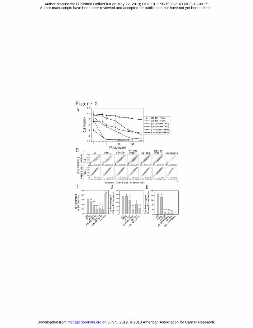
The Smac mimetics in combination with TRAIL selectively inhibit nasopharyngeal carcinoma
CSC growth and attenuate nasopharyngeal carcinoma CSCs
Based on the results above, we investigated whether two Smac mimetics, AT-406 and
SM-164, could selectively inhibit S18 cell growth in vitro in combination with TRAIL (Fig. 2A). MTT
assays to determine the cell growth inhibition properties of the two compounds showed that
both compounds, especially SM-164, could selectively inhibit S18 cell growth when used in
combination with TRAIL (Fig. 2A).
Whether AT-406 or SM-164 in combination with TRAIL could attenuate nasopharyngeal
carcinoma cancer stem cells was also tested. SP detection showed that treatment with AT-406 or
SM-164 decreased the proportions of SP cells in S18 cells, especially when used in combination
with TRAIL. For example, the percentages of SP cells decreased from 60% in the untreated group
to approximately 25% in the AT-406 or SM-164 + TRAIL treated group, whereas the percentage of
SP cells in a cisplatin-treated S18 cells population was up to 90% (Fig. 2B and C). The colony
formation assay and sphere formation assay also showed that AT-406 and SM-164 could inhibit
the colony formation and sphere formation abilities of S18 cells, especially when combined with
TRAIL (Fig. 2D, E and Supplementary Fig. S2A and B).
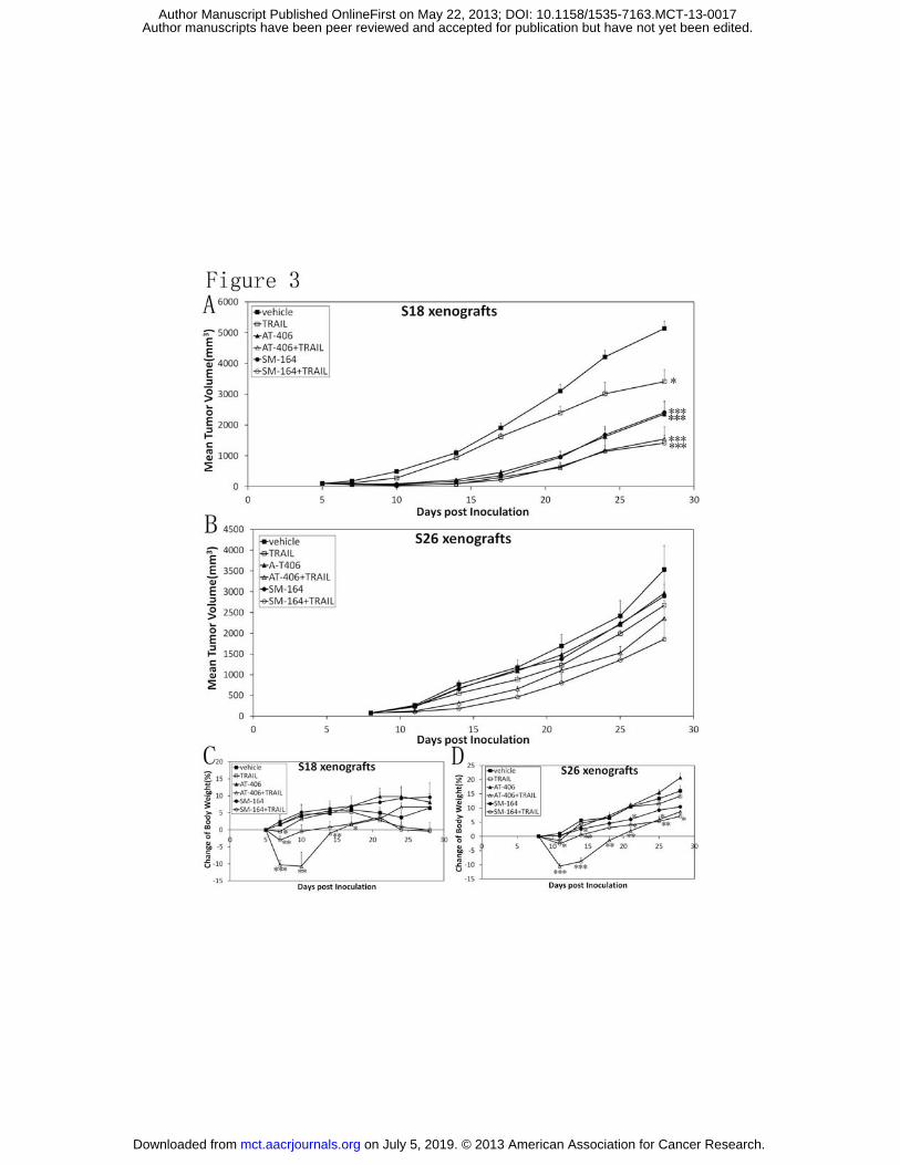
AT-406 and SM-164 sensitize nasopharyngeal carcinoma xenografts to TRAIL therapy
The above results indicated that treatment with AT-406 or SM-164 in combination with
TRAIL selectively targeted nasopharyngeal carcinoma CSCs in vitro. We then generated tumors in
mice and tested whether AT-406 and SM-164 could affect tumor growth when combined with
on July 5, 2019. © 2013 American Association for Cancer Research. mct.aacrjournals.org Downloaded from
Author manuscripts have been peer reviewed and accepted for publication but have not yet been edited. Author Manuscript Published OnlineFirst on May 22, 2013; DOI: 10.1158/1535-7163.MCT-13-0017
13
TRAIL in vivo.
Mice were injected with S18 or S26 cells, and when the resulting palpable tumors reached a
size of 100 mm3, the mice were treated with normal saline or with TRAIL (10 mg/kg, iv), AT-406
(100 mg/kg, po), and SM-164 (3 mg/kg, iv) or in combination 5 days per week for 3 weeks. Tumor
volume and body weight were measured 2 times per week. We found that the tumor volumes of
mice treated with AT-406 or SM-164 in combination with TRAIL were much smaller than those of
mice treated with normal saline (Fig. 3A and B). Interestingly, the two Smac mimetic compounds
seemed to be more effective against S18 cell xenografts than against S26 cell xenografts (Fig. 3A
and B). This result indicated that Smac mimetics also have anti-cancer stem cell properties in vivo.
There has not significant toxicity in mice treated with SM-164 in combination with TRAIL (Fig. 3C
and D). AT-406 in combination with TRAIL resulted in a weight loss in the first 10 days (Fig. 3C and
D). However, the changes were recovered after stopping the treatment (Fig. 3C and D). And the
T/C ratio were 27.7 and 26.6 when treated with Smac mimetics in combination with TRAIL in S18
xenografts, while T/C ratio were 71.2 and 47.2 in S26 xenografts (Table 3).
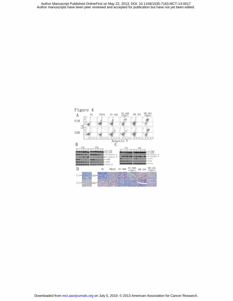
Smac mimetics in combination with TRAIL can induce IAPs degradation and lead to apoptosis in
tumor cells
As the smac mimetics are important inhibitors of IAP family. We also wanted to know
whether these compounds could induce cell apoptosis in vitro. Annexin V and PI staining showed
that both AT-406 and SM-164 in combination with TRAIL could induce apoptosis in
nasopharyngeal carcinoma cells (Fig. 4A). Western blotting analysis also showed treatment with
AT-406 or SM-164 in combination with TRAIL led to decreased levels of procaspase 3 and
on July 5, 2019. © 2013 American Association for Cancer Research. mct.aacrjournals.org Downloaded from
Author manuscripts have been peer reviewed and accepted for publication but have not yet been edited. Author Manuscript Published OnlineFirst on May 22, 2013; DOI: 10.1158/1535-7163.MCT-13-0017
14
pro-PARP, while the levels of cleaved caspase 3 and PARP were increased in nasopharyngeal
carcinoma cells (Fig. 4B). And the apoptosis induced by these two compounds was stronger in
S18 cells than in S26 cells (Fig. 4A and B). These observations indicated that the Smac mimetic
AT-406 and SM-164, in combination with TRAIL, have a selective cancer stem cell killing effects in
nasopharyngeal carcinoma.
Then the tissues from the compound-treated mice were evaluated to determine whether
these compounds could induce apoptosis in vivo. Increased levels of cleaved caspase 3 and PARP
protein determined by western blotting analysis of the tissue from mice showed that apoptosis
was induced in the compound-treated mice (Fig. 4C). TUNEL staining of the tissues from mice
also confirmed these results (Fig. 4D).
It has been reported that XIAP is the only IAP protein that can bind directly to and inhibit
caspases, indicating that XIAP must play a critical role in the apoptosis pathway (20, 24, 25). The
cIAP1 and cIAP2 proteins are two XIAP paralogs that may also play important role in apoptosis
(22, 23). cIAP1 and XIAP, which are expressed at higher levels in S18 cells, were degraded when
the cells were treated with Smac mimetics in combination with TRAIL, both in vitro and in vivo
(Fig. 4B and C). These observations indicated that the overexpression of the cIAP1, cIAP2 and
XIAP proteins in S18 cells led these cells to become much more sensitive to the inhibition of IAPs.
All of these observations indicate that the Smac mimetics AT-406 and SM-164 in combination
with TRAIL, can selectively target cancer stem cells in nasopharyngeal carcinoma by inhibiting the
cIAP1, cIAP2 and XIAP proteins.
Discussion
on July 5, 2019. © 2013 American Association for Cancer Research. mct.aacrjournals.org Downloaded from
Author manuscripts have been peer reviewed and accepted for publication but have not yet been edited. Author Manuscript Published OnlineFirst on May 22, 2013; DOI: 10.1158/1535-7163.MCT-13-0017
15
Although the 5-year overall survival (OS) of patients with nasopharyngeal carcinoma has
improved, the current treatments for nasopharyngeal carcinoma still have some drawbacks. Most
importantly, these treatment cannot prevent nasopharyngeal carcinoma relapse due to the
development of drug resistance and metastasis, and CSCs may play a critical role in these
processes (3, 12). Common chemotherapeutic drugs or radiotherapy may kill differentiated cells
but fail to eliminate CSCs. A drug that selectively targets CSCs may be an effective cure for
nasopharyngeal carcinoma when combined with common chemotherapeutic drugs or
radiotherapy.
There are many markers that allow the identification of CSCs in different cancers, such as
CD24, CD44, CD133, and the side population (SP) phenotype (6, 9, 13, 17). Here, we used the SP
phenotype to identify CSCs in nasopharyngeal carcinoma and found that S18, a clone of
nasopharyngeal carcinoma CNE2 cells, contains high levels of SP cells. S18 cells also exhibit strong
sphere and colony formation abilities. These cells are resistant to commonly used
chemotherapeutic drugs, and only a small number of cells are required to seed tumors. All of
these features indicate that S18 cells act as CSCs among nasopharyngeal carcinoma CNE2 cells.
Cancer stem cells have one common feature-the loss of apoptosis, and the overexpression of
IAP proteins may be involved in this phenotype (19, 20). IAPs are the last safeguards against
apoptosis pathway, and can inhibit the caspase activity, which result in promoting resistance to
apoptosis in cancer cells (20, 22). In this study, S18 cells were found to express higher levels of
cIAP1, cIAP2 and XIAP proteins than other clones. These results indicate that cIAP1, cIAP2 and
XIAP may play critical roles in nasopharyngeal carcinoma CSCs. Therefore, we treated
nasopharyngeal carcinoma cells with Smac mimetics in combination with TRAIL to test whether
on July 5, 2019. © 2013 American Association for Cancer Research. mct.aacrjournals.org Downloaded from
Author manuscripts have been peer reviewed and accepted for publication but have not yet been edited. Author Manuscript Published OnlineFirst on May 22, 2013; DOI: 10.1158/1535-7163.MCT-13-0017
16
these molecules can target nasopharyngeal carcinoma stem cells (35, 41).
Smac mimetics, designed as negative regulators of IAP proteins, can induce apoptosis in
cancer cells by antagonizing XIAP and cIAP1/2 (26, 27, 37, 38). Some of the smac mimetics are
currently evaluated in phase I clinical trials as potential therapeutic drugs in the treatment of
human tumors (40, 42). Tumor necrosis factor-related apoptosis inducing ligand (TRAIL), a TNF
family ligand that binds to the death receptor, is currently in development as an agent that
targets the apoptosis pathway (33, 34). Moreover, TRAIL- induced apoptosis seems to play an
important role in the malignant transformation(43). TRAIL kills transformed cells but not normal
cells, demonstrating that this compound has low toxicity(43). However, some cancers are
resistant to TRAIL, making it a prime candidate for combination with other safe agents for cancer
treatment(44). In a phase I dose-escalation study, TRAIL seem to be safe and well tolerated
(45).This research demonstrated that, unlike other molecules that target only differentiated
tumor cells, the Smac mimetics AT-406 and SM-164 in combination with TRAIL, may target cancer
stem cells. We observed that AT-406 and SM-164 have the ability to target S18 cells when
combined with TRAIL in vitro. The in vivo data confirm this result and show that these treatments
have no toxicity in mice.
It has been reported that XIAP is the only IAP protein that can directly bind to and inhibit
caspases (20, 24, 25); cIAP1/2 are two paralogs of XIAP(22, 23). In this study, we found that Smac
mimetics, especially when used in combination with TRAIL, can induce the degradation of cIAP1
and XIAP. This treatment also induces the cleavage of caspase 3 and PARP, indicating the
induction of the apoptosis pathway. Because nasopharyngeal carcinoma stem cells have higher
levels of cIAP1, cIAP2 and XIAP than other cells, these cells may be more sensitive to treatment
on July 5, 2019. © 2013 American Association for Cancer Research. mct.aacrjournals.org Downloaded from
Author manuscripts have been peer reviewed and accepted for publication but have not yet been edited. Author Manuscript Published OnlineFirst on May 22, 2013; DOI: 10.1158/1535-7163.MCT-13-0017
17
with Smac mimetics in combination with TRAIL.
Smac mimetic has been reported to sensitize cancer cells to TRAIL- induced apoptosis(35-37,
46), but this study is the first, to our knowledge, to show that smac mimetic in combination with
TRAIL can targeted CSCs in nasopharyngeal carcinoma. This combination treatment can lead to
the degradation of cIAP1 and XIAP, which result in the release of caspase inhibiton and induces
apoptosis in cancer stem cells. The usage of smac mimetic and TRAIL can be a potential
application for other tumor CSCs. Moreover, using Smac mimetics in combination with TRAIL as
an adjuvant therapy with common chemotherapeutic drugs or radiotherapy may provide a
promising new avenue for nasopharyngeal carcinoma therapy.
Grant Support
D.Yang, G.Wang, H.Wang, M.Wu, P.Min received financial support from the National Natural
Sciences Foundation (201281172107), the International cooperation project of the Ministry of
Science and Technology of China (2010DFB34090), "Key New Drug Creation" project of the major
science and technology program (2012ZX09401005), National "863" Grant (2012AA020305),
Jiangsu Provincial Science and Technology Innovation Team Grant, China (BE2010760) and Jiangsu
Provincial Key Laboratory Project Grant (BM2012114). M.Wu, Z.Zhao, Y.Liang, L.Chen, Q.Feng,
J.Bei, Y.X.Zeng received financial support from the National “973” Grant (2011CB504302 and
2012CB967002).
References
1. McDermott AL, Dutt SN, Watkinson JC. The aetiology of nasopharyngeal carcinoma. Clin
Otolaryngol. 2001;26:82-92.
2. Chan AT. Nasopharyngeal carcinoma. Annals of oncology : official journal of the European Society
for Medical Oncology / ESMO. 2010;21 Suppl 7:vii308-12.
on July 5, 2019. © 2013 American Association for Cancer Research. mct.aacrjournals.org Downloaded from
Author manuscripts have been peer reviewed and accepted for publication but have not yet been edited. Author Manuscript Published OnlineFirst on May 22, 2013; DOI: 10.1158/1535-7163.MCT-13-0017
18
3. Rottey S, Madani I, Deron P, Van Belle S. Modern treatment for nasopharyngeal carcinoma:
current status and prospects. Current opinion in oncology. 2011;23:254-8.
4. Chua DT, Ma J, Sham JS, Mai HQ, Choy DT, Hong MH, et al. Long-term survival after
cisplatin-based induction chemotherapy and radiotherapy for nasopharyngeal carcinoma: a pooled
data analysis of two phase III trials. Journal of clinical oncology : official journal of the American
Society of Clinical Oncology. 2005;23:1118-24.
5. Suarez C, Rodrigo JP, Rinaldo A, Langendijk JA, Shaha AR, Ferlito A. Current treatment options for
recurrent nasopharyngeal cancer. Eur Arch Otorhinolaryngol. 2010;267:1811-24.
6. Gupta PB, Onder TT, Jiang G, Tao K, Kuperwasser C, Weinberg RA, et al. Identification of selective
inhibitors of cancer stem cells by high-throughput screening. Cell. 2009;138:645-59.
7. Smith KM, Datti A, Fujitani M, Grinshtein N, Zhang L, Morozova O, et al. Selective targeting of
neuroblastoma tumour-initiating cells by compounds identified in stem cell-based small molecule
screens. EMBO molecular medicine. 2010;2:371-84.
8. Cheng L, Bao S, Rich JN. Potential therapeutic implications of cancer stem cells in glioblastoma.
Biochemical pharmacology. 2010;80:654-65.
9. Anido J, Saez-Borderias A, Gonzalez-Junca A, Rodon L, Folch G, Carmona MA, et al. TGF-beta
Receptor Inhibitors Target the CD44(high)/Id1(high) Glioma-Initiating Cell Population in Human
Glioblastoma. Cancer cell. 2010;18:655-68.
10. Dalerba P, Cho RW, Clarke MF. Cancer Stem Cells: Models and Concepts. Annual Review of
Medicine. 2007;58:267-84.
11. Steg AD, Bevis KS, Katre AA, Ziebarth A, Alvarez RD, Zhang K, et al. Stem cell pathways contribute
to clinical chemoresistance in ovarian cancer. Clinical cancer research : an official journal of the
American Association for Cancer Research. 2011.
12. Hadjipanayis CG, Van Meir EG. Tumor initiating cells in malignant gliomas: biology and
implications for therapy. J Mol Med (Berl). 2009;87:363-74.
13. Wang J, Guo LP, Chen LZ, Zeng YX, Lu SH. Identification of cancer stem cell-like side population
cells in human nasopharyngeal carcinoma cell line. Cancer research. 2007;67:3716-24.
14. Lee TK, Castilho A, Cheung VC, Tang KH, Ma S, Ng IO. Lupeol targets liver tumor-initiating cells
through phosphatase and tensin homolog modulation. Hepatology. 2011;53:160-70.
15. Li Y, Zhang T, Korkaya H, Liu S, Lee HF, Newman B, et al. Sulforaphane, a dietary component of
broccoli/broccoli sprouts, inhibits breast cancer stem cells. Clinical cancer research : an official journal
of the American Association for Cancer Research. 2010;16:2580-90.
16. Sachlos E, Risueno RM, Laronde S, Shapovalova Z, Lee JH, Russell J, et al. Identification of Drugs
Including a Dopamine Receptor Antagonist that Selectively Target Cancer Stem Cells. Cell.
2012;149:1284-97.
17. Forbes SJ, Alison MR. Side population (SP) cells: Taking center stage in regeneration and liver
cancer? Hepatology. 2006;44:23-6.
18. Naotsugu Haraguchi, Tohru Utsunomiya, Hiroshi Inoue, Fumiaki Tanaka, Mimori K, Graham F.
Barnard, et al. Characterization of a side population of cancer cells from human gastrointestinal
system. Stem Cells. 2006;24:506-13.
19. Hanahan. D, Weinberg. RA. The Hallmarks of Cancer. Cell. 2000:57-70.
20. Fulda S, Pervaiz S. Apoptosis signaling in cancer stem cells. The International Journal of
Biochemistry & Cell Biology. 2010;42:31-8.
21. Indran IR, Tufo G, Pervaiz S, Brenner C. Recent advances in apoptosis, mitochondria and drug
on July 5, 2019. © 2013 American Association for Cancer Research. mct.aacrjournals.org Downloaded from
Author manuscripts have been peer reviewed and accepted for publication but have not yet been edited. Author Manuscript Published OnlineFirst on May 22, 2013; DOI: 10.1158/1535-7163.MCT-13-0017
19
resistance in cancer cells. Biochimica et biophysica acta. 2011;1807:735-45.
22. Srinivasula SM, Ashwell JD. IAPs: what's in a name? Molecular cell. 2008;30:123-35.
23. Eckelman BP, Salvesen GS. The human anti-apoptotic proteins cIAP1 and cIAP2 bind but do not
inhibit caspases. The Journal of biological chemistry. 2006;281:3254-60.
24. Kashkar H. X-linked inhibitor of apoptosis: a chemoresistance factor or a hollow promise. Clinical
cancer research : an official journal of the American Association for Cancer Research.
2010;16:4496-502.
25. Eckelman BP, Salvesen GS, Scott FL. Human inhibitor of apoptosis proteins: why XIAP is the black
sheep of the family. EMBO reports. 2006;7:988-94.
26. Salvesen GS, Duckett CS. IAP proteins: blocking the road to death's door. Nature reviews
Molecular cell biology. 2002;3:401-10.
27. Yang QH, Du C. Smac/DIABLO selectively reduces the levels of c-IAP1 and c-IAP2 but not that of
XIAP and livin in HeLa cells. The Journal of biological chemistry. 2004;279:16963-70.
28. Sun H, Nikolovska-Coleska Z, Yang CY, Xu L, Tomita Y, Krajewski K, et al. Structure-based design,
synthesis, and evaluation of conformationally constrained mimetics of the second
mitochondria-derived activator of caspase that target the X-linked inhibitor of apoptosis
protein/caspase-9 interaction site. Journal of medicinal chemistry. 2004;47:4147-50.
29. Sun H, Nikolovska-Coleska Z, Lu J, Qiu S, Yang CY, Gao W, et al. Design, synthesis, and evaluation
of a potent, cell-permeable, conformationally constrained second mitochondria derived activator of
caspase (Smac) mimetic. Journal of medicinal chemistry. 2006;49:7916-20.
30. Zobel K, Wang L, Varfolomeev E, Franklin MC, Elliott LO, Wallweber HJ, et al. Design, synthesis,
and biological activity of a potent Smac mimetic that sensitizes cancer cells to apoptosis by
antagonizing IAPs. ACS chemical biology. 2006;1:525-33.
31. Chauhan D, Neri P, Velankar M, Podar K, Hideshima T, Fulciniti M, et al. Targeting mitochondrial
factor Smac/DIABLO as therapy for multiple myeloma (MM). Blood. 2007;109:1220-7.
32. Vince JE, Wong WW, Khan N, Feltham R, Chau D, Ahmed AU, et al. IAP antagonists target cIAP1 to
induce TNFalpha-dependent apoptosis. Cell. 2007;131:682-93.
33. Bellail. AC, Qi. L, Mulligan. P, Chhabra. V, Hao. C. TRAIL agonists on clinical trials for cancer
therapy- the promises and the challenges. Reviews on Recent Clinical Trails. 2009:34-41.
34. Carlo-Stella C, Lavazza C, Locatelli A, Vigano L, Gianni AM, Gianni L. Targeting TRAIL agonistic
receptors for cancer therapy. Clinical cancer research : an official journal of the American Association
for Cancer Research. 2007;13:2313-7.
35. Li L, Thomas RM, Suzuki H, De Brabander JK, Wang X, Harran PG. A small molecule Smac mimic
potentiates TRAIL- and TNFalpha-mediated cell death. Science. 2004;305:1471-4.
36. Fulda S, Wick W, Weller M, Debatin KM. Smac agonists sensitize for Apo2L/TRAIL- or anticancer
drug-induced apoptosis and induce regression of malignant glioma in vivo. Nature medicine.
2002;8:808-15.
37. Lu J, McEachern D, Sun H, Bai L, Peng Y, Qiu S, et al. Therapeutic potential and molecular
mechanism of a novel, potent, nonpeptide, Smac mimetic SM-164 in combination with TRAIL for
cancer treatment. Molecular cancer therapeutics. 2011;10:902-14.
38. Lu J, Bai L, Sun H, Nikolovska-Coleska Z, McEachern D, Qiu S, et al. SM-164: a novel, bivalent Smac
mimetic that induces apoptosis and tumor regression by concurrent removal of the blockade of
cIAP-1/2 and XIAP. Cancer research. 2008;68:9384-93.
39. Qian CN, Berghuis B, Tsarfaty G, Bruch M, Kort EJ, Ditlev J, et al. Preparing the "soil": the primary
on July 5, 2019. © 2013 American Association for Cancer Research. mct.aacrjournals.org Downloaded from
Author manuscripts have been peer reviewed and accepted for publication but have not yet been edited. Author Manuscript Published OnlineFirst on May 22, 2013; DOI: 10.1158/1535-7163.MCT-13-0017
20
tumor induces vasculature reorganization in the sentinel lymph node before the arrival of metastatic
cancer cells. Cancer research. 2006;66:10365-76.
40. Cai Q, Sun H, Peng Y, Lu J, Nikolovska-Coleska Z, McEachern D, et al. A potent and orally active
antagonist (SM-406/AT-406) of multiple inhibitor of apoptosis proteins (IAPs) in clinical development
for cancer treatment. Journal of medicinal chemistry. 2011;54:2714-26.
41. Petersen SL, Wang L, Yalcin-Chin A, Li L, Peyton M, Minna J, et al. Autocrine TNFalpha signaling
renders human cancer cells susceptible to Smac-mimetic-induced apoptosis. Cancer cell.
2007;12:445-56.
42. Houghton PJ, Kang MH, Reynolds CP, Morton CL, Kolb EA, Gorlick R, et al. Initial testing (stage 1)
of LCL161, a SMAC mimetic, by the Pediatric Preclinical Testing Program. Pediatric blood & cancer.
2012;58:636-9.
43. Wang S, El-Deiry WS. TRAIL and apoptosis induction by TNF-family death receptors. Oncogene.
2003;22:8628-33.
44. Kruyt FA. TRAIL and cancer therapy. Cancer Lett. 2008;263:14-25.
45. Herbst RS, Eckhardt SG, Kurzrock R, Ebbinghaus S, O'Dwyer PJ, Gordon MS, et al. Phase I
dose-escalation study of recombinant human Apo2L/TRAIL, a dual proapoptotic receptor agonist, in
patients with advanced cancer. Journal of clinical oncology : official journal of the American Society of
Clinical Oncology. 2010;28:2839-46.
46. Metwalli AR, Khanbolooki S, Jinesh G, Sundi D, Shah JB, Shrader M, et al. Smac mimetic reverses
resistance to TRAIL and chemotherapy in human urothelial cancer cells. Cancer biology & therapy.
2010;10:885-92.
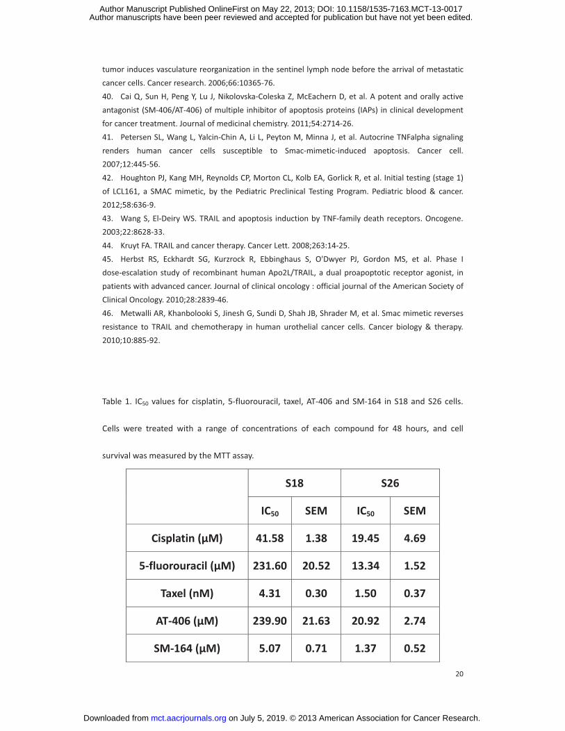
Table 1. IC50 values for cisplatin, 5-fluorouracil, taxel, AT-406 and SM-164 in S18 and S26 cells.
Cells were treated with a range of concentrations of each compound for 48 hours, and cell
survival was measured by the MTT assay.
S18 S26
IC50 SEM IC50 SEM
Cisplatin (�M) 41.58 1.38 19.45 4.69
5-fluorouracil (�M) 231.60 20.52 13.34 1.52
Taxel (nM) 4.31 0.30 1.50 0.37
AT-406 (�M) 239.90 21.63 20.92 2.74
SM-164 (�M) 5.07 0.71 1.37 0.52
on July 5, 2019. © 2013 American Association for Cancer Research. mct.aacrjournals.org Downloaded from
Author manuscripts have been peer reviewed and accepted for publication but have not yet been edited. Author Manuscript Published OnlineFirst on May 22, 2013; DOI: 10.1158/1535-7163.MCT-13-0017
21
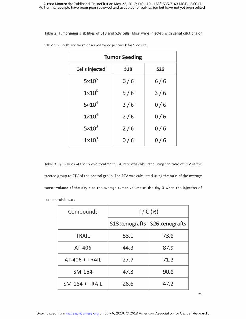
Table 2. Tumorgenesis abilities of S18 and S26 cells. Mice were injected with serial dilutions of
S18 or S26 cells and were observed twice per week for 5 weeks.
Tumor Seeding
Cells injected S18 S26
5×10⁵
1×10⁵
5×10⁴
1×10⁴
5×10³
1×10³
6 / 6
5 / 6
3 / 6
2 / 6
2 / 6
0 / 6
6 / 6
3 / 6
0 / 6
0 / 6
0 / 6
0 / 6
Table 3. T/C values of the in vivo treatment. T/C rate was calculated using the ratio of RTV of the
treated group to RTV of the control group. The RTV was calculated using the ratio of the average
tumor volume of the day n to the average tumor volume of the day 0 when the injection of
compounds began.
Compounds T / C (%)
S18 xenografts S26 xenografts
TRAIL 68.1 73.8
AT-406 44.3 87.9
AT-406 + TRAIL 27.7 71.2
SM-164 47.3 90.8
SM-164 + TRAIL 26.6 47.2
on July 5, 2019. © 2013 American Association for Cancer Research. mct.aacrjournals.org Downloaded from
Author manuscripts have been peer reviewed and accepted for publication but have not yet been edited. Author Manuscript Published OnlineFirst on May 22, 2013; DOI: 10.1158/1535-7163.MCT-13-0017
22
Figure 1. S18 cells act as cancer stem cells among nasopharyngeal carcinoma CNE2 cell lines.
(A) S18 and S26 cells were stained with Hoechst 33342 and FTC, incubated at 37°C in 5% CO2 in
the dark for 90 minutes and mixed every 15 minutes. Then, the cells were resuspended in PBS
and kept at 4°C in the dark before flow cytometric analysis.
(B) mRNA expression levels of cancer stem cells of nasopharyngeal carcinoma, ABCG2 and CD44,
in S18 and S26 cells. *, P<0.05; **, P<0.01; ***, P<0.001.
(C, D, E) SP detection (C), colony formation assay (D) and sphere formation assay (E) using S18
and S26 cells. Cells were counted, plated in 6-well plates and cultured for approximately 10 days.
*, P<0.05; **, P<0.01; ***, P<0.001.
(F) Western blotting for NAIP, cIAP1, cIAP2, XIAP, survivin, and livin in S18 and S26 cells.
(G) The cytotoxic effects of TRAIL on S18 and S26 cells. Cells were treated with TRAIL at a range of
concentrations for 48 hours, and cell survival was measured by the MTT assay.
Figure 2. Smac mimetics in combination with TRAIL attenuate cancer stem cells in vitro.
(A) The cytotoxic effect of Smac mimetics in combination with TRAIL on S18 and S26 cells.
Negative control, 5 �M AT-406, 0.1 �M SM-164 were mixed with a concentration gradient of
TRAIL and then added to the cells for 48 hours. Cell survival was measured by the MTT assay.
(B) SP detection of compound treated S18 cells using Hoechst 33342 with or without FTC. Cells
were treated with compounds for 48 hours and then evaluated by flow cytometry.
(C, D, E) SP detection (C), colony formation assays (D) and sphere formation assays(E) of treated
S18 cells. In the colony formation and sphere formation assays, cells were treated with
on July 5, 2019. © 2013 American Association for Cancer Research. mct.aacrjournals.org Downloaded from
Author manuscripts have been peer reviewed and accepted for publication but have not yet been edited. Author Manuscript Published OnlineFirst on May 22, 2013; DOI: 10.1158/1535-7163.MCT-13-0017
23
compounds for 48 hours, counted, plated in 6-well plates and cultured for approximately 10 days.
*, P<0.05; **, P<0.01; ***, P<0.001.
Figure 3. AT-406 and SM-164 sensitize nasopharyngeal carcinoma xenografts to TRAIL therapy.
(A and B) Tumor volumes of S18 (A) or S26 (B) xenografts treated with different compounds. Mice
were injected with 5×10⁶ S18 or S26 cells. When the xenograft tumors reached approximately
100 mm3 in size, the mice were randomly divided into six groups (for each cell line), with no
difference in tumor size between groups. Then, the mice were treated with normal saline, 100
mg/kg of AT-406, 3 mg/kg of SM-164, or 10 mg/kg of TRAIL alone or in combination 5 times per
week for 3 weeks. Tumor volume was measured 2 times per week. *, P<0.05; **, P<0.01; ***,
P<0.001.
(C and D) Body weights of mice with S18 (C) or S26 (D) xenografts that were treated with normal
saline, TRAIL (10 mg/kg), AT-406 (100 mg/kg), or SM-164 (3 mg/kg) alone or in combination.
Body weight was measured 2 times per week. *, P<0.05; **, P<0.01; ***, P<0.001.
Figure 4. Smac mimetics in combination with TRAIL induce apoptosis in vitro and in vivo.
(A) Cells were treated with negative control, 5 ng/ml TRAIL, 5 �M AT-406, 0.1 �M SM-164, 5 �M
AT-406 + 5 ng/ml TRAIL, or 0.1 �M SM-164 + 0.1 ng/ml TRAIL. After 48 hours, the cells were
double stained with propidium iodide (PI) and Annexin V and analyzed using flow cytometry to
evaluate the apoptosis.
(B and C) Western blotting for apoptosis related proteins, e.g. PARP, caspase3, cIAP1 and XIAP in
treated cells (B) or xenograft tumors (C). Cells were treated with compounds for 48 hours (B).NS,
on July 5, 2019. © 2013 American Association for Cancer Research. mct.aacrjournals.org Downloaded from
Author manuscripts have been peer reviewed and accepted for publication but have not yet been edited. Author Manuscript Published OnlineFirst on May 22, 2013; DOI: 10.1158/1535-7163.MCT-13-0017
24
normal saline; T, TRAIL; A, AT-406; S, SM-164; AT, AT-406 + TRAIL, and ST, SM-164 + TRAIL.
(D) TUNEL staining of compound-treated xenograft tumors. Mice were treated with normal saline,
or with TRAIL (10 mg/kg), AT-406 (100 mg/kg), or SM-164 (3 mg/kg) alone or in combination.
on July 5, 2019. © 2013 American Association for Cancer Research. mct.aacrjournals.org Downloaded from
Author manuscripts have been peer reviewed and accepted for publication but have not yet been edited. Author Manuscript Published OnlineFirst on May 22, 2013; DOI: 10.1158/1535-7163.MCT-13-0017

on July 5, 2019. © 2013 American Association for Cancer Research. mct.aacrjournals.org Downloaded from
Author manuscripts have been peer reviewed and accepted for publication but have not yet been edited. Author Manuscript Published OnlineFirst on May 22, 2013; DOI: 10.1158/1535-7163.MCT-13-0017
on July 5, 2019. © 2013 American Association for Cancer Research. mct.aacrjournals.org Downloaded from
Author manuscripts have been peer reviewed and accepted for publication but have not yet been edited. Author Manuscript Published OnlineFirst on May 22, 2013; DOI: 10.1158/1535-7163.MCT-13-0017
on July 5, 2019. © 2013 American Association for Cancer Research. mct.aacrjournals.org Downloaded from
Author manuscripts have been peer reviewed and accepted for publication but have not yet been edited. Author Manuscript Published OnlineFirst on May 22, 2013; DOI: 10.1158/1535-7163.MCT-13-0017
on July 5, 2019. © 2013 American Association for Cancer Research. mct.aacrjournals.org Downloaded from
Author manuscripts have been peer reviewed and accepted for publication but have not yet been edited. Author Manuscript Published OnlineFirst on May 22, 2013; DOI: 10.1158/1535-7163.MCT-13-0017
Published OnlineFirst May 22, 2013.Mol Cancer Ther Man-si Wu, Guang-feng Wang, Zhi-qiang Zhao, et al. cancer stem cells in nasopharyngeal carcinomaSmac mimetics in combination with TRAIL selectively target
Updated version
10.1158/1535-7163.MCT-13-0017doi:
Access the most recent version of this article at:
Material
Supplementary
http://mct.aacrjournals.org/content/suppl/2013/05/22/1535-7163.MCT-13-0017.DC1
Access the most recent supplemental material at:
Manuscript
Authoredited. Author manuscripts have been peer reviewed and accepted for publication but have not yet been
E-mail alerts related to this article or journal.Sign up to receive free email-alerts
Subscriptions
Reprints and
To order reprints of this article or to subscribe to the journal, contact the AACR Publications
Permissions
Rightslink site. Click on "Request Permissions" which will take you to the Copyright Clearance Center's (CCC)
.http://mct.aacrjournals.org/content/early/2013/05/22/1535-7163.MCT-13-0017To request permission to re-use all or part of this article, use this link
on July 5, 2019. © 2013 American Association for Cancer Research. mct.aacrjournals.org Downloaded from
Author manuscripts have been peer reviewed and accepted for publication but have not yet been edited. Author Manuscript Published OnlineFirst on May 22, 2013; DOI: 10.1158/1535-7163.MCT-13-0017