small molecules, big roles – the chemical manipulation of...
TRANSCRIPT
Journ
alof
Cell
Scie
nce
Small molecules, big roles – the chemical manipulationof stem cell fate and somatic cell reprogramming
Yu Zhang, Wenlin Li, Timothy Laurent and Sheng Ding*Gladstone Institute of Cardiovascular Disease, Department of Pharmaceutical Chemistry, University of California, San Francisco, CA 94158, USA
*Author for correspondence ([email protected])
Journal of Cell Science 125, 5609–5620� 2012. Published by The Company of Biologists Ltddoi: 10.1242/jcs.096032
SummaryDespite the great potential of stem cells for basic research and clinical applications, obstacles – such as their scarce availability and
difficulty in controlling their fate – need to be addressed to fully realize their potential. Recent achievements of cellular reprogramminghave enabled the generation of induced pluripotent stem cells (iPSCs) or other lineage-committed cells from more accessible andabundant somatic cell types by defined genetic factors. However, serious concerns remain about the efficiency and safety of current
genetic approaches to cell reprogramming and traditional culture systems that are used for stem cell maintenance. As a complementaryapproach, small molecules that target specific signaling pathways, epigenetic processes and other cellular processes offer powerful toolsfor manipulating cell fate to a desired outcome. A growing number of small molecules have been identified to maintain the self-renewalpotential of stem cells, to induce lineage differentiation and to facilitate reprogramming by increasing the efficiency of reprogramming
or by replacing genetic reprogramming factors. Furthermore, mechanistic investigations of the effects of these chemicals also providenew biological insights. Here, we examine recent achievements in the maintenance of stem cells, including pluripotent and lineage-specific stem cells, and in the control of cell fate conversions, including iPSC reprogramming, conversion of primed to naıve
pluripotency, and transdifferentiation, with an emphasis on manipulation with small molecules.
Key words: Stem cells, Small molecules, Self-renewal, Reprogramming
IntroductionStem cells, which are characterized by the ability to self-renew and
the potential to differentiate into diverse cell types (Box 1), have
essential roles in embryonic development and tissue homeostasis.
Owing to various promising applications in basic research, disease
modeling, drug screening and regenerative medicine, stem cells
have attracted enormous interest in the last three decades.
Ever since embryonic stem cells (ESCs) were first derived from
mouse embryos (Evans and Kaufman, 1981; Martin, 1981),
advances in stem cell biology and stem cell engineering have
resulted in a number of methods to maintain stem cell self-renewal
and direct lineage-specific differentiation, and to induce the
reprogramming of somatic cells either to a pluripotent state or into
another somatic cell type (Fig. 1). Despite this substantial
progress, a number of diverse challenges remain. For example,
defined culture conditions, which ideally are compatible with
clinical applications, remain highly desired to maintain the long-
term pluripotency of human ESCs (hESCs). Additionally, although
lineage-specific stem cells that reside in many adult tissues have
considerable self-renewal capacity under physiological or
pathological conditions, it is still technically challenging to
expand most types of lineage-specific stem cell ex vivo. The
advances in cell reprogramming have stimulated an increased
interest in generating patient-specific cell types from easily
accessible and healthy cell types. However, reprogramming
remains largely an inefficient and non-specific process, with
efficiencies of transduced cells becoming fully reprogrammed
induced pluripotent stem cells (iPSCs) lower than 0.01%
(Hasegawa et al., 2010; Takahashi et al., 2007; Yu et al., 2007).
Moreover, safety concerns – as well as other practical issues (e.g.
large-scale cell production) – still represent major challenges for
clinical translation. An improved understanding of the complex
regulation of stem cells through cell-intrinsic and -extrinsic signals
is essential to rationally devise appropriate conditions for
controlling stem cell fate, state and function.
Small molecules provide an attractive approach to addressing
these challenges, as they offer a number of compelling advantages.
First, the biological effects of small molecules are typically rapid,
reversible and dose-dependent, allowing precise control over
specific outcomes by fine-tuning their concentrations and
combinations. Second, the structural diversity that can be
provided by synthetic chemistry allows the functional
optimization of small molecules. Third, compared with genetic
interventions, the relative ease of the handling and administration
of small molecules make them more practical for in vitro and in
vivo applications, and for further therapeutic development.
However, small molecules have their own disadvantages. A
specific small molecule may have more than one target. Moreover,
unexpected toxicity or other side effects in vivo may interfere with
the clinical application of small molecules. However, the potential
of small molecules to advance the field of stem cell research
should not be underestimated.
In fact, phenotypic screening of chemical libraries, i.e. using
expression of markers or cellular functions as readouts of
biological effects, not only represents a powerful strategy for
identifying the conditions that maintain, differentiate or
reprogram cells, but also provides a chemical tool to dissect
the underlying molecular mechanisms of these phenomena
ARTICLE SERIES: Stem Cells Commentary 5609
Journ
alof
Cell
Scie
nce
(Boitano et al., 2010; Chen et al., 2006; Desbordes et al., 2008;Zhu et al., 2010). Owing to the explosion of interest in applying
chemical approaches to stem cell biology and regenerativemedicine (Ding and Schultz, 2004; Xu et al., 2008), manycompounds that regulate cell fate and function have been
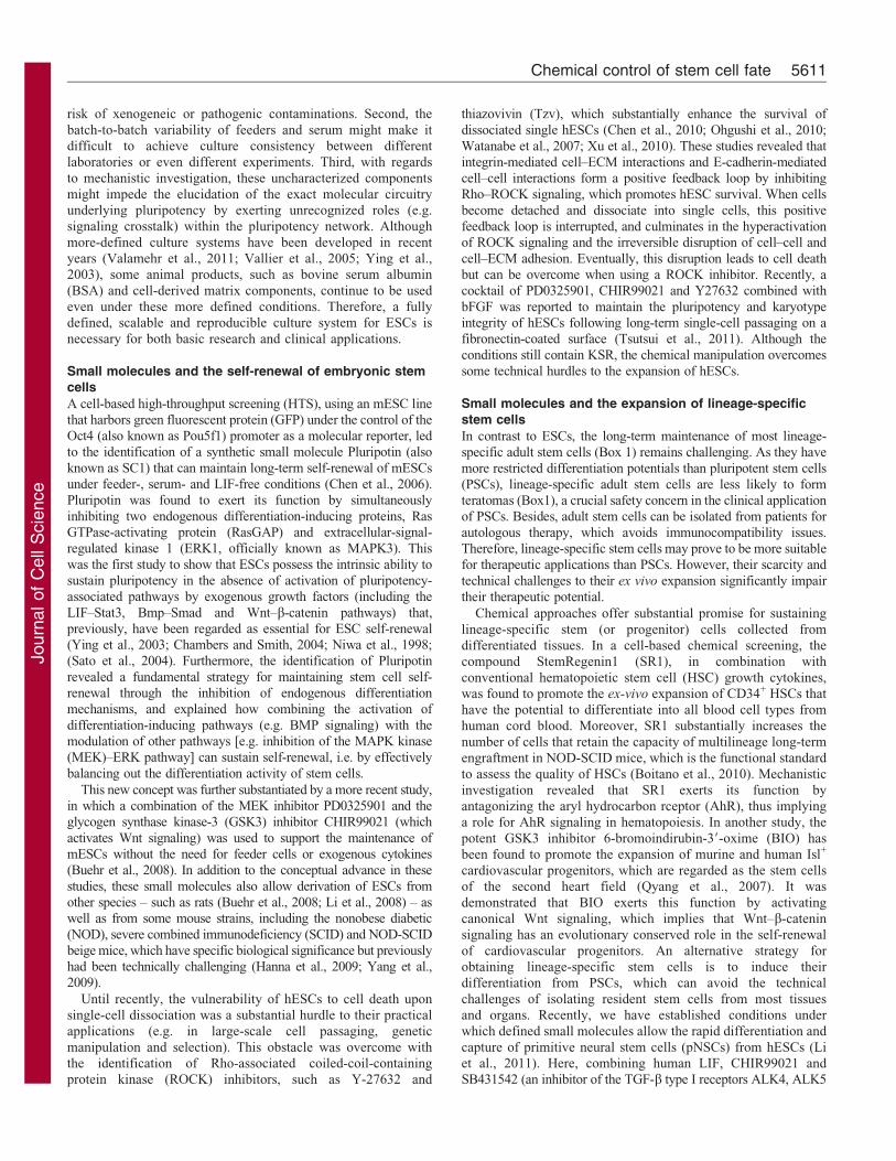
identified and characterized in recent years (summarized inTable 1; Fig. 2). For more general discussions of stem celldifferentiation, readers are encouraged to consult comprehensivereviews (Efe and Ding, 2011; Lyssiotis et al., 2011). In this
Commentary, we will focus on recent advances in the area ofstem cell maintenance and reprogramming, and place a specialemphasis on chemical strategies.
The role of small molecules in stem cellmaintenanceHere, we discuss strategies and new developments, particularlychemical approaches that have been employed to maintain the
self-renewal of ESCs or lineage-specific stem cells.
Embryonic stem cell culture systems
Conventionally, ESCs are cultured in the presence of feeder cells– typically human or mouse fibroblasts that have been growthinactivated through chemicals or c-irradiation – serum products,
e.g. fetal bovine serum (FBS), or knockout serum replacement(KSR), and growth factors. Until now, several crucial signalingpathways as well as related growth factors have been indentified
that participate in the maintenance of ESC pluripotency (Fig. 2).For mouse ESCs (mESCs), these include leukemia inhibitoryfactor (LIF)-signal transducer and activator for transcription 3
(STAT3) (Niwa et al., 1998), as well as bone morphogeneticprotein (BMP) (Chambers and Smith, 2004; Ying et al., 2003).Human ESCs rely on fibroblast growth factor 2 (FGF2, alsoknown as basic fibroblast growth factor) and Activin or NODAL
signaling (James et al., 2005; Vallier et al., 2005). In addition,Wnt signaling was reported to contribute to the maintenance ofboth mESCs and hESCs (Sato et al., 2004).
However, the presence of undefined culture components raisesa number of possible issues. First, feeder cells and other animalproducts including serum or serum replacements might entail the
Box 1. Introduction to stem cell biology
Stem cells are unspecialized cells that are characterized by their
capacity for self-renewal and differentiation. They can give rise
either to cells that bear characteristics identical to themselves and,
thus, maintain self-renewal, or to more specialized cells with more-
limited developmental potential, thereby resulting in differentiation.
Noticeably, stem cells exist not only in embryos but also in
adults throughout their whole life. Stem cells that are derived from
distinct developmental stages may display different developmental
potential.
Totipotent stem cells have the potential to generate an entire
functional organism, including not only the embryo but also the
extra-embryonic tissues. In mammals, the fertilized eggs and early
embryonic cells, such as blastomeres, are totipotent.
Pluripotent stem cells (PSCs) can give rise to all the cell types of
the entire embryo, including ectoderm, mesoderm and endoderm,
as well as germ cells, but not the extraembryonic tissues, such as
placenta. To date, several kinds of pluripotent stem cells (PSCs)
have been reported, including embryonic stem cells (ESCs) derived
from the inner cell mass of preimplantation embryos (Evans and
Kaufman, 1981; Martin, 1981; Thomson et al., 1998), epiblast stem
cells (EpiSCs) derived from the epiblast layer of the implanted
embryos (Brons et al., 2007; Tesar et al., 2007), and induced
pluripotent stem cells (iPSCs) generated from somatic cells by
reprogramming (Takahashi et al., 2007; Takahashi and Yamanaka,
2006; Yu et al., 2007). Their pluripotency can be evaluated by a
series of assays, such as the formation of teratomas or chimeras,
germline contribution or tetraploid complementation.
Multipotent stem cells have the ability to develop into different
cell types within the same cell lineage and are, therefore, also
referred to as lineage-specific stem cells or progenitors. These
cells have essential roles in maintaining tissue homeostasis under
both physiological and pathological conditions. For example,
hematopoietic stem cells in bone marrow can give rise to all
types of blood cell and replenish peripheral blood.
Induced pluripotent stem cells (iPSCs) are artificially
generated from somatic cells by ectopic expression of certain
pluripotency-related factors. They closely resemble natural PSCs
in many features, such as cellular biological properties,
pluripotency and epigenetic signatures.
ESCs iPSCs
EpiSCs
Unstable intermediates
Lineage-specific stem cellsLineage-specific stem cells
Self-renew
Terminally differentiated cells
Transdifferentiation I Transdifferentiation II
Plu
ripot
ent s
tem
cel
ls
Diff
eren
tiatio
n po
tent
ial
Mul
tipot
ent s
tem
cel
ls
Differentiation Reprogramming
Conversion
Key
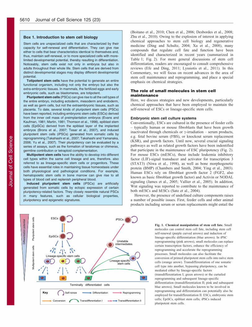
Fig. 1. Chemical manipulation of stem cell fate. Small
molecules can control stem cell fate, including stem cell
self-renewal (purple curved arrows) and induction of
lineage-specific differentiation (blue arrows). In iPSC
reprogramming (pink arrows), small molecules can replace
certain transcription factors, enhance the efficiency of
reprogramming and accelerate the reprogramming
processes. Small molecules can also facilitate the
conversion of primed pluripotent stem cells into naıve stem
cells (orange arrow). Transdifferentiation of one somatic
cell type into another, bypassing pluripotency, can be
mediated either by lineage-specific factors
(transdifferentiation I; green arrows) or the curtailed
reprogramming and subsequent lineage-specific
differentiation (transdifferentiation II; pink and subsequent
blue arrows). Small molecules known to be involved in
reprogramming and differentiation can potentially also be
employed for transdifferentiation II. ESCs, embryonic stem
cells; EpiSCs, epiblast stem cells; iPSCs induced
pluripotent stem cells.
Journal of Cell Science 125 (23)5610
Journ
alof
Cell
Scie
nce
risk of xenogeneic or pathogenic contaminations. Second, the
batch-to-batch variability of feeders and serum might make itdifficult to achieve culture consistency between differentlaboratories or even different experiments. Third, with regards
to mechanistic investigation, these uncharacterized componentsmight impede the elucidation of the exact molecular circuitryunderlying pluripotency by exerting unrecognized roles (e.g.signaling crosstalk) within the pluripotency network. Although
more-defined culture systems have been developed in recentyears (Valamehr et al., 2011; Vallier et al., 2005; Ying et al.,2003), some animal products, such as bovine serum albumin
(BSA) and cell-derived matrix components, continue to be usedeven under these more defined conditions. Therefore, a fullydefined, scalable and reproducible culture system for ESCs is
necessary for both basic research and clinical applications.
Small molecules and the self-renewal of embryonic stemcells
A cell-based high-throughput screening (HTS), using an mESC linethat harbors green fluorescent protein (GFP) under the control of theOct4 (also known as Pou5f1) promoter as a molecular reporter, ledto the identification of a synthetic small molecule Pluripotin (also
known as SC1) that can maintain long-term self-renewal of mESCsunder feeder-, serum- and LIF-free conditions (Chen et al., 2006).Pluripotin was found to exert its function by simultaneously
inhibiting two endogenous differentiation-inducing proteins, RasGTPase-activating protein (RasGAP) and extracellular-signal-regulated kinase 1 (ERK1, officially known as MAPK3). This
was the first study to show that ESCs possess the intrinsic ability tosustain pluripotency in the absence of activation of pluripotency-associated pathways by exogenous growth factors (including the
LIF–Stat3, Bmp–Smad and Wnt–b-catenin pathways) that,previously, have been regarded as essential for ESC self-renewal(Ying et al., 2003; Chambers and Smith, 2004; Niwa et al., 1998;(Sato et al., 2004). Furthermore, the identification of Pluripotin
revealed a fundamental strategy for maintaining stem cell self-renewal through the inhibition of endogenous differentiationmechanisms, and explained how combining the activation of
differentiation-inducing pathways (e.g. BMP signaling) with themodulation of other pathways [e.g. inhibition of the MAPK kinase(MEK)–ERK pathway] can sustain self-renewal, i.e. by effectively
balancing out the differentiation activity of stem cells.
This new concept was further substantiated by a more recent study,in which a combination of the MEK inhibitor PD0325901 and theglycogen synthase kinase-3 (GSK3) inhibitor CHIR99021 (which
activates Wnt signaling) was used to support the maintenance ofmESCs without the need for feeder cells or exogenous cytokines(Buehr et al., 2008). In addition to the conceptual advance in these
studies, these small molecules also allow derivation of ESCs fromother species – such as rats (Buehr et al., 2008; Li et al., 2008) – aswell as from some mouse strains, including the nonobese diabetic
(NOD), severe combined immunodeficiency (SCID) and NOD-SCIDbeige mice, which have specific biological significance but previouslyhad been technically challenging (Hanna et al., 2009; Yang et al.,2009).
Until recently, the vulnerability of hESCs to cell death uponsingle-cell dissociation was a substantial hurdle to their practicalapplications (e.g. in large-scale cell passaging, genetic
manipulation and selection). This obstacle was overcome withthe identification of Rho-associated coiled-coil-containingprotein kinase (ROCK) inhibitors, such as Y-27632 and
thiazovivin (Tzv), which substantially enhance the survival ofdissociated single hESCs (Chen et al., 2010; Ohgushi et al., 2010;
Watanabe et al., 2007; Xu et al., 2010). These studies revealed thatintegrin-mediated cell–ECM interactions and E-cadherin-mediatedcell–cell interactions form a positive feedback loop by inhibitingRho–ROCK signaling, which promotes hESC survival. When cells
become detached and dissociate into single cells, this positivefeedback loop is interrupted, and culminates in the hyperactivationof ROCK signaling and the irreversible disruption of cell–cell and
cell–ECM adhesion. Eventually, this disruption leads to cell deathbut can be overcome when using a ROCK inhibitor. Recently, acocktail of PD0325901, CHIR99021 and Y27632 combined with
bFGF was reported to maintain the pluripotency and karyotypeintegrity of hESCs following long-term single-cell passaging on afibronectin-coated surface (Tsutsui et al., 2011). Although theconditions still contain KSR, the chemical manipulation overcomes
some technical hurdles to the expansion of hESCs.
Small molecules and the expansion of lineage-specificstem cells
In contrast to ESCs, the long-term maintenance of most lineage-specific adult stem cells (Box 1) remains challenging. As they have
more restricted differentiation potentials than pluripotent stem cells(PSCs), lineage-specific adult stem cells are less likely to formteratomas (Box1), a crucial safety concern in the clinical applicationof PSCs. Besides, adult stem cells can be isolated from patients for
autologous therapy, which avoids immunocompatibility issues.Therefore, lineage-specific stem cells may prove to be more suitablefor therapeutic applications than PSCs. However, their scarcity and
technical challenges to their ex vivo expansion significantly impairtheir therapeutic potential.
Chemical approaches offer substantial promise for sustaining
lineage-specific stem (or progenitor) cells collected fromdifferentiated tissues. In a cell-based chemical screening, thecompound StemRegenin1 (SR1), in combination with
conventional hematopoietic stem cell (HSC) growth cytokines,was found to promote the ex-vivo expansion of CD34+ HSCs thathave the potential to differentiate into all blood cell types fromhuman cord blood. Moreover, SR1 substantially increases the
number of cells that retain the capacity of multilineage long-termengraftment in NOD-SCID mice, which is the functional standardto assess the quality of HSCs (Boitano et al., 2010). Mechanistic
investigation revealed that SR1 exerts its function byantagonizing the aryl hydrocarbon rceptor (AhR), thus implyinga role for AhR signaling in hematopoiesis. In another study, the
potent GSK3 inhibitor 6-bromoindirubin-39-oxime (BIO) hasbeen found to promote the expansion of murine and human Isl+
cardiovascular progenitors, which are regarded as the stem cellsof the second heart field (Qyang et al., 2007). It was
demonstrated that BIO exerts this function by activatingcanonical Wnt signaling, which implies that Wnt–b-cateninsignaling has an evolutionary conserved role in the self-renewal
of cardiovascular progenitors. An alternative strategy forobtaining lineage-specific stem cells is to induce theirdifferentiation from PSCs, which can avoid the technical
challenges of isolating resident stem cells from most tissuesand organs. Recently, we have established conditions underwhich defined small molecules allow the rapid differentiation and
capture of primitive neural stem cells (pNSCs) from hESCs (Liet al., 2011). Here, combining human LIF, CHIR99021 andSB431542 (an inhibitor of the TGF-b type I receptors ALK4, ALK5
Chemical control of stem cell fate 5611
Journ
alof
Cell
Scie
nce
Table 1. Known compounds that modulate stem cell fate and reprogramming
Compound name Identity Function References
Epigenetic-related compounds
Valproic acid (VPA) HDAC inhibitor Promotes MEF reprogramming efficiency,and enables Oct4- and Sox2-mediatedreprogramming of human fibroblasts;
(Huangfu et al., 2008a;Huangfu et al., 2008b)
facilitates proteins mediated reprogramming of MEFs (Zhou et al., 2009)Suberoylanilide hydroxamc
acid (SAHA)HDAC inhibitor Promotes MEF reprogramming efficiency (Huangfu et al., 2008a)
Trichostatin A (TSA) HDAC inhibitor Promotes MEF reprogramming efficiency (Huangfu et al., 2008a)Sodium butyrate (NaB) HDAC inhibitor Enhances reprogramming efficiency of human
adult or fetal fibroblasts;(Mali et al., 2010)
facilitates Oct4-only mediated reprogrammingwhen combined with A-83-01/PD0325901/PS48
(Zhu et al., 2010)
BIX-01294 G9a HMT inhibitor Enables NPC reprogramming mediated by Oct4 andKlf4, or substitutes for Oct4 in NPC reprogramming;
(Shi et al., 2008b)
promotes MEF reprogramming mediated by Oct4 and Klf4 (Shi et al., 2008a)RG108 DNMT inhibitor Promotes MEF reprogramming mediated by Oct4
when combined with BIX-01294(Shi et al., 2008a)
5-azazcytidine (5-aza) DNMT inhibitor Increases MEF reprogramming efficiency (Huangfu et al., 2008a;Mikkelsen et al., 2008)
Parnate LSD1 inhibitor Enables reprogramming of humankeratinocytes mediated by Oct4 and Klf4;
(Li et al., 2009b)
facilitates the conversion of mEpiSCs to naıvepluripotent state
(Zhou et al., 2010)
Signaling-pathway- or kinase-related compounds
PD0325901 MEK inhibitor Blocks differentiation pathway of ESCs andsupports self-renewal;
(Ying et al., 2008;Tsutsui et al.)
supports ESC derivation from refractorystrains or species;
(Nichols et al., 2009;Buehr et al., 2008;Li et al., 2008)
facilitates conversion of mEpiSCs and hESCsto naıve pluripotent state;
(Hanna et al., 2010;Zhou et al., 2010)
facilitates generation and maintenance ofmESC-like rat or human iPSCs;
(Li et al., 2009a)
facilitates rapid and efficient generation offully reprogrammed hiPSCs;
(Lin et al., 2009)
enables Oct4-mediated reprogramming whencombined with A-83-01/NaB/PS48
(Zhu et al., 2010)
CHIR99021 GSK3 inhibitor Supports ESCs self-renewal; facilitates ESCsderivation from refractory stains or species
(Ying et al., 2008;Tsutsui et al. 2011)
captures and maintains lineage-specific stem cells,like pNSCs; facilitates the conversion of mEpiSCsand hESCs to naıve pluripotent state;
(Nichols et al., 2009;Buehr et al., 2008;Li et al., 2008)
enables Oct4- and Klf4-mediated reprogramming ofMEFs or human primary keratinocytes with Parnate;
(Li et al., 2009a;Hanna et al., 2010;Zhou et al., 2010;Li et al., 2009b)
facilitates generation and maintenance ofmESC-like rat or human iPSCs;
(Li et al., 2009a)
facilitates the neural conversion of humanfibroblasts mediated by Ascl1 and Ngn2
(Ladewig et al., 2012)
6-bromoindirubin-39-oxime (BIO) GSK3 inhibitor Promotes self-renewal of ESCs and Isl+cardiovascular progenitors
(Sato et al., 2004;Qyang et al., 2007)
Kenpaullone GSK3 and CDK inhibitor Replaces Klf4 in MEF reprogramming (Lyssiotis et al., 2009)PD173074 FGF receptor inhibitor Supports mESC self-renewal; (Buehr et al., 2008)
facilitates the conversion of mEpiSCs tonaıve pluripotent state
(Zhou et al., 2010)
SU5402 FGF receptor inhibitor Supports mESC self-renewal (Buehr et al., 2008)A-83-01 ALK4, ALK5, ALK7
inhibitorFacilitates the conversion of mEpiSCs to
naıve pluripotent state;(Zhou et al., 2010)
enables generation and long-term maintenanceof mESC-like human iPSCs;
(Li et al., 2009a)
enables Oct4-mediated reprogramming whencombined with PD0325901/NaB/PS48
(Zhu et al., 2010)
SB431542 ALK4, ALK5, ALK7inhibitor
Captures and maintains pNSCs whencombined with CHIR99021;
(Li et al., 2011)
facilitates rapid and efficient generation offully reprogrammed human iPSCs;
(Lin et al., 2009)
Facilitates the neural conversion of humanfibroblasts mediated by Ascl1 and Ngn2
(Ladewig et al., 2012)
Table 1. Continued on next page
Journal of Cell Science 125 (23)5612
Journ
alof
Cell
Scie
nce
and ALK7) efficiently induces monolayer-cultured hESCs into
homogenous pNSCs. Addition of c-secretase inhibitor XXI (also
called compound E), further accelerates differentiation and leads to
complete neural induction within one week (.97% cells are SOX2
positive but OCT4 negative), which might be due to the suppression
of Notch signaling (Li et al., 2011). Importantly, in the presence of
LIF, CHIR99021 and SB431542, these pNSCs can stably self-renew
over long-term serial passages, while still maintaining their spatial
plasticity and high neurogenic differentiation propensity. This study
thus created a new paradigm in which hESCs can be differentiated
into homogenous lineage-committed progenitors that can be
stabilized and expanded under chemically defined conditions. Such
a strategy might make it possible to overcome some of the limitations
that are inherent in the step-wise uninterrupted differentiation
schemes, which result in the accumulation of undesired cells
in each differentiation step, eventually leading to increased
heterogeneity and a substantially lower yield of target cells.
Owing to their potency and flexibility in manipulating protein
functions, small molecules have become increasingly popular in
maintaining ESC self-renewal, inducing their lineage differentiation
and expanding lineage-specific stem cells. Unbiased chemical
screening could thus be an especially powerful approach to
interrogate the so-far unknown mechanisms that govern stem cell
self-renewal.
The role of small molecules in reprogrammingReprogramming of somatic cells towards pluripotency
The reversal of differentiation and the generation of PSCs from
somatic cells have fascinated researchers for years. Early studies
of somatic cell nuclear transfer (SCNT) revealed that a somatic
nucleus can be fully reprogrammed into a totipotent state by
factors from an enucleated egg, proceed to the generation of an
entire organism or be used to derive ESCs (Agarwal, 2006;
Campbell et al., 1996). Despite many advances in SCNT, the
process remains technically challenging and, in the human system,
there are ethical concerns to use SCNT to generate hESCs. Cell
fusion between somatic cells and ESCs to form heterokaryons can
also reprogram somatic nuclei into the pluripotent state, although
this typically results in the presence of extra sets of chromosomes
(Cowan et al., 2005; Ying et al., 2002). Another limitation of
SCNT and cell fusion is that, to mediate reprogramming, they use
largely undefined cellular contents, which makes it difficult to
investigate the underlying mechanisms.
In 2006, Yamanaka and colleagues demonstrated that virus-
mediated overexpression of Oct4, Sox2, Klf4 and Myc
(collectively termed OSKM) can convert mouse fibroblasts into
induced PSCs (iPSCs), which closely resemble mESCs in terms
of global gene expression, epigenetic state and developmental
potential (Takahashi and Yamanaka, 2006). Soon after this
discovery, human fibroblasts were reprogrammed into iPSCs by
overexpressing OSKM factors (Takahashi et al., 2007), or OCT4,
SOX2, NANOG and LIN28 (OSNL) (Yu et al., 2007). Compared
with SCNT and cell fusion approaches, the generation of iPSCs
relies on defined factors, is a much simpler process and is
unaffected by ethical controversy. However, there have been
concerns over the use of integrating retroviruses to deliver the
iPSC factors, which could potentially compromise the quality of
or even cause tumorigenicity in the resultant iPSCs.
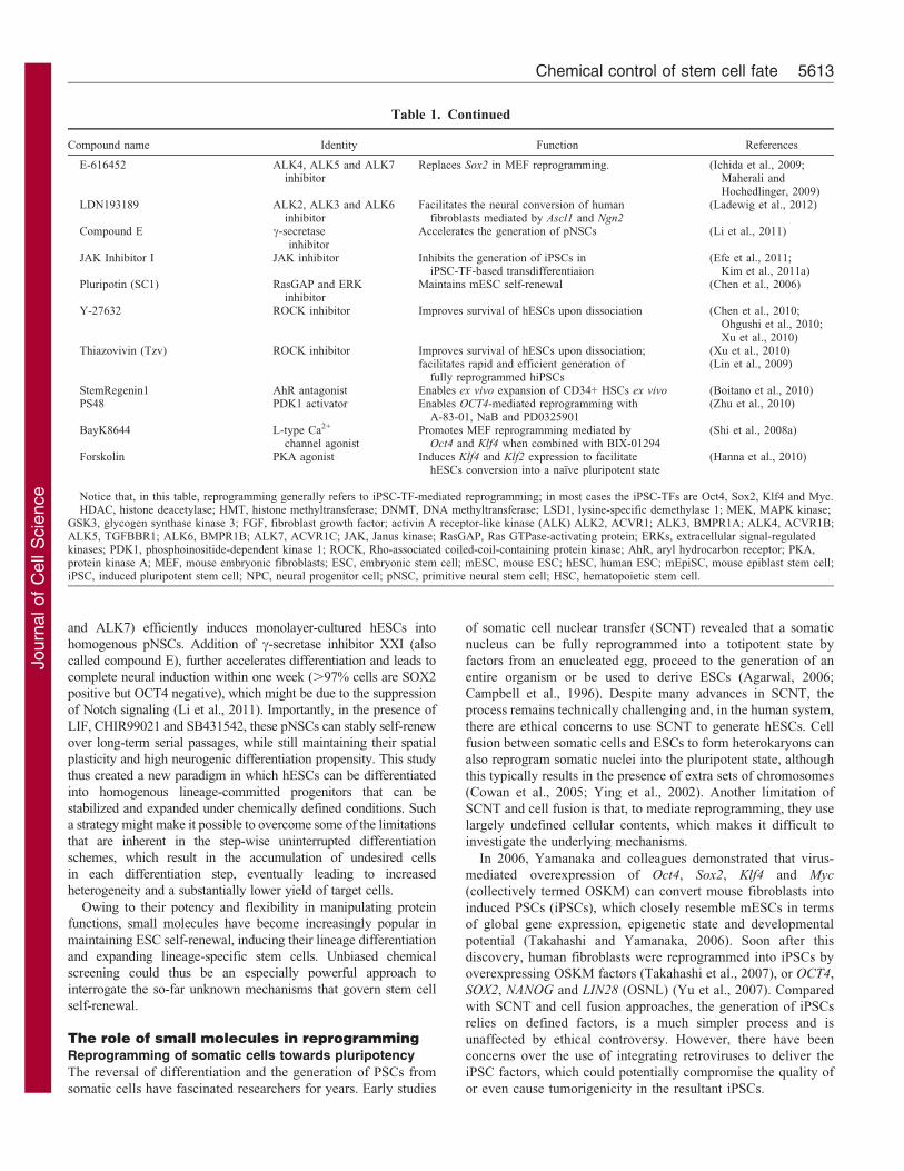
Table 1. Continued
Compound name Identity Function References
E-616452 ALK4, ALK5 and ALK7inhibitor
Replaces Sox2 in MEF reprogramming. (Ichida et al., 2009;Maherali andHochedlinger, 2009)
LDN193189 ALK2, ALK3 and ALK6inhibitor
Facilitates the neural conversion of humanfibroblasts mediated by Ascl1 and Ngn2
(Ladewig et al., 2012)
Compound E c-secretaseinhibitor
Accelerates the generation of pNSCs (Li et al., 2011)
JAK Inhibitor I JAK inhibitor Inhibits the generation of iPSCs iniPSC-TF-based transdifferentiaion
(Efe et al., 2011;Kim et al., 2011a)
Pluripotin (SC1) RasGAP and ERKinhibitor
Maintains mESC self-renewal (Chen et al., 2006)
Y-27632 ROCK inhibitor Improves survival of hESCs upon dissociation (Chen et al., 2010;Ohgushi et al., 2010;Xu et al., 2010)
Thiazovivin (Tzv) ROCK inhibitor Improves survival of hESCs upon dissociation; (Xu et al., 2010)facilitates rapid and efficient generation of
fully reprogrammed hiPSCs(Lin et al., 2009)
StemRegenin1 AhR antagonist Enables ex vivo expansion of CD34+ HSCs ex vivo (Boitano et al., 2010)PS48 PDK1 activator Enables OCT4-mediated reprogramming with
A-83-01, NaB and PD0325901(Zhu et al., 2010)
BayK8644 L-type Ca2+
channel agonistPromotes MEF reprogramming mediated by
Oct4 and Klf4 when combined with BIX-01294(Shi et al., 2008a)
Forskolin PKA agonist Induces Klf4 and Klf2 expression to facilitatehESCs conversion into a naıve pluripotent state
(Hanna et al., 2010)
Notice that, in this table, reprogramming generally refers to iPSC-TF-mediated reprogramming; in most cases the iPSC-TFs are Oct4, Sox2, Klf4 and Myc.HDAC, histone deacetylase; HMT, histone methyltransferase; DNMT, DNA methyltransferase; LSD1, lysine-specific demethylase 1; MEK, MAPK kinase;
GSK3, glycogen synthase kinase 3; FGF, fibroblast growth factor; activin A receptor-like kinase (ALK) ALK2, ACVR1; ALK3, BMPR1A; ALK4, ACVR1B;ALK5, TGFBBR1; ALK6, BMPR1B; ALK7, ACVR1C; JAK, Janus kinase; RasGAP, Ras GTPase-activating protein; ERKs, extracellular signal-regulatedkinases; PDK1, phosphoinositide-dependent kinase 1; ROCK, Rho-associated coiled-coil-containing protein kinase; AhR, aryl hydrocarbon receptor; PKA,protein kinase A; MEF, mouse embryonic fibroblasts; ESC, embryonic stem cell; mESC, mouse ESC; hESC, human ESC; mEpiSC, mouse epiblast stem cell;iPSC, induced pluripotent stem cell; NPC, neural progenitor cell; pNSC, primitive neural stem cell; HSC, hematopoietic stem cell.
Chemical control of stem cell fate 5613
Journ
alof
Cell
Scie
nce
Recent advances in iPSC technology have largely resolved the
concerns over genome modification through exogenous sequences
when new methods were introduced to deliver the reprogramming
factors that included the use of episomal plasmids (Yu et al., 2009)
or excisable expression systems (Soldner et al., 2009),
recombinant cell-penetrating reprogramming proteins (Kim et al.,
2009; Zhou et al., 2009) and reprogramming mRNAs (Warren
et al., 2010; Yakubov et al., 2010) or microRNAs (Anokye-Danso
et al., 2011; Miyoshi et al., 2011). For details regarding the
technical achievements in the reprogramming field, readers are
encouraged to examine more comprehensive reviews on this
subject (Gonzalez et al., 2011; Patel and Yang, 2010). Despite
these technical advancements, a challenging and more
fundamental issue is how to change the current iPSC
reprogramming procedure from a slow, inefficient and non-
deterministic process that involves stochastic events, to one that is
highly directed, specific and efficient. Another important question
to be answered is how to achieve reprogramming by using only
defined small molecules – an approach that is fundamentally
different from the exogenous transcription-factor-based
reprogramming that is employed in SCNT, cell fusion and
current iPSC methods. These advances would also address other
unresolved safety concerns with regards to the generation and use
of iPSCs, such as the potential effects on epigenetic memory and
other subtle genetic and epigenetic changes that might occur
during reprogramming (Kim et al., 2010; Ohi et al., 2011).
Using both phenotypic screening and hypothesis-driven
approaches, a growing number of compounds have been identified
that can functionally replace reprogramming transcription factors,
enhance efficiency of iPSC generation and accelerate the
reprogramming process (Table 1).
Given that reprogramming is accompanied by remodeling of
the epigenome, modulations of the epigenetic processes may
facilitate such conversion of cell fate by making cells more
permissive to these epigenenomic changes. Therefore, it is no
surprise to find that compounds that modulate epigenetic
enzymes, such as histone deacetylase (HDAC), histone
methyltransferase (HMT), histone demethylase (HDM) and
DNA methyltransferase (DNMT), can improve the efficiency of
reprogramming, or even replace the need to use certain
transcription factors. In a chemical screening, BIX-01294 (an
inhibitor of the HMT EHMT2; also known as G9a) was found to
substantially enhance Oct4–Klf4-mediated reprogramming of
neural progenitor cells (NPCs) into iPSCs to a level that is
AcAc
M
M
MM
DNMT inhibitor
Parnate
HMT inhibitor
BIX-01294
HDAC inhibitor
VPASAHATSANaB
Wnt BMP TGFβ/ActivinNodal
LIF FGF Growthfactors
Notch Integrinsα and β
LDN193189 SB431542A83-01
E-616452JAK
inhibitor IPD173074
SU5402
CHIR99021BIO
KenpaullonePluripotin
PD0325901
Compound E
Y27632Thiazovivin
PS48
Cytoskeleton
Metabolism
PDK1
MAPKERK1/2
MEK
RAF
PI3K
GTPGTPGTRASRAS
GDP
RAS GAP
JAKSMAD2/3SMAD1/5/8
STAT3
STAT3
SMAD4
4SMAD1/5/8
SMAD2/3
γ-secretase
NICDRhoA
GSK-3β
β-catenin
β-catenin
ALK2/3/6 ALK4/5/7
ROCK
RG1085-AZAHDM inhibitor
P
P
PP
P
PP
P
P
P
P
P
P
P
4
M M Ac PHistone methylation Acetylation PhosphorylationKey
Epigenetic status
Gene expression
LRP Frizzle
Methylation
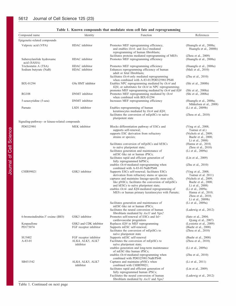
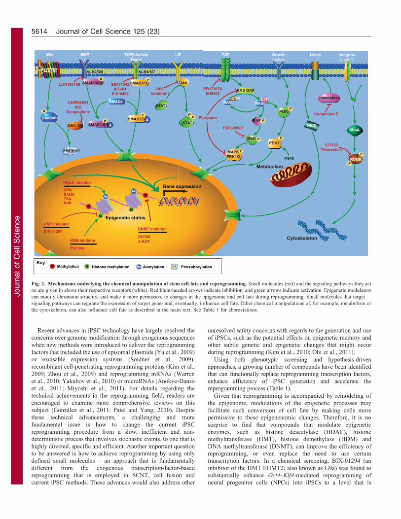
Fig. 2. Mechanisms underlying the chemical manipulation of stem cell fate and reprogramming. Small molecules (red) and the signaling pathways they act
on are given in above their respective receptors (white). Red blunt-headed arrows indicate inhibition, and green arrows indicate activation. Epigenetic modulators
can modify chromatin structure and make it more permissive to changes in the epigenome and cell fate during reprogramming. Small molecules that target
signaling pathways can regulate the expression of target genes and, eventually, influence cell fate. Other chemical manipulations of, for example, metabolism or
the cytoskeleton, can also influence cell fate as described in the main text. See Table 1 for abbreviations.
Journal of Cell Science 125 (23)5614
Journ
alof
Cell
Scie
nce
comparable with that when using OSKM factors, as well as toenable reprogramming mediated by only Klf4–Sox2–Myc without
the need for Oct4 (Shi et al., 2008b), albeit with much reducedefficiency. In another study, BIX-01294 was shown to enableOct4-Klf4-mediated reprogramming of mouse embryonicfibroblasts (MEFs), which could be further enhanced by
BayK8644 (an agonist of L-type Ca2+ channels) or RG108 (aninhibitor of DNMT) (Shi et al., 2008a). HDAC inhibitors [e.g.suberoylanilide hydroxamic acid (SAHA), Trichostatin A (TSA)
and valproic acid (VPA)] have also been shown to improvereprogramming efficiency (Huangfu et al., 2008a; Mikkelsenet al., 2008). In particular, VPA has been used in the
reprogramming of human fibroblasts with Oct4 and Sox2
(Huangfu et al., 2008b), in the reprogramming of MEFs withrecombinant cell-penetrating reprogramming proteins (Zhouet al., 2009), and in mir-302/367-mediated mouse fibroblasts
reprogramming (Anokye-Danso et al., 2011). Other compoundsthat affect epigenetic processes as well as their effects onreprogramming are summarized in Table 1.
As in the maintenance of pluripotency, some signalingpathways and their chemical modulators also help to re-establish pluripotency during reprogramming. The Wnt–b-
catenin signaling pathway has been reported to enhancereprogramming through alleviating the inhibitory effect of T-cell factor-3 (TCF3) on pluripotency (Niwa, 2011). Consistently,
CHIR99021, a GSK3 inhibitor that activates Wnt signaling,enables the reprogramming of MEFs into iPSCs by leading tooverexpression of Oct4 and Klf4 only, and also facilitates theOct4–Klf4-mediated reprogramming of human primary
keratinocytes when combined with Parnate, an inhibitor oflysine-specific demethylase1 (LSD1) (Li et al., 2009b).Kenpaullone, which inhibits GSK3 and several other kinases,
was identified from a HTS to be able to substitute for Klf4 inOSM-mediated reprogramming of MEFs (Lyssiotis et al., 2009).
TGF-b signaling is crucial to epithelial–mesenchymal transition
(EMT), an important hallmark of embryonic development (Xieet al., 2004b). TGF-b induces EMT through canonical Smadsignaling, non-canonical Ras–MEK–ERK MAP kinase signalingand Rho signaling (Xu et al., 2009). The reverse process,
mesenchymal–epithelial transition (MET), is a crucial earlyevent in reprogramming to pluripotency. It can thus beanticipated that small molecules that block TGF-b signaling or
its downstream effectors facilitate MET and enhancereprogramming. Consistent with this idea, inhibitors of TGF-breceptors, indeed, enhance reprogramming and can replace Sox2 in
the reprogramming of MEFs (Ichida et al., 2009; Maherali andHochedlinger, 2009). In another hypothesis-driven study, smallmolecules that are known to promote MET or inhibit EMT –
including SB431542, PD0325901 and Tzv, which inhibit TGF-breceptors, MEK and Rho-ROCK, respectively – were tested inhuman fibroblasts and found to not only substantially enhancereprogramming efficiency, but also accelerate the speed of
reprogramming, partially through derepression of the epithelialphenotype (Lin et al., 2009).
Recently, we have reported a chemical cocktail that enabled
the generation of human iPSCs from several human primarysomatic cell types when only Oct4 was exogenously expressed(Zhu et al., 2010). This cocktail contains sodium butyrate (NaB,
an inhibitor of HDAC), A-83-01 (an inhibitor of TGF-b type Ireceptors ALK4, ALK5 and ALK7) and PD0325901, as well asPS48 (an activator of 39-phosphoinositide-dependent kinase-1,
PDK1) (Zhu et al., 2010). Detailed mechanistic studies haverevealed that PS48 acts at the early phase of reprogramming in
order to facilitate a metabolic switch from mitochondrialoxidation (typically utilized by adult somatic cells) toglycolysis (almost exclusively used by PSCs) (Zhu et al.,2010). Other compounds that promote glycolytic metabolism
have also been shown to enhance reprogramming. They includefructose 2,6-bisphosphate (an activator of phosphofructokinase 1,a key rate-limiting enzyme of glycolysis) and N-oxaloylglycine
and quercetin, both activators of hypoxia-inducible factor-1 (Zhuet al., 2010). This study represents another significant steptowards the ultimate goal of chemical reprogramming and also
reveals metabolic modulation as another fundamental mechanismunderlying somatic cell reprogramming.
Conversion from the primed to naıve pluripotent stem cells
In addition to mESCs, which originate from the inner cell mass(ICM) of preimplantation mouse embryos, a different kind ofPSCs, mouse epiblast stem cells (mEpiSCs) can be derived from
late postimplantation epiblasts of mouse embryos (Brons et al.,2007; Tesar et al., 2007). mESCs and mEpiSCs exhibit distinctproperties in terms of gene expression, epigenetic profile, cell
behavior and their response to different signals (Table 2) (Bronset al., 2007; Guo et al., 2009; Tesar et al., 2007). Importantly,when mEpiSCs are injected into a preimplantation blastocyst,they contribute poorly to chimerism and are unable to transmit
through the germline in contrast to the robust chimera formationand germline transmission seen with mESCs (Brons et al., 2007).These different characteristics indicate that, consistent with their
origins, mEpiSCs represent a later or ‘primed’ state that isreminiscent of post-implantation epiblasts, whereas mESCsrepresent the ‘naıve’ state that corresponds to preimplantation
ICM (Nichols and Smith, 2009). This observation raises theinteresting question of whether it is feasible to convert mEpiSCsback into the mESC-like naıve pluripotent state. Indeed,
overexpression of Klf4, Nanog or nuclear receptor subfamily 5group A (Nr5a) has been shown to facilitate the reversion ofmEpiSCs into mESC-like cells (Guo and Smith, 2010; Guo et al.,2009; Silva et al., 2009). Concurrent with these genetic
approaches, we identified small-molecule conditions that alsoinduce this conversion, but in a more specific and efficientmanner. The combination of the inhibitors Parnate, A-83-01,
PD0325901, PD173074 and CHIR99021 (Table 1), whichantagonize LSD1, ALK5, MEK, FGF receptor and GSK3,respectively, can fully convert mEpiSCs back to the mESC-like
naıve state and restore their chimera competence (Zhou et al.,2010). Another group also demonstrated that the treatment ofPD0325901, CHIR99021 and LIF allow the conversion from
mEpiSCs to a mESC-like status that notably allowed for germlinetransmission (Greber et al., 2010).
Interestingly, even though the preparation of hESCs typicallystarts with preimplantation embryos, in many ways these cells
more closely resemble mEpiSCs than mESCs (Table 2),implying that hESCs represent a primed pluripotent state andnot the naıve state (Tesar et al., 2007). Technically, hESCs are
more resistant to gene targeting mediated by homogenousrecombination, which is routinely used in mESCs. This mightbe owing to the intolerance of hESCs to single-cell dissociation,
slow proliferation and a less-open chromatin structure (Li andDing, 2011). It has been speculated that naıve hPSCs – if theycould be generated – might have a number of advantages over
Chemical control of stem cell fate 5615
Journ
alof
Cell
Scie
nce
primed hPSCs in various applications (Li and Ding, 2011).
For example, a more robust growth and survival of naıve state
hPSCs would facilitate practical cell expansion and genetic
manipulation. In addition, the knowledge gained from studies in
mESCs might be more readily translatable to naıve state hPSCs.
From both scientific and technical viewpoints, it is of great
interest to generate naıve hESCs either from conventional hESCs
or directly from somatic cells by reprogramming. We first
demonstrated that mESC-like human PSCs can be generated and
stably expanded from human fibroblasts in an approach that
included genetic reprogramming (i.e. the overexpression of
OCT4, SOX2, NANOG and LIN28) in a culture medium
containing LIF, PD0325901, A-83-01 and CHIR99021 (Li et al.,
2009a). Another study combined the transient induction of Oct4,
Klf4 and Klf2 with a cocktail containing of PD0325901,
CHIR99021, LIF and forskolin (an agonist of protein kinase
A), which was able to convert hESCs into a naıve pluripotent
state and also facilitated the derivation of naıve hiPSCs that
shared many features with miPSCs (Hanna et al., 2010). Later,
naıve hPSCs were also generated directly from fibroblasts by
constitutively overexpressing OSKM factors and NANOG in the
presence of LIF (yielding the so-called hLR5 cells) (Buecker
et al., 2010), or by transient coexpression of retinoic acid receptor
c (RARG), liver receptor homolog 1 (Nr5a2) and OSKM factors
with subsequent culturing in a medium containing PD0325901,
CHIR99021 and LIF (Wang et al., 2011). These converted naıve
hESCs closely resemble mESCs in certain aspects, such as
morphology, signaling dependence and gene expression. More
importantly, as preliminarily identified in hLR5 cells, these naıve
hESCs allow for efficient gene targeting, thus making the genetic
manipulation of hESCs more feasible (Buecker et al., 2010).
However, it remains a substantial challenge to derive hPSCs at
the naıve pluripotent state and maintain them long-term without
the expression of any exogenous reprogramming factors.
Lineage-specific reprogramming – transdifferentiation
Unlike the reprogramming of iPSCs, transdifferentiation refers to
the direct reprogramming of one somatic cell type into another one
without passing through a pluripotent state. Transdifferentiation
may have several advantages over PSC-based strategies in
generating somatic cells, as it is potentially faster and more
efficient and can result in a higher yield of target cells.
Additionally, transdifferentiation might be safer for cell-based
therapeutic applications as it eliminates the risk of tumorigenesis
inherent to PSCs. To date, two distinct transdifferentiation
strategies have been developed, conventional transdifferentiation
mediated by lineage-specific factors and iPSC-transcription factor
(TF)-based transdifferentiation, which relies on curtailed or
hijacked reprogramming followed by lineage-specific culture
conditions to generate the desired cell type (Fig. 1).
Long before the development of iPSC technology, various
transdifferentiation procedures have been attempted. An early
example demonstrated the conversion of fibroblasts into skeletal
muscle cells by ectopic expression of MyoD, a master regulatory
transcription factor involved in myogenesis (Davis et al., 1987).
However, this conversion is considered incomplete, as the
acquired phenotype relies on a sustained overexpression of
MyoD. Other groups also reported the conversion of somatic
cells to other closely related cell types (Izumikawa et al., 2005;
Xie et al., 2004a; Zhou et al., 2008). Recently, several studies
have demonstrated that transdifferentiation between distantly
related cell types can be achieved by ectopic expression of
multiple lineage-specific factors. For example, mouse fibroblasts
can be converted into functional neurons by overexpressing
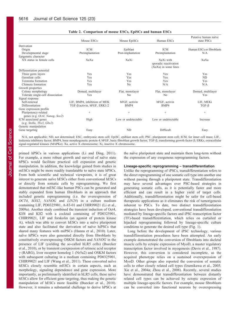
Table 2. Comparison of mouse ESCs, EpiSCs and human ESCs
Mouse ESCs Mouse EpiSCs Human ESCsPutative human naıve
state PSCs
DerivationOrigin ICM Epiblast ICM Human ESCs or fibroblastsDevelopmental stage Preimplantation Post-implantation Preimplantation N/A
Epigenetic characterXX status in female cells XaXa XaXi XaXi with
sporadic reactivation(XaXa) in some lines
XaXa
Differentiation potentialThree germ layers Yes Yes Yes YesGermline cells Yes Yes Yes NDTeratoma formation Yes Yes Yes YesChimera formation Yes No N/A N/A
Growth propertiesColony morphology Domed, multilayer Flat, monolayer Flat, monolayer Domed, multilayerTolerate single-cell dissociation Yes No No Yes
Signal responseSelf-renewal LIF, BMP4, inhibition of MEK bFGF, activin bFGF, activin LIF, MEKiDifferentiation TGF-b/activin, bFGF, ERK1/2 BMP4 BMP4 TGF-b
Gene expression profilePluripotency-related
genes (e.g. Oct4, Nanog, Sox2)+ + + +
ICM associated genes(e.g. Stella, Tbx3, Gbx2)
High Low or undetectable Low or undetectable Increase
Genetic manipulationGene targeting Easy ND Difficult Easy
N/A, not applicable; ND, not determined; ESC, embryonic stem cell; EpiSC, epiblast stem cell; PSC, pluripotent stem cell; ICM, for inner cell mass; LIF,leukemia inhibitory factor; BMP4, bone morphogenetic protein 4; bFGF, basic fibroblast growth factor; TGF-b, transforming growth factor b; ERKs, extracellularsignal-regulated kinases (MAPKs); Xa, active X chromosome; Xi, inactive X chromosome.
Journal of Cell Science 125 (23)5616
Journ
alof
Cell
Scie
nce
Brn2, Ascl1 and Myt1L (Vierbuchen et al., 2010) and intocardiomyocytes by overexpression of Gata4, Mef2c and Tbx5
(Ieda et al., 2010; Qian et al., 2012), and into hepatocyte-likecells by overexpressing Gata4, Hnf1a and Foxa3, while at thesame time inactivating p19Arf (Huang et al., 2011), or byoverexpressing Hnf4a and Foxa1, Foxa2 or Foxa3 (Sekiya and
Suzuki, 2011). In these studies, the transdifferentiated cellsclosely resembled native cell types in terms of morphology, geneexpression and functions in vitro or even in vivo. Of note, studies
from the Wernig laboratory verified that not only fibroblasts(Vierbuchen et al., 2010), but also terminally differentiatedhepatocytes can be converted into functional neurons by using
the same set of neuronal master factors (Marro et al., 2011).As shown in this work, converted cells can acquire thetranscriptional neuronal program and robustly silence thetranscriptional network of their cell type of origin. In addition,
several groups have independently reported that humanfibroblasts can be converted into functional neurons byoverexpressing different combinations of master factors, such
as miR-124, MYT1L and BRN2 (Ambasudhan et al., 2011), Brn2,Ascl1, Myt1L and NeuroD1 (Pang et al., 2011), or miR-9/9*, miR-
124 and NEUROD2 (Yoo et al., 2011). From these studies it is
apparent that several combinations or classes of masterfactor exist that can perform a similar, if not identical,transdifferentiation. In addition, some specific neural cell types,
such as spinal motor neurons (Son et al., 2011) and dopaminergicneurons (Caiazzo et al., 2011; Kim et al., 2011b; Pfisterer et al.,2011) could also be generated from both mouse and humanfibroblasts with the forced expression of cell-subtype-specific
transcription factors. More recently, it was reported that mousefibroblasts can also be converted into neural stem cells byoverexpression of Brn2, Sox2 and FoxG1 (Lujan et al., 2012), or
of Brn4, Sox2, Myc, Klf4 and Tcf3 (Han et al., 2012). Becausethese induced neural stem cells (iNSCs) retain the ability toproliferate and the potential to differentiate into neurons,
astrocytes and oligodendrocytes, they might be more desirablethan terminally differentiated cells for therapeutic cellreplacement, where proliferation and multilineage potential areneeded to repopulate the damaged tissue.
Collectively, in conventional transdifferentiation approaches,overexpression of lineage-specific master factors can drivesomatic cell conversion within or across germ layer boundaries.
This approach – although unlikely to generate iPSCs – has areduced risk of teratoma formation. However, to generate anydesired cell type, the required master factors need to be defined
for each type of target cell, and the underlying molecular andepigenetic mechanisms of each possible transdifferentiation needto be fully elucidated, which – undoubtedly – represents an
enormous research effort.
The second approach, iPSC-TF-based transdifferentiation, wasdeveloped on the basis of curtailed reprogramming and subsequentlineage differentiation. iPSC reprogramming is a non-deterministic
process that involves step-wise stochastic events, and only a verysmall percentage of induced cells eventually become iPSCs. We,therefore, hypothesized that it is possible to guide cells that have
initially been epigentically ‘activated’ through temporally restrictediPSC-TF expression, towards lineage-specific cell types, byswitching to different sets of signaling inputs without going
through the pluripotent state. Driven by this hypothesis, wetransiently expressed the OSKM factors that are able to induce theiPSC state, but for a period that is insufficient to generate iPSCs (Efe
et al., 2011; Kim et al., 2011a). This stage was followed by anexposure of the cells to signals that guide them to the desired
lineage, while simultaneously inhibiting the establishment ofpluripotency (Fig. 1). This procedure not only allows thegeneration of terminally differentiated somatic cells but alsoprovides a time window for capturing lineage-specific stem or
progenitor cells (Efe et al., 2011; Kim et al., 2011a). Following thetransient overexpression of the OSKM factors, we induced cardiacdifferentiation by treating cells with BMP4 and obtained
spontaneously contracting cardiomyocytes from MEFs as early asday 11 (Efe et al., 2011). Cardiac mesoderm precursor cells werealso observed during the transdifferentiation. In another study, we
captured neural progenitors from fibroblasts when cells were treatedwith FGFs and epidermal growth factor (EGF) following transientoverexpression of OSKM factors (Kim et al., 2011a). Using asimilar concept, another group demonstrated that the curtailed
reprogramming with transient Oct4 expression during the initialphase of reprogramming (within 5 days) converts fibroblasts intoexpandable and functional neural stem cells (NSCs) (Thier et al.,
2012). In another study, multilineage blood progenitors weregenerated from human dermal fibroblasts through the ectopicexpression of OCT4 and specific cytokine treatment, without going
through a pluripotent state (Szabo et al., 2010).
Compared with conventional transdifferentiation, iPSC-TF-based transdifferentiation provides a general platform withseveral practical advantages. First, it can be applied to induce
reprogramming towards several lineage-specific cell types byusing a single combination of transcription factors and well-established lineage induction conditions. Moreover, not only
terminally differentiated cells but also their lineage-committedprogenitors can be generated in one process. Second, because thismethod relies on ‘curtailed reprogramming’, advances in
reprogramming – such as small-molecule treatment and non-integrating factor delivery – can easily be implemented toimprove iPSC-TF-based transdifferentiation. This approach
might also represent an ideal platform to transfer the chemicalmanipulations in iPSC reprogramming and lineage differentiationdirectly into lineage-specific transdifferentiation.
Despite these advances of genetically based transdifferentiation,
it has the inherent problems of transgene expression, such as thegenomic intrgration of viral DNA fragments, as well as issues of lowefficiency or unclear epigenetic status that need to be addressed
before its clinical applications can be realized. For this, smallmolecules might provide a promising alternative for increasing theefficiency or replacing some, if not all, components needed for
transdifferentiation. Given the substantial resetting of epigenetic andgene expression patterns during transdifferentiation, it is reasonableto suspect that chemicals that affect epigenetic or signalingpathways have roles in this process. In fact, long before the
aforementioned genetically based transdifferentiations weredescribed, it had already been reported that 5-azazcytidine (5-aza)induces the conversion of a mesenchymal cell line into muscles,
adipocytes and chondroblasts (Taylor and Jones, 1979) bysuppressing DNA methylation (Jones and Taylor, 1980). In theiPSC-TF-based transdifferentiation approach described above, the
JAK inhibitor, which antagonizes the LIF–STAT3 pathway, anessential pathway for mESC maintenance, is used to suppress theestablishment of pluripotency, while at the same time facilitating the
generation of epigenetically plastic intermediate cells (Efe et al.,2011; Kim et al., 2011a). More recently, it was reported thatinhibition of GSK-3b and Smad by CHIR99021, SB431542 and
Chemical control of stem cell fate 5617
Journ
alof
Cell
Scie
nce
Noggin (a protein antagonist of BMP signaling) substantially
enhances the efficiency of Ascl- and Ngn2-driven conversion of
human fibroblasts into neural cells (Ladewig et al., 2012). It was
demonstrated that the concentration of Noggin can be reduced
significantly when used in combination with LDN193189 (a
chemical inhibitor of BMP type I receptors ALK2, ALK3 and
ALK6). As these small molecules act not only in reprogramming but
also in neural differentiation (Chambers et al., 2009), their exact role
during this conversion needs to be further elucidated. Although to
date there are only a few reports of the use of small molecules in the
nascent field of transdifferentiation, we expect to see more chemical
applications, especially those that have effects on iPSC
reprogramming and lineage differentiation, can be directly applied
to the transdifferentiation field. Additionally, in HTS or hypothesis-
driven studies, some small molecules have been shown to induce
certain conversions into specific cell types (Chen et al., 2004; Yau
et al., 2011) and these approaches might, therefore, provide
additional insight into the exact molecular mechanisms that
underlie transdifferentiation.
ConclusionsAlthough still very young, the field of stem cell research has
already offered enormous unprecedented opportunities for basic
research, disease modeling, drug screening and regenerative
medicine. Especially the recent achievements in reprogramming
and transdifferentiation have made it possible to generate highly
desired cell types from more accessible cell populations, and
bringing us closer to cell-based autotherapy. Complementary
to conventional strategies that include the overexpression
of reprogramming factors, chemical manipulation not only
represents a powerful tool for controlling stem cell fate, or
facilitating reprogramming or transdifferentiation, but also
provides the means for dissecting the underlying mechanisms.
There is no doubt that chemical approaches and the ongoing
discovery of new small molecules will continue to have essential
roles in the study and control of stem cell fate, state and function
towards the ultimate development of safe regenerative-medicine-
based treatments for various injuries and diseases.
AcknowledgementsS.D. is supported by funding from National Institute of Child Healthand Human Development, National Heart, Lung, and Blood Institute,National Eye Institute, and National Institute of Mental Health/National Institute of Health [grant numbers HD064610, HL107436,EY021374], California Institute for Regenerative Medicine, ProstateCancer Foundation, and the Gladstone Institute. We thank GaryHoward, and Anna Lisa Lucido for editing of this manuscript, andSaiyong Zhu, Jianghwan Kim, Tianhua Ma, Peng Liu, Baoming Nieand other members of the Ding lab for helpful discussions. Theauthors apologize to all scientists whose research could not bediscussed and/or cited in this Commentary to space limitations.Deposited in PMC for release after 12 month.
ReferencesAgarwal, S. (2006). Cellular Reprogramming. In Methods in Enzymology, Vol. 420 (ed.
K. Irina and L. Robert), pp. 265-283. New York, NY: Academic Press.
Ambasudhan, R., Talantova, M., Coleman, R., Yuan, X., Zhu, S., Lipton, S. A. and
Ding, S. (2011). Direct reprogramming of adult human fibroblasts to functional
neurons under defined conditions. Cell Stem Cell 9, 113-118.
Anokye-Danso, F., Trivedi, C. M., Juhr, D., Gupta, M., Cui, Z., Tian, Y., Zhang, Y.,
Yang, W., Gruber, P. J., Epstein, J. A. et al. (2011). Highly efficient miRNA-
mediated reprogramming of mouse and human somatic cells to pluripotency. Cell
Stem Cell 8, 376-388.
Boitano, A. E., Wang, J., Romeo, R., Bouchez, L. C., Parker, A. E., Sutton, S. E.,
Walker, J. R., Flaveny, C. A., Perdew, G. H., Denison, M. S. et al. (2010). Aryl
hydrocarbon receptor antagonists promote the expansion of human hematopoietic
stem cells. Science 329, 1345-1348.
Brons, I. G., Smithers, L. E., Trotter, M. W., Rugg-Gunn, P., Sun, B., Chuva de
Sousa Lopes, S. M., Howlett, S. K., Clarkson, A., Ahrlund-Richter, L., Pedersen,
R. A. et al. (2007). Derivation of pluripotent epiblast stem cells from mammalian
embryos. Nature 448, 191-195.
Buecker, C., Chen, H. H., Polo, J. M., Daheron, L., Bu, L., Barakat, T. S., Okwieka,
P., Porter, A., Gribnau, J., Hochedlinger, K. et al. (2010). A murine ESC-like state
facilitates transgenesis and homologous recombination in human pluripotent stem
cells. Cell Stem Cell 6, 535-546.
Buehr, M., Meek, S., Blair, K., Yang, J., Ure, J., Silva, J., McLay, R., Hall, J., Ying,
Q. L. and Smith, A. (2008). Capture of authentic embryonic stem cells from rat
blastocysts. Cell 135, 1287-1298.
Caiazzo, M., Dell’Anno, M. T., Dvoretskova, E., Lazarevic, D., Taverna, S., Leo, D.,
Sotnikova, T. D., Menegon, A., Roncaglia, P., Colciago, G. et al. (2011). Direct
generation of functional dopaminergic neurons from mouse and human fibroblasts.
Nature 476, 224-227.
Campbell, K. H. S., McWhir, J., Ritchie, W. A. and Wilmut, I. (1996). Sheep cloned
by nuclear transfer from a cultured cell line. Nature 380, 64-66.
Chambers, I. and Smith, A. (2004). Self-renewal of teratocarcinoma and embryonic
stem cells. Oncogene 23, 7150-7160.
Chambers, S. M., Fasano, C. A., Papapetrou, E. P., Tomishima, M., Sadelain,
M. and Studer, L. (2009). Highly efficient neural conversion of human ES and iPS
cells by dual inhibition of SMAD signaling. Nat. Biotechnol. 27, 275-280.
Chen, S., Zhang, Q., Wu, X., Schultz, P. G. and Ding, S. (2004). Dedifferentiation of
lineage-committed cells by a small molecule. J. Am. Chem. Soc. 126, 410-411.
Chen, S., Do, J. T., Zhang, Q., Yao, S., Yan, F., Peters, E. C., Scholer, H. R., Schultz,
P. G. and Ding, S. (2006). Self-renewal of embryonic stem cells by a small molecule.
Proc. Natl. Acad. Sci. USA 103, 17266-17271.
Chen, G., Hou, Z., Gulbranson, D. R. and Thomson, J. A. (2010). Actin-myosin
contractility is responsible for the reduced viability of dissociated human embryonic
stem cells. Cell Stem Cell 7, 240-248.
Cowan, C. A., Atienza, J., Melton, D. A. and Eggan, K. (2005). Nuclear
reprogramming of somatic cells after fusion with human embryonic stem cells.
Science 309, 1369-1373.
Davis, R. L., Weintraub, H. and Lassar, A. B. (1987). Expression of a single
transfected cDNA converts fibroblasts to myoblasts. Cell 51, 987-1000.
Desbordes, S. C., Placantonakis, D. G., Ciro, A., Socci, N. D., Lee, G., Djaballah,
H. and Studer, L. (2008). High-throughput screening assay for the identification of
compounds regulating self-renewal and differentiation in human embryonic stem
cells. Cell Stem Cell 2, 602-612.
Ding, S. and Schultz, P. G. (2004). A role for chemistry in stem cell biology. Nat.
Biotechnol. 22, 833-840.
Efe, J. A. and Ding, S. (2011). The evolving biology of small molecules: controlling
cell fate and identity. Philos. Trans. R. Soc. Lond. B Biol. Sci. 366, 2208-2221.
Efe, J. A., Hilcove, S., Kim, J., Zhou, H., Ouyang, K., Wang, G., Chen, J. and Ding,
S. (2011). Conversion of mouse fibroblasts into cardiomyocytes using a direct
reprogramming strategy. Nat. Cell Biol. 13, 215-222.
Evans, M. J. and Kaufman, M. H. (1981). Establishment in culture of pluripotential
cells from mouse embryos. Nature 292, 154-156.
Gonzalez, F., Boue, S. and Izpisua Belmonte, J. C. (2011). Methods for making
induced pluripotent stem cells: reprogramming a la carte. Nat. Rev. Genet. 12, 231-
242.
Greber, B., Wu, G., Bernemann, C., Joo, J. Y., Han, D. W., Ko, K., Tapia, N.,
Sabour, D., Sterneckert, J., Tesar, P. et al. (2010). Conserved and divergent roles of
FGF signaling in mouse epiblast stem cells and human embryonic stem cells. Cell
Stem Cell 6, 215-226.
Guo, G. and Smith, A. (2010). A genome-wide screen in EpiSCs identifies Nr5a nuclear
receptors as potent inducers of ground state pluripotency. Development 137, 3185-
3192.
Guo, G., Yang, J., Nichols, J., Hall, J. S., Eyres, I., Mansfield, W. and Smith,
A. (2009). Klf4 reverts developmentally programmed restriction of ground state
pluripotency. Development 136, 1063-1069.
Han, D. W., Tapia, N., Hermann, A., Hemmer, K., Hoing, S., Arauzo-Bravo, M. J.,
Zaehres, H., Wu, G., Frank, S., Moritz, S. et al. (2012). Direct reprogramming of
fibroblasts into neural stem cells by defined factors. Cell Stem Cell 10, 465-472.
Hanna, J., Markoulaki, S., Mitalipova, M., Cheng, A. W., Cassady, J. P., Staerk, J.,
Carey, B. W., Lengner, C. J., Foreman, R., Love, J. et al. (2009). Metastable
pluripotent states in NOD-mouse-derived ESCs. Cell Stem Cell 4, 513-524.
Hanna, J., Cheng, A. W., Saha, K., Kim, J., Lengner, C. J., Soldner, F., Cassady,
J. P., Muffat, J., Carey, B. W. and Jaenisch, R. (2010). Human embryonic stem
cells with biological and epigenetic characteristics similar to those of mouse ESCs.
Proc. Natl. Acad. Sci. USA 107, 9222-9227.
Hasegawa, K., Zhang, P., Wei, Z., Pomeroy, J. E., Lu, W. and Pera, M. F. (2010).
Comparison of reprogramming efficiency between transduction of reprogramming
factors, cell-cell fusion, and cytoplast fusion. Stem Cells 28, 1338-1348.
Huang, P., He, Z., Ji, S., Sun, H., Xiang, D., Liu, C., Hu, Y., Wang, X. and Hui, L.
(2011). Induction of functional hepatocyte-like cells from mouse fibroblasts by
defined factors. Nature 475, 386-389.
Journal of Cell Science 125 (23)5618
Journ
alof
Cell
Scie
nce
Huangfu, D., Maehr, R., Guo, W., Eijkelenboom, A., Snitow, M., Chen, A. E. and
Melton, D. A. (2008a). Induction of pluripotent stem cells by defined factors isgreatly improved by small-molecule compounds. Nat. Biotechnol. 26, 795-797.
Huangfu, D., Osafune, K., Maehr, R., Guo, W., Eijkelenboom, A., Chen, S.,Muhlestein, W. and Melton, D. A. (2008b). Induction of pluripotent stem cells fromprimary human fibroblasts with only Oct4 and Sox2. Nat. Biotechnol. 26, 1269-1275.
Ichida, J. K., Blanchard, J., Lam, K., Son, E. Y., Chung, J. E., Egli, D., Loh, K. M.,
Carter, A. C., Di Giorgio, F. P., Koszka, K. et al. (2009). A small-moleculeinhibitor of tgf-Beta signaling replaces sox2 in reprogramming by inducing nanog.Cell Stem Cell 5, 491-503.
Ieda, M., Fu, J. D., Delgado-Olguin, P., Vedantham, V., Hayashi, Y., Bruneau, B. G.and Srivastava, D. (2010). Direct reprogramming of fibroblasts into functionalcardiomyocytes by defined factors. Cell 142, 375-386.
Izumikawa, M., Minoda, R., Kawamoto, K., Abrashkin, K. A., Swiderski, D. L.,
Dolan, D. F., Brough, D. E. and Raphael, Y. (2005). Auditory hair cell replacementand hearing improvement by Atoh1 gene therapy in deaf mammals. Nat. Med. 11,271-276.
James, D., Levine, A. J., Besser, D. and Hemmati-Brivanlou, A. (2005). TGFbeta/activin/nodal signaling is necessary for the maintenance of pluripotency in humanembryonic stem cells. Development 132, 1273-1282.
Jones, P. A. and Taylor, S. M. (1980). Cellular differentiation, cytidine analogs andDNA methylation. Cell 20, 85-93.
Kim, D., Kim, C. H., Moon, J. I., Chung, Y. G., Chang, M. Y., Han, B. S., Ko, S.,Yang, E., Cha, K. Y., Lanza, R. et al. (2009). Generation of human inducedpluripotent stem cells by direct delivery of reprogramming proteins. Cell Stem Cell 4,472-476.
Kim, K., Doi, A., Wen, B., Ng, K., Zhao, R., Cahan, P., Kim, J., Aryee, M. J., Ji, H.,Ehrlich, L. I. R. et al. (2010). Epigenetic memory in induced pluripotent stem cells.Nature 467, 285-290.
Kim, J., Efe, J. A., Zhu, S., Talantova, M., Yuan, X., Wang, S., Lipton, S. A., Zhang,K. and Ding, S. (2011a). Direct reprogramming of mouse fibroblasts to neuralprogenitors. Proc. Natl. Acad. Sci. USA 108, 7838-7843.
Kim, J., Su, S. C., Wang, H., Cheng, A. W., Cassady, J. P., Lodato, M. A., Lengner,
C. J., Chung, C. Y., Dawlaty, M. M., Tsai, L. H. et al. (2011b). Functionalintegration of dopaminergic neurons directly converted from mouse fibroblasts. Cell
Stem Cell 9, 413-419.
Ladewig, J., Mertens, J., Kesavan, J., Doerr, J., Poppe, D., Glaue, F., Herms, S.,
Wernet, P., Kogler, G., Muller, F. J. et al. (2012). Small molecules enable highlyefficient neuronal conversion of human fibroblasts. Nat. Methods 9, 575-578.
Li, W. and Ding, S. (2011). Human pluripotent stem cells: Decoding the naıve state.Science translational medicine 3, 76ps10-76ps10.
Li, P., Tong, C., Mehrian-Shai, R., Jia, L., Wu, N., Yan, Y., Maxson, R. E., Schulze,
E. N., Song, H., Hsieh, C. L. et al. (2008). Germline competent embryonic stem cellsderived from rat blastocysts. Cell 135, 1299-1310.
Li, W., Wei, W., Zhu, S., Zhu, J., Shi, Y., Lin, T., Hao, E., Hayek, A., Deng, H. and
Ding, S. (2009a). Generation of rat and human induced pluripotent stem cells bycombining genetic reprogramming and chemical inhibitors. Cell Stem Cell 4, 16-19.
Li, W., Zhou, H., Abujarour, R., Zhu, S., Young Joo, J., Lin, T., Hao, E., Scholer,
H. R., Hayek, A. and Ding, S. (2009b). Generation of human-induced pluripotentstem cells in the absence of exogenous Sox2. Stem Cells 27, 2992-3000.
Li, W., Sun, W., Zhang, Y., Wei, W., Ambasudhan, R., Xia, P., Talantova, M., Lin,T., Kim, J., Wang, X. et al. (2011). Rapid induction and long-term self-renewal ofprimitive neural precursors from human embryonic stem cells by small moleculeinhibitors. Proc. Natl. Acad. Sci. USA 108, 8299-8304.
Lin, T., Ambasudhan, R., Yuan, X., Li, W., Hilcove, S., Abujarour, R., Lin, X.,Hahm, H. S., Hao, E., Hayek, A. et al. (2009). A chemical platform for improvedinduction of human iPSCs. Nat. Methods 6, 805-808.
Lujan, E., Chanda, S., Ahlenius, H., Sudhof, T. C. and Wernig, M. (2012). Directconversion of mouse fibroblasts to self-renewing, tripotent neural precursor cells.Proc. Natl. Acad. Sci. USA 109, 2527-2532.
Lyssiotis, C. A., Foreman, R. K., Staerk, J., Garcia, M., Mathur, D., Markoulaki, S.,
Hanna, J., Lairson, L. L., Charette, B. D., Bouchez, L. C. et al. (2009).Reprogramming of murine fibroblasts to induced pluripotent stem cells with chemicalcomplementation of Klf4. Proc. Natl. Acad. Sci. USA 106, 8912-8917.
Lyssiotis, C. A., Lairson, L. L., Boitano, A. E., Wurdak, H., Zhu, S. and Schultz,P. G. (2011). Chemical control of stem cell fate and developmental potential. Angew.
Chem. Int. Ed. Engl. 50, 200-242.
Maherali, N. and Hochedlinger, K. (2009). Tgfbeta signal inhibition cooperates in theinduction of iPSCs and replaces Sox2 and cMyc. Curr. Biol. 19, 1718-1723.
Mali, P., Chou, B. K., Yen, J., Ye, Z., Zou, J., Dowey, S., Brodsky, R. A., Ohm, J. E.,Yu, W., Baylin, S. B. et al. (2010). Butyrate greatly enhances derivation of humaninduced pluripotent stem cells by promoting epigenetic remodeling and the expressionof pluripotency-associated genes. Stem Cells 28, 713-720.
Marro, S., Pang, Z. P., Yang, N., Tsai, M. C., Qu, K., Chang, H. Y., Sudhof, T. C.and Wernig, M. (2011). Direct lineage conversion of terminally differentiatedhepatocytes to functional neurons. Cell Stem Cell 9, 374-382.
Martin, G. R. (1981). Isolation of a pluripotent cell line from early mouse embryoscultured in medium conditioned by teratocarcinoma stem cells. Proc. Natl. Acad. Sci.
USA 78, 7634-7638.
Mikkelsen, T. S., Hanna, J., Zhang, X., Ku, M., Wernig, M., Schorderet,
P., Bernstein, B. E., Jaenisch, R., Lander, E. S. and Meissner, A. (2008).Dissecting direct reprogramming through integrative genomic analysis. Nature 454,49-55.
Miyoshi, N., Ishii, H., Nagano, H., Haraguchi, N., Dewi, D. L., Kano, Y., Nishikawa,
S., Tanemura, M., Mimori, K., Tanaka, F. et al. (2011). Reprogramming of mouseand human cells to pluripotency using mature microRNAs. Cell Stem Cell 8, 633-638.
Nichols, J. and Smith, A. (2009). Naive and primed pluripotent states. Cell Stem Cell 4,487-492.
Niwa, H. (2011). Wnt: what’s needed to maintain pluripotency? Nat. Cell Biol. 13,1024-1026.
Niwa, H., Burdon, T., Chambers, I. and Smith, A. (1998). Self-renewal of pluripotentembryonic stem cells is mediated via activation of STAT3. Genes Dev. 12, 2048-2060.
Ohgushi, M., Matsumura, M., Eiraku, M., Murakami, K., Aramaki, T., Nishiyama,
A., Muguruma, K., Nakano, T., Suga, H., Ueno, M. et al. (2010). Molecularpathway and cell state responsible for dissociation-induced apoptosis in humanpluripotent stem cells. Cell Stem Cell 7, 225-239.
Ohi, Y., Qin, H., Hong, C., Blouin, L., Polo, J. M., Guo, T., Qi, Z., Downey, S. L.,
Manos, P. D., Rossi, D. J. et al. (2011). Incomplete DNA methylation underlies atranscriptional memory of somatic cells in human iPS cells. Nat. Cell Biol. 13, 541-549.
Pang, Z. P., Yang, N., Vierbuchen, T., Ostermeier, A., Fuentes, D. R., Yang, T. Q.,
Citri, A., Sebastiano, V., Marro, S., Sudhof, T. C. et al. (2011). Induction of humanneuronal cells by defined transcription factors. Nature 476, 220-223.
Patel, M. and Yang, S. (2010). Advances in reprogramming somatic cells to inducedpluripotent stem cells. Stem Cell Rev. 6, 367-380.
Pfisterer, U., Kirkeby, A., Torper, O., Wood, J., Nelander, J., Dufour,A., Bjorklund, A., Lindvall, O., Jakobsson, J. and Parmar, M. (2011). Directconversion of human fibroblasts to dopaminergic neurons. Proc. Natl. Acad. Sci. USA
108, 10343-10348.
Qian, L., Huang, Y., Spencer, C. I., Foley, A., Vedantham, V., Liu, L., Conway,
S. J., Fu, J. D. and Srivastava, D. (2012). In vivo reprogramming of murine cardiacfibroblasts into induced cardiomyocytes. Nature 485, 593-598.
Qyang, Y., Martin-Puig, S., Chiravuri, M., Chen, S., Xu, H., Bu, L., Jiang, X., Lin,L., Granger, A., Moretti, A. et al. (2007). The renewal and differentiation of Isl1+cardiovascular progenitors are controlled by a Wnt/beta-catenin pathway. Cell Stem
Cell 1, 165-179.
Sato, N., Meijer, L., Skaltsounis, L., Greengard, P. and Brivanlou, A. H. (2004).Maintenance of pluripotency in human and mouse embryonic stem cells throughactivation of Wnt signaling by a pharmacological GSK-3-specific inhibitor. Nat. Med.
10, 55-63.
Sekiya, S. and Suzuki, A. (2011). Direct conversion of mouse fibroblasts to hepatocyte-like cells by defined factors. Nature 475, 390-393.
Shi, Y., Desponts, C., Do, J. T., Hahm, H. S., Scholer, H. R. and Ding, S. (2008a).Induction of pluripotent stem cells from mouse embryonic fibroblasts by Oct4 andKlf4 with small-molecule compounds. Cell Stem Cell 3, 568-574.
Shi, Y., Do, J. T., Desponts, C., Hahm, H. S., Scholer, H. R. and Ding, S. (2008b).A combined chemical and genetic approach for the generation of induced pluripotentstem cells. Cell Stem Cell 2, 525-528.
Silva, J., Nichols, J., Theunissen, T. W., Guo, G., van Oosten, A. L., Barrandon,O., Wray, J., Yamanaka, S., Chambers, I. and Smith, A. (2009). Nanog is thegateway to the pluripotent ground state. Cell 138, 722-737.
Soldner, F., Hockemeyer, D., Beard, C., Gao, Q., Bell, G. W., Cook, E. G., Hargus,G., Blak, A., Cooper, O., Mitalipova, M. et al. (2009). Parkinson’s disease patient-derived induced pluripotent stem cells free of viral reprogramming factors. Cell 136,964-977.
Son, E. Y., Ichida, J. K., Wainger, B. J., Toma, J. S., Rafuse, V. F., Woolf, C. J. and
Eggan, K. (2011). Conversion of mouse and human fibroblasts into functional spinalmotor neurons. Cell Stem Cell 9, 205-218.
Szabo, E., Rampalli, S., Risueno, R. M., Schnerch, A., Mitchell, R., Fiebig-Comyn,A., Levadoux-Martin, M. and Bhatia, M. (2010). Direct conversion of humanfibroblasts to multilineage blood progenitors. Nature 468, 521-526.
Takahashi, K. and Yamanaka, S. (2006). Induction of pluripotent stem cells frommouse embryonic and adult fibroblast cultures by defined factors. Cell 126, 663-676.
Takahashi, K., Tanabe, K., Ohnuki, M., Narita, M., Ichisaka, T., Tomoda, K. and
Yamanaka, S. (2007). Induction of pluripotent stem cells from adult humanfibroblasts by defined factors. Cell 131, 861-872.
Taylor, S. M. and Jones, P. A. (1979). Multiple new phenotypes induced in 10T1/2 and3T3 cells treated with 5-azacytidine. Cell 17, 771-779.
Tesar, P. J., Chenoweth, J. G., Brook, F. A., Davies, T. J., Evans, E. P., Mack, D. L.,
Gardner, R. L. and McKay, R. D. (2007). New cell lines from mouse epiblast sharedefining features with human embryonic stem cells. Nature 448, 196-199.
Thier, M., Worsdorfer, P., Lakes, Y. B., Gorris, R., Herms, S., Opitz, T., Seiferling,D., Quandel, T., Hoffmann, P., Nothen, M. M. et al. (2012). Direct conversion offibroblasts into stably expandable neural stem cells. Cell Stem Cell 10, 473-479.
Thomson, J. A., Itskovitz-Eldor, J., Shapiro, S. S., Waknitz, M. A., Swiergiel, J. J.,Marshall, V. S. and Jones, J. M. (1998). Embryonic stem cell lines derived fromhuman blastocysts. Science 282, 1145-1147.
Tsutsui, H., Valamehr, B., Hindoyan, A., Qiao, R., Ding, X., Guo, S., Witte, O. N., Liu,X., Ho, C. M. and Wu, H. (2011). An optimized small molecule inhibitor cocktailsupports long-term maintenance of human embryonic stem cells. Nat. Commun 2, 167.
Valamehr, B., Tsutsui, H., Ho, C. M. and Wu, H. (2011). Developing defined culturesystems for human pluripotent stem cells. Regen. Med. 6, 623-634.
Vallier, L., Alexander, M. and Pedersen, R. A. (2005). Activin/Nodal and FGFpathways cooperate to maintain pluripotency of human embryonic stem cells. J. Cell
Sci. 118, 4495-4509.
Chemical control of stem cell fate 5619
Journ
alof
Cell
Scie
nce
Vierbuchen, T., Ostermeier, A., Pang, Z. P., Kokubu, Y., Sudhof, T. C. and Wernig,M. (2010). Direct conversion of fibroblasts to functional neurons by defined factors.Nature 463, 1035-1041.
Wang, W., Yang, J., Liu, H., Lu, D., Chen, X., Zenonos, Z., Campos, L. S., Rad, R.,
Guo, G., Zhang, S. et al. (2011). Rapid and efficient reprogramming of somatic cellsto induced pluripotent stem cells by retinoic acid receptor gamma and liver receptorhomolog 1. Proc. Natl. Acad. Sci. USA 108, 18283-18288.
Warren, L., Manos, P. D., Ahfeldt, T., Loh, Y. H., Li, H., Lau, F., Ebina, W.,Mandal, P. K., Smith, Z. D., Meissner, A. et al. (2010). Highly efficientreprogramming to pluripotency and directed differentiation of human cells withsynthetic modified mRNA. Cell Stem Cell 7, 618-630.
Watanabe, K., Ueno, M., Kamiya, D., Nishiyama, A., Matsumura, M., Wataya, T.,
Takahashi, J. B., Nishikawa, S., Nishikawa, S., Muguruma, K. et al. (2007). AROCK inhibitor permits survival of dissociated human embryonic stem cells. Nat.
Biotechnol. 25, 681-686.Xie, H., Ye, M., Feng, R. and Graf, T. (2004a). Stepwise reprogramming of B cells into
macrophages. Cell 117, 663-676.Xie, L., Law, B. K., Chytil, A. M., Brown, K. A., Aakre, M. E. and Moses, H. L.
(2004b). Activation of the Erk pathway is required for TGF-b1-induced EMT in vitro.Neoplasia 6, 603-610.
Xu, Y., Shi, Y. and Ding, S. (2008). A chemical approach to stem-cell biology andregenerative medicine. Nature 453, 338-344.
Xu, J., Lamouille, S. and Derynck, R. (2009). TGF-beta-induced epithelial tomesenchymal transition. Cell Res. 19, 156-172.
Xu, Y., Zhu, X., Hahm, H. S., Wei, W., Hao, E., Hayek, A. and Ding, S. (2010).Revealing a core signaling regulatory mechanism for pluripotent stem cell survivaland self-renewal by small molecules. Proc. Natl. Acad. Sci. USA 107, 8129-8134.
Yakubov, E., Rechavi, G., Rozenblatt, S. and Givol, D. (2010). Reprogramming ofhuman fibroblasts to pluripotent stem cells using mRNA of four transcription factors.Biochem. Biophys. Res. Commun. 394, 189-193.
Yang, W., Wei, W., Shi, C., Zhu, J., Ying, W., Shen, Y., Ye, X., Fang, L., Duo, S.,
Che, J. et al. (2009). Pluripotin combined with leukemia inhibitory factor greatlypromotes the derivation of embryonic stem cell lines from refractory strains. Stem
Cells 27, 383-389.
Yau, W. W., Tang, M. K., Chen, E., Yaoyao, Wong, I. W., Lee, H. S. and Lee, K. K.
(2011). Cardiogenol C can induce Mouse Hair Bulge Progenitor Cells to
Transdifferentiate into Cardiomyocyte-like Cells. Proteome Sci. 9, 3.
Ying, Q. L., Nichols, J., Evans, E. P. and Smith, A. G. (2002). Changing potency by
spontaneous fusion. Nature 416, 545-548.
Ying, Q. L., Nichols, J., Chambers, I. and Smith, A. (2003). BMP induction of Id
proteins suppresses differentiation and sustains embryonic stem cell self-renewal in
collaboration with STAT3. Cell 115, 281-292.
Ying, Q. L., Wray, J., Nichols, J., Batlle-Morera, L., Doble, B., Woodgett, J., Cohen,
P. and Smith, A. (2008). The ground state of embryonic stem cell self-renewal.
Nature 453, 519-523.
Yoo, A. S., Sun, A. X., Li, L., Shcheglovitov, A., Portmann, T., Li, Y., Lee-Messer,
C., Dolmetsch, R. E., Tsien, R. W. and Crabtree, G. R. (2011). MicroRNA-
mediated conversion of human fibroblasts to neurons. Nature 476, 228-231.
Yu, J., Vodyanik, M. A., Smuga-Otto, K., Antosiewicz-Bourget, J., Frane, J. L.,
Tian, S., Nie, J., Jonsdottir, G. A., Ruotti, V., Stewart, R. et al. (2007). Induced
pluripotent stem cell lines derived from human somatic cells. Science 318, 1917-
1920.
Yu, J., Hu, K., Smuga-Otto, K., Tian, S., Stewart, R., Slukvin, I. I. and Thomson,
J. A. (2009). Human induced pluripotent stem cells free of vector and transgene
sequences. Science 324, 797-801.
Zhou, Q., Brown, J., Kanarek, A., Rajagopal, J. and Melton, D. A. (2008). In vivo
reprogramming of adult pancreatic exocrine cells to beta-cells. Nature 455, 627-632.
Zhou, H., Wu, S., Joo, J. Y., Zhu, S., Han, D. W., Lin, T., Trauger, S., Bien, G., Yao,
S., Zhu, Y. et al. (2009). Generation of induced pluripotent stem cells using
recombinant proteins. Cell Stem Cell 4, 381-384.
Zhou, H., Li, W., Zhu, S., Joo, J. Y., Do, J. T., Xiong, W., Kim, J. B., Zhang, K.,
Scholer, H. R. and Ding, S. (2010). Conversion of mouse epiblast stem cells to
an earlier pluripotency state by small molecules. J. Biol. Chem. 285, 29676-
29680.
Zhu, S., Li, W., Zhou, H., Wei, W., Ambasudhan, R., Lin, T., Kim, J., Zhang, K.
and Ding, S. (2010). Reprogramming of human primary somatic cells by OCT4 and
chemical compounds. Cell Stem Cell 7, 651-655.
Journal of Cell Science 125 (23)5620