the roles of extracellular matrix molecules matrilins and
TRANSCRIPT

Aus der Klinik für Allgemeine, Unfall- und Wiederherstellungschirurgie
Klinik der Universität München
Vorstand/Direktor: Wolfgang Böcker
The Roles of Extracellular Matrix Molecules Matrilins and Aggrecan in
Bone Development and Articular Cartilage Functions
Dissertation
zum Erwerb des Doktorgrades der Humanbiologie
an der Medizinischen Fakultät der
Ludwig-Maximilians-Universität zu München
vorgelegt von
Ping Li
aus (Geburtsort)
Hebei, China
Jahr
2020
Mit Genehmigung der Medizinischen Fakultät der Universität München
PD Dr. Attila Aszodi
PD Dr. Jesus Buija
Prof Dr. Susanne Mayer
Prof Dr. Mohammad-Mehdi Shakibaei
Dr. hum,biol. Paolo Alberton
Prof. Dr. med. dent. Reinhard Hickel
______14.09.2020______________________
Berichterstatter:
Mitberichterstatter:
Mitbetreuung durch den Promovierten Mitarbeiter:
Dekan:
Tag der mündlichen Prüfung:

Dean’s Office Medical Faculty
Faculty of Medicine
Affidavit
Li, Ping
Surname, first name
I hereby declare, that the submitted thesis entitled
The Roles of Extracellular Matrix Molecules Matrilins and Aggrecan in Bone Development and
Articular Cartilage Functions
is my own work. I have only used the sources indicated and have not made unauthorised use of services
of a third party. Where the work of others has been quoted or reproduced, the source is always given.
I further declare that the submitted thesis or parts thereof have not been presented as part of an
examination degree to any other university.
Place, Date Signature doctoral candidate
Affidavit Human Biology Date: 15.09.2020
München, 16.03.2020 Ping Li

TABLE OF CONTENTS
TABLE OF CONTENTS ........................................................................................... I
ABBREVATIONS .................................................................................................. II
LIST OF PUBLICATIONS ...................................................................................... IV
1. INTRODUCTION .............................................................................................. 1
1.1. Bone and cartilage .............................................................................................21.1.1. Endochondral ossification .................................................................................................... 2
1.1.2. Articular cartilage..................................................................................................................5
1.2. Extracellular matrix ............................................................................................71.2.1. Collagens .............................................................................................................................8
1.2.2. Proteoglycans .......................................................................................................................9
1.2.3. Perifibrillar adapter proteins ..............................................................................................10
1.2.4. Matrilin family .................................................................................................................10
1.2.4.1. Expression pattern and functions of matrilins ............................................................11
1.3. Diseases related to matrilins and aggrecan in the skeletal system ...................................13
1.3.1. Chondrodysplasia ........................................................................................13
1.3.2. Osteoarthritis .......................................................................................................................14
1.4. Aim of thesis .....................................................................................................16
1.5. Own contributions ..............................................................................................17
2. SUMMARY ........................................................................................................18
PAPER I ............................................................................................................................................. 23
PAPER II ..............................................................................................................50
REFERENCES .......................................................................................................70
ACKNOWLEDGMENT............................................................................................80
CURRICULUM VITAE ..........................................................................................81
I
CONFIRMATION OF THE CO-AUTHORS..................................................................V
3. ZUSAMMENFASSUNG ........................................................................................18

ABBREVATIONS
AC Articular cartilage
ADAMTS A disintegrin and metalloproteinase with
thrombospondin motifs
AFM Atomic force microscopy
BHMED Bilateral hereditary micro-epiphyseal
dysplasia
CS Chondroitin sulphate
EO Endochondral ossification
COMP Cartilage oligomeric matrix protein
ECM Extracellular matrix
ER Endoplasmic reticulum
FACIT Fibril-associated collagens with interrupted
triple helix
GAGs Glycosaminoglycans
GP Growth plate
HA Hyaluronan
H&E Haematoxylin and Eosin
HSCs Hematopoietic stem cells
HZ Hypertrophic zone
IGD Interglobular domains
IGF-1 Insulin-like growth factor 1
ITM Interterritorial matrix
IO Intramembranous ossification
kDa Kilodalton
KS Keratan sulphate
Leucine-rich repeat LRR
LP Link protein
II

MED Multiple epiphyseal dysplasia
MMPs Matrix metalloproteinases
OA Osteoarthritis
Osteochondritis dissecans OCD
PCM Pericellular matrix
PG Proteoglycan
POC Primary ossification center
Ptc-1 Patched-1
PTHrP Parathyroid hormone related peptide
PRG4 Proteoglycan 4
PSACH Pseudoachondroplasia
PZ Proliferative zone
rER Rough endoplasmic reticulum
RZ Resting zone
SEMD Spondyloepimetaphyseal dysplasia
Small leucine-rich repeat proteoglycans SLRPs
Smoothened Smo
SOC Secondary ossification center
TM Territorial matrix
TSP Thrombospondin
UPR Unfolded protein response
VEGF Vascular endothelial growth factors
vWFA von Willebrand factor A
III

LIST OF PUBLICATIONS This thesis is based on the following publications
I. Mice lacking the Matrilin Family of Extracellular Matrix Proteins Develop Normal Skeleton but
are More Susceptible for Age-associated Osteoarthritis
Ping Li, Lutz Fleischhauer, Claudia Nicolae, Carina Prein, Zsuzsanna Farkas,Maximilian
Michael Saller, Wolf Christian Prall, Raimund Wagener, Juliane Heilig, Anja Niehoff, Hauke Clausen-
Schaumann, Paolo Alberton, Attila Aszodi*
International Journal of Molecular Sciences. 2020 Jan 19; 21(2). pii: E666.
II. Aggrecan Hypomorphism Compromises Articular Cartilage Biomechanical Properties and Is
Associated with Increased Incidence of Spontaneous Osteoarthritis.
Alberton Paolo, Dugonitsch HC, Hartmann B, Li Ping, Farkas Z, Saller MM, Clausen-Schaumann H,
Aszodi Attila.
International Journal of Molecular Sciences. 2019 Feb 26; 20(5). pii: E1008.
IV

1
1. Introduction
Bones are important constituents of the organ systems of the vertebrates providing body support,
physical protection for inner organs, movement facilitation, mineral storage and a niche for
hematopoiesis [1, 2]. In human, there are more than 200 bones, which derive through two
developmental pathways: 1) flat bones of the skull form directly from the condensation of the
skeletogenic mesenchymal cells in the process of intramembranous ossification (IO); 2) while long
bones of the appendicular and axial skeleton arise from intermediates of cartilaginous templates in the
process of endochondral ossification (EO) [3]. EO starts with the condensation of the skeletogenic
mesenchymal cells at the sites of the future bones, and the progenitor cells in these aggregates, under
the guidance of various factors, differentiate into chondrocytes forming the cartilage template.
Chondrocytes within the cartilage anlage begin to proliferate and synthesize cartilage-specific
extracellular matrix, which is rich in collagen II and aggrecan [3]. The cartilage templates
subsequently undergo maturation, hypertrophy, vascular invasion and mineralization, and the bones
grow both laterally and longitudinally [4, 5]. As a result of the morphogenetic processes, embryonic
cartilage is largely replaced by bone (transient cartilage), except the ends of long bones where it
remains intact and forms the permanent articular cartilage [6, 7].
Articular cartilage (AC) is a highly hydrated, strong, resilient, avascular, alymphatic and aneural
tissue. Covering the ends of long bones, AC is not only providing a lubricating, frictionless surface
for the synovial, diarthrodial joints, but is also essential to distribute the mechanical loading generated
during movement [8]. AC is composed of a relatively small number of chondrocytes, which lay down
a specialized extracellular matrix (ECM). The major constituents of the ECM are the organic
components (about 40%) and water (about 60%) [9, 10]. AC is characterized by a unique zonal
structure with varying structural and biochemical properties. Generally, the AC is divided into four
vertical layers: the superficial zone, the middle zone, the deep zone and the calcified cartilage
zones[11]. All these zones have characteristic mechanical behavior for loading stimuli [12].
The ECM of both the transient and permanent cartilage provides physical support for
chondrocytes, and acts as a sponge reserving growth factors and other cytokines, which in turn could
modulate cell proliferation and differentiation. The ECM is predominantly composed of fibrillary
2
collagens, proteoglycans, and non-collagenous molecules [6, 13, 14]. These constituents interact with each
other forming a unique protein network, which maintain the biochemical and biomechanical characters of the
cartilage. The collagen fibrils provide tensile strength, whereas proteoglycans account for the elasticity of the
tissue [13, 15]. The cartilage ECM is a dynamic network, which undergoes modeling and remodeling along
the whole life. Homeostasis of cartilage is maintained by complex mechanisms controlling the turnover
of the ECM by regulating the balance between anabolic and catabolic processes [16]. Mutations in matrix
proteins resulting in abnormal organization of the ECM could eventually affect the development of
1.1. Bone and cartilage
Bone is a dynamic organ, which form either endochondral ossification or intramembranous ossification [3,
20].viscero-cranium, and a part of the clavicle develop by IO, while most bones in the body including long
bones of the vertebral bodies and the ribs follow EO [7, 21]. In general, three types of cartilage can
be defined in vertebrates: hyaline cartilage, elastic cartilage and fibrocartilage. Elastic cartilage, composed
of collagen type II fibrils and elastic fibers, is found in the epiglottis, in the external ear, and in smaller
laryngeal cartilages. Fibrous cartilage is rich in collagen type I, in addition to collagen II, and is mostly
distributed in tissues with high shear stress such as the meniscus, the intervertebral discs and in
pubic symphysis [7, 22]. Hyaline cartilage is the most widely distributed cartilage, typical for
endochondral bone formation. The developing hyaline cartilage is surrounded by a perichondrium, and
its ECM can be divided into three compartments relative to the chondrocytes: pericellular matrix,
territorial matrix and interterritorial matrix. Hyaline cartilage stays permanent in the joints forming the
articular cartilage, which characterized by a lubricated surface reducing the friction between the opposing bones
making the movement more flexible [23].
1.1.1. Endochondral ossification
Endochondral bone formation (Figure 1) is initiated by the condensation of committed mesenchymal cells
at the places where the future bony elements form [24, 25]. Under the guidance of specific molecules including

3
transcription factors, morphogens, cell-cell and cell-matrix adhesion molecules, the condensing progenitors
form aggregates and most of them differentiate into chondrocytes, which synthetize cartilage-specific ECM and
form aggregates and most of them differentiate into chondrocytes, which synthetize cartilage-specific ECM and
form the initial cartilage template. Meanwhile, peripheral cells at the anlage elongate and make the
perichondrium surrounding the cartilaginous core [15, 24, 26]. The early cartilage molds of long bones grow
via the combination of chondrocyte proliferation and cartilage-specific ECM production, which is rich in
collagen II and the proteoglycan aggrecan. Then, the proliferative chondrocytes in the central part of the anlage
(diaphysis withdrawn from the cell-cycle, undergoing maturation and eventually transform into large,
hypertrophic chondrocytes [27]. The hypertrophic cells produce specific ECM components like collagen X
[28], express mineralization-inducing factors (e.g. alkaline phosphatase and matrix degrading enzymes (e.g.
matrix metalloproteinase-13, MMP-13, and secrete vascular endothelial growth factor (VEGF, which attracts
blood vessels invading the hypertrophic cartilage. The blood vessels recruit osteoclast/chondroclasts into the
cartilage, which partially digest the ECM of the hypertrophic core; and osteoblasts which lay dawn trabecular
bone on the remnants of the hypertrophic cartilage, thus forming the primary ossification center (POC at
the diaphysis of the long bones. Simultaneously, perichondrial cells surrounding the hypertrophic core
differentiate into osteoblasts, which secrete bone matrix and gradually form the bone collar. As the long bones
grow, the POC is continuously expanding towards the two epiphyseal ends. The steps of endochondral
ossification also occur later at the epiphyses establishing the secondary ossification centers (SOC) [29-31]. The
narrow cartilaginous structure between the POC and SOC is the growth plate (GP), which is essentially
responsible for spatially and timely coordinated maturation of the chondrocytes and for the longitudinal
elongation of the endochondral bones. According to the metabolic status of the chondrocytes, the GP is
organized into different horizontal zones: resting zone (RZ), proliferative zone (PZ) and hypertrophic zone (HZ)
(Figure 2). In the RZ, the chondrocytes are rounded and they constitute a “germ-pool” of the cartilage. These
progenitor cells after entering into the cell cycle generate the proliferative zone with chondrocytes characterized
by columnar organization and high mitotic activity. At the lower part of the PZ, the cells stop to proliferate,
mature and become hypertrophic. In the HZ, the chondrocytes enlarge, largely contributing for longitudinal
elongation of long bones in mammalians,and secrete factors, which prepare the ECM for mineralization,
vascular invasion, and for replacement of cartilage by bone. The last terminal layer of hypertrophic
chondrocytes either die by apoptosis or transdifferentiate into osteoblasts.

4
Figure 1: Endochondral bone formation. The figure is adapted and modified from Xie et.al, 2014 [32].
Plethora of molecules participate in the control of EO regulating mesenchymal condensation and aggregation,
chondrocyte differentiation, proliferation, maturation and death. Fibroblast growth factors (FGFs), Sry-related
high mobility group box (Sox) transcription factor 9 (Sox9) and Wnt signaling among the most important ones
which direct the condensation process and the differentiation of precursors towards chondrogenic lineage. Sox9
acts uninterruptedly during endochondral bone formation by regulating mesenchymal-chondrocyte transition,
chondrocyte proliferation and maturation [33, 34]. Sox5 and Sox6 are also expressed during the early
chondrocyte differentiation, but unlike Sox9, are not required for mesenchymal condensation [35, 36]. The
Indian hedgehog-Parathroid hormone-related protein (Ihh-PTHrP) signaling pathway plays the most critical
role during chondrocytes proliferation, chondrocyte and perichondreal osteoblast differentiation. Ihh is
produced by pre-hypertrophic chondrocytes, it binds to its receptor Patched-1 (Ptc-1) thus activating
Smoothened (Smo) and transcription factors of the Gli family, which then triggers transcription gene activation.
The expression of Ihh promotes synthesis of PTHrP in periarticular chondrocytes at the end of the epiphysis,
which diffuse back to the GP and prevent proliferative chondrocytes become hypertrophic. Hence, Ihh-PTHrP
signaling forms a negative feedback loop that regulates chondrocyte maturation [36-38]. Bone morphogenetic
protein (BMP) signaling another mechanism which is important during EO. BMPs could promote cell
proliferation and inhibit the terminal hypertrophic; meanwhile, BMPs could increase the expression of Ihh,
which is opposite to that of FGFs [26, 39]. Runx2 is well recognized for its functions in endochondral
ossification through modulating the proliferation and hypertrophic of chondrocytes and osteoblast, but different
from sox9, which initiates chondrogenesis, Runx2 is vital mainly for the late stages [33, 40-42].

5
Figure 2: Structure of the mouse epiphysis and the growth plate. AC, articular cartilage; GP,
growth plate; HZ, hypertrophic zone; PZ, proliferative zone; RZ, resting zone; SOC, secondary
ossification center.
1.1.2. Articular cartilage.
Articular cartilage is hydrated, strong and resilient avascular hyaline cartilage. It covers the ends of long
bones providing a lubricating, frictionless surface for the synovial joints and helps to distribute the loading
generated during moving [8]. AC is characterized by a specialized ECM, which is produced by a relatively
small number of chondrocytes. The ECM of the AC composed of the macromolecular assemblies of type II
collagen fibrils and the proteoglycan aggregates aggrecan, which networks are interconnected via multiple
adaptor glycoproteins [9]. The collagen network responsible for the tensile strength of the cartilage, while the
aggrecan aggregates retain a huge amount of water and provide resistance against compressive forces. The
chondrocytes are scattered throughout the ECM and, in contrast to cells in many other tissues, there is no direct
chondrocyte-chondrocyte interactions, thus the cellular communication relies solely on the extracellular matrix
[9, 10]. Throughout the AC, the orientation of the collagen fibrils, chondrocyte morphology and arrangement as
well as the proteoglycan composition of the ECM greatly vary, allowing the separation of four distinct AC
zones. In the uppermost superficial (or tangentional) zone (SZ), the chondrocytes are flattened and together
with the tightly packed collagen fibrils align parallel to the joint surface. Compared to the other zones, the
narrow SZ is further characterized by low proteoglycan content and by the expression of lubricating protein
lubricin. The middle (or transitional) zone (MZ), is populated by roundish chondrocytes and contains highly
intercrossed, obliquely oriented collagen fibrils. In the deep (or radial) zone (DZ), the chondrocytes form
column-like structures and the thick collagen fibers align perpendicular to the surface. The DZ has the highest

6
proteoglycan deposition and the lowest water content in the AC. The calcified zone (CZ) connects the
non-mineralized AC to the subchondral bone, and contains hypertrophic-like chondrocytes. Notably, the
interface between the DZ and the CZ, which is called the tidemark, has a peculiar high deposition of
proteoglycans and glycoproteins [43, 44] (Figure 3. The unique structure and composition of different
zones allow the AC to respond differently for loading stimuli. The SZ exhibits the lowest level of
hydrostatic pressure and the highest resistance to shear stress. In contrast, the DZ bears the highest
hydrostatic pressure and the greatest resistance to compression. The mechanical properties of the MZ range
between the two flanking zones [45].
Figure 3: Structure of the articular cartilage. SZ- Superficial zone, MZ- Middle zone, DZ- Deep zone, CZ- Calcified zone
SB- Subchondral bone. The figure is adapted from (Sam L. Francis, 2018)[46]
Damaged articular cartilage is generally inefficient to recover after injury or chronic diseases like
osteoarthritis (OA), primarily due to the lack of efficient regenerative capacity of the tissue [47, 48].
Owing to the lack of vessels and lymphatics, nutrients and waste exchange could only occur via
diffusion of ECM. Absence of nerve fibers makes it difficult to transmit the damage signals to
chondrocytes triggering adaptive changes. Furthermore, even though AC chondrocytes are metabolically
active, they have a very restricted proliferative capacity [9, 10]. Although previous studies have
identified a considerable number of progenitors at the SZ of articular cartilage, they are apparently unable to

7
rescue the tissue degradation [49]. Over the past decades, many efforts have been made in searching
for cartilage regenerative therapies, some are even applied in the clinic, but none of them was
found to be effective in the long term. Such a lack of success can be ascribed to the fact that
current methods fail in eliciting repair with an outcome which would properly mimic the architecture
and function of the native AC.
1.2. Extracellular matrix of the hyaline cartilage The ECM determines the structural, mechanical and physico-chemical properties of the hyaline articular
cartilage. The major components of the cartilage ECM are the collagen fibrils and the proteoglycans (PGs)
(Figure 4, which form a specialized macromolecular network providing tensile strength and resilience for the
tissue, respectively [13, 50]. Numerous non-collagenous proteins are also present in cartilage, which regulate
matrix assembly and involved in chondrocyte adhesion. Cartilage collagens may include fibrillar collagens
(collagen I, II, III, V, and XI), fibril-associated collagens with interrupted triple helix (FACITs) (collagens IX,
XII, XIV, XVI, and XXII), short chain collagens (collagen X), network-forming collagens (collagen IV), and
others (collagen VI, VII and XIII) [51]. Classical collagens build up the heterotypic collagen fibrils of the
cartilage composed of type II, IX and XI collagens. The collagen meshwork entraps the proteoglycan
aggregates composed of aggrecan molecules bound to a hyaluronan backbone via link protein. The two
supramolecular components of the ECM, the collagen fibrils and the aggrecan gel, are mainly responsible for
cartilage physical properties providing tensile strength and resistance against compressive forces. The
non-collagenous glycoproteins, e.g. matrilins and cartilage oligomeric matrix protein (COMP), could
bridge the collagen network and the hyaluronan-aggrecan gel together together contributing to the stability
and integrity of cartilage. The cartilage ECM not only provides physical support for the chondrocytes but
also functions as sponge reserving different kinds of growth factors and cytokines which modulate chondrocyte
activities [9, 52-54]. Cartilaginous ECM undergoes continuous remodeling by the combination of synthesis and
degradation, which in turn regulate cell behavior by modulating proliferation, survival and differentiation [55].
The ECM molecules in the cartilage are organized into different compartments according to the relative distance
from the cell membrane and to the structural organization and abundance of the individual ECM components
(Figure 4). The pericellular matrix (PM) is the nearest and narrowest ECM compartment, which surrounds each
chondrocyte. PM contains mainly proteoglycans, adhesive glycoproteins and a very fine collagen fibrillary

8
network and is primarily mediates cell-matrix interactions [56]. The territorial matrix (TM) is
situated around the PM of individual our grouped chondrocytes. This compartment is thicker than
the PM, and it characterized by a relatively fine collagen fiber network which protects the cells
from mechanical overload. The interterritorial matrix (ITM) makes the largest part of the
ECM with collagen bundles and proteoglycans, hence, predominantly responsible for the
mechanical properties of the cartilage [57, 58].
1.2.1. Collagens
Collagen II is the major component of the collagen fibrils and is ubiquitously present throughout of the
cartilage ECM zones and compartments [59, 60]. Collagen II is a homotrimer of three identical α chains encoded by
a single gene (COL2A1). The quantitatively minor collagen types IX and XI are also important constituents of the
heterotypic collagen II/IX/XI fibrils controlling fibrillogenesis and lateral growth. The collagen fibers in developing
cartilage are slightly different from that in permanent articular cartilage, characterized by changes from thin collagen
fibers, consisting of 10% collagen IX, 10% collagen XI and 80% collagen II, to much thicker collagen fibrils,
containing approximately 1% collagen IX, 3% collagen XI, and 90% collagen II [61, 62].
Mutations in collagen types II, IX and XI result in various chondrodysplasias ranging from lethal to mild
phenotype. The other collagen types, including collagens III, VI, X, XII and XIV, may exist in minor amount and
are considered as factors modulating matrix maturation [61, 63]. Specifically, collagen VI is enriched in PM
surrounding the chondrocytes. It is involved in cell-matrix communication and mechano-transduction, allowing
the chondrocytes to perceive its microenvironment changes and make adaptive reactions maintaining the tissue
homeostasis. [59, 64]. Collagen X is mainly synthesized by hypertrophic chondrocytes, and it is an important
differentiation marker for the hypertrophic zone and the calcified cartilage of the GP and the articular cartilage,
respectively [65, 66]. FACIT collagens could only bind to the surface of the fibrils, where they participates in the
stabilization and network organization of the fibrils [62, 63, 67-69].

9
Figure 4: Extracellular matrix of hyaline cartilage. The figure is adapted and modified from Heinegård, 2009 [70].
1.2.2. Proteoglycans
Proteoglycans (PGs) are complex molecules, which composed of a core protein and one or more
covalently attached glycosaminoglycan (GAG) chains. The GAG chains build up from various repeating
disaccharide units defining distinct GAGs: hyaluronan (HA), chondroitine sulphate (CS), keratine sulphate
(KS), dermatan sulphate and heparin sulphate. Expect HA, all other GAGs are directly attached to a core
protein and establishing the diverse superfamily of cartilage proteoglycans. The most abundant cartilage PG is
the aggrecan, which form macromolecular aggregates binding to the elongated, linear HA via cartilage link
protein (Figure 5. These PG aggregates occupy the space in between the collagen fibrils, due to their high
water binding capacity, and are regarded as a main structural constituent of the cartilage ECM [71-73]. The
aggrecan core protein carries two N-terminal globular domains, G1 and G2, an interglobular domain between
G1 and G2, an extended domain for KS and CS chains, and one C-terminal globular domain (G3). Among all
the composing unites, only G1 is involved in the aggregation processes with LP and HA [72, 74]. The function
of the G2 domain is largely unknown, while the G3 region containing several disulfide-bonded domains with
homology to epidermal growth factor (EGF), C-lectin and complements regulatory protein motifs is involved in
interaction of other cartilage ECM proteins [50, 75-77]. The extended domain between the G2 and G3 bears
more than hundred CS and about 30 KS chains. 100 CS and 30 KS chains. The high negative charge density of
the CS chains attracts counter ions and draw H2O into the tissue creating a positive osmotic pressure, which

10
endows articular cartilage to resist compressive forces. The KS chains mainly interact with the fibrillar
collagens [78]. Besides the structural role, the aggrecan–hyaluronan aggregates also play important roles in
cell-ECM crosstalk through their numerous CS chains. When the sulphation of aggrecan CS chains is impaired,
Ihh signaling is reduced, thereby affecting the normal morphogenesis of the cartilage [79]. Small leucine-rich
repeat proteoglycans (SLRPs) are another PGs present in the cartilage ECM. SLRPs belong to the leucine-rich
repeat (LRR) protein superfamily and characterized by a central LRR region stabilized by N- and C-terminal
cysteine bridges [80]. Decorin and biglycan are prominent members of the cartilage SLRPs, which able to bind
to collagen fibrils and carry only one or two CS chains at the N-terminal extension [81, 82].
1.2.3. Perifibrillar adapter proteins
Perifibrillar adapter proteins in the cartilage convey interactions between the collagen fibrils and the
aggrecan aggregates, thereby stabilize those two supramolecular networks (Figure 4). This diverse group of
ECM proteins includes collagens like the FACIT collagen type IX, SLRPs like decorin and biglycan, and other
non-collagenous matrix glycoproteins such as matrilins and cartilage oligomeric matrix protein (COMP). The
collagen IX molecule composed of three distinct polypeptide chains (α1/α2/α3) and it associates with the surface
of collagen II/XI fibrils in an anti-parallel orientation. Its short arm at the N terminus sticks out of the fibril and
provides binding sites for other perifibrillar adaptor proteins, whereas the long arm mediates covalent
interactions with type II collagen and other collagen IX molecules [83-85]. The pentameric glycoprotein COMP
belongs to the thrombospondin family and in the developing cartilage catalyzes collagen fibril formation. In
more maturated cartilage in which fibrillogenesis is limited, however, it is functioning as cross-linker, thereby
stabilizing the collagen network through binding to ECM proteins like collagen IX and matrilins. Furthermore,
COMP connects the collagen network to the PG gel by binding to aggrecan [86-88]. Members of the matrilin
family, such as matrilin-3, interact with collagen II/IX, COMP, biglycan, decorin and aggrecan forming a
multimolecular complex, which interconnect collagen fibrils and aggrecan/HA aggregates via collagen VI
microfibrils [89, 90]. In summary, perifibrillar adapter proteins are important for stabilization of the ECM by
connecting of its constituents and for fibrillogenesis by controlling fibril diameter [91, 92]. Mutations or
absences of perifibrillar adapter proteins results in a range of skeletal diseases including chondrodysplasias and
premature osteoarthritis [93] .
1.2.4. Matrilins
Matrilins are non-collagenous, oligomeric extracellular matrix proteins which form a family with four

11
members: matrilin-1, matrilin-2, matrilin-3 and matrilin-4. All the four members share a similar modular
protein structure containing one or two von Willebrand factor A-like (vWFA) domains, several epidermal
growth factor-like domains and a C- terminal coiled-coil domain [94, 95] (Figure 6). In addition, matrilin-2 and
matrilin-3 carry positively charged amino acids at the N termini, just before the vWFA domain, and matrilin-2
has a unique domain preceding the coiled-coil domain. This modular structure enables matrilins with multiple
molecular interactions [96, 97]. Through the C-terminal coiled-coil domain, matrilins form homo- or
heterooligomers: matrilin-1 and matrilin-4 can form homotrimers, matrilin-2 and matrilin-3 form
homotetramers, while only matrilin-1 and matrilin-3 able to form heterooligomers in cartilage with different
stoichiometry [96]. No direct interactions exist between matrilin-2 and matrilin-3 or matrilin-3 and
matrilin-4 [98]. Through their vWFA domain(s), matrilins bind to a diverse set of cartilage ECM
constituents including COMP, decorin and biglycan, collagens II, IX and XI, and aggrecan, and contribute
to matrix integrity and assembly [99-104]. Matrilin-3 consists of only one vWFA domain, while the other
matrilins composed of two vWFA domains interconnected by the EGF-like motifs. Through the EGF- like
domains, matrilins may interact with different growth factors such as bone morphogenetic protein-2
(BMP-2) and TGF-beta [96, 97]. Matrilins can form both collagen-dependent and independent networks in
the cartilage. The collagen-independent network is typically found in the pericellular matrix and it may
have a role in mechanotransduction of chondrocytes.
Figure 5: The domain structure of matrilins. The figure is adapted and modified from Wagener et al, 1998 [105].
1.2.4.1. Expression pattern and predicted functions of matrilins
According to tissue distributions in mice, matrilins can be sorted into two groups: matrilin-1 and
matrilin-3 are almost exclusively cartilage-specific proteins, while matrilin-2 and matrilin-4 are present
not only in cartilage but also in numerous extra-cartilaginous tissues.

12
Matrilin-1 (previously named cartilage matrix protein, CMP) first appearing around embryonic days 12.5
(E12.5) in the differentiated cartilage templates of long bones, and is enriched in the epiphyseal and growth
plate cartilages during limb development. At the layers of the forming articular cartilage matrilin-1 is absent. In
the adult joint, however, matrilin-1 is weakly expressed at the peripheral areas of the articular cartilage [106].
Matrilin-1 may take part in collagen fibril assembly and interact with the aggrecan core protein bridging the
two main ECM components together in cartilage [94, 97]. Matrilin-1 (Matn1-/-) knockout mice have no
obvious skeletal phenotype, although ultrastructural irregularities of the collagen fibrils, and increased stiffness
of the cartilage have been reported [98, 107]. Except for cartilaginous tissues, matrilin-1 exhibits minor
expression in the heart and the eye[108].
Matrilin-2 is the biggest member of matrilin family and in the skeletal system it is first
detectable in the developing limb cartilage at E13. Matrilin-2 is expressed in the proliferative
and upper hypertrophic zones of the growth plate, in the superficial layer of the forming articular
cartilage and in the perichondrium. Matrilin-2 is present in a broad range of extra-skeletal tissues
including tendons, dermis, heart, smooth muscles, alimentary canal and peripheral nerves [98, 106,
109]. The function of matrilin-2 in cartilaginous tissues largely remains unknown, hence Matn2-/-
knockout mice have no reported skeletal abnormalities. Outside of the skeleton, matrilin-2 has been
suggested to participate in peripheral nerve regeneration [110], muscle regeneration [111] and in the
control hepatocarciogenesis [112] and autoimmune neuroinflammation[113].
Matrilin-3 also starts appearing in cartilage at E12.5 and displays largely an overlapping expression
pattern with matrilin-1 [106, 109]. Matrilin-3 is expressed in the deep region of the developing articular
cartilage, but it is present only in minor amount of the peripheral areas of the permanent articular cartilage.
Similar to that of matrilin-1, matrilin-3 is primarily involved in connecting of the cartilage ECM networks and
could modulate of the collagen diameter during fibrogenesis [64, 114]. Matrilin-3 deficient mice (Matn3-/-)
generated in the Aszodi lab show onl ay slight ultrastructural abnormality of the collagen fibrils, while mice
generated in the Chen lab display transiently accelerated hypertrophic chondrocyte differentiation and knee
osteoarthritis in animals older than one year [115, 116]. Recently, a beneficial role of matrilin-3 for repair of
osteoarthritic lesions has been suggested [117].
Matrilin-4, the last member of the family being discovered, exhibits the most ubiquitous expression
pattern. Matrilin-4 is abundantly expressed in the growth plate and epiphyseal cartilage, and in the whole
surface of the forming articular cartilage. In the growth plate, the deposition of matrilin-4 in the hypertrophic

13
zone is weaker compared with that in the proliferative zone. In the adult articular cartilage, matrilin-4, just like
the other matrilins, is weakly expressed in the peripheral joint areas. Beyond the skeletal system, matrilin-4 can
be found in the heart, lung, kidney, central and peripheral nervous system [109]. Matrilin-4 knockout mice
(Matn4-/-) do not display obvious skeletal abnormalities by visual inspection but show abnormal proliferation of
hematopoietic stem cells upon stress conditions [118].
1.3. Skeletal diseases associated with matrilins and aggrecan The chemical composition and the architecture of the ECM are pivotal the normal physical properties of
the cartilage, for skeletal morphogenesis and for the normal functions of the AC. Defects in ECM assembly or
abnormal ECM composition may results in pathological conditions including chondrodyslasias and
degenerative joint diseases such as osteoarthritis.
1.3.1. Chondrodysplasia
Osteo-chondrodysplasias are heterogeneous genetic disorders with over 400 conditions affecting bone and
cartilage development. Chondrodysplasias often result in disproportionate dwarfism due to defects in genes
affecting growth plate structure and function. Mutations can alter the integrity of the cartilage matrix or disturb
cell-matrix interactions leading to misguidance of proliferating cells into columns, which are essential for the
elongation of long bones [119, 120]. Mutations in genes encoding various cartilage ECM proteins could induce
chondrodysplasia, including mutations in matrilin-3 and aggrecan [121].
Although all the four matrilins are present in the cartilage throughout endochondral bone formation, only
mutations in the human gene coding for matrilin-3 (MATN3) have been clearly assigned to any form of
chondrodysplasias. Mutations in MATN3 are linked to the autosomal dominant form of multiple epiphyseal
dysplasia (MED), a genetically heterogeneous, mild form of chondrodysplasia characterized by abnormal
ossification of the epiphysis, joint stiffness and early onset of osteoarthritis. MED-causing MATN3 mutations
are missense and are mainly located in β strands of the von Willebrand domain [122-125]. Mutations close to
the β-sheet regions lead to bilateral hereditary micro-epiphyseal dysplasia (BHMED), a skeletal condition
similar but still distinct to MED. Additional MED-like disorder is the spondylo-epi-metaphyseal dysplasia
(SEMD), which can be caused by mutations in the EGF domain of matrilin-3 [123, 126]. Mechanistically, most
MED and SEMD MATN3 mutations result in defective protein secretion and accumulation in the
endoplasmatic reticulum. Furthermore, a linkage of MATN3 to spinal disc degeneration has been reported
[127]. Mutations in the gene coding for matrilin-1 (MATN1) have been excluded from several types of

14
osteochondrodysplasias (including MED), while the association of MATN1 with idiopathic scoliosis,
mandibular prognathism and SEMD with joint laxity have been found [128]. To date, no genetic linkage
between MATN2 and MATN4 to any heritable skeletal disorder has been identified.
Mutations in the aggrecan gene (ACAN) cause various forms of skeletal dysplasias defining a
still growing group of genetic disorders collectivelly called as aggrecanopathies [71, 129, 130]. The
essential importance of aggrecan in cartilage development has been illustrated by the naturally
occouring mutations in various species including the embryonic lethal cartilage matrix deficiency
(cmd) in mouse and nanomelia in chicken, and the the recessive Dexter bulldog dwarfism. The cmd
-/- mice, which carry a functional null mutation of Acan, die shortly after birth, while the
heterozygotes mice are born normal but suffer from late-onset of spinal disorder [130, 131]. Further
ACAN mutations distributed all over the molecule cause SEMD, spondyloepiphyseal dysplasia
Kimberley type, osteochondritis dissecans and various idiopathic short stature syndromes. [132].
Recent genetic studies on the variation of human height have suggested ACAN as a potential
modulator of height [126]. Mechanistically, aggrecanopathies can be caused by mutations either
leading to haploinsuffiency or inducing a dominant-negative pathomechanism disrupting the normal
cartilage function[133].
1.3.2. Osteoarthritis
Osteoarthritis (OA) is a multifactor-induced disease characterized by progressive loss of the AC
associated with limited joint flexibility, swelling, severe pain and stiff joints. OA is the major cause of disability
in adults which seriously affects the life quality of middle-aged and elderly patients [134-136]. Besides AC
destruction, OA impacts multiple joint tissues including the synovial membrane, subchondral bone, menisci,
ligaments and tendons. Owing to the lack of effective therapies for rescuing the degraded cartilage, the long
disease duration and high cost for joint replacement make OA one of the most concerned musculoskeletal
disease worldwide [137-139]. Decades’ studies about the pathophysiology of OA have clarified that the OA is
raised from an imbalance of cartilage homeostasis shifting from anabolism to catabolism [140-142], which is
caused by multiple factors including genetic susceptibility, weight, inflammation and abnormal biomechanical
stimuli [143-145]. As a net effect, degradation of aggrecan complexes and disruption of the collagen fibrillary
network result in a structurally and biomechanically compromised tissue which eventually leads to cell death
and progressive erosion of the AC [146, 147].

15
Matrilins, as integrative players of the cartilage ECM, can be indirectly or directly associated with
osteoarthritis. MATN3 MED patients may have early onset osteoarthritis due to abnormal development and
shaping of the epiphysis. In addition, a mutation in the first EGF domain of MATN3 has been associated with
hand osteoarthritis but not with knee OA [148-150]. MATN3 expression is upregulated in the joint of OA
patients [151-154] and in vitro studies revealed that it may play a complex role in the control of anabolism-
catabolism in the cartilage [155-157]. The weak association of MATN1 with OA has been reported [158] and
the ectopic deposition of the matrilin-1 protein was demonstrated in the AC of human knee OA cartilage [159].
Dysregulation of MATN2 and MATN4 expression has been linked to the onset and progression of OA,
respectively [160, 161].
Aggrecan endows the cartilage with load-bearing properties and plays essential roles in mediating
acell-matrix interactions. Degradation of aggrecan is considered as a hallmark of early osteoarthritis
which mainly attributed to the proteolytic activities of MMPs and aggrecanases (ADAMTS-4 and -5)
[162, 163]. Decreased level of aggrecan molecules reduces the negative charge density of the sGAG
chains leading to decreased osmotic swelling pressure accompanied by increased constraining matrix
stress, and finally elevates the cartilage stiffness [164, 165]. Subsequently, collagenases (MMP-1 and
-13) degrade the collagen fibrils initiating tissue erosion [162, 166]. Missense mutations in the G3
domain of ACAN result in skeletal dysplasias such us idiopathic short stature, SEMD or familial
osteochondritis dissecans (OCD) [133, 167-170]. OCD is characterized by the separation of the
subchondral bone and the AC and early onset osteoarthritis. A missense V2303M mutation in the G3
domain of ACAN has been described which impairs protein interactions of aggrecan with other ECM
molecules [170].
16
1.4. Aim of the thesis The extracellular matrix is vital for the development of endochondral bones and for maintaining the
homeostasis of the articular cartilage. Disturbances in the secretion, matrix assembly or molecular interactions
of the composing macromolecules could induce a series of structural and metabolic abnormalities in the
cartilage, which may accelerate pathological processes in the skeletal system. Despite the increasing information
on the structural and functional importance of ECM constituents in cartilage physiology and pathophysiology,
still a significant gap exists between the molecular structure of a matrix protein and its biochemical and
biomechanical properties.
In this thesis, therefore, our ultimate goal was to further understand the biological roles of cartilage ECM in
the sequence of endochondral bone formation and articular cartilage physiology; and to elucidate its
contribution for pathological conditions of the skeleton such as osteochondrodysplasias and osteoarthritis using
genetically modified mice as model organism. Specifically, we aimed to investigate:
1. the role of matrilin family of cartilage adaptor proteins in morphogenesis and function of the skeleton
utilizing a set of single or multiple matrilin knockout mice;
2. the consequence of reduced aggecan expression on cartilage properties by analysing the hypomorphic
Agc1CreERT2/CreERT2 mouse line.
Particularly, the following milestones were defined:
1. clarifying possible functional compensation among matrilins in cartilage;
2. dissecting the role of matrilins in morphogenesis of the appendicular and axial skeleton;
3. understanding the role of matrilins for the structural biomechanical properties of the cartilage;
4. establishing the link between matrilin-deficiency and osteoarthritis;
5. assessing the contribution of aggrecan protein level to spontaneous, age-associated osteoarthritis.

17
1.5. Own contribution Paper I: I prepared all mouse tissue tissue samples and performed the whole-mount skeletal, histological,
histochemical, in situ hybridization and immunohistochemical analyses. I isolated proteins from cartilage and
performed the biochemical assays. I prepared all the samples for AFM measurement. I performed the
morphometric measurements and the statistical analyses. I assessed articular cartilage degradation of the knee
joint together with Paolo Alberton and Attila Aszodi. I wrote the manuscript together with Attila Aszodi and
Paolo Alberton.
Paper II: I performed the immunohistochemical analysis on articular cartilage samples. I did the OA scoring
together with Paolo Alberton and Attila Aszodi.

18
2. SUMMARYCartilage is composed of chondrocytes and an abundant extracellular matrix (ECM), which consists of
macromolecular assemblies of collagen fibrils and aggrecan-hyaluronan aggregates interconnected with adaptor
proteins. The cartilage ECM provides the proper microenvironment for chondrocytes and primarily determines
the architecture, biomechanical and functional properties of the tissue. Spatially- and temporally-controlled
ECM homeostasis through a sophisticated balance of protein synthesis and degradation is essential for both
endochondral bone formation and the normal function of the permanent articular cartilage. Disturbance of
cartilage ECM composition, assembly or turnover can lead to pathophysiological conditions such as
chondrodysplasias or osteoarthritis. For a better understanding of the role of ECM macromolecules in cartilage
physiology and pathology, we have focused our research on the characterization of the in vivo functions of the 1)
matrilin family of adaptor proteins, and the 2) major cartilage proteoglycan aggrecan.
Matrilins are structurally related, non-collagenous proteins belonging to the vWFA domain-containing
superfamily of macromolecules. Four matrilins are known, matrilin-1, -2, -3 and 4, and each are expressed in the
transient growth plate cartilage and, at low levels, in the permanent articular cartilage. Mutations in the human
gene encoding matrilin-3 (MATN3) are associated with autosomal dominant skeletal disorders, and all matrilins
(MATN1, MATN2, MATN3 and MATN4) have been implicated in human osteoarthritis. However, mouse
models carrying single null allele for matrilins display no or relatively mild skeletal abnormalities suggesting
functional compensation among the family members. In the first study (Paper I) we have challenged the
compensation theory of matrilins by generating and analyzing multiple matrilin knockout mice. First, we
demonstrated by protein analyses of knee cartilage of various compound knockout mice that MATN4 and
MATN2 are indeed upregulated in the absence of MATN1 and MATN4, respectively, evidencing the existence
of biochemical compensation mechanisms among matrilins. Second, we showed that quadruple knockout mice
(Matn1-4-/-) have normal size, exhibit no obvious abnormalities in the overall structural and biomechanical
properties of the growth plate, however, display a modestly increased mean diameter of the collagen fibrils.
Third, we provided biochemical evidences that the lack of all matrilins in cartilage only mildly affect the
extractability of their binding partners. Fourth, we showed that only Matn1-4-/- and Matn4-/- mice develop
age-associated spontaneous osteoarthritis suggesting an important role of MATN4 for maintaining the normal
physiological properties of the articular cartilage. Fifth, and most unexpectedly, we demonstrated that
matrilin-deficiency in the axial skeleton lead to sacralization of the last lumbar vertebra in Matn4-/- mice. Taken

19
together, our data indicate that matrilins are largely dispensable for the growth of the appendicular skeleton,
but are pivotal for correct patterning of the vertebral column and for preventing articular cartilage degradation.
Aggrecan is the essential proteoglycan of the cartilage ECM which, due to the high negative charge
density of its GAG chains, attracts water into the tissue allowing to resist compressive forces. The absence of
aggrecan in cartilage results in lethal chondrodysplasia, while mutations in the human aggrecan gene
(ACAN or AGC1) cause a broad range of skeletal disorders through dominant negative mechanisms
disrupting cartilage structure or by inducing reduced ECM deposition of the protein
(haploinsufficiency). The number of such aggrecanopathies still increasing, however, the relationship
between the disease-causing mutation and the cartilage phenotype is remains to be elucidated. We have,
therefore, investigated the impact of the reduced deposition of aggrecan on skeletal development and
articular cartilage function using the Agc1CreERT2/+ and Agc1CreERT2/CreERT2 mouse models (Paper II). We
demonstrated that both heterozygous and homozygous mutant mice are hypomorphic expressing aggrecan
mRNAs below the normal levels. We showed that diminished deposition of aggrecan protein into the
cartilage ECM leads to a dwarf phenotype in Agc1CreERT2/CreERT2 mice, which is persisted throughout the life.
Most importantly, we found that Agc1CreERT2/CreERT2 mice develop severe, age-associated degeneration of the
knee articular cartilage. The osteoarthritis-like phenotype was not due to upregulation of ECM degrading
enzymes but was accompanied by increased stiffening of the articular cartilage. Thus, our findings suggest that
a reduction in the normal collagen/aggrecan ratio in the articular cartilage makes harder the joint surface, which
eventually predisposes for degradation.

20
3. ZUSAMMENFASSUNGKnorpel setzt sich zusammen aus Chondrozyten und einer stark ausgeprägten extrazellulären Matrix (EZM),
welche aus makromolekularen Verflechtungen von Kollagenfibrillen und Aggrecan-Hyaluronan Molekülen
besteht. Die Aggrecan-Hyaluronan Moleküle sind untereinander über Adapter-Proteine verbunden. Die EZM des
Knorpels stellt nicht nur das angemessene mikrobiologische Milieu für die Chondrozyten breit, sondern ist zudem
maßgeblich für die biomechanischen, architektonischen und funktionellen Eigenschaften des Gewebes
verantwortlich. Die Homöostase der extrazellulären Matrix wird gewährleistet durch ein räumlich und zeitlich
aufeinander abgestimmtes Gleichgeweicht von Proteinsynthese und -degradierung, welches darüber hinaus für
eine regelrecht ablaufende enchondrale Ossifikation und Funktionsfähigkeit des artikulären Knorpels essentiell
ist. Kommt es zu einem Ungleichgewicht in der Zusammensetzung der EZM oder einer Störung im Aufbau und
Umsatz des Knorpels, kann dies zu schwerwiegenden Krankheitsbildern wie Chondrodysplasien oder
Osteoarthirtis führen. Um ein tieferes Verständnis für die Bedeutung der extrazellulären Makromoleküle an der
Physiologie und Pathophysiologie des Knorpels zu erlangen, haben wir unsere Forschungsschwerpunkte auf die
Charakterisierung der 1) Matrilin-Familie, einer Gruppe von Adapter-Proteinen, und 2) Aggrecan, einem
Hauptproteoglycan im Knorpel, gelegt.
Matriline bilden eine Familie von extrazellulären und strukturell verwandten Proteinen mit modulärem
Aufbau. Alle Matriline gehören zu einer Überfamilie der vWFA (von Willebrandfaktor A)-Domänen
beinhaltenden Gruppe von Makromolekülen. Bisher sind vier Matriline, Matrilin-1, -2, -3, und -4, bekannt und
alle werden im hohen Maße in der vorübergehenden Wachstumsplatte im Knorpel exprimiert. Geringe
Expressionsraten der Matriline konnten im ortsständigen artikulären Knorpel nachgewiesen werden. Einige
autosomal-dominant vererbte skelettale Erkrankungen sind assoziiert mit Mutationen im humanen Gen MATN3,
welches für das Protein Matrilin-3 codiert. Jedoch zeigten Mausmodelle mit einem einzelnen Nullallel für
Matriline keine, beziehungsweise, nur geringe Veränderungen des Skeletts. Diese Erkenntnisse lassen die
Hypothese zu, dass es durch andere Mitglieder der Familie zu einer funktionellen Kompensation kommt. In einer
vorherigen Studie (wissenschaftlicher Artikel I) haben wir diese Hypothese durch das Erstellen mehrerer Matrilin-
Knockout Mäuse genauer untersucht. Dazu haben wir zuerst mittels Proteinanalysemethoden von murinem
Knorpel nachgewiesen, dass MATN4 und MATN2, bei gleichzeitig vorliegendem Knockout des jeweils anderen
Gens, hochreguliert waren und somit einen funktionellen Kompensationsmechanismus unter den
Familienmitgliedern bewiesen. Weiterhin konnten wir mittels eines vierfachen Knockouts (Matn-1-4 -/-) zeigen,

21
dass betroffene Mäuse eine normale Größe aufwiesen und zudem keine offensichtlichen Veränderungen bezüglich
der strukturellen und biochemischen Eigenschaften der Wachstumsplatte zeigten. Ein Merkmal, welches
allerdings auffiel, war ein moderat erhöhter Durchmesser der Kollagenfibrillen. Darüber hinaus haben wir
aufgezeigt, dass ein Mangel an Matrilinen zu einer gering eingeschränkten Extrahierbarkeit der Bindungspartner
führt. Der Befund, dass nur Matn-1-4 -/- Knockout Mäuse eine altersabhängige und spontane einsetzende
Osteoarthritis aufwiesen, lässt auf eine wichtige Funktion von MATN4 für die Aufrechterhaltung normaler
physiologischer Eigenschaften des artikulären Knorpels schließen. Zu Letzt, jedoch sehr unerwartet wiesen Matn4
-/- Mäuse eine Fusion des letzten lumbalen Wirbelkörpers und des ersten sakralen Wirbelkörpers auf.
Zusammenfassend weisen unsere Ergebnisse darauf hin, dass die Matriline bei der Entwicklung und Ausbildung
des Extremitätenskeletts eine untergeordnete Rolle, jedoch bei der Untergliederung und Formierung der
Wirbelsäule eine Schlüsselrolle einzunehmen scheinen.
Aggrekan ist ein Proteoglykan und ein integraler Bestandteil der EZM des Knorpels, welches aufgrund der
hohen negativen Ladung der Glykosaminoglykane-Ketten, Wasser anzieht, im Gewebe bindet und diesem so eine
gewisse Elastizität verleiht. Fehlt Aggrekan gänzlich im Knorpel endet dies in letal verlaufenden
Chondrodysplasien. Wohingegen Mutationen im humanen Agrrekan-Gen (ACAN oder AGC1) durch einen
dominanten negativen Mechanismus zu einer Veränderung der Knorpelstruktur oder einer reduzierten EZM
Ablagerung führen und somit ein breites Spektrum an skelettalen Erkrankungsbildern hervorrufen
(Haploinsuffizienz). Obwohl die Anzahl an den neu endeckten Aggrekanopathien stetig zunimmt, bleibt die
Beziehung zwischen krankheitsauslösenden Mutationen und dem Knorpelphenotypen weiterhin ungeklärt. Um
diese Beziehung besser zu verstehen, haben wir den Einfluss einer verminderten Aggrekan Ablagerung auf die
skelettale Entwicklung und die funktionellen Eigenschaften des Gelenkknorpels mittels dafür entwickelten
Agc1CreERT2/+ und Agc1CreERT2/CreERT2 Mausmodellen untersucht (wissenschaftlicher Artikel II). Anhand dieser
Modelle konnten wir aufzeigen, dass die Aggrekan mRNA Spiegel bei heterozygoten und homozygoten
Mausmutanten unterexprimiert waren. Wir konnten zudem zeigen, dass eine verminderte Ablagerung von
Aggrekanmolekülen in die EZM des Knorpels zu einem Zwergphenotypen in Agc1CreERT2/CreERT2 Mäusen führt.
Dieser Phenotyp persistierte über die gesamte Lebensspanne der Mäuse. Besonders hervorzuheben ist, dass
Agc1CreERT2/CreERT2 Mäuse eine schwere und altersabhängige Degeneration des artikulären Knorpels der
Kniegelenke aufwiesen und, dass dieser Osteoarthiris ähnliche Phänotyp nicht einer Hochregulation von EZM
zersetzenden Enzymen zuzuschreiben war, sondern dass er mit einer erhöhten Steifheit des Knorpels resultierte.
Daraus lässt sich ableiten, dass eine Verringerung des normalen Kollagen/Aggrekan-Verhältnisses im artikulären

22
Knorpel zu einer erhöhten Härte in der Gelenkoberfläche führt und diese damit anfällig für degenerative
Veränderungen macht.

23
Paper I

Int. J. Mol. Sci. 2020, 21, 666; doi:10.3390/ijms21020666 www.mdpi.com/journal/ijms
Article
Mice Lacking the Matrilin Family of Extracellular Matrix Proteins Develop Mild Skeletal Abnormalities and Are Susceptible to Age-Associated Osteoarthritis Ping Li 1, Lutz Fleischhauer 1,2,3, Claudia Nicolae 4,†, Carina Prein 1,2,‡, Zsuzsanna Farkas 1, Maximilian Michael Saller 1, Wolf Christian Prall 1, Raimund Wagener 5, Juliane Heilig 6,7, Anja Niehoff 6,8, Hauke Clausen-Schaumann 2,3, Paolo Alberton 1 and Attila Aszodi 1,2,*
1 Experimental Surgery and Regenerative Medicine (ExperiMed), Department of General, Trauma and Reconstructive Surgery, Munich University Hospital, Ludwig-Maximilians-University, 80336 Munich, Germany; [email protected] (P.L.); [email protected] (L.F.); [email protected] (C.P.); [email protected] (Z.F.); [email protected] (M.M.S.); [email protected] (W.C.P.); [email protected] (P.A.)
2 Center for Applied Tissue Engineering and Regenerative Medicine, Munich University of Applied Sciences, 80533 Munich, Germany; [email protected]
3 Center for NanoScience, Ludwig-Maximilians University Munich, 80799 Munich, Germany 4 Department of Molecular Medicine, Max Planck Institute for Biochemistry, 82152 Martinsried, Germany;
[email protected] 5 Center for Biochemistry, Medical Faculty, and Center for Molecular Medicine, University of Cologne,
50923 Cologne, Germany; [email protected] 6 Cologne Center for Musculoskeletal Biomechanics, Faculty of Medicine and University Hospital of
Cologne, 50931 Cologne, Germany; [email protected] (J.H.); [email protected] (A.N.) 7 Center for Biochemistry, Faculty of Medicine, University of Cologne, 50931 Cologne, Germany 8 Institute of Biomechanics and Orthopaedics, German Sport University Cologne, 50933 Cologne, Germany * Correspondence: [email protected]; Tel.: +49-89-4400-55481† Current address: Department of Biochemistry and Molecular Biology, The Pennsylvania State University
College of Medicine, Hershey, PA 17033, USA. ‡ Current address: Department of Physiology and Pharmacology, Schulich School of Medicine and
Dentistry, The University of Western Ontario, London, ON N6A 3K7, Canada.
Received: 12 December 2019; Accepted: 15 January 2020; Published: 19 January 2020
Abstract: Matrilins (MATN1, MATN2, MATN3 and MATN4) are adaptor proteins of the cartilage extracellular matrix (ECM), which bridge the collagen II and proteoglycan networks. In humans, dominant-negative mutations in MATN3 lead to various forms of mild chondrodysplasias. However, single or double matrilin knockout mice generated previously in our laboratory do not show an overt skeletal phenotype, suggesting compensation among the matrilin family members. The aim of our study was to establish a mouse line, which lacks all four matrilins and analyze the consequence of matrilin deficiency on endochondral bone formation and cartilage function. Matn1-4−/− mice were viable and fertile, and showed a lumbosacral transition phenotype characterized by the sacralization of the sixth lumbar vertebra. The development of the appendicular skeleton, the structure of the growth plate, chondrocyte differentiation, proliferation, and survival were normal in mutant mice. Biochemical analysis of knee cartilage demonstrated moderate alterations in the extractability of the binding partners of matrilins in Matn1-4−/− mice. Atomic force microscopy (AFM) revealed comparable compressive stiffness but higher collagen fiber diameters in the growth plate cartilage of quadruple mutant compared to wild-type mice. Importantly, Matn1-4−/− mice developed more severe spontaneous osteoarthritis at the age of 18 months, which was accompanied by changes in the biomechanical properties of the articular cartilage. Interestingly, Matn4−/− mice also developed age-associated osteoarthritis suggesting a crucial role of MATN4 in maintaining the stability of the articular cartilage. Collectively, our data provide evidence that matrilins are
Paper I 24

Paper I 25
important to protect articular cartilage from deterioration and are involved in the specification of the vertebral column.
Keywords: matrilin; cartilage; bone development; articular cartilage; osteoarthritis
1. Introduction
Endochondral bone development is a complex process which requires the differentiation of chondrocytes and the production of a tissue-specific extracellular matrix (ECM) by forming cartilaginous templates of the future bones. Cartilage ECM provides physical support for chondrocytes maintaining the integrity and biomechanical properties of the cartilage, such as resistance against tensile strength and compressive forces. The typical transient hyaline cartilage of the developing bones and the permanent articular cartilage are composed of the heterotypic type II/IX/XI collagen fibrils and proteoglycans, mainly aggrecan, and numerous multi-domain adaptor proteins, which interconnect the two macromolecular networks. Among the perifibrillar adaptor proteins, matrilins (MATN) form a subfamily of modular, non-collagenous ECM proteins consisting of four members, namely matrilin-1, -2, -3, and -4 [1]. All matrilin members share similar structures containing one (matrilin-3) or two (matrilin-1, -3 and -4) von Willebrand factor A (VWA) domains, various numbers of epidermal growth factor (EGF) like domains and a coiled-coil (CC) α-helical oligomerization module. In mice, matrilin-1 (MATN1) and matrilin-3 (MATN3) are predominantly expressed in the developing epiphyseal and growth plate cartilages [2,3], while matrilin-2 (MATN2) and matrilin-4 (MATN4), besides cartilage, are also present in various extra-skeletal tissues [4,5]. Matrilins interacts with numerous cartilage ECM components including aggrecan (ACAN) [6], collagen II [7], collagen IX [8], cartilage oligomeric matrix protein (COMP) [9] and decorin [10], thereby may interconnect and stabilize the macromolecular networks of collagen fibrils and the aggregating proteoglycan aggrecan [1,10,11].
During skeletal development, MATN1, MATN3, and MATN4 display a largely overlapping expression pattern. These matrilins are abundant in epiphyseal and growth plate cartilage, whereas MATN2 is strongly expressed in the perichondrium/periosteum and moderately in the proliferative zone of the growth plate [12,13]. At the forming articular surface, the outermost superficial cell layers express MATN2 and MATN4, whereas the deeper cell layers of the articular surface express MATN3 and MATN4 but not MATN1 [13]. In more mature articular cartilage, all matrilins are expressed at very low levels [13].
To date, the association of a human connective tissue disorder has been only identified in the genes coding for matrilin-3 and matrilin-1. Multiple epiphyseal dysplasia (MED) is a clinically and genetically heterogeneous skeletal dysplasia characterized by joint pain and stiffness, and early onset of osteoarthritis (OA). Autosomal dominant forms of MED are caused by mutations in the genes encoding matrilin-3 (MATN3), collagen IX chains (COL9A1, COL9A2 and COL9A3) and cartilage oligomeric matrix protein (COMP) [14]. The MED mutations identified in MATN3 are missense and predominantly confined to the β-sheet regions of the VWA domain [15–17]. In vitro experiments suggest that these mutations lead to the retention of the mutant MATN3 in the rough endoplasmic reticulum, where it accumulates as an unfolded intermediate and activates unfolded protein response [18–20]. In addition to MED, mutations in MATN3 have been described in bilateral hereditary micro-epiphyseal dysplasia (BHMED) [21] and spondylo-epi-metaphyseal dysplasia (SEMD) [22] patients. Furthermore, a low occurrence of linkage of MATN3 to hand OA and spinal disc degeneration has been reported [23–25]. The association of MATN1 with osteoarthritis was described in the Dutch but not in the British population [26,27]. More recently, MATN1 was suggested as a candidate gene for idiopathic scoliosis [28] and mandibular prognathism [29], and as a genetic modifier of SEMD with joint laxity [30].
Despite the suggested integrative functions of matrilins in the cartilage ECM, ablation of matrilin genes in mice does not lead to an overt phenotype. Single knockout mice lacking matrilin-1 (Matn1−/−)

Paper I 26
or matrilin-3 (Matn3−/−) [31,32] were generated in our laboratory and these null mice showed no signs of chondrodysplasia or any other obvious skeletal phenotype. In contrast, subtle defects were identified in other matrilin mutant strains generated by independent laboratories. Abnormal collagen II fibrils and stiffness of the cartilage ECM were reported in the matrilin-1 deficient mice [33,34], while accelerated differentiation of embryonic hypertrophic chondrocytes in the growth plate, increased bone mineral density and higher incidence of knee osteoarthritis were found in matrilin-3 knockout mice [35]. Although, we were unable to detect these skeletal phenotypes in our single knockout lines, we have reported a mild increase of collagen fibrillar thickness in Matn1−/−, Matn3−/− and matrilin-1/matrilin-3 double deficient mice (Matn1−/−/Matn3−/−) by electron microscopy [36]. This finding implies that matrilin-1 and matrilin-3 may have only a minor role in the proper ultrastructural organization of collagen network in cartilage. Interestingly, matrilin-2 and matrilin-4 deficient mice, which also develop without obvious skeletal abnormalities [12,37], instead present extra-skeletal phenotypes. Matn2−/− mice display impaired functional recovery after femoral nerve lesion, indicating an essential role of matrilin-2 for peripheral nerve regeneration [38], while Matn4−/− mice show increased proliferation of hematopoietic stem cells upon myelosuppressive chemotherapy, inflammatory stress and transplantation [37].
The similar structure, function, and expression pattern of matrilins suggest compensation among the family members. Indeed, we previously demonstrated that matrilin-4 is up-regulated in the cartilage of Matn1−/− and Matn1−/−/Matn3−/− mice, providing the first experimental evidence that biochemical compensation could exist between matrilins in vivo [36]. In order to further extend our knowledge about the skeletal function of matrilins, herein we report the analysis of mice lacking all matrilins. Quadruple mutant mice (Matn1-4−/−) have a reduced number of lumbar vertebrae due to lumbosacral homeotic transition and osteoarthritic-like degeneration develops in mice older than 18 months. Interestingly, similar articular cartilage degeneration was observed in aged matrilin-4 deficient mice, indicating an unexpected role of matrilin-4 in protecting articular cartilage from age-associated, spontaneous osteoarthritis.
2. Results
2.1. Biochemical Compensation in Cartilage of Mice Lacking Matrilins
Previously we have demonstrated that knockout mice lacking matrilin-1 (Matn1−/−), matrilin-3 (Matn3−/−) or both matrilin-1 and matrilin-3 (Matn1−/−/Matn3−/−) exhibit only mild ultrastructural abnormalities of the collagenous fibrillar network of the cartilage without manifestation of any obvious skeletal defects [31,32,36]. We have also found, however, that matrilin-4 (MATN4) is upregulated in Matn1−/− and Matn1−/−/Matn3−/− mice but not in Matn3−/− mice in knee cartilage tissues sequentially extracted with high salt containing 10 mM ethylenediaminetetraacetic acid (EDTA) (fraction II) and 4 M guanidine hydrochloride (GuHCl) (fraction III), while matrilin-2 (MATN2) was deposited normally in those mutants compared with controls [36]. In the present study, we have analyzed further compound knockout mice lacking MATN1, MATN2 and MATN3 in various combinations (Matn2−/−/Matn3−/−; Matn1-3−/−), and we could confirm that the homotrimeric form of MATN4 was consistently upregulated in fractions II and III, but not in neutral salt extracts (fraction I) of animals lacking MATN1 and/or MATN2 (Figure 1C). MATN4 compensatory upregulation wasespecially prominent in mice, which lacked MATN2 in addition to MATN1 and/or MATN3, such asMatn2−/−/Matn3−/− and Matn1-3−/− mice. Importantly, these multiple knockouts including the triplemutant Matn1-3−/− mice had normal gross skeleton and displayed normal growth plate and articularcartilage histoarchitectures at birth and at various postnatal stages (Figure 1A,B and not shown).Similarly, mice lacking MATN4 developed a normal skeleton without apparent abnormalities of thezonal and columnar structure of the cartilaginous growth plate of the long bones (Figure 1D).Interestingly, immunohistochemical staining revealed an upregulation of MATN2 deposition in theproliferative and hypertrophic zones of the newborn growth plate cartilage in Matn4−/− mice (Figure1E). Using Western blots, we could confirm stronger signals for MATN2 in fractions II/III of matrilin-4 mutant cartilage extracts compared with wild type, while the levels of MATN1 and MATN3 did

Paper I 27
not change significantly (Figure 1F). The expression of Matn2 at mRNA levels was comparable between control and Matn4−/− mice in newborn limb cartilage (data not shown). Collectively, our data demonstrate that MATN2 and MATN4 biochemically compensate for the lack of MATN4 and MATN1, respectively, in the newborn mouse knee cartilage.
Figure 1. Biochemical compensation among matrilins. (A) Whole-mount skeletal staining at postnatal day 2 (P2) shows no obvious skeletal defects in mice lacking matrilin-1, -2 and -3 (Matn1-3−/−) compared to wild type. (B) HE staining of the proximal tibia (original magnification × 10) of the knee joint indicates normal growth plate and articular cartilage structures in Matn1-3−/− mice at 4 weeks of age. (C) Western blot analyses of sequential cartilage extracts (I-neutral salt; II-high salt/EDTA; III-GuHCl) from newborn mice indicates upregulation of matrilin-4 in mice lacking matrilin-1 (m1), matrilin-2 and -3 (m23), matrilin-1 and -3 (m13), and matrilin-1, -2, and -3 (m123). (D) HE staining of the proximal tibia (original magnification × 10) of the knee joint at 4 weeks of age demonstrates that mice lacking matrilin-4 (Matn4−/−) have a normal structure of long bones. (E) Immunohistochemistry (IHC, original magnification × 10) indicates the increased deposition of matrilin-2 in the growth plate of the humerus (rectangle, original magnification × 20) of Matn4−/− mice. (F) Western blot analyses show increased amounts of matrilin-2 in cartilage extracts of Matn4−/− mice (m4), while the levels of matrilin-1 and matrilin-3 are unchanged. Abbreviation: MW-molecular weight marker.
2.2. Loss of Matrilins Results in Modulation of Lumbosacral Identity of the Vertebrae
In order to assess the role of matrilins in skeletal development, we have generated quadruple knockout mice lacking all members of the protein family (Matn1-4−/−). Homozygous mutant breeding revealed normal litter size with offspring, which had normal life span and developed no apparent gross abnormalities. However, we have noticed by regular inspection of the cages, that Matn1-4−/− mice showed reduced fear and anxiety when they were picked up by the tail and, in general, were physically less active and motile in the cage compared with control mice. In this study, we have focused on the skeletal analysis of the mice, therefore, the behavioural abnormalities were not investigated further. Alcian blue and alizarin red double whole-mount skeletal staining of mutant

Paper I 28
and wild-type (control) mice at postnatal day 2 (P2) showed normal formation of the elements of the appendicular skeleton (Figure 2A). Closer inspection of the long bones on skeletal preparations or X-ray micrographs demonstrated very moderate but significantly increased lengths of the tibia, femur, and the humerus at P2 in the Matn1-4−/− mice compared with control mice (p < 0.05). However, the lengths of these skeletal elements were comparable at the ages of four weeks and four months (Figure 2A,C,E). Similarly, the whole body length (the distance between the nose and the tip of the tail) was comparable between wild-type and Matn1-4−/− animals at four weeks and four months of age (Figure 2F).
Figure 2. Skeletal phenotype in mice lacking all matrilins. (A) Skeletal staining at P2 showed normal formation of the elements of the appendicular skeleton. (B–D) Skeletal staining and X-ray analyses at P2 and 4 weeks of age revealed a highly penetrant homeotic transformation at the lumbar-sacral border of the axial skeleton. (B) Wild-type (control) mice have 6 lumbar (L1 to L6) and 4 sacral (S1 to S4) vertebrae, S1 articulates to the ilium and S1-S2-S3 are fused. In Matn1-4−/− mice, L6 is sacralized (S1*) resulting in 5 lumbar (L1 to L5) and 5 sacral vertebrae (S1 to S5). In the mutants, the sacralized L6 vertebra (S1*) gained the typical S1 wing shape (red arrows on B), S1* and S2 are articulate to the ilium and S1*/S2/S3/S4 are fused. The arrowheads depict the fusion between S3 and S4. The heterozygous offspring exhibit an intermediate pattern with only one side of L6 is sacralized (L6*) by gaining a wing-shaped transverse process and articulating to the ileum (L6*, red arrow), while the other side retained the lumbar identity. (C) Representative X-ray images of wild-type and Matn1-4−/− mice at 4 weeks. (D) Skeletal preparations at 4 weeks demonstrate the sacralization of L6 in the quadruple KO mice. (E) Measurements of the lengths of the appendicular skeletal elements indicate a moderate but significantly increased size of mutant long bones at P2. At 4 weeks and 4 months, there is no significant difference between the genotypes. Statistical significance calculated by Mann-

Paper I 29
Whitney U test where * p < 0.05. (F) Comparable body length of wild-type and Matn1-4−/− animals at 4 weeks and 4 months of age. Abbreviations: FL-forelimb; HL-hindlimb.
In contrast to the appendicular skeleton, skeletal staining and X-ray analyses revealed a highly penetrant homeotic transformation at the lumbar-sacral border of the axial skeleton (Figure 2B–D). The mouse axial skeleton typically consists of 30 pre-caudal vertebrae including 7 cervical (C), 13 thoracic (T), 6 lumbar (L) and 4 sacral (S) ones giving the formulation of C7/T13/L6/S4 [39,40]. We found that 87.5% of Matn1-4−/− mice have five lumbar and five sacral vertebrae (C7/T13/L5/S5), while only 6.7% of wild-type animals presented this vertebral pattern (Figure 2B,C and Table 1). Apparently, the missing L6 vertebrae in the Matn1-4−/− mice gained a sacral identity (S1* in Figure 2B,D) resulting a sacral pattern of S1*/S2/S3/S4/S5. In control, S1 articulates to the ilium, S1/S2 acquire wing-shaped transverse processes and S1/S2/S3 are fused. In the mutants, the sacralized L6 vertebra (S1*) and the true S1 (now S2) articulate to the ileum (red arrows on Figure 2B), S1*/S2/S3 have wing-shaped transverse processes and S1*/S2/S3/S4 are fused. Interestingly, when a Matn1-4−/− male was crossed with a C57/BL6 female, the heterozygous offspring exhibited either the normal L6/S4 pattern (three out of seven mice), the homeotic transformed L5/S5 pattern (two out of seven) or an intermediate, asymmetric L6*/S4 pattern (two out of seven mice). In the latter case, only one side of L6 is sacralized (L6*) by gaining a wing-shaped transverse process and articulating to the ileum (Figure 2B, Matn1-4+/−, L6*, arrows), while the other side retained the lumbar identity.
Table 1. Axial skeletal phenotypes in the respective genotypes.
Lumbosacral Pattern Control Matn1-4+/− Matn1-4−/− (n = 45) (n = 7) (n = 40)
L6/S4 42 (93.3%) 3 (42.8%) 6 (12.5%) L6*/S4 0 2 (28.6%) 0 L5/S5 3 (6.7%) 2 (28.6%) 34 (87.5%)
2.3. Lack of Matrilins Has No Adverse Effect on Structural and Functional Properties of the Cartilaginous Growth Plate in Long Bones
Next, we examined long bone development by histological tools at various developmental stages. Hematoxylin and eosin (HE) staining of the hindlimb at embryonic day 15 (E15) demonstrated that the length of the whole tibia and percentage of the hypertrophic zone relative to the entire cartilaginous mass were similar in Matn1-4−/− and control animals (Figure 3A,B). At E18, the proximal tibia exhibited comparable columnar organization of the growth plate (Figure 3C) and similar length of the proliferative and hypertrophic zones in control and Matn1-4−/− mice (Figure 3D). Similarly, the mineralization of the primary ossification center, judged by Safranin orange-von Kossa staining (Figure 3E), and the resorption at the chondro-osseous junction, visualized by tartrate resistant acid phosphatase (TRAP) activity staining (Figure 3F), were indistinguishable between control and quadruple mutant animals. At postnatal stage two weeks, the columnar organization of the growth plate was normal, and morphometric measurements of the lengths of the resting, proliferative and hypertrophic zones and the total growth plate showed no statistically significant difference between control and Matn1-4−/− mice (Figure 3G,H).
Chondrocyte differentiation was investigated in the growth plate of newborn tibia by in situ hybridization. We found no difference in the expression domains of the typical differentiation markers between control and Matn1-4−/− mice (Figure 4A). It has been previously suggested that matrilin-3 inhibits chondrocyte hypertrophy by suppressing BMP-2/SMAD-1 signaling [41]. Therefore, we have investigated the activation of SMADs in protein extracts of primary mouse chondrocytes isolated from newborn rib cages using a phospho-specific antibody. Western blotting displayed similar phospho-SMAD-1/5/8 levels between the genotypes (Figure 4B,C), arguing against significantly altered BMP-2 signaling in cartilage lacking matrilins.

Paper I 30
Figure 3. Normal cartilage development and growth plate structure in long bones of Matn1-4−/− mice. (A) HE-stained proximal tibiae at E15.5 show normal length, structure and hypertrophic zone (HZ)in Matn1-4−/− mice. (B) Percentage of the hypertrophic zone relative to the entire cartilaginous mass ofthe tibia was similar in Matn1-4−/− and in the control animals. (C) HE-stained proximal tibiae at E18and morphometric measurements of the proliferative (PZ) and hypertrophic (HZ) zones (D)demonstrate normal structure of the growth plate in the Matn1-4−/− mice. Safranin O-von Kossa (E)and TRAP staining (F) show comparable mineralization and chondroclast/osteoclast activity at thechondro-osseous junction in control and Matn1-4−/− mice. Quantification of the TRAP positive cells inthe tibial growth plate of control and Matn1-4−/− mice shows no difference at the chondro-osseousjunction. (G) HE-staining of the tibial growth plate at 2 weeks and morphometric analysis (H) of thelength of the entire growth plate (GP) and the separated growth plate zones (RZ-resting; PL-proliferative; HZ-hypertrophic) indicate normal columnar organization of the chondrocytes andnormal GP zonality in Matn1-4−/− mice. Original magnifications: × 10 for (A), (C) and (E); x 20 for (F)and (G).
Next, we analyzed the proliferation and survival of the chondrocytes in the tibial growth plate. Proliferation was assessed by bromodeoxyuridine (BrdU) incorporation assays, which revealed a similar proliferation rate in control and Matn1-4−/− mice at the newborn stage (Figure 4D,E) or at four and eight weeks of age (data not shown). We further investigated cell death by terminal deoxynucleotidyl transferase dUTP nick end labeling (TUNEL) assay in newborn samples and found comparable numbers of apoptotic chondrocytes at the chondro-osseous junctions in both control and quadruple mutant mice (Figure 4F,G).
Collectively, the data above demonstrate that chondrocyte differentiation, proliferation, and survival occur normally in the growth plate of long bones in mice lacking all matrilins.

Paper I 31
Figure 4. Chondrocyte differentiation, proliferation and apoptosis are not altered in Matn1-4−/− mice. (A) Non-radioactive in situ hybridization for collagen II (Col2a1), collagen X (Col10a1), indianhedgehog (Ihh), PTH/PTHrP receptor (Ppr) and matrix metalloproteinase-13 (Mmp13) show similarexpression pattern in newborn control and mutant mice. Western blotting (B) and densitometricquantification (C) indicate normal activation of SMAD-1/5/8 in mutant primary chondrocytes. (D)BrdU incorporation assay and quantification (E) at the newborn stage indicate normal chondrocyteproliferation in the Matn1-4−/− growth plate. TUNEL staining (F) and quantification (G) do not revealany difference in cell death at the chondro-osseous junctions (white boxes) in newborn control andMatn1-4−/− mice. Abbreviation: MW-molecular weight marker. Original magnifications: × 10 for (A),(D) and (F).
2.4. Normal Deposition but Altered Extractability of Binding Partners in the Cartilage ECM of Quadruple Knockout Mice
As matrilins are adaptor proteins of the cartilage ECM interacting with aggrecan, COMP, collagen fibrils, and small leucine-rich proteoglycans (e.g., biglycan and decorin) [1], we have investigated the expression and anchorage of some of these binding partners by immunohistochemistry and biochemical analysis. Immunostaining of the proximal tibia in newborn or four-week old limbs indicated no apparent differences in the deposition of collagen II, collagen VI, collagen IX, aggrecan and COMP between control and Matn1-4−/− mice (Figure 5A,B). Western blot analysis of sequential extracts of knee cartilage isolated from newborn animals showed slightly increased extractability of collagen II, collagen IX, aggrecan, collagen VI and COMP in fraction I (neutral salt) of Matn1-4−/− mice, when compared to wild-type controls. While the amount of matrilin

Paper I 32
interacting proteins did not change in fraction II (high salt with 10 mM EDTA), weaker signals for collagen II, collagen IX, and COMP were detected in fraction III (4 M GuHCl) isolated from the cartilage of quadruple knockout mice compared with controls (Figure 5C). At four weeks, the amount of aggrecan increased in all fractions, the amount of collagen IX slightly increased in fraction II, and the amount of COMP moderately decreased in fraction III in Matn1-4−/− mice compared to control (Figure 5D).
Figure 5. Immunohistochemical and biochemical analysis of the deposition and solubility of the binding partners of matrilins. Immunohistochemistry at newborn (A) and 4 weeks (B) stages indicates no apparent differences in the deposition of collagen II (COL II), collagen VI (COL VI), collagen IX (COL IX), aggrecan (ACAN) and cartilage oligomeric matrix protein (COMP) between control and Matn1-4−/− mice. Original magnifications: × 10 for (A) and (B); x 20 for inserts in (A). Western blot analysis of cartilage extracts in newborn (C) and 4 weeks old (D) animals. In newborn, slightly increased extractability of collagen II, collagen IX, aggrecan, collagen VI and COMP in buffer I and significantly weaker signals for collagen II, collagen IX and COMP were detected in fraction III isolated from the cartilage of quadruple knockout mice compared with controls (C). At 4 weeks, the amount of aggrecan is increased in all fractions, the amount of collagen IX is slightly increased in fraction II and the amount of COMP is moderately decreased in fraction III in M Matn1-4−/− mice compared with the control (D). Abbreviation: MW-molecular weight marker.
To assess the consequence of the lack of matrilins on the ultrastructural and biomechanical properties of the cartilaginous ECM, we applied atomic force microscopy (AFM) (Figure 6). We investigated the proliferative zone of the growth plate at two weeks of age on non-fixed, native sections and recorded high resolution images of the interterritorial matrix (ITM) representing the intercolumnar areas. Topographical images revealed an elaborated network of collagen fibrils in both control and Matn1-4−/− mice (Figure 6A). Quantification of the diameter of heterotypic collagen fibrils revealed a significantly increased thickness of the fibers (Figure 6B), similar to the results reported earlier in Matn1−/−/Matn3−/− mice [36]. The mean fibril diameter was 45.64 ± 7.29 nm in control and 61.50 ± 12.38 nm in Matn1-4−/− mice (p < 0.001). Interestingly, nanoindentation measurements indicated comparable compressive stiffness of the ITM. The frequency of the elastic moduli showed a bimodal distribution with a first peak (representing the proteoglycan moiety) at 45.17 ± 0.93 kPa in controls and at 47.91 ± 0.34 kPa in quadruple mutants, and with a second peak (representing the collagen fibrils) at 61.46 ± 7.79 kPa in controls and 59.48 ± 2.14 kPa in Matn1-4−/− mice (Figure 6C).

Paper I 33
Figure 6. Normal growth plate cartilage stiffness but altered fibril diameter in Matn1-4−/− mice. (A) At 2 weeks of age, high-resolution AFM images on tibial sections show comparable organization of the collagenous networks in the interterritorial matrix in control and the mutant animals. (B) Quantification of the fibrillar diameter demonstrates significant thickening of the collagen fibrils in the Matn1-4−/− mice (*** p < 0.001). (C) Histograms depicting a comparable, bimodal stiffness distribution between the genotypes determined by nano-scale AFM indentation. On each histogram, the solid line represents the sum of two Gaussian functions, whereas the dashed lines indicate individual fits representing proteoglycans (first peak, PG) and the collagen fibrils (second peak, COL).
Taken together, although the expression and distribution of the analyzed matrilin binding partners were not obviously altered in the growth plate cartilage of Matn1-4−/− mice, the solubility of these proteins in the ECM is moderately affected by the loss of all matrilins. Despite this mild biochemical phenotype and the increased diameter of collagen fibrils, the biomechanical behavior of the cartilage ECM, characterized by the compressive stiffness, is apparently not affected in the growth plate of adolescent Matn1-4−/− mice.
2.5. Depletion of All Matrilins in the Articular Cartilage Leads to Severe Spontaneous Osteoarthritis in Mice
Next, we analyzed the consequence of the ablation of matrilin genes on age-associated changes of the articular cartilage. All matrilins are expressed in low amounts in peripheral articular cartilage areas of the knee joint in wild-type mice at one year of age (Figure 7A) [13]. We examined articular cartilage degeneration on HE-stained sections of control and Matn1-4−/− knee joints at six, 12, and 18 months of age. We found no difference in articular cartilage degradation between the genotypes at six months of age (data not shown), however, histological signs of articular cartilage damage were observed in Matn1-4−/− mice at 12 and 18 months of age (Figure 7B). Applying a scoring system for articular cartilage degradation, ranging from normal appearance (score 0) to exposure of the subchondral bone (score 5) [36], the mean histological score was 1.4 for control (n = 8) and 2.5 for Matn1-4−/− mice (n = 10) at 12 months of age. At 18 months of age, the mean histology score was 1.3 for control (n = 8) and 2.7 for mutant mice (n = 11) (p < 0.05) (Figure 7C). These results demonstrate that the lack of the complete matrilin family in cartilage leads to an osteoarthritic-like phenotype in aging mice from one year on, and strongly suggest that matrilins are protective for spontaneous osteoarthritis.
Analyzing the expression of matrilins in healthy and osteoarthritic human knee cartilage by immunohistochemical staining further supported the participation of matrilins in OA progression (Figure 7D). In normal human articular cartilage, matrilins are weakly expressed in the middle and deep zones: MATN1 is localized to the nucleus; MATN2 and MATN4 are associated with the cytoplasm and diffusely with the ECM; while MATN3 displays mainly pericellular/territorial matrix deposition. All matrilins are upregulated in human OA cartilage samples with severe articular

Paper I 34
cartilage degeneration (Figure 7D), which may implicate an attempt of repair by enhancing the expression of matrilin family members in the diseased ECM.
Figure 7. Matn1-4−/− mice develop age-associated osteoarthritis. (A) Immunohistochemistry of knee joints demonstrates the moderate expression of matrilins in the articular cartilage of wild-type mice at 12 months of age. All matrilins have strong expression in the growth plate cartilage. Original magnifications: × 5 for overview pictures and x 20 for inserts. (B) HE staining of the knee joint at 12 and 18 months old control and Matn1-4−/− mice. Original magnification × 10. (C) Histological grading for cartilage degradation indicates higher incidence and severity of osteoarthritic-like erosion in Matn1-4−/− mice compared to age-matched controls (* p < 0.05). (D) Analyzing the expression of matrilins in healthy (control) and osteoarthritic human knee articular cartilage derived from the tibia by immunohistochemistry demonstrates upregulation of all matrilins in the OA tissue. The depicted zones: S-superficial; M-middle and D-deep. Original magnifications: × 5 for overview pictures and x 20 for inserts.
The increased severity of spontaneous osteoarthritis in the Matn1-4−/− mice might be a consequence of the compromised biomechanical properties of the articular cartilage ECM. Therefore, we have investigated the topography and stiffness of the different articular cartilage zones by AFM on native tissue sections in quadruple mutant and control animals. High-resolution images at four months depicted the well-formed, striated collagen fibrillar network in both genotypes (Figure 8A). Quantification of the collagen fibril diameters revealed fibril thickening in the Matn1-4−/− mice (Figure 8B) supporting the findings observed in the growth plate collagen fibrils. In the middle zone, the mean fibrillar diameter was 81.07 ± 17.23 nm in control and 95.31 ± 16.76 nm in Matn1-4−/− mice (p < 0.01). In the deep zone, the mean fibrillar diameter was 75.52 ± 14.88 nm in control and 93.22 ± 14.71 nm in Matn1-4−/− mice (p < 0.001). AFM nanoindentation in the superficial, middle and deep zones of the articular cartilage showed bimodal stiffness distribution for proteoglycans and collagen fibrils. At four months of age, we observed a stiffer superficial zone in the mutant compared with wild type, while the elastic moduli in the middle and deep zone were not changed significantly (Figure 8C). At 12 months, in contrast, both peaks of the elastic moduli indicated softer superficial and middle zones

Paper I 35
in the quadruple knockout mice compared to control (Figure 8D). Interestingly however, the deep zone was stiffer in the mutant articular cartilage than in the control (Figure 8D).
Figure 8. Altered biomechanical properties of articular cartilage in Matn1-4−/− mice. High resolution AFM images of articular cartilage at 4 months (A) showed comparable organization of the collagenous networks in control and mutant. (B) Significant thickening of the collagen fibrils was detected in the middle (** p < 0.01) and the deep zones (*** p < 0.001) of the articular cartilage of Matn1-4−/− mice. (C) Nanoindentation AFM demonstrated stiffer mutant matrix in the superficial zone (SZ) at four months. (D) At 1 year, softer ECM was detected in the superficial and middle (MZ) zones, whereas a stiffermatrix was observed in the deep zone (DZ) of the Matn1-4−/− mice. On each histogram, the solid linerepresents the sum of two Gaussian functions, whereas the dashed lines indicate individual fitsrepresenting proteoglycans (first peak, PG) and the collagen fibrils (second peak, COL).
To investigate whether matrilins may play a role in inflammation-mediated articular cartilage degradation, we performed an ex vivo explant culture experiment in which femoral heads were subjected or not to the influence of the pro-inflammatory cytokine interleukin-1 alpha (IL-1α). After four days in culture, the explants were investigated for proteoglycan loss by Safranin O staining on histological sections and for sulfated glycosaminoglycan (GAG) release into the medium (Figure 9A,B). Both the histochemical staining and the GAG-release assay indicated no difference in IL-1α—induced proteoglycan loss between the genotypes.
Figure 9. Matrilins are not involved in the interleukin-1-mediated proteoglycan loss of the articular cartilage. (A) Safranin-O and Fast Green staining of hip explants after culturing for 4 days (d4) with

Paper I 36
or without 10 ng/mL IL-1α. Note the similar loss of proteoglycans at the articular cartilage surface after IL-1α stimulation in control and Matn1-4−/− mice. (B) Quantification of the sulfated GAG release into the medium shows no difference after IL-1α stimulation between the genotypes.
2.6. MATN4-Deficiency Also Enhances Age-Associated Articular Cartilage Degradation
The development of spontaneous osteoarthritis in aged Matn1-4−/− mice raised the question about the contribution of the individual matrilins to the phenotype. Previously, we had not observed age-associated osteoarthritis in single MATN1, MATN2, MATN3, or double MATN1/MATN3 deficient mice [12,31,32,36]. However, the MATN4 knockout mice (Matn4−/−) have not been yet analyzed for articular cartilage degeneration. Thus, we investigated the pathology of the knee joint in 18–24 month old Matn4−/− mice on HE-stained histological sections (Figure 10). Matn4−/− animals, similar to the Matn1-4−/− mice, exhibited severe osteoarthritis-like phenotype at the age of 24 months often leading to complete erosion of the articular cartilage and exposure of the subchondral bone (Figure 10A). We found that the mean articular cartilage degeneration score was 1.70 in wild-type mice and was 3.67 in the Matn4−/− mice (p < 0.001) (Figure 10B), implying that MATN4 may protect against osteoarthritis. Interestingly, immunohistochemistry demonstrated that the deposition of MATN2 and MATN3, but not the expression of MATN1, is upregulated in the articular cartilage of the knee joint of aged mice (Figure 10C).
Figure 10. The lack of matrilin-4 leads to age-associated, spontaneous osteoarthritis of the knee joint. (A) HE-stained Matn4−/− knee joint (original magnification × 10) at 24 months of age exhibited severeosteoarthritis-like phenotype and (B) a significantly higher articular cartilage degeneration scorecompared to wild type (*** p < 0.001). (C) Immunohistochemistry demonstrates upregulated

Paper I 37
expression of MATN2 and MATN3 in MATN4-deficient articular cartilage. Original magnifications: × 10 for overview pictures and x 20 for inserts.
3. Discussion
Cartilage extracellular matrix consists of macromolecular suprastructures containing the aggregates of aggrecan and hyaluronan, which are embedded into the network of collagen types II/IX/XI fibrils [42,43]. In order to fulfil biological and biomechanical functions, these macromolecular assemblies are interconnected and stabilized by various adaptor proteins including the FACIT (fibril associated collagen with interrupted triple helices) collagen type IX, family members of thrombospondins (e.g., COMP or thrombospondin-5), small leucine-rich repeat proteoglycans (e.g., decorin and biglycan) and matrilins [8,10,11]. Matrilins are von Willebrand factor A domain containing multi-subunit adaptors with prominent expression in the skeleton, especially in the cartilage [1,13]. All matrilins (MATN1, MATN2, MATN3 and MATN4) are expressed in the growth plate and the articular surface suggesting important roles for both developing, transient and permanent cartilages [1,13]. Despite the anticipated importance of matrilins in the cartilaginous skeleton, no or relatively mild cartilage abnormalities have been reported in knockout mice carrying single or double deletions for matrilin genes, suggesting functional redundancy among the family members [12,32,33,36]. In this study, we have generated and analyzed further single and compound matrilin-deficient mouse lines. We found evidence for biochemical compensation within the matrilin family, which showed that matrilins are important for patterning of the vertebral column and they, especially MATN-4, protect articular cartilage against spontaneous, age-associated osteoarthritis.
Sequential extraction of cartilage tissue is a well-established biochemical tool to assess the protein expression and ECM anchorage of matrilins, and to test their possible compensatory regulations [44]. MATN1 and MATN3 are largely insoluble and require strongly denaturing agent, like 4M GuHCl (buffer III), to be extracted from the tissue, while MATN2 and MATN4 can be partially extracted by milder buffers (e.g., high salt/EDTA, buffer II) [36,44]. We have previously shown by immunoblotting [36] and reproduced in this study (Figure 1C) that MATN4 is upregulated in buffers II and III in mouse strains lacking the matrilin-1 protein (Matn1−/− and Matn1−/−/Matn3−/−). We found even higher level of MATN4 upregulation when MATN2 was additionally missing in the cartilage of Matn2−/−/Matn3−/− and Matn1-3−/− mice (Figure 1C). Numerous non-skeletal tissues co-express MATN2 and MATN4, including the brain, the eye, and the lung. Interestingly, in a previous study, we could not observe a similar upregulation of MATN4 protein in extracts of those tissues in Matn2−/− mice [12]. However, an increased level of Matn4 mRNA was reported in damaged sciatic nerves of Matn2−/− mice compared with control [38]. As we did not find enhanced Matn4 mRNA expression in cartilage of Matn1−/−, Matn3−/−, Matn1−/−/Matn3−/− [36] and Matn1-3−/− mice (data not shown), our data implicate that MATN4 may exhibit compensatory upregulation for MATN2 only at protein levels, and specifically in cartilage. Importantly, we showed in this study that only MATN2 is upregulated in cartilage of Matn4−/− mice, while the protein levels of MATN1 and MATN3 were unchanged in matrilin-4-deficient mice compared with control (Figure 1E,F). Taken together, accumulated evidence from our previous [36] and current studies demonstrate that MATN2 and MATN4 compensate each other mutually at the protein level in mouse cartilage tissue.
The prominent expression of matrilins in the developing long bones and the chondrodysplasia phenotypes associated with dominant-negative mutations of the human MATN3 gene suggest a role of matrilins for skeletal growth. We showed previously that mice lacking MATN1, MATN2, MATN3 and MATN1/MATN3 have normal skeleton, probably due to functional redundancy of the family members [12,31,32,36]. The lack of obvious cartilage abnormalities in Matn4−/− (Figure 1D) and Matn1-3−/− mice (Figure 1A,B) further support this hypothesis, which prompted us to analyze skeletal development and cartilage functions in details in mice lacking all matrilins. Surprisingly, Matn1-4−/− mice show normal growth indicated by the comparable body weights and the similar length of the skeletal elements of the appendicular skeleton in adolescent (four weeks) and adult (four months) Matn1-4−/− mice and control mice (Figure 2A,C,E,F). However, we observed slightly but significantly increased lengths of the femur, tibia, and humerus in Matn1-4−/− mice just shortly after birth (P2),

Paper I 38
which could possibly be explained by the differences in the genetic background. All matrilin single and multiple mutants were on C57BL/6 × 129/Sv background and Matn1-4−/− mice were kept in homozygous breeding. As control, we used wild-type mice on the same, mixed genetic background. However, we could not ensure the same ratio of the C57BL/6 and the 129/Sv genetic material in control and Matn1-4−/− mice. Inbred 129/Sv mice have longer gestation length, smaller litter size, and increased body weight at birth compared to inbred C57BL/6 mice [45], which suggest that even a mild shift towards higher 129/Sv contribution in Matn1-4−/− mice compared to wild type could result in an enlarged skeleton perinatally. In accordance with our observation that the lengths of skeletal elements were normal from four weeks of age in Matn1-4−/− mice, the two inbred strains display a similar body weight at 16 weeks [46].
Importantly, careful histological assessment of the development of the tibia at various embryonic and postnatal stages did not reveal any abnormalities of chondrocyte differentiation, growth plate structure and function in Matn1-4−/− mice (Figures 3 and 4). A previous in vitro study by Yang et al. showed that MATN3 binds to and inhibits BMP-2, which in turn suppresses SMAD-1/5 promoter activity, reduces SMAD-1 phosphorylation and inhibits the expression of the hypertrophic chondrocyte marker gene collagen X (Col10a1) in cultured chondrocytes [41]. It has been also reported that Matn3−/− mice, established in the Chen laboratory, display premature hypertrophic differentiation in the embryonic tibial growth plate at E16.5 and E17.5 [35], but not at the newborn stage, which was associated with increased SMAD1 activation in the proliferative zone of the growth plate at E18.5 [41]. Interestingly, in another study, MATN3 induced the chondrogenesis of murine ATDC5 chondroprogenitor cells by elevating the gene expression of aggrecan (Acan) and collagen II (Col2a1), but did not alter the expression of Col10a1 [47]. In contrast, MED or SEMD mutant MATN3 constructs abolished Acan and/or Col2a1 mRNAs expression and upregulated Col10a1 expression by interfering with TGF-β signaling [47]. Although these studies suggest a role of MATN3 for the modulation of hypertrophic differentiation of chondrocytes, Matn1-4−/− mice, however, do not show aberrant chondrogenic differentiation. Histomorphometry at different postnatal stages (Figure 3C,D,G,H) and in situ hybridization at P2 (Figure 4A) indicated normal lengths of growth plate zones and the normal expression of chondrocyte differentiation markers, respectively. At E15.5, the ratio of the total tibial length and the length of the hypertrophic core was comparable between wild-type and Matn1-4−/− animals indicating that the hypertrophic differentiation rate is not affected by the lack of matrilins during embryonic development of long bones (Figure 3A). Furthermore, the normal proliferation rate of growth plate chondrocytes, the proper and timely formation and mineralization of the chondro-osseous junctions, and the normal columnar structure in Matn1-4−/− mice clearly indicate that matrilins are dispensable for growth plate morphogenesis.
Matrilins interact through their VWA domains with other matrix constituents, including collagens (collagen types II, IX), proteoglycans (aggrecan, biglycan, decorin) [6], and COMP [9], and they can form both collagen-dependent and collagen-independent networks [3,48,49]. A growing body of evidence suggests that the disturbance of these interacting molecular assemblies could affect the integration of ECM molecules, collagen fibril formation and cartilage mechanical conditions. Ablation of collagen IX in mice results in abnormal perinatal organization of the growth plate architecture [50,51] associated with reduced integrations of COMP, MATN1, MATN3, and MATN4 into the ECM [8,52–54], softer cartilage matrix [54], and increased collagen fibril diameter [8,51]. COMP deficiency mildly impacts growth plate structure [55] and moderately influences the deposition and fibrillar integration of MATN3, but has no apparent effect on collagen fibrillogenesis [8,51]. While collagen fibrils with larger diameters were also reported in Matn1−/− and Matn1−/−/Matn3−/− knockout mice [33,36], the solubility of the matrilin interacting partners biglycan, decorin, COMP and collagen II were not altered in the cartilage of Matn1−/−/Matn3−/− double deficient mice [36]. In the epiphyseal cartilage of Matn1-4−/− knockout mice, we did not observe an obvious difference in the deposition of ECM molecules such as aggrecan, COMP and collagen types II, IX, and VI using immunohistochemistry (Figure 5A,B). However, alterations in their extractability were noticed by Western blotting. At the newborn stage, when the cartilage ECM is less mature, all investigated proteins showed increased solubility in neutral salt (buffer I), while collagens II, IX and

Paper I 39
COMP were present in a clearly reduced extractable amount in chaotropic buffer containing GuHCl (buffer III) (Figure 5C). At four weeks, when the cartilage matrix is undergoing a maturation process, the amount of extractable aggrecan was increased in all fractions of the cartilage of Matn1-4−/− mice compared with that in the control, whereas the solubility of the other ECM proteins did not change significantly (Figure 5D). These results suggest that matrilins may have a matrix-stabilizing role by supporting the firm anchorage of their interaction partners into the ECM when the cartilage undergoes extensive perinatal growth. Using high-resolution AFM imaging, we found collagen fibrils with increased diameters in the growth plate cartilage of two-week old Matn1-4−/− mice (Figure 6B), confirming the established role of MATN1 and/or MATN3 in the control of lateral growth of collagen fibrils [33,36]. Nanoscale AFM indentation, however, demonstrated normal stiffness of the growth plate cartilage in Matn1-4−/− mice, which implies that matrilins are dispensable for cartilage biomechanics, at least at this stage of postnatal development.
In humans, chondrodysplasia-causing mutations in COMP, collagen IX, or MATN3 are frequently associated with premature osteoarthritis. Matrilin-3 is present at low levels in the joint articular surface and its deposition is upregulated in cartilage and in the synovial fluid of patients with OA as a consequence of cartilage degradation [56–60]. Studies with recombinant MATN3 and human primary chondrocytes have revealed that MATN3 exhibits a context-dependent anabolic or catabolic function by influencing the expression of pro-inflammatory cytokines, ECM degradation enzymes, and ECM synthesis [59,61–63] through the modulation of protein kinase B (AKT) [59], interleukin-6 [62], and interleukin-1 [63,64] signaling pathways. The neoexpression of MATN1 was also observed in the cartilage of OA or rheumatoid arthritis patients [65,66], and MATN2 was recently observed in total knee arthroplasty and suggested as a biomarker for OA [67]. In contrast to other matrilins, down-regulation of MATN4 gene expression was reported to be associated with knee OA progression [68]. In the present study, we have performed, for the first time, a comprehensive immunohistochemical study to assess the deposition of matrilins in normal and osteoarthritic human knee articular cartilage (Figure 7D). We observed that matrilins are present at various amounts in the healthy articular cartilage, preferentially in the middle and deep zones. MATN1 shows a very weak, cell-associated staining pattern; MATN3 displays predominantly pericellular expression; MATN2 and MATN4 exhibit moderate cellular staining. In severely damaged OA cartilage, all matrilins are upregulated, indicating that each member of the matrilin family participates in the cellular response for the advanced disease as an attempt to protect the tissue from further degradation.
Indeed, the most striking phenotype of the Matn1-4−/− mice was the development of severe osteoarthritis in aged mice (Figure 7B,C). In mice, matrilins are differentially expressed in the developing and mature articular cartilage. MATN2 and MATN4 are present, whereas MATN1 and MATN3 are absent at the superficial zone of the growing epiphysis [12,13,31,69,70]. In adult joints, immunohistochemistry demonstrated a very moderate deposition of matrilins in the articular cartilage [13] (Figure 7A). Interestingly, a lineage tracing experiment, in which the matrilin-1-Cre (Matn1-Cre) knock-in mice were crossed with the floxed ROSA26-LacZ reporter mouse line (R26R), demonstrated the lack of beta-galactosidase signal at the joint surface, suggesting that matrilin-1 is not expressed in the mature articular cartilage [70]. The OA phenotype in the Matn1-4−/− mice was accompanied by increased collagen fibril diameter (Figure 8A,B), confirming our similar results obtained on the growth plate cartilage, and changes in the nanomechanical properties of the knee joint cartilage. Nano-scale indentation-type AFM recorded the typical bimodal stiffness distribution of the articular cartilage (representing proteoglycans and collagens) [71,72] and showed higher stiffness of both macromolecular assemblies at the superficial zone of the articular cartilage in Matn1-4−/− mice compared to control already at 4 months of age before any histological sign of cartilage degeneration (Figure 8C). As the stiffness of the middle and deep zones were in the normal range at this age, it seems that matrilin-deficiency primarily affects ECM biomechanics in the outermost zone where the collagen fibrils are oriented parallel to the surface. Of note, a recent study found increased elastic moduli at the articular cartilage surface in Matn1−/− mice [34]. Furthermore, stiffening of the articular cartilage before the onset of OA has been also recently observed in hypomorphic aggrecan mutant mice [71]. When OA is histologically visible in one-year old Matn1-4−/− mice, the superficial

Paper I 40
and middle zones exhibited a reduced Young’s modulus for both proteoglycans and collagens (Figure 8D), probably reflecting the advanced deterioration of the ECM [73]. The increased stiffness in the deep zone may be the consequence of a mechanical adaptation mechanism to the weakened upper zones. Importantly, the lack of matrilins did not influence GAG release in IL-1α stimulated hip explant culture (Figure 9), therefore reasonable to speculate that matrilins protect against OA through biomechanical stabilization of the articular cartilage ECM.
Based on mouse models, the participation of the individual matrilins in OA is controversial. Spontaneous articular cartilage degradation was observed in 1 year old Matn3−/− mice generated by Chen and his colleagues [35], while Matn3−/− and Matn1−/−/Matn3−/− mice established in our laboratory showed no obvious signs of accelerated articular cartilage degradation in aged animals [32,36]. Similarly, no OA-related phenotype was reported in mouse strains carrying a human MATN3 MED mutation in wild-type [74] or in Matn1 null background [75]. Interestingly, a recent study demonstrated that the microRNA miR-483-5p targeted Matn3 and Timp2 (tissue inhibitor of metalloproteinase 2), which in turn accelerated articular cartilage degradation in mice with experimental OA [76]. Age-related, spontaneous OA has not been reported in mice lacking MATN1 [31,33] or MATN2 [12]. However, more severe articular cartilage degeneration was observed after surgical destabilization of the medial meniscus in Matn1−/− mice compared to wild-type control [34]. Since the role of MATN4 in OA has not been investigated, we have also evaluated spontaneous articular cartilage degradation in aged Matn4−/− mice. MATN4 deficiency had a dramatic effect on the joint by exacerbating OA-like erosion of the articular cartilage at a similar level as we observed in the Matn1-4−/− mice (Figure 10). Consistent with the observation in the growth plate, we have also found a prominent upregulation of MATN2 expression in the MATN4 deficient articular cartilage (Figure 10C) which, however, was unable to compensate for the lack of MATN4. These findings indicate for the first time that MATN4 is essential to maintain the integrity of the articular cartilage and protect the joints against age-associated osteoarthritis.
The most surprising phenotype what we have observed in the Matn1-4−/− mice was the patterning defect of the axial skeleton manifested by the highly penetrant (nearly 90%) transition of the L6 vertebra into a sacral identity (Figure 2B–D and Table 1). Disturbance of the vertebral column specification at the lumbosacral junction was also present in Matn1-4+/− mice demonstrated by partial (asymmetric) or complete sacralization of L6 in two-thirds of the investigated animals (n = 6). Congenital skeletal anomalies including sacralization of 6th lumbar vertebra (or 26th pre-caudal vertebra) are common in some inbred strains including the strain 129 [77,78]. Depending on environmental factors, the three vertebral types (L6, L6 to S1, asymmetric L6 to S1) occur about the same frequency in this strain [40,77], and the proportion of the normal L6 identity is significantly increased when 129 animals were raised in 129 x C57BL hybrid females upon ovary transplantation [78]. As we have not observed patterning defects of the vertebral columns in previous single or double knockout matrilin mutant mice [12,31,32,36] maintained on the mixed C57BL/6 × 129/Sv genetic background, and the Matn1-4−/− mice do not display other skeletal abnormalities of the 129 strain (e.g., accessory sternebrae), we are convinced that the phenotype is the consequence of the lack of all matrilins in the axial skeleton. The molecular basis of how matrilins regulate vertebral specification is not clear and warrant further studies. Interestingly, it has been recently shown that mice lacking the nucleus accumbens-associated protein 1 (NAC1) also exhibit the L6 sacralization phenotype accompanied by the reduced mRNA expression of matrilins in Nacc1−/− chondrocytes, which is especially significant in case of Matn3 and Matn4 [79]. Furthermore, all matrilin family members are expressed in the developing vertebral bodies [12,13], and MATN1 and MATN3 have been implicated in chondrogenic differentiation in vitro [80].
4. Materials and Methods
4.1. Knockout Mice
Outbred mice (C57BL/6 × 129/Sv) knockout for matrilin-1 (Matn1−/−), matrilin-2 (Matn2−/−), matrilin-3 (Matn3 −/−), and matrilin-4 (Matn 4−/−) were previously generated in our laboratory

Paper I 41
[12,31,32,37]. Double knockout mice for matrilin-1 and -3 (Matn1−/−/Matn3−/−) [36], matrilin-2 and -3 (Matn2−/−/Matn3−/−), triple knockout mice lacking matrilin-1, -2, and -3 (Matn1-3−/−), and quadruple knockout mice lacking all matrilins (Matn1-4−/−) [37] were generated by intercrossing single and multiple matrilin deficient mice. Wild-type littermates were used as control for single and double knockout mice. Triple and quadruple knockout mice were maintained in homozygous mutant breeding. Age-matched wild-type non-littermates on the same C57BL/6 x 129/Sv background were used as control for the Matn1-4−/− and Matn1-3−/− mice. Mice were kept under 12 h light/dark cycle, constant temperature, in individually ventilated cages in the Central Animal Facility at the Medical Faculty of the Ludwig-Maximilians-University. Mice were housed in groups of 2–5 per cage and received food and water ad libitium. The handling and breeding of all mouse strains have been approved by the government of Upper Bavaria (Application number: 55.2-1-54-2532-15-2016).
4.2. Human Samples
Human tibial plateau were obtained from patients undergoing total knee arthroplasty after written consent according to the ethical approval no. 238-15. The whole tissue explant was collected in phosphate buffered saline (PBS) pH 7.4 in the operation room of the Schön Klinik (Munich, Germany) and immediately delivered to our laboratory. Afterwards, cylindrical osteochondral plugs from differently degenerated or non-degenerated areas of the plateau were harvested with the aid of a 7 mm diameter trephine drill, washed once in PBS and fixed in 4% paraformaldehyde (PFA)/PBS overnight at 4 °C. Osteochondral plugs were decalcified in 10% formic acid/dH2O for 3 days at RT. After that, plugs were thoroughly washed in PBS, immersed in 20% sucrose/PBS for 24 h at 4 °C and embedded in Tissue-Tek cryomedia (Sakura Finetek, Alphen aan den Rijn, The Netherlands) and gradually frozen on a chilled copper plate placed on dry ice. Sagittal sections of 10 µm were cut using a cryotome (HM500 cryostat, Thermo Fischer Scientific, Waltham, MA, USA) and collected on Superfrost Plus glass slides (Thermo Fischer Scientific, Waltham, MA, USA).
4.3. Antibodies
For immunohistochemistry and Western blots, the following primary antibodies were used: Rabbit polyclonal antibodies against matrilin-1, matrilin-2, matrilin-3, matrilin-4, cartilage oligomeric protein (COMP) and collagen VI were described previously [36]. Antibodies against collagen IX were gifts of Susanne Grässel (University of Regensburg, Germany) and Frank Zaucke (Orthopaedic University Hospital Friedrichsheim, Germany). Rabbit polyclonal anti-aggrecan antibody (ab#1031) was obtained from Merck Millipore (Billerica, MA, USA); mouse monoclonal anti-collagen II antibody (CIIC1) was purchased from the Developmental Studies Hybridoma Bank (Iowa, IL, USA); and the rabbit monoclonal antibodies specific for SMAD-1 (D59D7, #6944) and for phospho-SMAD-1/5/8 (D5B10, #13820) were obtained from Cell Signaling Technology (Beverly, MA, USA). Primary antibodies were diluted 1:400 for immunohistochemistry and 1:1000 for immunoblotting.
4.4. Whole-Mount Skeletal Staining and X-ray Analysis
For skeletal staining, 2-day and 4-week old mice were euthanized with carbon dioxide. De-skinned and eviscerated specimens were fixed in 95% ethanol for 3 days and transferred into acetone for additional 2 days. The skeleton was stained with 0.6% Alcian Blue (for cartilage) and 0.02% Alizarin Red (for bone) (both Sigma-Aldrich, St. Louis, MO, USA) in 90% ethanol and 5% acetic acid for 3 days in a 37 °C incubator with continuous shaking. Samples were cleared by incubation in descending potassium hydroxide and ascending glycerol solutions, and were finally preserved in 100% glycerol. To determine the length of the long bones, the humerus, the femur, and the tibia were dissected from five control and five Matn1-4−/− mice, photographed with a Stemi 1000 stereo microscope (Carl Zeiss, Jena, Germany) and measured with the ZEN software (Carl Zeiss, Jena, Germany).
For X-ray analysis, 4-week, 4-month, and 1-year old mice were euthanized, and radiographs were taken using a sealed X-ray cabinet (FAXITRON 43855 A) at 35 kV, 2 mA, and 2 s exposure time.
Paper I 42
Total body length (from the snout to the end of the tail) and the length of the skeletal elements (tibia, femur, and humerus) were analyzed with the distance measurement plug-in of the syngo Imaging XS-VA60B software (Siemens, Erlangen, Germany).
4.5. Histology, Immunohistochemistry and In Situ Hybridization
Mouse limbs were dissected from 15.5 and 18 day-old embryos (E15.5 and E18), and from newborn, 2-day (P2), 2-week, 4-week, 2-month, 4-month, 8-month, and 15–24 month old animals. The specimens were routinely fixed in 4% PFA/PBS at 4 °C for 12–24 h. Additionally, forelimbs were fixed in 95% ethanol and 1% acetic acid for immunohistochemical analysis. Samples from 2 weeks of age were decalcified in 15% ethylenediaminetetraacetic acid (EDTA) (Sigma-Aldrich, St. Louis, MO, USA) dissolved in PBS (pH 8.0) for 1–3 weeks. Specimens were processed either for standard paraffin or for cryo embedding. All the embedded tissues were cut into 8-µm thick sections using a microtome or cryotome. For routine histology, the sections were stained with hematoxylin and eosin (HE), Safranin orange and fast green (SO/FG), and Safranin orange-von Kossa (SO/vK) according to the standard protocols. Morphometric analyses of the growth plate zones were performed as described previously [81]. TRAP (tartrate resistant acid phosphatase) staining to visualize chondroclasts/osteoclasts at the chondro-osseous junction was performed with the leukocyte acid phosphatase kit (Sigma-Aldrich, St. Louis, MO, USA) according to manufacturer instructions. To assess articular cartilage degeneration on HE stained sections, a histological grading score for structural alteration was applied as follow: 0-normal articular cartilage; 1-surface irregularities; 2-cleft to transition zone; 3-cleft to radial zone; 4-cleft extending to calcified zone; 5-exposure of subchondral bone [36].
For immunohistochemistry, paraffin sections were rehydrated in descending ethanol series, rinsed in PBS and treated with bovine testicular hyaluronidase (Sigma-Aldrich, St. Louis, MO, USA) (2 mg/mL in PBS, pH 5.0) for 30 min at 37 °C to facilitate antibody penetration. Primary antibodies were incubated overnight at 4 °C and the subsequent immunohistochemical detection was performed using the corresponding Vectastain ABC Elite kit (Vector Laboratories, Burlingame, CA, USA) and 3,3-diaminobenzidine (DAB, Sigma-Aldrich, St. Louis, MO, USA) as chromogenic substrate.
For non-radioactive in situ hybridization, deparaffinized sections were rinsed in Tris-buffered saline (TBS) pH 7.5, acetylated with 0.25% acetic anhydride (pH 8.0) for 10 min, rinsed in TBS and dehydrated in ethanol. Sections were hybridized at 70 °C overnight with digoxigenin (DIG)-UTP-labelled antisense riboprobes specific for mouse collagen II (Col2a1), collagen X (Col10a1), indian hedgehog (Ihh), parathyroid hormone/parathyroid hormone-related peptide receptor (Ppr) and matrix metalloproteinase-13 (Mmp-13) as previously described [32]. After hybridization and subsequent washing in 2 X sodium citrate-chloride buffer (SSC) at 70 °C for 30 min, the sections were blocked with 2% goat serum for 1 h and incubated with a 1:500 diluted alkaline phosphatase-coupled anti-DIG antibody (Roche, Penzberg, Germany) for 2 h. Hybridization signals were detected using the p-nitroblue tetrazolium chloride/5-bromo-4-chloro-3-indolyl phosphate (NBT/BCIP) solution (Roche, Penzberg, Germany) according to the recommendation of the manufacturer. Brightfield microscopy images were acquired with an AxioObserver microscope (Carl Zeiss, Jena, Germany).
4.6. Cell Proliferation and Cell Death Assays
In vivo chondrocyte proliferation was analysed using the 5-bromo-2´-deoxyuridine (BrdU) incorporation assay as described previously [32]. Briefly, mice were injected intraperitoneally (50 µg/g of body weight) with BrdU solution (10 mg/mL in PBS, pH 7.4). After 2 h, the animals were sacrificed, knee samples were dissected and embedded into paraffin. Deparaffinized sections were treated with 2 M HCl for 30 min, washed in PBS, and incubated with a peroxidase-conjugated antibody against BrdU (Roche, Penzberg, Germany). Detection of proliferative cells in the S phase of the cell cycle was performed with DAB as chromogen substrate. Analysis of apoptotic chondrocytes was carried out using the TUNEL assay according to the manufacturer’s instructions (In Situ Cell Death Detection Kit, Roche, Penzberg, Germany).

43 Paper I
4.7. Protein Extraction and Western Blotting
Knee joint cartilage was dissected from newborn and 4-week old Matn1-4−/− and control mice (n = 10). Specimens were weighed, cut into small pieces, and incubated in ten volumes of chilled extraction buffer I (0.15 M NaCl, 50 mM Tris, pH 7.4) overnight at 4 °C with continuous stirring. Next day, the mixture was centrifuged at 14,000 rpm for 1 h and the supernatant was stored at −20 °C, and the obtained pellet was incubated as above in buffer II (1 M NaCl, 10 mM EDTA, 50 mM Tris, pH 7.4) and subsequently in buffer III (4 M guanidine hydrochloride, 10 mM EDTA, 50 mM Tris, pH 7.4). All extraction buffers contained EDTA-free proteinase inhibitor cocktail (Roche, Penzberg, Germany). Aliquots (100 µL) of all extracts were precipitated with 96% ethanol and the pellets were processed and re-suspended in non-reducing SDS-PAGE (sodium dodecyl sulfate–polyacrylamide gel electrophoresis) sample buffer as described previously [32]. Samples were applied to 4–15% pre-casted SDS-PAGE gels (Bio-Rad, Berkeley, CA, USA) and electrophoresis was performed using a Bio-Rad apparatus. For Western blotting, proteins were transferred to a PVDF membrane (GE Healthcare, Chicago, USA) and incubated with primary antibodies diluted either in 5% skim milk (Sigma-Aldrich, St. Louis, MO, USA) or 5% bovine serum albumin (BSA) (Sigma-Aldrich, St. Louis, MO, USA) in 1X TBST (0.5% Tween 20 in TBS). Bound antibodies were detected by HRP-conjugated secondary antibodies, and the signal was developed using Luminata ECL Forte (Merck Millipore) and acquired on the GE Healthcare imaging system ImageQuant LAS 4000.
Primary chondrocytes were isolated from newborn ribcage as described [71]. Single cell suspensions were lysed in RIPA buffer with phosphatase (PhosSTOP, Roche, Penzberg, Germany) and protease inhibitors. Total protein was normalized using the BCA assay (Themo Scientific, Waltham, MA, USA) and equal amounts of protein were subjected to 10% SDS-PAGE, transferred to PVDF membrane and imaged as above.
4.8. Explant Culture
Femoral heads were dissected from 4-week-old control and Matn1-4−/− mice and cultured in serum-free Dulbecco’s modified Eagle’s medium (DMEM) supplemented with streptomycin-penicillin for 4 days at 37 °C and 5% CO2 in the absence or presence of the catabolic cytokine IL-1α (10 ng/mL) (R&D Systems, Minneapolis, MN, USA). After the culture period, femoral heads were fixed in 4% PFA, cryo-embedded, cut, and stained with SO to examine the loss of proteoglycans. Conditioned culture medium was also collected and analyzed for released sulfated glycosaminoglycan (sGAG) using the Blyscan B1000 GAG assay (Biocolor Ltd., Carrickfergus, UK) according to the instructions of the manufacturers.
4.9. Atomic Force Microscopy (AFM)
Knee joints from 2-week, 4-month, and 1-year-old control and Matn1-4−/− animals were dissected and immediately immersed into Tissue-Tek and snap frozen in a liquid nitrogen-cooled bath of isopentane. Sagittal sections (30 µm) of three animals per genotype were cut using two supportive tapes (one double adhesive and an adhesive) and a cryostat, and placed on Super Frost Plus glass slides as previously described [82]. Sections were stored at −20 °C until the analysis. AFM imaging and indentation measurements were carried out on sections equilibrated at room temperature using the NanoWizard 1 AFM (JPK Instruments, Berlin, Germany) in combination with an inverse optical microscope (Axiovert 200, Carl Zeiss, Göttingen, Germany) as previously described [82]. Briefly, for the growth plate measurements the Young’s modulus was determined at each indentation position by fitting a modified Hertz model (pyramidal indenter) to the respective approach curve, using the JPK data processing software (versions 4.2.20 and 5.0.96; JPK Instruments). The contact point was determined manually for each force curve, and the fit range was limited to a maximum indentation depth of 500 nm. For the articular cartilage measurements, the contact point was determined by fitting the modified Hertz model to the entire force range first and then using this fix contact point to fit only the first 500 nm indentation depth. To generate histograms and fitting Gaussian distributions, the data analysis software Origin 8.0 (OriginLab Corporation, Northampton, MA, USA) and Igor Pro

Paper I 44
6.37 (Wavemetrix, London, UK) were used for growth plate and articular cartilage data, respectively. AFM imaging of the middle and deep zone of the articular cartilage was performed on 20 µm thick, PFA-fixed sections.
4.10. Statistical Analysis
Data are presented as mean ± SD (standard deviation). Statistical significance was assessed using the Mann-Whitney U test, p values less than 0.05 were considered significant. Histology data are representative of a minimum of six animals for each group.
5. Conclusions
In summary, our data imply that the matrilin family of adaptor proteins is dispensable for the growth of the cartilaginous skeleton, but required for vertebral column specification and articular cartilage stability. Biochemical analyses of cartilages from various matrilin-deficient mouse lines showed compensation among the family members, and indicated that the lack of all four matrilins moderately changes the extractability of binding partners. Atomic force microscopy revealed that matrilins control collagen fibril diameters in the cartilage ECM and modulate the stiffness of the articular cartilage. Matn1-4−/− and Matn4−/− mice develop similar age-associated osteoarthritis suggesting a novel function of matrilin-4 for preventing articular cartilage degradation in the murine knee joint.
Author Contributions: All authors have read and agree to the published version of the manuscript. Study conception and design: P.L., P.A., H.C.-S. and A.A.; methodology and investigation, P.L., C.N., L.F., C.P., Z.F. and J.H.; formal analysis, M.M.S.; resources, W.C.P.; writing—original draft preparation, P.L., P.A. and A.A; writing—review and editing, L.F., C.N., M.M.S., W.C.P., A.N., H.C.-S. and R.W.; supervision, H.C.-S., R.W., A.N., P.A. and A.A.; project administration, H.C.-S, R.W., A.N. and A.A. All authors have read and agreed tothe published version of the manuscript.
Funding: This study was supported by the German Research Council to R.W. (FOR 2722-B1) and to A.A. (AS 150/1-1 and AS 150/1-2).
Conflicts of Interest: The authors declare no conflict of interest.
Abbreviations
ACAN Aggrecan AFM Atomic force microscopy BMP-2 Bone morphogenetic protein-2 BrdU Bromodeoxyuridine COL Collagen COMP Cartilage oligomeric matrix protein ECM Extracellular matrix EDTA Ethylenediaminetetracetic acid GAG Glycosaminoglycan HE Hematoxylin and eosin IL-1 Interleukin-1 ITM Interterritorial matrix MATN Matrilin MED Multiple epiphyseal dysplasia OA Osteoarthritis PBS Phosphate buffered saline PFA Paraformaldehyde P2 Postnatal day 2 SDS-PAGE Sodium dodecyl sulfate-polyacrylamide gel electrophoresis SEMD Spondylo-meta-epiphyseal dysplasia TRAP Tartrate-resistant acid phosphatase

Paper I 45
TUNEL Terminal deoxynucleotidyl transferase dUTP nick end labeling
References
1. Klatt, A.R.; Becker, A.K.; Neacsu, C.D.; Paulsson, M.; Wagener, R. The matrilins: Modulators ofextracellular matrix assembly. Int. J. Biochem. Cell. Biol. 2011, 43, 320–330.
2. Aszodi, A.; Hauser, N.; Studer, D.; Paulsson, M.; Hiripi, L.; Bosze, Z. Cloning, sequencing and expressionanalysis of mouse cartilage matrix protein cDNA. Eur. J. Biochem. 1996, 236, 970–977.
3. Klatt, A.R.; Nitsche, D.P.; Kobbe, B.; Morgelin, M.; Paulsson, M.; Wagener, R. Molecular structure and tissuedistribution of matrilin-3, a filament-forming extracellular matrix protein expressed during skeletaldevelopment. J. Biol. Chem. 2000, 275, 3999–4006.
4. Piecha, D.; Muratoglu, S.; Morgelin, M.; Hauser, N.; Studer, D.; Kiss, I.; Paulsson, M.; Deak, F. Matrilin-2, alarge, oligomeric matrix protein, is expressed by a great variety of cells and forms fibrillar networks. J. Biol.Chem. 1999, 274, 13353–13361.
5. Klatt, A.R.; Nitsche, D.P.; Kobbe, B.; Macht, M.; Paulsson, M.; Wagener, R. Molecular structure, processing,and tissue distribution of matrilin-4. J. Biol. Chem. 2001, 276, 17267–17275.
6. Hauser, N.; Paulsson, M.; Heinegård, D.; Mörgelin, M. Interaction of Cartilage Matrix Protein withAggrecan. Increased covalent cross-linking with tissue maturation. J. Biol. Chem. 1996, 271, 32247–32252.
7. Winterbottom, N.; Tondravi, M.M.; Harrington, T.L.; Klier, F.G.; Vertel, B.M.; Goetinck, P.F. Cartilagematrix protein is a component of the collagen fibril of cartilage. Dev. Dyn. 1992, 193, 266–276.
8. Budde, B.; Blumbach, K.; Ylostalo, J.; Zaucke, F.; Ehlen, H.W.; Wagener, R.; Ala-Kokko, L.; Paulsson, M.;Bruckner, P.; Grassel, S. Altered integration of matrilin-3 into cartilage extracellular matrix in the absenceof collagen IX. Mol. Cell. Biol. 2005, 25, 10465–10478.
9. Mann, H.H.; Ozbek, S.; Engel, J.; Paulsson, M.; Wagener, R. Interactions between the cartilage oligomericmatrix protein and matrilins. Implications for matrix assembly and the pathogenesis of chondrodysplasias.J. Biol. Chem. 2004, 279, 25294–25298.
10. Wiberg, C.; Klatt, A.R.; Wagener, R.; Paulsson, M.; Bateman, J.F.; Heinegard, D.; Morgelin, M. Complexesof matrilin-1 and biglycan or decorin connect collagen VI microfibrils to both collagen II and aggrecan. J.Biol. Chem. 2003, 278, 37698–37704.
11. Wagener, R.; Ehlen, H.W.; Ko, Y.P.; Kobbe, B.; Mann, H.H.; Sengle, G.; Paulsson, M. The matrilins—Adaptor proteins in the extracellular matrix. FEBS Lett. 2005, 579, 3323–3329.
12. Mates, L.; Nicolae, C.; Morgelin, M.; Deak, F.; Kiss, I.; Aszodi, A. Mice lacking the extracellular matrixadaptor protein matrilin-2 develop without obvious abnormalities. Matrix Biol. 2004, 23, 195–204.
13. Klatt, A.R.; Paulsson, M.; Wagener, R. Expression of matrilins during maturation of mouse skeletal tissues.Matrix Biol. 2002, 21, 289–296.
14. Briggs, M.D.; Chapman, K.L. Pseudoachondroplasia and multiple epiphyseal dysplasia: Mutation review,molecular interactions, and genotype to phenotype correlations. Hum. Mutat. 2002, 19, 465–478.
15. Chapman, K.L.; Mortier, G.R.; Chapman, K.; Loughlin, J.; Grant, M.E.; Briggs, M.D. Mutations in the regionencoding the von Willebrand factor A domain of matrilin-3 are associated with multiple epiphysealdysplasia. Nat. Genet. 2001, 28, 393–396.
16. Mabuchi, A.; Haga, N.; Maeda, K.; Nakashima, E.; Manabe, N.; Hiraoka, H.; Kitoh, H.; Kosaki, R.;Nishimura, G.; Ohashi, H.; et al. Novel and recurrent mutations clustered in the von Willebrand factor Adomain of MATN3 in multiple epiphyseal dysplasia. Hum. Mutat. 2004, 24, 439–440.
17. Jackson, G.C.; Barker, F.S.; Jakkula, E.; Czarny-Ratajczak, M.; Makitie, O.; Cole, W.G.; Wright, M.J.;Smithson, S.F.; Suri, M.; Rogala, P.; et al. Missense mutations in the beta strands of the single A-domain ofmatrilin-3 result in multiple epiphyseal dysplasia. J. Med. Genet. 2004, 41, 52–59.
18. Otten, C.; Wagener, R.; Paulsson, M.; Zaucke, F. Matrilin-3 mutations that cause chondrodysplasiasinterfere with protein trafficking while a mutation associated with hand osteoarthritis does not. J. Med.Genet. 2005, 42, 774–779.
19. Cotterill, S.L.; Jackson, G.C.; Leighton, M.P.; Wagener, R.; Makitie, O.; Cole, W.G.; Briggs, M.D. Multipleepiphyseal dysplasia mutations in MATN3 cause misfolding of the A-domain and prevent secretion ofmutant matrilin-3. Hum. Mutat. 2005, 26, 557–565.
20. Nundlall, S.; Rajpar, M.H.; Bell, P.A.; Clowes, C.; Zeeff, L.A.; Gardner, B.; Thornton, D.J.; Boot-Handford,R.P.; Briggs, M.D. An unfolded protein response is the initial cellular response to the expression of mutantmatrilin-3 in a mouse model of multiple epiphyseal dysplasia. Cell Stress Chaperones 2010, 15, 835–849.
Paper I 46
21. Mostert, A.K.; Dijkstra, P.F.; Jansen, B.R.; van Horn, J.R.; de Graaf, B.; Heutink, P.; Lindhout, D. Familialmultiple epiphyseal dysplasia due to a matrilin-3 mutation: Further delineation of the phenotype including40 years follow-up. Am. J. Med. Genet. A 2003, 120, 490–497.
22. Borochowitz, Z.U.; Scheffer, D.; Adir, V.; Dagoneau, N.; Munnich, A.; Cormier-Daire, V. Spondylo-epi-metaphyseal dysplasia (SEMD) matrilin 3 type: Homozygote matrilin 3 mutation in a novel form of SEMD.J. Med. Genet. 2004, 41, 366–372.
23. Stefansson, S.E.; Jonsson, H.; Ingvarsson, T.; Manolescu, I.; Jonsson, H.H.; Olafsdottir, G.; Palsdottir, E.;Stefansdottir, G.; Sveinbjornsdottir, G.; Frigge, M.L.; et al. Genomewide scan for hand osteoarthritis: Anovel mutation in matrilin-3. Am. J. Hum. Genet. 2003, 72, 1448–1459.
24. Min, J.L.; Meulenbelt, I.; Riyazi, N.; Kloppenburg, M.; Houwing-Duistermaat, J.J.; Seymour, A.B.; van Duijn,C.M.; Slagboom, P.E. Association of matrilin-3 polymorphisms with spinal disc degeneration andosteoarthritis of the first carpometacarpal joint of the hand. Ann. Rheum. Dis. 2006, 65, 1060–1066.
25. Pullig, O.; Tagariello, A.; Schweizer, A.; Swoboda, B.; Schaller, P.; Winterpacht, A. MATN3 (matrilin-3)sequence variation (pT303M) is a risk factor for osteoarthritis of the CMC1 joint of the hand, but not forknee osteoarthritis. Ann. Rheum. Dis. 2007, 66, 279–280.
26. Meulenbelt, I.; Bijkerk, C.; Breedveld, F.C.; Slagboom, P.E. Genetic linkage analysis of 14 candidate geneloci in a family with autosomal dominant osteoarthritis without dysplasia. J. Med. Genet. 1997, 34, 1024–1027.
27. Loughlin, J.; Dowling, B.; Mustafa, Z.; Smith, A.; Sykes, B.; Chapman, K. Analysis of the association of thematrillin-1 gene (CRTM) with osteoarthritis: Comment on the article by Meulenbelt et al. Arthritis Rheum.2000, 43, 1423–1425.
28. Montanaro, L.; Parisini, P.; Greggi, T.; Di Silvestre, M.; Campoccia, D.; Rizzi, S.; Arciola, C.R. Evidence of alinkage between matrilin-1 gene (MATN1) and idiopathic scoliosis. Scoliosis 2006, 1, 21.
29. Jang, J.Y.; Park, E.K.; Ryoo, H.M.; Shin, H.I.; Kim, T.H.; Jang, J.S.; Park, H.S.; Choi, J.Y.; Kwon, T.G.Polymorphisms in the Matrilin-1 gene and risk of mandibular prognathism in Koreans. J. Dent. Res. 2010,89, 1203–1207.
30. Chimusa, E.R.; Beighton, P.; Kumuthini, J.; Ramesar, R.S. Detecting genetic modifiers ofspondyloepimetaphyseal dysplasia with joint laxity in the Caucasian Afrikaner community. Hum. Mol.Genet. 2019, 28, 1053–1063.
31. Aszodi, A.; Bateman, J.F.; Hirsch, E.; Baranyi, M.; Hunziker, E.B.; Hauser, N.; Bosze, Z.; Fassler, R. Normalskeletal development of mice lacking matrilin 1: Redundant function of matrilins in cartilage? Mol. Cell.Biol. 1999, 19, 7841–7845.
32. Ko, Y.; Kobbe, B.; Nicolae, C.; Miosge, N.; Paulsson, M.; Wagener, R.; Aszodi, A. Matrilin-3 is dispensablefor mouse skeletal growth and development. Mol. Cell. Biol. 2004, 24, 1691–1699.
33. Huang, X.; Birk, D.E.; Goetinck, P.F. Mice lacking matrilin-1 (cartilage matrix protein) have alterations intype II collagen fibrillogenesis and fibril organization. Dev. Dyn. 1999, 216, 434–441.
34. Chen, Y.; Cossman, J.; Jayasuriya, C.T.; Li, X.; Guan, Y.; Fonseca, V.; Yang, K.; Charbonneau, C.; Yu, H.;Kanbe, K.; et al. Deficient mechanical activation of anabolic transcripts and post-traumatic cartilagedegeneration in Matrilin-1 knockout mice. PLoS ONE 2016, 11, e0156676.
35. van der Weyden, L.; Wei, L.; Luo, J.; Yang, X.; Birk, D.E.; Adams, D.J.; Bradley, A.; Chen, Q. Functionalknockout of the matrilin-3 gene causes premature chondrocyte maturation to hypertrophy and increasesbone mineral density and osteoarthritis. Am. J. Pathol. 2006, 169, 515–527.
36. Nicolae, C.; Ko, Y.P.; Miosge, N.; Niehoff, A.; Studer, D.; Enggist, L.; Hunziker, E.B.; Paulsson, M.; Wagener,R.; Aszodi, A. Abnormal collagen fibrils in cartilage of matrilin-1/matrilin-3-deficient mice. J. Biol. Chem.2007, 282, 22163–22175.
37. Uckelmann, H.; Blaszkiewicz, S.; Nicolae, C.; Haas, S.; Schnell, A.; Wurzer, S.; Wagener, R.; Aszodi, A.;Essers, M.A. Extracellular matrix protein Matrilin-4 regulates stress-induced HSC proliferation via CXCR4.J. Exp. Med. 2016, 213, 1961–1971.
38. Malin, D.; Sonnenberg-Riethmacher, E.; Guseva, D.; Wagener, R.; Aszodi, A.; Irintchev, A.; Riethmacher,D. The extracellular-matrix protein matrilin 2 participates in peripheral nerve regeneration. J. Cell. Sci. 2009,122, 995–1004.
39. Green, E.L. Genetic and non-genetic factors which influence the type of the skeleton in an inbred strain ofmice. Genetics 1941, 26, 192–222.

Paper I 47
40. Mc, L.A.; Michie, D. Factors affecting vertebral variation in mice. 4. Experimental proof of the uterine basisof a maternal effect. Development 1958, 6, 645–659.
41. Yang, X.; Trehan, S.K.; Guan, Y.; Sun, C.; Moore, D.C.; Jayasuriya, C.T.; Chen, Q. Matrilin-3 inhibitschondrocyte hypertrophy as a bone morphogenetic protein-2 antagonist. J. Biol. Chem. 2014, 289, 34768–34779.
42. Gentili, C.; Cancedda, R. Cartilage and bone extracellular matrix. Curr. Pharm. Des. 2009, 15, 1334–1348.43. Hedlund, H.; Hedbom, E.; Heineg rd, D.; Mengarelli-Widholm, S.; Reinholt, F.P.; Svensson, O. Association
of the aggrecan keratan sulfate-rich region with collagen in bovine articular cartilage. J. Biol. Chem. 1999,274, 5777–5781.
44. Paulsson, M.; Wagener, R. Matrilins. Methods Cell. Biol. 2018, 143, 429–446.45. Murray, S.A.; Morgan, J.L.; Kane, C.; Sharma, Y.; Heffner, C.S.; Lake, J.; Donahue, L.R. Mouse gestation
length is genetically determined. PLoS ONE 2010, 5, e12418.46. Reed, D.R.; Bachmanov, A.A.; Tordoff, M.G. Forty mouse strain survey of body composition. Physiol. Behav.
2007, 91, 593–600.47. Jayasuriya, C.T.; Zhou, F.H.; Pei, M.; Wang, Z.; Lemme, N.J.; Haines, P.; Chen, Q. Matrilin-3
chondrodysplasia mutations cause attenuated chondrogenesis, premature hypertrophy and aberrantresponse to TGF-beta in chondroprogenitor cells. Int. J. Mol. Sci. 2014, 15, 14555–14573.
48. Chen, Q.; Johnson, D.M.; Haudenschild, D.R.; Tondravi, M.; Goetinck, P. Cartilage matrix protein forms atype II collagen-independent filamentous network: Analysis in primary cell cultures with a retrovirusexpression system. Mol. Biol. Cell 1995, 6, 1743–1753.
49. Chen, Q.; Zhang, Y.; Johnson, D.M.; Goetinck, P.F. Assembly of a novel cartilage matrix protein filamentousnetwork: Molecular basis of differential requirement of von Willebrand factor A domains. Mol. Biol. Cell1999, 10, 2149–2162.
50. Dreier, R.; Opolka, A.; Grifka, J.; Bruckner, P.; Grässel, S. Collagen IX-deficiency seriously compromisesgrowth cartilage development in mice. Matrix Biol. 2008, 27, 319–329.
51. Blumbach, K.; Bastiaansen-Jenniskens, Y.; DeGroot, J.; Paulsson, M.; Van Osch, G.; Zaucke, F. Combinedrole of type IX collagen and cartilage oligomeric matrix protein in cartilage matrix assembly: Cartilageoligomeric matrix protein counteracts type IX collagen–induced limitation of cartilage collagen fibrilgrowth in mouse chondrocyte cultures. Arthritis Rheum. 2009, 60, 3676–3685.
52. Blumbach, K.; Niehoff, A.; Paulsson, M.; Zaucke, F. Ablation of collagen IX and COMP disrupts epiphysealcartilage architecture. Matrix Biol. 2008, 27, 306–318.
53. Brachvogel, B.; Zaucke, F.; Dave, K.; Norris, E.L.; Stermann, J.; Dayakli, M.; Koch, M.; Gorman, J.J.; Bateman,J.F.; Wilson, R. Comparative proteomic analysis of normal and collagen IX null mouse cartilage revealsaltered extracellular matrix composition and novel components of the collagen IX interactome. J. Biol. Chem.2013, 288, 13481–13492.
54. Kamper, M.; Hamann, N.; Prein, C.; Clausen-Schaumann, H.; Farkas, Z.; Aszodi, A.; Niehoff, A.; Paulsson,M.; Zaucke, F. Early changes in morphology, bone mineral density and matrix composition of vertebraelead to disc degeneration in aged collagen IX−/− mice. Matrix Biol. 2016, 49, 132–143.
55. Posey, K.; Hecht, J. The role of cartilage oligomeric matrix protein (COMP) in skeletal disease. Curr. DrugTargets 2008, 9, 869–877.
56. Pullig, O.; Weseloh, G.; Klatt, A.; Wagener, R.; Swoboda, B. Matrilin-3 in human articular cartilage:Increased expression in osteoarthritis. Osteoarthr. Cartil. 2002, 10, 253–263.
57. Firner, S.; Zaucke, F.; Michael, J.; Dargel, J.; Schiwy-Bochat, K.-H.; Heilig, J.; Rothschild, M.A.; Eysel, P.;Brüggemann, G.-P.; Niehoff, A. Extracellular Distribution of Collagen II and Perifibrillar Adapter Proteinsin Healthy and Osteoarthritic Human Knee Joint Cartilage. J. Histochem. Cytochem. 2017, 65, 593–606.
58. Hosseininia, S.; Önnerfjord, P.; Dahlberg, L.E. Targeted proteomics of hip articular cartilage in OA andfracture patients. J. Orthop. Res. 2019, 37, 131–135.
59. Vincourt, J.B.; Etienne, S.; Grossin, L.; Cottet, J.; Bantsimba-Malanda, C.; Netter, P.; Mainard, D.; Libante,V.; Gillet, P.; Magdalou, J. Matrilin-3 switches from anti- to pro-anabolic upon integration to theextracellular matrix. Matrix Biol. 2012, 31, 290–298, doi:10.1016/j.matbio.2012.03.004.
60. Vincourt, J.B.; Vignaud, J.M.; Lionneton, F.; Sirveaux, F.; Kawaki, H.; Marchal, S.; Lomazzi, S.; Plenat, F.;Guillemin, F.; Netter, P. Increased expression of matrilin-3 not only in osteoarthritic articular cartilage butalso in cartilage-forming tumors, and down-regulation of SOX9 via epidermal growth factor domain 1-dependent signaling. Arthritis Rheum. 2008, 58, 2798–2808.
Paper I 48
61. Klatt, A.R.; Klinger, G.; Paul-Klausch, B.; Kuhn, G.; Renno, J.H.; Wagener, R.; Paulsson, M.; Schmidt, J.;Malchau, G.; Wielckens, K. Matrilin-3 activates the expression of osteoarthritis-associated genes in primaryhuman chondrocytes. FEBS Lett. 2009, 583, 3611–3617.
62. Klatt, A.; Paul-Klausch, B.; Klinger, G.; Hillebrand, U.; Kühn, G.; Kobbe, B.; Renno, J.; Johannis, W.;Paulsson, M.; Wagener, R. The matrilin-3 VWA1 domain modulates interleukin-6 release from primaryhuman chondrocytes. Osteoarthr. Cartil. 2013, 21, 869–873.
63. Jayasuriya, C.T.; Goldring, M.B.; Terek, R.; Chen, Q. Matrilin-3 induction of IL-1 receptor antagonist isrequired for up-regulating collagen II and aggrecan and down-regulating ADAMTS-5 gene expression.Arthritis Res. Ther. 2012, 14, R197.
64. Yang, H.; Wu, D.; Li, H.; Chen, N.; Shang, Y. Downregulation of microRNA-448 inhibits IL-1β-inducedcartilage degradation in human chondrocytes via upregulation of matrilin-3. Cell. Mol. Biol. Lett. 2018, 23,7.
65. Okimura, A.; Okada, Y.; Makihira, S.; Pan, H.; Yu, L.; Tanne, K.; Imai, K.; Yamada, H.; Kawamoto, T.;Noshiro, M. Enhancement of cartilage matrix protein synthesis in arthritic cartilage. Arthritis Rheum. 1997,40, 1029–1036.
66. Ohno, S.; Murakami, K.; Tanimoto, K.; Sugiyama, H.; Makihira, S.; Shibata, T.; Yoneno, K.; Kato, Y.; Tanne,K. Immunohistochemical study of matrilin-1 in arthritic articular cartilage of the mandibular condyle. J.Oral Pathol. Med. 2003, 32, 237–242.
67. Zhang, S.; Peng, J.; Guo, Y.; Javidiparsijani, S.; Wang, G.; Wang, Y.; Liu, H.; Liu, J.; Luo, J. Matrilin-2 is awidely distributed extracellular matrix protein and a potential biomarker in the early stage of osteoarthritisin articular cartilage. BioMed Res. Int. 2014, 2014:986127.
68. Chou, C.-H.; Lee, M.T.M.; Song, I.-W.; Lu, L.-S.; Shen, H.-C.; Lee, C.-H.; Wu, J.-Y.; Chen, Y.-T.; Kraus, V.B.;Wu, C.-C. Insights into osteoarthritis progression revealed by analyses of both knee tibiofemoralcompartments. Osteoarthr. Cartil. 2015, 23, 571–580.
69. Aszódi, A.; Módis, L.; Páldi, A.; Rencendorj, A.; Kiss, I.; Bösze, Z. The zonal expression of chicken cartilagematrix protein in the developing skeleton of transgenic mice. Matrix Biol. 1994, 14, 181–190.
70. Hyde, G.; Dover, S.; Aszodi, A.; Wallis, G.A.; Boot-Handford, R.P. Lineage tracing using matrilin-1 geneexpression reveals that articular chondrocytes exist as the joint interzone forms. Dev. Biol. 2007, 304, 825–833.
71. Alberton, P.; Dugonitsch, H.C.; Hartmann, B.; Li, P.; Farkas, Z.; Saller, M.M.; Clausen-Schaumann, H.;Aszodi, A. Aggrecan hypomorphism compromises articular cartilage biomechanical properties and isassociated with increased incidence of spontaneous osteoarthritis. Int. J. Mol. Sci. 2019, 20, 1008.
72. Gronau, T.; Krüger, K.; Prein, C.; Aszodi, A.; Gronau, I.; Iozzo, R.V.; Mooren, F.C.; Clausen-Schaumann, H.;Bertrand, J.; Pap, T. Forced exercise-induced osteoarthritis is attenuated in mice lacking the small leucine-rich proteoglycan decorin. Ann. Rheum. Dis. 2017, 76, 442–449.
73. Mieloch, A.A.; Richter, M.; Trzeciak, T.; Giersig, M.; Rybka, J.D. Osteoarthritis Severely Decreases theElasticity and Hardness of Knee Joint Cartilage: A Nanoindentation Study. J. Clin. Med. 2019, 8, 1865.
74. Leighton, M.P.; Nundlall, S.; Starborg, T.; Meadows, R.S.; Suleman, F.; Knowles, L.; Wagener, R.; Thornton,D.J.; Kadler, K.E.; Boot-Handford, R.P. Decreased chondrocyte proliferation and dysregulated apoptosis inthe cartilage growth plate are key features of a murine model of epiphyseal dysplasia caused by a matn3mutation. Hum. Mol. Genet. 2007, 16, 1728–1741.
75. Bell, P.A.; Pirog, K.A.; Fresquet, M.; Thornton, D.J.; Boot-Handford, R.P.; Briggs, M.D. Loss of matrilin 1does not exacerbate the skeletal phenotype in a mouse model of multiple epiphyseal dysplasia caused bya Matn3 V194D mutation. Arthritis Rheum. 2012, 64, 1529–1539.
76. Wang, H.; Zhang, H.; Sun, Q.; Wang, Y.; Yang, J.; Yang, J.; Zhang, T.; Luo, S.; Wang, L.; Jiang, Y. Intra-articular delivery of antago-miR-483-5p inhibits osteoarthritis by modulating matrilin 3 and tissue inhibitorof metalloproteinase 2. Mol. Ther. 2017, 25, 715–727.
77. Runner, M.N. Inheritance of susceptibility to congenital deformity—Embryonic instability. J. Natl. CancerInst. 1954, 15, 637–649.
78. Russell, W.L. Maternal influence on number of lumbar vertebrae in mice raised from transplanted ovaries.Genetics 1948, 33, 627.
79. Yap, K.L.; Sysa-Shah, P.; Bolon, B.; Wu, R.-C.; Gao, M.; Herlinger, A.L.; Wang, F.; Faiola, F.; Huso, D.;Gabrielson, K. Loss of NAC1 expression is associated with defective bony patterning in the murinevertebral axis. PLoS ONE 2013, 8, e69099.

Paper I 49
80. Pei, M.; Luo, J.; Chen, Q. Enhancing and maintaining chondrogenesis of synovial fibroblasts by cartilageextracellular matrix protein matrilins. Osteoarthr. Cartil. 2008, 16, 1110–1117.
81. Aszodi, A.; Hunziker, E.B.; Brakebusch, C.; Fassler, R. Beta1 integrins regulate chondrocyte rotation, G1progression, and cytokinesis. Genes Dev. 2003, 17, 2465–2479.
82. Prein, C.; Warmbold, N.; Farkas, Z.; Schieker, M.; Aszodi, A.; Clausen-Schaumann, H. Structural andmechanical properties of the proliferative zone of the developing murine growth plate cartilage assessedby atomic force microscopy. Matrix Biol. 2016, 50, 1–15.
© 2020 by the authors. Licensee MDPI, Basel, Switzerland. This article is an open access article distributed under the terms and conditions of the Creative Commons Attribution (CC BY) license (http://creativecommons.org/licenses/by/4.0/).

50
Paper II

International Journal of Molecular Sciences
Article
Aggrecan Hypomorphism CompromisesArticular Cartilage Biomechanical Propertiesand Is Associated with Increased Incidenceof Spontaneous Osteoarthritis
Paolo Alberton 1,2,*, Hans Christian Dugonitsch 1, Bastian Hartmann 1,2,3 , Ping Li 1,Zsuzsanna Farkas 1, Maximilian Michael Saller 1 , Hauke Clausen-Schaumann 2,3
and Attila Aszodi 1,2
1 Laboratory of Experimental Surgery and Regenerative Medicine, Clinic for General,Trauma and Reconstructive Surgery, Ludwig-Maximilians University, 80336 Munich, Germany;[email protected] (H.C.D.); [email protected] (B.H.);[email protected] (P.L.); [email protected] (Z.F.);[email protected] (M.M.S.); [email protected] (A.A.)
2 Center for Applied Tissue Engineering and Regenerative Medicine, Munich University of Applied Sciences,80533 Munich, Germany; [email protected];
3 Center for NanoScience, Ludwig-Maximilians University Munich, 80799 Munich, Germany* Correspondence: [email protected]; Tel.: +49-89-4400-55485
Received: 1 February 2019; Accepted: 21 February 2019; Published: 26 February 2019�����������������
Abstract: The gene encoding the proteoglycan aggrecan (Agc1) is abundantly expressed in cartilageduring development and adulthood, and the loss or diminished deposition of the protein results in awide range of skeletal malformations. Furthermore, aggrecan degradation is a hallmark of cartilagedegeneration occurring in osteoarthritis. In the present study, we investigated the consequences of apartial loss of aggrecan in the postnatal skeleton and in the articular cartilage of adult mice. We tookadvantage of the previously described Agc1tm(IRES-CreERT2) mouse line, which allows for conditionaland timely-regulated deletion of floxed, cartilage-expressed genes. As previously reported, theintroduction of the CreERT2 cassette in the 3’UTR causes a disruption of the normal expression ofAgc1 resulting in a hypomorphic deposition of the protein. In homozygous mice, we observed adwarf phenotype, which persisted throughout adulthood supporting the evidence that reducedaggrecan amount impairs skeletal growth. Homozygous mice exhibited reduced proteoglycanstaining of the articular cartilage at 6 and 12 months of age, increased stiffening of the extracellularmatrix at six months, and developed severe cartilage erosion by 12 months. The osteoarthritis in thehypomorph mice was not accompanied by increased expression of catabolic enzymes and matrixdegradation neoepitopes. These findings suggest that the degeneration found in homozygous mice islikely due to the compromised mechanical properties of the cartilage tissue upon aggrecan reduction.
Keywords: aggrecan; articular cartilage; osteoarthritis; atomic force microscopy
1. Introduction
Osteoarthritis (OA) is a degenerative disorder of the synovial joints causing significant disabilityand morbidity in the aging population. OA is the most common cause of disability in elderly with anestimated prevalence of 10–15% of adults aged over 60. According to the United Nations, 130 millionpeople will suffer from OA by 2050, and among them one-third being severely disabled. Genetic andnumerous environmental risk factors (e.g., obesity, trauma, joint overuse) are responsible for the
Int. J. Mol. Sci. 2019, 20, 1008; doi:10.3390/ijms20051008 www.mdpi.com/journal/ijms
Paper II 51

Paper II 52
etiology of OA. Despite a widespread awareness of the disease, the pathogenesis of OA is notcompletely understood. It is generally accepted that uncontrolled homeostasis of the extracellularmatrix (ECM) produced by chondrocytes is critical for the onset of the condition. Changes in thecomposition and structural organization of cartilage ECM proteins may activate catabolic processesdegrading proteoglycans and the heterotypic collagen II/IX/XI fibrils, which in turn alter thebiomechanical properties of the tissue compromising the correct function of articular cartilage (AC),therefore laying the foundations for the establishment of the disorder.
The large aggregating protein aggrecan is the most abundant proteoglycan of the typicalcartilaginous structures such us the growth plate and the articular cartilage (reviewed in [1,2]).The 250 kDa aggrecan protein core is composed of three globular (G1, G2 and G3), and two interglobulardomains. The large interglobular region between the G2 and G3 domains carries about 100 chondroitinsulphate (CS) and 30 keratan sulphate (KS) chains [3]. The high negative charge density of the CSchains attracts counter ions and draw water molecules into the tissue, thus creating a positive osmoticpressure which endows articular cartilage to resist compressive forces. Aggrecan molecules formhuge tertiary complexes in the cartilage ECM through interaction of the N-terminal G1 domain withhyaluronan and link protein. The KS chains interact with fibrillar collagens [4], and the C-terminal G3domain binds to multiple ECM proteins including tenascins and fibulins [1]. Apart from creating thebasis for the viscoelastic properties of cartilage, aggrecan–hyaluronan aggregates with their numerousglycosaminoglycan chains are playing important roles in cell-ECM crosstalk, binding and release ofgrowth factors and morphogens [5,6]. In normal cartilage, aggrecan gene expression is sustained andits protein turnover relatively rapid [7,8].
The critical importance of aggrecan for endochondral bone formation and the function ofpermanent cartilage structures is demonstrated by numerous mutations identified in the gene codingfor aggrecan in both human and various animal species (reviewed in [9]). Mutations in the humanaggrecan gene (ACAN or AGC1) lead to a broad range of non-lethal skeletal dysplasia includingspondyloepimetaphyseal dysplasia (SEMD) [10], osteochondritis dissecans with early onset of OA [11],and various short stature syndromes with accelerated bone maturation [12]. It has been recentlysuggested that human aggrecanopathies are mechanistically caused by AGC1 mutations resultingeither in haploinsufficiency or disruption of the cartilage structure [9]. In animals including mice,chicken and cattle, naturally occurring homozygous null or functional null mutations result inembryonic lethality, while heterozygous animals display milder skeletal dysplasias [1,9]. In mice, thecmd (cartilage matrix deficiency) and the cmd–bc deletion mutations lead to truncated aggrecan moleculesor the lack of aggrecan, respectively, and the lethal phenotype is characterized by disproportionatedwarfism, short snout, protruding tongue and enlarged abdomen [13,14]. Heterozygous cmd/+ miceare hypomorphic showing a reduced aggrecan mRNA level in embryonic limb cartilage (81% of thewild type), and develop postnatal dwarfism, spinal misalignment and age-associated intervertebraldisc degeneration [15].
In modern functional genetics, generation of mouse lines which allow inducible gene deletionis of great importance for studying gene function and generating animal models for humandisorders. Recently, the Agc1tm(IRES-CreERT2) mouse line, further referred to here as Agc1CreERT2,has been established for inducible, conditional inactivation of genes floxed with loxP elements inchondrocytes [16]. In this model, a transgene encoding the fusion polypeptide of the Cre recombinaseand a mutant ligand binding domain of the estrogen receptor (CreERT2) has been introduced (knockedin) into the 3’ untranslated region (UTR) of the endogenous mouse aggrecan gene (Agc1). These micerepresent a precious tool, allowing the inducible inactivation of genes involved in cartilage degenerativedisorders in a spatially and temporally coordinated manner after tamoxifen administration [17–26].However, we have noticed during breeding of the line in our animal facility that the homozygousknock-in mice develop a postnatal dwarf phenotype. Indeed, it has been recently published thatAgc1CreERT2/CreERT2 mice have reduced body weight and length, and shorter endochondral bonescompared to wild type and heterozygous mice [27]. Histological analysis indicated reduced height

Paper II 53
of the growth plate and the articular cartilage. Further molecular and biochemical investigationrevealed that the homozygous animals express about 50% of both aggrecan mRNA and proteinin cartilage compared to the normal levels in wild type [27]. It was suggested that the decreasedAgc1 expression may have been caused by the deletion of a 760 bp region of the 3’UTR, disruptingputative regulatory regions or by the negative effect of the targeting construct-derived pgk promoteron the activity of the endogenous Agc1 promoter [27]. As a consequence of the disturbed skeletalgrowth in Agc1CreERT2/CreERT2 mice, only heterozygous Agc1CreERT2/+ mice were suggested to be usedfor conditional deletion of the floxed genes in chondrocytes [27].
In this study, we extended the analysis of the Agc1CreERT2 mice for the function of the articularcartilage. We found that Agc1CreERT2/CreERT2 mice develop severe cartilage erosion of the knee joint at12 months of age. Importantly, heterozygous Agc1CreERT2/+ mice also exhibited a tendency for increasedarticular cartilage destruction compared with wild type animals. Applying nanoscale indentation-typeatomic force microscopy (IT-AFM) to characterize cartilage biomechanics, we demonstrated thatAgc1CreERT2/CreERT2 mice have stiffer cartilaginous ECM in the superficial, middle and deep zones ofthe AC compared to control and heterozygous mice. We believe that this alteration in biomechanicalproperties compromises the basic cushioning function of AC, eventually predisposing mice to developspontaneous OA. Thus, the Agc1CreERT2/CreERT2 mouse is a valuable model to investigate articularcartilage degradation in dependence of aggrecan level.
2. Results
2.1. Agc1CreERT2/CreERT2 Dwarf Phenotype Persist through the Adulthood
In our breeding colony, we have observed that, after two weeks of age, both male and femaleAgc1CreERT2/CreERT2 mice were consistently smaller compared to wild type confirming the previouslyreported early postnatal dwarfism between one and three months [27]. We did not notice any othergross visual abnormalities up to one year of age: the Agc1CreERT2/CreERT2 mice had apparently normalgait and physical activity. X-ray analysis confirmed the dwarf phenotype of Agc1CreERT2/CreERT2 mice at6 and 12 months of age without an apparent sign of spinal misalignment (Figure 1A).

Paper II 54
Figure 1. Agc1CreERT2/CreERT2 mice develop dwarfism which is maintained after skeletal maturation.(A) representative X-ray images of Agc1+/+, Agc1CreERT2/+ and Agc1CreERT2/CreERT2 mice at 6 and 12 months ofage. (B–F) analysis of body weight (B), body length (C), L4 vertebra length (D), tibia length (E) and humeruslength (F) at 6 and 12 months of age. Bars represent the mean ± standard deviation (SD). n > Agc1+/+: 6(m)/4(f);Agc1Cre/+: 6(m)/4(f); Agc1Cre/Cre: 6(m)/4(f) at six months; and n > Agc1+/+: 6(m)/6(f); Agc1Cre/+: 7(m)/7(f); Agc1Cre/Cre:7(m)/5(f) at 12 months. Statistical significance calculated by one-way ANOVA where * p < 0.5; ** p < 0.01; *** p< 0.001.

Paper II 55
In order to assess body parameters after the skeletal maturation, we have analyzed body weight,body length, the length of the fourth lumbar (L4) vertebra, and the length of long bones of theappendicular skeleton (tibia, femur and humerus) of homozygous Agc1CreERT2/CreERT2, heterozygousAgc1CreERT2/+ and wild type Agc1+/+ animals at ages of 6 and 12 months. The body weight of bothgenders was significantly lower in Agc1CreERT2/CreERT2 mice compared to Agc1+/+ mice (males: 9% and29% reduction at 6 and 12 months, respectively; females: 18% and 27% reduction at 6 and 12 months,respectively) (Figure 1B). Heterozygous Agc1CreERT2/+ mice showed the tendency for reduced weight,which, however, only reached significance in 6-month-old males (7.6% reduction compared to wildtype). X-ray micrographs were used to measure the length of the body (from snout to the end of the tail),the L4 vertebra, tibia, femur, and humerus, as characteristics for the size of the skeleton (Figure 1C,Fand not shown). The body length of both gender was significantly reduced in Agc1CreERT2/CreERT2 micecompared to Agc1+/+ mice (males: 17% and 22% reduction at 6 and 12 months, respectively; females:21% and 20% reduction at 6 and 12 months, respectively) (Figure 1C). Similarly, we found a statisticalsignificant decrease in the length of the L4 vertebra when comparing Agc1CreERT2/CreERT2 homozygousto control mice of both sex (males: 16% and 14% reduction at 6 and 12 months, respectively; females: 9%and 14% reduction at 6 and 12 months, respectively) (Figure 1D). Regarding long bones, the length ofthe total tibia was significantly decreased in both male and female Agc1CreERT2/CreERT2 mice comparingto Agc1+/+ mice (males: 17% and 22% reduction at 6 and 12 months, respectively; females: 21% and20% reduction at 6 and 12 months, respectively) (Figure 1E). A similar trend was also observed in thelength of the femur (data not shown). In the forelimbs, the length of the humerus was significantlyreduced again in both male and female homozygous Agc1CreERT2/CreERT2 mice compared to controls(males: 15% and 19% reduction at 6 and 12 months, respectively; females: 16% reduction at both 6and 12 months) (Figure 1F). Interestingly, we found that heterozygous Agc1CreERT2/+ mice occasionallyexhibited significantly reduced skeletal parameters compared to Agc1+/+ animals: 6% decreased bodylength in females at six months; 5.7% decreased body length in males at 12 months; 3.4% reducedtibia length in 12-month-old males (Figure 1C,E). Taken together, our results indicate that the dwarfphenotype of the Agc1CreERT2/CreERT2 mice is maintained after the end of skeletal maturation (aboutfour months of age) and there is no compensatory catch up during skeletal growth. It is importantto note that heterozygous Agc1CreERT2/+ mice displayed the tendency for reduced body weight, bodylength and tibia length at almost all investigated postnatal stages.
2.2. Agc1CreERT2/CreERT2 Mice Are Characterized by Reduced Aggrecan Expression
As the expression level of aggrecan could crucially influence the phenotype, we have analyzedaggrecan mRNA and protein by quantitative RT-PCR and immunohistochemistry, respectively, in allgenotypes. We have isolated total RNA from newborn rib cartilage (n = 3 for each genotype) andperformed qPCR using Agc1-specific TaqMan probe. The mRNA expression was reduced to 74% and64% of the control in Agc1CreERT2/+ and Agc1CreERT2/CreERT2 mice, respectively, indicating that insertionof the CreERT2 transgene into the 3´UTR affect total aggrecan gene expression in both homozygous andheterozygous knock-in animals (Figure 2A).
Next, we performed immunohistochemical staining in knee joints of 6- and 12-month-old animalsusing antibody against mouse aggrecan core protein (Figure 2B). The articular cartilage of the femurand tibia showed strong, predominantly pericellular/territorial matrix localization of the aggrecanprotein in Agc1+/+ mice at 6 and 12 months of age. In Agc1CreERT2/CreERT2 mice, aggrecan was greatlyreduced at 6 and 12 months of age, whereas, in Agc1CreERT2/+ mice, aggrecan immunoreactivity wascomparable to wild type at six months and decreased at 12 months. As the level of aggrecan coreprotein in the tissue correlates with the number of CS and KS chains, we also stained the knee jointsections with Toluidine blue at pH = 2.5 to detect sulphated glycosaminoglycans (sGAGs) (Figure 2B).The staining was intense in Agc1+/+ and Agc1CreERT2/+ cartilage, while the signal was reduced inAgc1CreERT2/CreERT2 cartilage. Expression of cartilage fibril component type II collagen in the articularcartilage was comparable in each genotype.

Paper II 56
Taken together, our expression analyses confirmed that Agc1CreERT2/CreERT2 mice are hypomorph, characterized by reduced Agc1 RNA expression in primary chondrocytes isolated from newborn rib cartilage and greatly diminished aggrecan protein deposition in adult articular cartilage.
Agc1CreERT2/CreERT2, mice. The mean values ± SD from three independent animals of each genotype are
with
Figure 2. Expression of aggrecan in cartilage of mice carrying the CreERT2 transgene. (A) relative
shown; (B) sagittal sections of the knee joint were stained antibodies against aggrecan, collagen
type II, and with Toluidine blue. n = 5 mice per genotype

Paper II 57
2.3. Aggrecan Depletion Results in Stiffening of the Articular Cartilage
Reduced deposition of aggrecan and sulphated GAGs in the cartilage of Agc1CreERT2/CreERT2 micev may result in alteration of the biomechanical properties of the tissue. In order to assess this possibility, we applied nano-scaled indentation-type atomic force microscopy (IT-AFM) on native knee sections and recorded the compressive stiffness in the various zones (superficial, middle and deep) of the AC at six months of age (Figure 3A,B). For all measurements, we observed a bimodal distribution of the Young’s moduli, where the first peak (E1) on the histograms represents the softer proteoglycan moiety and the second peak (E2) is attributed to the stiffer collagen fibrils (Figure 3B) [28–30]. We found that, in the Agc1CreERT2/CreERT2 mice, both peaks were shifted towards 60%–80% higher values in each AC zone compared to Agc1+/+ mice indicating a general stiffening of the cartilage extracellular matrix. The values in wild type and Agc1CreERT2/CreERT2 superficial zone were E1 = 285.47/E2 = 484.61 kPa vs. E1 = 485.95 kPa/E2 = 800.04 kPa; in the middle zone were E1 = 374.30 kPa/E2 = 673.63 kPa vs. E1 = 699.07 kPa/E2 = 1.08 MPa; in the deep zone were E1 = 614.58 kPa/E2 = 1.01 MPa vs. E1 = 1.06 MPa/E2 = 1.84 MPa. In contrast, Agc1CreERT2/+ mice showed comparable stiffness values with Agc1+/+ mice, in a range of 4% to 15% difference. All together, these data indicate that reduced amount of aggrecan in Agc1CreERT2/CreERT2 mice severely compromises nanomechanical properties of the matrix, making the articular cartilage stiffer.
Figure 3. IT-AFM reveals stiffening of articular cartilage in Agc1CreERT2/CreERT2 mice at six months of age. (A) Safranin Orange /Fast Green staining and an overview image of a native articular cartilage section show the zones assessed by IT-AFM (40× magnification); (B) histograms showing the stiffness distribution in various zones of the tibial plateau cartilage determined by nano-indentation (n = 3 animals in each group). On each histogram, the solid line represents the sum of two Gaussian functions, whereas the dashed lines indicate individual fits. The first and the second stiffness peaks (E1 and E2) represent the proteoglycan gel and the collagen fibrils, respectively. n = 3 animals per genotype
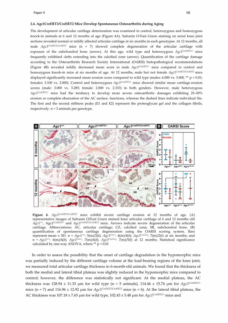
Paper II 58
2.4. Agc1CreERT2/CreERT2 Mice Develop Spontaneous Osteoarthritis during Aging
The development of articular cartilage deterioration was examined in control, heterozygous and homozygous knock-in animals at 6 and 12 months of age (Figure 4A). Safranin O-Fast Green staining on serial knee joint sections revealed normal or mildly affected articular cartilage at six months in each genotypes. At 12 months, all male Agc1CreERT2/CreERT2 mice (n = 7) showed complete degeneration of the articular cartilage with exposure of the subchondral bone (arrow). At this age, wild type and heterozygous Agc1CreERT2/+ mice frequently exhibited clefts extending into the calcified zone (arrow). Quantification of the cartilage damage according to the Osteoarthritis Research Society International (OARSI) histopathological recommendations (Figure 4B) revealed mildly decreased mean score in male Agc1CreERT2/+ mice compared to control and homozygous knock-in mice at six months of age. At 12 months, male but not female Agc1CreERT2/CreERT2 mice displayed significantly increased mean erosion score compared to wild type (males: 6.000 vs. 3.000, ** p < 0.01; females: 3.100 vs. 2.000). Control and heterozygous Agc1CreERT2/+ mice showed similar mean cartilage erosion scores (male: 3.000 vs. 3.285; female: 2.000 vs. 2.333) in both genders. However, male heterozygous Agc1CreERT2/+ mice had the tendency to develop more severe osteoarthritic damages exhibiting 25–50% erosion or complete eburnation of the AC surface. functions, whereas the dashed lines indicate individual fits. The first and the second stiffness peaks (E1 and E2) represent the proteoglycan gel and the collagen fibrils, respectively. n = 3 animals per genotype.
Figure 4. Agc1CreERT2/CreERT2 mice exhibit severe cartilage erosion at 12 months of age. (A) representative images of Safranin O/Fast Green stained knee articular cartilage of 6 and 12 months old Agc1+/+, Agc1CreERT2/+ and Agc1CreERT2/CreERT2 mice. Arrows indicate severe degeneration of the articular cartilage. Abbreviations: AC, articular cartilage; CZ, calcified zone; SB, subchondral bone. (B) quantification of spontaneous cartilage degeneration using the OARSI scoring system. Bars represent mean ± SD. n = Agc1+/+: 5(m)/2(f); Agc1Cre/+: 4(m)/4(f); Agc1Cre/Cre: 7(m)/2(f) at six months; and n = Agc1+/+: 6(m)/4(f); Agc1Cre/+: 7(m)/6(f); Agc1Cre/Cre: 7(m)/5(f) at 12 months. Statistical significance calculated by one-way ANOVA, where ** p < 0.01
In order to assess the possibility that the onset of cartilage degradation in the hypomorphic mice was partially induced by the different cartilage volume at the load-bearing regions of the knee joint, we measured total articular cartilage thickness in 6-month-old animals. We found that the thickness of both the medial and lateral tibial plateau was slightly reduced in the hypomorphic mice compared to control; however, the difference was statistically not significant. At the medial plateau, the AC thickness was 128.94 ± 11.33 µm for wild type (n = 5 animals), 114.46 ± 15.74 µm for Agc1CreERT2/+
mice (n = 7) and 116.96 ± 12.92 µm for Agc1CreERT2/CreERT2 mice (n = 6). At the lateral tibial plateau, the AC thickness was 107.18 ± 7.65 µm for wild type, 102.43 ± 5.48 µm for Agc1CreERT2/+ mice and

Paper II 59
105.54 ± 10.33 µm for Agc1CreERT2/CreERT2 mice. These data indicate that articular cartilage thickness isapparently not affected by the decreased aggrecan levels, and the thickness of the cartilage does notcontribute to cartilage degeneration in the hypomorphic mice per se.
2.5. Higher Incidence of OA in Agc1CreERT2/CreERT2 Mice Is Not Correlated with Increased AC Catabolism
The reduced amount of aggrecan and the stiffer biomechanical environment in the articularcartilage might disturb metabolism inducing upregulation of catabolic enzymes which breakdownthe ECM. Therefore, we have analyzed the expression of several proteases, which are able to degradecollagens or aggrecan by immunohistochemistry at 12 months of age. We found comparable expressionof Matrix Metalloproteinase-13 (MMP-13), a typical collagenase upregulated during OA, and theaggrecanase A Disintegrin and Metalloproteinase with Thrombospondin Motifs-5 (ADAMTS-5) ineach genotype (Figure 5A). Similarly, immunostaining for MMP-3, MMP-9 and ADAMTS-4 showednodifference in the expression of these proteases in the articular cartilage of the Agc1CreERT2/CreERT2,Agc1CreERT2/+ and Agc1+/+ mice (not shown). Immunostaining with the Collagen Type I and II Cleavage(C1,2C) antibody (Figure 5A) and the Collagen Type II Cleavage (C2C) Elisa assay (Figure 5B), whichcan detect collagen degradation products on tissue sections and in the urine, respectively, indicated nodifference for collagen breakdown between the genotypes. Interestingly, when we stained the articularcartilage sections with an antibody recognizing the MMP-driven aggrecan degradation neo-epitopeVIDIPEN, we found gradually decreasing immunoreactivity in Agc1CreERT2/+ and Agc1CreERT2/CreERT2
mice compared to wild type.

Paper II 60
Figure 5. Agc1CreERT2/CreERT2 mice develop dwarfism which is maintained after skeletal maturation. (A) representative X-ray images of Agc1+/+, Agc1CreERT2/+ and Agc1CreERT2/CreERT2 mice at 6 and 12 months of age. (B–F) analysis of body weight (B), body length (C), L4 vertebra length (D), tibia length (E) and humerus length (F) at 6 and 12 months of age. Bars represent the mean ± standard deviation (SD). n > Agc1+/+: 6(m)/4(f); Agc1Cre/+: 6(m)/4(f); Agc1Cre/Cre: 6(m)/4(f) at six months; and n > Agc1+/+: 6(m)/6(f); Agc1Cre/+: 7(m)/7(f); Agc1Cre/Cre: 7(m)/5(f) at 12 months. Statistical significance calculated by one-way ANOVA where * p < 0.5; ** p < 0.01; *** p < 0.001.

Paper II 61
and permanent cartilaginous tissues. In this study, we have investigated the consequence of decreasedaggrecan expression for articular cartilage function by analyzing the hypomorphic Agc1CreERT2 mouseline. We found that reduced aggrecan leads to increased stiffness of the articular cartilage, which inturn results in severe age-associated osteoarthritis.
Mutations in the human aggrecan gene lead to aggrecanopathies, an increasing group ofskeletal dysplasias ranging from mild idiopathic short stature to severe chondrodysplasias [9].Missense mutations typically found in the C-terminal G3 domain of the aggrecan core proteinare likely to disrupt interactions with other cartilage ECM proteins. The phenotypic spectrum ofthese neomorphic mutations includes mild short stature with accelerated bone maturation [12];osteochondritis dissecans with short stature and early-onset osteoarthritis [11]; and extreme shortstature with craniofacial and spinal abnormalities [10]. Heterozygous nonsense mutations accumulatedat the G1–G2 and CS1–CS2 domains introduce premature termination codon resulting in the formationof truncated proteins or haploinsufficiency due to nonsense-mediated mRNA degradation [9,31].These dominant mutations lead to spondyloepiphyseal dysplasia (SED) Kimberly type characterizedby proportionate short stature and severe premature osteoarthritis [32] or idiopathic short statureassociated with early-onset OA [12,33]. Null or functional null loss of function mutations in mice(cmd, cmd–bc) [13,14], chicken (nanomelia) [34,35] or cattle (Dexter bulldog dwarfism) [36] are perinatallethal due to severely abnormal skeletal growth caused by the lack of aggrecan in the cartilage ECM.
Although the findings in human and other species indicate that the absence or reduction of theaggrecan in the cartilage ECM leads to chondrodysplasias and osteoarthritis, the correlation betweenthe level of aggrecan expression and the clinical manifestation of the diseases is not clear. To date,only two mouse models have been investigated in order to link aggrecan gene expression to skeletalphenotype. The naturally occurring cartilage matrix deficiency (cmd) is caused by a 7 bp deletion inexon 5 of Agc1 which leads to a premature stop codon in exon 6 and formation of a short, truncatedprotein [13]. Homozygous (cmd/cmd) mice die at birth due to severe chondrodysplasia characterizedby a cleft palate, abnormal tracheal cartilage formation and dwarfism [13] caused by the loss of thenormal columnar structure of the growth plate [37]. Heterozygous cmd/+ mice, however, surviveand develop moderate proportional dwarfism at around one month of age. Furthermore, cmd/+ miceexhibit misalignment of the spine at one year of age, which eventually results in premature death at19 months due to eating difficulties [15]. In the Agc1CreERT2 mouse model, the 3´ UTR of the aggrecangene was disrupted by knocking in a Cre-ERT2 construct to generate an inducible, cartilage-specificdeleter mouse line [16]. Although the original publication did not describe a phenotype, it has laterbeen reported that Agc1CreERT2/CreERT2 mice develop dwarfism by one month of age associated withshortening of the growth plate [27]. Now, here we show that the dwarf phenotype exists after theend of skeletal maturation having about 10–20% smaller skeletal elements of the Agc1CreERT2/CreERT2
mice compared to wild type at 6 and 12 months of age (Figure 1). The size reduction affected boththe axial and the appendicular skeleton. Interestingly, we observed a mild, but sometimes statisticallysignificant, 3% to 6% reduction of the skeleton and body weight of Agc1CreERT2/+ mice indicating thatthe skeletally mature heterozygous animals also exhibit a very slight dwarfism.
Insertion of an exogenous targeting construct with selection cassette into intronic or untranslatedregions of an endogenous gene could disrupt important regulatory domains, affect splicing or directlyinfluence promoter activity of the endogenous or neighboring genes [38–42]. The interference with theendogenous gene activity leads to hypomorphic alleles with reduced mRNA and protein expression,which is proved to be useful to study the effect of gene doses in various disorders [38,39,43]. It hasbeen previously reported that insertion of the CreERT2 targeting construct into the Agc1 gene causesthe lack of 760 bp of the 3´-UTR. This could affect RNA stability resulting in 20% and 50% reduction ofaggrecan mRNA in adult, three-month-old knee epiphyseal cartilage of heterozygous and homozygousknock-in mice, respectively [27]. We found that Agc1 mRNA levels were reduced in newborn ribcartilage to ~74% in Agc1CreERT2/+ mice and to 64% in Agc1CreERT2/CreERT2 mice compared to the wildtype levels (Figure 2A). These observations indicate that disruption of the 3´UTR by the transgene
Paper II 62
insertion causes a similar reduction of the Agc1 mRNA levels in chondrocytes regardless of the ageor location of the cartilage tissue. In the cmd model, the Agc1 mRNA in embryonic limb cartilage isreduced to 41% in cmd/cmd mice and to 81% in cmd/+ mice [15]. Although the direct comparisonbetween the haploinsufficiency cmd/+ model and the hypomorph Agc1CreERT2 model is difficult dueto the different nature of the causative mutations, it is reasonable to speculate that about 25–50%reduction in the normal Agc1 mRNA level is sufficient to generate a mild dwarf phenotype in mice.The reduction of mRNA expression in this range affects the deposition of the aggrecan protein into theECM as demonstrated by 13% decreased the CS level in spinal cartilage from cmd/+ mice [15], ~50%less aggrecan in knee joint extracts from Agc1CreERT2/CreERT2 mice [27] and the greatly reduced aggrecanimmunoreactivity in the articular cartilage of Agc1CreERT2/CreERT2 knee joints (Figure 2B).
The most dramatic phenotype we found in the adult Agc1CreERT2/CreERT2 mice is the severedegeneration of the knee articular cartilage typical for late stage osteoarthritis (Figure 4). SinceOA was not described in aging cdm/+ mice, we hypothetise that Agc1CreERT2/CreERT2 mice have agreater loss of aggrecan in the articular cartilage which impairs its function and eventually leads tothe complete erosion of the AC surface in males by 12 months of age. The aggrecan–hyaluronanaggregates have a crucial role for maintaining the biomechanical properties of the articular cartilage byimmobilizing a vast amount of negative charges and generating an osmotic swelling pressure, whichprovides the tissue with resistance against compressive forces [3]. Agc1CreERT2/CreERT2 mice displayedreduced aggrecan and GAG content in the knee joint suggesting alterations in the biomechanicsof the articular cartilage. Applying nanoscale IT-AFM, we observed prominent stiffening of theAC from Agc1CreERT2/CreERT2 mice (Figure 3B) at six months of age, when histological differences ofAC degeneration were not evident between the genotypes (Figure 4B). The increased stiffness wasdetected in the superficial, middle and deep zones implying a systemic hardening of the wholearticular cartilage.
In normal cartilage, the swelling pressure is balanced by the tensile force of the collagennetwork and modifying the collagen fibril-hydrated proteoglycan ratio could affect the swellingand biomechanical properties of the tissue. Earlier studies have shown that porcine articular cartilageincubated in hypotonic salt solution or digested with proteoglycan-degrading enzymes has increasedcartilage nanostiffness [28,44]. Accordingly, the reduced negative charge density of the sGAG chainsdue to the decreased aggrecan level in the homozygous hypomorphic mice could result in reducedtissue hydration, decreased swelling pressure and increased constraining matrix stress, which in turnelevates cartilage stiffness. It is important to note, however, that the imbalance between osmoticswelling pressure and the tensile forces apparently does not affect cartilage volume, as the thickness ofthe tibial articular cartilage was not significantly altered in the Agc1CreERT2/CreERT2 mice.
We have previously shown that nanoscale indentation is a sensitive tool to assess cartilagebiomechanical properties of the growth plate, the articular cartilage and the intervertebraldisc [29,30,45–47]. We showed that IT-AFM depicts subtle biomechanical differences between wildtype and genetically modified mice carrying mutations, which affect the collagen network [45–47]or the proteoglycans [30]. The method is capable of resolving the biomechanical properties of thecollagen fibrils and the entrapped proteoglycan gel resulting in a bimodal stiffness distribution onthe recorded histograms [28,29]. We observed a shift for the stiffness of both the collagens andthe proteoglycans in the Agc1CreERT2/CreERT2 mice compared to wild type and Agc1CreERT2/+ mice.This general increase in stiffness of the major components of the cartilage matrix could lead to arigid AC surface, which is more susceptible for mechanical load-induced degradation. We havepreviously found that mice lacking collagen IX, an essential component of the heterotypic cartilagecollagen fibrils, also display increased nanostiffness of the hip articular cartilage before obvious signsof joint degeneration and the onset of osteoarthritis [45]. In human AC, nanostiffness is increasing byage due to GAG loss-induced dehydration of the proteoglycan moiety [45] or stiffening of the collagenfibrils [48] and could predispose for OA. However, disintegration of the cartilage extracellular matrix

Paper II 63
(e.g., the collagen meshwork) during the onset and progression of OA is accompanied by decreasedcompressive stiffness in both animal models and human patients [45,49,50].
During the course of OA, usually degradation of aggrecan followed by breakdown of the collagennetwork are critical steps for progressive cartilage destruction. In Agc1CreERT2/CreERT2 mice, the reducedlevel of aggrecan apparently does not affect cartilage metabolism as demonstrated by the normalexpression of MMP-13, ADAMTS-5 and collagen and aggrecan degradation neoepitopes (Figure 5).These findings suggest a physical activity-induced “wear and tear” mechanism for destruction of themechanically abnormal AC in the Agc1CreERT2/CreERT2 mice.
Epidemiological studies have shown that the prevalence, incidence and severity of osteoarthritisare higher in postmenopausal women than men due to hormonal, anatomical and genetic factors [51,52].In murine, however, sex hormones, body weight, behavior and activity differences betweengenders lead to higher incidence of joint degeneration in male mice in both age-associated andexperimentally-induced osteoarthritis models [53–55]. In line with this evidence, the cartilagedestruction was more severe in male Agc1CreERT2/CreERT2 mice than in female mice implying thatphysical activities (e.g., fighting) and high body weight could worsen the condition.
In summary, we have demonstrated that the hypomorphic Agc1CreERT2/CreERT2 mice developage-associated OA characterized by severe erosion of the articular cartilage by 12 months of age.The phenotype was associated with stiffening of the AC, indicating that aggrecan level crucially affectsbiomechanical properties of the cartilage matrix. As heterozygous mice occasionally show a tendencyfor reduced length of skeletal elements and increased cartilage destruction, we advise the cartilagefield for a careful interpretation of experiments with cartilage specific-deletion of floxed genes usingthe Agc1CreERT2 line. On the other hand, these hypomorphic mice are ideal models to study aggrecangene doses and skeletal phenotype correlation through the entire postnatal life with implications forhuman aggrecanopathies.
4. Materials and Methods
4.1. Mouse Model and Husbandry
Agc1tm(IRES-CreERT2) [16] were purchased from the Jackson Laboratory (JAX stock #019148).Upon arrival, mice were bred with 129/Sv outbred mice for at least two generations. Mice werehoused in groups of 2–4 per cage and received food and water ad libitum. They were maintained ina temperature- and air- controlled environment, with 12 h light/dark cycle. Animals were handledand housed according to the federal and institutional guidelines for the care and use of laboratoryanimals, approved by the Central Animal Facility of the LMU Munich and the government of UpperBavaria (Application number: 55.2-1-54-2532-15-2016). For the sake of simplicity, control, heterozygousand homozygous mice are denoted as Agc1+/+, Agc1CreERT2/+ and Agc1CreERT2/CreERT2, respectively.Newborn, 6- and 12-month-old male and female mice were included in the study. Genotyping wasperformed according to the Jackson Laboratory protocol. Mice were sacrificed via cervical dislocationand tissue collection performed post mortem.
4.2. Radiography and Morphometry
X-ray images of euthanized mice were taken with a Faxitron X-ray cabinet (model 43855A) at 35 kV,2 mA and 2 sec exposure time. Total body length (from the snout to the end of the tail) and the length ofthe skeletal elements (4th lumbar vertebra, tibia, femur and humerus) were analyzed with the distancemeasurement plug-in of the syngo Imaging XS-VA60B software (Siemens, Erlangen, Germany).
4.3. Sample Preparation
Hind limbs of euthanized mice were de-skinned and partially cleaned from the muscles aroundthe knee joints. For histology and immunohistochemistry, knee joints from the left side were fixed in4% paraformaldehyde (PFA) in phosphate buffered saline (PBS) pH 7.4 (Sigma-Aldrich, Taufkirchen,

Paper II 64
Germany), overnight at 4 ◦C. The next day, samples were washed 3 times in PBS and afterwardsdecalcified for approximately 4 weeks in a 20% ethylenediaminetetraacetic acid (EDTA)/PBS pH8.0 solution (Sigma-Aldrich). Decalcified specimens were washed 3 times in PBS, then dehydratedthrough an ascending row of ethanol, cleared in xylol and finally embedded in paraffin. Samples werecut on a Rotary Microtome HM360 in 8 µm sagittal sections and placed on Superfrost plus glass slides(Thermo Scientific, Madison, WI, USA). For AFM measurements, the correspondent right knee jointswere directly enclosed in Tissue-Tek cryomedia (Sakura, Zoeterwoude, NL, USA) on the base of plasticdisposable cassettes, and gradually frozen by transferring them on a chilled copper plate placed ondry ice. Sagittal sections of 30 µm were cut with the aid of adhesive tape (as described in [29]) using aMicrom HM500 cyostat (Thermo Scientific) and kept at −20 ◦C until use.
4.4. Histology
For the histological examination of proteoglycan content, Toluidine blue and Safranin O/FastGreen staining were performed. For Toluidine blue staining, tissue sections were deparaffinized,rehydrated and incubated for 10 min in 0.1% Toluidine blue in toluidine buffer pH 2.5. Excess stain,was adsorbed with Whatman paper and then slides were immersed in a 2% K3Fe(CN6)/dH2O solutionfor 3 min. Finally, sections were directly mount with Roti-histokit (Roth, Karlsruhe, Germany).For Safranin O/Fast Green staining a 0.5% hematoxylin staining solution (Roth) was used for 2 minand then washed with tap water for 5 min. After dipping in 1% acetic alcohol (Merck, Darmstadt,Germany). slides were incubated in a 0.02% fast green solution (Sigma-Aldrich) for 1 min. Next, tissuesections were immersed in 1% acetic acid for 30 s and then a 1% Safranin O solution (Sigma-Aldrich)was applied for 30 min. After three changes of 95% ethanol, and two of 100% ethanol, sections werecleared in xylol and mounted with Roti-Histokit. The thickness of the tibial articular cartilage in6-month-old animals was measured as previously described [56]. Briefly, representative micrographsof Safranin O/Fast Green sections from the lateral and medial tibial plateau were analyzed by usingthe linear measure tool of the ZEN 2.3 Lite image capturing and processing software (Carl Zeiss, Jena,Germany). In each section, three regions of the articular cartilage extending from the surface to thesubchondral bone were measured. The mean of the three values was used to characterize the totalthickness of the articular cartilage. At least five animals of each genotype were evaluated.
4.5. Scoring of Articular Cartilage Erosion
The pathological scoring of AC has been performed on Safranin O/Fast Green stained sagittaltissue sections. To have a full representation of the entire knee, about 10 slides, harvested atapproximately 80 µm intervals, were chosen. Slides were evaluated by two independent observersin a blinded fashion. To assess cartilage degeneration, the OARSI scoring system was used [57].Evaluation of articular cartilage structure comprehends eight grades as follows: 0—normal cartilage;0.5—loss of proteoglycan stain without cartilage damage; 1—Surface fibrillations; 2—Cleft extendingjust below the superficial zone; 3—Cleft/erosion to the calcified cartilage extending to < 25%of the articular surface; 4—25–50% loss of articular surface; 5—50–75% loss of articular surface;6—Eburnation with > 75% loss of cartilage. The means of the two independent observers of themaximal histopathological scores obtained across the entire knee joints were presented
4.6. Immunohistochemistry
For immunohistochemistry, paraffin embedded sections were deparaffinized, rehydrated anddigested with 2 mg/mL bovine testicular hyaluronidase/PBS pH 5.0 at 37 ◦C for 30 min. Endogenousperoxidase activity was quenched with a 0.3% H2O2/methanol solution for 30 min. Sections wereblocked with 1% bovine serum albumin/PBS for 60 min at room temperature. Afterwards, slideswere incubated overnight at 4 ◦C with the following antibodies diluted in blocking solution: aggrecan(Millipore ab1031, 1:200, Beverly, MA, USA), collagen type II (DSHB CIICI, 5 µg/mL), MMP-13(Sigma Aldrich MAB13424, 1:100), ADAMTS-5 (Abcam ab13976, 1:500, Cambridge, MA, USA),

Paper II 65
VIDIPEN (a gift of Amanda Fosang, University of Melbourne, 1:1000), and C1,2C (IBEX 50-1035, 1:400).The next day, sections were incubated with the correspondent biotinylated secondary antibodiesand immunostained by the avidin-biotin complex (Vectastain ABC Elite kit) method or the VectorM.O.M kit (both Vector Laboratories, Burlingame, CA, USA) and the 3,3’-diaminobenzidine (Sigma)as chromogenic substrate. Finally, sections were counterstained with hematoxylin and mountedwith Roti-Histokit.
4.7. RNA Isolation and Quantitative RT-PCR
Total RNA was isolated from primary chondrocytes of newborn mice. Briefly, rib cages fromeuthanized mice were dissected, cleaned from the attached tissues with forceps and digestedfor 30 min in a solution of 2 mg/mL collagenase type 2 (Worthington, Malvern, PA, USA) inDulbecco’s Modified Eagle’s (DMEM)/Ham’s F12 1:1 (Thermo Fischer Scientific) supplemented with1× penicillin/streptomycin (Biochrom, Berlin, Germany). After the first enzymatic digestion, theperichondrium was removed and cleaned ribs were re-placed in a new collagenase solution for 3 h at37 ◦C in a humidified incubator with 5% CO2. The obtained cell suspension was centrifuged at 500 gfor 5 min and washed with PBS. Total RNA isolation and cDNA synthesis were performed with theQiagen RNeasy Mini kit (Qiagen, Hilden, Germany) and the Transcriptor First-Strand cDNA Synthesiskit (Roche, Basel, Switzerland), respectively, according to the manufacturer’s instructions. QuantitativeRT-PCR was performed using the gene specific TaqMan probes from Integrated DNA Technologies forglyceraldehyde 3-phosphate dehydrogenase (GAPDH) (Mm.PT.39a.1) and aggrecan (Mm.PT.58.10174685).PrimeTime Gene Expression Master Mix, dH2O and cDNA samples were pipetted in triplicates in a96-well plate and a PCR reaction was carried out in a LightCycler 96 thermocycler provided with theLightCycler 96 Software (Roche). The relative gene expression is calculated against the housekeepinggene GAPDH by the comparative ∆Ct method. Results in graph show the mean ± standard deviationof two PCR runs from three mice per genotypes.
4.8. Atomic Force Microscopy
Nano-scale indentation-type atomic force microscopy (IT-AFM) was carried out using a JPKNanoWizard®1 (JPK Instruments, Berlin, Germany) mounted on an inverse Axiovert 200 opticalmicroscope (Carl Zeiss, Jena, Germany) to enable a precise positioning of the AFM tip on the region ofinterest. For measurements, silicon nitride cantilevers (MLCT, Cantilever E, Bruker) with a nominalspring constant of 0.1 N/m and pyramidal tips with a nominal radius of 20 nm were used. All IT-AFMmeasurements were carried out in PBS at pH 7.4. In order to determine the actual cantilever springconstant, each cantilever was individually calibrated using the thermal noise method [58]. Every springconstant determination was repeated three times and the mean value was used for data analysis. In eachcartilage zone (superficial, middle and deep) an area of 3 × 3 µm, 25 × 25 force-indentation curveswere recorded with a constant vertical tip velocity of 15 µm/s and a sampling rate of 5 kHz. For dataanalysis, the JPK Data Processing Software was used (Version 6.1.102, JPK Instruments). The Young’smodulus was extracted from the approach section of the force-indentation curves from the contactpoint up to a maximum indentation depth of 500 nm, using a modified Hertz model for a pyramidalindenter as described in [29,59]. For comparison, stiffness distributions (histograms) were calculatedfor each developmental stage and genotype. Two sections per animal were scanned with a total of3 × 625 force-indentation curves for each cartilage zone. To locate the maxima of these histograms,a linear combination of two Gaussian distributions was fitted to each histogram using the Igor Prosoftware (Version 6.3.7.2, WaveMetrics, Portland, OR, USA).
4.9. Enzyme-Linked Immunosorbent Assay
In order to detect the levels of collagen type 2 degradation products, the commercially availableMouse Collagen Type 2 Cleavage ELISA kit (MyBioSource, MBS725817, San Diego, CA, USA) wasused according to the manufacturer instructions. Urine samples were collected from 12-month-old

Paper II 66
mice on the day of sacrification and kept at −80 ◦C until use. C2C levels are expressed in ng/mLand were normalized to urinary creatinine concentration. Quantitative determination of creatininein urine samples was assessed with the Mouse Creatinine kit from Crystal Chem (Cat. 80350) as permanufacturer protocol.
4.10. Statistical Analysis
Statistical significance was calculated after determination of the Gaussian distribution usinga one-way ANOVA test (GraphPad Prism, San Diego, CA, USA) with appropriate post hoc tests.Statistical significance was assumed at a p-value of ≤ 0.05. All values represent the mean and thestandard deviation (SD). In all experiments, at least three animals were used (a list of the precisenumber of animals used in each experiment is stated in figure legends).
5. Conclusions
In conclusion, our data demonstrate that Agc1CreERT2/CreERT2 mice have smaller skeleton, stifferarticular cartilage and are prone to spontaneous, age-associated osteoarthritis owing to reduceddeposition of aggrecan in cartilage. Therefore, the homozygous Agc1CreERT2/CreERT2 mice should notbe used for conditional gene ablation experiments assessing gene function in the articular cartilage.However, the Agc1CreERT2/CreERT2 mice could serve as valuable model for studying aggrecanopathiescaused by reduced aggrecan expression in the cartilaginous skeleton.
Author Contributions: Study conception, design and manuscript writing: P.A., H.C.-S. and A.A. Performedexperiments: P.A., H.C.D, B.H., P.L. and Z.F. Data visualization and statistical analysis: M.M.S. and B.H.
Funding: This work was funded by the German Research Foundation (DFG) as part of Subproject 1 (AA 150/11-1and CL 409/4-1) of the Research Consortium ExCarBon / FOR2407/1.
Acknowledgments: The authors would like to thank Marvin Althaus for technical help on tissues sectioning andthe Department of Radiology (LMU Munich) for the help in X-ray micrograph downloading.
Conflicts of Interest: The authors confirm that there are no conflicts of interest.
References
1. Aspberg, A. The Different Roles of Aggrecan Interaction Domains. J. Histochem. Cytochem. 2012, 60, 987–996.[CrossRef] [PubMed]
2. Aspberg, A. Cartilage Proteoglycans. In Cartilage; Springer International Publishing: Cham, Switzerland,2016; pp. 1–22.
3. Kiani, C.; Chen, L.; Wu, Y.J.; Yee, A.J.; Yang, B.B. Structure and function of aggrecan. Cell Res. 2002, 12, 19–32.[CrossRef] [PubMed]
4. Hedlund, H.; Hedbom, E.; Heinegrd, D.; Mengarelli-Widholm, S.; Reinholt, F.P.; Svensson, O. Association ofthe aggrecan keratan sulfate-rich region with collagen in bovine articular cartilage. J. Biol. Chem. 1999, 274,5777–5781. [CrossRef] [PubMed]
5. Cortes, M.; Baria, A.T.; Schwartz, N.B. Sulfation of chondroitin sulfate proteoglycans is necessary for properIndian hedgehog signaling in the developing growth plate. Development 2009, 136, 1697–1706. [CrossRef][PubMed]
6. Mouw, J.K.; Ou, G.; Weaver, V.M. Extracellular matrix assembly: A multiscale deconstruction. Nat. Rev. Mol.Cell Biol. 2014, 15, 771–785. [CrossRef] [PubMed]
7. Chambers, M.G.; Kuffner, T.; Cowan, S.K.; Cheah, K.S.E.; Mason, R.M. Expression of collagen and aggrecangenes in normal and osteoarthritic murine knee joints. Osteoarthr. Cartil. 2002, 10, 51–61. [CrossRef][PubMed]
8. Maroudas, A.; Bayliss, M.T.; Uchitel-Kaushansky, N.; Schneiderman, R.; Gilav, E. Aggrecan turnover inhuman articular cartilage: Use of aspartic acid racemization as a marker of molecular age. Arch. Biochem.Biophys. 1998, 350, 61–71. [CrossRef] [PubMed]
9. Gibson, B.G.; Briggs, M.D. The aggrecanopathies; an evolving phenotypic spectrum of human geneticskeletal diseases. Orphanet J. Rare Dis. 2016, 11, 86. [CrossRef] [PubMed]
Paper II 67
10. Tompson, S.W.; Merriman, B.; Funari, V.A.; Fresquet, M.; Lachman, R.S.; Rimoin, D.L.; Nelson, S.F.;Briggs, M.D.; Cohn, D.H.; Krakow, D. A Recessive Skeletal Dysplasia, SEMD Aggrecan Type, Resultsfrom a Missense Mutation Affecting the C-Type Lectin Domain of Aggrecan. Am. J. Hum. Genet. 2009, 84,72–79. [CrossRef] [PubMed]
11. Stattin, E.-L.; Tegner, Y.; Domellöf, M.; Dahl, N. Familial osteochondritis dissecans associated with earlyosteoarthritis and disproportionate short stature. Osteoarthr. Cartil. 2008, 16, 890–896. [CrossRef] [PubMed]
12. Nilsson, O.; Guo, M.H.; Dunbar, N.; Popovic, J.; Flynn, D.; Jacobsen, C.; Lui, J.C.; Hirschhorn, J.N.; Baron, J.;Dauber, A. Short stature, accelerated bone maturation, and early growth cessation due to heterozygousaggrecan mutations. J. Clin. Endocrinol. Metab. 2014, 99, E1510–E1518. [CrossRef] [PubMed]
13. Watanabe, H.; Kimata, K.; Line, S.; Strong, D.; Gao, L.Y.; Kozak, C.A.; Yamada, Y. Mouse cartilage matrixdeficiency (cmd) caused by a 7 bp deletion in the aggrecan gene. Nat. Genet. 1994, 7, 154–157. [CrossRef][PubMed]
14. Bell, L.; Juriloff, M.; Harris, M.J. A new mutation at the cmd locus in the mouse. J. Hered. 1986, 77, 205–206.[CrossRef] [PubMed]
15. Watanabe, H.; Nakata, K.; Kimata, K.; Nakanishi, I.; Yamada, Y. Dwarfism and age-associated spinaldegeneration of heterozygote cmd mice defective in aggrecan. Proc. Natl. Acad. Sci. USA 1997, 94, 6943–6947.[CrossRef] [PubMed]
16. Henry, S.P.; Jang, C.-W.; Deng, J.M.; Zhang, Z.; Behringer, R.R.; de Crombrugghe, B. Generation ofaggrecan-CreERT2 knockin mice for inducible Cre activity in adult cartilage. Genesis 2009, 47, 805–814.[CrossRef] [PubMed]
17. Alvarez-Garcia, O.; Matsuzaki, T.; Olmer, M.; Miyata, K.; Mokuda, S.; Sakai, D.; Masuda, K.; Asahara, H.;Lotz, M.K. FOXO are required for intervertebral disk homeostasis during aging and their deficiency promotesdisk degeneration. Aging Cell 2018, 17, e12800. [CrossRef] [PubMed]
18. Wang, C.; Abu-Amer, Y.; O’Keefe, R.J.; Shen, J. Loss of Dnmt3b in Chondrocytes Leads to DelayedEndochondral Ossification and Fracture Repair. J. Bone Miner. Res. 2018, 33, 283–297. [CrossRef] [PubMed]
19. Diekman, B.O.; Sessions, G.A.; Collins, J.A.; Knecht, A.K.; Strum, S.L.; Mitin, N.K.; Carlson, C.S.; Loeser, R.F.;Sharpless, N.E. Expression of p16INK4ais a biomarker of chondrocyte aging but does not cause osteoarthritis.Aging Cell 2018, 17, e12771. [CrossRef] [PubMed]
20. Alkhatib, B.; Liu, C.; Serra, R. Tgfbr2 is required in Acan-expressing cells for maintenance of the intervertebraland sternocostal joints. JOR Spine 2018, 1, e1025. [CrossRef] [PubMed]
21. Liao, L.; Zhang, S.; Gu, J.; Takarada, T.; Yoneda, Y.; Huang, J.; Zhao, L.; Oh, C.D.; Li, J.; Wang, B.; et al.Deletion of Runx2 in Articular Chondrocytes Decelerates the Progression of DMM-Induced Osteoarthritis inAdult Mice. Sci. Rep. 2017, 7, 2371. [CrossRef] [PubMed]
22. Henry, S.P.; Liang, S.; Akdemir, K.C.; De Crombrugghe, B. The postnatal role of Sox9 in cartilage. J. BoneMiner. Res. 2012, 27, 2511–2525. [CrossRef] [PubMed]
23. Couasnay, G.; Bon, N.; Devignes, C.S.; Sourice, S.; Bianchi, A.; Véziers, J.; Weiss, P.; Elefteriou, F.; Provot, S.;Guicheux, J.; et al. PiT1/Slc20a1 Is Required for Endoplasmic Reticulum Homeostasis, Chondrocyte Survival,and Skeletal Development. J. Bone Miner. Res. 2018, 34, 387–398. [CrossRef] [PubMed]
24. Liao, L.; Jiang, H.; Fan, Y.; Lu, R.S.; Wei, C.; Takarada, T.; He, S.; Chen, D. Runx2 is required for postnatalintervertebral disc tissue growth and development. J. Cell. Physiol. 2019, 234, 6679–6687. [CrossRef][PubMed]
25. Zhang, Y.; Sheu, T.; Hoak, D.; Shen, J.; Hilton, M.J.; Zuscik, M.J.; Jonason, J.H.; O’Keefe, R.J. CCN1 RegulatesChondrocyte Maturation and Cartilage Development. J. Bone Miner. Res. 2016, 31, 549–559. [CrossRef][PubMed]
26. Zhou, X.; von der Mark, K.; Henry, S.; Norton, W.; Adams, H.; de Crombrugghe, B. ChondrocytesTransdifferentiate into Osteoblasts in Endochondral Bone during Development, Postnatal Growth andFracture Healing in Mice. PLoS Genet. 2014, 10, e1004820. [CrossRef] [PubMed]
27. Rashid, H.; Chen, H.; Hassan, Q.; Javed, A. Dwarfism in homozygous Agc1CreERT mice is associated withdecreased expression of aggrecan. Genesis 2017, 55, e23070. [CrossRef] [PubMed]
28. Loparic, M.; Wirz, D.; Daniels, A.U.; Raiteri, R.; Vanlandingham, M.R.; Guex, G.; Martin, I.; Aebi, U.; Stolz, M.Micro- and nanomechanical analysis of articular cartilage by indentation-type atomic force microscopy:Validation with a gel-microfiber composite. Biophys. J. 2010, 98, 2731–2740. [CrossRef] [PubMed]

Paper II 68
29. Prein, C.; Warmbold, N.; Farkas, Z.; Schieker, M.; Aszodi, A.; Clausen-Schaumann, H. Structural andmechanical properties of the proliferative zone of the developing murine growth plate cartilage assessed byatomic force microscopy. Matrix Biol. 2016, 50, 1–15. [CrossRef] [PubMed]
30. Gronau, T.; Krüger, K.; Prein, C.; Aszodi, A.; Gronau, I.; Iozzo, R.V.; Mooren, F.C.; Clausen-Schaumann, H.;Bertrand, J.; Pap, T.; et al. Forced exercise-induced osteoarthritis is attenuated in mice lacking the smallleucine-rich proteoglycan decorin. Ann. Rheum. Dis. 2017, 76, 442–449. [CrossRef] [PubMed]
31. Hauer, N.N.; Sticht, H.; Boppudi, S.; Büttner, C.; Kraus, C.; Trautmann, U.; Zenker, M.; Zweier, C.;Wiesener, A.; Jamra, R.A.; et al. Genetic screening confirms heterozygous mutations in ACAN as a majorcause of idiopathic short stature. Sci. Rep. 2017, 7, 12225. [CrossRef] [PubMed]
32. Gleghorn, L.; Ramesar, R.; Beighton, P.; Wallis, G. A mutation in the variable repeat region of the aggrecangene (AGC1) causes a form of spondyloepiphyseal dysplasia associated with severe, premature osteoarthritis.Am. J. Hum. Genet. 2005, 77, 484–490. [CrossRef] [PubMed]
33. Gkourogianni, A.; Andrew, M.; Tyzinski, L.; Crocker, M.; Douglas, J.; Dunbar, N.; Fairchild, J.; Funari, M.F.A.;Heath, K.E.; Jorge, A.A.L.; et al. Clinical Characterization of Patients With Autosomal Dominant ShortStature due to Aggrecan Mutations. J. Clin. Endocrinol. Metab. 2017, 102, 460–469. [CrossRef] [PubMed]
34. Primorac, D.; Stover, M.L.; Clark, S.H.; Rowe, D.W. Molecular basis of nanomelia, a heritablechondrodystrophy of chicken. Matrix Biol. 1994, 14, 297–305. [CrossRef]
35. Vertel, B.M.; Grier, B.L.; Li, H.; Schwartz, N.B. The chondrodystrophy, nanomelia: Biosynthesis andprocessing of the defective aggrecan precursor. Biochem. J. 1994, 301, 211–216. [CrossRef] [PubMed]
36. Cavanagh, J.A.L.; Tammen, I.; Windsor, P.A.; Bateman, J.F.; Savarirayan, R.; Nicholas, F.W.; Raadsma, H.W.Bulldog dwarfism in Dexter cattle is caused by mutations in ACAN. Mamm. Genome 2007, 18, 808–814.[CrossRef] [PubMed]
37. Lauing, K.L.; Cortes, M.; Domowicz, M.S.; Henry, J.G.; Baria, A.T.; Schwartz, N.B. Aggrecan is required forgrowth plate cytoarchitecture and differentiation. Dev. Biol. 2014, 396, 224–236. [CrossRef] [PubMed]
38. Fritsch, A.; Loeckermann, S.; Kern, J.S.; Braun, A.; Bösl, M.R.; Bley, T.A.; Schumann, H.; von Elverfeldt, D.;Paul, D.; Erlacher, M.; et al. A hypomorphic mouse model of dystrophic epidermolysis bullosa revealsmechanisms of disease and response to fibroblast therapy. J. Clin. Investig. 2008, 118, 1669–1679. [CrossRef][PubMed]
39. Mohn, A.R.; Gainetdinov, R.R.; Caron, M.G.; Koller, B.H. Mice with reduced NMDA receptor expressiondisplay behaviors related to schizophrenia. Cell 1999, 98, 427–436. [CrossRef]
40. Nagy, A.; Moens, C.; Ivanyi, E.; Pawling, J.; Gertsenstein, M.; Hadjantonakis, A.K.; Pirity, M.; Rossant, J.Dissecting the role of N-myc in development using a single targeting vector to generate a series of alleles.Curr. Biol. 1998, 8, 661–664. [CrossRef]
41. Pan, Y.; Zhang, L.; Liu, Q.; Li, Y.; Guo, H.; Peng, Y.; Peng, H.; Tang, B.; Hu, Z.; Zhao, J.; et al. Insertionof a knockout-first cassette in Ampd1 gene leads to neonatal death by disruption of neighboring genesexpression. Sci. Rep. 2016, 6, 35970. [CrossRef] [PubMed]
42. Pham, C.T.; MacIvor, D.M.; Hug, B.A.; Heusel, J.W.; Ley, T.J. Long-range disruption of gene expression by aselectable marker cassette. Proc. Natl. Acad. Sci. USA 1996, 93, 13090–13095. [CrossRef] [PubMed]
43. Raffai, R.L.; Weisgraber, K.H. Hypomorphic apolipoprotein E mice: A new model of conditional generepair to examine apolipoprotein E-mediated metabolism. J. Biol. Chem. 2002, 277, 11064–11068. [CrossRef][PubMed]
44. Stolz, M.; Raiteri, R.; Daniels, A.U.; VanLandingham, M.R.; Baschong, W.; Aebi, U. Dynamic elastic modulusof porcine articular cartilage determined at two different levels of tissue organization by indentation-typeatomic force microscopy. Biophys. J. 2004, 86, 3269–3283. [CrossRef]
45. Stolz, M.; Gottardi, R.; Raiteri, R.; Miot, S.; Martin, I.; Imer, R.; Staufer, U.; Raducanu, A.; Düggelin, M.;Baschong, W.; et al. Early detection of aging cartilage and osteoarthritis in mice and patient samples usingatomic force microscopy. Nat. Nanotechnol. 2009, 4, 186–192. [CrossRef] [PubMed]
46. Aro, E.; Salo, A.M.; Khatri, R.; Finnilä, M.; Miinalainen, I.; Sormunen, R.; Pakkanen, O.; Holster, T.;Soininen, R.; Prein, C.; et al. Severe Extracellular Matrix Abnormalities and Chondrodysplasia in MiceLacking Collagen Prolyl 4-Hydroxylase Isoenzyme II in Combination with a Reduced Amount of Isoenzyme I.J. Biol. Chem. 2015, 290, 16964–16978. [CrossRef] [PubMed]

Paper II 69
47. Kamper, M.; Hamann, N.; Prein, C.; Clausen-Schaumann, H.; Farkas, Z.; Aszodi, A.; Niehoff, A.; Paulsson, M.;Zaucke, F. Early changes in morphology, bone mineral density and matrix composition of vertebrae lead todisc degeneration in aged collagen IX -/- mice. Matrix Biol. 2016, 49, 132–143. [CrossRef] [PubMed]
48. Wen, C.-Y.; Wu, C.-B.; Tang, B.; Wang, T.; Yan, C.-H.; Lu, W.W.; Pan, H.; Hu, Y.; Chiu, K.-Y. Collagen fibrilstiffening in osteoarthritic cartilage of human beings revealed by atomic force microscopy. Osteoarthr. Cartil.2012, 20, 916–922. [CrossRef] [PubMed]
49. Kleemann, R.U.; Krocker, D.; Cedraro, A.; Tuischer, J.; Duda, G.N. Altered cartilage mechanics and histologyin knee osteoarthritis: Relation to clinical assessment (ICRS Grade). Osteoarthr. Cartil. 2005, 13, 958–963.[CrossRef] [PubMed]
50. Wilusz, R.E.; Zauscher, S.; Guilak, F. Micromechanical mapping of early osteoarthritic changes in thepericellular matrix of human articular cartilage. Osteoarthr. Cartil. 2013, 21, 1895–1903. [CrossRef] [PubMed]
51. O’Connor, M.I. Sex differences in osteoarthritis of the hip and knee. J. Am. Acad. Orthop. Surg. 2007, 15,S22–S25. [CrossRef] [PubMed]
52. Palazzo, C.; Nguyen, C.; Lefevre-Colau, M.-M.; Rannou, F.; Poiraudeau, S. Risk factors and burden ofosteoarthritis. Ann. Phys. Rehabil. Med. 2016, 59, 134–138. [CrossRef] [PubMed]
53. Sokoloff, L. Natural history of degenerative joint disease in small laboratory animals. I. Pathological anatomyof degenerative joint disease in mice. AMA Arch. Pathol. 1956, 62, 118–128. [PubMed]
54. van Osch, G.J.; van der Kraan, P.M.; Vitters, E.L.; Blankevoort, L.; van den Berg, W.B. Induction ofosteoarthritis by intra-articular injection of collagenase in mice. Strain and sex related differences.Osteoarthr. Cartil. 1993, 1, 171–177. [CrossRef]
55. Ma, H.-L.; Blanchet, T.J.; Peluso, D.; Hopkins, B.; Morris, E.A.; Glasson, S.S. Osteoarthritis severity is sexdependent in a surgical mouse model. Osteoarthr. Cartil. 2007, 15, 695–700. [CrossRef] [PubMed]
56. Raducanu, A.; Hunziker, E.B.; Drosse, I.; Aszodi, A. B1 Integrin Deficiency Results in Multiple Abnormalitiesof the Knee Joint. J. Biol. Chem. 2009, 284, 23780–23792. [CrossRef] [PubMed]
57. Glasson, S.S.; Chambers, M.G.; Van Den Berg, W.B.; Little, C.B. The OARSI histopathologyinitiative—Recommendations for histological assessments of osteoarthritis in the mouse. Osteoarthr. Cartil.2010, 18, S17–S23. [CrossRef] [PubMed]
58. Butt, H.-J.; Jaschke, M. Calculation of thermal noise in atomic force microscopy. Nanotechnology 1995, 6.[CrossRef]
59. Sneddon, I.N. The relation between load and penetration in the axisymmetric boussinesq problem for apunch of arbitrary profile. Int. J. Eng. Sci. 1965, 3, 47–57. [CrossRef]
© 2019 by the authors. Licensee MDPI, Basel, Switzerland. This article is an open accessarticle distributed under the terms and conditions of the Creative Commons Attribution(CC BY) license (http://creativecommons.org/licenses/by/4.0/).

70
REFERENCES 1. Buck DW, 2nd, Dumanian GA: Bone biology and physiology: Part I. The fundamentals. Plastic
and reconstructive surgery 2012, 129(6):1314-1320.2. Kothari ML, Mehta LA, Natrajan M: The nature of bones and joints: a new perspective. Journal
of postgraduate medicine 1990, 36(3):143-146.3. Caplan AI: Bone development. Ciba Foundation symposium 1988, 136:3-21.4. Mackie EJ, Ahmed YA, Tatarczuch L, Chen KS, Mirams M: Endochondral ossification: how
cartilage is converted into bone in the developing skeleton. The international journal ofbiochemistry & cell biology 2008, 40(1):46-62.
5. Hunter WL, Arsenault AL: Vascular invasion of the epiphyseal growth plate: analysis ofmetaphyseal capillary ultrastructure and growth dynamics. The Anatomical record 1990,227(2):223-231.
6. Reddi AH: Bone and cartilage differentiation. Curr Opin Genet Dev 1994, 4(5):737-744.7. Zaleske DJ: Cartilage and bone development. Instr Course Lect 1998, 47:461-468.8. Hayes AJ, MacPherson S, Morrison H, Dowthwaite G, Archer CW: The development of
articular cartilage: evidence for an appositional growth mechanism. Anatomy andembryology 2001, 203(6):469-479.
9. Kuettner KE, Schleyerbach R, Hascall VC: Articular cartilage biochemistry. In.: Raven Press, NewYork, NY; 1986.
10. Kuettner KEJCb: Biochemistry of articular cartilage in health and disease. 1992, 25(3):155-163.11. Cohen NP, Foster RJ, Mow VCJJoO, Therapy SP: Composition and dynamics of articular
cartilage: structure, function, and maintaining healthy state. 1998, 28(4):203-215.12. Buckwalter J, Mankin HJJoB, surgery j: Articular cartilage: part I. 1997, 79(4):600.13. Theocharis AD, Skandalis SS, Gialeli C, Karamanos NK: Extracellular matrix structure.
Advanced drug delivery reviews 2016, 97:4-27.14. Velleman SG: The role of the extracellular matrix in skeletal development. Poultry science
2000, 79(7):985-989.15. Suzuki F: Roles of cartilage matrix proteins, chondromodulin-I and -II, in endochondral
bone formation: a review. Connect Tissue Res 1996, 35(1-4):303-307.16. Thompson JR, Oegema JTJTJob, volume jsA: Metabolic activity of articular cartilage in
osteoarthritis. An in vitro study. 1979, 61(3):407-416.17. Kuivaniemi H, Tromp G, Prockop DJJHm: Mutations in fibrillar collagens (types I, II, III, and XI),
fibril‐associated collagen (type IX), and network‐forming collagen (type X) cause a spectrum ofdiseases of bone, cartilage, and blood vessels. 1997, 9(4):300-315.
18. Mundlos S, Olsen BJTFj: Heritable diseases of the skeleton. Part II: Molecular insights intoskeletal development-matrix components and their homeostasis. 1997, 11(4):227-233.
19. Patterson SE, Dealy CN: Mechanisms and models of endoplasmic reticulum stressin chondrodysplasia. Developmental dynamics : an official publication of the AmericanAssociation of Anatomists 2014, 243(7):875-893.
20. Berendsen AD, Olsen BR: Bone development. Bone 2015, 80:14-18.21. Carter DR, Orr TE: Skeletal development and bone functional adaptation. Journal of bone
and mineral research : the official journal of the American Society for Bone and Mineral Research1992, 7 Suppl 2:S389-395.
22. Hoenderop JG, van Leeuwen JP, van der Eerden BC, Kersten FF, van der Kemp AW, MerillatAM,

71
Waarsing JH, Rossier BC, Vallon V, Hummler E et al: Renal Ca2+ wasting, hyperabsorption, and reduced bone thickness in mice lacking TRPV5. The Journal of clinical investigation 2003, 112(12):1906-1914.
23. Sugiki T, Uyama T, Toyoda M, Morioka H, Kume S, Miyado K, Matsumoto K, Saito H,Tsumaki N, Takahashi Y et al: Hyaline cartilage formation and enchondral ossification modeledwith KUM5 and OP9 chondroblasts. J Cell Biochem 2007, 100(5):1240-1254.
24. Yang Y: Skeletal morphogenesis during embryonic development. Critical reviews in eukaryoticgene expression 2009, 19(3):197-218.
25. Long F, Ornitz DM: Development of the endochondral skeleton. Cold Spring Harbor perspectivesin biology 2013, 5(1):a008334.
26. Horton WA, Degnin CR: FGFs in endochondral skeletal development. Trends in endocrinologyand metabolism: TEM 2009, 20(7):341-348.
27. Gerstenfeld L, Shapiro FJJocb: Expression of bone‐specific genes by hypertrophicchondrocytes: Implications of the complex functions of the hypertrophic chondrocyte duringendochondral bone development. 1996, 62(1):1-9.
28. Shen G: The role of type X collagen in facilitating and regulating endochondral ossificationof articular cartilage. Orthodontics & craniofacial research 2005, 8(1):11-17.
29. Gabner S, Hausler G, Bock P: Vascular Canals in Permanent Hyaline Cartilage:Development, Corrosion of Nonmineralized Cartilage Matrix, and Removal of MatrixDegradation Products. Anatomical record (Hoboken, NJ : 2007) 2017, 300(6):1067-1082.
30. Blumer MJ, Longato S, Fritsch H: Structure, formation and role of cartilage canals in thedeveloping bone. Annals of anatomy = Anatomischer Anzeiger : official organ of the AnatomischeGesellschaft 2008, 190(4):305-315.
31. Blumer MJ, Longato S, Richter E, Perez MT, Konakci KZ, Fritsch H: The role of cartilagecanals in endochondral and perichondral bone formation: are there similarities between thesetwo processes?Journal of anatomy 2005, 206(4):359-372.
32. Xie Y, Zhou S, Chen H, Du X, Chen LJJome: Recent research on the growth plate:Advances in fibroblast growth factor signaling in growth plate development and disorders. 2014,53(1):T11-T34.
33. Bell DM, Leung KK, Wheatley SC, Ng LJ, Zhou S, Ling KW, Sham MH, Koopman P, Tam PP,Cheah KS: SOX9 directly regulates the type-II collagen gene. Nature genetics 1997, 16(2):174-178.
34. Dy P, Wang W, Bhattaram P, Wang Q, Wang L, Ballock RT, Lefebvre V: Sox9 directshypertrophic maturation and blocks osteoblast differentiation of growth plate chondrocytes.Developmental cell 2012, 22(3):597-609.
35. Akiyama H, Chaboissier MC, Martin JF, Schedl A, de Crombrugghe B: The transcription factor Sox9has essential roles in successive steps of the chondrocyte differentiation pathway and isrequired for expression of Sox5 and Sox6. Genes & development 2002, 16(21):2813-2828.
36. Smits P, Li P, Mandel J, Zhang Z, Deng JM, Behringer RR, de Crombrugghe B, Lefebvre V:The transcription factors L-Sox5 and Sox6 are essential for cartilage formation. Developmentalcell 2001, 1(2):277-290.
37. Suda N: Parathyroid hormone-related protein (PTHrP) as a regulating factor of endochondralbone formation. Oral diseases 1997, 3(4):229-231.
38. Adams SL, Cohen AJ, Lassova L: Integration of signaling pathways regulatingchondrocyte differentiation during endochondral bone formation. Journal of cellularphysiology 2007, 213(3):635-641.

72
39. Lu C, Wan Y, Cao J, Zhu X, Yu J, Zhou R, Yao Y, Zhang L, Zhao H, Li H et al: Wnt-mediatedreciprocal regulation between cartilage and bone development during endochondral ossification.Bone 2013, 53(2):566-574.
40. Yamashiro T, Wang XP, Li Z, Oya S, Aberg T, Fukunaga T, Kamioka H, Speck NA, Takano-Yamamoto T, Thesleff I: Possible roles of Runx1 and Sox9 in incipient intramembranousossification. Journal of bone and mineral research : the official journal of the American Society forBone and Mineral Research 2004, 19(10):1671-1677.
41. Yoshida CA, Yamamoto H, Fujita T, Furuichi T, Ito K, Inoue K, Yamana K, Zanma A, Takada K, ItoY et al: Runx2 and Runx3 are essential for chondrocyte maturation, and Runx2 regulateslimb growth through induction of Indian hedgehog. Genes & development 2004, 18(8):952-963.
42. Ding M, Lu Y, Abbassi S, Li F, Li X, Song Y, Geoffroy V, Im HJ, Zheng Q: Targeting Runx2expression in hypertrophic chondrocytes impairs endochondral ossification during earlyskeletal development. Journal of cellular physiology 2012, 227(10):3446-3456.
43. Huber M, Trattnig S, Lintner FJIr: Anatomy, biochemistry, and physiology of articular cartilage.2000, 35(10):573-580.
44. Buckwalter J, Mankin HJIcl: Articular cartilage: tissue design and chondrocyte-matrixinteractions. 1998, 47:477-486.
45. Vanderploeg EJ, Wilson CG, Levenston MEJO, cartilage: Articular chondrocytes derived fromdistinct tissue zones differentially respond to in vitro oscillatory tensile loading. 2008,16(10):1228-1236.
46. Francis SL, Di Bella C, Wallace GG, Choong PFM: Cartilage Tissue Engineering Using StemCells and Bioprinting Technology—Barriers to Clinical Translation. 2018, 5(70).
47. Carballo CB, Nakagawa Y, Sekiya I, Rodeo SA: Basic Science of Articular Cartilage. Clinics insports medicine 2017, 36(3):413-425.
48. Sakata R, Iwakura T, Reddi AH: Regeneration of Articular Cartilage Surface: Morphogens,Cells, and Extracellular Matrix Scaffolds. Tissue engineering Part B, Reviews 2015, 21(5):461-473.
49. Armiento AR, Stoddart MJ, Alini M, Eglin D: Biomaterials for articular cartilage tissueengineering: Learning from biology. Acta biomaterialia 2018, 65:1-20.
50. Watanabe H, Yamada Y, Kimata K: Roles of aggrecan, a large chondroitin sulfateproteoglycan, in cartilage structure and function. Journal of biochemistry 1998, 124(4):687-693.
51. Hay ED: Cell biology of extracellular matrix: Springer Science & Business Media; 2013.52. Hedlund H, Hedbom E, Heinegård D, Mengarelli-Widholm S, Reinholt FP, Svensson OJJoBC:
Association of the aggrecan keratan sulfate-rich region with collagen in bovine articularcartilage. 1999, 274(9):5777-5781.
53. Cortes M, Baria AT, Schwartz NBJD: Sulfation of chondroitin sulfate proteoglycans is necessaryfor proper Indian hedgehog signaling in the developing growth plate. 2009, 136(10):1697-1706.
54. Nia HT, Ortiz C, Grodzinsky A: Aggrecan: approaches to study biophysical andbiomechanical properties. In: Glycosaminoglycans. Springer; 2015: 221-237.
55. Bonnans C, Chou J, Werb ZJNrMcb: Remodelling the extracellular matrix in development anddisease. 2014, 15(12):786-801.
56. Alexopoulos LG, Setton LA, Guilak FJAb: The biomechanical role of the chondrocytepericellular matrix in articular cartilage. 2005, 1(3):317-325.
57. Waldman SD, Spiteri CG, Grynpas MD, Pilliar RM, Kandel RAJJoor: Long‐term intermittentshear deformation improves the quality of cartilaginous tissue formed in vitro. 2003,21(4):590-596.
58. Allen DM, Mao JJJJosb: Heterogeneous nanostructural and nanoelastic properties of pericellularand

73
interterritorial matrices of chondrocytes by atomic force microscopy. 2004, 145(3):196-204.59. Rojas FP, Batista MA, Lindburg CA, Dean D, Grodzinsky AJ, Ortiz C, Han L: Molecular
adhesion between cartilage extracellular matrix macromolecules. Biomacromolecules 2014,15(3):772-780.
60. Wilusz RE, Sanchez-Adams J, Guilak FJMb: The structure and function of the pericellularmatrix of articular cartilage. 2014, 39:25-32.
61. Responte DJ, Natoli RM, Athanasiou KA: Collagens of articular cartilage: structure, function,and importance in tissue engineering. Critical reviews in biomedical engineering 2007,35(5):363-411.
62. Eyre D: Collagen of articular cartilage. Arthritis research 2002, 4(1):30-35.63. Eyre DR: Collagens and cartilage matrix homeostasis. Clinical orthopaedics and related
research 2004(427 Suppl):S118-122.64. Budde B, Blumbach K, Ylostalo J, Zaucke F, Ehlen HW, Wagener R, Ala-Kokko L, Paulsson M,
Bruckner P, Grassel S: Altered integration of matrilin-3 into cartilage extracellular matrix inthe absence of collagen IX. Molecular and cellular biology 2005, 25(23):10465-10478.
65. Jansen ID, Hollander AP, Buttle DJ, Everts V: Type II and VI collagen in nasal and articularcartilage and the effect of IL-1alpha on the distribution of these collagens. Journal of molecularhistology 2010, 41(1):9-17.
66. Linsenmayer TF, Long F, Nurminskaya M, Chen Q, Schmid TM: Type X collagen and other up-regulated components of the avian hypertrophic cartilage program. Progress in nucleic acidresearch and molecular biology 1998, 60:79-109.
67. Hjorten R, Hansen U, Underwood RA, Telfer HE, Fernandes RJ, Krakow D, Sebald E, Wachsmann-Hogiu S, Bruckner P, Jacquet R et al: Type XXVII collagen at the transition of cartilage tobone during skeletogenesis. Bone 2007, 41(4):535-542.
68. Athens AA, Makris EA, Hu JC: Induced collagen cross-links enhance cartilage integration.PloS one 2013, 8(4):e60719.
69. Dreier R, Opolka A, Grifka J, Bruckner P, Grassel S: Collagen IX-deficiency seriouslycompromises growth cartilage development in mice. Matrix biology : journal of the InternationalSociety for Matrix Biology 2008, 27(4):319-329.
70. Lindahl AJPTotRSBBS: From gristle to chondrocyte transplantation: treatment of cartilageinjuries. 2015, 370(1680):20140369.
71. Watanabe H: [Cartilage proteoglycan aggregate: structure and function]. Clinical calcium2004, 14(7):9-14.
72. Knudson CB, Knudson W: Cartilage proteoglycans. Seminars in cell & developmental biology2001, 12(2):69-78.
73. Roughley PJ, Lee ER: Cartilage proteoglycans: structure and potential functions. Microscopyresearch and technique 1994, 28(5):385-397.
74. Miwa HE, Gerken TA, Huynh TD, Flory DM, Hering TM: Mammalian expression of full-lengthbovine aggrecan and link protein: formation of recombinant proteoglycan aggregates andanalysis of proteolytic cleavage by ADAMTS-4 and MMP-13. Biochimica etbiophysica acta 2006, 1760(3):472-486.
75. Nia HT, Ortiz C, Grodzinsky A: Aggrecan: approaches to study biophysical andbiomechanical properties. Methods in molecular biology (Clifton, NJ) 2015, 1229:221-237.
76. Dudhia J: Aggrecan, aging and assembly in articular cartilage. Cellular and molecular lifesciences : CMLS 2005, 62(19-20):2241-2256.
77. Alberton P, Dugonitsch HC, Hartmann B, Li P, Farkas Z, Saller MM, Clausen-Schaumann H,Aszodi A:

74
Aggrecan Hypomorphism Compromises Articular Cartilage Biomechanical Properties and Is Associated with Increased Incidence of Spontaneous Osteoarthritis. International journal of molecular sciences 2019, 20(5).
78. Hedlund H, Hedbom E, Heineg rd D, Mengarelli-Widholm S, Reinholt FP, Svensson O: Associationof the aggrecan keratan sulfate-rich region with collagen in bovine articular cartilage. TheJournal of biological chemistry 1999, 274(9):5777-5781.
79. Cortes M, Baria AT, Schwartz NB: Sulfation of chondroitin sulfate proteoglycans is necessaryfor proper Indian hedgehog signaling in the developing growth plate. Development (Cambridge,England) 2009, 136(10):1697-1706.
80. Flannery CRJAr, therapy: Usurped SLRPs: novel arthritis biomarkers exposed by catabolism ofsmall leucine-rich proteoglycans? 2006, 8(2):106.
81. Roughley PJ, Lee ERJMr, technique: Cartilage proteoglycans: structure and potential functions.1994, 28(5):385-397.
82. Bianco P, Fisher LW, Young MF, Termine JD, Robey PGJJoH, Cytochemistry: Expression andlocalization of the two small proteoglycans biglycan and decorin in developing human skeletaland non-skeletal tissues. 1990, 38(11):1549-1563.
83. Eyre DR, Pietka T, Weis MA, Wu JJ: Covalent cross-linking of the NC1 domain of collagen typeIX to collagen type II in cartilage. The Journal of biological chemistry 2004, 279(4):2568-2574.
84. Brachvogel B, Zaucke F, Dave K, Norris EL, Stermann J, Dayakli M, Koch M, Gorman JJ,Bateman JF, Wilson R: Comparative proteomic analysis of normal and collagen IX null mousecartilage reveals altered extracellular matrix composition and novel components of the collagenIX interactome. The Journal of biological chemistry 2013, 288(19):13481-13492.
85. Olsen BR: Collagen IX. The international journal of biochemistry & cell biology 1997, 29(4):555-558.86. Hedbom E, Antonsson P, Hjerpe A, Aeschlimann D, Paulsson M, Rosa-Pimentel E, Sommarin Y,
Wendel M, Oldberg A, Heinegård DJJobc: Cartilage matrix proteins. An acidic oligomericprotein (COMP) detected only in cartilage. 1992, 267(9):6132-6136.
87. ZAUCKE F, DINSER R, MAURER P, PAULSSON MJBJ: Cartilage oligomeric matrix protein(COMP) and collagen IX are sensitive markers for the differentiation state of articularprimary chondrocytes. 2001, 358(1):17-24.
88. Blumbach K, Niehoff A, Paulsson M, Zaucke FJMb: Ablation of collagen IX and COMPdisrupts epiphyseal cartilage architecture. 2008, 27(4):306-318.
89. Wu W, Mwale F, Tchetina E, Kojima T, Yasuda T, Poole AR: Cartilage matrixresorption in skeletogenesis. Novartis Foundation symposium 2001, 232:158-166; discussion166-170.
90. Tan K, Duquette M, Joachimiak A, Lawler J: The crystal structure of the signature domain ofcartilage oligomeric matrix protein: implications for collagen, glycosaminoglycan and integrinbinding. Faseb j 2009, 23(8):2490-2501.
91. DiCesare PE, Morgelin M, Carlson CS, Pasumarti S, Paulsson M: Cartilage oligomeric matrixprotein: isolation and characterization from human articular cartilage. Journal oforthopaedic research : official publication of the Orthopaedic Research Society 1995, 13(3):422-428.
92. Rock MJ, Holden P, Horton WA, Cohn DH: Cartilage oligomeric matrix protein promotescell attachment via two independent mechanisms involving CD47 and alphaVbeta3 integrin.Molecular and cellular biochemistry 2010, 338(1-2):215-224.
93. Firner S, Zaucke F, Michael J, Dargel J, Schiwy-Bochat KH, Heilig J, Rothschild MA, Eysel P,Bruggemann GP, Niehoff A: Extracellular Distribution of Collagen II and Perifibrillar AdapterProteins in Healthy

75
and Osteoarthritic Human Knee Joint Cartilage. The journal of histochemistry and cytochemistry : official journal of the Histochemistry Society 2017, 65(10):593-606.
94. Klatt AR, Becker AK, Neacsu CD, Paulsson M, Wagener R: The matrilins: modulators of extracellular matrix assembly. The international journal of biochemistry & cell biology 2011,43(3):320-330.
95. Klatt AR, Paulsson M, Wagener R: Expression of matrilins during maturation of mouse skeletal tissues. Matrix biology : journal of the International Society for Matrix Biology 2002, 21(3):289-296.
96. Deak F, Wagener R, Kiss I, Paulsson M: The matrilins: a novel family of oligomeric extracellular matrix proteins. Matrix biology : journal of the International Society for Matrix Biology 1999,18(1):55-64.
97. Schubert A, Schminke B, Miosge N: Fibulins and matrilins are novel structural components of the periodontium in the mouse. Archives of oral biology 2017, 82:216-222.
98. Huang X, Birk DE, Goetinck PF: Mice lacking matrilin-1 (cartilage matrix protein) have alterations in type II collagen fibrillogenesis and fibril organization. Developmental dynamics : an official publication of the American Association of Anatomists 1999,216(4-5):434-441.
99. Hauser N, Paulsson M, Heinegård D, Mörgelin MJJoBC: Interaction of Cartilage Matrix Protein with Aggrecan INCREASED COVALENT CROSS-LINKING WITH TISSUE MATURATION. 1996, 271(50):32247-32252.
100. Winterbottom N, Tondravi MM, Harrington TL, Klier FG, Vertel BM, Goetinck PFJDd: Cartilage matrix protein is a component of the collagen fibril of cartilage. 1992, 193(3):266-276.
101. Budde B, Blumbach K, Ylöstalo J, Zaucke F, Ehlen HW, Wagener R, Ala-Kokko L, Paulsson M, Bruckner P, Grässel SJM et al: Altered integration of matrilin-3 into cartilage extracellular matrix in the absence of collagen IX. 2005, 25(23):10465-10478.
102. Mann HH, Özbek S, Engel J, Paulsson M, Wagener RJJoBC: Interactions between the Cartilage Oligomeric Matrix Protein and Matrilins IMPLICATIONS FOR MATRIX ASSEMBLY AND THE PATHOGENESIS OF CHONDRODYSPLASIAS. 2004,279(24):25294-25298.
103. Wiberg C, Klatt AR, Wagener R, Paulsson M, Bateman JF, Heinegård D, Mörgelin MJJoBC: Complexes of matrilin-1 and biglycan or decorin connect collagen VI microfibrils to both collagen II and aggrecan. 2003, 278(39):37698-37704.
104. Wagener R, Ehlen HW, Ko Y-P, Kobbe B, Mann HH, Sengle G, Paulsson MJFl: The matrilins–adaptor proteins in the extracellular matrix. 2005, 579(15):3323-3329.
105. Wagener R, Kobbe B, Paulsson MJFl: Matrilin‐4, a new member of the matrilin family of extracellular matrix proteins 1. 1998, 436(1):123-127.
106. Segat D, Frie C, Nitsche PD, Klatt AR, Piecha D, Korpos E, Deak F, Wagener R, Paulsson M, Smyth N: Expression of matrilin-1, -2 and -3 in developing mouse limbs and heart. Matrix biology : journal of the International Society for Matrix Biology 2000, 19(7):649-655.
107. Aszodi A, Bateman JF, Hirsch E, Baranyi M, Hunziker EB, Hauser N, Bosze Z, Fassler R: Normal skeletal development of mice lacking matrilin 1: redundant function of matrilins in cartilage?Molecular and cellular biology 1999, 19(11):7841-7845.
108. Deák F, Wagener R, Kiss I, Paulsson MJMB: The matrilins: a novel family of oligomeric extracellular matrix proteins. 1999, 18(1):55-64.
109. Paulsson M, Wagener R: Matrilins. Methods in cell biology 2018, 143:429-446.110. Malin D, Sonnenberg-Riethmacher E, Guseva D, Wagener R, Aszódi A, Irintchev A, Riethmacher DJJocs:
The extracellular-matrix protein matrilin 2 participates in peripheral nerve regeneration. 2009, 122(7):995-1004.
111. Deák F, Mátés L, Korpos É, Zvara Á, Szénási T, Kiricsi M, Mendler L, Keller-Pintér A, Ózsvári B, Juhász

76
HJJCS: Extracellular matrilin-2 deposition controls the myogenic program timing during muscle regeneration. 2014:jcs. 141556.
112. Fullar A, Baghy K, Deak F, Peterfia B, Zsak Y, Tatrai P, Schaff Z, Dudas J, Kiss I, Kovalszky IJPo:Lack of Matrilin-2 favors liver tumor development via Erk1/2 and GSK-3β pathways in vivo.2014, 9(4).
113. Jonas A, Thiem S, Kuhlmann T, Wagener R, Aszodi A, Nowell C, Hagemeier K, Laverick L,Perreau V, Jokubaitis VJTJoci: Axonally derived matrilin-2 induces proinflammatory responsesthat exacerbate autoimmune neuroinflammation. 2014, 124(11):5042-5056.
114. Otten C, Hansen U, Talke A, Wagener R, Paulsson M, Zaucke F: A matrilin-3 mutation associatedwith osteoarthritis does not affect collagen affinity but promotes the formation of widercartilage collagen fibrils. Human mutation 2010, 31(3):254-263.
115. Nicolae C, Ko Y-P, Miosge N, Niehoff A, Studer D, Enggist L, Hunziker EB, Paulsson M,Wagener R, Aszodi AJJobc: Abnormal collagen fibrils in cartilage of matrilin-1/matrilin-3-deficient mice. 2007, 282(30):22163-22175.
116. van der Weyden L, Wei L, Luo J, Yang X, Birk DE, Adams DJ, Bradley A, Chen QJTAjop:Functional knockout of the matrilin-3 gene causes premature chondrocyte maturation tohypertrophy and increases bone mineral density and osteoarthritis. 2006, 169(2):515-527.
117. Wang H, Zhang H, Sun Q, Wang Y, Yang J, Yang J, Zhang T, Luo S, Wang L, Jiang Y et al: Intra-articular Delivery of Antago-miR-483-5p Inhibits Osteoarthritis by Modulating Matrilin 3 andTissue Inhibitor of Metalloproteinase 2. Molecular therapy : the journal of the American Society ofGene Therapy 2017, 25(3):715-727.
118. Uckelmann H, Blaszkiewicz S, Nicolae C, Haas S, Schnell A, Wurzer S, Wagener R, Aszodi A,Essers MAGJJoEM: Extracellular matrix protein Matrilin-4 regulates stress-induced HSCproliferation via CXCR4. 2016, 213(10):1961-1971.
119. Spranger JW: Metaphyseal chondrodysplasia. Postgraduate medical journal 1977, 53(622):480-487.120. Rimoin DL, Cohn D, Krakow D, Wilcox W, Lachman RS, Alanay Y: The skeletal
dysplasias: clinical-molecular correlations. Annals of the New York Academy of Sciences 2007,1117:302-309.
121. Olsen BR: Mutations in collagen genes resulting in metaphyseal and epiphyseal dysplasias.Bone 1995, 17(2 Suppl):45s-49s.
122. Otten C, Wagener R, Paulsson M, Zaucke F: Matrilin-3 mutations that causechondrodysplasias interfere with protein trafficking while a mutation associated with handosteoarthritis does not. Journal of medical genetics 2005, 42(10):774-779.
123. Jayasuriya CT, Zhou FH, Pei M, Wang Z, Lemme NJ, Haines P, Chen Q: Matrilin-3chondrodysplasia mutations cause attenuated chondrogenesis, premature hypertrophy andaberrant response to TGF-beta in chondroprogenitor cells. International journal ofmolecular sciences 2014, 15(8):14555-14573.
124. Zaucke F, Grassel S: Genetic mouse models for the functional analysis of the perifibrillarcomponents collagen IX, COMP and matrilin-3: Implications for growth cartilagedifferentiation and endochondral ossification. Histology and histopathology 2009,24(8):1067-1079.
125. Cotterill SL, Jackson GC, Leighton MP, Wagener R, Makitie O, Cole WG, Briggs MD: Multipleepiphyseal dysplasia mutations in MATN3 cause misfolding of the A-domain and preventsecretion of mutant matrilin-3. Hum Mutat 2005, 26(6):557-565.
126. Borochowitz Z, Scheffer D, Adir V, Dagoneau N, Munnich A, Cormier-DaireV: Spondylo-epi-metaphyseal dysplasia (SEMD) matrilin 3 type: homozygote matrilin 3mutation in a novel form of SEMD. Journal of medical genetics 2004, 41(5):366-372.

77
127. van der Weyden L, Wei L, Luo J, Yang X, Birk DE, Adams DJ, Bradley A, Chen Q: Functionalknockout of the matrilin-3 gene causes premature chondrocyte maturation to hypertrophy andincreases bone mineral density and osteoarthritis. The American journal of pathology 2006,169(2):515-527.
128. Hyde G, Dover S, Aszodi A, Wallis GA, Boot-Handford RP: Lineage tracing using matrilin-1gene expression reveals that articular chondrocytes exist as the joint interzone forms.Developmental biology 2007, 304(2):825-833.
129. Watanabe H, Yamada Y: Chondrodysplasia of gene knockout mice for aggrecan and linkprotein. Glycoconjugate journal 2002, 19(4-5):269-273.
130. Watanabe H, Nakata K, Kimata K, Nakanishi I, Yamada Y: Dwarfism and age-associatedspinal degeneration of heterozygote cmd mice defective in aggrecan. Proceedings of the NationalAcademy of Sciences of the United States of America 1997, 94(13):6943-6947.
131. Watanabe H, Kimata K, Line S, Strong D, Gao LY, Kozak CA, Yamada Y: Mouse cartilagematrix deficiency (cmd) caused by a 7 bp deletion in the aggrecan gene. Nature genetics 1994,7(2):154-157.
132. Gleghorn L, Ramesar R, Beighton P, Wallis G: A mutation in the variable repeat region of theaggrecan gene (AGC1) causes a form of spondyloepiphyseal dysplasia associated withsevere, premature osteoarthritis. American journal of human genetics 2005, 77(3):484-490.
133. Gibson BG, Briggs MDJOjord: The aggrecanopathies; an evolving phenotypic spectrum ofhuman genetic skeletal diseases. 2016, 11(1):86.
134. Gardner DL: The nature and causes of osteoarthrosis. British medical journal (Clinical researched) 1983, 286(6363):418-424.
135. Schafer M, Dreinhofer K: [Sports and osteoarthrosis]. Zeitschrift fur Rheumatologie2009, 68(10):804-810.
136. Cicuttini FM, Spector TD: Genetics of osteoarthritis. Ann Rheum Dis 1996, 55(9):665-667.137. Bierma-Zeinstra SM, Koes BW: Risk factors and prognostic factors of hip and knee osteoarthritis.
Nat Clin Pract Rheumatol 2007, 3(2):78-85.138. Hawker GA, Mian S, Bednis K, Stanaitis I: Osteoarthritis year 2010 in review: non-
pharmacologic therapy. Osteoarthritis and cartilage 2011, 19(4):366-374.139. Lane NE, Shidara K, Wise BL: Osteoarthritis year in review 2016: clinical. Osteoarthritis and
cartilage 2017, 25(2):209-215.140. Taruc-Uy RL, Lynch SA: Diagnosis and treatment of osteoarthritis. Primary care 2013,
40(4):821-836, vii.141. Rahmati M, Nalesso G, Mobasheri A, Mozafari M: Aging and osteoarthritis: Central role of
the extracellular matrix. Ageing research reviews 2017, 40:20-30.142. Miyaki S, Lotz MK: Extracellular vesicles in cartilage homeostasis and osteoarthritis. Current
opinion in rheumatology 2018, 30(1):129-135.143. Burton-Wurster N, Vernier-Singer M, Farquhar T, Lust G: Effect of compressive loading and
unloading on the synthesis of total protein, proteoglycan, and fibronectin by canine cartilageexplants. Journal of orthopaedic research : official publication of the Orthopaedic Research Society1993, 11(5):717-729.
144. Bader DL, Salter DM, Chowdhury TT: Biomechanical influence of cartilage homeostasis in healthand disease. Arthritis 2011, 2011:979032.
145. Torzilli PA, Grigiene R, Borrelli J, Jr., Helfet DL: Effect of impact load on articularcartilage: cell metabolism and viability, and matrix water content. Journal of biomechanicalengineering 1999, 121(5):433-441.
146. Loening AM, James IE, Levenston ME, Badger AM, Frank EH, Kurz B, Nuttall ME, Hung HH,Blake SM,
78
Grodzinsky AJ et al: Injurious mechanical compression of bovine articular cartilage induces chondrocyte apoptosis. Archives of biochemistry and biophysics 2000, 381(2):205-212.
147. Milentijevic D, Torzilli PA: Influence of stress rate on water loss, matrix deformation andchondrocyte viability in impacted articular cartilage. Journal of biomechanics 2005, 38(3):493-502.
148. Stefánsson SE, Jónsson H, Ingvarsson T, Manolescu I, Jónsson HH, Ólafsdóttir G, PálsdóttirE, Stefánsdóttir G, Sveinbjörnsdóttir G, Frigge MLJTAJoHG: Genomewide scan for handosteoarthritis: a novel mutation in matrilin-3. 2003, 72(6):1448-1459.
149. Min JL, Meulenbelt I, Riyazi N, Kloppenburg M, Houwing-Duistermaat JJ, Seymour AB, van DuijnCM, Slagboom PEJAotrd: Association of matrilin-3 polymorphisms with spinal discdegeneration and osteoarthritis of the first carpometacarpal joint of the hand. 2006,65(8):1060-1066.
150. Pullig O, Tagariello A, Schweizer A, Swoboda B, Schaller P, Winterpacht AJAotrd: MATN3(matrilin-3) sequence variation (pT303M) is a risk factor for osteoarthritis of the CMC1 joint ofthe hand, but not for knee osteoarthritis. 2007, 66(2):279-280.
151. Pullig O, Weseloh G, Klatt A, Wagener R, Swoboda BJO, cartilage: Matrilin-3 in humanarticular cartilage: increased expression in osteoarthritis. 2002, 10(4):253-263.
152. Firner S, Zaucke F, Michael J, Dargel J, Schiwy-Bochat K-H, Heilig J, Rothschild MA, Eysel P,Brüggemann, Niehoff AJJoH et al: Extracellular distribution of collagen II and perifibrillaradapter proteins in healthy and osteoarthritic human knee joint cartilage. 2017, 65(10):593-606.
153. Vincourt JB, Vignaud JM, Lionneton F, Sirveaux F, Kawaki H, Marchal S, Lomazzi S, Plénat F,Guillemin F, Netter PJA et al: Increased expression of matrilin‐3 not only in osteoarthriticarticular cartilage but
also in cartilage‐forming tumors, and down‐regulation of SOX9 viaepidermal growth factor
domain 1–dependent signaling. 2008, 58(9):2798-2808.
154. Vincourt J-B, Gillet P, Rat A-C, Guillemin F, Netter P, Mainard D, Magdalou JJO, cartilage:Measurement
of matrilin-3 levels in human serum and synovial fluid using acompetitive enzyme-linked
immunosorbent assay. 2012, 20(7):783-786.
155. Vincourt J-B, Etienne S, Grossin L, Cottet J, Bantsimba-Malanda C, Netter P, Mainard D, Libante V,Gillet
P, Magdalou JJMB: Matrilin-3 switches from anti-to pro-anabolic uponintegration to the
extracellular matrix. 2012, 31(5):290-298.
156. Klatt AR, Klinger G, Paul-Klausch B, Kühn G, Renno JH, Wagener R, Paulsson M, Schmidt J,Malchau G,
Wielckens KJFl: Matrilin‐3 activates the expression of osteoarthritis‐associatedgenes in primary
human chondrocytes. 2009, 583(22):3611-3617.
157. Jayasuriya CT, Goldring MB, Terek R, Chen QJAr, therapy: Matrilin-3 induction of IL-1receptor
antagonist is required for up-regulating collagen II and aggrecan and down-
regulating ADAMTS-5 gene expression. 2012, 14(5):R197.158. Meulenbelt I, Bijkerk C, De Wildt S, Miedema H, Valkenburg H, Breedveld F, Pols H, Te Koppele J,
Sloos
V, Hofman AJA et al: Investigation of the association of the CRTM and CRTL1genes with
radiographically evident osteoarthritis in subjects from the Rotterdam
study. 1997, 40(10):1760-1765.159. Okimura A, Okada Y, Makihira S, Pan H, Yu L, Tanne K, Imai K, Yamada H, Kawamoto T, Noshiro
MJA et
al: Enhancement of cartilage matrix protein synthesis in arthritic cartilage. 1997,
40(6):1029-1036.160. Zhang S, Peng J, Guo Y, Javidiparsijani S, Wang G, Wang Y, Liu H, Liu J, Luo JJBri: Matrilin-2 is a
widely
distributed extracellular matrix protein and a potential biomarker in the early stage ofosteoarthritis in articular cartilage. 2014, 2014.
161. Chou C-H, Lee MTM, Song I-W, Lu L-S, Shen H-C, Lee C-H, Wu J-Y, Chen Y-T, Kraus VB, Wu C-CJO et al:
79
Insights into osteoarthritis progression revealed by analyses of both knee tibiofemoral compartments. 2015, 23(4):571-580.
162. Rizkalla G, Reiner A, Bogoch E, Poole AR: Studies of the articular cartilage proteoglycanaggrecan in health and osteoarthritis. Evidence for molecular heterogeneity and extensivemolecular changes in disease. The Journal of clinical investigation 1992, 90(6):2268-2277.
163. Fraser A, Fearon U, Billinghurst RC, Ionescu M, Reece R, Barwick T, Emery P, Poole AR,Veale DJ: Turnover of type II collagen and aggrecan in cartilage matrix at the onset ofinflammatory arthritis in humans: relationship to mediators of systemic and localinflammation. Arthritis and rheumatism 2003, 48(11):3085-3095.
164. Wen CY, Wu CB, Tang B, Wang T, Yan CH, Lu WW, Pan H, Hu Y, Chiu KY: Collagen fibrilstiffening in osteoarthritic cartilage of human beings revealed by atomic force microscopy.Osteoarthritis and cartilage 2012, 20(8):916-922.
165. Stolz M, Gottardi R, Raiteri R, Miot S, Martin I, Imer R, Staufer U, Raducanu A, Duggelin M,Baschong W et al: Early detection of aging cartilage and osteoarthritis in mice and patientsamples using atomic force microscopy. Nature nanotechnology 2009, 4(3):186-192.
166. Roughley PJ, Mort JS: The role of aggrecan in normal and osteoarthritic cartilage.Journal of experimental orthopaedics 2014, 1(1):8.
167. Matyas JR, Huang D, Chung M, Adams MEJA, Rheumatology ROJotACo: Regionalquantification of cartilage type II collagen and aggrecan messenger RNA in joints withearly experimental osteoarthritis. 2002, 46(6):1536-1543.
168. Nilsson O, Guo MH, Dunbar N, Popovic J, Flynn D, Jacobsen C, Lui JC, Hirschhorn JN, Baron J,Dauber AJTJoCE et al: Short stature, accelerated bone maturation, and early growthcessation due to heterozygous aggrecan mutations. 2014, 99(8):E1510-E1518.
169. Tompson SW, Merriman B, Funari VA, Fresquet M, Lachman RS, Rimoin DL, Nelson SF, Briggs MD,Cohn DH, Krakow DJTAJoHG: A recessive skeletal dysplasia, SEMD aggrecan type, results froma missense mutation affecting the C-type lectin domain of aggrecan. 2009, 84(1):72-79.
170. Stattin E-L, Wiklund F, Lindblom K, Önnerfjord P, Jonsson B-A, Tegner Y, Sasaki T,Struglics A, Lohmander S, Dahl NJTAJoHG: A missense mutation in the aggrecan C-type lectindomain disrupts extracellular matrix interactions and causes dominant familialosteochondritis dissecans. 2010, 86(2):126-137.

80
ACKNOWLEDGEMENTS This work would not have been possible without the financial support of the China scholarship Council.
I am especially indebted to PD. Dr. Attila Aszodi, Head of the ExperiMed and my dear supervisor, who have
been supportive of my career goals and provide me with the best conditions to pursue those goals. I am really
appreciating for your patience and extensive professional guidance about scientific research.
I would like to show my great appreciates to Mrs. Zsuzsanna Farkas, who provides me with great technical
assistance for all experiment with great patience and she is always there when we need her.
I would especially like to thank Dr. Paolo Alberton, our diligent postdoctor and genius artist. He has shown me
what a good scientist should be and a positive attitude to life. Nobody has been more important to me in the
pursuit of this project than the members of my family.
I would like to thank my parents, who stand together with me in whatever decision I made. And I would like to
apologize for could not always go back to visit you. I miss you!!
Most importantly, I wish to thank my loving and supportive Mr.Yang, who provide me with a great future and
Visa cards without limitations, and thanks my baby cat Feizhai who helps me to release stress during the whole
Doctoral study.