small vitro packaging bacteriophage
TRANSCRIPT

72. A. D. Walsh,J. Chem. Soc. 1953, 2266 (1953).73. J. J. Valentini, M. J. Coggiola, Y. T. Lee, J. Am. Chem. Soc. 98, 853 (1976);
Faraday Discuss. Chem. Soc. 62, 232 (1977).74. B. A. Blackwell, J. C. Polanyi, J. J. Sloan, Chem. Phys. 24, 25 (1977).75. D. L. Thompson, J. Chem. Phys. 56, 3570 (1972).76. J. C. Polanyi, Faraday Discuss. Chem. Soc. 55, 389 (1973).77..-, J. L. Schreiber, J. J. Sloan, Chem. Phys. 9, 403 (1975).78. J. C. Polanyi, J. J. Sloan, J. Wanner, ibid. 13, 1 (1976).79. K. G. Anlauf, D. H. Maylotte, J. C. Polanyi, R. B. Bernstein,J. Chem. Phys. 51,
5716 (1969).80. J. C. Polanyi and D. C. Tardy, ibid., p. 5717.81. D. S. Perry, J. C. Polanyi, C. Woodrow Wilson, Jr., Chem. Phys. Lett. 24, 484
(1974).82. T. J. Odiorne, P. R Brooks, J. V. V. Kasper,J. Chem. Phys. 55, 1980 (1971); J. G.
Pruett, F. R. Grabiner, P. R Brooks, ibid. 63, 3335 (1974); ibid., p. 1173.83. A. Gupta, D. S. Perry, R. N. Zare, ibid. 72, 6237 (1980); ibid., p. 6250.84. F. Heismann and H. J. Loesch, Chem. Phys. 64, 43 (1982).85. D. J. Douglas, J. C. Polanyi, J. J. Sloan,J. Chem. Phys. 59, 6679 (1973).86. F. E. Bartoszek, B. A. Blackwell, J. C. Polanyi, J. J. Sloan, ibid. 74, 3400 (1981).87. B. A. Blackwell, J. C. Polanyi, J. J. Sloan, Chem. Phys. 30, 299 (1978).88. H. H. Dispert, M. W. Geis, P. R. Brooks,J. Chem. Phys. 70, 5317 (1979).89. M. Hoffmeister, L. Potthast, H. J. Loesch, Book ofAbstracts, Twelfh International
Conference on the Physics ofEkctronic and Atomic Collisions (Gatlinburg, 1981).90. J. C. Polanyi and W. H. Wong,J. Chem. Phys. 51, 1439 (1969).91. M. H. Mok and J. C. Polanyi, ibid. 53, 4588 (1970).92. G. S. Hammond, J. Am. Chem. Soc. 77, 334 (1955).93. J. C. Polanyi,J. Chem. Phys. 31, 1338 (1959).94. T. F. George,J. Phys. Chem. 86, 10 (1982), and references therein; A. M. F. Lau,
Phys. Rev. A 13, 139 (1976); ibid. 14, 279 (1976); Phys. Rev. Lett. 43, 1009(1978); Phys. Rev.A 22, 614 (1980); V. S. Dubov, L. I. Gudzenko, L. V. Gurvich,
S. I. Yakovlenko, Chem. Phys. Lett. 45, 330 (1977); S. I. Yakovlenko, Sov. J.Quantum Electron. 8, 151 (1977); A. E. Orel and W. H. Miller, Chem. Phys. Lett.57, 362 (1979);J. Chem. Phys. 70, 4393 (1979); J. C. Light and A. Altenberger-Siczek, ibid., p. 4108; J. C. Polanyi, Faraday Discuss. Chem. Soc. 67, 129 (1979);
and R. J. Wolf, J. Chem. Phys. 75, 5951 (1981); H. R. Mayne, R. A.Poirier, J. C. Polanyi, ibid. 80, 4025 (1984); H. R. Mayne, J. C. Polanyi, N.Sathyamurthy, S. Raynor, ibid. 88, 4064 (1984); V. Engel, Z. Bacic, R. Schike,M. Shapiro, ibid. 82, 4844 (1985); V. Engel and R. Schinke, Chem. Phys. Lett.122, 103 (1985); P. M. Agrawal, V. Mohan, N. Sathyamurthy, ibid. 114, 343(1985).
95. P. Arrowsmith et al.,J. Chem. Phys. 73, 5895 (1980); H. J. Foth, J. C. Polanyi, H.H. Telle, J. Phys. Chem. 86, 5027 (1982); P. Arrowsmith, S. H. P. Bly, P. E.Charters, J. C. Polanyi,J. Chem. Phys. 79,283 (1983); P. Hering, P. R. Brooks, R.F. Curl, Jr., R. S. Judson, R. S. Lowe, Phys. Rev. Lett. 44, 57 (1980); P. R.Brooks, R. F. Curl, T. C. Maguire, Ber. Bunsenges. Phys. Chem. 86,401 (1982); H.P. Grieneisen, H. Xue-Jing, K. L. Kompa, Chem. Phys. Lett. 82, 421 (1981); J. K.Ku, G. Inoue, D. W. Setser,J. Phys. Chem. 87, 2989 (1983); T. C. Maguire, P. R.Brooks, R. F. Curl, J. H. Spence, S. Ulrick,J. Chem. Phys. 85, 844 (1986); Phys.Rev. A 34, 4418 (1986); P. D. Kleiber, A. M. Lyyra, K. M. Sando, S. P.Heneghan, W. C. Stwalley, Phys. Rev. Lett. 54, 2003 (1985); P. D. Kleiber, A. M.Lyyra, K. M. Sando, V. Zafiropolos, W. C. Stwalley, J. Chem. Phys. 85, 5493(1986).
96. E. B. D. Bourdon et al.,J. Phys. Chem. 88, 6100 (1984); E. B. D. Bourdon et al.,Faraday Discuss. Chem Soc. 82, (1986).
97. The research described here insofar as it related to the work of this laboratory wasperformed over a 30-year period at the University of Toronto. It is a pleasure toexpress indebtedness to my colleagues at this university, most especially to the lateD. J. LeRoy, who fostered this work from its inception, and to my students andpostdoctoral associates whose talents, generosity, and friendship have made thisundertaking possible and fiulfilling.
* A
A Small Viral RNA Is Required forin Vitro Packaging of Bacteriophage 29 DNA
PEIxUAN Guo, STEPHEN ERICKSON, DWIGHT ANDERSON
A small RNA of Bacillus subtilis bacteriophage +29 isshown to have a novel and essential role in viral DNApackaging in vitro. This requirement for RNA in theencapsidation of viral DNA provides a new dimension ofcomplexity to the attendant protein-DNA interactions.The RNA is a constituent of the viral precursor shell ofthe DNA-packaging machine but is not a component ofthe mature virion. Studies of the sequential interactionsinvolving this RNA molecule are likely to provide newinsight into the structural and possible catalytic roles ofsmall RNA molecules. The +29 assembly in extracts and+29 DNA packaging in the defined in vitro system werestrongly inhibited by treatment with the ribonucleases Aor Ti. However, phage assembly occurred normally in thepresence of ribonuclease A that had been treated with aribonuclease inhibitor. An RNA of approximately 120nucleotides co-purified with the +29 precursor proteinshell (prohead), and this particle was the target ofribonu-clease action. Removal of RNA from the prohead byribonuclease rendered it inactive for DNA packaging. ByRNA-DNA hybridization analysis, the RNA was shownto originate from a viral DNA segment very near the leftend of the genome, the end packaged first during in vitroassembly.
P ROKARYOTIC RNA SPECIES HAVE BEEN CATEGORIZED INTO
transfer RNA (tRNA), ribosomal RNA (rRNA), messengerRNA (mRNA), and small RNA groups, although some
RNA species fit into more than one category (1). The heteroge-neous small RNAs of approximately 200 nucleotides include regula-tors, enzymes, primers, fragments from processing reactions, andmolecules of unknown function. For example, a 4.5S RNA isneeded to develop or maintain ribosome function in the initiation ofprotein synthesis in Escherichia coli (2). We show here that a smallRNA transcript of the Bacillus subtilis bacteriophage +29 has a noveland essential finction in viral DNA packaging. This RNA ofapproximately 120 nucleotides is not assembled into the maturevirion but is a component of the precursor protein shell (prohead)into which the 18-kilobase pair (kbp) 4+29 DNA-gene product 3complex (DNA-gp3) is packaged during morphogenesis. This find-ing may have general significance because common mechanisms areused in packaging double-stranded DNAs of the tailed bacterio-phages (3). In addition, the observation increases an awareness thatmolecules other than proteins may be involved in the assembly ofDNA viruses. Indeed,, this role for RNA as a transient structuralcomponent and possible catalyst in viral morphogenesis extends the
Peixuan Guo is a graduate student, Stephen Erickson is a technician, and DwightAnderson is a professor in the Departments of Microbiology and Dentistry, Universityof Minnesota, Minneapolis, MN 55455. The perrnanent address of Peixuan Guo isSouth China Agricultural University, Guangzhou (Canton), People's Republic ofChina.
SCIENCE, VOL. 236690
on
Oct
ober
16,
201
1w
ww
.sci
ence
mag
.org
Dow
nloa
ded
from

evolvement of novel and multiple biochemical functions for RNA.An advantage of 4)29 for studies on the mechanism of DNA
packaging is that genome encapsidation occurs efficiently in vitro inextracts of phage-infected cells (4-8) or in an adenosine triphos-phate (ATP)-dependent, completely defined reaction aided by thepurified gene product 16 (gpl6) (9). Using these systems, we showbelow that the DNA-gp3 packaging phase of 4)29 morphogenesis issensitive to the ribonucleases (RNases) A and Ti but that phageassembly occurs in the presence of RNase that has been treated withan RNase inhibitor (from human placenta, Boehringer Mannheim).Specifically, RNase treatment inactivates proheads by removing anRNA component. The RNA is similar to 5S rRNA in electrophoret-ic mobility and is shown by RNA-DNA hybridization to originatefrom sequences very near the left end of the viral DNA, the endpackaged first during in vitro assembly (6).Our in vitro 4)29 assembly systems have been described (4-9).
Briefly, extracts were prepared from B. subtilis SpoA12 (the suphost) infected with the suppressor-sensitive (sus) mutantssusl6(300)-susl4(1241) or sus7(614)-sus8(769)-susl4(1241),which are defective in this host for production of the DNApackaging protein gpl6 or proheads, respectively. Gene product 7(gp7) of 4)29 is the prohead scaffold, gene product 8 (gp8) is themajor shell protein, and the mutation susl4(1241) provides in-creased yields of proteins by delaying lysis. Extracts derived frominfections of the sup- (nonpermissive) bacterial host with the16-14- and 7-8-14- mutants are designated "prohead donor" and"gpl6 donor" extracts, respectively. Exogenous [3H]DNA-gp3 canbe packaged in a mixture of these extracts on an equal basis withendogenous 4)29 DNA and appear in filled heads that are separatedfrom unpackaged DNA by sucrose gradient centrifugation. The gp3of DNA-gp3 is covalently attached to the 5' ends of the DNA andfunctions both as a primer for DNA replication (10) and as anessential component in packaging (6). The completely defined invitro packaging system includes purified DNA-gp3, proheads, andgpl6 in a reaction that requires ATP (9). To initiate packaging inthe defined system, gpl6 first binds to, and is modified by, theprohead. The prohead-gpI6 complex then binds DNA-gp3, result-ing in a second conformational change in gpl6 that permitstrapping and hydrolysis of ATP (11). Packaging of the 18-kbpgenome consumes 9 x 10³ ATP molecules, or one ATP per 2 bp ofDNA. The gpl6 protein is a prohead-dependent and DNA-gp3-dependent adenosinetriphosphatase (ATPase) that contains both A-and B-type ATP-binding consensus sequences and a potentialmagnesium-binding domain (12). The efficiency of 4)29 DNA-gp3packaging in vitro, both in extracts and in the defined system,approaches in vivo assembly (4-9).RNases inhibited +29 assembly in extracts and in the defined
in vitro system. Assembly of 4)29 in extracts was sensitive totreatment with the RNases Ti or A (Tables 1 and 2). RNase Ti(Bethesda Research Laboratories or Boehringer Mannheim Bio-chemicals) and RNase A (Sigma or Boehringer Mannheim) gavesimilar results. Treatment of extracts or purified proheads withmicrogram quantities of these RNases reduced phage assembly(measured as plaque-forming units per milliliter) by several orders ofmagnitude and to background levels. Prior treatment of the RNaseA with RNase inhibitor blocked the effect of the enzyme (Table 2),indicating that the inhibition of assembly was due specifically to theaction of RNase rather than another contaminating enzyme. TheRNases were boiled for 10 minutes before use and had no degrada-tive effect on 4)29 DNA, as described below.The extract supplying the prohead was determined to be the
target of RNase. Portions ofthe prohead donor extracts or the gpl6donor extracts were treated with the RNases A or TI and mixedwith complementary untreated extracts. When the RNase-treated
8 MAY I987
Table 1. Inhibition of in vitro 4)29 assembly by RNase Ti. RNase Ti[Boehringer Mannheim, 0.3 mg (105 unit)/ml] was diluted in TMS. Phagewere assembled by mixing a prohead donor extract with a gpl6 donorextract or by replacing the prohead donor extract with purified proheads.Prior to complementation, individual extracts or purified proheads (10 ,ul)were treated with RNase (3 ,ul) to give the indicated concentrations; after 20minutes at room temperature, complementation mixtures were made andincubated for 90 minutes at room temperature. Assembly was measured asthe number of plaque-forming units (pfu) per milliliter. Experimentsrepresent separate complementations, each with quadruple platings, with thesame components. The background was determined by complementingDNase I-treated extracts (10 pg/mi for 20 minutes) or by replacing purifiedproheads with TMS for complementation. Growth of bacteria, phageinfections, and extract preparation have been described (4, 9). Briefly, toprepare extracts, infected cells (2 x 10⁸ per milliliter) were centrifuged, andprotoplasts were produced by resuspending the cells at 5 x 10⁹ per milliliterin double strength (2x) Difco antibiotic medium No. 3 containing 0.5Msucrose and 1 percent bovine serum albumin (BSA). After 5 to 10 minutes at37°C, the protoplasts were diluted tenfold in this medium without lysozyme,collected by centrifugation, and lysed at 1010 cell equivalents per milliliter inreaction buffer containing TMS, 10 mM ATP, 6mM spermidine, and 3mM2-mercaptoethanol. To prepare proheads, protoplasts produced fromsusl6(300)-susl4(1241)-infected B. subtilis SpoA12 were collected andresuspended at 2 x 10¹⁰ cell equivalents per milliliter in the above protectivemedium containing 0.02M MgCI2 and DNase I (10 ,ug/ml), and lysed bydialysis against TMS for 90 minutes at 4°C. The lysate was clarified by threecentrifugations, each at 10,000g for 10 minutes at 4°C, and proheads wereisolated by successive centrifugations in 10 to 30 percent and 5 to 20 percentlinear sucrose gradients.
RNase (jig/ml) Assembly* (109 pfui/ml) Nt
Extracts ofprohead donor andgpl6 donor0 864 ± 87 20.012 380 ± 25 40.12 42 ± 5 41.2 2.7 ± 0.4 4
12 0.3 ± 0.1 4Background 0.5 ± 0.2 2
Purified proheads and extract ofgpl6 donor0 192 ± 18 20.012 88.2 ± 7.3 40.12 7.3 ± 0.5 41.2 0.50 ± 0.05 4
12 0.085 ± 0.011 4Background 0.066 ± 0.010 2
*Mean ± SD tNumber of experiments.
prohead donor extracts were complemented with the untreatedgpi6 donor extracts (Table 3, items c and f), assembly, which wasmeasured in plaque-forming units per milliliter, was similar to thatof the complementations in which both extracts were treated withRNase (Table 3, items b and e). Alternatively, when the RNase-treated gpi6 donor extracts were complemented with the untreatedprohead donor extracts (Table 3, items d and g), the number ofplaque-forming units per milliliter was similar to that of the normalcontrol (Table 3, item a). These results indicated that the target ofRNase action was the prohead in the prohead donor extract. Thelow concentrations of RNase used in these extracts, which containmany RNA species, were critical. After RNase incubation with thegpl6 donor extracts (Table 3, items d and g), the remaining effectiveRNase was insufficient to inactivate the proheads in the comple-menting untreated prohead donor extract before packaging ofDNA-gp3. RNA may only be needed for an initiation phase ofpackaging, and the rate of bacteriophage DNA packaging in vitrocan be very rapid (11, 13).RNase also inhibited the DNA-gp3 packaging in the defined-in
vitro assembly system. When purified proheads, [3H]DNA-gp3,and the DNA packaging protein gpi6 are mixed and incubated inthe presence of ATP, 30 to 50 percent of the labeled DNA is
RESEARCH ARTICLES 69I
on
Oct
ober
16,
201
1w
ww
.sci
ence
mag
.org
Dow
nloa
ded
from
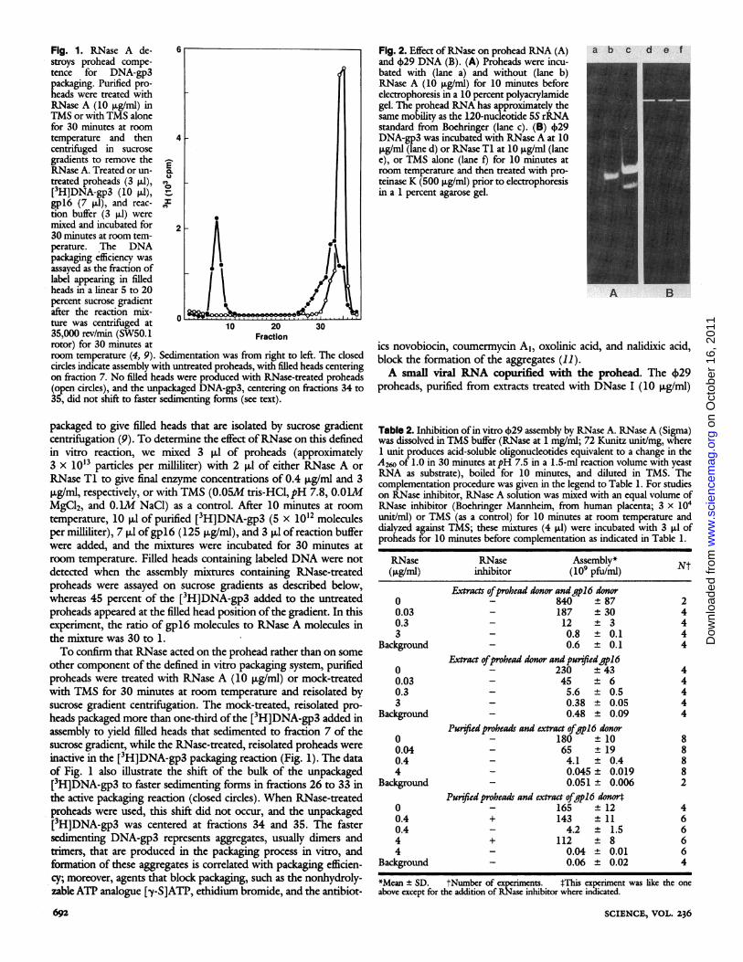
Fig. 1. RNase A de- 6stroys prohead compe-tence for DNA-gp3packaging. Purified pro-heads were treated withRNase A (10 ,ug/ml) inTMS or with TMS alonefor 30 minutes at roomtemperature and then 4centrifuged in sucrosegradients to remove theRNase A. Treated or un- E.treated proheads (3 ,ul), O[3H]DNA-gp3 (10 .1),-gpl6 (7 p.1), and reac- vtion buffer (3 [l1) weremixed and incubated for 230 minutes at room tem-perature. *The DNApackaging efficiency wasassayed as the fraction oflabel appearing in filledheads in a linear 5 to 20percent sucrose gradientafter the reaction mix-
0 L&ture was centrifuged at 10 20 3035,000 rev/min (SW50.1 Fractionrotor) for 30 minutes atroom temperature (4, 9). Sedimentation was from right to left. The closedcircles indicate assembly with untreated proheads, with filled heads centeringon fraction 7. No filled heads were produced with RNase-treated proheads(open circles), and the unpackaged DNA-gp3, centering on fractions 34 to35, did not shift to faster sedimenting forms (see text).
packaged to give filled heads that are isolated by sucrose gradientcentrifuigation (9). To determine the effect ofRNase on this definedin vitro reaction, we mixed 3 ,ul of proheads (approximately3 x 10'3 particles per milliliter) with 2 ,ul of either RNase A orRNase Ti to give final enzyme concentrations of 0.4 ,ug/ml and 3ig/ml, respectively, or with TMS (0.05M tris-HCI, pH 7.8, 0.O1MMgCl2, and O.IM NaCl) as a control. After 10 minutes at roomtemperature, 10 p,1 of purified [3H]DNA-gp3 (5 x 10¹² moleculesper milliliter), 7 pul ofgpl6 (125 jig/ml), and 3 pll of reaction bufferwere added, and the mixtures were incubated for 30 minutes atroom temperature. Filled heads containing labeled DNA were notdetected when the assembly mixtures containing RNase-treatedproheads were assayed on sucrose gradients as described below,whereas 45 percent of the [3H]DNA-gp3 added to the untreatedproheads appeared at the filled head position ofthe gradient. In thisexperiment, the ratio of gpl6 molecules to RNase A molecules inthe mixture was 30 to 1.To confirm that RNase acted on the prohead rather than on some
other component of the defined in vitro packaging system, purifiedproheads were treated with RNase A (10 pug/mil) or mock-treatedwith TMS for 30 minutes at room temperature and reisolated bysucrose gradient centrifugation. The mock-treated, reisolated pro-heads packaged more than one-third ofthe [3H]DNA-gp3 added inassembly to yield filled heads that sedimented to fraction 7 of thesucrose gradient, while the RNase-treated, reisolated proheads wereinactive in the [3H]DNA-gp3 packaging reaction (Fig. 1). The dataof Fig. 1 also illustrate the shift of the bulk of the unpackaged[3H]DNA-gp3 to faster sedimenting forms in fractions 26 to 33 inthe active packaging reaction (closed circles). When RNase-treatedproheads were used, this shift did not occur, and the unpackaged[3H]DNA-gp3 was centered at fractions 34 and 35. The fastersedimenting DNA-gp3 represents aggregates, usually dimers andtrimers, that are produced in the packaging process in vitro, andformation of these aggregates is correlated with packaging efficien-cy; moreover, agents that block packaging, such as the nonhydroly-zable ATP analogue [y-S]ATP, ethidium bromide, and the antibiot-
Fig. 2. Effect of RNase on prohead RNA (A) a b c d e fand )29 DNA (B). (A) Proheads were incu-bated with (lane a) and without (lane b)RNase A (10 ,uglml) for 10 minutes beforeelectrophoresis in a 10 percent polyacrylamidegel. The prohead RNA has approximately thesame mobility as the 120-nucleotide 5S rRNAstandard from Boehringer (lane c). (B) 029DNA-gp3 was incubated with RNase A at 10,ug/ml (lane d) or RNase TI at 10 ,ug/ml (lanee), or TMS alone (lane f) for 10 minutes atroom temperature and then treated with pro-teinase K (500 ,ug/ml) prior to electrophoresisin a 1 percent agarose gel.
A B
ics novobiocin, coumermycin A1, oxolinic acid, and nalidixic acid,block the formation of the aggregates (11).A small viral RNA copurified with the prohead. The 4)29
proheads, purified from extracts treated with DNase I (10 ,ug/ml)
Table 2. Inhibition of in vitro 429 assembly by RNase A. RNase A (Sigma)was dissolved in TMS buffer (RNase at 1 mg/mi; 72 Kunitz unit/mg, where1 unit produces acid-soluble oligonucleotides equivalent to a change in theA260 of 1.0 in 30 minutes atpH 7.5 in a 1.5-mi reaction volume with yeastRNA as substrate), boiled for 10 minutes, and diluted in TMS. Thecomplementation procedure was given in the legend to Table 1. For studieson RNase inhibitor, RNase A solution was mixed with an equal volume ofRNase inhibitor (Boehringer Mannheim, from human placenta; 3 x 10⁴unit/ml) or TMS (as a control) for 10 minutes at room temperature anddialyzed against TMS; these mixtures (4 p1) were incubated with 3 RI ofproheads for 10 minutes before complementation as indicated in Table 1.
RNase RNase Assembly* Nt(ng/ml) inhibitor (109 pfu/nil)
Extrats ofprohead donor and gpl6 donor0 - 840 ± 87 20.03 - 187 ± 30 40.3 - 12 ± 3 43 - 0.8 ± 0.1 4
Background - 0.6 ± 0.1 4Extract ofprobead donor and purifiedgpl6
0 - 230 ± 43 40.03 - 45 ± 6 40.3 - 5.6 ± 0.5 43 - 0.38 ± 0.05 4
Background - 0.48 ± 0.09 4Purified proheads and extract ofgpl6 donor
0 - 180 ± 10 80.04 - 65 ± 19 80.4 - 4.1 ± 0.4 84 - 0.045 ± 0.019 8
Background - 0.051 ± 0.006 2Puified proheads and extract ofgpl6 donort
0 - 165 ± 12 40.4 + 143 ± 11 60.4 - 4.2 ± 1.5 64 + 112 ± 8 64 - 0.04 ± 0.01 6
Background - 0.06 ± 0.02 4
*Mean ± SD. tNumber of experiments. 4This experiment was like the oneabove except for the addition of RNase inhibitor where indicated.
SCIENCE, VOL. 236692
on
Oct
ober
16,
201
1w
ww
.sci
ence
mag
.org
Dow
nloa
ded
from

A
U.- CUIwZ=
C
3000 bpI1I1I1I 1 1I I
_ =
o.-CU
a1
D
Fig. 3. Hybridization of prohead RNA to restriction fragments of 4)29DNA. (A) Restriction maps of the left end of 4)29 DNA for Eco RI, HindIII, Hae II, and Bcl I (17, 18); (B) restriction fragment designations; (C)separation of restriction fragments of 029 DNA in a 1 percent agarose gel;(D) autoradiograph of a Southern blot of the fragments of (C) afterhybridization with [32P]RNA purified from proheads.
(see legend to Table 1 for prohead purification), were examineddirectly for RNA content by electrophoresis in a 1 percent agarosegel. Without prior treatment, the proheads released nucleic acidapproximately the size of the 5S rRNA standard. Release of thenucleic acid from 4)29 proheads during electrophoresis in tris-borate-EDTA buffer, pH 8.3, resembles RNA escape from plantviruses under alkaline conditions or in the absence of Ca2+, Mg2+,or KC1 (14).The size of the prohead nuleic acid was confirmed by electropho-
resis in a 10 percent polyacrylamide gel in the presence ofthe RNaseinhibitor diethyl pyrocarbonate (Fig. 2A). Again, the bulk of the
Table 3. Effect of RNase on the extract containing the 4)29 prohead.Individual prohead donor or gpl6 donor extracts were treated with RNasesA or Ti and complemented with untreated extracts to determine thesensitive extract. The methods used are described in the legends to Tables 1and 2, and the in text.
Mixtures of RNase treatment Assembly*prohead donor and of extract (lo pfu/n-d) Ntgp16 donor extracts or extracts
a None 860 ± 170 4RibonudeaseA (0.3 pglml)
b Both 9.7 ± 0.1 5c Prohead donor 14.2 ± 3.1 5d gp16 donor 740 ± 130 5
Ribonuclease Ti (1.2 pg/ml)e Both 1.3 ± 0.1 3f Prohead donor 3.2 ± 0.6 3g gpl6 donor 665 ± 70 3
*Mean ± SD. tNumber of experiments.
8 MAY I987
Table 4. Reconstruction of RNA-free proheads with purified proheadRNA. The gpl6 donor extract was treated with RNase A (0.4 ,ug/ml, 10minutes, room temperature) and then with RNase inhibitor (from humanplacenta; 400 units/ml, 10 minutes, room temperature). The complementa-tion procedures are described in the legends of Tables 1 and 2.
Complementation Purifiedin RNA-free RNA-free prohead Phage assembly Nt
gpl6 donor extract proheads RNA (10 pfu/ml)
a - - 0.7± 0.1 8b + - 0.8 ± 0.1 4c - + 0.7± 0.1 2d + + 140 ± 20 4
*Mean t SD. tNumber of experiments.
prohead nucleic acid had a mobility similar to the 120-nucleotide 5SrRNA (compare lanes b and c). The faster band in the 5S rRNAstandard was due to secondary structure, since glyoxal-treated 5SrRNA ran at the position of the prohead RNA as a single band.Proheads treated with RNase A at 10 ,ug/ml for 30 minutes at roomtemperature showed no bands (lane a), confirming that the proheadnucleic acid was RNA. The boiled RNase A used in this experimenthad no effect on 4)29 DNA, nor did RNase Ti (Fig. 2B).The RNA in purified proheads was quantified by analyzing
prohead concentration by SDS-polyacrylamide gel electrophoresis(SDS-PAGE) and prohead RNA concentration by PAGE. Thehead-to-neck connector of the 4)29 particle is a dodecamer of geneproduct 10 (gplO) (15), and bands of this protein stained withCoomassie blue were compared by densitometry to stained bands ofa bovine serum albumin standard. Similarly, bands of prohead RNAstained with ethidium bromide were compared to stained bands ofthe 5S rRNA standard. If the bulk of the proheads contained RNA,each particle had approximately two copies.The prohead RNA was a transcript from the left end of 4)29
DNA. Purified proheads were treated with SDS (1 percent) andproteinase K (500 ,ug/ml) for 30 minutes at 55°C, and the RNA wasextracted with phenol and chloroform. With [a-32P]ATP as sub-strate, a poly(A) tail was added to the RNA in the presence ofATP:RNA adenyltransferase [poly(A) polymerase] from Eschenchiacoli (New England Nuclear) (16) and used as a hybridization probewith 4)29 restriction enzyme fragments that were separated byagarose gel electrophoresis, denatured, and transferred to Zeta-Probe (Bio-Rad Laboratories) by electroblotting. The [32P]RNAhybridized specifically to denatured restriction fragments from theleft end of 4)29 DNA (17, 18), including Eco RI-A (-9000 bp),Hind III-B (-2900 bp), Hae II-H (416 bp), and Bcl I-B (-5700bp), but not to the HAE II-I (176 bp), Hae II-J (61 bp), or Bcl I-C(73 bp) fragments (Fig. 3). Thus the prohead RNA is a viraltranscript from a 4)29 DNA segment near, but not extending to, theleft end of the genome. The transcript may originate from the B.subtilis RNA polymerase binding site Al (19) and promoter PE1with its "Pribnow-box" at positions 328 to 333 (18) from the leftend ofthe genome. The Al site may be a weak initiation site in vivobecause early transcription initiating at this site was not confirmedby S1 mapping experiments with 5' end-labeled RNA (20). Sincethe prohead RNA was about 120 nucleotides long and was homolo-gous to sequences of the Hae II-H fragment, it might include openreading frame 2 (18). RNA of 120 nucleotides would have amolecular size of about 40,000 daltons. This corresponds to the sizeof an early RNA produced in 4)29-infected cells in the presence ofchloramphenicol (21).The purified prohead RNA can restore inactive R.NA-free
proheads for phage assembly in extracts. RNA can be isolatedfrom the purified proheads in the presence of 2 mM EDTA. TheRNA-free proheads were inactive in a prohead donor extract treated
RESEARCH ARTICLES 693
0II Il l l l1
1000 2000
Il 11 1 1IIII
Eco RI
Hind III
Hae II
Bcl I
A
B
I H Pi G D
|-C B
- -
w _
we I S m
on
Oct
ober
16,
201
1w
ww
.sci
ence
mag
.org
Dow
nloa
ded
from

with RNase A and then with RNase inhibitor (Table 4, item b).Purified RNA alone did not increase phage production (Table 4,item c). However, the inactive RNA-free proheads and the purifiedprohead RNA can be reconstructed, resulting in phage production(Table 4, item d), albeit at a relatively low efficiency compared tocomplementations of normal extracts and proheads (Tables 1 and2). It is not clear whether this lower efficiency of assembly is due toalteration ofthe extracts with RNase and RNase inhibitor treatmentor the quality of the purified RNA or RNA-free proheads.
In conclusion, we have demonstrated that a small RNA transcriptof about 120 nucleotides from the far left end of the 4)29 genomewas a component of the 4)29 prohead and that it had an essentialrole in 4)29 DNA packaging in vitro. Some data have been obtainedfor systematic investigations of the possible role or roles ofthe smallRNA in the initiation events of DNA-gp3 packaging (11) and theformation of the prohead-dependent and DNA-gp3-dependentATPase (12) (as mentioned earlier). Treated proheads do notproduce the aggregated forms of DNA-gp3 (Fig. 1), which arecorrelated with packaging efficiency (11), suggesting that an initia-tion step in DNA-gp3 packaging is blocked.
REFERENCES AND NOTES1. T. C. King, R. Sirdeskmukh, D. Schiessinger, Microbiol. Rev. 50, 428 (1986).2. D. B. Bourgaize and M. J. Fournier, Nature (London) 325, 281 (1987).3. S. Casjens, in Virus Structure and Assembly, S. Casjens, Ed. (Jones and Bartlett,
Portola Valley, CA, 1985), pp. 75-147.4. M. A. Bjomsti et al., Proc. Natl. Acad. Sci. USA. 78, 5861 (1981).5. __,J. Virol. 41, 508 (1982).6. , ibid. 45, 383 (1983).7. ibid. 50, 766 (1984).8. , ibid. 53, 858 (1985).9. P. Guo, S. Grimes, D. Anderson, Proc. Natl. Acad. Sci. U.SA. 83, 3505 (1986).
10. M. Salas, Current TopicsMicrobiol. Immunol. 109, 89 (1983).11. P. Guo, C. Peterson, D. Anderson,J. Mol. Biol., in press.12. __, ibid., inpress.13. R. Gope and P. Serwer,J. Virol. 47, 96 (1983).14. P. Argos and J. E. Johnson, in Biological Maroemolcuks and Assemblies, 1: Virus
Structure, F. A. Jurnak, A. McPherson, Eds. (Wiley, New York, 1984), pp. 1-43.15. J. L. Carrascosa et al.,J. Mol. Bul. 154, 311 (1982).16. A. E. Sippel, Eur. J. Biocbcm. 37, 31 (1973).17. J. Ito, J. Virol. 28, 895 (1978).18. H. Yoshikawa and J. Ito, Gene 17, 323 (1982).19. J. M. Sogo et al., J. Mol. Biol. 127, 411 (1979).20. I. Barthelemy, M. Salas, R. P. Mellado,J. Virol. 60, 874 (1986).21. D. J. Loskutoff and J. J. Pene, ibid. 11, 87 (1973).22. We thank C. Peterson, M. A. Gomes, and L. Lu for technical assistance; C. Church
for typing the manuscript; and N. Anderson for the drawings of Fig. 3, A and B.Supported by NIH grant DE-3606.9 February 1987; accepted 10 April 1987
Bacterial Resistance to -Lactam Antibiotics:Crystal Structure of fi-Lactamase from
Staphylococcus aureus PCi at 2.5 A Resolution
OSNAT HERZBERG AND JOHN MOULT
,B-lactamases are enzymes that protect bacteria from thelethal effects of P-lactam antibiotics, and are therefore ofconsiderable clinical importance. The crystal structure ofI-lactamase from the Gram-positive bacterium Staphylo-coccus aureus PC1 has been determined at 2.5 angstromresolution. It reveals a molecule of novel topology, madeup of two closely associated domains. The active site islocated at the interface between the domains, with the key
M ANY TYPES OF DEFENSE MECHANISMS EXIST IN THE
biological world, and relatively lowly life forms oftenexhibit a surprising degree of sophistication in these
matters. One such system that has been used to advantage by man isthe defense that some fungi mount, through the production of I-lactam antibiotics, against bacterial attack. These compounds killbacterial cells by irreversibly inhibiting a number of enzymesconcemed with cell wall synthesis and repair (1). A degree ofcomplexity is added by the ability of some bacteria to defeat thisfungal defense mechanism, primarily through the production of69
catalytic residue Ser70 at the amino terminus of a buriedhelix. Examination of the disposition of the functionallyimportant residues within the active site depression leadsto a model for the binding of a substrate and a functionalanalogy to the serine proteases. The unusual topology ofthe secondary structure units is relevant to questionsconcerning the evolutionary relation to the I-lactamtarget enzymes of the bacterial cell wall.
membrane-bound and excreted enzymes which hydrolyze ,B-lactamrings. These enzymes are known as 1-lactamases or penicillinases(E.C. 3.5.2.6) (2). A variety of ,-lactams are produced by fungi, andin tumm, there are at least three different classes of 1-lactamasesproduced by bacteria (3). A further layer of complexity is providedby the production of inhibitors of 3-lactamases by some fungi (4).
The authors are members ofthe Medical Research Council ofCanada Group in ProteinStructure and Function, Department of Biochemistry, University of Alberta, Edmon-ton, Canada, T6G 2H7.
SCIENCE, VOL. 236
on
Oct
ober
16,
201
1w
ww
.sci
ence
mag
.org
Dow
nloa
ded
from