some applications of phase-contrast microscopy · pi. i, figs. 2 and 3, closely resembles that in...
TRANSCRIPT

Some Applications of Phase-contrast MicroscopyBY
ROBERT BARER
(From the Department of Human Anatomy, Oxford)
With four Plates and three Text-figures
THE purpose of this paper is to present a preliminary report on certainless obvious applications of phase-contrast microscopy which may be of
some biological interest. The principles of this method of microscopy arenow too well known to necessitate any detailed description (Zernike, 1942;Burch and Stock, 1942).
Briefly, the method sets out to render transparent objects visible by convert-ing changes in phase, which cannot be appreciated by the human eye or bythe photographic plate, into changes of amplitude, which can be so appre-ciated. The theory, as worked out by Zernike (1942), shows that for the caseof a grating composed of non-absorbing bars of different refractive indices,the final image is exactly that which would ordinarily be produced by a grat-ing composed of absorbing bars, provided that the changes in phase produced bythe grating are small. Perhaps the most remarkable feature of Zernike's theory,as applied to a grating, is that it shows the enormous superiority of phase-contrast illumination over other methods for examining transparent material.Without going into details, the image produced by an absorbing grating canbe represented by the sum of an infinite number of sine and cosine waves.The image of a transparent grating can be represented by a different series ofwaves. Ideally we should like to convert this second series into the first, andthis is exactly what phase-contrast illumination does! All the other commonmethods of observation—oblique illumination, central and oblique darkground illumination, or observation with reduced substage aperture produceimages which are represented by different sine and cosine series, which whilein some cases showing certain similarities to the 'ideal' series, in general fallfar short of it. Roughly speaking, we may say that the effect of phase contrastillumination is as if we stained the object with a dye which stained each pointwith an intensity proportional to the product of its thickness and refractiveindex. To this extent, and subject to the performance of the microscope,phase-contrast illumination gives a true representation of what is actuallypresent. All other methods commonly employed to render transparentobjects visible produce an image which is often a mere caricature of theobject.
It must be pointed out that this almost exact theory has only been workedout for very special objects, such as gratings, the elements of which produce
[Q.J.M.S., Vol. 88, Third Series, No. 41 Agi

492 Barer—Some Applications of Phase-contrast Microscopy
very small changes of phase. It is reasonable to ask how small must thesechanges be, and what happens if they become large. It is not easy to givean exact answer to these questions since the mathematics involved is rathercomplicated. Fortunately, we can tackle the whole question in a much moregeneral way, and without assuming any particular type of object.
Following Zernike's treatment the object (or the image) can be representedby a vector diagram as in Text-fig. la.
Each point is represented in phase and amptitude by a vector. Areas whichabsorb light without affecting its phase will be represented by vectors lying
along the line OM. Areas which absorb no light but merely alter its phase(i.e. transparent objects) will be represented by points on the circumferenceof a circle. Now according to the Abbe theory of the microscope, the finalimage is produced by interference between the central undiffracted image ofthe object and a series of diffraction images. In order to obtain phase contrastit is necessary to change the phase of the central .pencil relative to the phasesof the diffracted pencils. In practice this is usually accomplished by insertinga phase-retarding or phase-advancing strip into the back focal plane of theobjective, and an illuminating diaphragm of corresponding shape in the sub-stage. The phase strip is usually constructed to produce a 90° change ofphase in the central image. The reason for this is that the exact theory forthe case of a grating requires such a phase change in order to transform a'phase-grating' into an 'amplitude grating'. In practice, however, and in deal-ing with a more general type of object, this phase change need not be exactly900. Indeed in some cases it is advantageous to produce a phase change otherthan 900.
Returning to our vector diagram (Text-fig. la) the effect of the centralpencil alone can be represented by a vector which is the average of all thevectors representing the object. Let us suppose it to be represented by theline OC. The effect of the phase strip is to rotate this vector through 900,or in other words to shift the origin of the system from O to O' (positive phasecontrast) or O" (negative phase contrast). We can now see that non-absorbing

Barer—Some Applications of Phase-contrast Microscopy 493
points, which are represented by vectors such as P and Q on the circum-ference of the circle, will appear to have very different intensities when theorigin is shifted to O' or O". Whereas with normal illumination the intensitiesof such points were represented by (OP)2 and (OQ)2, i.e. equal and thereforeshowing no contrast, with phase-contrast illumination the intensities become(O'P)2 and (O'Qf or (O"P)2 and {O"Qf. Restricting ourselves to positivephase contrast (i.e. origin at O') we see that highly refracting points willappear darker, less refracting points lighter, than the mean illumination.
Now suppose the object contains many points which produce large changesin phase. The effect of such points will be represented by vectors OPX, OP2,OP3, OPit &c, around the circumference of the circle. If these points aresufficiently numerous the point representing the average of the whole systemwill approximate to O. Thus phase-contrast illumination would be of novalue for such an object. At the same time we observe that the intensity ofa point producing a phase change 6 will be the same as the intensity for pointsproducing phase changes of 2mr-\-d, i.e. an extremely refractile region maynot always appear any darker than a less refractile region.
OBSERVATIONS ON LIVING INTACT ANIMALS
In practice, according to Zernike (1942), phase-contrast illumination is mostuseful for objects which produce phase changes of less than 45° (one-eighth ofa wave-length) since objects thicker than this can be seen reasonably well byother methods. In most cases this means that phase contrast is best used withvery thin objects. Single cells such as protozoa or tissue cultures are ideallysuited for this method of examination. The excellent films produced byMichel (on the development of grasshopper spermatocytes) and by Hughes(on the growth of cells in tissue cultures) have already shown the enormouspotentialities of the method for this type of material. Unfortunately, theinsistence on the use of thin objects has rather tended to obscure the pointthat it is not thickness per se that is important, but the amount of phase change.Phase-contrast illumination may be quite useful for examining relativelythick but very transparent objects. Now the larva of Chaoborus (the phantomlarva) has long been known for its transparency, and it was thought that itwould provide good material for examination by phase-contrast illumination.The larva is usually several millimetres in diameter but can be flattened some-what by mounting in water under a cover slip. At the same time thisimmobilizes the insect, but if carried out gently does not appear to harm it,for the latter will swim away quite normally when released after examination.Now it must be admitted that many of the cells and tissues of Chaoborus canbe observed by the familiar method of reducing the, substage iris to a verysmall diameter and altering the focus. As a matter of fact this is one method(though a very poor one) of producing phase contrast. Resolving power islost owing to the small substage aperture, the method only works properlywhen the object is slightly out of focus, diffraction effects are accentuated,and the image is a very poor representation of what is actually present. With

494 Barer—Some Applications of Phase-contrast Microscopy
properly adjusted phase-contrast illumination the results are very different.It is possible to state with reasonable confidence that we can now observeliving cells in the living intact multicellular organism with greater clarity, andwith a greater chance of seeing what is actually present, than ever before.PI. I, fig. i, shows a low-power general view of part of a living Chaoboruslarva, focused on two ganglia with the intervening nerve-cord. PI. I, figs. 2and 3, are higher power views of single ganglia, taken with the \ in. objective.These photographs show one large nerve-cell on each side of the ganglion.The nuclei and nucleoli are clearly seen and it will be observed that the cyto-plasm is highly granular. These granules are particularly well seen in PI. I,fig. 3. It is not yet possible to establish their nature with certainty. At firstsight they might be taken for Nissl granules, and indeed their appearance inPI. I, figs. 2 and 3, closely resembles that in fixed sections stained with Borrel'smethylene blue, a recognized Nissl stain. It is possible, however, that thegranules seen in the living cells may be part of the Golgi apparatus, which isoften very easily seen in other living cells by phase-contrast illumination.Another region of some interest is shown in PI. I, fig. 4. It is necessary tospeak with caution of such material as the tissues of living insect larvae, butas a result of prolonged observations I am of the opinion that PI. I, fig. 4,is a photograph of a living motor nerve-ending in a muscle-fibre. (All themuscles of Chaoborus larvae are composed of single fibres.) The pyramidalaccumulation of sarcoplasm containing one or more nuclei with what appearsto be a nerve-fibre entering at the apex corresponds to the description of the'Doyeres hillock' given in the entomological literature (see Morison, 1927).PI. II, fig. 5, shows another view of these end-plates taken at the samemagnification but from a smaller specimen. Two end-plates are seen (theylay on slightly different planes and a compromise focus had to be chosen inorder to show both on the same photograph) and they appear to be inter-connected by two nerve-fibres which seem to emerge from a common junc-tion, at which there appears to be some sort of cell, possibly a peripheralnerve-cell, but more probably a neurilemmal cell. Numerous sarcoplasmicgranules are also seen. Other insect larvae are also suitable for study by
this method, e.g. young mosquito and Chironomus larvae (PL II, fig. 6).The giant salivary gland chromosomes in the intact Chironomus larva canoften be seen quite well. Numerous possibilities exist for the study of secre-tory and excretory activity of cells in insect larvae. It is very much to behoped that suitable vertebrate material may be found. I have carried outpreliminary observations on the tissues of a tadpole's tail and the results werequite promising. Unfortunately, these observations were made rather latein the spring when frog tadpoles were very scarce and rather large. The trans-parent tails of very young tadpoles should be quite suitable. The mesenteriesof small vertebrates have not on the whole been found satisfactory, thoughvery clear views of the capillary endothelium have sometimes been obtained.It is possible that a very thin transparent chamber inserted in the ear of arabbit may prove satisfactory.

Quart. Journ. Micr. Sci., Third Series, Vol. 88
FIG. I FIG. 2
FIG. 3 FIG. 4
R. BARER.—PLATE I

Quart. Journ. Micr. Sci., Third Series, Vol. 88
FIG. FIG. 6
FIG. 7 FIG. 8
R. BARER.—PLATE II

Barer—Some Applications of Phase-contrast Microscopy 495
THE EXAMINATION OF STAINED SECTIONS
It is generally stated that phase-contrast illumination is of no value forexamining stained objects. This opinion seems to have been based mainlyon theoretical consideration of an over-simplified ideal case. In Text-fig. 1we saw that Zernike represented all absorbing points as lying on the line OM,and all phase-changing points as lying on the circumference of the circle.When the origin is shifted from O to O', the absorbing points on OM becomeroughly equidistant from O' and therefore lose contrast. Now a stainedsection will contain points like U and V (Text-fig. 16) which both absorb andproduce changes of phase. The intensities of such points will be proportionedto (O'C/)2 and (O'F)2 and they will appear to have good contrast, whereaswith ordinary illumination (origin at O) they will appear equally stained. Wethus have the theoretical possibility that phase-contrast illumination mayafter all be of some value for examining stained objects. To put the mattervery crudely we should expect to lose much of the contrast due to the staining,but we might gain a different contrast due to differences in refractive indexin both the object and the stains. (In a private communication Dr. Burchhas suggested that stains may have anomalous refractive indices in the regionof their absorption bands, and that it would be interesting to examine stainedsections by phase-contrast illumination using monochromatic light of dif-ferent wave-lengths, in order to vary staining contrast.) It must be admittedat once that this simple theory is far from adequate. Unfortunately, the exacttreatment bristles with difficulties, and, indeed, it is unlikely that the problemwill ever be solved for any but very special cases. In the circumstances it wasjudged best to try the method in practice. A wide range of routine normaland pathological material was examined, but a systematic examination ofdifferent stains and mounting media has not yet been made.
There are several ways in which phase-contrast illumination may be ofvalue for examining stained sections. Perhaps its most obvious use is torender visible details of structure which have not been properly stained by thedyes employed. Such cases occur especially in certain histochemical reac-tions. Text-fig. 2fl shows a section of bone marrow stained by the Feulgentechnique. Only the nuclei are visible. Text-fig. 26 shows the identicalfield as seen by phase-contrast illumination. The cytoplasm as well as thenuclei of the cells is now rendered visible.
Other examples of the way in which unstained or poorly stained details canbe made clearer are shown in Pis. II and III, figs. 7-10. It should be pointedout that it is often extremely difficult to demonstrate the superiority of phase-contrast illumination over ordinary illumination by means of photographs.The reason for this is that one automatically tries to get the best possiblephotograph out of any given material. Details which are seen with difficultythrough the microscope can be rendered clearer by filters, differences inexposure, development, and printing. Strictly speaking one ought to treatboth phase-contrast and ordinary pictures identically, but this is very difficult

496 Barer—Some Applications of Phase-contrast Microscopy
as the former require longer exposures. A direct comparison under themicroscope is far more convincing than any series of photographs. Were itnot for a natural tendency to 'load the dice' against oneself the photographicsuperiority of phase-contrast would be even more clear-cut.
Neurological material offers a particularly interesting field for study bymeans of phase-contrast microscopy. It is well known that scarcely any twomethods of staining nervous tissues will produce the same result. As regards
TEXT-FIG. 2a. TEXT-FIG. 26.
the various methods of silver staining, one may almost say that the samemethod in the hands of two different workers will produce different results.It is, therefore, of some interest to know what an entirely unstained nerve-cell looks like. The answer is seen in PI. I l l , figs, n , 12.
These cells were almost invisible by ordinary illumination, even when thesubstage iris was almost closed, and were quite impossible to photograph,except by phase-contrast illumination. The appearance is more or less whatone might expect to see if it were possible to use a mixture of silver stainingand Nissl staining. It is the purpose of this paper to describe the possibleapplications of a new technique rather than to discuss results, but for thepresent we may say that the nerve-cell, free from staining artifact (but not,of course, free from fixation artifact), may show appearances suggestive ofNissl granules, Golgi apparatus, boutons terminaux, and numerous extremelyfine cell processes. A further investigation on frozen-dried material and tissuecultures is being undertaken.
Returning, however, to the question of stained nervous tissue, in general

Quart. Journ. Mia: Set., Third Series, Vol. 88
FIG. 9 FIG. IO
FlC. 12
R. BARER.—PLATE III
Quart. Joiirn. Micr. Set'., Third Series, Vol. SS
Fie. 13 FIG. 14
FIG. IS
R. BARER.—PLATE TV

Barer—Some Applications of Phase-contrast Microscopy 497
we may say that any given stain is fairly specific for either nerve-cells or nerveprocesses. Thus toluidin blue, methylene blue, and cresyl violet stain nucleiand Nissl granules but not nerve-fibres, whereas the reverse is on the wholetrue of most silver stains. We thus have the possibility of rendering the miss-ing element visible by phase-contrast illumination.
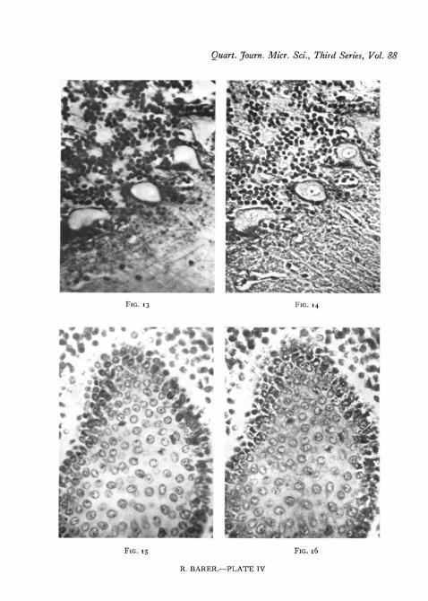
PL IV, fig. 13, shows the Purkinje cells of the cerebellum seen in a Biel-schowsky preparation. The nerve-fibres are very clear but the cells seem almostdevoid of detail. PL IV, fig. 14, shows the same field by phase-contrast
TEXT-FIG. 3a. TEXT-FIG. 36.
illumination. The nuclear structure is clearly visible. Conversely it is oftenpossible to render the nerve-fibres more visible in Nissl preparations bymeans of phase-contrast. Finally, it is necessary to mention the variousmyelin stains, used for studying nerve-tracts. TKese usually leave the nerve-cells unstained and the latter can be rendered visible by phase-contrastillumination. Text-figs. 3 a, 36 show this effect in a Weigert-Pal preparation(X 150) of a transverse section of the spinal cord (monkey).
The main disadvantage of the method is that it very often renders too muchdetail visible. Most stains are to a certain extent selective, but phase-contrastillumination may reveal so many minute fibrils that it is very easy to miss thewood for the trees. A considerable amount of further study will be necessarybefore any exact evaluation of the method as applied to neurological materialcan be made.
Many types of granules are extremely easily seen by means of phase-contrast illumination. PL III, figs. 9, 10, shows a section of liver from a caseof pernicious anaemia. Iron-containing pigment granules (haemosiderin) are

498 Barer—Some Applications of Phase-contrast Microscopy
present but very difficult to see by ordinary methods. Phase contrast revealsthe presence of myriads of these granules with remarkable clarity. Even suchnormally well-seen pigment granules as melanin are rendered clearer by phasecontrast. PL IV, figs. 15,16, from a section of a melanoma of the skin illustratethis point. The method has also been used with success for demonstratingsecretory granules. It should be mentioned that all the photographs of fixedmaterial shown were from routine paraffin sections, 8-12 /x thick, mounted inCanada balsam.
DISCUSSION AND CONCLUSIONS
It is necessary to emphasize once more that it is not the purpose of thispaper to discuss results in detail, but rather to suggest certain applications ofphase-contrast microscopy which require further study. As regards theexamination of living cells and tissues anyone who has ever seen such materialby properly adjusted phase-contrast illumination would readily acknowledgeits superiority over the older methods. The main problem here is to find suit-able material. Isolated cells such as bacteria, protozoa, tissue cultures, orspermatozoa are of course ideal, but multicellular organisms are in many waysmore interesting and important. Insect larvae and other transparent creaturessuch as certain medusae may yield useful information, but it would be veryvaluable to be able to extend the range of available material, especially tovertebrate tissues. The examination of stained sections by phase-contrastillumination is still in its infancy. Time and further investigation alone willshow its uses and limitations. All that can be said at present is that it mayprove to be a useful adjunct to the usual methods for examination of certaintypes of material. It is not possible to predict what will be seen by its aid.In some cases certain details may be rendered more clearly visible, in othersthe method will be found of no value. One practical point should be empha-sized : the appearance of a stained section under the low power (•§ in. objec-tive) is often bizarre and disappointing. The investigator should not bedisturbed by this but should always proceed to examine the section with the\ in. objective, when the appearance is often greatly improved. I am atpresent unable to offer any satisfactory explanation for this phenomenon,but recent experiments suggest that appearances can be greatly improved byproper choice of colour filters. One last word: it is of course very importantthat any new method of microscopy should be received with caution. It mustbe admitted that there are many theoretical and practical points about phase-contrast illumination that require much further study. Nevertheless thetheoretical basis underlying the method is sufficiently well understood to enableone to say that the image seen by its use bears at least as close a relation towhat is actually present as that seen by more orthodox methods. Indeed, itis necessary to point out that the theory underlying the observation of stainedobjects by ordinary illumination is very far from complete. The theory under-lying the use of oblique or central dark ground illumination is even less per-fectly understood, but this has not prevented the extensive use of these

Barer—Some Applications of Phase-contrast Microscopy 499
methods. The proper course to pursue for the present is to use the methodcautiously as an adjunct to other routine methods and to seek out its specialapplications and limitations.
I wish to thank Professor W. E. Le Gros Clark, F.R.S., for his encourage-ment and advice. I have had several valuable discussions on histologicalmatters with Drs. A. Brodal, P. Glees, and G. Bourne. Mr. A. W. Dent andMr. L. G. Cooper have provided technical assistance. Finally, I wish toexpress my special gratitude to Dr. C. R. Burch, F.R:S., for many stimulatingdiscussions on all aspects of phase-contrast microscopy.
SUMMARY
Some extensions of the simple theory of phase-contrast microscopy areconsidered. It is emphasized that transparency, rather than thickness, is thelimiting factor for the successful employment of the method. Certain trans-parent insect larvae (Chaoborus, Chironomus) can be observed in the livingstate by phase-contrast illumination.
The statement that the method is of no value for the examination of fixedand stained sections is based on consideration of an ideal physical case. Inpractice the method may be a valuable adjunct to routine examination of suchmaterial. Examples are given of the application of phase-contrast microscopyto normal and pathological stained sections.
REFERENCESBURCH, C. R., and STOCK, J. P. P., 1942. J. sci. Inst., 19, 71.MORISON, G. D., 1927. Quart. J. micr. Sci., 71, 395.ZERNIKE, F., 1943. Physica, 9, 686, 974.
EXPLANATION OF PLATESAll the figures are untouched photomicrographs taken with the Cooke,
Troughton, and Simms phase-contrast equipment. Green filter. Ortho-chromatic film.
PLATE I
Fig. 1. Low power view (phase contrast x 150) of two segments of a living Chaoboruslarva. Focused at the level of the nerve-cord and ganglia. Two large nerve-cells are visiblein the lower ganglion.
Fig. z. Higher power view ( X 500) of a single ganglion showing two nerve-cell nuclei andnucleoli.
Fig. 3. Another example ( X 500) showing the granules in the cytoplasm of the large nerve-cell on the right.
Fig. 4. Motor nerve-ending (phase contrast X 500) in striated muscle-fibre of a Chaoboruslarva. Note the nucleus.
PLATE II
Fig. 5. Another example of motor nerve-endings; same magnification as Fig. 4 ( x 500) butfrom a smaller specimen. The two muscle-fibres lay on slightly different levels and a com-promise focus was chosen. Note the sarcoplasmic granules and the apparent inter-connexionof the nerve-fibres.