sonographic spectrum of first-trimester fetal cephalocele: … · 30 sepulveda et al. cases of...
TRANSCRIPT
Ultrasound Obstet Gynecol 2015; 46: 29–33Published online 4 June 2015 in Wiley Online Library (wileyonlinelibrary.com). DOI: 10.1002/uog.14661
Sonographic spectrum of first-trimester fetal cephalocele:review of 35 cases
W. SEPULVEDA*, A. E. WONG*, E. ANDREEVA†, N. ODEGOVA†, P. MARTINEZ-TEN‡and S. MEAGHER§*Fetalmed–Maternal-Fetal Diagnostic Center, Las Condes, Santiago, Chile; †Medical-Genetics Department, Moscow Regions ResearchInstitute of Obstetrics and Gynecology, Moscow, Russia; ‡Delta–Ultrasound Diagnostic Center for Obstetrics and Gynecology, Madrid,Spain; §Monash Ultrasound for Women, Melbourne, Australia
KEYWORDS: cephalocele; fetal ultrasound; first-trimester screening; Meckel–Gruber syndrome; open neural tube defects;prenatal diagnosis
ABSTRACT
Objective To describe the sonographic features of fetalcephalocele diagnosed at the time of first-trimesterultrasound screening for aneuploidy.
Methods This was a retrospective review of cases ofcephalocele diagnosed in the first trimester at four fetalmedicine referral centers. Once diagnosis was suspected,a transvaginal ultrasound examination was offered toimprove depiction of the cranial defect and enhanceexamination of fetal anatomy, with special attention givento the location, size and content of defects. To assureconsistency in diagnosis, representative pictures andvideoclip sequences of the cranial defect were obtainedand reviewed by at least two authors. Cases were classifiedand compared with the assessment made at diagnosis.
Results Of the 35 affected fetuses identified, 33 were of asingleton pregnancy and two were of twin pregnanciesin which the other fetus was unaffected. The lesionwas classified as a cranial meningocele in 13 (37%)cases and as an encephalocele in 22 (63%). The bonedefect was occipital in 27 (77%), frontal in three (9%),parietal in three (9%) and non-classifiable in two (6%).Twelve (34%) were considered as small in size, 11(31%) as medium and 12 (34%) as large. There wereno reported cases of aneuploidy; however, four (11%)cases were associated with Meckel–Gruber syndrome,two (6%) with a disruptive syndrome and one (3%) withskeletal dysplasia. Eight (23%) pregnancies were lost tofollow-up. Parents opted for termination of pregnancyin 21 of the 27 remaining cases and, of the six ongoingpregnancies, four patients miscarried or the fetus died inutero during the second trimester, one liveborn infant diedshortly after delivery and one underwent neonatal surgeryfor an isolated cranial meningocele and is currentlydoing well.
Correspondence to: Prof. W. Sepulveda, Estoril 50, Room 515, Las Condes, Santiago 7591047, Chile (e-mail: [email protected])
Accepted: 25 August 2014
Conclusions First-trimester sonographic diagnosis ofcephalocele is accomplished easily with a detailedexamination of the skull contour at the time of routineassessment of the axial and sagittal views of the headfor measurement of the biparietal diameter and nuchaltranslucency, respectively. However, the sonographicfeatures are highly variable. A significant proportion ofcases are associated with genetic or disruptive syndromes.Prenatal diagnosis of cephalocele in the first trimester wasassociated with a high rate of termination of pregnancyand early intrauterine fetal demise. Only one fetus in thisseries survived and is neurologically intact; therefore, theprognosis of this condition remains poor. Copyright ©2014 ISUOG. Published by John Wiley & Sons Ltd.
INTRODUCTION
Cephaloceles are relatively rare open neural tubedefects characterized by protrusion of brain tissue ormeninges through a congenital cranial defect. Thesedefects are classified, according to their content, intothose containing only meninges (cranial meningocele),brain tissue (encephalocele) or brain and part of thelateral ventricles (encephalocystocele)1,2. The prenatalsonographic diagnosis of this condition is, in most cases,straightforward, particularly if the defect is medium orlarge in size2,3. Indeed, there is a plethora of reports in theultrasound literature describing the sonographic findings,antenatal course and pregnancy outcome in pregnanciescomplicated by a cephalocele diagnosed in the second andthird trimesters2–8. In contrast, reports of cases diagnosedin the first trimester are scarce and are usually single casereports or small case series9–12.
With the widespread incorporation of sonographicscreening for aneuploidy at 11 − 13 weeks of gestationinto clinical practice13,14, an increasing number of
Copyright © 2014 ISUOG. Published by John Wiley & Sons Ltd. ORIGINAL PAPER

30 Sepulveda et al.
cases of cephalocele are expected to be diagnosed atthis early gestational age. Nevertheless, there is stilllimited information regarding the sonographic features,associated findings and clinical implications in fetusesdiagnosed as having cephalocele in the first trimester. Theaim of this report was to describe these characteristicsbased on a large number of fetuses with cephalocelediagnosed in the first trimester.
METHODS
This was a retrospective review of fetuses with cephalo-cele diagnosed in the first trimester of pregnancy at fourfetal medicine referral centers. In all cases, sonographicexaminations were performed for clinical reasons fol-lowing the recommendations of the UK Fetal MedicineFoundation13,14, by operators with extensive experiencein first-trimester sonographic screening and under pro-tocols approved by the corresponding local institutionalreview boards.
The diagnosis of cephalocele was considered when adefect in the skull associated with protrusion of meningesor brain tissue was identified in both the axial and sagittalsections of the fetal head on routine ultrasound exam-ination for aneuploidy screening. At our institutions, theaxial view is obtained routinely at the first-trimester scanin order to measure the biparietal diameter and visualizethe choroid plexuses15, and the sagittal view is obtainedfor nuchal translucency (NT) measurement and nasalbone assessment16. Once the diagnosis of cephalocelewas considered, the parents were offered transvaginalultrasound for improved depiction of the cranial defectand to enhance examination of the fetal anatomy. Specialattention was given to the location, size and contentof the defect, and to differentiate large encephalocelesfrom acrania and cranial meningoceles from cystichygroma colli by demonstration of the fetal skull and
the bony defect, respectively2. Cephaloceles diagnosed inassociation with disruptive syndromes, such as body stalkanomaly or amniotic band syndrome, were also includedin the analysis. Once the diagnosis was confirmed,available pregnancy management options were discussedextensively with the parents. If the parent decided to con-tinue the pregnancy, serial ultrasound scans were offered.In addition, fetal karyotyping was offered and performedprenatally or postnatally according to the parentalwishes.
To assure consistency in the diagnosis, representativepictures and videoclip sequences, if available, of thecranial defect were reviewed by at least two of the authors(W.S. and S.M.). The cases were classified independentlyaccording to the location (occipital, parietal or frontal),content (cranial meningocele or encephalocele) and size(large if > 30%, medium if 10–30% and small if < 10%of the cranial content), and compared with the assessmentmade at the time of diagnosis. If different opinions weregiven among reviewers or operators, the final assessmentwas established by consensus.
Information regarding maternal demographics,first-trimester sonographic findings, subsequent antenatalcourse and pregnancy outcome was obtained fromthe ultrasound reports and medical records, from thereferring obstetricians or from the patients themselves.Owing to the retrospective nature of this review,ethical permission for this study was waived by thecorresponding institutional review boards.
RESULTS
A total of 35 cases from 34 patients diagnosed between2002 and 2014 were analyzed; one patient had twoconsecutive affected fetuses within the study period.Two (6%) cases occurred in a twin pregnancy (onemonochorionic–monoamniotic17 and one dichorionic)
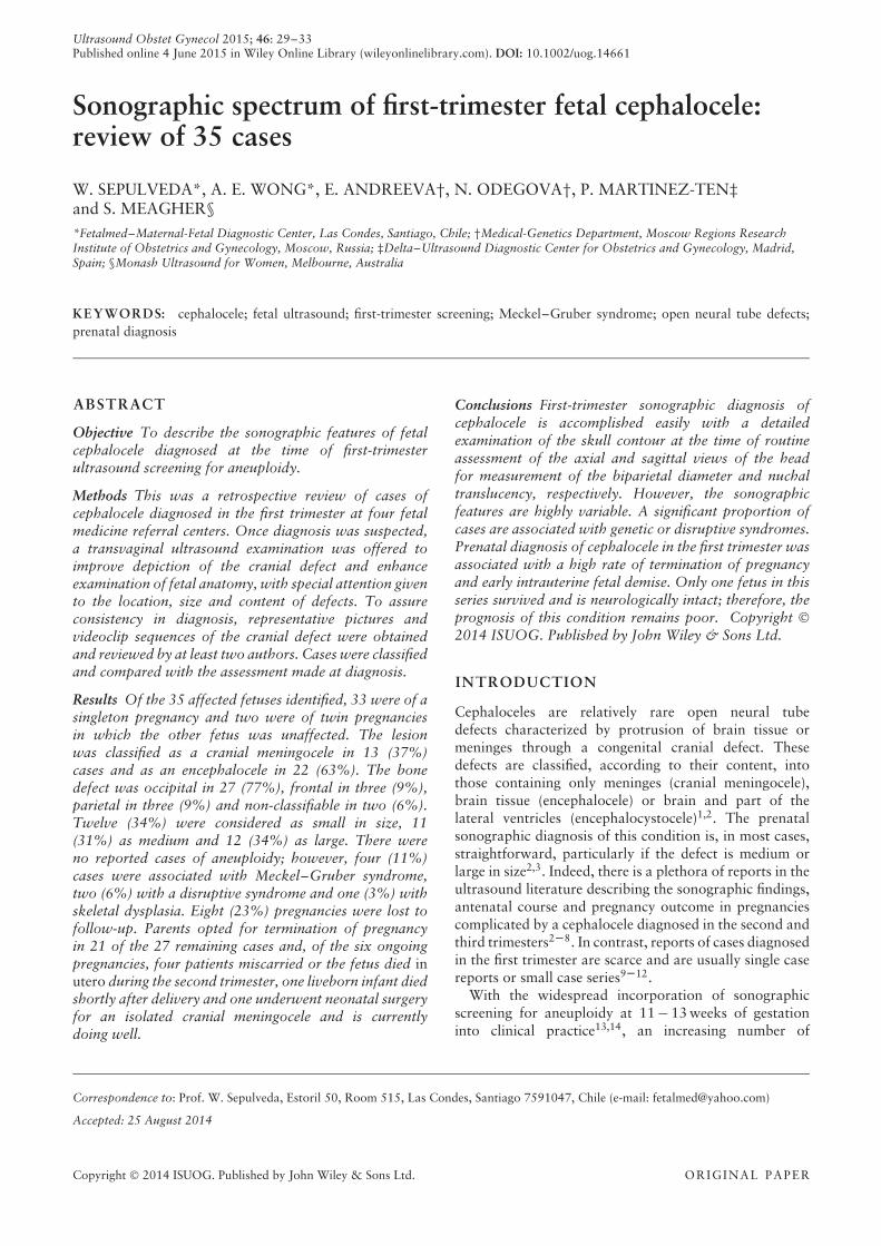
Figure 1 Axial transvaginal ultrasound images of fetal head showing a cranial cephalocele at 13 + 4 weeks (a) and an encephalocele at12 + 6 weeks (b). Note enlarged cisterna magna, enlarged third ventricle and absent brain tissue within the defect in (a). Arrow depicts braintissue within the defect in (b).
Copyright © 2014 ISUOG. Published by John Wiley & Sons Ltd. Ultrasound Obstet Gynecol 2015; 46: 29–33.

Cephalocele in the first trimester 31
Figure 2 Frontal encephalocele in a fetus at 11 + 2 weeks: two-dimensional ultrasound showing a large cranial defect with protrusion ofbrain tissue (a); sagittal (b) and axial (c) three-dimensional sonographic views of the fetal head showing a cranial defect containing braintissue.
Figure 3 Fetal two-dimensional ultrasound (a) and three-dimensional ultrasound using the HDlive technique (b) showing a medium-sizedoccipital cranial cephalocele at the time of first-trimester referral. (c) The defect was larger and contained brain tissue at ultrasoundexamination 2 weeks later.
Figure 4 First-trimester ultrasound images of a fetus with: cephalocele associated with Meckel–Gruber syndrome (note bilateral polycystickidneys) (a); isolated cephalocele showing normal kidneys (b); and postaxial polydactyly, with the arrow depicting an extra toe (c).
Figure 5 (a) Ultrasound image of cranial cephalocele, diagnosed at 13 + 2 weeks, with calipers depicting a cranial defect (courtesy of DrPaula Vargas). (b) Axial view of fetal head showing the defect at 16 weeks. (c) Posterior aspect of the head after surgical repair in the onlysurvivor in this series.
Copyright © 2014 ISUOG. Published by John Wiley & Sons Ltd. Ultrasound Obstet Gynecol 2015; 46: 29–33.

32 Sepulveda et al.
in which only one of the fetuses was affected and theother was normal anatomically. Information on thefirst-trimester sonographic findings was available forall 35 cases, but eight (23%) pregnancies were lost tofollow-up or the records were missing. Demographic datawere not obtained in six (17%) cases because of missingmedical records. Of the remaining 29 cases, the medianmaternal age was 29 (range, 21–40) years, the mediangestational age at evaluation in our centers was 12 (range,11–14) weeks, and the median crown–rump length was63 (range, 45–80) mm. All patients had an otherwiseuncomplicated antenatal course. However, one patientwas receiving antidepressive medication at the time ofthe scan and two had an obstetric history of fetuses withMeckel–Gruber syndrome.
Thirteen (37%) cases of cephalocele were classifiedas cranial meningocele and 22 (63%) as encephalocele(Figure 1). The defect was located in the occipital areain 27 (77%) cases, in the parietal area in three (9%), inthe frontal area in three (9%) (Figure 2) and was partof a disruptive lesion and involved several areas of theskull in two (6%) cases, and was therefore assessed asnon-classifiable. The defect was classified as large in 12(34%) cases, medium in 11 (31%) and small in 12 (34%).There was good agreement (n = 33; 94%) betweenthe operator and the reviewers regarding the location,size and content of the cephalocele. In three cases, afollow-up scan was performed in the first trimester;changes in defect appearance, both in content and size,were documented in all three cases, despite the shortperiod of time between examinations (Figure 3). All threeof these pregnancies were terminated.
Five (14%) cases were associated with NT thicknesses> 95th percentile for gestational age. Of note, in somecases of occipital cephalocele, NT thickness was difficultto measure because of the defect extending significantlyinto the cranial aspect of the neck, hampering accuratemeasurement. Nevertheless, chromosomal analysis wasperformed in 17 of the 27 cases with complete informa-tion, and no cases of aneuploidy were reported. Therewere four (11%) cases associated with Meckel–Grubersyndrome (Figure 4), two (6%) with a disruptive syn-drome and one (3%) with a skeletal dysplasia. Of theremaining 28 cases with non-recognizable syndromes,five had additional malformations, including congen-ital heart defects in two and one each of congenitaldiaphragmatic hernia, enlarged posterior fossa andomphalocele. All cases of large encephalocele wereassociated with microcephaly.
Among the 27 cases for which follow-up was available,21 were terminated after counseling of the parents. Of thesix ongoing pregnancies, four resulted in miscarriage orfetal demise during the second trimester, one liveborninfant died shortly after delivery and one underwentneonatal surgery for an isolated cranial meningocele andis currently doing well at 8 months of age (Figure 5).In four of the six ongoing pregnancies, the sonographicfollow-up showed an increase in the size and an alterationin the content of the cephalocele as pregnancy advanced.
Finally, there was no recollection of false-negative cases,as determined by the second-trimester scan or at delivery,in any of the referral centers.
DISCUSSION
This is the largest series of fetal cephalocele diagnosed inthe first trimester reported to date. Until now, most infor-mation on the condition at this early stage in gestation hasbeen obtained from single case reports or small series. Therelatively large number of cases reported in this study waspossible by reviewing the collected experience from fourfetal medicine referral centers, which provided us with theopportunity of compiling and analyzing data that can beuseful to characterize this condition in early pregnancy.
Our study demonstrates that most cases of cephalocelecontain brain tissue (63%), are located preferentially inthe occipital region (77%) and, more often than not,present as an isolated anomaly on the first-trimesterscan (66%). However, a major limitation of this studywas the lack of information regarding antenatal courseand associated findings that were undetectable duringthe first trimester because of the high rate of pregnancytermination and early intrauterine fetal demise in thispopulation. Special consideration should be given tothe observation obtained from the few cases in whicha sonographic follow-up was available; in a largeproportion of these cases, an increase in size and changein content of the defect was documented with advancinggestation. This information should be included in parentalcounseling, as it may influence prognosis and thereforesubsequent management options.
There were no cases of aneuploidy in our series;however, as the use of microarray-based comparativegenomic hybridization is becoming the standard of carein many fetal medicine centers, this technique could helpin the identification of submicroscopic chromosomalimbalances that may be associated with the underlyingcause in some cases. Of note, four fetuses in this series hadMeckel–Gruber syndrome, an autosomal-recessive con-dition characterized by the triad of occipital cephalocele,bilateral polycystic kidneys and axial polydactyly2,18. Therisk of recurrence of these cases is 25%; therefore, carefulevaluation of the kidneys should be performed everytime an occipital cephalocele is detected, to rule out thisgenetic condition. Because the severe oligohydramniosassociated with fetuses affected by Meckel–Gruber syn-drome frequently impairs detailed anatomic evaluationin the second trimester2, the diagnosis of the syndrome iseasier in the first trimester when there remains sufficientamniotic fluid surrounding the fetus18. Two other casesof cephalocele were detected in association with a dis-ruptive syndrome: one with body stalk anomaly and theother with amniotic band syndrome, conditions closelyrelated to early amniotic rupture sequence and thereforeassociated with a poor prognosis. The main first-trimestersonographic features observed in these cases include addi-tional disruptive malformations, such as facial clefting,anterior abdominal wall defects, kyphoscoliosis and limb
Copyright © 2014 ISUOG. Published by John Wiley & Sons Ltd. Ultrasound Obstet Gynecol 2015; 46: 29–33.

Cephalocele in the first trimester 33
amputations19,20. Our series also included three (9%)cases of frontal cephalocele, which is a rare location forthis condition in the Caucasian population2,3. Prognosisin these cases is almost invariably poor because conse-quences of the skull defect and brain protrusion in thisarea are devastating. In cases with frontal cephalocele inour series, one patient decided to continue the pregnancybut the neonate died soon after delivery and the othertwo opted for an early termination of pregnancy.
In our centers, there was no recollection of false-negative cases, as demonstrated by the second-trimesterscan or examination of the newborn at the time ofdelivery. Therefore, it can be inferred that, with a detailedexamination of the fetal skull contour, all cases ofcephaloceles are potentially amenable to an early prenataldiagnosis. This is particularly true in cases of medium- andlarge-sized cephaloceles because small cephaloceles areobviously more difficult to detect. The main differentialdiagnoses in the latter case were a scalp cyst, cranial lym-phangioma and hemangioma, which are developmentalabnormalities that usually present later on in pregnancy2.
In summary, cephaloceles can be diagnosed readily inthe first trimester of pregnancy during the 11–13-weekscan. The sonographic appearance is similar to thatdescribed for affected cases in the second trimester,although diagnosis of Meckel–Gruber syndrome canbe facilitated in the first trimester as a result of thenormal volume of amniotic fluid present. Because of thepoor prognosis associated with cephaloceles, an earlyprenatal diagnosis can lead to earlier, and therefore safer,termination of pregnancy.
ACKNOWLEDGMENTS
This work was presented as an oral presentation at the13th World Congress in Fetal Medicine, June 29 − July3, 2014, in Nice, France. W.S. was supported by anunrestricted research grant from the Sociedad Profesionalde Medicina Fetal ‘Fetalmed’ Limitada, Chile.
REFERENCES
1. Botto LD, Moore CA, Khoury MJ, Erickson JD. Neural-tube defects. N Engl J Med1999; 341: 1509–1519.
2. McGahan JP, Pilu G, Nyberg DA. Neural tube defects and the spine. In DiagnosticImaging of Fetal Anomalies, Nyberg DA, McGahan JP, Pretorius DH, Pilu G (eds).Lippincott Williams & Wilkins: Philadelphia, PA, 2003; 291–334.
3. Pilu G, Nicolaides KH, Meizner I, Romero R, Sepulveda W. Prenatal diagnosisof fetal anomalies. In European Practice in Gynaecology and Obstetrics Series:Ultrasound in Obstetrics and Gynaecology, Wladimoroff JW, Eik-Nes SH (eds).Elsevier: Edinburgh, 2009; 157–208.
4. Jeanty P, Shah D, Zaleski W, Ulm J, Fleischer A. Prenatal diagnosis of fetalcephalocele: a sonographic spectrum. Am J Perinatol 1991; 8: 144–149.
5. Goldstein RB LaPidus AS, Filly RA. Fetal cephaloceles: diagnosis with ultrasound.Radiology 1991; 180: 803–808.
6. Winniger SJ, Donnenfeld AE. Syndromes identified in fetuses with prenatal diagnosedcephaloceles. Prenat Diagn 1994; 14: 839–843.
7. Budorick NE, Pretorius DH, McGahan JP, Grafe MR, James HE, Slivka J.Cephalocele detection in utero: sonographic and clinical features. Ultrasound ObstetGynecol 1995; 5: 77–85.
8. Boyd PA, Wellesley DG, De Walle HE, Tenconi R, Garcia-Minaur S, ZandwijkenGR, Stoll C, Clementi M. Evaluation of the prenatal diagnosis of neural tube defectsby fetal ultrasonographic examination in different centres across Europe. J MedScreen 2000; 7: 169–174.
9. Souka AP, Nicolaides KH. Diagnosis of fetal abnormalities at the 10–14-week scan.Ultrasound Obstet Gynecol 1997; 10: 429–442.
10. Hindryckx A, De Catte L, Van Esch H, Fryns JP, Moerman P, Devlieger R.First trimester prenatal diagnosis of 13q-syndrome presenting with increased nuchaltranslucency, Dandy-Walker malformation and small parietal encephalocoele. PrenatDiagn 208; 28: 445–446.
11. Blaas HG, Eik-Nes SH. Sonoembryology and early prenatal diagnosis of neuralanomalies. Prenat Diagn 2009; 29: 312–325.
12. Tonni G, Grisolia G. Dilated intracranial translucency and Blake’s pouchcyst: first-trimester ultrasound markers of occipital cephalocele diagnosedusing novel three-dimensional reslicing technique. J Clin Ultrasound 2014;42: 157–161.
13. Snijders RJ, Noble P, Sebire N, Souka A, Nicolaides KH. UK multicenter projecton assessment of risk of trisomy 21 by maternal age and fetal nuchal-translucencythickness at 10-14 weeks of gestation. Fetal Medicine Foundation First TrimesterScreening Group. Lancet 1998; 352: 343–346.
14. Nicolaides KH. Screening for fetal aneuploidies at 11 to 13 weeks. Prenat Diagn2011; 31: 7–15.
15. Sepulveda W, Wong AE. First trimester screening for holoprosencephaly with choroidplexus morphology (‘butterfly’ sign) and biparietal diameter. Prenat Diagn 2013; 33:1233–1237.
16. Sepulveda W, Wong AE, Dezerega V. First-trimester ultrasonographic screening fortrisomy 21 using fetal nuchal translucency and nasal bone. Obstet Gynecol 2007;109: 1040–1045.
17. Sepulveda W, Wong AE, Sandoval R, Aiello H, Otano L. Monoamniotic twinpregnancy discordant for lethal open cranial defect: management dilemmas. PrenatDiagn 2011; 31: 578–582.
18. Sepulveda W, Sebire NJ, Souka A, Snijders RJ, Nicolaides KH. Diagnosis of theMeckel–Gruber syndrome at eleven to fourteen weeks’ gestation. Am J ObstetGynecol 1997; 176: 316–319.
19. Sepulveda W, Wong AE, Fauchon DE. Fetal spinal anomalies in a first-trimestersonographic screening program for aneuploidy. Prenat Diagn 2011; 31: 107–114.
20. Daskalakis G, Sebire NJ, Jurkovic D, Snijders RJ, Nicolaides KH. Body stalk anomalyat 10-14 weeks of gestation. Ultrasound Obstet Gynecol 1997; 10: 416–418.
Copyright © 2014 ISUOG. Published by John Wiley & Sons Ltd. Ultrasound Obstet Gynecol 2015; 46: 29–33.