sonoran desert drosophila - proceedings of the national academy
TRANSCRIPT
Proc. Natl. Acad. Sci. USAVol. 89, pp. 11998-12002, December 1992Ecology
Involvement of cytochrome P450 in host-plant utilization bySonoran Desert Drosophila
(polysubstrate monooxygenase/allelochemical detoxicatlon/alkalold resistance/alkaloid metabolism/larval viability)
MICHAEL R. FRANK AND JAMES C. FOGLEMAN*Department of Biological Sciences, University of Denver, Denver, CO 80208
Communicated by Bruce Wallace, September 17, 1992
ABSTRACT The four Drosophila species endemic to theSonoran Desert (Drosophila mettleri, Drosophila mojavensis,Drosophila nigrospiracula, and Drosophila pachea) utilize te-crotic cactus tissue or soil soaked by rot exudate as breedingsubstrates. Each Drosophila species uses a different cactusspecies as its primary host. D. pachea is limited to senita cactusby a biochemical dependency on unusual sterols available onlyin that cactus. For the other Drosophila species, no suchchemical dependencies exist to explain the relationships withtheir primary host plants. Each cactus species has a differentarray of allelochemicals that have detrimental effects on non-resident fly species. We have hypothesized that the desertfly-cactus associations are due, in part, to differences betweenthe fly species in their allelochemical detoxication enzymes, thecytochrome P450 system. To test whether P450s are involved inthe detoxication of cactus allelochemicals, several experimentswere done. (i) The effect of a specific P450 inhibitor, piperonylbutoxide, on larval survival through eclosion on each cactussubstrate was investigated. (it) In vitro metabolism of cactusalkaoids was determined for each Drosophila species. Theeffects of specific inducers and inhibitors were included in theseexperiments. (fii) The basal and induced content ofcytochromeP450 in each species was determined. The results support thehypothesis that P450 enzymes are involved in host-plant utili-zation by these Sonoran Desert Drosophila species.
For >20 yr, the interrelationships of columnar cacti andDrosophila ofthe Sonoran Desert ofthe southwestern UnitedStates and northwestern Mexico have provided an excellentmodel system with which to study relevant questions inevolution, ecological genetics, and chemical ecology. Thesecactophilic Drosophila use the necrotic pockets that form inthe stems of the columnar cacti or in soil soaked by rotexudate for all stages of their life cycle. To do this, the fliesmust be able to locate suitable rot pockets, assimilate re-quired nutrients, and tolerate the array of toxic allelochem-icals in the cactus tissue. Chemical interactions between thecacti and the flies, therefore, are of major importance indetermining the pattern of host-plant-Drosophila relation-ships that exist in the Sonoran Desert (1).The four Drosophila species that are endemic to the
Sonoran desert are Drosophila nigrospiracula, Drosophilamettleri, Drosophila mojavensis, and Drosophila pachea.The four species are not closely related phylogenetically,although all are members ofthe major virilis-repleta radiationof the genus Drosophila. These species appear to haveadapted independently to the desert environment (2). Themajor population center of the nearest relative of eachspecies is found outside the desert or overlaps only slightlywith the desert. Each of the endemic species also uses adifferent cactus species as a host, in a nearly one-to-one
relationship, as demonstrated by extensive rearing recordsand collection of adults from naturally occurring rots (1). Inany particular location, each Drosophila species generallyutilizes the necrotic tissue of only one cactus species. Due todifferences in the geographic distribution of the cacti and onecase of behavioral preference, three of the four fly speciesexhibit a shift in host plants between the Baja Peninsula andthe mainland Sonoran Desert. D. nigrospiracula uses saguaro(Carnegiea gigantea) on the mainland and card6n (Pachy-cereus pringlei) on the peninsula. Saguaro cactus is onlyfound on the mainland, and card6n is primarily restricted tothe Baja peninsula. The two cacti are morphologically andchemically very similar and have low concentrations (1%dry weight) of simple isoquinoline alkaloids (e.g., carnegineand gigantine in saguaro) as their primary allelochemicals.The host plants for D. mojavensis are agria (Stenocereusgummosus) and organ pipe (Stenocereus thurberi), which aresympatric on the peninsula. D. mojavensis shows a clearpreference for agria and utilizes organ pipe on the mainland,where agria is all but absent. Agria and organ pipe do notcontain alkaloids, but both have sterol diols, medium-chain(CS-C12) fatty acids, and triterpene glycosides. These com-pounds have been shown to reduce fitness as measured bylarval viability, development time, adult longevity, and/orthorax size (3-5). D. pachea has a nutritional dependency onunusual sterols found only in senita cactus (Lophocereusschottii) and so undergoes no host shift (3). Senita containsfairly high concentrations (3-15% dry weight) of more com-plex isoquinoline alkaloids (lophocereine and its trimers,pilocereine and piloceredine) that are toxic to both adults andlarvae of D. nigrospiracula, D. mojavensis, and to speciesfound on the margin of the desert, including Drosophilamelanogaster (6).D. mettleri exhibits a trait that is very unusual in the genus
Drosophila: oviposition in soaked soil. Only two other spe-cies display this behavior. Drosophila heedi is a known soilbreeder and is found in a xeric area of the island of Hawaii.Drosophila micromettleri, which is the closest relative of D.mettleri, is thought to utilize soaked soil produced by col-umnar cacti in the Caribbean (2, 7). Because D. mettlerioviposits in soil soaked by rot exudate and this situationoccurs more frequently with saguaro and card6n than withthe other cacti due to differences in the size of the cactusstems, D. mettleri exhibits the same host-plant associationand host shift as D. nigrospiracula. D. mettleri can toleratethe allelochemicals found in senita, agria, and organ pipe butis limited to soil breeding by a strong behavioral preference(7). In the soil, this species may encounter all the secondaryplant compounds in concentrations significantly higher thanin the necrotic tissue due to water evaporation (8, 9). Al-though D. nigrospiracula, D. mojavensis, and D. pachea cantolerate the secondary plant compounds in the concentration
Abbreviation: PBO, piperonyl butoxide.*To whom reprint requests should be addressed.
11998
The publication costs of this article were defrayed in part by page chargepayment. This article must therefore be hereby marked "advertisement"in accordance with 18 U.S.C. §1734 solely to indicate this fact.

Proc. Natl. Acad. Sci. USA 89 (1992) 11999
encountered in the tissue of their respective hosts, none cantolerate the higher concentrations found in soaked soil.
In contrast to the ability to tolerate these diverse alle-lochemicals that the Sonoran Drosophila exhibit, D. mela-nogaster is unable to survive in the necrotic tissue of agria,organ pipe, senita, or saguaro cactus (6). Furthermore, D.melanogaster has never been reared out of naturally occur-ring rots of the first three cacti, and only a few adult D.melanogaster have been reared from saguaro (10).The response of insects to toxic allelochemicals may be
behavioral adaptations, modified physiological processes, orbiochemical resistance mechanisms. Although the first twoare important, there is reasonable agreement that biochem-ical resistance mechanisms are of primary importance in thedevelopment of patterns of host utilization by insects (11).Within this category, there are several enzyme systems thathave been implicated in the detoxication of allelochemicals.Of these, the most intensively studied is the cytochrome P450enzyme system, considered to be the major detoxicationsystem, due to the ability ofcytochrome P450s to metabolizean extremely broad range of substrates. The cytochromeP450 enzyme system is also called the polysubstrate mono-oxygenase system or the mixed-function oxidase system.The cytochrome P450 enzymes, made up of the terminal
oxidase cytochrome P450 and NADPH-cytochrome P450reductase, are located in the endoplasmic reticulum. Theyfunction primarily by converting lipophilic compounds intomore hydrophilic forms that are more easily metabolized orexcreted by the insect. P450s are involved in processingpheromones, such as disparlure and monocrotaline, andsteroids, such as ecdysone (12). The same enzyme system isalso involved in the metabolism of insecticides and drugs andin the metabolism and/or activation of mutagens and carcin-ogens (13-16). In insects, the system has been well-characterized in houseflies (Musca domestica) and manyeconomically and agriculturally important insects, especiallylepidopterans. For excellent reviews of this aspect of thecytochrome P450 system, see Hodgson (17) and Brattstenand Ahmad (18). In terms of host-plant utilization, cy-tochrome P450s have been shown to metabolize many sec-ondary plant compounds implicated in plant-defense mech-anisms against herbivory-such as alkaloids, phenolics, qui-nones, sterols, and terpenes (19). The cytochrome P450enzymes are specified by multiple genetic loci and aredifferentially inducible. Exposure to a substrate or otherinducer may result in elevated levels of a particular isozymeor elevated activity or both (20-23). In Drosophila, only thecytochrome P450 system ofD. melanogaster has been stud-ied, and the emphasis for that species has been on pheromone(ecdysone) processing (24, 25), insecticide resistance, andmutagen metabolism and activation (14-16, 26-38).Although P450 activity and inducibility were initially
thought to correlate with degree of polyphagy in insects (12,39, 40), recent investigations have indicated that, at least inlepidopterans, activity of P450 more closely correlates withthe classes and content of allelochemicals of the host plant(41). The Sonoran Desert Drosophila represents a goodmodel system with which to test whether cytochrome P450activity is involved in host-plant utilization via the detoxi-cation of allelochemicals. Furthermore, because the repletagroup species are generally cactophilic, the P450 system inthis species group and flies ancestral to it may containspecific features that enabled them to colonize the cactusniche. Comparing the desert flies to D. melanogaster, whichis in a different subgenus of Drosophila, provides an evolu-tionary context.The experiments reported herein were designed to inves-
tigate the role of P450 in host use by desert Drosophila. (i)Larval viability on each of the cactus substrates was inves-tigated in the presence of various concentrations ofpiperonyl
butoxide (PBO), a specific cytochrome P450 inhibitor. (ii) Invitro metabolism of the relevant allelochemicals by cy-tochrome P450, both induced and uninduced, was evaluated.(iii) Determination of basal cytochrome P450 content andinducibility by phenobarbital and cactus allelochemicalswere determined.
MATERIALS AND METHODSDrosophila Strains and Larval Induction. Stocks of D.
nigrospiracula, D. mettleri, D. mojavensis, and D. melano-gaster (strains Canton-S and Hikone-R) had been maintainedin the laboratory for >4 yr. D. nigrospiracula, D. mettleri,and D. mojavensis were multi-female lines collected from themainland section of the Sonoran Desert. Due to the difficultyof maintaining D. pachea in the laboratory, it was excludedfrom the present study. All species were raised on yeastedinstant Drosophila medium at ambient temperature and hu-midity. Larval induction was accomplished by adding 1.0 g ofpowdered cactus tissue to the medium surface, rehydrated byadding deionized water. This amount of rehydrated tissueproduced a layer "6 mm thick on the medium surface.Induction by phenobarbital was similarly accomplished byadding 1 ml of 2% sodium phenobarbital (pH 9). Inductionwas carried out for a period of48 hr before harvest. InductionofD. nigrospiracula by senita tissue was not done due to theextreme toxicity of senita alkaloids. to D. nigrospiraculalarvae.Microsomes. Microsomes were obtained from mid-third-
instar larvae. Larvae were collected by flotation in 20%sucrose and rinsed in deionized water. Twenty-gram portionsof larvae were used to prepare microsomes by standardmethods (27, 42). Microsomal protein concentration wasdetermined with the Pierce Coomassie protein assay reagentkit and resuspended at 2 or 5 mg of microsomal protein perml. P450 concentration was determined spectrophotometri-cally by the method ofOmura and Sato (43), except reductionwas by NADPH rather than by sodium dithionite. Cy-tochrome P450 content was expressed as nmol ofP450 permgof microsomal protein.
Alkaloid Extraction. Alkaloids were extracted from senitaor saguaro tissue by using standard techniques (9, 44), andalkaloid identities were verified by mass spectrometry.
Larval Viability. Cactus substrates were prepared by add-ing PBO in ether to dried cactus powder, such that afterrehydration the concentrations were 0.01, 0.05, 0.1, 1.0, and10.0 mg of PBO per g of tissue. Control plates without PBOwere made similarly. After evaporation of ether, 4 g of thePBO-treated powder was rehydrated in a plastic Petri platewith 16 ml of-a microbial suspension containing cactophilicyeast and bacteria originally isolated from natural rots. Threereplicate sets were prepared with various concentrations ofPBO. All concentrations were not used for each species.First-instar larvae were obtained from oviposition plates (2%water agar with a 1-cm spot of yeast paste in the center). Onehundred first-instar larvae were transferred to a l-cm2 pieceoffilter paper in the middle ofeach substrate plate, and larvaekilled in transfer were replaced. In most cases, the samebatch of larvae was used to set up all replicates of a concen-tration series for a particular Drosophila species. Eclosionrates were recorded, and the mean eclosion ofeach replicateset (of a specific PBO concentration) was expressed aspercent of the mean eclosion in the control (no PBO) plates.D. melanogaster was tested in a similar manner on un-
treated cactus substrates rotted with the same microbialcommunity. Additionally, to determine whether or not thealkaloids are the toxic component of saguaro and senitacactus, D. melanogaster Canton-S larvae were transferred toplates containing artificial medium treated with 0.0%6, 10,or 5.0%6 purified senita alkaloids. All D. melanogaster ex-
Ecology: Frank and Fogleman

12000 Ecology: Frank and Fogleman
Table 1. Average (± SD) percent viability of resident cactophilic Sonoran Drosophila andD. melanogaster larvae on cactus substrates
Average larval Average larvalNormal resident viability of viability of
Substrate Drosophila species normal resident,* % D. melanogaster, %
Saguaro D. nigrospiracula 68.4 ± 9.8 6.2 ± 1.6Agria D. mojavensis 73.0 ± 5.7 0.2 ± 0.5Organ pipe D. mojavensis 77.2 ± 13.0 1.6 ± 1.7Senita D. pachea 71.4 + 6.0 0.0 ± 0.0Control medium 89.6 + 3.7
Five replicates of 100 larvae were used in each case.*Data are from J. Fogleman (45).
periments were done with five replicates per treatment.Eclosion data were collected and analyzed as above.In Vitro Metabolism of Alkaloids. One-milliliter reaction
aliquots were standardized to 1.6 mg of microsomal protein.Microsomes containing cytochrome P450 enzymes were in-cubated with 50 ,.M (approximate final concentration)saguaro or senita alkaloids (delivered in 7 1.l of ethyleneglycol monomethyl ether), 167 Aul of 50 mM Tris HCI/50 mMpotassium phosphate/i mM EDTA/1 mM magnesium chlo-ride buffer, pH 7.4, and 500 ,uM NADPH for 2 hr at 37°C withshaking. Both minus-NADPH and minus-alkaloid controlswere set up in parallel to the experimental replicates. Inhi-bition ofalkaloid metabolism was investigated by the additionof PBO (1.0 ,uM, final concentration) in some experiments.Each treatment was always done in triplicate. After incuba-tion, protein was precipitated by adding 25 ,l of72% trichlo-roacetic acid and then centrifuging for 15 min in a bench-topcentrifuge. The supernatant was added to 200 ,ul of strongammonia solution. The unmetabolized substrate was ex-tracted with ether (two extractions of 2.0 ml each, pooled)and then dried under N2. The extract was resuspended in 100,l of chloroform, and the alkaloids were quantified bycapillary gas chromatography. A Hewlett-Packard 5890A gaschromatograph equipped with a flame ionization detector anda Hewlett-Packard Ultra 2 column (crosslinked 5% phenyl-methyl silicone, 25 m x 0.2 mm x 0.33-,um film thickness)was used. The carrier gas was hydrogen at 9 lb of headpressure (1 psi = 6.9 kPa). Injector and detector temperatureswere 300°C, and the sample was run for 8 min at 220°C. Peakidentity of carnegine was confirmed by comparison of theretention time of a commercial standard (Pfaltz & Bauer).The amount of alkaloid metabolized was determined bysubtracting the area count of the alkaloid peak of the exper-imental tube from the area count of the same peak in- theminus-NADPH control tube. These values were expressed interms of area counts metabolized per mg of microsomalprotein. The minus-alkaloid controls served to ensure that thealkaloid peaks did not overlap with peaks representing co-extracted compounds. The induced versus uninduced andinhibited versus uninhibited cases were tested by one-wayANOVA. When appropriate, the Student-Newman-Keulsprocedure was also used.
RESULTS AND DISCUSSIONIn Vivo Inhibition of Aflelochemical Detoxication. If cy-
tochrome P450 enzymes are involved in larval tolerance tocactus allelochemicals, addition of the P450 inhibitor PBO tothe cactus substrate should reduce viability. The viability ofeach species on its normal host is shown in Table 1 and isrelatively high, averaging =70%. All of the cactus substratesare toxic to D. melanogaster, as can be seen in that table.Because cytochrome P450s are involved in essential phys-
iological pathways (e.g., steroid processing), high PBO con-centrations are likely to result in reduced larval viability,even in the absence ofallelochemicals. Fig. 1 shows that none
of the larvae survived in substrates containing the highestPBO concentration (10 mg of PBO per g of substrate), and,in general, the lowest concentration (0.01 mg/g) did notreduce viability of the larvae. The exception to this was D.mettleri on senita, where the lowest concentration resulted inonly 10% larval viability. The most interesting effects wereseen at the PBO concentration of 1 mg/g (log 1 = 0). At thisconcentration, both D. mojavensis and D. mettleri had zeroviability on saguaro cactus, but their viability in organ pipecactus was not significantly reduced compared with controlvalues. The main difference between these two cacti is thatsaguaro contains isoquinoline alkaloids, whereas organ pipecontains other allelochemicals that are unrelated to alkaloids.D. mettleri was also tested on senita cactus, which containsmuch higher concentrations of alkaloids than saguaro, andthe effect of PBO was correspondingly more extreme. Thelarval viability of D. mettleri was zero at all concentrations>0.01 mg/g of PBO in senita. D. nigrospiracula was onlytested on treated saguaro, as it cannot survive on untreatedsenita, organ pipe, or agria cactus. On saguaro cactus, D.nigrospiracula had zero viability at 0.1 mg/g ofPBO but hadnearly 100l% viability at 0.01 mg/g of PBO.The differences seen here in the larval viability of each
Drosophila species on the different cactus substrates at thesame concentration of PBO suggest that cytochrome P450 isinvolved in larval tolerance to the alkaloids of saguaro andsenita. Furthermore, differences between Drosophila specieswith respect to alkaloid-PBO inhibitor interactions insaguaro follow known trends in general tolerance to alle-lochemicals-e.g., D. nigrospiracula is the least tolerantbecause it encounters only low concentrations of only oneclass of allelochemical (i.e., alkaloids). D. mojavensis and D.mettleri interact with more diverse host species and a widerarray of allelochemicals and are much more tolerant. The
-1 0log(PBO conc. in rng/g substrate)
FIG. 1. In vivo inhibition of allelochemical detoxication by PBO.Mean larval viability (% control) of three replicates of 100 larvae pertreatment. Species were as follows: D. nigrospiracula (Nig), D.mettleri (Met), and D. mojavensis (Moj). Substrates were as follows:agria (Ag), organ pipe (Op), saguaro (Sag), and senita (Sen). Species-substrate data are listed as Nig-Sag (o), Met-Sag (o), Met-Sen (O),Met-Op (X), Moj-Sag (+), Moj-Ag (A), and Moj-Op (*). PBOconcentration is expressed as log (mg of PBO per g of substrate).
Proc. Natl. Acad. Sci. USA 89 (1992)

Proc. Natl. Acad. Sci. USA 89 (1992) 12001
effect of PBO on larval viability of D. mojavensis in organpipe and agria is not as pronounced as in substrates contain-ing alkaloids. This can clearly be seen at the PBO concen-tration of 1 mg/g. The viability ofD. mojavensis in organ pipedoes not significantly differ from the control value. Althoughviability is reduced in agria, it is not reduced to zero, as it isin saguaro or senita. It is possible that tolerance to theallelochemicals in agria and organ pipe involve enzymesother than the cytochrome P450s or alternate mechanismssuch as target-site insensitivity (46). For this reason, themeasurement of in vitro metabolism with and without induc-tion or inhibition was limited to activity on cactus alkaloids.That the alkaloids of senita and saguaro are, in fact, the
toxic components of these cacti is supported by the results ofthe experiment involving D. melanogaster Canton-S larvaeon senita alkaloid-treated artificial medium. No larvae sur-vived on the 5% senita alkaloid plates for >24 hr, and only asingle individual, out of an initial 500 first-instar larvae,survived to eclosion on the 1% alkaloid plates (averageviability ± SD = 0.4 ± 0.093). Thus, even relatively lowconcentrations of purified senita alkaloids are effective inreducing the viability of this species to <1% of the controlmean.In Vitro Metabolism of Alkaloids. If cytochrome P450s are
involved in the detoxication of alkaloids, incubating purifiedalkaloids with microsomal P450s and required cofactors invitro should decrease alkaloid concentration as the alkaloidsare metabolized. Additionally, induction and inhibition ofthis activity should be possible. These effects are demon-strated in Fig. 2. Basal (uninduced) metabolism of thesaguaro alkaloids, carnegine and gigantine, was seen in all thespecies tested, and the differences in uninduced activitybetween the Drosophila species were statistically significant(one-way ANOVA; carnegine: F. = 14.757; df = 3,17; P <0.001; gigantine: F. = 4.202; df= 3,17; P < 0.05). The trendin basal activity was D. melanogaster < D. mojavensis < D.nigrospiracula < D. mettleri.The in vitro metabolism of saguaro alkaloids, by both D.
mettleri and D. mojavensis, was significantly induced bysenita tissue. In the most extreme case, a 21.0-fold increasein carnegine metabolism and a 49.2-fold increase in gigantinemetabolism was observed for D. mojavensis. It is importantto point out that this result represents cross-induction-i.e.,exposure to senita alkaloids increases the metabolism of
saguaro alkaloids. Significant induction of P450 activity bysaguaro tissue was seen only for D. mojavensis. The differ-ences in inducibility between saguaro and senita appear to befrom the differences in alkaloid concentration between thetwo cacti (1% in saguaro vs. 3-15% in senita) becauseinduction ofD. mojavensis activity by 10-times concentratedpurified saguaro alkaloids was highly significant and compa-rable to induction by senita alkaloids (Fig. 2). Significantinduction of saguaro alkaloid metabolism by phenobarbitalwas observed in all three cactophilic Drosophila species.When the P450 inhibitor PBO was included in the in vitro
metabolism experiments, significant decreases in carneginemetabolism were observed in every case. In most cases, asimilar decrease was observed in gigantine metabolism.
This pattern of significant induction by senita tissue andinhibition by PBO was also seen in experiments on metabo-lism of the senita alkaloid lophocereine by D. mettleri. A3.8-fold induction was observed (P < 0.001), and inhibitionby 1 mM PBO reduced metabolism to 67% of the averageinduced level (P < 0.01). No metabolism ofthe senita trimers,pilocereine and piloceredine, was detected under the gaschromatography conditions used.Although metabolism of alkaloids in these experiments is
seen through reduction in quantity over time, possible me-tabolites were not identified. At least two new unidentifiedpeaks were seen in the senita-induced extracts (as comparedto minus-NADPH controls), and these peaks may representproducts of saguaro alkaloid metabolism.Cytochome P450 Content and Indib. The basal cy-
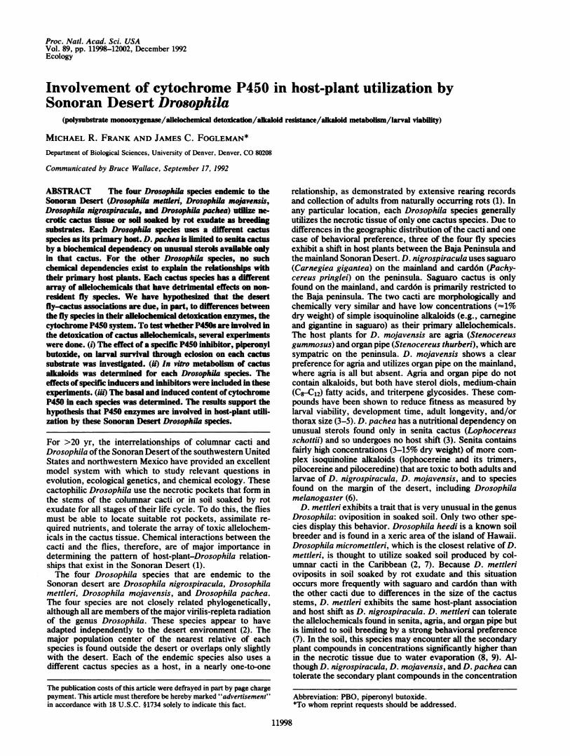
tochrome P450 content, seen in Fig. 3, differs significantly ineach of the species examined (F. = 37.704; df = 4,17; P <0.001). The species ranked by P450 content are as follows: D.melanogaster (Hikone-R > Canton-S) > D. mojavensis > D.nigrospiracula > D. mettleri. The value for D. melanogasterstrain Canton-S is similar to published values ofP450 contentfor this strain-e.g., 0.17 + 0.03 (32). Exposure to pheno-barbital resulted in highly significant increases in P450 con-tent for all species, compared with uninduced controls.Exposure of D. nigrospiracula to saguaro tissue, its normalhost, had no significant effect. Although P450 content in D.mettleri was significantly induced by saguaro and senitatissue, these increases are relatively minor compared with theincrease from phenobarbital induction. P450 content in D.mojavensis was significantly induced by agria tissue, senita
1Cn -4-
= CO)os5:0 o 100
~E :(n cLa 8
32o c
0) 60.oO 6)
0)LUcoE co 40
20E
0Species:Inducer:
PBO mM:
- * Carnegine 1.........................................*..... *.[ GiantineL~~~~~~~~~~~~~~
I~~ ~ ~ ~~~~~~~~~~~*_x,* *1
T~~~~~~* * i* Atr
CS Nig Nig Nig NigN Met Met Met Met Met Met Met ml Moj Mlo Moj MojMoiMCIojMoWj-- SagSag PB PB SagSagSenSen PB PB -- SagSagSenSen 1 OS 1 OS PB PB
- ---1-1 --1-1-1 d--
FIG. 2. In vitro metabolism of saguaro alkaloids. Asterisks above induced experiments indicate significant differences from the uninducedlevel in the same species (*, P < 0.05; **, P < 0.01; ***, P < 0.001). Asterisks above inhibited experiments indicate significant differences fromthe uninhibited (induced) experiments. Species were as follows: D. melanogaster strain Canton-S (CS), D. nigrospiracula (Nig), D. mettleri(Met), and D. mojavensis (Moj). Inducers were as follows: saguaro (Sag), senita (Sen), 10-times-concentrated purified saguaro alkaloids (lOS),and phenobarbital (PB). !, Truncated value due to complete metabolism of substrate.
Ecology: Frank and Fogleman
12002 Ecology: Frank and Fogleman
._ 1.._
a ***a*
__At-----------k^>.. ~~~...... ........................-..-- -----...........*....* .......8 G
l~~~~~~~~~~frimH-.4. ..~~~~~mll
HR HR-- PB5 4
Nig Nig Nig Nig-- OP Sag PB4 3 9 8
Met Met Met Met Met-- OP SagSen PB7 3 5 3 6
Moj Moj Moj Moj Moj Moj MoiAg Op Sag Sen 1OS PB4.(
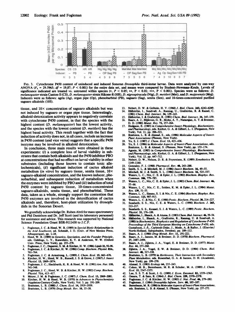
FIG. 3. Cytochrome P450 content of uninduced and induced Sonoran Drosophila third-instar larvae. Data were analyzed by one-way
ANOVA (F. = 29.5965; df = 19,87; P < 0.001) for the entire data set, and means were compared by Student-Newman-Keuls. Levels ofsignificance indicated are treated vs. untreated within species (*, P < 0.05; **, P < 0.01; ***, P < 0.001). Species were as follows: D.melanogaster strain Canton-S (CS), D. melanogaster strain Hikone-R (HR), D. nigrospiracula (Nig), D. mettleri (Met), andD. mojavensis (Moj).Inducers were as follows: agria (Ag), organ pipe (Op), phenobarbital (PB), saguaro (Sag), senita (Sen), and 10-times-concentrated purifiedsaguaro alkaloids (lOS).
tissue, and lOx concentration of saguaro alkaloids but wasnot induced by saguaro or organ pipe tissue. Interestingly,alkaloid-detoxication activity appears to negatively correlatewith cytochrome P450 content, in that the species with thehighest content (D. melanogaster) has the lowest activity,and the species with the lowest content (D. mettleri) has thehighest basal activity. This result together with the fact thatinduction ofactivity does not, in all cases, include an increasein P450 content (and vice versa) suggests that a specific P450isozyme may be involved in alkaloid detoxication.
In conclusion, three main results were obtained in theseexperiments: (i) a complete loss of larval viability in sub-strates that contain both alkaloids and the P450 inhibitorPBOat concentrations that had no effect on larval viability in othersubstrates (including those known to contain toxic alle-lochemicals), (ii) significant induction of cactus alkaloidmetabolism (in vitro) by saguaro tissue, senita tissue, lOx
saguaro-alkaloid concentration, and the known inducer, phe-nobarbital, and subsequent reduction of metabolism whenthe enzymes were inhibited by PBO and (iii) induction oftotalP450 content by saguaro tissue, 10-times-concentratedsaguaro-alkaloids, senita tissue, and phenobarbital. Thesedata, taken as a whole, strongly support the contention thatP450 enzymes are involved in the detoxification of cactusalkaloids and, therefore, host-plant utilization by drosoph-ilids in the Sonoran Desert.
We gratefully acknowledge Dr. Ruben Abril for mass spectrometryand Phil Danielson and Dr. Jeff Scott (and his laboratory personnel)for assistance and advice. This research was supported by NationalScience Foundation Grant BSR-9111430 to J.C.F.
1. Fogleman, J. C. & Heed, W. B. (1989) in Special Biotic Relationships inthe Arid Southwest, ed. Schmidt, J. 0. (Univ. of New Mexico Press,Albuquerque), pp. 1-24.
2. Heed, W. B. (1989) in Genetics, Speciation, and the Founder Principle,eds. Giddings, L. V., Kaneshiro, K. Y. & Anderson, W. W. (OxfordUniv. Press, New York), pp. 253-278.
3. Fogleman, J. C., Dupenret, S. M. & Kircher, H. W. (1986 LOpid 21, 92-%.4. Fogleman, J. C. & Kircher, H. W. (1986 Comp. Biochem. Physiol. 83A,
761-764.5. Fogleman, J. C. & Armstrong, L. (1989) J. Chem. Ecol. 15, 663-676.6. Kircher, H. W., Heed, W. B., Russeli, J. S. & Grove, J. (1967) J. Insect
Physiol. 13, 1869-1874.7. Fogleman, J. C., Hackbarth, K. R. & Heed, W. B. (1981) Am. Nat. 118,
541-548.8. Fogleman, J. C., Heed, W. B. & Kircher, H. W. (1982) Comp. Biochem.
Physiol. 71A, 413-417.9. Meyer, J. M. & Fogleman, J. C. (1987) J. Chem. Ecol. 13, 2069-2081.
10. Kircher, H. W. & Heed, W. B. (1970) in Advances in Phytochemistry(Appleton-Century-Crofts, New York), Vol. 3, pp. 191-209.
11. Brattsten, L. B. (1988) J. Chem. Ecol. 14, 1919-1939.12. Brattsten, L. B. (1979) Drug Metab. Rev. 10, 35-58.
13. Nebert, D. W. & Gelboin, H. V. (1968) J. Biol. Chem. 243, 6242-6249.14. Hillstr6m, I., Sundvall, A., Rannug, U., Grafstr6m, R. & Ramel, C.
(1981) Chem. Biol. Interact. 34, 129-143.15. Hillstr6m, I. & Grafstr6m, R. (1981) Chem. Biol. Interact. 34, 145-159.16. Baars, A. J., Blijeven, G. R., Mohn, A. T., Natajan, A. T. & Breimer,
D. D. (1980) Mutat. Res. 72, 257-264.17. Hodgson, E. (1985) in Comprehensive Insect Physiology, Biochemistry,
andPharmacology, eds. Kerkut, G. A. & Gilbert, L. I. (Pergamon, NewYork), Vol. 11, pp. 206-321.
18. Brattsten, L. B. & Ahmad, S., eds. (1986) Molecular Aspects ofInsect-Plant Associations (Plenum, New York).
19. Yu, S. J. (1987) J. Chem. Ecol. 13, 423-436.20. Yu, S. J. (1986) in Molecular Aspects ofInsect-Plant Associations, eds.
Brattsten, L. B. & Ahmad, S. (Plenum, New York), pp. 153-174.21. Agosin, M. (1985) in Comprehensive Insect Physiology, Biochemistry,
and Pharmacology, eds. Kerkut, G. A. & Gilbert, L. (Pergamon, New
York), Vol. 12, pp. 647-712.22. Nebert, D. W., Nelson, D. R. & Feyereisen, R. (1989) Xenobiotica 19,
1149-1160.23. Gonzalez, F. J. (1988) Pharmacol. Rev. 40, 243-288.24. Smith, S. L. & Mitchell, M. J. (1986) Insect Biochem. 16, 49-55.25. Mitchell, M. J. & Smith, S. L. (1986) Insect Biochem. 16, 525-537.26. Waters, L. C., Nix, C. E. & Epler, J. L. (1982) Biochem. Biophys. Res.
Commun. 1X, 779-785.27. Waters, L. C., Nix, C. E. & Epler, J. L. (1983) Chem. Biol. Interact. 46,
55-66.28. Waters, L. C., Nix, C. E., Solden, K. M. & Epler, J. L. (1984) Mutat.
Res. 139, 51-55.29. Waters, L. C., Simms, S. I. & Nix, C. E. (1984) Biochem. Biophys. Res.
Commun. 123, 907-913.30. Waters, L. C. & Nix, C. E. (1988) Pestic. Biochem. Physiol. 30, 214-227.31. Sundseth, S. S., Nix, C. E. & Waters, L. C. (1990) Biochem. 1. 265,
213-217.32. Sundseth, S. S., Kennel, S. J. & Waters, L. C. (1989) Pestic. Biochem.
Physiol. 33, 176-188.33. Hfillstrdm, I., Blanck, A. &Atuma, S. (1983) Chem. Biol. Interact. 46,39-54.34. H~llstr6m, I., Blanck, A., Graftrsdm, R., Rannug, U. & Sundvall, A.
(1980) in Biochemistry, Biophysics and Regulation ofCytochrome P450:Proceedings of the Third European Meeting on Cytochrome P450, eds.Gustafsson, J.-A., Carlstedt-Duke, J., Mode, A. & Rafter, J. (Elsevier/North-Holland, Saltsjobaden, Sweden), pp. 109-112.
35. Baars, A. J. (1980) Drug Metab. Rev. 11, 191-221.36. Baars, A. J., Jansen, M. & Breimer, D. D. (1978) Biochem. Pharmacol.
27, 2487-2494.37. Baars, A. J., Zijlstra, J. A., Vogel, E. & Breimer, D. D. (1977) Mutat.
Res. 44, 257-268.38. Zijlstra, J. A., Vogel, E. W. & Breimer, D. D. (1984) Chem. Biol.
Interact. 48, 317-338.39. Brattsten, L. B. (1979) in Herbivores, Their Interaction with Secondary
Plant Metabolites, eds. Rosenthal, G. A. & Janzen, D. H. (Academic,New York), pp. 199-270.
40. Ahmad, S. (1983) Ecology 64, 235-243.41. Cohen, M. B., Berenbaum, M. R. & Schuler, M. A. (1989) J. Chem.
Ecol. 15, 2347-2355.42. Lee, S. S. T. & Scott, J. G. (1989) J. Econ. Entomol. 82, 1559-1563.43. Omura, T. & Sato, R. (1964) J. Biol. Chem. 239, 2370-2378.44. Fogleman, J. C. & Kircher, H. W. (1983) J. Nat. Prod. 46, 279-282.45. Fogleman, J. (1984) Drosophila Inform. Serv. 68, 105-107.46. Berenbaum, M. R. (1986)in MolecularAspects ofInsect-Plant Associations,
eds. Brattsten, L. B. & Ahmad, S. (Plenum, New York), pp. 257-272.
o sU)0.LO irk
CU)0 EC u0Co -
±: -
m: E
Q
0.5
0.4
0.2
0.1
L0
Species:nducer:
CS CS-- PB
n. 3 5
Proc. Nad. Acad. Sci. USA 89 (1992)