south central chapter of the society of toxicology
TRANSCRIPT
2014 ANNUAL FALL MEETING OCTOBER 23-24, 2014
THE UNIVERSITY OF MISSISSIPPI OXFORD, MS
South Central Chapter of the
Society of Toxicology
SOUTH CENTRAL CHAPTER OF THE SOCIETY OF TOXICOLOGY 2014 ANNUAL FALL MEETING
OCTOBER 23-24, 2014 UNIVERSITY OF MISSISSIPPI OXFORD, MS THAD COCHRAN RESEARCH CENTER/JACKSON AVENUE CENTER
“Toxicological Research- Inspiring the Next Generation of Toxicologists”
Thursday, October 23 5:00 Registration at Thad Cochran Center (University of Mississippi) 5:30–6:30 Dinner served (Taylor Grocery) 5:30 Judges’ meeting 6:00 Toxicology “Trivia Pursuit” 6:30 Welcoming remarks by Dr. Alice Clark (Office of Research and Sponsored
Programs) and introduction of keynote speaker by Dr. Larry Walker (National Center for Natural Products Research)
6:45 Keynote Speaker- Dr. Frederick Guengerich (Vanderbilt University) “Cytochrome P450 Enzymes and Natural Products: Roles in Toxicity and Drug Development”
Friday, October 24 (Jackson Ave. Center) 8:00-8:30 Continental breakfast 8:30-8:45 Welcoming remarks by Dean David Allen (UM School of Pharmacy) and Dr. William Slikker, Jr. (National Center for Toxicological Research) and
introduction of keynote speaker by Dr. Kristie Willett (UM BioMolecular Sciences)
8:45-9:45 Keynote Speaker- “The results of an intentional exposure to a career in Toxicology and Risk Assessment, with a heavy focus on Dose” Dr. John Lipscomb- (US EPA)
9:45-10:00 Break Platform Session 10:00-11:00 Platforms 1-4 11:00-11:15 Coffee Break 11:15-12:00 Platforms 5-7 12:00-1:00 Welcome high school attendees- “What is Toxicology” Dr. John Lipscomb (US EPA) “K-12 Toxicologist” Luncheon and Lunch with an Expert 1:00-3:30 Poster session 3:30 Award presentations/Adjournment
1

Special Thanks To: 2014 Meeting Sponsors
2
Office of Research
National Center for Natural Products Research
Department of BioMolecular Sciences
Environmental Toxicology Research Program

Regulatory Science Awards
Three Regulatory Science Awards will be given to the best research presented at the Society of Toxicology South Central Chapter Annual Meeting that will be held in Oxford, MS, October 23-24, 2014. Two awards will be given to Student Researchers ($50 and $150 value) and one to Postdoctoral Researcher ($200).
These awards are sponsored by the Regulatory Science Program at University of Arkansas for Medical (Little Rock, AR). The UAMS Graduate Certificate in Regulatory Sciences provides an extension to the Ph.D. student’s existing toxicology/pharmacology training. The Certificate in Regulatory Sciences focus provides post-doctoral fellows or graduate students a unique component to their training that sets them apart from other classically trained scientists when seeking employment opportunities, whether they seek jobs in governmental regulatory agencies, regulated industries, or academia. The Certificate in Regulatory Sciences serves both full-time and part-time students interested in expanding their knowledge of regulatory science. The training provides a more competitive background for regulatory science-based careers. The need for increased training in Regulatory Sciences is highlighted in a recent Institute of Medicine (IOM) report entitled, “Strengthening a Workforce for Innovative Regulatory Science in Therapeutics Development: Workshop Summary” (2011).
A primary goal of the UAMS program is to provide students with insights into the complexities of the laws, regulations, policies, risk assessments, risk-benefit analyses and risk management processes. This training provides graduates with a working knowledge of regulatory science and provides leaders in regulatory science for industry, government, and academia. To learn more about the UAMS Regulatory Sciences certificate program, go to http://publichealth.uams.edu/academics/certificates/certificate-in-regulatory-science/.
3
SOUTH CENTRAL CHAPTER OF THE SOCIETY OF TOXICOLOGY 2014 ANNUAL FALL MEETING
OCTOBER 23, 2014 UNIVERSITY OF MISSISSIPPI OXFORD, MS THAD COCHRAN RESEARCH CENTER
“Toxicological Research- Inspiring the Next Generation of Toxicologists”
Thursday, October 235:00 Registration at Thad Cochran Center (University of Mississippi) 5:30–6:30 Dinner served (Taylor Grocery) 5:30 Judges’ meeting 6:00 Toxicology “Trivia Pursuit” 6:30 Welcoming remarks by Dr. Alice Clark (Office of Research and Sponsored
Programs) and introduction of keynote speaker by Dr. Larry Walker (National Center for Natural Products Research Center)
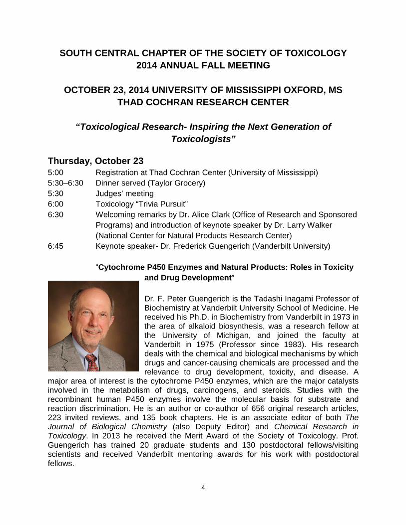
6:45 Keynote speaker- Dr. Frederick Guengerich (Vanderbilt University)
“Cytochrome P450 Enzymes and Natural Products: Roles in Toxicity and Drug Development”
Dr. F. Peter Guengerich is the Tadashi Inagami Professor of Biochemistry at Vanderbilt University School of Medicine. He received his Ph.D. in Biochemistry from Vanderbilt in 1973 in the area of alkaloid biosynthesis, was a research fellow at the University of Michigan, and joined the faculty at Vanderbilt in 1975 (Professor since 1983). His research deals with the chemical and biological mechanisms by which drugs and cancer-causing chemicals are processed and the relevance to drug development, toxicity, and disease. A
major area of interest is the cytochrome P450 enzymes, which are the major catalysts involved in the metabolism of drugs, carcinogens, and steroids. Studies with the recombinant human P450 enzymes involve the molecular basis for substrate and reaction discrimination. He is an author or co-author of 656 original research articles, 223 invited reviews, and 135 book chapters. He is an associate editor of both The Journal of Biological Chemistry (also Deputy Editor) and Chemical Research in Toxicology. In 2013 he received the Merit Award of the Society of Toxicology. Prof. Guengerich has trained 20 graduate students and 130 postdoctoral fellows/visiting scientists and received Vanderbilt mentoring awards for his work with postdoctoral fellows.
4

Notes:
5
SOUTH CENTRAL CHAPTER OF THE SOCIETY OF TOXICOLOGY 2014 ANNUAL FALL MEETING
OCTOBER 24, 2014 UNIVERSITY OF MISSISSIPPI OXFORD, MS
JACKSON AVENUE CENTER “Toxicological Research- Inspiring the Next Generation of
Toxicologists”
Friday, October 24 8:00-8:30 Continental breakfast 8:30-8:45 Welcoming remarks by Dean David Allen (UM School of Pharmacy) and Dr. William Slikker, Jr. (National Center for Toxicological Research) and
introduction of keynote speaker by Dr. Kristie Willett (UM BioMolecular Sciences)
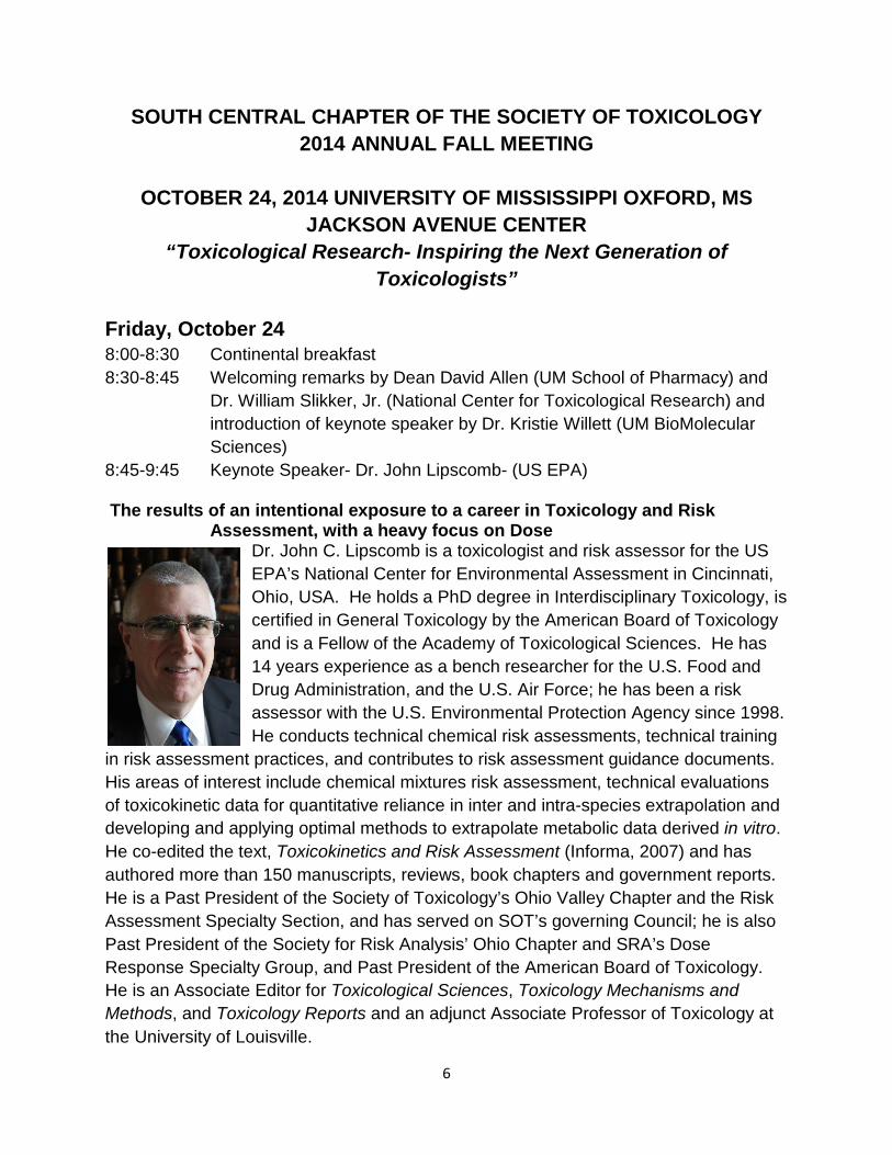
8:45-9:45 Keynote Speaker- Dr. John Lipscomb- (US EPA) The results of an intentional exposure to a career in Toxicology and Risk
Assessment, with a heavy focus on Dose Dr. John C. Lipscomb is a toxicologist and risk assessor for the US EPA’s National Center for Environmental Assessment in Cincinnati, Ohio, USA. He holds a PhD degree in Interdisciplinary Toxicology, is certified in General Toxicology by the American Board of Toxicology and is a Fellow of the Academy of Toxicological Sciences. He has 14 years experience as a bench researcher for the U.S. Food and Drug Administration, and the U.S. Air Force; he has been a risk assessor with the U.S. Environmental Protection Agency since 1998. He conducts technical chemical risk assessments, technical training
in risk assessment practices, and contributes to risk assessment guidance documents. His areas of interest include chemical mixtures risk assessment, technical evaluations of toxicokinetic data for quantitative reliance in inter and intra-species extrapolation and developing and applying optimal methods to extrapolate metabolic data derived in vitro. He co-edited the text, Toxicokinetics and Risk Assessment (Informa, 2007) and has authored more than 150 manuscripts, reviews, book chapters and government reports. He is a Past President of the Society of Toxicology’s Ohio Valley Chapter and the Risk Assessment Specialty Section, and has served on SOT’s governing Council; he is also Past President of the Society for Risk Analysis’ Ohio Chapter and SRA’s Dose Response Specialty Group, and Past President of the American Board of Toxicology. He is an Associate Editor for Toxicological Sciences, Toxicology Mechanisms and Methods, and Toxicology Reports and an adjunct Associate Professor of Toxicology at the University of Louisville.
6

9:45-10:00 Break
Platform Session 10:00-10:15 Platform 1: A.W. Alund, Arkansas Children’s Nutrition Center/
University of Arkansas for Medical Sciences, “Protection Against Ethanol-Induced Osteopenia in Female Mice by Dietary Antioxidants”
10:15-10:30 Platform 2: A. Sreedhar, LSU-Health Science Center, “Knockout of UCP2 Suppresses Skin Carcinogenesis”
10:30-10:45 Platform 3: K. Alharthy, University of Mississippi, “Effects of Benzo[a]pyrene and CYP19b Knockdown on Early Zebrafish Development”
10:45-11:00 Platform 4: S.L. Hudson, University of Alabama at Birmingham, “Interaction Between Early-Life, Low-Level Methylmercury Exposure and a Low-Iron Diet in Daphnia Pulex”
11:00-11:15 Coffee Break 11:15-11:30 Platform 5: C. Wang, US FDA/National Center for Toxicological
Research, “In Vivo Monitoring of Sevoflurane-Induced Neuronal Injury in Neonatal Nonhuman Primates Using Small-Animal Positron Emission Tomography”
11:30-11:45 Platform 6: Z. Zhang, National Center for Toxicological Research, “Mechanistic Study of Ginkgo biloba Leaf Extract Induced DNA Damage in Human Hepatic Cells”
11:45-12:00 Platform 7: X. Guo, National Center for Toxicological Research, “In Vitro Investigation of the Mutagenic Potential of Aloe Vera Extracts”
12:00- 1:00 “K-12 Toxicologist” Luncheon and Lunch with an Expert Welcome high school attendees “What is Toxicology” Dr. John Lipscomb (US EPA)
1:00-3:30 Poster session 3:30 Award presentations/Adjournment
7

Notes:
8

South Central Chapter of the Society of Toxicology 2014 Annual Meeting
“Toxicological Research - Inspiring the Next Generation of Toxicologists”
K-12 Toxicologist Luncheon with an Expert
12:15pm – 1:00 p.m., Friday, October 24, 2014 Jackson Avenue Center
University of Mississippi Oxford, Ms
Program
Greetings and Introductions ……………………………………………………..… Dr. Yunfeng Zhao
President, SCC-SOT Acknowledgement of K-12 Visitors ……….……………..……………………… Dr. Wesley G. Gray Professor of Chemistry, Southern University Introduction of Students and Remarks ………….………………..…… K-12 Teacher and Students
Lunch is served Introduction of Speaker …………………………………………………………....… Dr. Kristie Willett
Conference Chair, University of Mississippi
“Road to Becoming a Toxicologist” Dr. John Lipscomb
US EPA’s National Center for Environmental Assessment in Cincinnati, Ohio, USA
Closing Remarks ……………………………………………...…….……………… Dr. William Slikker
National Center for Toxicological Research
Participants from Ms. Deborah Jones’s Lafayette High School class include: Harrison Atkinson, Katie Barnes, Kayla Hall, Elizabeth Hubbard, Devin Johnson, Linsey Lawrence, Maren OHaver, Garrett Reed, Vanndrika Robinson, and Addison Roush.
9

PLATFORM 1 PROTECTION AGAINST ETHANOL-INDUCED OSTEOPENIA IN FEMALE MICE BY DIETARY ANTIOXIDANTS A.W. Alund1,2, K.E. Mercer1,2, C. F. Pulliam1, T. N. Turner1, L.J. Suva2, T.M. Badger1,2, and M.J. Ronis1,2 1Arkansas Children’s Nutrition Center and the 2University of Arkansas for Medical Sciences, Little Rock, AR, USA Chronic alcohol consumption leads to increased fracture risk and an elevated risk of osteoporosis by decreasing bone mineral density through increasing osteoclast activity, decreasing osteoblast activity, and increasing senescence. Our lab has shown this to be mediated by reactive oxygen species (ROS) produced by NADPH oxidases (NOX). We hypothesized that different dietary antioxidants, Curcumin (120mg/kg/d), N-acetyl cysteine (NAC) (1.2mg/kg/d)), and Vitamin E (α-tocopherol) (60mg/kg/d)) would attenuate osteopenia due to chronic alcohol intake. Female mice received a high fat (35%) Lieber DeCarli liquid diet supplemented with an antioxidant with or without EtOH at 30% of total calories for 8 weeks. MicroCT analysis showed protection from trabecular bone loss in the EtOH+NAC and EtOH+α-tocopherol groups compared to the EtOH group (P<0.05). A significant decrease in bone volume (BV/TV) and trabecular number (Tb.N) (P<.05), along with a significant increase in trabecular spacing (Tb.Sp) was observed in the EtOH compared to PF (P<.05), while the EtOH+NAC and EtOH+α-tocopherol did not statistically differ from their respective PF controls. Femur mRNA analysis revealed a significant increase in the osteoblastic marker, osteocalcin, and decreases in the osteoclast marker, TRAP, and in AP2 expression, a marker of bone marrow adiposity, in the EtOH+NAC and EtOH+α-tocopherol groups compared to EtOH (P<.05). p21 expression, a senescence marker, was elevated by EtOH and blocked by NAC and α-tocopherol. Supported in part by R01 AA18282 (M.J.R.).
10

PLATFORM 2 KNOCKOUT OF UCP2 SUPPRESSES SKIN CARCINOGENESIS *A. Sreedhar, C. Zhang and Y. Zhao. Department of Pharmacology, Toxicology & Neuroscience, LSU-Health Sciences Center, Shreveport. UCP2 is a mitochondrial uncoupling protein present in the inner mitochondrial membrane whose main function is to reduce the mitochondrial membrane potential and thereby decreasing the production of ROS.
UCP2 is generally up-regulated in cancer cells to fight against increased levels of oxidative stress. However, there is no direct evidence supporting UCP2 contributes to carcinogenesis.
Using UCP2 homozygous knockout mice and UCP2 overexpressed cells, our data have demonstrated that knockout of UCP2 suppressed skin carcinogenesis. Overexpression of UCP2 enhanced skin cell transformation and oncogenic activation. In addition, genipin, a known UCP2 inhibitor, was able to inhibit oncogenic activation during skin cell transformation, even in UCP2 overexpressed cells. Finally, although knockout of UCP2 increased the basal levels of protein oxidation, it did not enhance protein oxidation in mouse skin epidermal tissues during skin carcinogenesis.
In summary, our data have provided direct evidence supporting that UCP2 promotes carcinogenesis, and targeting UCP2 may serve as a translational approach for chemoprevention.
11

PLATFORM 3 EFFECTS OF BENZO[A]PYRENE AND CYP19B KNOCKDOWN ON EARLY ZEBRAFISH DEVELOPMENT *K. Alharthy, F. Booc, J. Corrales, C. Thornton, K. L. Willett; Department of BioMolecularSciences, Division of Pharmacology and ETRP, School of Pharmacy, University of Mississippi, University, MS. USA
Benzo[a]pyrene (BaP) is a ubiquitous environmental contaminant that is both an endocrine disruptor and carcinogen. Aromatase (CYP19b) is a key enzyme in steroidogenesis playing a key role in the hypothalamus-pituitary-gonad feedback loop. Here, we consider whether the toxicities observed following BaP exposure are comparable to those following a transient CYP19b knockdown during early development. One-cell zebrafish embryos were injected with a CYP19b morpholino or the Genetools control-MO. Other non-injected embryos were exposed to nominal waterborne concentrations of BaP (0, 10 & 50 μg/L) for 96 hours post-fertilization (hpf). Real time PCR showed both BaP doses significantly decreased CYP19b expression in 96 hpf zebrafish larvae. Cumulative mortality of zebrafish larvae was significantly increased following BaP exposure and CYP19b knockdown, compared to controls. In a treatment-blinded morphological assessment of larvae at 96 hpf, several phenotypes were negatively impacted by high dose BaP and CYP19b knockdown such as body length, optic vesicle, swim bladder inflation, pericardial and abdominal edema, incidence of normal larval body, tail and pectoral fin shape. Ability of exogenous estrogen (E2; 0.01µM) to alleviate toxicities resulting from CYP19b knockdown was evaluated. E2 significantly decreased mortality incidence, abdominal and pericardial edema in CYP19b knockdown larvae. Results suggest BaP effects in zebrafish embryos are similar to the effects observed by aromatase knockdown. Supported by NIEHS R03 ES018962.
12

PLATFORM 4 INTERACTION BETWEEN EARLY-LIFE, LOW-LEVEL METHYLMERCURY EXPOSURE AND A LOW-IRON DIET IN DAPHNIA PULEX *S.L. Hudson, D.A. Doke, and J.M. Gohlke. Department of Environmental Health Sciences,School of Public Health, University of Alabama at Birmingham, Birmingham, AL.
Methylmercury (MeHg) is a known neurotoxicant and bioaccumulates in fish, with exposure to humans in utero being of highest concern. Anemia is a global health concern, and is particularly problematic when occurring during pregnancy and early life. Iron deficiency (ID) is responsible for approximately 50% of anemia cases worldwide. Previous studies have shown that ID exacerbates toxicity associated with exposure to lead (Pb), manganese (Mn), and cadmium (Cd). The overall purpose of this research is to investigate the interaction between a low iron (Fe) diet and exposure to MeHg in Daphnia pulex. We hypothesized when D. pulex are fed a low-Fe diet, the toxicity associated with MeHg will increase. We fed D. pulex standard and low-Fe diets of Ankistrodesmus falcatus, and administered MeHg(1.6 ug/l) for 24 hrs in the first 72 hrs. of life. Results suggest that D. pulex fed a standard Fe diet had no significant difference in lifespan or reproduction (average brood size and reproduction rate) compared to Daphnia on a low-Fe diet. There was no difference found between vehicle and MeHg exposed Daphnia fed either standard or low iron diet. Subsequent trials confirmed maturation time is longer in Daphnia fed a low-Fe versus standard Fe diet. Image analysis of lipids stained with Oil Red O suggests significantly lower percent lipids in Daphnia fed a low-Fe diet and a significant increase in percent lipids in the MeHg exposed Daphnia. (Support: ILSI N.A.)
13
PLATFORM 5 IN VIVO MONITORING OF SEVOFLURANE-INDUCED NEURONAL INJURY IN NEONATAL NONHUMAN PRIMATES USING SMALL-ANIMAL POSITRON EMISSION TOMOGRAPHY C. Wang, X. Zhang, G.D. Newport, S. Liu, M.G. Paule, R. Callicott, James Thompson, F. Liu, M.S. Berridge, S.M. Apana, C.C. Brown, W. Slikker, Jr., National Center for Toxicological Research, U.S. Food and Drug Administration Jefferson, Arkansas 72079; Background: In surgical procedures for human infants, sevoflurane is currently the most widely used volatile general anesthetic. Early exposure to sevoflurane may induce neuronal cell death in the developing brain. In this study, in vivo microPET/CT imaging with 18F-labeled fluoroethoxybenzyl-N-(4-phenoxypyridin-3-yl) acetamide (FEPPA), a sensitive biomarker for the activation of glia, was utilized as a surrogate marker of sevoflurane-induced neuronal injury in neonatal rhesus monkeys. Methods: On postnatal day (PND) 5 or 6, animals (4-6/group) were exposed for 8 hours to 2.5% sevoflurane mixed with oxygen, or this mixture plus acetyl-L-carnitine (ALC) (100mg/kg given i.p.). Control monkeys were exposed to room air only, with or without ALC. One day later, [18F]-FEPPA (56 MBq) was injected into the lateral saphenous vein and microPET/CT images were obtained over the next 2 hours. MicroPET/CT scans were repeated for each monkey one week, three weeks and 6 months after the anesthetic exposure. Results: The radiotracer quickly distributed into the brains of both treated and control monkeys on all scan days. One day after anesthetic exposure the uptake of [18F]-FEPPA was significantly increased in the frontal and temporal lobes. One week after exposure the uptake of [18F]-FEPPA in the frontal lobe of treated animals remained significantly greater than in controls. No significant increases were found in radiotracer uptake in the brains of treated monkeys three weeks or 6 months after exposure. Co-administration of ALC effectively blocked the increase in FEPPA uptake in both the temporal and frontal lobes. Sevoflurane-induced neural damage in the frontal was confirmed morphologically by EM observations in a separate group of animals. Conclusions: These findings suggest that early exposure to sevoflurane may trigger neurotoxicity in the nonhuman primate brain. Volatile anesthetic-induced brain damage in different brain regions can be dynamically detected using microPET imaging. In addition, ALC appears to be a potential protective agent against at least some of the adverse effects associated with such exposures.
14

PLATFORM 6 MECHANISTIC STUDY OF GINKGO BILOBA LEAF EXTRACT INDUCED DNA DAMAGE IN HUMAN HEPATIC CELLS Z. Zhang1, S. Chen2, J. Xuan2, X. Guo1, L. Couch2, L. Guo2, N. Mei1 1Division of Genetic and Molecular Toxicology, 2Division of Biochemical Toxicology, National Center for Toxicological Research, Jefferson, AR Ginkgo biloba extract has been used as dietary supplement for a wide range of remedies. Ginkgo biloba extract consists of various constituents including flavonol glycosides and terpene lactones. Ginkgo biloba extract has been shown to increase an incidence in liver tumors in mice in a standard 2-year bioassay evaluated by the National Toxicology Program. In this study, the DNA damaging effects of Ginkgo biloba extract and many of its constituents were evaluated in HepG2 cells and the underlying mechanism was determined. A molecular docking study revealed that quercetin showed a higher potential to interact with topoisomerase II (Topo II) than did the other Ginkgo biloba constituents, and this in silico predication has been confirmed by using a biochemical assay to study Topo II enzyme inhibition. Moreover, as measured by the Comet assay and the induction of γ-H2A.X, quercetin, followed by keampferol, appeared to be the most potent DNA damage inducer in HepG2 cells. In Topo II knockdown cells, DNA damage triggered by quercetin was dramatically decreased, indicating that DNA damage is directly associated with Topo II. In addition, the Topo II inhibitory effect and DNA damage were also observed when cells were treated with commercially available Ginkgo biloba extracts. Our findings suggest that quercetin- and Ginkgo biloba-induced genotoxicity and tumorigenicity may be the result of Topo II inhibition.
15

PLATFORM 7 IN VITRO INVESTIGATION OF THE MUTAGENIC POTENTIAL OF ALOE VERA EXTRACTS *X. Guo, S. Zhang, S.L. Dial, M.D. Boudreau, Q. Xia, P.P. Fu, D.D. Levy, M.M. Moore and N.Mei. Division of Genetic and Molecular Toxicology, National Center for Toxicological Research, 3900 NCTR Road, Jefferson, AR 72079.
A 2-year cancer bioassay in rodents with a preparation of Aloe vera whole leaf extract administered in drinking water showed clear evidence of carcinogenic activity. To provide insight into the identity and mechanisms associated with mutagenic components of the Aloe vera extracts, we used the mouse lymphoma assay to evaluate the mutagenicity of Aloe vera whole leaf extract (WLE) and Aloe vera decolorized whole leaf extract (WLD). The WLD extract was obtained by subjecting WLE to activated carbon-adsorption. HPLC analysis indicated that the decolorization process removed many components from the WLE extract, including anthraquinones. Both WLE and WLD extracts showed cytotoxic and mutagenic effects in mouse lymphoma cells but at different concentration ranges, and WLD induced about 3-fold higher levels of intracellular reactive oxygen species (ROS) than WLE. Molecular analysis of mutant colonies from cells treated with WLE and WLD revealed that the primary type of damage from both treatments was largely due to chromosome mutations (deletions and/or mitotic recombination). The fact that the samples were mutagenic at different concentrations suggests that while some mutagenic components of WLE were removed by activated carbon filtration, components with pro-oxidant activity and mutagenic activity remained. The results demonstrate the utility of the mouse lymphoma assay as a tool to characterize the mutagenic activity of fractionated complex botanical mixtures to identify bioactive components. (The views presented in this report do not necessarily reflect those of the U.S. Food and Drug Administration)
16
Undergraduate Student Posters 1-3 Poster 1: EVALUATION OF PURKINJE NEURONS AS POTENTIAL TARGET OF ALCOHOLISM USING JAPANESE MEDAKA (ORYZIAS LATIPES) AS ANIMAL MODEL *John F. Franklin1, Ikhlas A Khan2, Asok K .Dasmahapatra 2, 3
1Sally McDonnell-Barksdale Honors College, 2National Center for Natural Product Research, 3Department of Biomolecular Sciences- Pharmacology Division, School of Pharmacy, University of Mississippi.
Purkinje cells (PK) are considered as the neurons with large cell bodies which are found in the central nervous system and are mostly distributed in the cerebellar region of the hind brain. Several novel markers for PK cells have been developed and are used for characterization of neurobehavioral disorders. The present study was aimed to identify endocannabinoid receptor (CB receptor) as a potential marker of PK cells and their evaluation as a molecular target of cerebellar functions using Japanese medaka (Oryzias latipes) as an animal model. Previously, we have observed that medaka genome consist three CB receptor paralogs; two of them (cnr1a and cnr1b) showed structural identity with human CB1 and another with human CB2. Moreover, we have also observed that cnr mRNAs (cnr1a, 1b, and cnr2) are highly expressed in medaka embryos and adult brain. Expression of cnr1a mRNA in medaka embryos was disrupted by developmental ethanol exposure. Moreover, adult male medaka when exposed to ethanol (300 mM) waterborne is able to alter swimming behavior within 1 h of exposure. We expect that these alterations are probably mediated by disruption of the expression of cnr1a receptor in the brain especially in the PK cells of the cerebellum. Preliminary data indicate that the average sizes of the cell body of PK neurons in adult medaka were increased during ethanol exposure which may be related with the expression of cnr1a mRNA in PK cells. Poster 2: LOCALIZATION OF RAT CYP2E1 IMPACTS MECHANISMS FOR SUBSTRATE METABOLISM *H.C. Martin1, J.H. Hartman2, G.P. Miller2
1Dept. of Chemistry, University of Central Arkansas, Conway, AR; 2Dept. of Biochemistry and Molecular Biology, University of Arkansas for Medical Sciences, Little Rock, AR
An appropriate estimation of the health risk posed by CYP2E1 metabolic activation of pollutants requires an understanding the kinetic mechanism and constants describing these processes. The current emphasis on CYP2E1 localized to the endoplasmic reticulum (microsomal) ignores the potential contributions of mitochondrial CYP2E1. The subcellular compartmentalization leads to different redox partners that play a role in CYP2E1 metabolism. Consequently, we hypothesized that CYP2E1 localization to the endoplasmic reticulum and mitochondrion yields distinctly different kinetic mechanisms and constants describing the metabolic clearance of substrates. We tested this hypothesis through steady-state kinetic assays for aniline and 4-nitrophenol metabolism using subcellular fractions isolated from Sprague Dawley rats. Microsomal kinetic studies with the marker substrate, 4-nitrophenol, revealed substrate inhibition kinetics with a Ks of 24 μM and Kss of 2000 μM, while parallel studies with mitochondrial CYP2E1 revealed Michaelis-Menten kinetics with a comparable Km of 28 μM. Mitochondrial kinetic studies with aniline also displayed Michaelis-Menten kinetics with a Km of 11 μM, which contrast with the previously reported negative cooperative mechanism observed for microsomal reactions. These findings demonstrate that CYP2E1 localization alters substrate metabolism and thus confound the interpretation of the consequences of CYP2E1 metabolism as a function of time and concentration.
17

Poster 3: DIGLYCOLIC ACID, THE TOXIC METABOLITE OF DIETHYLENE GLYCOL INDUCES MITOCHONDRIAL DYSFUNCTION *R.H. Nichols1, T. Conrad2, and K.E. McMartin2. 1Department of Biology, King University, Bristol,TN and 2Department of Pharmacology, Toxicology, and Neuroscience, LSU Health Sciences Center-Shreveport, LA
Diethylene glycol is a well-known acute nephrotoxicant and diglycolic acid (DGA) is the primary metabolite that accumulates and produces acute nephrotoxicity in the proximal tubule of the kidneys. Although necrosis is the dominant feature of this toxicity, the specific mechanism for these effects is not fully understood. Previous studies have suggested that DGA decreases the mitochondrial membrane potential and reduces mitochondrial oxygen consumption, suggesting that it induces mitochondrial dysfunction. To determine the extent of this mitochondrial inhibition, kidney mitochondria from the cortex of Wistar or Fischer rats were isolated and then exposed to increasing concentrations of DGA (0.5, 5, 50 and 100 mM). Electron Transport Chain (ETC) complex activities were measured at the respective mitochondrial complexes (I-IV) via spectrophotometric analysis. Positive inhibitory controls for the complexes included rotenone (I), malonate (II), antimycin A (III) and KCN (IV). The results indicated that DGA selectively inhibited Complex II, but had no effect on the other ETC complexes. The effects of DGA on complex II were significant, albeit less marked than that of malonate. The similarities in the chemical structure of DGA and succinate, the substrate for complex II, likely plays a role in this specificity. These results could explain how DGA inhibits oxidative phosphorylation in kidney cells, thereby leading to necrotic cell death and eventually to the acute kidney injury displayed by diethylene glycol.
18

Graduate Student Posters 4-21 Poster 4: EFFECTS OF CROCETIN AND SAFRANAL, CONSTITUENTS OF SAFFRON, USING IN VITRO AND IN VIVO MODELS OF HUMAN PROSTATE CANCER. *F. F. Albaqami1, M. A. Ibrahim2, I. Muhammad2, and K. L. Willett1. 1Department of BioMolecularSciences, Division of Environmental Toxicology and Research Program, School of Pharmacy, University of Mississippi; 2The National Center for Natural Products Research, School of Pharmacy, University of Mississippi
Natural products are a promising source to develop new treatment modalities that work as chemopreventative for prostate cancer development and/or as cytotoxic agents to treat existing cancer cells. Saffron extract and its active constituents may meet these aims. Extracts possess antioxidant properties and have tumoricidal activity. There is a critical need to study saffron’s actual mechanisms against the development, progression, and metastasis of prostate cancer. Normal prostate PNT1A cells and the androgen responsive 22Rv1 prostate cancer cells were used to determine saffron constituents’ activity. Safranal had selective cytotoxicity (IC50 142 µM in 22Rv1 cells) toward prostate cancer cells and acted through an apoptotic mechanism. Furthermore, safranal inhibited prostate cancer cell migration. We conducted HPLC and mass spectrometry on Iranian saffron extracts for profiling, extraction, and in vivo assessment purposes. Ongoing studies are investigating the in vivo effects of saffron extracts on zebrafish xenograft model by transplanting human prostate cancer cells into transgenic zebrafish. According to our results, saffron constituents should be explored extensively to prevent and/or treat prostate cancer. This work is supported by Saudi Arabian Ministry of Higher Education and Salman bin Abdulaziz University.
19

Poster 5: IMMUNITY AGAINST DISEASE IN THE HAWAIIAN CORAL MONTIPORA CAPITATA *M.S. Ansley, Environmental Toxicology Research Program, University of Mississippi, Oxford, MS; D.J. Gochfeld, National Center for Natural Products Research and ETRP, University of Mississippi; M. Slattery, Dept. of BioMolecular Sciences and ETRP, University of Mississippi; L. Mydlarz, Dept. of Biology, University of Texas at Arlington, Arlington, TX; G.S. Aeby, Hawaii Institute of Marine Biology, Kaneohe, HI Disease and anthropogenic stressors, such as nutrient runoff, have caused a global decline in coral reefs. Corals possess innate immune defenses that may protect them from pathogenesis, including physical barriers, enzyme activity, and antimicrobial chemical defenses. Some of these defenses are known to be inducible in response to stress. Montipora capitata is one of the predominant corals in Hawaii, and is susceptible to Montipora White Syndrome [MWS], characterized by tissue loss that exposes the coral’s white skeleton. Within Kaneohe Bay, Oahu, MWS prevalence varies, with lower abundance in the north Bay than the south Bay. This may be due to variability in nutrient runoff and sedimentation. M. capitata occurs in two color morphs, which have different susceptibilities to MWS, possibly due to differences in immune response to pathogenesis. To assess baseline differences in immune function, red and orange healthy colonies of M. capitata were collected from the north and south Bay. To test whether immune defenses are inducible, orange and red colonies from the north and south Bay were exposed to a bacterial pathogen associated with MWS in a laboratory challenge experiment. Overall, healthy colonies from the north Bay had higher baseline levels of antioxidant activity than those from the south bay. Orange colonies exhibit higher baseline levels of melanin than red colonies. These differences in M. capitata immune levels may explain, in part, the variability in disease prevalence throughout Kaneohe Bay. (Support: NSF-OCE 0961384 and NOAA/NIUST NA16RU1496)
20

Poster 6: THE STEROIDAL SAPONIN, DIOSCIN, ISOLATED FROM WILD YAM (Dioscorea villosa) ROOT EXTRACT, HAS THE POTENTIAL TO MODULATE HUMAN BREAST CANCER METASTASIS IN VITRO. *P. Aumsuwan 1, 2, S. I. Khan 1,3, I. A. Khan 1,3, L. A. Walker 1,2, Z. Ali1, B. Avula 1, A.K.Dasmahapatra 1,2 1National Center for Natural Product Research, 2 Department of BioMolecular sciences, Division of Pharmacology, 3 Department of BioMolecular sciences, Division of Pharmacognosy. School of Pharmacy, University of Mississippi, University, MS 38677.
Previously, we have observed that wild yam root extract (WYRE) is able to activate GATA binding protein-3 (GATA3) gene in human breast cancer cells, MCF-7(GATA3-positive, non-invasive, epithelial) and MDA-MB-231(GATA3-negative, invasive, mesenchymal), targeting epigenome. The present study aims to find out bioactive molecules of WYRE which can modulate GATA3 gene functions as well as prevent metastasis in human breast cancer cell lines at the molecular level. We identified eleven saponins and one sapogenin from WYRE and found that four of them have the anticancer activity. In this study we have evaluated diosin (DS), a steroidal saponin which showed anticancer activity, for preventing metastatic potential of cancer cells using MDA-MB-231 cells. Our data indicate that DS like WYRE is able to reduce cell viability and induce GATA3 mRNA expression in both MCF-7 and MDA-MB-231 cells in a concentration-dependent manner. The calculated IC50 is 3.85 µM for MCF-7 cells and 2.07 µM for MDA-MB-231 cells. Invasion analyses based on cell migration, invasion, and wound-healing assays indicate that DS is able to inhibit metastasis of MDA-MB-231 cells in vitro. In contrast to metastastis, GATA3 protein expression as evidenced by western blot and immunocytochemisty was enhanced by DS in MDA-MB-231 cells. To verify the effects of DS on metastasis at the molecular level we have evaluated the expression of four other genes which have significant influence on GATA3 expression as well as metastasis. The mRNAs of ZFPM2, a member of FOG family, and E-Cad which binds to the GATA motifs, were increased by DS (5.76 µM) while VIM, a downstream gene of GATA3, and MMP9 which is considered as a cellular marker for epithelial to mesenchymal transition (EMT), were decreased by DS. These findings indicate that DS has the potential to be used as a breast cancer preventive agent targeting metastasis.
21

Poster 7: SHORTENED IN VIVO BIOCONCENTRATION FACTOR TESTING IN CYPRINUS CARPIO * M.A. Cantu and D.B. Huggett. Institute of Applied Sciences, University of North Texas,Denton, TX.
Bioconcentration factor testing serves as the most valuable surrogate for the assessment of bioaccumulation. The assessment of potentially harmful chemicals is crucial to not only the health of aquatic environments, but to humans as well. Chemicals that possess the ability to persist in the environment or that have the potential to bioaccumulate, pose a greater risk to organisms that are exposed to these chemicals. The Organization for Economic Cooperation and Development Guideline 305 outlines specific protocols to run an accurate and reliable aquatic flow-through test. However, since its adoption in 1996, very few changes have been made to accommodate the endeavor to lowering the amount of test species to run one of these said tests. Running an aquatic flow-through test, according to 305, takes much time and money as well as numerous amounts of fish. Such burdens can be eliminated through simple modifications to the standard protocols. In this study, we propose an abbreviated study design for aquatic bioconcentration testing which effectively alleviates the burdens of running a flow-through test. Four chemicals were used individually to evaluate the usefulness of the proposed shortened design; 4-Nonyphenol, Chlorpyrifos, Musk Xylene, and DDT. The study consisted of exposing Cyprinus carpio for 7 days followed by 7 days of depuration, for a total of a 14-day study. Our results for each of the four compounds are consistent with literature values, thus, demonstrating that BCFk can be accurately predicted in an abbreviated in vivo test.
Poster 8: STEROIDOGENIC EFFECTS OF SERTRAILNE ON JUVENILE FATHEAD MINNOW (PIMEPHALES PROMELAS) *D.R. Carty, W.B. Steele IV, D. Hala, and D.B. Huggett. Department of BiologicalSciences, Institute of Applied Sciences, University of North Texas, Denton, TX
Sertraline was the third most prescribed drug in 2011 with over 37 million prescriptions and generally enters the environment through post-consumer use. Due to the abundance of sertraline in waste water effluents and surface waters, it is imperative to determine the chronic effects of low-level exposure of sertraline. Given that serotonin transporters (SERT) in teleost fish, compared to humans, have nearly 75% sequence similarity, a teleost model to study sertraline toxicity is appropriate. Fathead minnows and other teleost fish are widely used as model organisms for various toxicological/pathophysiological conditions, including endocrine disruption. In this study, early-life stage steroidogenic effects of sertraline on juvenile fathead minnows (FHM) were analyzed via RT-qPCR. Larval FHM were exposed to 0.1, 1, and 10 μg/L sertraline for 28 days and screened for differential expression of 11β-Hydroxysteroid dehydrogenase (11β-HSD), 20β-Hydroxysteroid dehydrogenase (20β-HSD), aromatase (CYP19), and nuclear thyroid receptor alpha (TRα) mRNA. Larval FHM exposed to 0.1 µg/L sertraline had a significant upregulation of 20β-HSD (log(2) = 0.84) and TRα (log(2) = 0.41). In addition to mRNA transcript analysis, FHM survival and weight were also taken into consideration. Survival rates of exposed fish were >80% in all exposure groups and while a positive correlation between weight and sertraline dose was observed, no statistical significance was found. This data presents additional insight into the negative ramifications SSRIs pose on aquatic life and offers caution to possible downstream effects SSRIs may have on human health.
22

Poster 9: REPRODUCTIVE EFFECTS OF DIETARY BENZO[A]PYRENE EXPOSURE IN ZEBRAFISH *Dhawan, T., Thornton, C., Corrales, J., Burkett, A., Shore, J., White, M.B., and Willett,K.L. Department of BioMolecular Sciences, Division of Pharmacology and ETRP, University of Mississippi, University, MS. USA
Benzo[a]pyrene (BaP) is a ubiquitous environmental contaminant that is known to cause negative impacts on reproduction and development when organisms are directly exposed. However, little is known about the multi- or trans-generational effects of BaP. In this study, a parental-only dietary (BaP) exposure was conducted in adult zebrafish (Danio rerio). Fish were exposed to 0, 0.21, 2.3, or 20 µg BaP/g fish for 22 days (n=10 tanks/treatment; 2 males + 2 females/tank). Non-exposed F1, F2, and F3 generations were then reared. Histology of ovaries in the F0 generation revealed a significant decrease in corpus atreticum (F0) following 20 µg BaP/g fish, however, the percentage of previtellogenic, vitellogenic, and mature oocytes were not significantly altered. Accordingly, egg production was not significantly affected following BaP exposure in any of the generations. Despite a significant decrease in the number of fertilized eggs following high dose BaP exposure in the F0 generation, testis histology was not significantly altered in the F0 generation following BaP exposure. Fertilization success was not affected in the F1, F2, or F3 generations. However, in the F1 generation, a significant decrease in spermatid area and a significant increase in percentage of spermatogonia per phase were observed following a parental high dose BaP exposure. These results indicate that reproductive impairment following BaP exposure may be due to male sensitivity. (Supported by NIEHS R21ES019940)
Poster 10: COMPARISON OF MYELOSTIMULATION BY ECHINACEA PURPUREA EXTRACTS WITH ANALYTICAL FINGERPRINTS TO IDENTIFY ACTIVE CONSTITUENTS *H.F.A. Hussin1, K.A. El Sayed2 and S.A. Meyer1. Departments of Toxicology1 and BasicPharmaceutical Sciences2, University of Louisiana at Monroe, Monroe, LA
We have previously described a myelostimulatory activity of Echinacea herbal supplement (Spring Valley Natural Whole Herb, Idea Sphere Inc., American Fork, UT; E. purpurea aerial part) (Ramasahayam et al. 2011. Planta Med. 77:1883-9). A 75% ethanol extract p.o. for 7d at 50-100 mg/kg/d to female SD rats yielded a 70% increase in femur myeloid progenitors, i.e., CFU-GMs, upon culture in methylcellulose with csf2, IL-3 and SCF (HALO assay, Hemogenix, Colorado Springs, CO). To determine that bioactivity was due to plant-derived material, we obtained plant source material (CP) of the supplement (Ray Jaglowski, Twinlab Corp., Grand Rapids, MI), extracted with ethanol (EtCP) and tested for CFU-GMs. Since Echinacea aerial part includes N-alkylamides as a primary constituent, we also determined myelostimulatory activity of n-hexane-washed EtCP (HxEtCP). Analytical fingerprints of EtCP and HxEtCP constituents were acquired with HPTLC and 1HNMR (400 MHz, 20 mg/700 ul MeOH-d4). CFU-GMs of EtCP were comparable to herbal supplement and HxEtCP activity, normalized to source EtCP (~95% yield), was 2 times more potent than EtCP. Hexane wash fraction (HxW) did not affect CFU-GMs. 1HNMR spectra of HxW contained prominent peaks from β-sitosterol verified with a pure standard (ChromDex,Irvine, CA). Similarly, 1HNMR spectral peaks with shifts equivalent to those of dodeca-2,4,8,10-tetraenoic acid isobutylamide (PhytoLab, Vestenbergsgreuth, Germany) were evident in HxW. HPTLC confirmed that these lipophilic constituents were removed from EtCP by washing with n-hexane. In conclusion, these studies demonstrated that myelostimulation of supplement Echinacea was due to phytomaterial other than lipophilic constituents.
23
Poster 11: INTRAVENOUS ADENO-ASSOCIATED VIRUS GENE TRANSFER TO ADULT AND NEONATAL RATS. *K.L. Jackson, R. D. Dayton, A. Richard, R. L. Klein. Department of Pharmacology, Toxicology,and Neuroscience, Louisiana State University Health Sciences Center, Shreveport, LA 71130.
Neonatal intravenous administration of adeno-associated virus (AAV) allows for widespread central nervous system (CNS) transduction. This provides a powerful tool for modeling CNS diseases like amyotrophic lateral sclerosis (ALS) that affect multiple areas within the nervous system. However, most neurodegenerative diseases are associated with aging, thus an adult model would more closely mimic these diseases. Reports of intravenous AAV gene transfer to adults have shown conflicting results. In this study, we compare CNS transduction efficiency of AAV serotypes 1, 8, 9, and rh10 in adult and neonatal rats. Additionally, we evaluate the effect of gender on transduction efficiency. We hypothesize that AAV serotype 9 will have the highest transduction efficiency in both adults and neonates, but that neonatal administration will transduce the CNS more efficiently than adult administration. We also hypothesize that males will have a higher transduction efficiency than females. Preliminary results suggest a decreased transduction efficiency in adults compared to neonates at the same per kg dose and a neuronal pattern of transduction in both neonates and adults. These results suggest that intravenous AAV gene transfer may be feasible for modeling neurodegenerative diseases such as ALS in adults.
Poster 12: P,P’-DDE ENHANCES ADIPOGENESIS IN 3T3-L1 ADIPOCYTES AND ALTERS CYCLOOXYGENASE-2 (COX-2) ACTIVITY IN J774A.1 AND THP-1 MACROPHAGE CELLS L.H. Mangum, G.E. Howell, S.B. Pruett, M.K. Ross, J.E. Chambers. Center for Environmental Health Sciences, Mississippi State University, MS State, MS.
Exposure to p,p’-DDE, a metabolite of p,p’-DDT, is associated with obesity, dyslipidemia, insulin resistance and incidence of metabolic syndrome. DDE has been shown to have immunomodulatory properties, affecting macrophage and T cell populations. Obesity leads to immune cell infiltration into the adipose tissue (AT) causing local inflammation. Potential mechanisms were studied by which DDE could modulate adipocyte and immune cell function and facilitate an increased risk of obesity and immune dysregulation, potentially through COX-2. Human THP-1 and murine J774a cell lines and 3T3-L1 preadipocytes were studied for the effects of DDE on prostaglandin (PG) production and adipogenesis. Macrophage cell lines were exposed to 20µM DDE or 10µM NS398, a specific COX-2 inhibitor, for 18hr before treatment with an inflammatory challenge of 0.25µg/ml lipopolysaccharide (LPS) or 200µM palmitic acid (PA). PGE2, PGD2, PGF2α, and arachidonic acid (AA) were analyzed in cell culture supernatants. The effect of DDE or NS398 on COX-2 activity was also measured indirectly in a cell-free system through addition of AA to cell lysates of LPS (1µg/ml) stimulated macrophages. In J774a and THP-1 cells, DDE or NS398 followed by immune challenge reduced cellular PG secretion and reduced PG production in a cell free system, suggesting that DDE may interfere with COX-2 activity. 3T3-L1 cells were induced to differentiate to adipocytes using a sub-optimal differentiation cocktail with increasing concentrations of DDE (0.5µM-100µM). DDE increased adipogenesis in a dose-dependent manner in mature adipocytes as determined by Oil Red O staining. As PGE2 and PGF2α inhibit adipogenesis, decreased PG synthesis by resident AT macrophages may lead to increased adipogenesis and contribute to development of obesity. These results suggest that DDE exposure may contribute to increased adipogenesis and altered PG production, potentially mediated through a COX-2 dependent mechanism. (Support: Center for Environmental Health Sciences)
24
Poster 13: BIOACTIVE LIPID METABOLISM BY CARBOXYLESTERASE 1 (CES1) IN MACROPHAGES Lee C. Mangum*, Abdolsamad Borazjani, and Matthew K. Ross. Department of Basic Sciences and Center for Environmental Health Sciences, College of Veterinary Medicine, Mississippi State University, Mississippi State, MS 39762 Human carboxylesterases (CES) play an emerging role in lipid metabolism. CES1 has been shown to be a cholesteryl ester, triacylglycerol, and 2-arachidonoylglycerol (2-AG) hydrolase, which may be relevant to cardiovascular disease and other pathologies related to lipid metabolism dysfunction. 2-AG is an endogenous ligand of the cannabinoid receptors (CB1 and CB2), which are responsible for the regulation of numerous physiological processes, including lipogenesis, inflammation, and ROS production by immune cells. 2-AG–mediated activation of CB1/2 receptors is also suspected to regulate cholesterol influx and efflux in macrophages. To further characterize the cholesterol homeostatic functions of CES1, its expression was stably knocked-down in a THP-1 monocyte/macrophage line (designated as CES1KD/THP-1) with a lentivirus harboring CES1 shRNA, and the extent of in situ 2-AG catabolism or [3H]-cholesterol efflux from foam cells was evaluated. In addition, expression levels of several genes involved in cholesterol and 2-AG homeostasis, including ABCA1, ABCG1, CES1, CES3, DAGLβ, CB1 and CB2 were determined following CES1 knockdown. Successful CES1 knockdown by lentivirus transduction was evident by marked reductions in CES1 mRNA transcript, protein content, and enzyme activity. Stimulation of CES1KD/THP-1 cells with ionomycin caused in situ 2-AG levels to be elevated 3-fold compared to wildtype cells, whereas cholesterol efflux from CES1KD/THP-1 foam cells was unaltered. A compensatory upregulation (3-fold) of CES3 transcript was observed in CES1KD/THP-1 cells compared to wildtype cells following cholesterol loading; however, functional enzymatic activity of CES3 was not evident following treatment of cell lysates with serine hydrolase activity-based probe fluorophosphonate(FP)-biotin. Collectively these data suggest that perturbed CES activity does not influence macrophage cholesterol efflux, although our previous work indicated that cholesterol mass is altered following CES1 inhibition. However, enhanced 2-AG tonic levels resulting from reduced hydrolytic activity in macrophages may activate CB1/2 receptor signaling pathways in a paracrine/autocrine manner leading to altered cholesterol homeostasis. This hypothesis is currently being examined. [Supported by NIH 1R15ES015348-02]
25

Poster 14: INCREASED OXIDATIVE STRESS ENHANCES ENDOCANNABINOID TONE Anberitha T. Matthews*, Abdolsamad Borazjani, Ran Wang§, and Matthew K. Ross. Department of Basic Sciences and Center for Environmental Health Sciences, College of Veterinary Medicine, Mississippi State University, Mississippi State, MS 39762; §Institute of Food Safety, Jiangsu Academy of Agricultural Sciences, Nanjing, China, 210014
Cardiovascular disease (CVD) has been characterized as a chronic inflammatory disease that has become prevalent in industrialized societies. NADPH oxidase contributes to atherosclerosis through the activation of macrophages leading to the internalization of oxidized low-density lipoproteins (oxLDL). Chronic inflammation is caused in part by monocytes invading the vasculature leading to the formation of lipid-laden macrophage foam cells with increased flux of oxygen/nitrogen radicals and subsequent chemical modification of extracellular LDL. Endogenous cannabinoids (eCB), such as 2-arachidonoylglycerol (2-AG), may be a link between oxidative stress and atherosclerosis. We hypothesize that 2-AG biosynthesis is enhanced following CD36 ligation by oxLDL and subsequent activation of diacylglycerol lipase β (DAGLβ), the key biosynthetic enzyme of 2-AG, via upregulated NADPH oxidase activity. Treatment of murine J774 macrophages and human THP1 macrophages/monocytes with either extracellular xanthine/xanthine oxidase or phorbol 12-myristate 13-acetate (PMA) caused an increase in superoxide (O2•−) levels and enhanced 2-AG biosynthesis (2.3-fold and 2.8-fold, respectively) compared to vehicle controls. These treatments were not cytotoxic. These data suggest a significant positive correlation between oxygen radical flux and 2-AG biosynthesis in macrophages. Increased 2-AG biosynthesis may be an adaptive response to elevated oxidative stress because of antioxidant and anti-inflammatory actions associated with this bioactive lipid. Therefore, the pathogenesis of atherosclerosis within the vessel wall intima may be reduced by enhancing eCB tone. [Supported in part by NIH R15ES015348-02]
Poster 15: ALTERED EMOTIONAL REACTIVITY IN RATS FOLLOWING EXPOSURE TO LOW LEVELS OF CHLORPYRIFOS DURING DEVELOPMENT A.N. Mohammed, N.H. Armstrong, A.T. Buchanan, M.K. Ross, C.A. Nail, and R.L. Carr. Center for Environmental Health Sciences, College of Veterinary Medicine, Mississippi State University, Mississippi State, MS, USA.
Traditionally, chlorpyrifos (CPS) mediates its toxicity through inhibition of cholinesterase (ChE). However, in recent years, the toxicological effects of developmental CPS exposure have been attributed to an unknown non-cholinergic mechanism of action. We hypothesize that the endocannabinoid system may be an important target because of its vital role in nervous system development. We have previously reported that repeated exposure to 0.5 mg/kg CPS results in the inhibition of fatty acid amide hydrolase (FAAH), the enzyme that metabolizes the endocannabinoid anandamide (AEA), without measurable inhibition of ChE. This exposure resulted in the accumulation of AEA in the forebrain of juvenile rats. Current literature indicates that FAAH activity and AEA levels play an important role in the regulation of emotional reactivity. However, it is unclear if our observed changes in these parameters results in any changes in emotional reactivity. To determine this, 10 day old rat pups were exposed daily for 7 days to either corn oil or 0.5, 0.75, or 1.0 mg/kg CPS by oral gavage. On PND25, the rats were placed into a dark container in a novel open field and the latency to emerge from the container was measured. In this test, rats that stay in the dark for a long time are considered emotionally reactive. All CPS treated groups spent significantly less time in the dark prior to emerging as compared to control suggesting a decreased level of emotional reactivity induced by CPS exposure. Although not directly correlated, our data suggest that the alteration of endocannabinoid signaling during developmental exposure can lead to alteration of behavioral function.
26

Poster 16: RNA-SEQ ANALYSIS REVEALS THE DIFFERENTIAL EXPRESSION OF HOXA1 TARGET GENES IN MOUSE ES CELLS IN RESPONSE TO RETINOIC ACID. *D. Refuge, X. Yi, and E. Martinez‐Ceballos. Environmental Toxicology Department, Southern University and A&M College, Baton Rouge, LA. The homeobox (Hox) family of transcription factors comprises important regulators of embryonic patterning and organogenesis. In mammals, the Hox genes are located in four separate chromosome clusters, Hoxa, Hoxb, Hoxc and Hoxd and their expression depends on their position in the chromosomal cluster: genes positioned at the 3’ end are expressed earlier and more anteriorly than 5’ end genes. In addition, Hox genes can be activated sequentially by retinoic acid (RA) in a manner that resembles their positions in the clusters, e.g. 3’ genes are activated by RA before 5’ genes. In vertebrate embryos, alterations of the normal pattern of Hox gene expression result in homeotic transformations and malformations. In mice, disruption of the Hoxa1 gene results in abnormal ossification of the skull, hindbrain, inner ear deficiencies, and neonatal death; however, little is known about the molecular events that occur downstream of Hoxa1 gene activation. In an attempt to elucidate the molecular mechanism of Hoxa1 action in mouse Embryonic Stem (ES) cells, gene expression profiling was carried out on Wild type versus Hoxa1-/- mouse ES cells using RNA-seq. Overall, transcriptome profiling revealed significant changes in the expression of 2842 genes. Of these, 1979 genes were upregulated by RA in Wt ES cells and 863 were downregulated in these cells as compared to Hoxa1-/- ES cells. The gene ontology of the differentially expressed genes is discussed further. These results provide an insight into the mechanism of Hoxa1 action in differentiating mouse ES cells. (Support: INBRE) Poster 17: ENDOCANNABINOID RECEPTOR EXPRESSIONS IN JAPANESE MEDAKA (ORYZIAS LATIPES) IS ORGAN-SPECIFIC S. K. Singha1, I.A. Khan2, 3, A.K. Dasmahapatra.1, 3 1Department of BioMolecular Sciences, Division of Pharmacology, 2Division of Pharmacognosy, 3National Center for Natural Product Research, School of Pharmacy, University of Mississippi, University, MS. Japanese medaka (Oryzias latipes) genome consists of three endocannabinoid receptor paralogs; two of them (cnr1a and cnr1b) showed structural identity with human CB1 receptor (CNR1) and another (cb2) with human CB2 receptor (CNR2). The present study was aimed to find out whether these CB receptors were expressed in major organs of Japanese medaka and what is the significance of the presence of two cnr1 paralogs in medaka genome. Four adult reproductively active male medaka maintained in the fish culture facility of the Department of BioMolecular Sciences of the University of Mississippi were used for tissue collections. Brain, eye, gill, heart, kidney, liver, spleen and testis were quickly dissected out, weighed to the nearest mg and used for RNA extraction by Trizol reagent. Purified RNA was reverse transcribed into cDNA and used for quantitative PCR (qPCR) using elongation factor alpha (ef1α) as internal control. Gene-specific primers were used for amplification of cnr1a (CB1 receptor A, CB1A), cnr1b (CB1 receptor B) and cnr2 (CB2) mRNA. Our data indicate that expression of CB receptors is organ-specific in Japanese medaka. Significant expression of cnr1a mRNA was observed in brain, eye, spleen and testis, however, expression of cnr1b was observed in brain, eye, gill, heart and spleen. In contrast, cnr2 was expressed in all most all organs used in this study except eye and testis. This study indicates that like human, cnr1a is probably most operative in brain and cnr2 is in immune organs.
27

Poster 18: INFERRING THE CANCER-RELATED HOXA1 GENE REGULATORY NETWORK Augusta Smith, Dr. Xiaoping Yi, Dr. Eduardo Martinez-Ceballos. Health Research Center, Department of Environmental Toxicology. Southern University and A&M College
HoxA1 is a member of the homeobox (Hox) family of transcription factors, which are important regulators of embryonic organogenesis. In humans, HoxA1 mutations have been described in association with various Central Nervous System (CNS) disorders and its overexpression has been associated with cancer development. Although HoxA1 has been recognized as an important oncogene, little is known about the molecular mechanism by which this transcription factor promotes cell proliferation. To address this problem, we sought to construct the HoxA1 Gene Regulatory Network (GNR) using mouse Embryonic Stem (ES) cells since we hypothesize that using an ES model would identify more HoxA1 targets than a specific cancer cell line. Thus, we performed high-throughput RNA sequencing (RNA-seq) analyses on Wild type vs. HoxA1 mutant ES cells treated with Retinoic Acid, an inducer of HoxA1 in cells, for different periods of time. The time-course RNA-seq data was examined using a noise and redundancy reduction technique called NARROMI to infer the Cancer-related HoxA1 GNR. Preliminary analysis of the inferred network indicates that short series of RNA-seq data provide biological insights on the oncogenic mechanism of HoxA1 action.
Poster 19: CONSTRUCTION OF THE HOXA1 GENE REGULATORY NETWORK USING GENE EXPRESSION DATA *Dominique Townsend, Xiaoping Yi, and Eduardo Martinez-Ceballos. Health Research Center,Department of Environmental Toxicology, Southern University A&M College, Louisiana
Hoxa1 is a transcription factor known to regulate embryonic patterning, development, cell proliferation and/or differentiation. Although the relevance of Hoxa1 during embryonic development has been realized for over two decades, little is known about the molecular mechanism of action of this important transcription factor. The human Hoxa1 protein has also been shown to act as an oncogene in various cell types. Hoxa-1 encodes proteins that bind specifically to a DNA sequence and regulate the expression of DNA. However, the gene regulatory interactions of Hoxa-1 have not been well characterized due to a limited number of known target genes. The goal of this work is to identify functional relationships between Hoxa1 and related genes combined in groups based on similarities in their expression profiles using clustering and computational network modeling techniques. Analysis of protein interactions among gene pairs and their functional relationships will help determine strong Hoxa1 gene associations. The identification of differentially expressed Hoxa1 target genes will enable further studies of the consequences of aberrant expression of Hoxa1 during embryonic development and/or cellular transformation. In this study, we investigated Hoxa1 gene interactions in mouse Embryonic Stem (ES) cells using pathway analyses, and validated replicated commonalities amongst research data sets. The time progressions of these gene profiles were analyzed to yield gene expression networks. GRNs were constructed using preliminary RNA-sequencing results. Web-based tools for GRN construction were employed and the results were compared. Genes such as Gbx2, Hand1, Cidea, and Sox17 were inferred to have direct interaction with Hoxa1, whereas Wnt5a, Lamb3, and Mmp19 were found to have indirect interactions with Hoxa1. These observations suggest putative mechanisms of action of Hoxa1 and its associated target genes. Furthermore, the constructed GRN infers the sequential mechanisms by which Hoxa1 directs the differentiation of mouse ES cells. These interactions were verified with published data.
28
Poster 20: EFFECT OF ORGANOCHLORINE COMPOUND EXPOSURE ON APOLIPOPROTEIN B SECRETION BY RAT HEPATOMA CELLS. *A.B. Ward, M.B. Dail, J.E. Chambers. Center for Environmental Health Sciences, College of Veterinary Medicine, Mississippi State University, Mississippi State, MS Dyslipidemia occurs in type 2 diabetes mellitus (T2D). During dyslipidemia there is increased production of lipids from the liver including apolipoprotein B (Apo B) containing very low density lipoproteins (VLDL) and low density lipoproteins (LDL). There is an increasing epidemiological association between diabetes and higher levels of serum organochlorine compounds (OC). OC exposure was found to be a predictor of dyslipidemia in diabetes and non-diabetes. The liver is a target for OC metabolism and plays a role in dyslipidemia and T2D. Therefore, the effect of a group of select OC on the dyslipidemic risk factor, Apo B, in liver cells was investigated. McArdle-RH7777 (McA) rat hepatoma liver cells were treated with 0.4 mM oleic acid (OA) for 16 hr to induce Apo B secretion. McA cells were exposed to 10 µM dichlorodiphenyldichloroethylene (DDE), dichlorodiphenyltrichloroethane (DDT), dieldrin, or chlordane simultaneously with 0.4 mM OA for 16 hr to induce Apo B secretion. McA cells were exposed to 1, 10, or 100 µM DDE simultaneously with 0.4 mM OA for 16 hr to investigate a dose-response of Apo B secretion. Immunoblotting of lipoproteins secreted into the cell media yielded significantly higher levels of secreted Apo B for exposed versus non-exposed liver cells. Immunoblotting of cell lysates revealed that intracellular Apo B levels were significantly different for OC and OA induced cells compared to non-induced cells. The OC exposed cells exhibited a trend of higher Apo B protein levels than OA induced cells. These results suggest a role of OC in lipid transport and secretion of Apo B containing lipids rather than in Apo B synthesis that is induced by lipogenic fatty acids such as OA. Based on these results, OC exposure may be associated with T2D by increasing a dyslipidemic risk factor of T2D. (Support: Center for Environmental Health Sciences, College of Veterinary Medicine, Mississippi State University)
Poster 21: DIFFUSION OF FUNCTIONALIZED DENDRIMERS INTO MOUSE ES CELL AGGREGATES *Niharika Mente, 1Humberto Munoz, and 2Eduardo Martinez-Ceballos *Department of Biology, Chemistry, and Environmental Toxicology 1Department of Physics and Mathematics 2Southern University and A&M College, Louisiana The controlled release and delivery of drugs with target specificity is the main aim of pharmaceutics involved with drug delivery. A safe and targeted delivery is essential to improve the efficacy and efficiency of a particular drug as it minimizes the undesired side effects. Nano biotechnology has helped foster the development of nano-scale pharmaceutical delivery of the drugs and has been employed for the directed differentiation of stem cells along specific cell lineages. Liposomes, cyclodextrins, dendrimers, etc., have been extensively used in recent times as drug delivery models due to their ability to alter the physiochemical, kinetic, body distribution, delivery properties of hydrophobic drugs. Dendrimers are of special interest since they possess different surface functionalities and the ability to encapsulate a wide group of guest molecules. We were particularly interested in studying the diffusion of Generation 5 (G5) dendrimers into mouse Embryonic Stem (mES) cell aggregates known as Embryoid Bodies or EBs. For this, we treated EBs with chromophore-functionalized dendrimers and examined their diffusion by confocal microscopy. We observed that dendrimers could readily penetrate various EB cell layers in as short as 24 hours after application. In some cases, labeled dendrimers could be detected in the cell nucleus, which supports their use for the delivery of ligands for nuclear receptors. Further discussion on the diffusion properties of functionalized dendrimers into EBs is presented here. Our studies may have implications on areas such as ES Cell Differentiation and Regenerative Medicine.
29

Non-Student Posters 22-38 Poster 22: PURIFICATION, DETECTION AND DOSE DEPENDENT ACUTE TOXICITY STUDY OF ABRIN IN SWISS ALBINO MALE MICE *Krishna Chaturvedia, Om Kumara, Yangchen Doma Bhutiaa, Sunil E. Jadhava, Ramesh KumarKaula, Nalini Shrivastavab aDivision of Pharmacology and Toxicology, Defense Research Development and Establishment, bDepartment of Biochemistry, Jiwaji University,Gwalior,474002, India
Abrin is a highly toxic (an estimated human fatal dose of 0.1-1 µg/kg) ribosome inactivating protein present in the seeds of Abrus precatorius plant. There is no antidote available against abrin. The present study is aimed at the purification of abrin from Abrus seeds and its toxicological characterization and detection using immunological methods. Abrin was purified by lactamyl-sepharose affinity chromatography and its purity was confirmed by mass spectrometric analysis. Antibodies against abrin were raised in rabbit and used to detect abrin using western blotting. Further, dose dependent toxicity studies were carried out in swiss albino male mice at three-time points: 1st, 3rd and 7th day. The median lethal concentration (LD50) value was determined for mice through intraperitoneal route. Abrin was administered at the doses of 0.5 and 1.0 LD50 intra-peritoneally in mice, and biochemical, histopathological, and oxidative stress parameters were measured. The levels of liver enzymes such as SGOT and SGPT were increased significantly at both the doses, whereas levels of alkaline phosphatase and bilirubin increased significantly at higher dose only. Blood urea and uric acid levels were increased significantly at both the doses. The level of liver GSH decreased significantly whereas the level of lipid peroxidation increased significantly in a dose dependent manner. Histopathological studies showed degenerative changes in liver, spleen, and kidney. Western blot was prominent even at 20ng/µl. These results indicate that abrin purified from our laboratory is highly pure and toxic. It can be further used for the screening of antidotes and the development of sensitive detection systems.
Poster 23: THE ROLE OF ER STRESS AND STORE OPERATED CALCIUM ENTRY IN USNIC ACID-INDUCED TOXICITY IN HEPATIC CELLS S Chen, Y Wu, Z Zhang, L Guo. National Center for Toxicological Research/ U.S. FDA, Jefferson, AR 72079
Usnic acid and usnic acid containing products have been used for antimicrobial, antiviral, antiparasitic, antimycotic, and antiproliferative purposes and marketed in the United State as dietary supplements aiding in weight loss. Previously, we have demonstrated that usnic acid caused hepatic cytotoxicity, with autophagy induction being a defensive mechanism. In this study, we investigated and characterized molecular mechanisms of usnic acid induced toxicity in human hepatoma HepG2 cells. Usnic acid caused ER stress as demonstrated by an increased expression of ER stress markers by real-time PCR and Western blots. The expression level of typical ER stress markers such as CHOP, ATF-4, p-eIF2α, and spliced XBP1 was significantly increased. Usnic acid inhibited the secretion of Gluc, an established ER stress reporter. Moreover, an ER stress inhibitor 4-phenylbutyrate attenuated usnic acid-induced apoptosis. In addition, usnic acid significantly increased the cytosolic free Ca2+ concentration. Exposure to usnic acid markedly upregulated the protein expression of calcium release-activated calcium channel protein 1 (ORAI1) and stromal interaction molecule 1 (STIM1), the two major molecular components of store operated calcium entry (SOCE). Furthermore, ORAI1 contributed to the process of usnic acid-induced ER stress and hepatotoxicity. Taken together, our results suggest that usnic acid induced ER stress in HepG2 cells at least partially via activation of the Ca2+ channel of SOCE.
30

Poster 24: PON 1 ENZYME ACTIVITIES IN A MISSISSIPPI POPULATION VARY WITH SUBSTRATE, GENOTYPE AND RACE *M.B.Dail1, C.McDaniel1, R.W.Wills1, H.W.Chambers2, J.E.Chambers1
1Coll. Vet. Med., 2Dept. Biochem., Mol. Biol., Entom., Plant Path., Mississippi State University,Mississippi State, MS
Paraoxonase (PON1) hydrolyzes paraoxon (PO) and diazoxon (DZO), the active metabolites of the insecticides parathion and diazinon, respectively. The human PON1 gene has several single nucleotide polymorphisms (SNPs). The SNP involving arginine (R) to glutamine (Q) substitution at codon 192 is associated with catalytic efficiency while that involving methionine (M) to leucine (L) substitution at codon 55 is associated with serum concentration. PON1 polymorphisms and activities have been linked to cardiovascular and other diseases. Since Mississippians have high disease rates, 186 cardiology clinic patients were evaluated. Hydrolysis rates of PO (POase), DZO (DZOase), dihydrocoumarin (lactonase) and phenyl acetate (arylesterase) were evaluated for associations with race, gender, age, and PON1 55/192 SNP genotypes. The variables were analyzed both individually and in combination. African Americans had higher levels (p<0.02) of POase and arylesterase than Caucasians but Caucasians had higher DZOase and lactonase values (p<0.05). Since lactonase activity may be the most critical PON1 enzyme activity for LDL oxidation prevention, this might partially explain the health disparity paradox of why African Americans have higher POase and arylesterase activities than Caucasians yet still have higher rates of coronary disease. Significant (p<0.0001) differences in arylesterase activities were seen amongst the genotypes with QQMM having the lowest and RRLL the highest activities. This opposes the prevailing belief that arylesterase is unaffected by genotype and suggests that this activity cannot be used to quantify PON1 protein. The differences in PON1 activities indicate the need to specify substrate and demographic data for enzyme assays. (Support NIH ES015170)
31

Poster 25: CANNABIDIOL (CBD) INDUCES CD25 AND FOXP3 ON CD4 CELLS IN RESPONSE TO SUBOPTIMAL ACTIVATION. Saphala Dhital, John Stokes, Barbara L. Kaplan. Department of Basic Sciences, Center for Environmental Health Sciences, College of Veterinary Medicine, Mississippi State University, Starkville, MS
Cannabis sativa-derived cannabinoid compounds are being investigated as potential immunosuppressive agents and important drug candidates for immune related diseases. Our previous data demonstrated that under conditions of suboptimal T cell activation, CBD could increase interleukin-2 (IL-2) production. Initially this result was surprising given that cannabinoids are immunosuppressive, but IL-2 can also contribute to regulatory T cell (Treg) induction. We tested the hypothesis that part of the mechanism for immunosuppression by CBD is induction of a T regulatory (Treg) population in response to suboptimal stimulation. Splenocytes from C57BL6 mice were treated with CBD, or vehicle (VH; 0.1% ethanol) for 30 minutes, then stimulated with PMA/Io (PMA - phorbol-12-myristate-13-acetate; Io -ionomycin) overnight. After supernatants were harvested for enzyme-linked immunosorbent assay (ELISA) analysis, the cells were treated with Brefeldin A for four hours before staining for fluorescently-activated cell sorting (FACS) analysis for intracellular IL-2 and TGF-β. For the measurement of CD4, CD25, and Foxp3, cells were stained for extracellular markers, CD4 and CD25, and intracellular marker, for Foxp3. Fixable viability dye was used to stain dead cells before extracellular staining. Our observations showed that the percentage of CD25+Foxp3+ cells increased in response to suboptimal activation plus CBD. Interestingly, in addition to an increase in IL-2 with suboptimal activation plus CBD, there was also an increase in IL-2+TGF-β+ population, both of which contribute Treg induction. Measuring IL-2 by ELISA in the culture supernatant also demonstrated increased IL-2 in response with the increased CBD concentrations of 1, 5 and 10 µM. All these results demonstrate that CBD induces cellular markers consistent with a T reg population in response to suboptimal activation. Thus, these data suggest that the mechanism by which CBD is immunosuppressive might involve induction of Tregs under conditions in which T cells are not maximally activated (Funding provided by MSU College of Veterinary Medicine.)
32

Poster 26: PRELIMINARY RESULTS FROM SEGMENT I FERTILITY STUDY OF OXYBENZONE IN RATS D.K. Hansen1, N. Nakamura1, B. McIntyre2, P.M.D. Foster2, A.L. Inselman1 1 Division of Systems Biology, FDA/NCTR, Jefferson, AR 2 Toxicology Branch, NTP/ NIEHS Research Triangle Park, NC
The UV filter, oxybenzone, is used in cosmetics and other products where it may prevent photodecomposition. Analysis of samples collected through NHANES indicated widespread exposure to this agent, and both in vitro and in vivo assays have suggested that it may be an endocrine disruptor. The purpose of this study was an evaluation of effects on fertility using a Segment I study design. Twenty-five Harlan Sprague-Dawley male and female rats were assigned to each treatment group; treatment groups were 0, 3000, 10,000 or 30,000 ppm oxybenzone added to low phytoestrogen 5K96 chow. Ethinyl estradiol (EE2) was used as a reference estrogen in this study and was administered in the chow at 0.05 ppm. Males were dosed for 10 weeks and females for 2 weeks prior to mating. One male and one female from each treatment group were placed together for mating, and the day that sperm were observed in the vaginal lavage was counted as gestation day (GD) 0. Dosing of all animals continued to GD 6, and animals were returned to control chow until sacrifice on GD 15. Both males and females in the 10,000 and 30,000 oxybenzone groups and the EE2 animals weighed less than control animals through most of the treatment period. The observed decrease in weight was not due to decreased food consumption. Oxybenzone produced few adverse effects on organ weights in either sex, had no effect on sperm count, motility or morphology and had no effect on hormone levels in males; there was an increase in the number of males with minimal to mild mammary gland hyperplasia. There were no differences in the time to plug or the overall pregnancy rate, and no effects on the numbers of corpora lutea, implantation sites, resorptions or live fetuses in the females at GD 15. Based on the doses used in this study, oxybenzone does not appear to significantly impact fertility in male and female rats when treated as adults. This work was funded by an Interagency Agreement between FDA (224-12-0003) and NIEHS (AES12013).
Poster 27: GENOTOXICITY OF GINKGO BILOBA LEAF EXTRACT AND ITS CONSTITUENTS IN MOUSE LYMPHOMA CELLS N Mei. Division of Genetic and Molecular Toxicology, National Center for Toxicological Research, Jefferson, AR
Ginkgo biloba leaf extract is a complex mixture with many constituents, including flavonol glycosides and terpene lactones. Gingko products are widely available in commercial products as dietary supplements and top the list of the best-selling herbal products. The NTP 2-year cancer bioassay found that Ginkgo biloba leaf extract targets the liver, thyroid gland, and nose of rodents; however, the mechanism of Ginkgo biloba leaf extract-associated carcinogenicity remains unclear. In this study using mouse lymphoma L5178Y cells, we investigated the genotoxicity of Ginkgo biloba leaf extract and its eight constituents. Ginkgo biloba leaf extract induced mutations in the mouse lymphoma assay and DNA damage in the Comet assay at the dose of 1 mg/ml, and had a pro-oxidative effects with increased intracellular ROS levels and decreased GSH levels when applied at the doses over 0.4 mg/ml. Considering the percentages of eight constituents tested in this study, it is tempting to attribute these observations with whole leaf extracts to the two components of quercetin and kaempferol. Because gingko products are derived from botanical products using different methods of preparation, the final product could vary considerably in its chemical composition. Therefore, more studies on the mechanism of genotoxicity and carcinogenicity of Ginkgo biloba leaf extract are warranted.
33

Poster 28: SOY FEEDING PREVENTS ETOH-MEDIATED TUMOR PROGRESSION IN A DIETHYLNITROSAMINE-TREATED HCC MOUSE MODEL. K.E. Mercer; C.F. Pulliam; L. Hennings; K. Lai, M.A.Cleves, E.E. Jones; R.R. Drake; and M. J. Ronis., University of Arkansas for Medical Sciences, Arkansas Children’s Nutrition Center, Little Rock, AR 72202, Medical University of South Carolina Proteomic Center, Charleston, SC 29425
In this study, DEN-treated male mice were assigned to 5 groups: a 35% high fat ethanol diet (EtOH/cas) and corresponding high fat pair-fed diet (PF/cas) using casein as sole source protein, and a high fat EtOH liquid diet (EtOH/soy) and high fat PF diet (PF/soy) using soy protein isolate as sole source protein and a chow+DEN group. EtOH feeding continued for 16 wks. EtOH increased the number of tumor foci present in the livers of the EtOH/cas group compared to the PF/cas group and the chow+DEN group, p<0.05. Tumor multiplicity and adenoma incidence were reduced in the EtOH/soy group compared to the EtOH/cas group, p<0.05. These findings corresponded to reductions in serum ALT concentrations, and decreased hepatic TNFα, CXCl-2 and CD-14 expression in the EtOH compared to the EtOH/soy group, p<0.05. Detection of sphingolipids using high resolution MALDI-FTICR Imaging mass spectrometry revealed increased accumulation of long acyl chain ceramide species, sphingosine-1 phosphate, and glucosylceramide in the EtOH/cas group that were significantly reduced in the EtOH/soy group. Gene expression of β-catenin targets, including cyclin D1, MMP7 and glutamine synthetase, were also reduced in the EtOH/soy group compared to the EtOH/cas group, p<0.05. These findings suggest that soy prevents tumorigenesis by reducing both pro-inflammatory signaling and hepatocyte proliferation. Moreover, these mechanisms may involve sphingolipid signaling. Supported in part by NCI R21 CA169389 (M.J.R.).
Poster 29: EFFECTS OF THE TYPE OF PARTICULATE MATTER ON THE EPIGENETIC RESPONSE. *IR Miousse, MG Chalbot, E Nzabarushimana, IG Kavouras, I KoturbashDepartment of Environmental and Occupational Health, University of Arkansas for Medical Sciences, Little Rock, AR
Particulate matter (PM), a major component of outdoor air pollution, has been recently classified as a human lung carcinogen by the International Agency for Research on Cancer. Previous studies by our laboratory demonstrated that ambient particulate matter induced epigenetic alterations in mouse macrophages after 24h in vitro exposure. The objective of the current study was to examine the effects of particle types on the cellular epigenome. To achieve this, we determined the levels of repetitive elements-associated DNA methylation and expression DNA methylation machinery genes in response to exposure to six different particle types: traffic, wood burning, road dust, agricultural dust, natural dust, and pollen. Levels were assessed for two doses of PM; 5 and 50 µg per mL of cell culture media, and at two time points; 24h and 72h of exposure in murine macrophages and primary human small airway epithelial cells. The chemical composition of atmospheric aerosol was determined by NMR analysis. We show that PM of various sources cause changes in DNA methyltransferase gene expression. This was followed by the hypomethylation of repetitive elements after 72h of exposure to PM. Our data suggest that the extent of the epigenetic response was dependent on the type of PM.
34
Poster 30: SUPPRESSING EFFECT OF HSA-MIR-29A-3P ON CYP2C19 EXPRESSION IN HUMAN LIVER CELLS *B. Ning, D. Yu, B. Green, J. Fuscoe and I. Pogribny. National Center for ToxicologicalResearch, US Food and Drug Administration, Jefferson, AR, USA; University of Arkansas for Medical Sciences, AR, USA.
Cytochrome P450 2C19 (CYP2C19) is involved in the metabolism of many clinical drugs. Extensive studies have demonstrated that the genetic variants and environmental factors play important roles in the inter-individual variability of CYP2C19 expression; however, the regulatory mechanisms of microRNA affecting CYP2C19 expression are still unclear. In this present study, we performed in silico analysis to search the potential miRNAs that might control CYP2C19 expression, and then applied a series of biochemical assays to elucidate the underlying functional mechanisms in regulation of CYP2C19 by microRNAs. In silico analysis identified that hsa-miR-23A-3p and hsa-miR-29A-3p could target the coding region of CYP2C19, with binding affinities of –27.5 kcal/mol and –23.3 kcal/mol, respectively. RNA electrophoresis mobility shift assays showed either hsa-miR-23A-3p or hsa-miR-29A-3p was able to directly bind the cognate target in the CYP2C19 transcript. Further, the exogenous CYP2C19 expression in HepG2 or 293T cells, or the endogenous CYP2C19 expression in HepaRG cells was suppressed by transfection of hsa-miR-29A-3p, but not by the transfection of hsa-miR-23A-3p. In addition, chemical-induced up-regulation of hsa-miR-29A-3p was significantly inversely correlated with CYP2C19 expression. These results demonstrated the suppressing role of hsa-miR-29A-3p on CYP2C19 expression. (Support: NCTR/FDA)
35

Poster 31: GENDER-SPECIFIC ALTERATION OF LUNG FUNCTIONS IN ADULT MICE EXPOSED IN UTERO TO SECOND-HAND SMOKE *A. Noël1; R. Xiao1; L. R. Petersen1; Z. Perveen1; M. McNulty1; V. Le Donne2 and A. L. Penn1.1Department of Comparative Biomedical Sciences and 2Pathological Sciences, School of Veterinary Medicine, Louisiana State University, Baton Rouge, LA 70803.
Introduction: An increasing number of epidemiological and experimental studies have associated exposure to second-hand smoke (SHS) during pregnancy with adverse outcomes in the newborns. As we previously shown, in utero exposure to SHS, at critical stages of the fetal development, can result in increased disease susceptibility upon re-exposure to irritants (SHS or ovalbumin) in adulthood. In this study, we tested whether the in utero SHS exposure alone is sufficient to initiate inflammation and alter lung function in adult mice. Methods: Pregnant Balb/c mice were exposed from days 6 to 19 of pregnancy to 10 mg/m3 of SHS or HEPA-filted air. Male and female offspring (n = 15 per group) were sacrificed at 15 weeks of age. The broncho-alveolar lavage fluid (BALF) was collected for cytology analysis. A FlexiVent system and whole body plethysmography (Busco System) were used to measure lung function though invasive and non-invasive techniques, respectively. Results: Lung function testing measured through whole body plethysmography showed that tidal volume and minute volume were significantly decreased (p < 0.05) in male mice exposed in utero to SHS compared to their respective filtered-air controls. These results were confirmed by micro computed tomography imaging of lungs. In addition, the inspiratory capacity, measured by the FlexiVent system was also significantly decreased (P < 0.05) in the in utero SHS exposed male group compared to the air controls. As expected, no significant difference was observed in BALF cytology, indicating that there was no inflammation. Interestingly, no difference in lung function and BALF cytology was observed for the female mice. Conclusion: In utero SHS exposure alters lung functions through adulthood (15 weeks of age) in male mice. In addition, this effect seems to be gender specific, since no pronounced effect was observed in females. These data suggest that males might be more susceptible to in utero SHS effects.
36
Poster 32: NEUROPROTECTIVE EFFECT OF THE TETRAMETHYL PYRAZINE (TMP) IN A CHRONIC MPTP-INDUCED PARKINSON’S DISEASE MOUSE MODEL Sumit Sarkara, James Raymickb, Srinivasulu Chigurupatia , Joseph P. Hanigc, and Merle G. Paulea; a Division of Neurotoxicology, National Center for Toxicological Research/FDA, Jefferson, AR-72079; cOffice of Testing & Research, CDER/FDA, White Oak, MD-20993. bToxicologic Pathology Associates, Jefferson, AR- 72079
Parkinson's disease (PD) is a progressive motor disease of unknown etiology in the majority of cases. The clinical features of PD emerge due to selective degeneration of dopamine (DA) neurons in the substantia nigra pars compacta (SNc), which project to the caudate putamen (CPu) where they release DA. In the current in vivo mouse model study, we tested TMP for its neuroprotective role against 1-methyl-4-phenyl-1, 2, 3, 6-tetrahydropyridine (MPTP)-induced damage to DA neurons. TMP is a superoxide scavenger and can be found in medicinal plant Ligusticum wallichat franchat, which is widely used to treat coronary artery and cerebral vascular diseases. Recent studies suggest that TMP has the potential to reduce the cerebral ischemia and reperfusion injury through suppression of inflammatory cell activation and pro-inflammatory cytokine production. TMP is also been shown to prevent neuronal apoptosis and neuronal loss in vivo. In the present study we investigated whether TMP treatment can ameliorate the pathology associated in a chronic MPTP-induced PD mouse model. We studied four treatment groups: saline only (control); probenecid only (250mg/kg, i.p; control), MPTP (25mg/kg-10 doses) +probenecid (250mg/kg-10doses), and MPTP (25mg/kg-10 doses) + TMP (20mg/kg-daily i.p. for 38 days). MPTP-induced losses in tyrosine hydroxylase (TH) and DA transporter immunoreactivity in the ventral midbrain SNc and CPu were significantly reduced by TMP. In addition, microglial activation and astrocytic hypertrophy induced by MPTP were significantly reduced by TMP, indicating protection against neuroinflammation. This study demonstrates TMP protects MPTP-induced TH loss and suggesting that TMP therapy may have the potential to slow or ameliorate the progression of PD pathology. This study was supported by NCTR protocol E751201.
37

Poster 33: EFFECTS OF LIFELONG ACRYLAMIDE EXPOSURE ON PREPULSE INHIBITION BEHAVIOR OF MALE AND FEMALE RATS *M. E. Smith, J. D. Garey, S. A. Ferguson, and M. G. PauleDivision of Neurotoxicology, National Center for Toxicological Research/FDA, Jefferson, Arkansas.
Acrylamide (ACR) is a known neurotoxicant found in many foods, especially carbohydrate-containing foods cooked at high temperatures. Here, pregnant Fischer 344 rats (n=8-9/group) were gavaged with 0, 0.1, 0.3, 1.0, or 5.0 mg/kg/day ACR on gestational days 6 – 22. From postnatal day (PND) 1 through weaning on PND 21, pups were directly gavaged with the same dose as the dams after which ACR treatment continued via drinking water. One/sex/litter was assessed for prepulse inhibition on PNDs 186 and 368 and males continued additional assessments on PNDs 550 and 732. Females were not tested at those later ages due to early, non-ACR related deaths. At each test age, each subject was exposed to five trial types within a 60 trial test session: 1) no stimulus (n=8), 2) pulse-alone trials (a single burst of 120 dB) (n=22), 3) one of three prepulse trials in which a prepulse stimulus of 72, 75, or 82 dB was presentedfollowed by the 120 dB startle stimulus (n=10 each). Prepulse inhibition for each prepulse stimulus level was calculated as: 100*((response magnitude on pulse-alone trials – response magnitude on prepulse trials)/response magnitude on pulse-alone trials).Data for males and females were analyzed separately with a separate ANOVA for each of the five trial types and each included factors of dose, PND, and the interaction. Analysis of female response on the pulse-alone trials indicated a significant main effect of dose (p=0.003); however no ACR group differed significantly from the control group. There were no significant effects for any of the other trial types in females. Analysis of male responses indicated significant main effects of PND on most trial types, indicating that responses varied with age. The analysis of no stimulus trials indicated a significant effect of dose (p=0.006) and pairwise comparisons indicated that the response of the 5.0 mg/kg ACR group was increased on those trials relative to controls (p<0.001). Analysis of the 82 dB prepulse trial types indicated a significant effect of dose (p=0.046); however, pairwise comparisons did not indicate that any ACR group was significantly different from the control group. These data demonstrate that lifelong ACR treatment had few effects on prepulse inhibition testing throughout the lifespan. (Supported by NTP #224-93-001 and ORISE)
38

Poster 34: TOXICOLOGY STUDY OF THE SAFFLOWER (CARTHAMUS TINCTORIUS L) AND CAMEL THORN (ALHAGI KIRGHISORUM) GROWN IN KAZAKHSTAN. *A.A. Turgumbayeva 1.2 , G.O. Ustenova 1, and Samir A Ross.2..1Kazakh National Medical University name of S.D.Asfendiyarov,Tole bi 050011, Almaty,Kazakhstan; 2Department of Pharmacognosy, School of Pharmacy, University of Mississppi,University, MS 38677,USA
Carthamus tinctorius L. and Alhagi kirghisorum are two important kinds of Iranian manna. They have several therapeutic applications in adults and children. The most important ingredient of Carthamus tinctorius L. Due to the scarcity of toxicological studies on these compounds. A composition of volatile oil was obtained from dried flowers of Carthamus tinctorius L. growing in Kazakhstan. The aim was designed to study a biological activity and chemical composition of the essential oil. Phytochemical study essential oil of the extracts, gained from safflower ( Carthamus tinctroius) with Kazakh origin «Ak mai», which antimicrobial, antimalarial and anti-leishmanial curative effects, and with using of most new method of gas chromatography-mass spectrometry (GC-MS). The preclinical study essential oil, gained from safflower flowers ( Carthamus tinctroius L.) with Kazakh origin «Ak mai» for purpose of safety determination of developed medical plants. Taking Carthamus tinctroius L.and Alhagi kirghisorum in doses which produce concentrations less than 50 mg/ml and 5 μg/ml, respectively, is safe. The harmful effects of non sugary components might be considered in the toxicity caused by these compounds.
Poster 35: VALIDATION OF A GENOMICS-BASED HYPOTHETICAL ADVERSE OUTCOME PATHWAY: 2,4-DINITROTOLUENE PERTURBS PPAR SIGNALING THUS IMPAIRING ENERGY METABOLISM AND EXERCISE ENDURANCE. *M.S. Wilbanks1, K.A. Gust1, S. Atwa2, I. Sunesara3, D. Johnson4, C.Y. Ang5, S.A. Meyer2, andE.J. Perkins1. 1Army Engineer Research and Development Center, Vicksburg, Mississippi; 2University of Louisiana at Monroe, Monroe, Louisiana; 3University of Mississippi Medical Center, Jackson, Mississippi; 4Conestoga-Rovers & Associates, Dallas, Texas; and 5Badger Technical Services, San Antonio, Texas.
2,4-dinitrotoluene (2,4-DNT) is a nitroaromatic used in industrial dyes and explosives manufacturing processes that is found as a contaminant in the environment. Previous studies have implicated antagonism of PPARα signaling as a principal process affected by 2,4-DNT. Here, we test the hypothesis that 2,4-DNT-induced perturbations in PPARα signaling and resultant downstream deficits in energy metabolism, especially from lipids, cause organism-level impacts on exercise endurance. PPAR nuclear activation bioassays demonstrated inhibition of PPARα signaling by 2,4-DNT whereas PPARγ signaling increased. PPARα (-/-) and wild-type (WT) female mice were exposed for 14 days to vehicle or 2,4-DNT (134 mg/kg/day) and performed a forced swim to exhaustion 1 day after the last dose. 2,4-DNT significantly decreased body weights and swim times in WTs, but effects were significantly mitigated in PPARα (-/-) mice. 2,4-DNT decreased transcript expression for genes downstream in the PPARα signaling pathway, principally genes involved in fatty acid transport. Results indicate that PPARγ signaling increased resulting in enhanced cycling of lipid and carbohydrate substrates into glycolytic/gluconeogenic pathways ffavoring energy production versus storage in 2,4-DNT-exposed WT and PPARα (-/-) mice. PPARα (-/-) mice appear to have compensated for the loss of PPARα by shifting energy metabolism to PPARα-independent pathways resulting in lower sensitivity to 2,4-DNT when compared with WT mice. Our results validate 2,4-DNT-induced perturbation of PPARα signaling as the molecular initiating event for impaired energy metabolism, weight loss, and decreased exercise performance. (Support: U.S. Army Environmental Quality and Installations applied research program.)
39

Poster 36: IDENTIFICATION OF ARYL HYDROCARBON RECEPTOR (AHR) POSITIVE T CELLS INDUCED BY EXPERIMENTAL AUTOIMMUNE ENCEPHALOMYELITIS (EAE) IN THE ABSENCE OF PERTUSSIS TOXIN *E.J. Yang, J. Stokes and B. Kaplan. Center for Environmental Health Sciences, Department ofBasic Sciences, Mississippi State University, Starkville, Mississippi State
The ligand-activated transcription factor aryl hydrocarbon receptor (AhR) was originally determined as a mediator of the toxicity of environmental pollutants, such as 2,3,7,8-tetrachlorodibenzo-p-dioxin (TCDD). Recently, it has been investigated that AhR activation by various AhR ligands affects pro-inflammatory IL-17-secreting CD4+ T (TH17) and regulatory T (Treg) cells, demonstrating its important role in immune homeostasis. In this study, we used the mouse model of multiple sclerosis, experimental autoimmune encephalomyelitis (EAE), which is characterized by the stimulation of myelin-specific CD4+ T cells, to characterize other AhR-expressing T cells. For EAE induction, C57BL/6 mice were immunized with 100 μg myelin oligodendrocyte glycoprotein (MOG) 35-55 with complete Freund’s adjuvant (CFA) in the absence of pertussis toxin (PTx), which is often used to worsen the disease, but also inhibits G protein-coupled receptors that might also be important in immune homeostasis. CD4+, CD8+, and γδT cells exhibited MOG-specific IL-17A+AhR+ and IFN-γ+AhR+ populations in splenocytes restimulated with 100 μg/mL MOG overnight. These results demonstrated that MOG-mediated AhR expression was increased in the stimulated T cell subsets in a time-dependent manner, not only IL-17A and IFN-γ-producing CD4+ and γδT cells, but also CD8+ T cells, which leads to neuroinflammation and damage of central nervous system, even in the absence of PTx. We also examined T cell populations in vitro and identified that AhR was detected in IL-17A and IFN-γ-producing CD8+ T cells. These results identify the AhR+ T cell populations during EAE development without PTx, suggesting that they could be targeted by alternative nontoxic AhR ligands to inhibit neuroinflammation. (Support: Funding provided by MSU College of Veterinary Medicine)
40

Poster 37: MODULATION OF CYP2C9 BY MICRORNA HSA-MIR-128-3P IN HUMAN LIVER CELLS *D. Yu, B. Green, A. Marrone, Y. Guo, S. Kadlubar, J. Fuscoe, I. Pogribny and B. Ning.National Center for Toxicological Research, US Food and Drug Administration, Jefferson, AR, USA; Beijing Children’s Hospital, Capital Medical University, Beijing, China; University of Arkansas for Medical Sciences, AR, USA.
Published studies have identified genetic variants, somatic mutations, and changes in gene expression profiles that are associated with hepatocellular carcinoma (HCC), particularly involving genes that encode drug metabolizing enzymes (DMEs). CYP2C9, one of the most abundant and important DMEs, is involved in the metabolism of many carcinogens and drugs and is down-regulated in HCC. To investigate the molecular mechanisms that control CYP2C9 expression, we applied integrative approaches including in silico, in vitro, and in vivo analysis to elucidate the role of microRNA hsa-miR-128-3p in the regulation of CYP2C9 expression and translation. RNA electrophoresis mobility shift assays demonstrated a direct interaction between hsa-miR-128-3p and its cognate target, the CYP2C9 transcript. Furthermore, the expression of a luciferase reporter gene containing the 3’-UTR of CYP2C9 and the endogenous expression of CYP2C9 were suppressed by transfection of hsa-miR-128-3p. Importantly, chemically-induced up- or down-regulation of hsa-miR-128-3p correlated inversely with the expression of CYP2C9. Finally, an association analysis revealed that the expression of hsa-miR-128-3p is significantly inversely correlated with the expression of CYP2C9 in HCC tumor tissues. Altogether, the study partially elucidated the mechanism of CYP2C9 regulation by hsa-miR-128-3p, and their inverse association in HCC. (Support: NCTR/FDA)
Poster 38: SHIKONIN SUPPRESSES SKIN CARCINOGENESIS VIA INHIBITING ATF2 AND CELL PROLIFERATION C.J. Zhang1, W.J. Li1,A.Ren1, T.Li1, X.Gu2, R.H. Shi3, Y.F. Zhao1,*. 1Department of Pharmacology, Toxicology & Neuroscience, 2Department of Pathology, 3Feist-Weiller Cancer Center, LSU Health Sciences Center in Shreveport, Shreveport, LA
The M2 isoform of pyruvate kinase M2 (PKM2) has been shown to be up-regulated in human skin cancers. To test whether PKM2 may be a target for chemoprevention, shikonin (SKN), a natural product from the root of Lithospermum erythrorhizon and a specific inhibitor of PKM2, was used in a chemically-induced mouse skin carcinogenesis study. The results revealed that SKN treatment suppressed both tumor incidence and multiplicity. Morphological examinations of the skin epidermal cells suggested that SKN inhibited both cell proliferation and apoptosis. Although SKN alone suppressed PKM2 activity, it did not suppress tumor promoter-induced PKM2 activation in the skin epidermal tissues. To reveal the potential chemopreventive mechanism of SKN, an antibody array analysis was performed, and the results showed that 8 proteins involved in cell proliferation and migration (ATF2- activating transcription factor 2, Actopaxin, and JNK), cell cycle (Cdk4-cdc activates cyclin-dependent kinases), and apoptosis (Bcl-10, caspase 3, 6, 7) were up-regulated by chemical carcinogen treatment; whereas these up-regulations were suppressed by SKN. Among these promitotic proteins, ATF2 is the only one that itself is inhibited by SKN. Knockdown of ATF2 led to decreased expression of Cdk4 and Fra-1 (a key subunit of the activator protein 1). Nuclear localization of ATF2 and Fra-1, and the increased mRNA levels of CDk4 and cyclinD1 upon chemical carcinogen treatment were attenuated by SKN. The results suggest that SKN, rather than inhibiting PKM2 in vivo, suppresses the key cell proliferation pathways in skin carcinogenesis. (Support: NIH Grant Number 1R03CA167689)
41

Lodging:
A block of rooms was reserved for attendees at the:
Holiday Inn Express at a rate of $129.00 plus tax for a single or double occupancy standard room. The rate includes a continental breakfast and complimentary wireless internet access.
Holiday Inn Express 112 Heritage Drive Oxford, MS. 38655 (662) 236-2500 http://www.ihg.com/holidayinnexpress/hotels/us/en/oxford/oxfms/hoteldetail
Additional hotels in close proximity to the Jackson Ave Center:
Hampton Inn Comfort Inn 110 Heritage Drive 1808 Jackson Ave. W. Oxford, MS. 38655 Oxford, MS. 38655 (662) 232-2442 (662) 234-6000
Driving directions:
Oxford is located east of Batesville off of Hwy 6. All the hotels listed above are located just off Hwy 6 on West Jackson Ave.
To reach the Jackson Ave. Center from Hwy 6 on Friday morning, take the W. Jackson Ave. exit. Go 1.6 miles and take a left at the red light at the old mall. Jackson Ave Center will be on the far right of the old mall (past the Malco). Signs will direct you to the registration area for the meeting.
The reception on Thursday evening, October 23 from 6-10 PM will be held at the Thad Cochran Research Center located on the University of Mississippi campus. From West Jackson Ave. turn onto Fraternity row. Continue straight onto Coliseum Dr. At the University Ave intersection, turn left into the Circle. The Thad Cochran Research Center is the 2nd building on the left.
42

Attendee list:
Name Affiliation Email Address Faisal Albaqami University of Mississippi [email protected]
Khalid Alharthy University of Mississippi [email protected]
Alexander Alund University of Arkansas for Medical Sciences [email protected]
Melinda Ansley University of Mississippi [email protected]
Pranapda Aumsuwan University of Mississippi [email protected]
William Benson US EPA [email protected]
Mark Cantu University of North Texas [email protected]
Russell Carr MSU-CVM [email protected]
Dennis Carty University of North Texas [email protected]
Krishna Chaturvedi University of Mississippi [email protected]
Amar Chittiboyina University of Mississippi [email protected]
Mary Beth Dail MSU-CVM [email protected]
Asok Dasmahapatra University of Mississippi [email protected]
Trisha Dhawan University of Mississippi [email protected]
Saphala Dhital MSU-CVM [email protected]
Dorcas Falodun Southern University and A&M College [email protected]
John Franklin University of Mississippi [email protected]
Kathleen Gilbert University of Arkansas for Medical Sciences [email protected]
Julia Gohlke University of Alabama at Birmingham [email protected]
Wesley Gray Southern University [email protected]
Lei Guo NCTR/FDA [email protected]
Xiaoqing Guo NCTR/FDA [email protected]
Deborah Hansen NCTR/FDA [email protected]
Jessica Hartman University of Arkansas for Medical Sciences [email protected]
Sherri Hudson University of Alabama at Birmingham [email protected]
Hanin Hussin University of Louisiana Monroe [email protected]
Kasey Jackson LSUHSC [email protected]
Jagrati Jain University of Mississippi [email protected]
Surendra Jain University of Mississippi [email protected]
Igor Koturbash University of Arkansas for Medical Sciences [email protected]
Lauren Mangum MSU [email protected]
Lee Mangum MSU-CVM H.C. Martin University of Central Arkansas [email protected]
Anberitha Matthews MSU-CVM Kenneth McMartin LSUHSC [email protected]
Nan Mei NCTR/FDA [email protected]
Niharika Mente Southern University and A&M College [email protected]
Kelly Mercer Arkansas Children's Nutrition Center [email protected]
Sharon Meyer University of Louisiana Monroe [email protected]
Isabelle Miousse University of Arkansas for Medical Sciences [email protected]
43

A.N. Mohammed MSU-CVM [email protected]
Royce Nichols King University [email protected]
Baitang Ning NCTR/FDA [email protected]
Alexandra Noel LSU [email protected]
Barbara Parsons NCTR/FDA [email protected]
Merle Paul NCTR [email protected]
Orville Phillip Southern University and A&M College [email protected]
Casey Pulliam Arkansas Children's Hospital [email protected]
Danielle Refuge Southern University and A&M College [email protected]
Martin Ronis University of Arkansas for Medical Sciences [email protected]
Sumit Sarkar NCTR [email protected]
Santu Singha University of Mississippi [email protected]
William Slikker NCTR/FDA [email protected]
Augusta Smith Southern University and A&M College [email protected]
Melody Smith NCTR [email protected]
Annapoorna Sreedhar LSUHSC [email protected]
Dominique Townsend Southern University and A&M College [email protected]
Aknur Turgumbayeva Kazakh National Medical University [email protected]
David Wallace Oklahoma State University Center for Health Sciences [email protected]
Cheng Wang NCTR [email protected]
Antonio Ward Mississippi State University [email protected]
Kristie Willett University of Mississippi [email protected]
Eun-Ju Yang MSU [email protected]
Dianke Yu NCTR [email protected]
Chunjing Zhang LSUHSC [email protected]
Zhuhong Zhang NCTR/FDA [email protected]
Yungfeng Zhao LSU [email protected]
44

South Central Chapter of the Society of Toxicology 2014 Awards Ceremony:
Outstanding Graduate Student Oral Presentation:
1st Place-
2nd Place-
3rd Place-
Outstanding Undergraduate Student Poster Presentation:
1st Place-
2nd Place-
3rd Place-
Outstanding Graduate Student Poster Presentation:
1st Place-
2nd Place-
3rd Place-
Outstanding Postdoc/Non-Student Poster Presentation:
1st Place-
2nd Place-
3rd Place-
UAMS Student Regulatory Science Awards:
1st Place-
2nd Place-
UAMS Postdoc/Non-Student Regulatory Science Award:
1st Place-
45

THANKS!!!!
This meeting was made possible by the help of many individuals but most especially Ms. Cammi Thornton. She will not be present at the meeting because she just gave birth to a gorgeous baby girl, but any success of the meeting was influenced by her. Both Jennifer Taylor and Annette Ford have been instrumental in details and logistical arrangements and their expertise is greatly appreciated. Ms. Dorcas Falodun willingly organized the trivial pursuit game during the opening reception. Dr. Wesley Gray spearheaded the K-12 luncheon invitations and proposal funding. Thanks also to the judges of the student awards, and Dr. Asok Dasmahapatra who helped with abstract submissions. Critically needed funding was graciously provided by a number of organizations as noted on the sponsor page.
We hope you have had a productive meeting and look forward to seeing you all again in San Diego! - Kristie Willett
46