sox2 is required to maintain cancer stem cells in a mouse...
TRANSCRIPT

Tumor and Stem Cell Biology
Sox2 Is Required to Maintain Cancer Stem Cells in a MouseModel of High-Grade Oligodendroglioma
Rebecca Favaro1, Irene Appolloni5, Serena Pellegatta2,3, Alexandra Badiola Sanga1, Pierfrancesco Pagella1,Eleonora Gambini5, Federica Pisati4, Sergio Ottolenghi1, Maria Foti1, Gaetano Finocchiaro2, PaoloMalatesta5,and Silvia K. Nicolis1
AbstractThe stem cell–determining transcription factor Sox2 is required for the maintenance of normal neural stem
cells. In this study, we investigated the requirement for Sox2 in neural cancer stem-like cells using a conditionalgenetic deletion mutant in a mouse model of platelet-derived growth factor–induced malignant oligodendro-glioma. Transplanting wild-type oligodendroglioma cells into the brain generated lethal tumors, but micetransplanted with Sox2-deleted cells remained free of tumors. Loss of the tumor-initiating ability of Sox2-deletedcells was reversed by lentiviral-mediated expression of Sox2. In cell culture, Sox2-deleted tumor cells were highlysensitive to differentiation stimuli, displaying impaired proliferation, increased cell death, and aberrantdifferentiation. Gene expression analysis revealed an early transcriptional response to Sox2 loss. The observedrequirement of oligodendroglioma stem cells for Sox2 suggested its relevance as a target for therapy. In support ofthis possibility, an immunotherapeutic approach based on immunization of mice with SOX2 peptides delayedtumor development and prolonged survival. Taken together, our results showed that Sox2 is essential for tumorinitiation by mouse oligodendroglioma cells, and they illustrated a Sox2-directed strategy of immunotherapy toeradicate tumor-initiating cells. Cancer Res; 74(6); 1833–44. �2014 AACR.
IntroductionThe cancer stemcell (CSC) hypothesis provides a novel point
of view on the mechanisms of tumor development and ontherapeutic approaches. CSC can reinitiate tumor develop-ment following conventional therapeutic approaches (towhich they are resistant) and following experimental trans-plantation into mouse brain. Neural tumors were among thefirst tumors in which CSCs were identified (1–4). Definingthe gene regulatory networks that control the maintenance ofthe malignant phenotype of CSCs is thus a fundamentalobjective to understand tumor pathogenesis and to developnovel approaches to targeted therapy.
Neural CSCs are proposed to originate from normal neuralstem cells (NSC) or from the "reprogramming" of more differ-entiated cells to a stem cell state (5–7).
Sox2 is a transcription factor functionally important fornormal stemcells, including pluripotent and tissue-specific stemcell types (8–12). In the nervous system, Sox2 is active in NSCsand progenitors of the embryo and adult (8, 12). By conditionalknockout in mouse, we found that Sox2 is required to maintainself-renewal of NSCs in vitro and in vivowithin specific postnatalbrain regions (e.g., hippocampus; ref. 13). Sox2 also "reprograms"differentiated fibroblasts to pluripotent iPS cells, acting togetherwith a small number of other molecules (14, 15).
Sox2 is expressed in many neural tumors (including glio-mas/glioblastomas, medulloblastomas, and ependymomas),and its expression is consistently detected in the cell fractiondisplaying properties of CSCs (2, 3, 16, 17).
Oligodendroglioma is a type of glioma consisting primarily ofcells resembling oligodendroglia and it is the second mostcommonmalignant brain tumor in adults (18); patients affectedby high-grade (anaplastic) oligodendroglioma have a mediansurvival of 3 to 4 years (19). Alteration of platelet-derived growthfactor (PDGF)-B signaling is a common molecular lesion inoligodendrogliomas, and in gliomas in general, and PDGF-Bexperimental overexpression in neural stem/progenitor cells ofthe mouse brain generates neural tumors, in particular oligo-dendrogliomas (20, 21). We generated a mouse genetic model ofoligodendroglioma by overexpression of PDGF-B in mouseembryonic neural precursors, by retroviral transduction ofembryonic brains in utero (22); these tumors (PDGF-inducedhigh-grade gliomas, pHGG hereafter) display a homogeneous
Authors' Affiliations: 1Department of Biotechnology and Biosciences, Uni-versity of Milano-Bicocca; 2Department of Molecular Neuro-Oncology, Fon-dazione I.R.C.C.S. IstitutoNeurologicoC.Besta; 3DepartmentofExperimentalOncology, European Institute of Oncology at IFOM-IEO Campus; 4TissueProcessingUnit, TheFIRC InstituteofMolecularOncologyFoundation-IFOM,IFOM-IEO Campus, Milano; and 5IRCCS Azienda Ospedaliera UniversitariaSan Martino, IST Istituto Nazionale per la Ricerca sul Cancro, Genova, Italy
Note: Supplementary data for this article are available at Cancer ResearchOnline (http://cancerres.aacrjournals.org/).
R. Favaro and I. Appolloni contributed equally to this work.
Corresponding Author: Silvia K. Nicolis, Department of Biotechnologyand Biosciences, University of Milano-Bicocca, piazza della Scienza 2,20126 Milano, Italy. Phone: 390-2644-83339; Fax: 390-2644-83314;E-mail: [email protected]; andPaoloMalatesta, IRCCSAziendaOspe-daliera Universitaria San Martino – IST Istituto Nazionale per la Ricerca sulCancro Largo Rosanna Benzi 10 16132 Genova, Italy. Phone: 39-010-5558403; Fax: 39-010-5558405; E-mail: [email protected]
doi: 10.1158/0008-5472.CAN-13-1942
�2014 American Association for Cancer Research.
CancerResearch
www.aacrjournals.org 1833
on February 14, 2019. © 2014 American Association for Cancer Research. cancerres.aacrjournals.org Downloaded from
Published OnlineFirst March 5, 2014; DOI: 10.1158/0008-5472.CAN-13-1942

character of oligodendroglioma, express Sox2 (IA/PM, unpub-lished data; see Figs. 1 and 2), and reproducibly develop after alatency of several weeks (22, 23). pHGGs contain CSCs that willreform the same tumor type following in vivo transplantation ofdissociated tumor tissue or in vitro cultured pHGG cells (22, 23).
Here, we ask whether Sox2 is required by oligodendrogliomastemcells,mirroring its requirement for normalNSCs.Weusedour Sox2flox conditional mutation (13), in combinationwith thepHGGmousemodel (22), to address the effects of Sox2 ablationon tumor reinitiation following tumor cell transplantation intobrain. Mice transplanted with Sox2-deleted cells remainedtumor-free throughout the time window in which controlsdeveloped lethal tumors. Loss of tumorigenesis of Sox2-ablatedcells is prevented by transductionwith a Sox2-expressing virus.Microarray analysis identifies early gene expression changesfollowing Sox2 deletion. Finally, vaccination with Sox2 pep-tides elicits a response that significantly delays tumor devel-opment, pointing to Sox2 itself as a possible therapeutic target.
Materials and MethodsTumor induction in Sox2flox/flox mice
Sox2flox/flox (13) E14.5 embryos from homozygous Sox2flox/flox
matings were injected in the ventricular space with PDGF-B-IRES-GFP-encoding retrovirus, as in ref. 23. Tumors (pHGGs)arising after 90 days were retransplanted and cultured asdescribed (22).
Lentiviral constructs and infectionsCre-encoding virus was obtained from Cre-IRES-GFP (a gift
from S. Brunelli, The University of Milan-Bicocca, Milan, Italy;ref. 24) by GFP deletion (using BstXI/SalI); this avoided GFPtoxicity observed while superinfecting oligodendrogliomacells (which synthesize PDGF-B-GFP) with the originalGFP-expressing virus. The control mCherry-expression virus(1070.935.hPGK.dNGFR.minhCMV.mCherry.SV40PolyA) wasa gift from L. Naldini, San Raffaele Telethon Institute for GeneTherapy, San Raffaele Scientific Institute, Milan, Italy. TheSox2-expressing lentivirus was obtained from a Sox2-IRES-GFP lentivirus (25) by SalI/BstxI GFP deletion.
For Cre- or control-lentiviral transduction, cells were platedin 24-well plates at 50,000 cells per well on Matrigel andtransduced 4hours after plating,with amultiplicity of infection(MOI) of 7. Medium was changed 15 hours after transduction.In some experiments (Fig. 2), pHGG cells were initially trans-duced with Sox2 lentivirus at MOI 7, tested for expression oftransduced Sox2 (Supplementary Material), passaged, andfurther transduced with the Cre virus, as above.
Sox2 PCR, quantitative reverse transcription PCR, andimmunofluorescence
PCR primers and procedures are described in Supplemen-tary Materials. Sox2 immunofluorescence was performed asdescribed in ref. 25.
Figure 1. Obtainment of Sox2flox/flox-induced oligodendrogliomas and Sox2 deletion via lentiviral Cre recombinase. A, schematic representationof the experimental procedure followed to obtain and studySox2flox/flox oligodendrogliomas (pHGGs; modified from ref. 23). B, PCR assay of Sox2flox deletionon DNA, with primers detecting a non-mutated DNA sequence for normalization (DNA), the nondeleted (flox), or Cre-deleted (Dflox) Sox2flox alleles. Primers(depicted in the bottomdiagram) are on genomic sequences just upstream anddownstream to the loxP site, absent in thewild-type control DNA (wt), thus giving aband of slightly lower size compared with Sox2flox (Sox2fl/fl). The small amount of nondeleted DNA in Cre-treated cells typically represents less than 10%.C, qRT-PCRassayofSox2mRNA innontransduced (nt; set¼ 100), controlmCherry, or Cre lentivirus-transducedpHGGcells. D,SOX2 immunofluorescence (red)of untransduced (nt) and Cre-transduced (lenti-Cre) pHGG cells. 40,6-diamidino-2-phenylindole (DAPI; blue) stains nuclei. Scale bar, 20 mm. Sox2 is widelyexpressed in pHGG cells and efficiently ablated by lenti-Cre. Right, quantification of Sox2-positive cells (���, P < 0.0001; Fisher exact two-tailed test).
Favaro et al.
Cancer Res; 74(6) March 15, 2014 Cancer Research1834
on February 14, 2019. © 2014 American Association for Cancer Research. cancerres.aacrjournals.org Downloaded from
Published OnlineFirst March 5, 2014; DOI: 10.1158/0008-5472.CAN-13-1942
Transplantation of virally transduced cells into mousebrainTwenty thousand cells were transplanted into the brain of
C57Bl/6J mice (23) 36 to 40 hours after viral transduction. Thedata in Figs. 2 and 6 were obtained in different laboratories(Malatesta, Genova and Finocchiaro, Milano, respectively).
In vitro assaysFor in vitro assays, pHGG cells were transduced, collected
after 96 hours, and plated: (i) at clonal density (60 cells/100
mL/well) in 96-well plates in normal growth medium (22)without Matrigel, to allow the formation of well-individu-alized clones, with or without EGF and basic fibroblastgrowth factor (bFGF; Fig. 3A); and (ii) at a density of 5,000cells per well in Matrigel-coated 4-well chambered slides, inmedium devoid of EGF and bFGF, supplemented with 2%fetal calf serum (Fig. 3B). A total of 10 mmol/L EdU (A10044Molecular Probes, Invitrogen) was administered for 30minutes before 4% paraformaldehyde fixation and EdU-positive nuclei were detected by a Click-IT EdU Alexa Fluor549 HCS Assay Kit (Molecular Probes, Invitrogen). Terminaldeoxynucleotidyl transferase–mediated dUTP nick endlabeling (TUNEL) analysis and GFAP IF were performed asdescribed in ref. 25. O4 and GalC IF used anti-O4 and anti-GalC hybridomas (undiluted supernatant), a gift from C.Taveggia.
Gene expression analysisTotal RNA was prepared as described (22) from triplicate
independent cultures of Sox2flox/flox pHGG cells transducedwith Cre virus or control (nontransduced, or transduced, withmCherry virus). Cre-transduced cells were harvested 40 or 96hours after transduction and mCherry-transduced and non-transduced control cells were harvested 40 hours aftertransduction.
RNA extraction, microarray hybridization, and analysis wereperformed as described previously (for details, data analysisand Gene Ontology annotation, see Supplementary Files;ref. 22).
Sox2 peptide design and vaccinationFor SOX2 immunotherapy, we used four SOX2 peptides:
TLMKKDKYTL (26), SGPVPGTAI (Score 21); VSALQYNS(Score 14); GGGGNATA (Score 16), and four OVA controlpeptides: OVA257-264 SIINFEKL (Sigma Aldrich), OVA55-62
KVVRFDKL (Score 22); OVA107-114 AEERYPIL (Score 22);OVA176-183 NAIVFKGL (Score 22) that were expected to bindthe murine MHC class I (H-2Db). The new peptides weredesigned using SYFPEITHI (http://www.syfpeithi.de/) andBIMAS (http://www-bimas.cit.nih.gov/molbio/hla_bind/)binding-motif algorithms and were synthesized by Primmsrl (Milano).
C57BL/6N (5-week-old females) mice were injected (day 0)with 20,000 tumor cells (stereotactic coordinates with respectto the bregma: 1 mm anterior, 1.5 mm left lateral, and 2.5 mmdeep).
Injected mice were treated with temozolomide (SigmaAldrich) and/or peptide vaccinations. Temozolomide wasadministered by intraperitoneal injections (5 mg/kg). Pep-tides were emulsified with Montanide ISA 51 VG (1:1;SEPPIC) and administered by subcutaneous injections ofthe four peptides separately (15 mg/peptide) into differentareas of the flank.
We tested four different conditions: group I: vehicle only(montanide); group II: three peptide vaccinations spaced 1week apart (days 14, 21, 28); group III: temozolomide alone, fivedaily injections (day 10–14); group IV: peptide vaccinationcombined with temozolomide. Groups I, II, and IV also
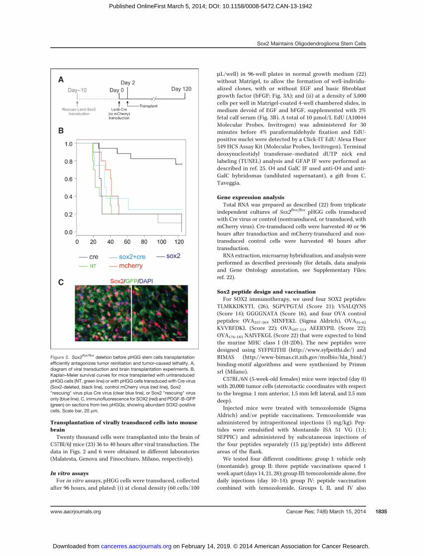
Figure 2. Sox2flox/flox deletion before pHGG stem cells transplantationefficiently antagonizes tumor reinitiation and tumor-caused lethality. A,diagram of viral transduction and brain transplantation experiments. B,Kaplan–Meier survival curves for mice transplanted with untransducedpHGG cells (NT, green line) or with pHGG cells transduced with Cre virus(Sox2-deleted, black line), control mCherry virus (red line), Sox2"rescuing" virus plus Cre virus (clear blue line), or Sox2 "rescuing" virusonly (blue line). C, immunofluorescence for SOX2 (red) and PDGF-B-GFP(green) on sections from two pHGGs, showing abundant SOX2-positivecells. Scale bar, 20 mm.
Sox2 Maintains Oligodendroglioma Stem Cells
www.aacrjournals.org Cancer Res; 74(6) March 15, 2014 1835
on February 14, 2019. © 2014 American Association for Cancer Research. cancerres.aacrjournals.org Downloaded from
Published OnlineFirst March 5, 2014; DOI: 10.1158/0008-5472.CAN-13-1942

received a total of 3 mg of recombinant murine granulocytemacrophage colony-stimulating factor as described (27).Cumulative survival curves were obtained using the Kaplan–Meier method (MedCalc 12.7).
For cytotoxicity assay, isolation of tumor-infiltrating lym-phocytes (TIL) and flow cytometry, see ref. 27 and Supple-mentary Materials.
ResultsWe generated oligodendrogliomas in mouse by transduc-
tion of a PDGF-B-IRES-GFP-encoding retrovirus within thebrain at embryonic day (E) 14.5 (22, 23). Embryos were homo-zygous carriers of a Sox2flox mutation, allowing subsequentSox2 excision via Cre recombinase (Fig. 1A; ref. 13). Tumorsdeveloped, at different times after birth: early-onset, showinglow-grade tumor features and late-onset, displaying high-gradeglioma characteristics, as expected fromour previous data (23).We focused on tumors arising at least 90 days after birth, as ourprevious analyses showed that low-grade tumors arising beforeday 90 can be hardly grown in culture and are not tumorigenic(23). Indeed, 4 of 7 tumors appearing after 90 days reinitiatedtumorigenesis following transplantation into adult mousebrain. We cultured in vitro three of these secondary tumors(pHGGs), in conditions allowing the long-termmaintenance ofTICs, i.e., presence of EGF and bFGF and absence of serum(16, 22), and we used for subsequent analyses one cell popu-lation derived from such tumor.
Sox2 deletion impairs tumor reinitiation by pHGG cellsfollowing in vivo transplantation in the brain
To evaluate the role of Sox2 in tumor initiation, wedeleted the "floxed" Sox2 gene from tumor-derived cells bytransduction with lentiviruses expressing Cre recombinaseor mCherry as a control (Fig. 1A). Transduction of Crerecombinase (but not of control virus) induced efficientdeletion of Sox2 (>95% by DNA analysis; Fig. 1B), leading toloss of Sox2 mRNA (>90% by real-time RT-PCR; Fig. 1C) andprotein (>95% by immunofluorescence) by 36 hours aftertransduction (Fig. 1D). We then transplanted Cre-trans-duced, or control mCherry-transduced, or nontransducedcells into the brain of adult C57/Bl6 mice, 36 hours afterviral transduction (Fig. 2). Control mCherry-transduced andnontransduced cells caused the development of tumors (17/20 mice, 85%; of which 8/10 mCherry, 9/10 nontransduced),resulting in an overall median survival of the control mice of40 days, consistent with previous reports with similartumor-derived cells (Fig. 2B; Table 1; ref. 23). However,mice injected with Cre-transduced cells were almost allalive (13 of 17; 76.5%) at day 118 after transduction (Fig. 2B;log-rank test, P < 10�4). When sacrificed and analyzed at day121 � 3, these mice were found tumor-free. Analysis of the 4mice injected with Cre-transduced cells that had died (2 byday 50, 1 on day 64, 1 on day 117; Table 1) showed that theyhad developed tumors that demonstrated a nondeletedstatus of the Sox2flox gene upon genotyping (not shown),quantitative reverse transcription (qRT)-PCR (Supplementary
Figure 3. Sox2flox/flox deletion reduces in vitro growth of pHGG stem cells. A, pHGG oligodendroglioma clone numbers obtained in EGFþbFGF-containingmedium (EGF_FGF), or factor-free medium (NO Factors), with nontransduced cells (NT), or cells transduced with control (mCherry) or Cre-expressing(CRE) virus. The number of clones obtained with NT cells (representing >1,400 clones counted for each experiment) was set at 100%. More than 1,400clones were counted for each experiment replicate. The results shown are the average of n ¼ 2 independent experiments performed in duplicate(���, P < 0.0001; ns, nonsignificant; P > 0.05; Wilcoxon test). The images show examples of clones. Scale bar, 100 mm. B, pHGG cell numbers, obtained withnontransduced cells (NT) or following transduction with control (mCherry) or Cre virus (CRE), after 7 days in serum-containing medium. The cellnumber obtained with untransduced (NT) cells (>1,000 cells counted for each experiment) is set at 100%. The results shown are the average of n ¼ 2independent experiments performed in duplicate (���, P < 0.0001; Fisher exact test). Representative images showing cell density at day 3 and 7 are shown.
Favaro et al.
Cancer Res; 74(6) March 15, 2014 Cancer Research1836
on February 14, 2019. © 2014 American Association for Cancer Research. cancerres.aacrjournals.org Downloaded from
Published OnlineFirst March 5, 2014; DOI: 10.1158/0008-5472.CAN-13-1942

Fig. S1), and immunofluorescence (Fig. 2C), indicating theirlikely origin from the few non–Sox2-deleted cells.To address whether the loss of TIC properties was spe-
cifically due to loss of Sox2, rather than to nonspecific effects(Cre-toxicity, etc.) we performed a control experiment.Before Cre transduction, we transduced the tumor-derivedcells with a Sox2-encoding lentivirus (Fig. 2A and B; ref. 13).After subsequent ablation of endogenous Sox2 by Cre (lead-ing to loss of endogenous Sox2 mRNA, as verified withspecific primers, see Supplementary Materials), we trans-planted the cells into host mouse brains and compared theirsurvival with that of controls, i.e., cells transduced with Sox2virus but not with Cre, or with the control mCherry virus, oruntransduced (Fig. 2B). Sox2-transduced cells demonstratedtumorigenic ability similar to that of controls, with tumorsdeveloping in 6 of 8 mice (75%) within 120 days and a mediansurvival of 34 days (Fig. 2B).We conclude that the tumor-initiating ability of PDGF-B–
induced oligodendroglioma cells requires Sox2 function.
Consequences of Sox2 deletion on in vitro growth ofpHGG cellsTo obtain information on the mechanisms of loss of tumor-
initiating ability of the Sox2-deleted pHGG cells, we studiedthe in vitro growth of intact or Sox2-deleted cells (Figs. 3and 4). We tested cells in three growth conditions: the firstone optimized for maintenance of stem cell properties, andcorresponding to the initial condition in which the cultureshad been derived (with EGF and bFGF and without serum:þEGF/bFGF); a second one in the same medium with noadded growth factors (no factors); and a third one in mediumwithout added factors, but with 2% serum (no factors þserum). The latter represents "differentiating" conditionsnormally used to obtain terminal differentiation of normalNSC (neurospheres; refs. 1, 25, 28, 29). Cells were plated atclonal density (96 hours after viral transduction) and scoredfor clone numbers after 7 days. In EGF/bFGF, the number ofclones obtained with Sox2-deleted cells was only slightly,
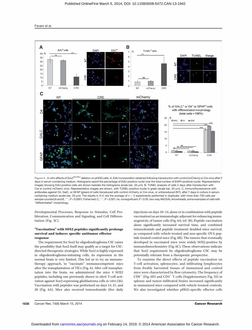
although significantly, decreased compared with undeletedcontrols; however, when plated without factors, Sox2-deletedcells were reduced to less than 50% of controls (Fig. 3A). Cellsplated in serum adhered to the substrate and did not formindividualized clones. We thus plated the cells, 96 hours aftertransduction, in 2% serum-containing medium withoutadded factors, at nonclonal density, on Matrigel, allowingmore efficient growth. Under these conditions, untreatedpHGG cells continue to grow, although to a rate somewhatlower (20%–30% increase in duplication time) than in medi-um with added growth factors. Following Sox2 deletion, thetotal cell number at day 7 was strongly reduced relative tocontrols (<20% that obtained with untransduced or mCherrytransduced cells; Fig. 3B). We also assessed proliferativeability (at day 2 and 7) by administering EdU for 30 minutesand measuring the percentage of cells that incorporated EdU(Fig. 4A). While controls (mCherry transduced or nontrans-duced) cells had similar high levels of EdU incorporation(30%–35%), Sox2-deleted cells showed significant reductionof EdU incorporation (to about 10%; Fig. 4A). We thenevaluated apoptosis by the TUNEL assay (Fig. 4B). TUNEL-positive cells were more than 4-fold increased following Sox2deletion relative to controls (Fig. 4B). Immunofluorescencefor differentiation markers of oligodendroglia, O4 and GalC,and astroglia, GFAP, revealed widespread positivity, togetherwith an altered morphology of Sox2-deleted cells, with fea-tures suggestive of aberrant differentiation (branching, flat-tening), as compared with the relatively undifferentiatedmorphology of undeleted pHGG cells (Fig. 4C). We concludethat in "differentiating" growth conditions, Sox2 ablationleads to progressive exhaustion of in vitro cell proliferation,increased apoptotic cell death, and morphologic changes,suggesting aberrant differentiation.
Sox2 deletion causes alterations in the gene expressionprogram of pHGG cells
The dependence on Sox2 of tumor-initiating properties ofoligodendroglioma cells raises the hypothesis that Sox2 mayact by regulating the transcription of critical downstreamgenes. We analyzed gene expression in Sox2-deleted andcontrol cells (nontransduced, m-Cherry virus-transduced) bymicroarray analysis, at 40 and 96 hours following Cre trans-duction (Fig. 5A; Supplementary Table S1). The gene expres-sion profile of Sox2flox/flox pHGG cells closely matched the"oligodendroblast" program that we previously reported forseveral independent PDGF-B–induced oligodendrogliomas(Fig. 5B; refs. 22, 23). At 96 hours, the expression of 146 geneswas substantially deregulated (more that 2-fold); at 40 hours,few, if any, gene changed its expression level significantly(Fig. 5A). This suggests that our analysis at 96 hours likelyincludes the earliest changes in gene expression that followSox2 loss, presumably including those genes that directlyrely on Sox2. Following Sox2 ablation, 12 genes are down-regulated, compatibly with an activator function of Sox2; 134genes are upregulated (Fig. 5A and D), possibly reflecting arepressor function of Sox2 (25), or indirect effects. Geneontology analysis of the deregulated genes indicated asignificant enrichment in the functional categories of
Table 1. Transplanted animals and observedtumors
Transplanted cellsLethal tumors/transplanted animals
NT 9/10 (90%)Lenti-mCherry 8/10 (80%)Lenti-Sox2 5/5 (100%)Lenti-Sox2; Lenti-CRE 6/8 (75%)Lenti-CRE 4a/17b (23.5%)
aThe four tumors were tested by PCR for the presence of theundeleted and deleted Sox2 locus; all four were Sox2-pos-itive (undeleted Sox2); one also presented a band for thedeleted Sox2 locus, indicating that some Sox2 deleted cellsare part of the tumor mass.bThe 13 surviving mice were tumor-free at day 120.
Sox2 Maintains Oligodendroglioma Stem Cells
www.aacrjournals.org Cancer Res; 74(6) March 15, 2014 1837
on February 14, 2019. © 2014 American Association for Cancer Research. cancerres.aacrjournals.org Downloaded from
Published OnlineFirst March 5, 2014; DOI: 10.1158/0008-5472.CAN-13-1942
Developmental Processes, Response to Stimulus, Cell Pro-liferation, Communication and Signaling, and Cell Differen-tiation (Fig. 5C).
"Vaccination" with SOX2 peptides significantly prolongssurvival and induces specific antitumor effectorresponse
The requirement for Sox2 by oligodendroglioma CSC raisesthe possibility that Sox2 itself may qualify as a target for CSC-directed therapeutic strategies. While Sox2 is highly expressedin oligodendroglioma-initiating cells, its expression in thenormal brain is very limited. This led us to try an immuno-therapy approach, to "vaccinate" immunocompetent miceafter the transplantation of TICs (Fig. 6). After cell transplan-tation into the brain, we administered the mice 4 SOX2peptides, including one previously shown to elicit T-cell acti-vation against Sox2-expressing glioblastoma cells in vitro (26).Vaccination with peptides was performed on days 14, 21, and28 (Fig. 6A). Mice also received temozolomide (five daily
injections on days 10–14, alone or in combination with peptidevaccination) as an immunologic adjuvant for enhancing immu-nogenicity of tumor cells (Fig. 6A; ref. 30). Peptide vaccinationalone significantly increased survival time, and combinedtemozolomide and peptide treatment doubled mice survival,as compared with vehicle-treated and non-specific OVA pep-tide-treated control mice (Fig. 6B). The tumors that eventuallydeveloped in vaccinated mice were widely SOX2-positive byimmunohistochemistry (Fig. 6C). These observations indicatethat Sox2 requirement by oligodendroglioma CSC may bepotentially relevant from a therapeutic perspective.
To examine the direct effects of peptide vaccination onT-cell activation, splenocytes, and infiltrating lymphocytesfrom freshly harvested tissues of immunized and controlmice were characterized by flow cytometry. The frequency ofCD8þ (Fig. 6D) and CD4þ T cells (Supplementary Fig. S2) inspleens and tumor-infiltrated brains increased significantlyin immunized mice compared with vehicle-treated controls.We also investigated whether pHGG-specific effector cells
Figure 4. In vitro effects of Sox2flox/flox deletion on pHGG cells. A, EdU incorporation obtained following transduction with control (mCherry) or Cre virus after 2days in serum-containing medium. Histograms report the percentage of EdU-positive nuclei over the total number of (DAPI-positive) nuclei. Representativeimages showing EdU-positive cells are shown besides the histograms (scale bar, 20 mm). B, TUNEL analysis of cells 2 days after transduction withCre or control mCherry virus. Representative images are shown, with TUNEL-positive nuclei in green (scale bar, 20 mm). C, immunofluorescence withantibodies against O4, GalC, or GFAP (green) of cells transduced with control mCherry or Cre virus, or untransduced (NT), after 7 days in culture in serum-containing medium (scale bar, 20 mm). The results in A–C are the average of n ¼ 2 experiments performed in duplicate, with more than 700 cells persample counted (AandB, ���,P<0.0001; Fisher test;C, ���,P<0.001; ns, nonsignificant;P>0.05, two-wayANOVA). Arrowheads, someexamplesof cellswith"differentiated" morphology.
Favaro et al.
Cancer Res; 74(6) March 15, 2014 Cancer Research1838
on February 14, 2019. © 2014 American Association for Cancer Research. cancerres.aacrjournals.org Downloaded from
Published OnlineFirst March 5, 2014; DOI: 10.1158/0008-5472.CAN-13-1942
were generated in response to SOX2 peptide vaccination.Prestimulated splenocytes were assayed for in vitro cytotoxicactivity against pHGG cells or NIH 3T3 cells (negativecontrol) using a cytotoxicity MTT assay. The splenocytesfrom immunized mice, but not from vehicle-treated mice,displayed cytotoxic activity against tumor cells (Fig. 6E). Thespecificity of the effector immune response was confirmedby the absence of cytotoxicity against NIH 3T3 cells (Fig. 6E).
DiscussionWe report that Sox2 is required by oligodendroglioma stem
cells to reinitiate tumor development within the transplantedmouse brain, and, in some conditions, for in vitro growth.Cells cultured from PDGF-B–induced mouse oligodendro-glioma will reform a lethal tumor following transplantation inmouse brain; however, the majority of animals transplantedwith Sox2-deleted cells remain tumor-free. Transduction ofSox2-deleted tumor cells with a Sox2-expressing lentivirusmaintains tumor-initiating capacity, confirming that this isdependent on Sox2 activity. Finally, vaccination against Sox2significantly delays tumor development, pointing to Sox2(and its downstream targets) as a potential therapeutictarget.In adult mouse, Sox2 is expressed only in a minority of cells,
mainly stem/progenitor cells within various tissues (8). Incontrast, Sox2 is expressed in many tumor types, both in thebrain and in other organs (mammary gland, lung, esophagus,bone; refs. 31–33).In the majority of these tumors, Sox2 is not primarily
altered/mutated, with the exception of its amplification inlung and esophageal squamous cell carcinoma (31). Sox2deregulation is, in rare cases, the immediate downstreamconsequence of the primary lesion (34); more frequently, it ispart of the altered transcriptional program of the tumor. AsSox2 is important for pluripotency and for reprogramming,these observations suggests an analogy between the roleof Sox2 in CSC and in the normal development of stem cells (6).
Sox2 is required for the propagation of CSC inoligodendrogliomaIn our oligodendroglioma model, Sox2 is necessary for the
maintenance of CSC, in agreement with its requirement innormal NSC (13). Previous work showed that in human glio-blastoma-derived cell lines, Sox2 downregulation by shRNAsimpaired tumorigenesis following transplantation (35). Inother patient-derived glioblastoma cells, Sox2 was describedto be downstream to Sox4 in a TGF-b signaling-dependenttumorigenicity pathway and was required for in vitro mainte-nance of tumorigenic cells although its in vivo requirement wasnot tested (36). Interestingly, TGF-b promotes proliferation oftumors, including gliomas and osteosarcomas, through induc-tion of PDGF-B (37).While our results agree with those of Gangemi and collea-
gues (35), in that both glioblastoma and oligodendrogliomarequire Sox2 for in vivo tumorigenicity, some important differ-ences should be noted. Sox2 ablation in glioblastoma (byshRNA) causes significant loss of cell proliferation in vitro, inmedia with added growth factors; instead, in oligodendro-
glioma,wenoted only a small decrease in the presence of addedgrowth factors in the proliferation of Sox2-ablated cells, both inclonal tests (Fig. 3) and in mass culture (not shown). However,omission of growth factors, and particularly combined addi-tion of serum, a condition favoring NSC differentiation in vitro(25, 28, 29), strongly decreased Sox2-ablated oligodendro-glioma cell proliferation, increased cell death, and causedimportant morphologic changes. These culture conditionsmight mimic conditions more similar to those encounteredby tumor cells in the brain, with absence of abundantamounts of EGF/bFGF and presence of various cytokinesand factors. Sox2-deleted cells showed morphologic changes(branching and flattening), together with marked positivityfor differentiation markers (Fig. 3). Immunopositivity foroligodendrocyte differentiation markers was also observedin vivo within the very small number of Sox2-deleted tumorcells found 10 days after transplantation (SupplementaryFig. S3). "Priming" by Sox2 of "differentiation" genes in NSCswas reported (38), and NSCs expressing reduced levels ofSox2 (from mouse hypomorphic mutants) showed morpho-logic and gene expression abnormalities when induced todifferentiation (25). Thus, as in normal NSCs (13, 25), Sox2may be required in pHGGs in the presence of differentiationstimuli to prevent abnormal differentiation and apoptosis.Prodifferentiative stimuli (bone morphogenetic proteins,BMP, especially BMP4) efficiently antagonize glioblastomadevelopment in mice (39) and targeting of molecules main-taining an undifferentiated state, such as EphA2 receptor,induced differentiation and loss of tumor-initiating capacityin mouse glioblastoma (40). Also in pHGGs, we previouslydocumented a correlation between loss of tumor-initiatingability (following Pax6 overexpression) and the acquisitionof differentiated features (41).
Overall, these results suggest that mechanisms causingtumor cell loss after Sox2 ablation may differ between differ-ent tumors (glioblastoma and oligodendroglioma), pointingto multiple molecular mechanisms of action of Sox2 in thesecells.
On the other hand, Sox2 expression in tumor cells does notalways correlate with a strict functional requirement fortumorigenesis. Sox2 is expressed in medulloblastoma, acerebellar tumor most frequent in childhood; medulloblas-toma CSCs express Sox2 in humans, and in mouse models(3, 42, 43). A class of medulloblastomas is associated withmutations activating the SHH pathway; these includeSmoM2, a mutation in the SHH-receptor Smo leading to itsconstitutive activation; in mice, Cre-mediated activation of aSmoM2 transgene leads to medulloblastoma development(44). In these mice, we concomitantly deleted Sox2 (Sox2flox)by Cre; yet, Sox2-negative medulloblastoma still developed(42). The discrepancy with our present work might beexplained in several ways. First, Sox2 might act upstreamto Smo signaling; indeed, Sox2 was found to activate SHHexpression in NSCs and neural cells (13, 45). Second, the closehomolog Sox3 is expressed in medulloblastoma and mightact redundantly with Sox2 in CSC maintenance (42). Third, inthis system, SmoM2-induced medulloblastoma developmentin vivomay likely arise from multiple SmoM2-expressing cells
Sox2 Maintains Oligodendroglioma Stem Cells
www.aacrjournals.org Cancer Res; 74(6) March 15, 2014 1839
on February 14, 2019. © 2014 American Association for Cancer Research. cancerres.aacrjournals.org Downloaded from
Published OnlineFirst March 5, 2014; DOI: 10.1158/0008-5472.CAN-13-1942
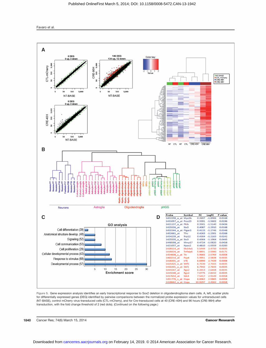
Figure 5. Gene expression analysis identifies an early transcriptional response to Sox2 deletion in oligodendroglioma stem cells. A, left, scatter plotsfor differentially expressed genes (DEG) identified by pairwise comparisons between the normalized probe expression values for untransduced cells(NT-BASE), control mCherry virus-transduced cells (CTL-mCherry), and for Cre-transduced cells at 40 (CRE-40H) and 96 hours (CRE-96H) aftertransduction, with the fold change threshold of 2 (red dots). (Continued on the following page.)
Favaro et al.
Cancer Res; 74(6) March 15, 2014 Cancer Research1840
on February 14, 2019. © 2014 American Association for Cancer Research. cancerres.aacrjournals.org Downloaded from
Published OnlineFirst March 5, 2014; DOI: 10.1158/0008-5472.CAN-13-1942

and it is possible that additional mutations in a subset ofthese cells allow to bypass Sox2 requirement. Finally,although Sox2 activity was not strictly required for medul-loblastoma development, experimental increase of Sox2
levels was found to correspondingly affect medulloblastomacell proliferation (42).
The differences between neural tumors with respect to thedegree of their Sox2 requirement are reminiscent of the
Figure 6. Vaccination with Sox2 peptides causes a significant delay in tumor development and lethality following transplantation. A, schedule of Sox2 peptideand temozolomide (TMZ) administration and cell transplantation. Peptide vaccinations (vacc) were on day 14, 21, 28; temozolomide: five daily injections ondays 10 to 14. B, Kaplan–Meier survival curves for mice treated with: vehicle (n ¼ 5, mean � SD, 23.6 � 1.8; median, 24); OVA peptides (n ¼ 5; mean � SD,25.2� 0.9;median, 25); temozolomide (n¼ 5;mean�SD, 30.8� 1.9;median, 31); temozolomideþOVApeptides (n¼ 5;mean�SD, 32.2� 0.5;median, 32);Sox2 peptides (n ¼ 5; mean � SD, 38.0 � 2.5; median, 38); temozolomide þ SOX2 peptides (n ¼ 5; mean � SD, 45.2 � 6.7; median, 41; �, P < 0.001;��, P < 0.005; ���, P < 0.001 temozolomide þ SOX2 peptides vs. vehicle or OVA peptides). C, hematoxylin and eosin (H&E) staining and Sox2immunohistochemistry (brown) of sections from tumors obtained after the indicated treatments. D, flow cytometry on splenocytes (top) and TILs (bottom;n ¼ 4 mice per group; data reported in dot plots as the mean% � SD; P ¼ 0.0003 and P ¼ 0.003 for temozolomide þSOX2 peptides versus vehicle insplenocytes and TIL, respectively). E, in vitro MTT cytotoxicity assay performed using splenocytes from mice treated with SOX2 peptide with orwithout temozolomide, temozolomide, and vehicle as effector cells and pHGG or NIH 3T3 cells as target using different effector:target (E:T) ratios(10:1, 25:1, and 50:1).
(Continued.) Data represent the mean of probe expression values of the replicates samples in the considered condition. Right, heatmap diagram of geneexpression changes (red, increased expression; blue, reduced expression) in Cre-treated cells, as compared with the indicated controls. Probesets (rows)and samples (columns) are clustered on the basis of their similarity by hierarchical clustering using complete linkage (Euclideandistance). The topdendrogram(x-axis) indicates the pairwise comparisons between the cell types identified by the different colors. NT, nontransduced cells; CTL, control mCherry-transduced cells; CRE40 and CRE96, Cre-transduced cells at 40 and 96 hours after transduction. B, dendrogram representation of the results of thehierarchical clustering analysis between the gene expression profiles of our pHGG cells (pHGG¼ untransduced, pHGG-mCherry or pHGG-Cre-transduced),and previously analyzed pHGGs (pHGG-2, 3, 4; ref. 22), as well as neurons, astroglia, oligodendroglia, and OPC gene expression profiles as described in ref.22. C, analysis of Gene Ontology (GO) biologic processes enriched in DEGs. The most representative GO functional annotations for DEGs from eachexperimental condition are identified by determining the probability of random occurrence of functional terms (hyper geometric distribution). On the basis ofthis probability ranking, only the top eight statisticallymost significant annotation terms are reported. The enrichment scores identify the functional categoriesthat are overrepresented. Enrichment scores<6 indicates enrichmentP values of 10�6, scores between 5and13P values of 10�7, scores >15P values of 10�8.D, list of the 10 top-down (blue) and top-upregulated (red) genes following Sox2 deletion. FC, fold change as compared with undeleted cells.
Sox2 Maintains Oligodendroglioma Stem Cells
www.aacrjournals.org Cancer Res; 74(6) March 15, 2014 1841
on February 14, 2019. © 2014 American Association for Cancer Research. cancerres.aacrjournals.org Downloaded from
Published OnlineFirst March 5, 2014; DOI: 10.1158/0008-5472.CAN-13-1942

differences in Sox2 requirement between different regions ofthe normal, developing nervous system. Sox2 is expressedubiquitously in neural stem/progenitor cells, yet its deletionin vivo has region- and stage-specific effects in the brain(hippocampus, ventral telencephalon; refs. 13, 46). Theseobservations point to specificities in the downstream geneexpression networks controlled by Sox2 in tumorigenic as wellas in normal neural (stem) cells.
Oligodendrogliomas may arise within the committed oligo-dendrocyte lineage, by "reprogramming" to a CSC state. Oli-godendrocyte precursor cells (OPC) can be "reprogrammed" toa neural stem-like state, by sequential treatment with PDGFand EGF, and this process requires Sox2 reactivation (47). Afuture in-depthmolecular investigation of Sox2 function in ourmodel system may uncover if Sox2 regulates genes critical forreprogramming committed cells to a stem cell status, acting asa pioneer factor in ways related to its action in iPS cellgeneration (6).
Sox2 as a potential therapeutic targetThe requirement for Sox2 by CSC raises the possibility that
Sox2 itself may qualify as a target for therapeutic intervention.Targeting CSC may be a strategy to increase the potentialefficacy of immunotherapy (27). Sox2 vaccination significantlyprolongs survival enhancing systemic and local immuneresponse (Fig. 6). Sox2 is localized in the nucleus, and is thusnot, a priori, the most accessible molecule to target. However,recent data suggest that intracellular oncoproteins can betargeted by vaccination, as some intracellular antigens maybe released and expressed on the surface of cancer cells (48).Antibodies and T-cell immune responses against SOX2 havebeen detected in patients with monoclonal gammopathy(MGUS), a premalignant condition to myeloma, where Sox2expression marks the clonogenic compartment (49), and,recently, in about 50% of patients with non–small cell lungcarcinoma (NSCLC; ref. 50). Cellular anti-SOX2 immunityinhibited the growth of MGUS cells in vitro and the presenceof anti-SOX2 T cells predicted favorable clinical outcome (49);in NSCLC, T-cell response against SOX2 was associated withNSCLC regression upon immunotherapy with anti-PD-1 anti-bodies (50). These observations suggest that the immunesystem may be able to "discover" tumor-associated SOX2.Furthermore, an immune reaction by T cells elicited bySOX2-derived peptides (one of which was used here) wasreported to lyse human glioblastoma-derived cells in culture(26). Finally, we previously found that vaccination againstGLAST, a protein retaining significant expression in the adultbrain, elicited an immune reaction specifically targeted to thetumor, not damaging the surrounding tissue (27).
The fact that late-arising tumors that eventually developedin vaccinated animals were widely Sox2-positive (Fig. 6) is
consistent with the hypothesis of a failure of the immunesystem to completely eradicate Sox2-positive tumor cells,rather than with escape mechanisms developed by the tumor,allowing it to develop without Sox2. Collectively, these obser-vations suggest that targeting Sox2-expressing cells may pro-vide a basis for therapeutic approaches. Complementing Sox2immunotherapy with action directed against some down-stream Sox2 targets in oligodendroglioma might furtherincrease the efficacy of this approach.
The observations about Sox2 requirement in neural tumorsare extended by the reported requirement for Sox2 in a widersample of tumor types. These include tumors of the osteoblastlineage, as shown in osteosarcoma cell lines (32); here, Sox2 isrequired also in the normal tissue stem cell counterpart,osteoblast stem/progenitor cells (10), as seen with neural cells.CSC from mammary tumors cultured as tumorigenic "mam-mospheres" express Sox2 and Sox2 knockdown impairs mam-mosphere formation and delays tumor formation followingtransplantation (33).
We conclude that targeting Sox2, likely in combination withselected downstream targets, may provide an effective strategyto antagonize the development of oligodendroglioma, and,perhaps, other tumor types.
Disclosure of Potential Conflicts of InterestNo potential conflicts of interest were disclosed.
Authors' ContributionsConception and design: R. Favaro, S. Ottolenghi, P. Malatesta, S.K. NicolisDevelopment of methodology: R. Favaro, G. Finocchiaro, S.K. NicolisAcquisition of data (provided animals, acquired and managedpatients, provided facilities, etc.): R. Favaro, I. Appolloni, S. Pellegatta,A.B. Sanga, P. Pagella, E. Gambini, F. Pisati, M. Foti, G. Finocchiaro, P. Malatesta,S.K. NicolisAnalysis and interpretation of data (e.g., statistical analysis, biostatistics,computational analysis): R. Favaro, I. Appolloni, A.B. Sanga, M. Foti,G. Finocchiaro, P. Malatesta, S.K. NicolisWriting, review, and/or revision of the manuscript: R. Favaro, I. Appolloni,S. Pellegatta, S. Ottolenghi, M. Foti, G. Finocchiaro, P. Malatesta, S.K. NicolisStudy supervision: S.K. Nicolis
AcknowledgmentsThe authors thank L. Naldini for the mCherry-encoding lentivirus, S. Brunelli
for the Cre-encoding lentivirus, and C. Taveggia for the anti-GalC and anti-O4antibodies.
Grant SupportThis study was supported by grants from Associazione Italiana per la Ricerca
sul Cancro (AIRC, grant IG-5801) and Fondazione Cariplo (grant 2010-0673; S.K.Nicolis), AIRC grant NUSUG 1180 and Italian Ministry of Health grant (GR-2008-1135643; P. Malatesta). A.B. Sanga was the recipient of a Fondazione CariploInterbiomed PhD fellowship.
The costs of publication of this article were defrayed in part by the payment ofpage charges. This article must therefore be hereby marked advertisement inaccordance with 18 U.S.C. Section 1734 solely to indicate this fact.
Received July 16, 2013; revised December 3, 2013; accepted December 17, 2013;published OnlineFirst March 5, 2014.
References1. Galli R, Binda E, Orfanelli U, Cipelletti B, Gritti A, De Vitis S, et al.
Isolation and characterization of tumorigenic, stem-like neuralprecursors from human glioblastoma. Cancer Res 2004;64:7011–21.
2. Ignatova TN, Kukekov VG, Laywell ED, Suslov ON, Vrionis FD, Steind-ler DA. Human cortical glial tumors contain neural stem-like cellsexpressing astroglial and neuronal markers in vitro. Glia 2002;39:193–206.
Favaro et al.
Cancer Res; 74(6) March 15, 2014 Cancer Research1842
on February 14, 2019. © 2014 American Association for Cancer Research. cancerres.aacrjournals.org Downloaded from
Published OnlineFirst March 5, 2014; DOI: 10.1158/0008-5472.CAN-13-1942
3. Singh SK, Hawkins C, Clarke ID, Squire JA, Bayani J, Hide T, et al.Identification of human brain tumour initiating cells. Nature 2004;432:396–401.
4. Vescovi AL, Galli R, Reynolds BA. Brain tumour stem cells. Nat RevCancer 2006;6:425–36.
5. Friedmann-Morvinski D, Bushong EA, Ke E, Soda Y, Marumoto T,Singer O, et al. Dedifferentiation of neurons and astrocytes by onco-genes can induce gliomas in mice. Science 2012;338:1080–4.
6. SuvaML, Riggi N, Bernstein BE. Epigenetic reprogramming in cancer.Science 2013;339:1567–70.
7. Visvader JE. Cells of origin in cancer. Nature 2011;469:314–22.8. Arnold K, Sarkar A, Yram MA, Polo JM, Bronson R, Sengupta S,
et al. Sox2(þ) adult stem and progenitor cells are important fortissue regeneration and survival of mice. Cell Stem Cell 2011;9:317–29.
9. Avilion AA, Nicolis SK, Pevny LH, Perez L, Vivian N, Lovell-Badge R.Multipotent cell lineages in earlymousedevelopment dependonSOX2function. Genes Dev 2003;17:126–40.
10. Basu-Roy U, Ambrosetti D, Favaro R, Nicolis SK, Mansukhani A,Basilico C. The transcription factor Sox2 is required for osteoblastself-renewal. Cell Death Differ 2010;17:1345–53.
11. Campolo F, Gori M, Favaro R, Nicolis S, Pellegrini M, Botti F, et al.Essential role of sox2 for the establishment and maintenance of thegerm cell line. Stem Cells 2013;31:1408–21.
12. Pevny LH, Nicolis SK. Sox2 roles in neural stem cells. Int J BiochemCell Biol 2010;42:421–4.
13. Favaro R, Valotta M, Ferri AL, Latorre E, Mariani J, Giachino C, et al.Hippocampal development and neural stem cell maintenancerequire Sox2-dependent regulation of Shh. Nat Neurosci 2009;12:1248–56.
14. Orkin SH, Hochedlinger K. Chromatin connections to pluripotency andcellular reprogramming. Cell 2011;145:835–50.
15. Takahashi K, Yamanaka S. Induction of pluripotent stem cells frommouse embryonic and adult fibroblast cultures by defined factors. Cell2006;126:663–76.
16. Lee J, Kotliarova S, Kotliarov Y, Li A, SuQ,DoninNM, et al. Tumor stemcells derived from glioblastomas cultured in bFGF and EGF moreclosely mirror the phenotype and genotype of primary tumors thando serum-cultured cell lines. Cancer Cell 2006;9:391–403.
17. Pollard SM, Yoshikawa K, Clarke ID, Danovi D, Stricker S, Russell R,et al. Glioma stem cell lines expanded in adherent culture have tumor-specificphenotypes andare suitable for chemical andgenetic screens.Cell Stem Cell 2009;4:568–80.
18. Bettegowda C, Agrawal N, Jiao Y, Sausen M, Wood LD, Hruban RH,et al. Mutations in CIC and FUBP1 contribute to human oligodendro-glioma. Science 2011;333:1453–5.
19. Reis-Filho JS, Faoro LN, Carrilho C, Bleggi-Torres LF, Schmitt FC.Evaluation of cell proliferation, epidermal growth factor receptor, andbcl-2 immunoexpression as prognostic factors for patients with WorldHealth Organization grade 2 oligodendroglioma. Cancer 2000;88:862–9.
20. Jackson EL, Garcia-Verdugo JM, Gil-Perotin S, Roy M, Quinones-Hinojosa A, VandenBerg S, et al. PDGFR alpha-positive B cellsare neural stem cells in the adult SVZ that form glioma-like growthsin response to increased PDGF signaling. Neuron 2006;51:187–99.
21. Shih AH, Holland EC. Platelet-derived growth factor (PDGF) and glialtumorigenesis. Cancer Lett 2006;232:139–47.
22. Appolloni I, Calzolari F, Tutucci E, Caviglia S, Terrile M, Corte G,et al. PDGF-B induces a homogeneous class of oligodendrogliomasfrom embryonic neural progenitors. Int J Cancer 2009;124:2251–9.
23. Calzolari F, Appolloni I, Tutucci E, Caviglia S, Terrile M, Corte G, et al.Tumor progression and oncogene addiction in a PDGF-B-inducedmodel of gliomagenesis. Neoplasia 2008;10:1373–82.
24. Borello U, Berarducci B, Murphy P, Bajard L, Buffa V, Piccolo S, et al.The Wnt/beta-catenin pathway regulates Gli-mediated Myf5 expres-sion during somitogenesis. Development 2006;133:3723–32.
25. Cavallaro M, Mariani J, Lancini C, Latorre E, Caccia R, Gullo F, et al.Impaired generation of mature neurons by neural stem cells fromhypomorphic Sox2 mutants. Development 2008;135:541–57.
26. Schmitz M, Temme A, Senner V, Ebner R, Schwind S, Stevanovic S,et al. Identification of SOX2 as a novel glioma-associated antigen andpotential target for T cell-based immunotherapy. Br J Cancer2007;96:1293–301.
27. Cantini G, Pisati F, Pessina S, Finocchiaro G, Pellegatta S. Immuno-therapy against the radial glia marker GLAST effectively triggersspecific antitumor effectors without autoimmunity. Oncoimmunology2012;1:884–93.
28. Gritti A, Parati EA, Cova L, Frolichsthal P, Galli R, Wanke E, et al.Multipotential stem cells from the adult mouse brain proliferate andself-renew in response to basic fibroblast growth factor. J Neurosci1996;16:1091–100.
29. Reynolds BA, Weiss S. Generation of neurons and astrocytes fromisolated cells of the adult mammalian central nervous system. Science1992;255:1707–10.
30. Tesniere A, Panaretakis T, KeppO, Apetoh L, Ghiringhelli F, Zitvogel L,et al. Molecular characteristics of immunogenic cancer cell death. CellDeath Differ 2008;15:3–12.
31. Bass AJ, Watanabe H, Mermel CH, Yu S, Perner S, Verhaak RG, et al.SOX2 is an amplified lineage-survival oncogene in lung and esoph-ageal squamous cell carcinomas. Nat Genet 2009;41:1238–42.
32. Basu-RoyU, Seo E, Ramanathapuram L, Rapp TB, Perry JA, Orkin SH,et al. Sox2 maintains self renewal of tumor-initiating cells in osteo-sarcomas. Oncogene 2012;31:2270–82.
33. Leis O, Eguiara A, Lopez-Arribillaga E, Alberdi MJ, Hernandez-GarciaS, Elorriaga K, et al. Sox2 expression in breast tumours and activationin breast cancer stem cells. Oncogene 2012;31:1354–65.
34. Riggi N, Suva ML, De Vito C, Provero P, Stehle JC, Baumer K, et al.EWS-FLI-1 modulates miRNA145 and SOX2 expression to initiatemesenchymal stemcell reprogramming towardEwing sarcomacancerstem cells. Genes Dev 2010;24:916–32.
35. Gangemi RM, Griffero F, Marubbi D, Perera M, Capra MC, MalatestaP, et al. SOX2 silencing in glioblastoma tumor-initiating cells causesstop of proliferation and loss of tumorigenicity. Stem Cells 2009;27:40–8.
36. Ikushima H, Todo T, Ino Y, Takahashi M, Miyazawa K, Miyazono K.Autocrine TGF-beta signaling maintains tumorigenicity of glioma-ini-tiating cells through Sry-related HMG-box factors. Cell Stem Cell2009;5:504–14.
37. Bruna A, Darken RS, Rojo F, Ocana A, Penuelas S, Arias A, et al. HighTGFbeta-Smad activity confers poor prognosis in glioma patients andpromotes cell proliferation depending on themethylation of the PDGF-B gene. Cancer Cell 2007;11:147–60.
38. Lodato MA, Ng CW, Wamstad JA, Cheng AW, Thai KK, Fraenkel E,et al. SOX2 co-occupies distal enhancer elements with distinct POUfactors in ESCs and NPCs to specify cell state. PLoS Genet 2013;9:e1003288.
39. Piccirillo SG, Reynolds BA, Zanetti N, Lamorte G, Binda E, Broggi G,et al. Bone morphogenetic proteins inhibit the tumorigenic potential ofhuman brain tumour-initiating cells. Nature 2006;444:761–5.
40. Binda E, Visioli A, Giani F, Lamorte G, Copetti M, Pitter KL, et al. TheEphA2 receptor drives self-renewal and tumorigenicity in stem-liketumor-propagating cells fromhumanglioblastomas.CancerCell 2012;22:765–80.
41. Appolloni I, Calzolari F, Barilari M, Terrile M, Daga A, Malatesta P.Antagonistic modulation of gliomagenesis by Pax6 and Olig2in PDGF-induced oligodendroglioma. Int J Cancer 2012;131:E1078–87.
42. Ahlfeld J, Favaro R, Pagella P, Kretzschmar HA, Nicolis S, Schuller U.Sox2 requirement in Sonic hedgehog-associated medulloblastoma.Cancer Res 2013;73:3796–807.
43. Sutter R, Shakhova O, Bhagat H, Behesti H, Sutter C, Penkar S,et al. Cerebellar stem cells act as medulloblastoma-initiating cellsin a mouse model and a neural stem cell signature characterizesa subset of human medulloblastomas. Oncogene 2010;29:1845–56.
44. Schuller U, Heine VM, Mao J, Kho AT, Dillon AK, Han YG, et al.Acquisition of granule neuron precursor identity is a critical determi-nant of progenitor cell competence to form Shh-inducedmedulloblas-toma. Cancer Cell 2008;14:123–34.
Sox2 Maintains Oligodendroglioma Stem Cells
www.aacrjournals.org Cancer Res; 74(6) March 15, 2014 1843
on February 14, 2019. © 2014 American Association for Cancer Research. cancerres.aacrjournals.org Downloaded from
Published OnlineFirst March 5, 2014; DOI: 10.1158/0008-5472.CAN-13-1942
45. Zhao L, Zevallos SE, Rizzoti K, Jeong Y, Lovell-Badge R, Epstein DJ.Disruption of SoxB1-dependent Sonic hedgehog expression in thehypothalamus causes septo-optic dysplasia. Dev Cell 2012;22:585–96.
46. Ferri A, Favaro R, Beccari L, Bertolini J, Mercurio S, Nieto-Lopez F,et al. Sox2 is required for embryonic development of the ventraltelencephalon through the activation of the ventral determinantsNkx2.1 and Shh. Development 2013;140:1250–61.
47. KondoT,RaffM.Chromatin remodeling andhistonemodification in theconversion of oligodendrocyte precursors to neural stem cells. GenesDev 2004;18:2963–72.
48. Guo K, Li J, Tang JP, Tan CP, Hong CW, Al-Aidaroos AQ, et al.Targeting intracellular oncoproteins with antibody therapy or vacci-nation. Sci Transl Med 2011;3:99ra85.
49. Spisek R, Kukreja A, Chen LC, Matthews P, Mazumder A, Vesole D,et al. Frequent and specific immunity to the embryonal stem cell-associated antigen SOX2 in patients with monoclonal gammopathy.J Exp Med 2007;204:831–40.
50. Dhodakpar KM, Gettinger SN, Das R, Zebroski H, Dhodakpar MV.SOX2-specific adaptive immunity and response to immunotherapyin non-small cell lung cancer. Oncoimmunology 2013;2:e25205.
Favaro et al.
Cancer Res; 74(6) March 15, 2014 Cancer Research1844
on February 14, 2019. © 2014 American Association for Cancer Research. cancerres.aacrjournals.org Downloaded from
Published OnlineFirst March 5, 2014; DOI: 10.1158/0008-5472.CAN-13-1942

2014;74:1833-1844. Published OnlineFirst March 5, 2014.Cancer Res Rebecca Favaro, Irene Appolloni, Serena Pellegatta, et al. of High-Grade OligodendrogliomaSox2 Is Required to Maintain Cancer Stem Cells in a Mouse Model
Updated version
10.1158/0008-5472.CAN-13-1942doi:
Access the most recent version of this article at:
Material
Supplementary
http://cancerres.aacrjournals.org/content/suppl/2014/01/29/0008-5472.CAN-13-1942.DC1
Access the most recent supplemental material at:
Cited articles
http://cancerres.aacrjournals.org/content/74/6/1833.full#ref-list-1
This article cites 50 articles, 15 of which you can access for free at:
Citing articles
http://cancerres.aacrjournals.org/content/74/6/1833.full#related-urls
This article has been cited by 2 HighWire-hosted articles. Access the articles at:
E-mail alerts related to this article or journal.Sign up to receive free email-alerts
Subscriptions
Reprints and
To order reprints of this article or to subscribe to the journal, contact the AACR Publications Department at
Permissions
Rightslink site. Click on "Request Permissions" which will take you to the Copyright Clearance Center's (CCC)
.http://cancerres.aacrjournals.org/content/74/6/1833To request permission to re-use all or part of this article, use this link
on February 14, 2019. © 2014 American Association for Cancer Research. cancerres.aacrjournals.org Downloaded from
Published OnlineFirst March 5, 2014; DOI: 10.1158/0008-5472.CAN-13-1942