spalt activity in embryonic pns1999a), also characterised by malformations in the nervous system,...
TRANSCRIPT
INTRODUCTION
The evolutionary conserved spaltgenes encode for C2H2zinc finger transcription factors involved in numerousdevelopmental processes. In Drosophila melanogaster, twomembers of this family have been identified, spalt (sal) andspalt-related(salr), which are highly similar at the levels ofsequence, regulation and function (Barrio et al., 1999; Barrioet al., 1996; Reuter et al., 1996). During tracheal development,sal represses tracheal placode formation, and ensures thecorrect migration and fusion of the dorsal tracheal trunk(Kühnlein and Schuh, 1996). In the wing imaginal disc, salandsalr are necessary for vein patterning in the pouch (de Celisand Barrio, 2000; de Celis et al., 1996; Lecuit et al., 1996;Nellen et al., 1996; Sturtevant et al., 1997), while in regions ofthe disc forming the thorax, they regulate bristle formationthrough the regulation of pro-neural gene expression (de Celiset al., 1999).
The conservation of their expression in nervous tissue ofevery species studied leads to the presumption that theseproteins function in the development of the nervous system.Although the presence of sal members in the nervous systemhave also been shown in frogs, mice and fish (Buck et al., 2000;Hollemann et al., 1996; Köster et al., 1997; Ott and Schutz,1996), their function in this tissue has only been studied in
the nematode C. elegansand humans. In C. elegans, the salhomologue sem-4 is required for correct development ofneurons, vulva and mesoderm, including sensory organ celllineages (Basson and Horvitz, 1996; Grant et al., 2000).Mutations in sem-4result in cell-fate transformations as wellas failures in nuclear morphology, axonal outgrowth and cellmigration. In humans, mutations in a sal homologue, SALL1,are associated with the Townes-Brock syndrome (Kohlhaseet al., 1999b; Kohlhase et al., 1998). This genetic disorderleads to malformations in the anus and limbs, as well assensoryneural hearing loss and mental retardation. In addition,the newly identified SALL3gene has been suggested tocontribute to the 18q deletion syndrome (Kohlhase et al.,1999a), also characterised by malformations in the nervoussystem, mental retardation, hearing loss and facial and limbabnormalities.
In Drosophilanothing is known about whether these genesmight function in the development of the embryonic sensoryorgans in the peripheral nervous system (PNS). The knowledgeabout the cell lineages, the existence of many molecularmarkers at the cellular level and known functional requirementsfor specific genes make the PNS an excellent choice forstudying gene function during the development of the nervoussystem. We therefore chose to address the function of the spaltgenes in the embryonic PNS. The Drosophila PNS comprises
711Development 128, 711-722 (2001)Printed in Great Britain © The Company of Biologists Limited 2001DEV7852
Genes of the spaltfamily encode nuclear zinc fingerproteins. In Drosophila melanogaster, they are necessaryfor the establishment of head/trunk identity, correcttracheal migration and patterning of the wing imaginaldisc. Spalt proteins display a predominant pattern ofexpression in the nervous system, not only in Drosophilabut also in species of fish, mouse, frog and human,suggesting an evolutionarily conserved role for theseproteins in nervous system development. Here we show thatSpalt works as a cell fate switch between two EGFR-induced cell types, the oenocytes and the precursors of thepentascolopodial organ in the embryonic peripheralnervous system. We show that removal of spalt increases
the number of scolopodia, as a result of extra secondaryrecruitment of precursor cells at the expense of theoenocytes. In addition, the absence of spalt causes defectsin the normal migration of the pentascolopodial organ. Thedual function of spalt in the development of this organ,recruitment of precursors and migration, is reminiscent ofits role in tracheal formation and of the role of a spalthomologue, sem-4, in the Caenorhabditis elegansnervoussystem.
Key words: Drosophila melanogaster, spalt, Chordotonal organ,dEGFR, PNS, Cell migration, Oenocytes
SUMMARY
Spalt modifies EGFR-mediated induction of chordotonal precursors in the
embryonic PNS of Drosophila promoting the development of oenocytes
Tor Erik Rusten 1, Rafael Cantera 2, Joachim Urban 3, Gerhard Technau 3, Fotis C. Kafatos 1 and Rosa Barrio 1,*,‡
1European Molecular Biology Laboratory, Meyerhofstrasse, D-69117 Heidelberg, Germany2Zoology Department, Stockholm University, 10691 Stockholm, Sweden3Department of Genetics, University of Mainz, Saarstrasse 21, D-55122 Mainz, Germany*Present address: Centro de Biologia Molecular Severo Ochoa, Universidad Autonoma de Madrid, 28049 Madrid, Spain‡Author for correspondence (e-mail: [email protected])
Accepted 15 December 2000; published on WWW 7 February 2001

712
approximately 600 neurons and 1200 associated cells,organised in a segment-specific pattern (Campos-Ortega andHartenstein, 1997; Ghysen et al., 1986). Each abdominalhemisegment (A1-A7) contains 44 neurons organised in threeclusters along the dorsoventral axis (ventral, lateral and dorsal).The PNS sensory organs can be divided into two types, external(es) and internal. The es organs are often mechanoreceptors inthe cuticle. The internal receptors, called chordotonal organs(ch), are subepidermal stretch receptors consisting of oneneuron (n), one ligament (l), one cap (c) and one sheath cell (s)belonging to the same cell lineage (Fig. 1B; Brewsterand Bodmer, 1995; Brewster and Bodmer, 1996; Okabe andOkano, 1997). Eight chordotonal organs arise in eachabdominal hemisegment. At a lateral position five associatedscolopodia constitute a prominent compound structure, thepentascolopodial organ (lch5). In contrast, the correspondingorgan in the thoracic segments consists of only three scolopodia(dch3) which, in this case, are located dorsally (Fig. 1A).
It was previously shown that the lch5 is generated in twodistinct steps (Lage et al., 1997; Okabe and Okano, 1997). Inthe first step, the proneural gene atonal(ato) is required for thedevelopment of three primary sensory organ precursors (C1,C2 and C3; Fig. 1A, green circles; Jarman et al., 1994; Lageet al., 1997; Okabe and Okano, 1997). At the same time, theprimary precursors for other chordotonal organs are alsorecruited (C4 for v′ch1 and C5 for vchA/B; Fig. 5A). In asecond step, occurring only in abdominal segments, theprimary SOPs express high levels of rhomboid(rho) and signalvia spitz (spi) to the ectoderm through the EGFR receptor(dEGFR) to recruit secondary SOPs (Fig. 1A, red circles; Lageet al., 1997; Okabe and Okano, 1997; Price et al., 1989;Rutledge et al., 1992; Schejter and Shilo, 1989; Schweitzer etal., 1995). The ectodermal cells receiving the EGFR signalexpress several target genes including pointed(pnt; O’Neill etal., 1994), argos(aos; Freeman et al., 1992; Okano et al., 1992)and sprouty(spry; Kramer et al., 1999) but, interestingly, only
two of these cells will become secondary SOPs for the lch5and one of them for the vchA/B cluster. Conversely, in thethoracic segments the primary SOPs show a lower expressionof rhomboid and do not signal at the same level to theectoderm, thus secondary recruitment does not take place(Lage et al., 1997).
In this study we investigated the role of the Drosophila salgenes on lch5 development. While salr is not expressed inthis organ, sal is present at two different stages of lch5development. First, salis expressed in a subset of the cellsthat surround the primary chordotonal SOPs and receiveEGFR signalling. Later on, Sal appears in the support cellsof the lch5, but not in the neurons. Through loss- and gain-of-function studies, we show that salplays a dual role on lch5development. On the one hand, it restricts the number ofscolopodia to five per abdominal organ. On the other hand, itensures the correct location of this chordotonal organ alongthe dorsoventral axis. Furthermore, through genetic analysis,we demonstrate that salcontrols the number of lch5scolopodia by restricting the capacity of EGFR-responsiveectodermal cells to become secondary SOPs. We present amodel in which the extra scolopodia observed in the salmutants develop at the expense of the neighbouringoenocytes.
MATERIALS AND METHODS
Drosophila melanogaster strainsFlies were raised on standard Drosophilamedium at 25ºC. We usedthree sal alleles: sal445, which expresses a truncated Spalt protein(Kühnlein et al., 1994), DF(2L)32FP-5, a small deletion whichremoves sal andsalr (Barrio et al., 1999), and the hypomorphic alleleFCK-68, which carries a translocation breakpoint between saland salrand behaves as a salhypomorph, probably because of the loss ofregulatory regions (Barrio et al., 1999). The null alleles spi1, flbIK35
and SIIN were obtained from the Bloomington Stock Centre and ato1
T. E. Rusten and others
Fig. 1. Schematic representation of chordotonal organdevelopment. (A) Development of the chordotonal organs dch3(second and third thoracic segments, exemplified here by T3)and lch5 (abdominal segments A1-A7, exemplified by A1) instage 11 to 16 embryos. At early stage 11, the three primarydch3 and lch5 SOPs are born in similar dorsoventral positions(green circles). EGFR signalling from the primary SOPs to theoverlying ectoderm results in recruitment of two additionalsecondary SOPs only in abdominal segments (red circles).Soon after delamination, processes of cell division, migrationand differentiation take place (shown in green). At stage 16,dch3 is located in a dorsal position with the dendrites pointingventrally, while lch5 is located in a lateral position withdorsally pointing dendrites (see Fig. 3A). (B) Schematicrepresentation of a SOP cell lineage that gives rise to onescolopodium (Brewster and Bodmer, 1995; Brewster andBodmer, 1996). This consists of one neuron (n) and threesupport cells, the sheath (s), ligament (l) and cap (c) cells. Twoof the five abdominal SOPs also give rise to one accessory cell(a). Cell-specific markers for the differentiated lch5 used in thisstudy are shown. Note that salis expressed in all the support and accessory cells of the SOP lineage (filled red circles), but not in the neuron.For additional markers in early steps of development see Fig. 5A.

713Spalt activity in embryonic PNS
from A. Jarman (Jarman et al., 1994). The UAS-sal was describedpreviously (de Celis et al., 1996). For misexpression experiments inthe neuroectoderm, as well as early neuroblasts and SOPs, we usedthe driver-lines scabrous-GAL4 (sca-GAL4; Mlodzik et al., 1990) andKrüppel-GAL4 (Kr-GAL4; Castelli-Gair et al., 1994). For themisexpression experiments in the neural cells after delamination ofSOPs we used the GAL4 driver-lines MZ1407-GAL4 (Sweeney etal., 1995), and asense-GAL4 (ase-GAL4; Hoch et al., 1994). TheP[lac,ry+]A18, and P[lac,ry+]A37, referred to as A18 and A37(Ghysen and O’Kane, 1989), label most cells, if not all, in thedeveloping embryonic PNS. The RX-drf-lacZ insertion linereproduces ventral veins lacking/drifter(vvl/drf) pattern of expressionin the oenocytes (Anderson et al., 1995). Information about strains notdescribed in the text and balancer chromosomes have been describedpreviously (Lindsley and Zimm, 1992).
ImmunohistochemistryImmunohistochemistry was performed in whole-mount embryosusing the following primary antibodies: anti-Sal rat and rabbit antisera(1:1000 and 1:250 dilution, respectively; de Celis et al., 1999); anti-Salr rat antiserum (1:250 dilution; Barrio et al., 1996); anti-Ato rabbitantiserum (1:1000 dilution; Jarman et al., 1994); rabbit anti-β-galactosidase (β-Gal) antiserum (1:5000 dilution; Cappel); anti-Couch potato (Cpo) rabbit antiserum (1:4000 dilution; Bellen et al.,1992); anti-dpERK (activated MAPK/Rolled) mouse monoclonalantibody (1:500 dilution; SIGMA; Gabay et al., 1997); anti-Reversedpolarity (Repo) rabbit antiserum (1:300 dilution; Halter et al., 1995);anti-Embryonic lethal abnormal vision (Elav) monoclonal antibodies(9F8A9, 1:1000 dilution; (O’Neill et al., 1994) and 22C10monoclonal antibody (1:20 dilution; Fujita et al., 1982) were obtainedfrom the Developmental Studies Hybridoma Bank at the Universityof Iowa. Fluorescent Cy2- and Cy3-conjugated secondary antibodies(Jackson Immunoresearch) were used at 1:1000 dilution. HRP-conjugated goat anti-rabbit and goat anti-mouse antibodies (Promega)were used at 1:250.
Embryos were staged according to Campos-Ortega and Hartenstein(Campos-Ortega and Hartenstein, 1997) and were fixed and processedfor whole-mount antibody staining using standard techniques (Patel,1994). Stained embryos were cleared in 80% glycerol, mounted andexamined on a Zeiss Axiophot. Alternatively, fluorescent embryoswere analysed by confocal microscopy using a Zeiss LSM 510microscope.
Electron microscopyDf(2L)32FP-5/sal445 and wild-type embryos were fixed in a doublealdehyde mixture (2.5% glutaraldehyde, 4% paraformaldehyde)prepared in 0.1 M sodium cacodylate buffer, pH 7.4. The embryoswere first dechorionated by hand and mounted in halocarbon oil ona microscope slide for determination of genotype and developmentalstage under fluorescence and Nomarski optics. Embryos were fixedindividually once they reached early stage 16. The halocarbon oilwas cleaned off by gently rolling the embryo over a cleanmicroscope slide and the fixation started by moving the embryo intoa drop of fixative (50 µl). After 5 minutes, the embryo was movedout of the fixative, the vitelline membrane was ruptured with aneedle and the embryo placed into 2 ml of ice-cold, fresh fixativefor 5 hours. The aldehydes were washed out with a few changes inthe same buffer before post-fixing for 1 hour in ice-cold 2% osmiumtetroxide dissolved in distilled water. The embryos were thendehydrated in an ethanol series, embedded in EPON andpolymerized for 48 hours at 60°C according to standard routines.Semithin sections in the transverse plane were cut at 2 µm andstained with boracic Toluidine Blue for localization of abdominalchordotonal organs. Ultrathin sections (‘silver’) were contrasted byserial incubation in lead citrate, uranyl acetate and lead citrate, andmounted on copper grids for examination on a JEOL 100 CXelectron microscope operated at 60 mV. Serial sections of three
different abdominal segments were examined in 4 embryos of eachgenotype.
RESULTS
Spalt and Spalt-related are expressed in a partiallyoverlapping pattern during embryonic peripheralnervous system developmentIn order to understand the role of sal and salrduring PNSdevelopment, we carried out a detailed analysis of theirexpression pattern in the trunk region during embryonic stages.We performed double immunostaining using anti-Sal antibodiestogether with different markers for the developing PNS in stage16 embryos. To identify neuronal cells, we used the monoclonalantibody 22C10 that labels all PNS neurons (Fujita et al., 1982;Hummel et al., 2000; Roos et al., 2000). We also usedantibodies specific for Elav, an RNA binding protein located inthe nuclei of all neuronal cells (O’Neill et al., 1994). Finally,we used the A18 and A37 lacZ insertion lines, as well as anti-Cpo antibodies which are all markers for most if not all PNScells (Bellen et al., 1992; Ghysen and O’Kane, 1989).
Sal-positive cells are located in the three abdominal PNSclusters, ventral, lateral and dorsal (Fig. 2A; nomenclatureaccording to Brewster and Bodmer, 1995). In the lateralcluster sal is expressed in the sheath cell of the singlechordotonal organ, v′ch1 (s′; Fig. 2C) as well as all thepentascolopodial support cells (s, l, c), but not thepentascolopodial neuron (n; Fig. 2B,D). Moreover, sal is alsoexpressed in the two accessory cells associated with lch5 (a;Fig. 2C; Brewster and Bodmer, 1996; Ghysen and O’Kane,1989). In the ventral cluster we identified neurons v′esA andv′esB as Sal positive (Fig. 2E,F), two unidentified cells inclose proximity (u, Fig. 2E,F), as well as the sheath cells ofthe vchA and vchB chordotonal organs (data not shown). Inthe dorsal cluster salis expressed in the dorsal bipolar neuron(dbp) and its associated glia (PG3), as well as anotherunidentified neuron (dn, Fig. 2B).
In addition to the PNS, other cells in the region stainprominently with anti-Sal antibodies. These cells are theoenocytes (oe), which are situated between the epidermis andlch5 in late embryos (Fig. 2A-F; Barrio et al., 1996; Kühnleinet al., 1994). Little is known about the development of theseputative nephrocytes, except that they are located exclusively inabdominal segments and are of ectodermal origin. The analysisof a number of lacZlines show that oenocytes originate in theepidermis of stage 11 embryos (Hartenstein et al., 1992).
Using the expression pattern of sal as reference, theexpression of salrwas analysed by in situ hybridisation (resultsnot shown) and double immunostaining using anti-Sal and anti-Salr specific antisera (Fig. 2D-F). Salr was first detected atstage 13 in the oenocytes, where it colocalises with Sal and atstage 14 in some ventral cells. These are likely to be v′esA andv′esB since they are positive for Sal and Salr later indevelopment. At stage 16, Salr is expressed in the oenocytes,the dbp neuron, v′esA and v′esB, but it is absent from otherPNS organs in the abdomen (Fig. 2D-F). In summary, Sal andSalr are expressed in a partially overlapping pattern in the PNS.However, Sal and not Salr is expressed in distinct support cellsof lch5, indicating that salr may not play an important role inthe development of this organ.

714
Loss of spalt increases the number of scolopodia inthe lch5 organTo investigate the role of sal in the development of the PNS,we performed immunostainings using the monoclonal antibody22C10 (Fujita et al., 1982) in Df(2L)32FP-5/Df(2L)32FP-5,FCK-68/Df(2L)32FP-5, and Df(2L)32FP-5/sal445 alleliccombinations. These mutants show obvious malformations andchanges in number of neurons in the lch5 organ. In wild-typeembryos the lch5 scolopodia appear in a lateral position andare organised in an array with prominent dorsally pointingdendrites (Fig. 3A). In all mutant combinations analysed, thelch5 organs are mostly in a dorsal position (approx. 70% of thecases) and appear disorganised with ventrally pointingdendrites (Fig. 3B,C). Furthermore, the lch5 organ frequentlyhas 1 to 3 supernumerary neurons (Fig. 3C,E) per hemisegment(Table 1). In the dorsal cluster the dbp neuron presentsabnormally directed and shortened axonal projections (resultsnot shown), but this could be a secondary consequence of other
unrelated malformations seen in the mutants (e.g. failure oftracheal fusion). Otherwise, the dorsal and ventral clustersappear normal.
The increase in number of neurons could in theory be theresult of cell fate transformation at the expense of support cells(such as ligament or cap cells), within the chordotonal lineage(Brewster and Bodmer, 1995; Brewster and Bodmer, 1996).Such a phenotype has been shown in glial cells missing (gcm)mutants, where the ligament cells are transformed into neurons(Jones et al., 1995), and in numboverexpression backgroundswhere sheath cells are transformed into neurons (Chien et al.,1998). In the case of sal mutants, one of the support cells(normally expressing sal) could be transformed into oneneuron (normally lacking sal). Therefore, we closely examinedpossible cell fate changes within the SOP lineage by electronmicroscopy, given the fact that each cell type of thescolopodium has characteristic microstructures (Carlson et al.,1997a; Carlson et al., 1997b; Hartenstein, 1988). In all the
T. E. Rusten and others
Fig. 2. spaltand spalt relatedare expressed in a subset of peripheral neurons and support cells in the embryo. (A) Summary of the expressionpattern of Sal (red) or Sal and Salr (yellow). The cartoon represents one abdominal segment of stage a 16 embryo (adapted from Brewster andBodmer, 1995). All the cells forming the ventral, lateral and dorsal clusters are depicted. In the dorsal cluster, the dorsal bipolar neuron (dbp) ispositive for both Sal and Salr, while its associated glial cell (PG3) is only positive for Sal. In the lateral cluster, sal is expressed in two organs,v′ch1 and lch5. In vch′1 it appears only in the sheath cell (s′) but not in the neuron (n′). In lch5, Sal is detected in all the support cells: ligament(l), sheath (s), cap (c) and accessory epidermal cells (a) but, as in vch′1, not in the neurons (n). In close proximity to lch5, the oenocyte cells(oe) are positive for both factors. In the ventral cluster, the neurons of the v′esA and v′esB are Sal and Salr positive, and the sheath cells of thevchA and vchB organs are positive for Sal. (B-F) Single confocal sections of stage 16 embryos (anterior to the left and dorsal up) showingdouble immunostaining using anti-Sal (red) and either anti-Cpo (green in B and C) or anti-Salr antibodies (green in D-F) antibodies.Abbreviations are as in A. (B,C) Two consecutive single confocal sections show Cpo expression, which highlights all cells in the PNS. Stackedconfocal sections were combined at the green and red lines generating orthogonal sections that are displayed at the top (green) and left (red) ofeach panel, respectively. The Sal-, but not Cpo-expressing cells (red) are oenocytes. Some clusters of neurons and support cells are outlined. Inthe dorsal cell cluster salis also expressed in one unidentified dorsal neuron (dn). (D-F) Three consecutive single confocal sections show thatSal and Salr overlap in the dpb neuron and the oenocytes (yellow). Salr is not detected in the lch5. According to the relative positions shown bydouble staining with anti-Elav, 22C10 and anti-Cpo antibodies (data not shown) the cells in the ventral position are v′esA and v′esB, with 2closely associated Sal-positive unidentified cells (u). In the latter, Salr is expressed at a significantly higher level than Sal.

715Spalt activity in embryonic PNS
cases examined, together with supernumerary neurons wecould identify associated supernumerary sheath and capcells (Fig. 3E). Moreover, we never observed more than oneneuron inserted into a single sheath cell, as is the case in gcmmutants. Furthermore, all the cells in each scolopodium havenormal ultrastructure, indicating that even the supernumeraryscolopodia are fully differentiated (Fig. 3F).
The number of ligament cells is difficult to score by EManalysis at late stages of embryonic development. These cells,considered as glial cells, are located at different dorsoventrallevels and their nuclei appear distant from the rest of the cellsin the organ. Therefore, we performed immunostaining in stage13 embryos using antibodies against the glial marker Repo(Campbell et al., 1994; Halter et al., 1995; Xiong et al., 1994).At this stage, all the cells of the chordotonal organs are alreadyformed and are differentiating (Carlson, 1997; Carlson et al.,1997; Hartenstein, 1988), so they can be detected beforeperipheral and exit glial cells migrate out from the CNS andpopulate the PNS (Halter et al., 1995). In the sal mutantbackgrounds we observed supernumerary glial cells in theposition where the ligament cells are expected to be (Fig. 3H).These cells are clustered, supporting the idea that they are bonafide ligament cells. Furthermore, we never observed fewer than5 ligament cells, excluding the possibility that supernumeraryneurons derive from transformed ligament cells. Indeed, we
frequently found clusters of 6 ligament cells (36 out of 96hemisegments observed) or even 7 (8 out of 96), with anaverage of 5.4 ligament cells per cluster (Table 2).
Fig. 3. Loss of spaltleads to supernumeraryscolopodia in the sensory organ lch5.Immunostaining (A-C,G,H) and electronmicroscopy (D-F) analysis of the embryonic PNSin wild-type embryos (A,D,G) andtransheterozygous DF(2L)32FP-5/sal445embryos(B,C,E,F,H). (A,B) Lateral view of stage 16embryos with the neuronal marker 22C10 evidentin segments T3-A3. Arrowheads point to thedendrites of the dorsally located dch3 organ in thethorax and of the laterally located abdominallch5. The arrows indicate the v´ch1 organ in oneabdominal segment. The lch5 of the salmutantshows misplacement and supernumerary neurons,while thoracic dch3 does not show any detectableabnormality. (C) Enlargement of one mutant lch5organ, boxed in B. Arrowheads indicate sixventrally pointing dendrites. (D-F) Electronmicroscopy analysis of abdominal segmentsreveals supernumerary and irregularly arrangedlch5 scolopodia in stage 16 salmutant embryos.The arrowheads indicate the ciliary dendrites.The five scolopodia of a wild-type embryo (D)are arranged in a linear array separated from theepidermis (ep) by the v′ch1 organ and theoenocytes (oe), and exterior to the mesodermallyderived fat body (fb). In E, a mutant embryoshows up to seven irregularly arrangedscolopodia (arrowheads). Each scolopodiumpresents correctly differentiated sheath and capcells, distinguishable by their characteristicultrastructural composition. (F) Ultrastructure ofa chordotonal organ sectioned at the level of thedendrite. The color code indicates the differentcell types; yellow marks the neuronal dendritewith its central cilium, blue the sheath cell, and purple the cap cell. (G,H) Lateral views of T3-A2 segments of stage 13 embryos showing stainingfor Repo in the ligament cells (arrowheads) of the dch3 and lch5 organs. Other Repo-positive glial cells (SDBD and PG3) are also detectable. InH, an increased number of ligament cells is shown in the mutant abdominal lch5, while the thoracic dch3 does not show differences.
Table 1. Peripheral nervous system defects in variousgenetic backgrounds in late embryos
Dorsalised No. of No. of Genotype lch5 neurons* hemisegments‡
sal445/Df(2L)32FP-5 + 6.4±0.13 58 (6)sca-Gal4;UAS-sal§ − 3±0.04 25 (2)kr-Gal4;UAS-sal¶ − 2.9±0.11 9 (3)sal445;S + 3.05±0.20 76 (6)sal445;spi + 3±0.08 15 (4)aop‡‡ − 5.3±0.10 42 (4)S‡‡ − 3.04±0.2 76 (9)spi‡ − 3.04±0.04 23 (2)dEGFR‡‡ − 3.03±0.03 36 (3)
*Number of neurons per lch5 organ and standard error.‡Number of hemisegments analysed. Between brackets, number of
embryos analysed.§Other lateral and ventral clusters of neurons were affected. Sporadic
mislocation of lch5 was observed (3 cases). v′ch1 is sometimes eliminated.dbp axons fail to extend anterior-posterior axonal projections.
¶Only abdominal segments A1-A3 were scored. Occasional loss of ventraland lateral neurons was observed.
‡‡Homozygous mutant embryos analysed.
716
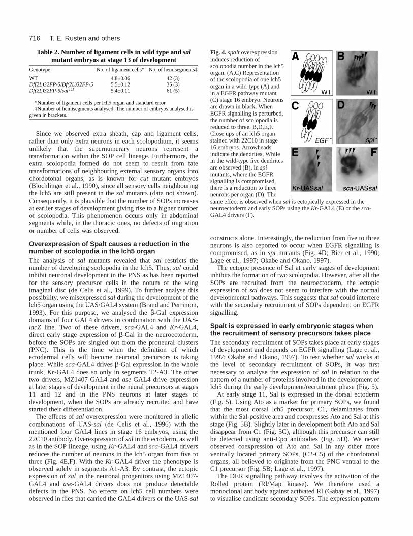
Since we observed extra sheath, cap and ligament cells,rather than only extra neurons in each scolopodium, it seemsunlikely that the supernumerary neurons represent atransformation within the SOP cell lineage. Furthermore, theextra scolopodia formed do not seem to result from fatetransformations of neighbouring external sensory organs intochordotonal organs, as is known for cut mutant embryos(Blochlinger et al., 1990), since all sensory cells neighbouringthe lch5 are still present in the sal mutants (data not shown).Consequently, it is plausible that the number of SOPs increasesat earlier stages of development giving rise to a higher numberof scolopodia. This phenomenon occurs only in abdominalsegments while, in the thoracic ones, no defects of migrationor number of cells was observed.
Overexpression of Spalt causes a reduction in thenumber of scolopodia in the lch5 organThe analysis of salmutants revealed that salrestricts thenumber of developing scolopodia in the lch5. Thus, sal couldinhibit neuronal development in the PNS as has been reportedfor the sensory precursor cells in the notum of the wingimaginal disc (de Celis et al., 1999). To further analyse thispossibility, we misexpressed salduring the development of thelch5 organ using the UAS/GAL4 system (Brand and Perrimon,1993). For this purpose, we analysed the β-Gal expressiondomains of four GAL4 drivers in combination with the UAS-lacZ line. Two of these drivers, sca-GAL4 and Kr-GAL4,direct early stage expression of β-Gal in the neuroectoderm,before the SOPs are singled out from the proneural clusters(PNC). This is the time when the definition of whichectodermal cells will become neuronal precursors is takingplace. While sca-GAL4 drives β-Gal expression in the wholetrunk, Kr-GAL4 does so only in segments T2-A3. The othertwo drivers, MZ1407-GAL4 and ase-GAL4 drive expressionat later stages of development in the neural precursors at stages11 and 12 and in the PNS neurons at later stages ofdevelopment, when the SOPs are already recruited and havestarted their differentiation.
The effects of saloverexpression were monitored in alleliccombinations of UAS-sal(de Celis et al., 1996) with thementioned four GAL4 lines in stage 16 embryos, using the22C10 antibody. Overexpression of sal in the ectoderm, as wellas in the SOP lineage, using Kr-GAL4 and sca-GAL4 driversreduces the number of neurons in the lch5 organ from five tothree (Fig. 4E,F). With the Kr-GAL4 driver the phenotype isobserved solely in segments A1-A3. By contrast, the ectopicexpression of salin the neuronal progenitors using MZ1407-GAL4 and ase-GAL4 drivers does not produce detectabledefects in the PNS. No effects on lch5 cell numbers wereobserved in flies that carried the GAL4 drivers or the UAS-sal
constructs alone. Interestingly, the reduction from five to threeneurons is also reported to occur when EGFR signalling iscompromised, as in spimutants (Fig. 4D; Bier et al., 1990;Lage et al., 1997; Okabe and Okano, 1997).
The ectopic presence of Sal at early stages of developmentinhibits the formation of two scolopodia. However, after all theSOPs are recruited from the neuroectoderm, the ectopicexpression of saldoes not seem to interfere with the normaldevelopmental pathways. This suggests that salcould interferewith the secondary recruitment of SOPs dependent on EGFRsignalling.
Spalt is expressed in early embryonic stages whenthe recruitment of sensory precursors takes placeThe secondary recruitment of SOPs takes place at early stagesof development and depends on EGFR signalling (Lage et al.,1997; Okabe and Okano, 1997). To test whether sal works atthe level of secondary recruitment of SOPs, it was firstnecessary to analyse the expression of sal in relation to thepattern of a number of proteins involved in the development oflch5 during the early development/recruitment phase (Fig. 5).
At early stage 11, Sal is expressed in the dorsal ectoderm(Fig. 5). Using Ato as a marker for primary SOPs, we foundthat the most dorsal lch5 precursor, C1, delaminates fromwithin the Sal-positive area and coexpresses Ato and Sal at thisstage (Fig. 5B). Slightly later in development both Ato and Saldisappear from C1 (Fig. 5C), although this precursor can stillbe detected using anti-Cpo antibodies (Fig. 5D). We neverobserved coexpression of Ato and Sal in any other moreventrally located primary SOPs, (C2-C5) of the chordotonalorgans, all believed to originate from the PNC ventral to theC1 precursor (Fig. 5B; Lage et al., 1997).
The DER signalling pathway involves the activation of theRolled protein (Rl/Map kinase). We therefore used amonoclonal antibody against activated Rl (Gabay et al., 1997)to visualise candidate secondary SOPs. The expression pattern
T. E. Rusten and others
Table 2. Number of ligament cells in wild type and salmutant embryos at stage 13 of development
Genotype No. of ligament cells* No. of hemisegments‡
WT 4.8±0.06 42 (3)Df(2L)32FP-5/Df(2L)32FP-5 5.5±0.12 35 (3)Df(2L)32FP-5/sal445 5.4±0.11 61 (5)
*Number of ligament cells per lch5 organ and standard error. ‡Number of hemisegments analysed. The number of embryos analysed is
given in brackets.
Fig. 4. spaltoverexpressioninduces reduction ofscolopodia number in the lch5organ. (A,C) Representationof the scolopodia of one lch5organ in a wild-type (A) andin a EGFR pathway mutant(C) stage 16 embryo. Neuronsare drawn in black. WhenEGFR signalling is perturbed,the number of scolopodia isreduced to three. B,D,E,F.Close ups of an lch5 organstained with 22C10 in stage16 embryos. Arrowheadsindicate the dendrites. Whilein the wild-type five dendritesare observed (B), in spimutants, where the EGFRsignalling is compromised,there is a reduction to threeneurons per organ (D). Thesame effect is observed when salis ectopically expressed in theneuroectoderm and early SOPs using the Kr-GAL4 (E) or the sca-GAL4 drivers (F).

717Spalt activity in embryonic PNS
of activated Rl corresponds well to the patterns reported foraosand pnt (Lage et al., 1997; Okabe and Okano, 1997; Okabeet al., 1996) and appears around the tracheal pits, in cellssurrounding C1, and in cells overlying the more ventral C2-C5SOPs (Fig. 5A,E). Only a ring of 4-5 cells with elongatednuclei around the C1 precursor coexpress Sal and activated Rl.Interestingly, Rl-positive cells in the ectoderm not expressingSal overly the SOPs, C2-C5, and their number corresponds tothe number of secondary SOPs recruited to the abdominalchordotonal organs (two for the lch5 and one for the vchA/Bcluster; Fig. 5E). Later in development, Sal-positive cellssurrounding C1 migrate ventrally and end up in a lateralposition close to the lch5, strongly suggesting that they are thedeveloping oenocytes. Later on, Sal expression can be detectedat stage 13 in the lch5 lineage, at a time when all cell divisionshave already occurred and differentiation and migration of thelch5 has initiated (Carlson et al., 1997a; Carlson et al., 1997b;Hartenstein, 1988). At stage 13, four additional Sal-positivecells in the lateral region are likely to include the v′esA and Bneurons since these are Sal-positive in stage 16 embryos (Fig.2D-F). In addition to the C1 precursor of lch5, some otherSOPs were identified as Sal positive. These include the SOP
of the dorsal bipolar neuron (Fig. 5B) and a precursor notidentified (data not shown), which probably corresponds to theSOP of the Sal-positive neuron in the dorsal cluster of lateembryos (dn in Fig. 2B). At stage 16 of embryogenesis theepidermal expression of Sal fades away.
Since EGFR signalling is involved in recruitment ofsecondary SOPs, an excess of potential secondary SOPs exist(Fig. 5A). We have shown that loss of salgenerates more SOPs,and that among the candidates only those that surround the C1express Sal. This leads to the hypothesis that the candidatesoverlying the C2-C5 and not expressing Sal, are the actualprecursors of secondary SOPs. To prove this hypothesis it isnecessary to show that the extra SOPs generated by loss ofsal are dependent on EGFR signalling, and thus representsecondary SOPs.
The supernumerary scolopodia observed in spaltmutants are dependent on EGFR signallingThe PNS phenotype displayed by sal mutants is reminiscentof that of the increased EGFR signalling observed in aosmutants (Freeman et al., 1992; Kretzschmar et al., 1992;Okano et al., 1992), anterior open(aop; Lai and Rubin,
Fig. 5. spaltis expressed in theectoderm during early lch5development. A. The cartoonsummarises the expression patternof various markers in the differentcell types during early lch5development. C1-C5 are theprimary chordotonal sensory organprecursors (Lage et al., 1997;Okabe and Okano, 1997), whereC1-C3 (thick black outline) arethought to contribute to lch5, andC4 and C5 are thought to contributeto v′ch1 and one of the ventralvchA or vchB organs (Lage et al.,1997). The primary SOPs expressproteins such as Ato, Rho and Cpo.The putative secondary precursorcells (S) transduce signals andexpress the target genes pnt andaos. Two of these cells will berecruited to form the lch5 and oneto the vchA/B organs. Theexpression of salis localised in theprimary precursor C1 and in thecells that surround it (red). Thehorizontal line indicates the limit ofsal-expressing cells in the ectodermand the oval ring the tracheal pit.(B-E) Confocal sections stainedwith color-coded antibodies. B′, D′, E′ and B′′, D′′, E′′ are single channel images of the same section. (B) Confocal sections of two consecutivesegments in early stage 11 embryos showing the expression pattern of Ato (green). Ato is restricted to the C1 precursor of the lch5 and the SOPof the dbp, while it is still broadly expressed in the proneural cluster (PNC) that gives rise to the other SOPs of the chordotonal organs (C2-C5,compare with Fig. 2a in Lage et al., 1997). The C1 precursor delaminates within a Sal-positive ectodermal area (red), while the PNC arisesventral to the Sal domain. (C) Later on, at late stage 11, both Sal and Ato disappear from the C1 lineage (Lage et al., 1997). Ato is restricted toother chordotonal precursors (C3-C5, compare with Fig. 2b in Lage et al., 1997) where sal is not expressed. (D) A different late stage 11embryo showing the C1 precursor expressing Cpo. Although Sal is not present in the C1 precursor, it is strongly expressed in the cellssurrounding it, which have elongated nuclei. (E) Putative secondary chordotonal organ precursors showing active EGFR signalling visualisedwith anti-Rl antibodies (green). Sal and activated Rl coincide in the cells with elongated nuclei surrounding C1 (S). Activated Rl also labelsthree putative chordotonal precursors (S) ventral to the Sal domain, seemingly overlying the position of the primary signalling SOPs, as well asthe tracheal pits (outlined).
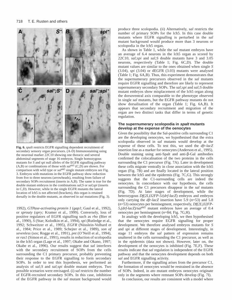
718
1992), GTPase-activating protein 1(gap1; Gaul et al., 1992),or sprouty (spry; Kramer et al., 1999). Conversely, loss ofpositive regulators of EGFR signalling such as rho (Bier etal., 1990), S (Star; Kolodkin et al., 1994), spi (Rutledge et al.,1992; Schweitzer et al., 1995), EGFR (Nüsslein-Volhard etal., 1984; Price et al., 1989; Schejter et al., 1989), son ofsevenless(sos; Rogge et al., 1991), pnt(O’Neill et al., 1994),or ras1 (Simon et al., 1991), results in reduction of scolopodiain the lch5 organ (Lage et al., 1997; Okabe and Okano, 1997;Okabe et al., 1996). Our results suggest that sal interfereswith the secondary recruitment of SOPs from the cellssurrounding the C1 primary precursor, probably preventingtheir response to the EGFR signalling to form secondarySOPs. In order to test this hypothesis, we performed ananalysis of sal;Sand sal;spidouble mutant embryos. Twopossible scenarios were envisaged. (i) salrestricts the numberof EGFR-recruited secondary SOPs. In this case, inhibitionof the EGFR pathway in the salmutant background would
produce three scolopodia. (ii) Alternatively, salrestricts thenumber of primary SOPs for the lch5. In this case doublemutants where EGFR signalling is perturbed in the salmutant background would produce more than 3 neurons orscolopodia in the lch5 organ.
As shown in Table 1, while the salmutant embryos havean average of 6.4 neurons in the lch5 organ as scored by22C10, sal;spi and sal;S double mutants have 3 and 3.05neurons, respectively (Table 1; Fig. 6C,D). The doublemutant values are similar to the ones obtained when single S(3.04), spi (3.04) or dEGFR (3.03) mutants were analysed(Table 1; Fig. 6A,B). Thus, this experiment demonstrates thatthe supernumerary precursors observed in the salmutantsrequire EGFR signalling and therefore are likely to representsupernumerary secondary SOPs. The sal;spiand sal;Sdoublemutant embryos show misplacement of the lch5 organ alongthe dorsoventral axis comparable to the phenotype observedin single salmutants, but the EGFR pathway mutants do notshow dorsalisation of the organ (Table 1; Fig. 6A,B). Itappears that secondary recruitment and migration of theorgan are two distinct tasks that differ in terms of geneticregulation.
The supernumerary scolopodia in spalt mutantsdevelop at the expense of the oenocytesGiven the possibility that the Sal-positive cells surrounding C1are the developing oenocytes, we hypothesised that the extrascolopodia observed in sal mutants would develop at theexpense of these cells. To test this, we used the dfr-lacZinsertion line as a marker for oenocytes (Anderson et al., 1995).Double staining using anti-Spalt and anti-β-Gal antibodiesconfirmed the colocalisation of the two proteins in the cellssurrounding the C1 precursor (Fig. 7A). Later in developmentthese cells migrate ventrally in close association with the lch5organ (Fig. 7B) and are finally located in the lateral positionbetween the lch5 and the epidermis (Fig. 7C,G). This stronglysuggests that the C1-surrounding cells are indeed theoenocytes. In concordance with our hypothesis, the cellssurrounding the C1 precursors disappear in the sal mutants(Fig. 7D). At later stages of development, while theheterozygous Df(2L)32FP-5/(drf-lacZ) embryos and embryosonly carrying the dfr-lacZinsertion have 5.9 (n=53) and 5.8(n=53) oenocytes per hemisegment, respectively, Df(2L)32FP-5,(drf-lacZ)/sal445 mutant embryos have an average of 0.4oenocytes per hemisegment (n=84; Fig. 7G,H).
In analogy with the developing lch5, we then hypothesisedthat the oenocytes require EGFR signalling for properdevelopment. We therefore analysed embryos mutants for Sand spi at different stages of development. Interestingly, instage 11 embryos the sal pattern of expression remainsunaltered in the cells surrounding the C1 precursor, as well asin the epidermis (data not shown). However, later on, thedevelopment of the oenocytes is inhibited (Fig. 7E,F). Theseresults indicate that salregulation is independent of the EGFRpathway and that the oenocytes development depends on bothsal and EGFR signalling activity.
Furthermore, if the signalling arises from the precursor C1,the formation of oenocytes would be restrained in the absenceof SOPs. Indeed, in atomutant embryos oenocytes originateonly in the segments where remnant SOPs develop (Fig. 7I).
In conclusion, our results are consistent with a model where
T. E. Rusten and others
Fig. 6. spaltrestricts EGFR signalling dependent recruitment ofsecondary sensory organ precursors. (A-D) Immunostaining usingthe neuronal marker 22C10 showing one thoracic and severalabdominal segments of stage 16 embryos. Single homozygousmutants for Sand spinull alleles of the EGFR signalling pathway(A,B) or combinations of those with sal445 (C,D) are shown. Forcomparison with wild type or sal445 single mutant embryos see Fig.3. Embryos with mutations in the EGFR pathway show reductionfrom five to three neurons (arrowheads), resulting from failure ofsecondary SOPs recruitment (inserts in A,B). The same is true for thedouble mutant embryos in the combinations sal;Sor sal;spi (insertsin C,D). However, while in the single EGFR mutants the laterallocation of lch5 is not affected (brackets), this organ is retaineddorsally in the double mutants, as observed in salmutations (Fig. 3).

719Spalt activity in embryonic PNS
sal restricts the ability of C1-surrounding cells, receivingEGFR signalling, to adopt sensory organ precursor cell fate;these cells then develop as oenocytes rather than chordotonalorgans (Fig. 8).
DISCUSSION
The work presented here has documented the expressionpattern of salin the peripheral nervous system and associatedcells in the Drosophilaembryo. It also revealed the functionof this gene in the formation of the pentascolopodial organs ofthe PNS and the associated oenocytes. It has shown that salinteracts with the EGFR signalling pathway, acting as a switchbetween the secondary SOP fate (which it restricts) and theoenocyte fate (which it promotes).
Spalt restricts the EGFR mediated recruitment ofSOPs in the developing lch5The EGFR pathway is involved in a number of cellularprocesses such as cell survival, proliferation, patterning,migration, and cell fate decision (Dominguez et al., 1998). Inparticular, the EGFR pathway is implicated in the developmentof the chordotonal organs in Drosophila melanogaster(Lageet al., 1997; Okabe and Okano, 1997; Okabe et al., 1996). Thepathway is necessary for the second step of recruitment ofSOPs from ectodermal precursors, and for the consequentincrease of number of scolopodia in the lch5 and in the vchA/Borgans. Thus, during development of the lch5 organ, where twosecondary SOPs are recruited, removal of positive EGFRpathway components like rho, S, spi, pnt, sos, Drk, or DER
itself, reduces the number of scolopodia in the lch5 from fiveto three. Conversely, mutations in negative regulators of EGFRsignalling like argos, gap1or spry result in an increase of
Fig. 7. The spalt-expressing cells surrounding C1 arethe precursor of the oenocytes. (A-F,I) Immunostainingsshowing Sal (red) and β-Gal or Cpo (green) expressionin embryonic stages 11 (A,D), 13 (B,E) and 16 (C,F,I).Abbreviations are as in Figs 1 and 2. A. The cellssurrounding the precursor C1 express both saland thedfr-lacZ transgene, and are the putative secondarysensory organ precursors (S) that receive the EGFRsignal (Fig. 5). Later in development, these cells migrateventrally together with lch5 (B) and at stage 16 they arelocated between the epidermis and the pentascolopodialorgan, and continue expressing Sal and β-Gal (C). In salmutants (D), these cells are not detectable at stage 11. Inspimutants, the cells that migrate together with lch5 arenot longer visible (E), in correlation with the loss ofoenocytes observed in the late embryo (F).(G,H) Enzymatic stainings using anti-β-Gal antibodiesexpressed by the dfr-lacZ transgene, reveal the loss ofoenocytes in salmutant background. Only a few cellsare formed (arrows). Oenocytes do not form inhomozygous atomutants, where the formation ofprimary SOPs is compromised in most of the segments.In I, three consecutive hemisegments are shown.Sometimes, oenocytes (in red) develop in thehemisegments where a primary SOP has singled out, asrevealed by the presence of the neuron, sheath, ligamentand cap cells from one scolopodium (left segment).However, this is not always observed (middle segment),indicating that EGFR signalling to the ectoderm arisesfrom some primary SOPs but not from others.
Fig. 8. Function of spaltin abdominal lateral chordotonal organdevelopment. (A) In early stage 11 of embryogenesis, Sal isexpressed in the dorsal ectodermal region (red) from which the C1primary SOP is selected. (B) At late stage 11, the primary SOPs (C1-C5) delaminate and signal via the EGFR pathway to the overlyingectoderm. The receiving cells (S) respond to EGFR signalling, asshown by activated Rl staining. Only three are recruited as secondarySOPs (green). Thus, the presence of Sal in the other receiving cells(yellow) prevents them from becomimg sensory cells. C. Thechordotonal organs are then formed: lch5 (C1-C3 plus two secondarySOPs), v′ch1 (C4; no secondary recruited SOPs), and the vchA/Borgan (C5 plus one secondary SOP). The epidermal cells around C1adopt the alternative oenocyte fate (Oe). D. Model in which EGFRsignalling is necessary for the formation of both SOPs andoenocytes, while Sal acts as a switch restricting the sensory precursorin favour of the oenocyte fate.
720
secondary recruited SOPs in the thorax as well as in theabdominal segments.
Here, we show that the zinc finger transcription factor Salplays a role in the formation of the lch5 in parallel with theEGFR signalling pathway: the absence of Sal generatessupernumerary scolopodia, while the overexpression of Salreduces the number of scolopodia from five to three. Ourresults are consistent with the model proposed in Fig. 8: thatunder wild-type conditions, Sal modifies the EGFR signallingoutput in the cells surrounding the primary precursor C1,which instead of becoming secondary SOPs adopt theoenocytes cell fate. Five lines of evidence support thisidea. First, supernumerary support cells accompany thesupernumerary neurons observed in salmutants. Thus, thephenotype is not caused by cell fate transformation within theSOP lineage. Second, the C1-surrounding cells receive theEGFR signal (shown by the antibody staining for activatedRl/MAPK) and, therefore, are capable of becomingsecondary precursors. These cells are Sal positive while theother potential secondary precursors, also showing activatedRl and overlying the more ventrally located C2-C5, are not.Given that the number of cells receiving the EGFR signal islarger than the number of cells that become secondary SOPs(two for lch5 and one for vchA/B), the output of the EGFRpathway must be modified in the rest of the cells receivingthe signal. Third, the analysis of allelic combinationsbetweensal and EGFR pathway mutants reveals that thesupernumerary neuronal phenotype observed in the absenceof Sal is EGFR dependent. Fourth, the oenocyte precursorsdepend on sal and EGFR signalling to develop. And fifth, inthe absence of primary precursors oenocytes do not develop,as shown in atomutants.
The effects of salloss- and gain-of-function are similar, butnot identical, to the ones exhibited by corresponding changesin negative regulators of EGFR signalling. There are at leasttwo important differences between the role of these regulatorsand sal. First, aos, pntand spryare expressed in all the cellsreceiving the EGFR signal from the primary SOPs, while salis expressed only in a subset of them. Consistent with this, theloss of function of these regulators affects the secondaryrecruitment of SOPs to other chordotonal organs, like vchA/Band v′ch1, while salseems to modify only lch5. Second, theincrease of scolopodia numbers in lch5 is moderate in the spryand aosmutants, while in salmutants we have observed up toeight scolopodia. In conclusion, differently from the EGFRpathway regulators that are involved in the development of allthe chordotonal organs, salis involved specifically in theformation of lch5.
According to our observations, the cells surrounding C1migrate along the dorsoventral axis closely associated with thepentascolopodial organ. These cells are easy to recognise bythe elongated shape of their nuclei and the strong salexpression that they display. These cells occupy the location ofoenocytes in late embryonic stages. It is then likely that salplays a role in deciding the fate of the EGFR responding cellssurrounding the C1 precursor. In the presence of Sal these cellswill become oenocytes while in the absence of Sal (as is truefor the presumptive secondary precursors overlying C2, C3, C4and C5), the cells will become sensory organ precursors. Sincethe putative precursors of the oenocyte cells need EGFRsignalling to accomplish some aspects of their development,
sal would act as a selector gene being necessary to direct themto their correct fate.
The correct location of the lch5 organ iscompromised in spalt mutant embryosIn addition to the extra recruitment phenotype, sal mutantshave aberrantly located lch5 along the dorsoventral axis. In thewild type, lch5 precursors are recruited in a dorsal position andthen migrate ventrally (Salzberg et al., 1994). In the mutant,the ventral migration does not seem to take place. Thephenotype is similar, but not identical to that of Homothorax,Abdominal-Aor extradenticlemutants, where the lch5 organremains in a dorsal position and scolopodial numbers arereduced to three (Kurant et al., 1998). The involvement of salin other migration process has been reported previously fortracheal development (Kühnlein and Schuh, 1996). There, incells of the dorsal tracheal trunk, sal is required foranteroposterior migration and morphogenesis. Furthermore, ithas been shown that sal is necessary for the correct location ofsome neurons in the CNS (T. E. R., R. C., J. U., G. T., F. C.K. and R. B., unpublished results). The molecular mechanismsinvolved in the specification of migration are largely unknown,and whether the same mechanism applies in the threementioned cases remains unexplored.
The pleiotropic functions that Spalt proteins exert duringdevelopment are remarkable. In C. elegans, sem-4phenotypesinclude cell fate changes, cell death, defects in axonalmorphologies, extra cell divisions or migration. The same istrue in Drosophila, where salgenes play a role in establishinghomeotic identities in the blastoderm (Casanova, 1989;Jurgens, 1988), positioning the wing veins (de Celis andBarrio, 2000; de Celis et al., 1996), localising sensory organclusters (de Celis et al., 1999) and affecting the migration ofthe dorsal tracheal trunk (Chen et al., 1998; Kühnlein andSchuh, 1996). It therefore appears that the Spalt proteins canfunction with different signalling pathways and act incombination with other transcription factors to serve diverseroles during development.
We thank A. Ephrussi, A. Ghysen, A. Jarman, W. A. Johnson andJ. Castelli-Gair for reagents and fly stocks. We thank J. F. de Celisfor critically reading the manuscript. We would like to thank ouranonymous referees for their help in improving the quality of themanuscript. We also thank B. Miñana for technical assistance. T. E.R. is supported with a fellowship from the Norwegian ResearchCouncil and R. C. was supported by a Short Term EMBOFellowship.
REFERENCES
Anderson, M. G., Perkins, G. L., Chittick, P., Shribley, R. J. and Johnson,W. A. (1995). drifter, a DrosophilaPOU-domain transcription factor, isrequired for correct differentiation and migration of tracheal cells andmidline glia. Genes Dev.9, 123-137.
Barrio, R., de Celis, J. F., Bolshakov, S. and Kafatos, F. C.(1999).Identification of regulatory regions driving the expression of the Drosophilaspaltcomplex at different developmental stages. Dev. Biol.215, 33-47.
Barrio, R., Shea, M. J., Carulli, J., Lipkow, K., Gaul, U., Frömmer, G.,Schuh, R., Jäckle, H. and Kafatos, F. C.(1996). The spalt-relatedgeneof Drosophila melanogasteris a member of an ancient family, defined bythe adjacent, region-specific homeotic gene spalt. Dev. Genes Evol.206,315-325.
Basson, M. and Horvitz, H. R.(1996). The Caenorhabditis elegansgene sem-
T. E. Rusten and others

721Spalt activity in embryonic PNS
4 controls neuronal and mesodermal cell development and encodes a zincfinger protein. Genes Dev.10, 1953-65.
Bellen, H. J., Kooyer, S., D’Evelyn, D. and Pearlman, J.(1992). TheDrosophila Couch potato protein is expressed in nuclei of peripheralneuronal precursors and shows homology to RNA-binding proteins. GenesDev.6, 2125-2136.
Bier, E., Jan, L. Y. and Yan, Y. N.(1990). rhomboid, a gene required fordorsoventral axis establishment and peripheral nervous system developmentin Drosophila melanogaster. Genes Dev. 4, 190-203.
Blochlinger, K., Bodmer, R., Jan, L. Y. and Jan, Y. N.(1990). Patterns ofexpression of Cut, a protein required for external sensory organdevelopment in wild-type and cutmutant Drosophilaembryos. Genes Dev.4, 1322-31.
Brand, A. H. and Perrimon, N. (1993). Targeted gene expression as a meansof altering cell fates and generating dominant phenotypes. Development,118,401-415.
Brewster, R. and Bodmer, R.(1995). Origin and specification of type IIsensory neurons in Drosophila. Development121, 2923-36.
Brewster, R. and Bodmer, R.(1996). Cell lineage analysis of the Drosophilaperipheral nervous system. Dev. Genet.18, 50-63.
Buck, A., Archangelo, L., Dixkens, C. and Kohlhase, J.(2000). Molecularcloning, chromosomal localization, and expression of the murine SALL1ortholog Sall1. Cytogenet. Cell Genet.89, 150-153.
Campbell, G., Goring, H., Lin, T., Spana, E., Andersson, S., Doe, C. Q.and Tomlinson, A. (1994). RK2, a glial-specific homeodomain proteinrequired for embryonic nerve cord condensation and viability in Drosophila.Development120, 2957-66.
Campos-Ortega, J. A. and Hartenstein, V. (1997). The EmbryonicDevelopment of Drosophila melanogaster. Berlin: Springer-Verlag.
Carlson, S. D., Hilgers, S. L. and Juang, J. L.(1997a). Ultrastructure andblood-nerve barrier of chordotonal organs in the Drosophilaembryo. J.Neurocyt. 26, 377-388.
Carlson, S. D., Hilgers, S. L. and Juang, J. L.(1997b). First developmentalsigns of the scolopale (glial) cell and neuron comprising the chordotonalorgan in the Drosophilaembryo. Glia19, 269-74.
Casanova, J.(1989). Mutations in the spaltgene ofDrosophilacause ectopicexpression of Ultrabithoraxand Sex combs reduced. Wilhelm Roux’s Arch.Dev. Biol.198, 137-140.
Castelli-Gair, J., Greig, S., Micklem, G. and Akam, M.(1994). Dissectingthe temporal requirements for homeotic gene function. Development120,1983-95.
Chen, C. K., Kühnlein, R. P., Eulenberg, K. G., Vincent, S., Affolter, M.and Schuh, R. (1998). The transcription factors KNIRPS and KNIRPSRELATED control cell migration and branch morphogenesis duringDrosophilatracheal development. Development125, 4959-4968.
Chien, C. T., Wang, S., Rothenberg, M., Jan, L. Y. and Jan, Y. N.(1998).Numb-associated kinase interacts with the phosphotyrosine binding domainof Numb and antagonizes the function of Numb in vivo. Mol. Cell. Biol.18,598-607.
de Celis, J. F. and Barrio, R.(2000). Function of the spalt/spalt-relatedgenecomplex in positioning the veins in the Drosophila wing. Mech. Dev.91,31-41.
de Celis, J. F., Barrio, R. and Kafatos, F. C.(1996). A gene complex actingdownstream of dppin Drosophilawing morphogenesis. Nature381, 421-424.
de Celis, J. F., Barrio, R. and Kafatos, F. C.(1999). Regulation of thespalt/spalt-relatedgene complex and its function during sensory organdevelopment in the Drosophilathorax. Development126, 2653-2662.
Dominguez, M., Wasserman, J. D. and Freeman, M.(1998). Multiplefunctions of the EGFR receptor in Drosophilaeye development. Curr. Biol.8, 1039-1048.
Freeman, M., Klambt, C., Goodman, C. S. and Rubin, G. M.(1992). Theargosgene encodes a diffusible factor that regulates cell fate decisions inthe Drosophilaeye. Cell 69, 963-975.
Fujita, S. C., Zipursky, S. L., Benzer, S., Ferrus, A. and Shotwell, S. L.(1982). Monoclonal antibodies against the Drosophilanervous system.Proc. Natl. Acad. Sci. USA79, 7929-33.
Gabay, L., Seger, R. and Shilo, B. Z.(1997). In situ activation pattern ofDrosophila EGFR receptor pathway during development. Science277,1103-1106.
Gaul, U., Mardon, G. and Rubin, G. M. (1992). A putative Ras GTPaseactivating protein acts as a negative regulator of signaling by the Sevenlessreceptor tyrosine kinase. Cell68, 1007-1019.
Ghysen, A., Dambly-Chaudière, C., Aceves, E., Jan, L. Y. and Jan, Y. N.
(1986). Sensory neurons and peripheral pathways in Drosophilaembryos.Roux’s Arch. Dev. Biol.195, 281-289.
Ghysen, A. and O’Kane, C.(1989). Neural enhancer-like elements as specificcell markers in Drosophila. Development105, 35-53.
Grant, K., Hanna-Rose, W. and Han, M.(2000). sem-4 promotes vulval cell-fate determination in Caenorhabditis elegansthrough regulation of lin-39Hox. Dev. Biol.224, 496-506.
Halter, D. A., Urban, J., Rickert, C., Ner, S. S., Ito, K., Travers, A. A. andTechnau, G. M. (1995). The homeobox gene repo is required for thedifferentiation and maintenance of glia function in the embryonic nervoussystem of Drosophila melanogaster. Development121, 317-32.
Hartenstein, A. Y., Rugendorff, A., Tepass, U. and Hartenstein, V.(1992).The function of the neurogenic genes during epithelial development in theDrosophilaembryo. Development, 116, 1203-1220.
Hartenstein, V. (1988). Development of Drosophilalarval sensory organs:spatiotemporal pattern of sensory neurons, peripheral axonal pathways andsensilla differentiation. Development102, 869-886.
Hoch, M., Broadie, K., Jäckle, H. and Skaer, H.(1994). Sequential fates ina single cell are established by the neurogenic cascade in the Malpighiantubules of Drosophila. Development120, 3439-3450.
Hollemann, T., Schuh, R., Pieler, T. and Stick, R.(1996). Xenopus Xsal-1,a vertebrate homolog of the region specific homeotic gene spalt ofDrosophila. Mech. Dev.55, 19-32.
Hummel, T., Krukkert, K., Roos, J., Davis, G. and Klambt, C. (2000).Drosophila Futsch/22C10 is a MAP1B-like protein required for dendriticand axonal development. Neuron26, 357-370.
Jarman, A. P., Grell, E. H., Ackerman, L., Jan, L. Y. and Jan, Y. N.(1994).Atonal is the proneural gene for Drosophila photoreceptors. Nature369,398-400.
Jones, B. W., Fetter, R. D., Tear, G. and Goodman, C. S.(1995). glial cellsmissing: a genetic switch that controls glial versus neuronal fate. Cell 82,1013-1023.
Jurgens, G. (1988). Head and tail development of the Drosophila embryoinvolves spalt, a novel homeotic gene. EMBO J.7, 189-196.
Kohlhase, J., Hausmann, S., Stojmenovic, G., Dixkens, C., Bink, K.,Schulz-Schaeffer, W., Altmann, M. and Engel, W.(1999a). SALL3, anew member of the humanspalt-like gene family, maps to 18q23.Genomics62, 216-222.
Kohlhase, J., Taschner, P. E., Burfeind, P., Pasche, B., Newman, B., Blanck,C., Breuning, M. H., ten Kate, L. P., Maaswinkel-Mooy, P., Mitulla, B.et al. (1999b). Molecular analysis of SALL1 mutations in Townes-Brockssyndrome. Am. J. Hum. Genet.64, 435-445.
Kohlhase, J., Wischermann, A., Reichenbach, H., Froster, U. and Engel,W. (1998). Mutations in the SALL1putative transcription factor gene causeTownes- Brocks syndrome. Nat. Genet.18, 81-83.
Kolodkin, A. L., Pickup, A. T., Lin, D. M., Goodman, C. S. and Banerjee,U. (1994). Characterization of Starand its interactions with sevenlessandEGFR receptor during photoreceptor cell development in Drosophila.Development120, 1731-1745.
Köster, R., Stick, R., Loosli, F. and Wittbrodt, J.(1997). Medaka spaltactsas a target gene of hedgehogsignalling. Development124, 3147-3156.
Kramer, S., Okabe, M., Hacohen, N., Krasnow, M. A. and Hiromi, Y.(1999). Sprouty: a common antagonist of FGF and EGFR signalingpathways in Drosophila. Development126, 2515-2525.
Kretzschmar, D., Brunner, A., Wiersdorff, V., Pflugfelder, G. O.,Heisenberg, M. and Schneuwly, S.(1992). Giant lens, a gene involved incell determination and axon guidance in the visual system of Drosophilamelanogaster. EMBO J.11, 2531-2539.
Kühnlein, R. P., Frömmer, G., Friedrich, M., Gonzalez-Gaitán, M., Wever,A., Wagner-Bernholz, J. F., Gehring, W. J., Jäckle, H. and Schuh, R.(1994). spalt encodes an evolutionarily conserved zinc finger protein ofnovel structure which provides homeotic gene function in the head and tailregion of the Drosophilaembryo. EMBO J.13, 168-179.
Kühnlein, R. P. and Schuh, R.(1996). Dual function of the region-specifichomeotic gene spaltduring Drosophila tracheal system development.Development122, 2215-2223.
Kurant, E., Pai, C. Y., Sharf, R., Halachmi, N., Sun, Y. H. and Salzberg,A. (1998). Dorsotonals/homothorax, the Drosophila homologue of meis1,interacts with Extradenticle in patterning of the embryonic PNS.Development125, 1037-1048.
Lage, P., Jan, Y. N. and Jarman, A. P.(1997). Requirement for EGFRreceptor signalling in neural recruitment during formation of Drosophilachordotonal sense organ clusters. Curr. Biol. 7, 166-175.
Lai, Z. C. and Rubin, G. M. (1992). Negative control of photoreceptor

722
development in Drosophilaby the product of the yangene, an ETS domainprotein. Cell 70, 609-620.
Lecuit, T., Brook, W. J., Ng, M., Calleja, M., Sun, H. and Cohen, S. M.(1996). Two distinct mechanisms for long-range patterning byDecapentaplegic in the Drosophilawing. Nature381, 387-393.
Lindsley, D. and Zimm, G. G. (1992). The genome of Drosophilamelanogaster. San Diego, USA: Academic Press Inc.
Mlodzik, M., Baker, N. E. and Rubin, G. M. (1990). Isolation and expressionof scabrous, a gene regulating neurogenesis in Drosophila. Genes Dev.4,1848-1861.
Nellen, D., Burke, R., Struhl, G. and Basler, K.(1996). Direct and long rangeaction of a DPP morphogen gradient. Cell 85, 357-368.
Nüsslein-Volhard, C., Wieschaus, E. and Kluding, H.(1984). Mutationsaffecting the pattern of the larval cuticle in Drosophila melanogaster. RouxArch. Dev. Biol.193, 267-282.
O’Neill, E. M., Rebay, I., Tjian, R. and Rubin, G. M. (1994). Theactivities of two Ets-related transcription factors required for Drosophilaeye development are modulated by the Ras/MAPK pathway. Cell 78,137-47.
Okabe, M. and Okano, H.(1997). Two-step induction of chordotonal organprecursors in Drosophilaembryogenesis. Development124, 1045-53.
Okabe, M., Sawamoto, K. and Okano, H.(1996). The function of theDrosophila argos gene product in the development of embryonicchordotonal organs. Dev. Biol.175, 37-49.
Okano, H., Hayashi, S., Tanimura, T., Sawamoto, K., Yoshikawa, S.,Watanabe, J., Iwasaki, M., Hirose, S., Mikoshiba, K. and Montell, C.(1992). Regulation of Drosophilaneural development by a putative secretedprotein. Differentiation52, 1-11.
Ott, T. and Schutz, G. (1996). The mouse homolog of the region-specifichomeotic gene spaltof Drosophila is expressed in the developing nervoussystem and in mesoderm-derived structures. Mech. Dev.56, 117-128.
Patel, N. H. (1994). Imaging neuronal subsets and other cell types in whole-mount Drosophilaembryos and larvae using antibody probes. Methods CellBiol. 44, 445-487.
Price, J. V., Clifford, R. J. and Schüpbach, T.(1989). The maternalventralizing locus torpedoIs allelic to faint little ball, an embryonic lethal,and encodes the DrosophilaEGFR receptor homolog. Cell 56, 1085-1092.
Reuter, D., Kühnlein, R. P., Frommer, G., Barrio, R., Kafatos, F. C. and
Jäckle, H. (1996). Regulation, function and potential origin of theDrosophilagene spalt adjacent, which encodes a secreted protein expressedin the early embryo. Chromosoma104, 445-454.
Rogge, R. D., Karlovich, C. A. and Banerjee, U.(1991). Genetic dissectionof a neurodevelopmental pathway: Son of sevenless functions downstreamof the Sevenless and EGFR receptor tyrosine kinases. Cell 64, 39-48.
Roos, J., Hummel, T., Ng, N., Klambt, C. and Davis, G. W.(2000).Drosophila Futsch regulates synaptic microtubule organization and isnecessary for synaptic growth. Neuron26, 371-382.
Rutledge, B. J., Zhang, K., Bier, E., Jan, Y. N. and Perrimon, N.(1992).The Drosophila spitzgene encodes a putative EGFR-like growth factorinvolved in dorsal-ventral axis formation and neurogenesis. Genes Dev.6,1503-1517.
Salzberg, A., D’Evelyn, D., Schulze, K. L., Lee, J. K., Strumpf, D., Tsai,L. and Bellen, H. J.(1994). Mutations affecting the pattern of the PNS inDrosophilareveal novel aspects of neuronal development. Neuron13, 269-287.
Schejter, E. D. and Shilo, B. Z.(1989). The DrosophilaEGFR receptorhomolog (DER) gene is allelic to faint little ball, a locus essential forembryonic development. Cell56, 1093-104.
Schweitzer, R., Shaharabany, M., Seger, R. and Shilo, B. Z.(1995).Secreted Spitz triggers the DER signaling pathway and is a limitingcomponent in embryonic ventral ectoderm determination. Genes Dev.9,1518-1529.
Simon, M. A., Bowtell, D. D., Dodson, G. S., Laverty, T. R. and Rubin, G.M. (1991). Ras1and a putative guanine nucleoside exchange factor performcrucial steps in signaling by the Sevenless protein tyrosine kinase. Cell 67,701-716.
Sturtevant, M. A., Biehs, B., Marin, E. and Bier, E.(1997). The spaltgenelinks the A/P compartment boundary to a linear adult structure in theDrosophilawing. Development124, 21-32.
Sweeney, S. T., Broadie, K., Keane, J., Niemann, H. and O’Kane, C. J.(1995). Targeted expression of tetanus toxin light chain in Drosophilaspecifically eliminates synaptic transmission and causes behavioral defects.Neuron14, 341-351.
Xiong, W. C., Okano, H., Patel, N. H., Blendy, J. A. and Montell, C.(1994).repo encodes a glial-specific homeo domain protein required in theDrosophilanervous system. Genes Dev.8, 981-994.
T. E. Rusten and others