structural malformations growth retardation functional impairment death of the organism...
TRANSCRIPT

Structural malformations
Growth retardation
Functional impairment
Death of the organism
Developmental toxicology

Teratology 1. the study of malformations or serious deviations from the normal type in organisms
2. the branch of science concerned with the production, development, anatomy, and classification of malformed fetuses.
TeratogenAny agent that causes a birth defectAfter Greek “monster creating”

•Environmental conditions (1200)
•Maternal nutritional deficiencies (1930)
•Rubella virus infection (1941)
•Thalidomide (1961)

(65%)
35-40 /4100 chemicals alter prenatal development, Table 10-1

Thalidomide

Thalidomide Thalidomide was released in 1956 as a mild seda
tive used to combat nausea in pregnant women. It was later (1961) withdrawn from the market once it was discovered thalidomide was a human teratogen. As little as one dose could cause a significant birth defect. Approximately 5,000-7,000 malformed infants were born to women who ingested thalidomide during pregnancy.
Symptoms:malformed intestines, hearing defects, absent ears, and/or ocular and renal anomalies. However, the most striking phenotype is phocomelia: severe limb malformations in which the long bones of the limb are either greatly reduced in length or absent all together.

Teratogenic between 20-36 days after fertilization
Mechanisms –unknown?
Proposed mechanisms (more than 30)
Angiogenesis
Integrin regulation
Oxidative DNA damage
growth factor antagonism
Now approved for Oral ulcer for AIDS, erythema nodosum leprosum-new anticancer drug? Anti-angiogenesis

Diethylstilbesterol (DES)DES was prescribed between 1940 and 1970 to prevent miscarriages in high risk pregnancies. This was accomplished by DES increasing estrogen and progesterone synthesis by the placenta. In the mid 1970 cases of vaginal adenocarcinoma in women ages 16-20 were linked to fetal exposure through maternal DES ingestion early in the pregnancy. Approximately 1 in 1000 pregnancies were exposed, 75% of which resulted in female children with vaginal and cervical carcinomas as well as uterine anomalies. Male offspring had epididymal cyst, hypotrophic testes, decreased semen volume and poor semen quality.

Alcohol (Ethanol)
Fetal Alchohol Syndrome (FAS)
Fetal Alchohol Effects (FAE)
•Cranial facial dysmorphism•Intrauterine and postnatal growth retadation•Retarded psychomotor and intellectual development•IQ 68

Tobacco Nicotine restricts uterine blood vessels and restricts blood
flow to the fetus resulting in chronic hypoxia and
malnutrition leading to birth defects. On average, offspring
of smoking women weigh 170-200 g less at birth as
compared to a non smoker’s child. There is a dose
dependence in that the child weight decreases in
proportion to number of cigarettes smoked by the mother.
There is also a reduction in overall fetal length, reduced
head circumference, intrauterine growth retardation as
well as behavioral alterations after birth.

Possible outcomes of smoking during preganancy include:
•spontaneous abortion•perinatal deaths•increase risk of sudden infant death syndrome•increased risk of learning, behavioral, and attention disorders.
Perinatal exposure to tobacco smoke can affect branching morphogenesis and maturation of the lung.Smoking during pregnancy increases the risk for premature delivery, abruption placenta, placenta previa and perinatal mortality.

Cocaine is an anesthetic and vasoconstrictor. Cocaine is thought to induce birth defects by disrupting the vasculature in the placenta thereby inducing intrauterine hypoxia and malnutrition
Cocaine
These pregnancies are at risk for premature labor, spontaneous abortion, increased perinatal mortality and fetal death.
Exposed fetuses often have intrauterine growth retardation, microcephaly, altered presencephalic development, decreased birth weight, a neonatal neurologic syndrome of abnormal sleep, tremor, poor feeding, irritability, and occasional seizures.

Retinoic Acid

Retinoic acid is the active ingredient in “Accutane”, a drug used to treat severe acne. Since its introduction in September of 1982, an estimated 160,000 women of child bearing age have ingested the drug. Between 1982 and 1987, approximately 900-1300 malformed children, 700-1000 spontaneous abortions and 5000-7000 elective abortions are due to Accutane exposure. Exposed children may have hydrocephaly, ear malformations, cardiovascular defects and decreased IQ. Accutane carries a pregnancy category X warning, meaning it is a known human teratogen.
Retinoic acid

MechanismA proposed mechanism is that biologically activ
e retinoic acid binds retinoic acid receptors which in t
urn bind DNA enhancer elements such as the retinoi
c acid response elements. Several Hox genes (respo
nsible for early patterning of the embryo) contain this
enhancer element in their promotors. Therefore, Hox
signaling may be altered due to increased retinoic aci
d concentrations resulting in multiple birth defects.

Retinoid Actions in vivo
Myeloid differentiationMyeloid differentiation Epithelial growth – keratinocytesEpithelial growth – keratinocytes Embryo developmentEmbryo development Anti-oxidantsAnti-oxidants

Retinoid Therapies
UseUse DrugsDrugs
PsoriasisPsoriasis Tazartene (Zorac),Tazartene (Zorac),
Etritinate (Tegison)Etritinate (Tegison)
AcneAcne Adapalene (Differin),Adapalene (Differin),
Tretinoin (Renova),Tretinoin (Renova),
Isotretinoin (Accutane)Isotretinoin (Accutane)
LeukemiaLeukemia Tretinoin/ATRA (Vesanoid)Tretinoin/ATRA (Vesanoid)

Retinoid Excess in Embryogenesis Retinoids are teratogensRetinoids are teratogens Embryos exposed to excess RA develoEmbryos exposed to excess RA develo
p p posterior neural tube defectsposterior neural tube defects Particularly affected are the retina, spinParticularly affected are the retina, spin
al chord and hind brainal chord and hind brain Posteriorization of anterior structuresPosteriorization of anterior structures
Marshall et al., FASEB J, 1996

RAR and RXR (Simple Version) Nuclear Receptors (like ER, PPAR, VDNuclear Receptors (like ER, PPAR, VD
R and others)R and others) RXR/RAR Heterodimer is functional unitRXR/RAR Heterodimer is functional unit Bind selectively to REs in genomeBind selectively to REs in genome Act as transcription factorsAct as transcription factors Up-regulate or Repress the expression Up-regulate or Repress the expression
of particular genesof particular genes

Molecular biology
S17
Target cell
ROH = Retinol
RA = Retinoic acid
RBP = Retinol bindingprotein
CRBP = Cellular retinol binding protein
CRABP = Cellular retinoic acidbinding protein
RXR, RAR = Nuclear retinoic acidreceptors
RBP–ROH
Extracellular
Regulatedgenetranscription D
NA
Receptor
ROH–CRBP
ROH RA Nucleus
RA–RXR RAR–RA
RA
RA–CRABP
Intracellular

Hormonal Targeting of Nuclear Complexes to Chromatin
SIGMA-ALDRICH.com/rbi

RAR and RXR types
GeneGene Major IsoformsMajor Isoforms Endogenous LigEndogenous Ligandand
RARRAR 1, 1, 22 ATRA, ATRA, 9-cis9-cisRARA
RARRAR 1, 1, 2, 2, 3, 3, 44 ATRA, ATRA, 9-cis9-cisRARA
RARRAR 1, 1, 22 ATRA, ATRA, 9-cis9-cisRARA
RXRRXR 1, 1, 22 9-cis 9-cis RARA
RXRRXR 1, 1, 22 9-cis 9-cis RARA
RXRRXR 1, 1, 22 9-cis 9-cis RARA
Summarized from: Chambon, FASEB J., 1996

Valproic Acid
Valproic acid was released in 1967 in Europe and in
1978 in the United States to treat epilepsy. Approximately 1
1,500 epileptic women become pregnant each year, many o
f which use valproic acid. By 1980, publications began linkin
g malformed children to in utero exposure to valproic acid (g
reater than 500 mg/day).

These children were born with lumbosacral spina bifida with menigomyelocele or menigocele, often accompanied by midfacial hypoplasia, deficient orbital ridge, prominent forehead, congenital heart disease and decreased postnatal growth. The proposed mechanism of action is that valproic acid influences folate metabolism, thereby altering the closure of the spinal column resulting in spina bifida.

Congenital Minamata DiseaseMethylmercury was used in the past as a fungicide on wheat and grains. Cases have been documented in Iraq (1971-1972), Sweden, Japan and New Mexico of birth defects due to maternal ingestion of bread made with contaminated grain. There have also been documented cases in Canada, New York and Sweden of paper mill contaminants polluting the water with inorganic mercury. This mercury is converted to the biologically active methylmercury by microbes that live on the bottom of the lakes. It is then concentrated in the flesh of fish. Here fetal damage may occur by maternal intake of fish and shellfish containing methylmercury. Exposure in utero may result in sensory and motor impairments, cerebral palsy, mental retardation and behavioral damage.

Between 1953 and 1965 there were over a hundred adult men and women developing symptoms of central nervous system disorders such as ataxia, alterations in gait, tremors, altered sight and sensation. In 1955 in the Minamata Bay area of Kyushu, Japan, there was a large influx of cases of severe neurological disorders in newborn children. There were cases of cerebral palsy, some children were diplegic and others were tetraplegic. They were all mentally handicapped. Some villages had 6-12% of their newborns affected. Together, these disorders are now known as Congenital Minamata Syndrome. In 1959, it was found that methylmercury was being dumped into the bay by a plant of the Chisso Corporation.

Children with Congential Minamata Syndrome seem to be normal at birth and begin to present symptoms at approximately six months of age. They have instability of the neck, convulsions, reduced IQ, microcephaly, malformed limbs, restricted growth and an altered cerebellum. In utero exposure to methylmercury induces general brain atrophy and hypoplasia.

The bombing of the Japanese cities of Hiroshima and Nagasaki (in 1945) induced an increase in newborns with microcephaly and mental retardation. There was also a marked increase in fetal and neonatal death. Studies have since been able to link the incidence of microcephaly directly with the distance of the mother from the explosion of the bomb.

The 6 Principles of Teratology1. Susceptibility to teratogenesis depends on the genotype
of the conceptus and the manner in which this interacts with environmental factors.
2. Susceptibility to teratogenic agents varies with the developmental stage at the time of exposure.
3. Teratogenic agents act in specific ways (mechanisms) on developing cells and tissues to initiate abnormal embryogenesis (pathogenesis).
4. The final manifestations of abnormal development are death, malformation, growth retardation, and functional disorder.
5. The access of adverse environmental influences to developing tissue depends on the nature of the influences (agent).
6. Manifestations of deviant development increase in degree as dosage increases from the no-effect to the totally lethal level.

Principle of developmental toxicology
1. genetic influences- 由於遺傳差異,引起個體對致畸作用的敏感度不同
2. critical periods- 不同時期對致畸作用的敏感度不同3. initiating mechanism- 致畸原以特定的機轉對細胞組織作
用引發一連串的不正常發育4. access to embryo and fetus- 致畸原的特性決定其與胚胎
接近的難易5. abnormal development- 不正常的發育的結果→ death,
malformation, growth retardation, and functional disorder
6. dose-response relationship-however, 一般致畸原存在 threshold level
Attribution of threshold1. high restorative growth potential of mammalian embry
o2. cellular homeostatic mechanisms3. maternal metabolic defenses

Critical periods of susceptibility and endpoints of toxicity
1. Gametogenesis and Fertilization Mechanism unclear, may be related to imprintingCytosine methylation and change in chromotin con
formation受精後 6hr 暴露 ethylene oxide, ethylmethane s
ulfonate, ethylnitrosourea→malformed fetus

Early development: ovulation to implantation

DNA MethylationMethyl groups may be attached
to cytosine (C5 position)• Methyltransferases
Methyl groups provide a tag Concentrated in CG-rich domains, often in promoter regions
Maintains a gene in inactive state rather than initiating gene repression – Example:
•Inactivation of genes of one X chromosome in female mammals occurs prior to a wave of methylation•Implantation – a new wave of methylation occurs•Early Zygote – most methylation tags removed

DNA Methylation – Genomic ImprintingDNA Methylation – Genomic Imprinting
Certain genes are active or inactive during early deCertain genes are active or inactive during early developmentvelopment
•Depending on whether they are paternal or materDepending on whether they are paternal or maternal genesnal genes
•Eg – IGF-2 is only active in the gene from the malEg – IGF-2 is only active in the gene from the male parente parent
•The gene is The gene is imprintedimprinted according to parental origin according to parental origin
Mammalian genome has > 100 imprinted genes in cMammalian genome has > 100 imprinted genes in clusterslusters
Imprinted due to selective methylation of one of the Imprinted due to selective methylation of one of the allelesalleles

2.Preimplantation 著床前期 (blastocyst)
囊胚形成,細胞分裂到 1000 個細胞,僅 3 個細胞將發育成胎兒,餘發育成胎盤等支持組織,在此期暴露,理論上不影響或稍微影響胎兒生長,不然就導致死胎。
DDT, nicotine, methylmethane→body and/or brain weight deficits and embryo lethality but not malformation
然而 , Methylnitrosourea, cyproterone→malformation

BlastocystThe developing embryo becomes ahollow ball of cells and is called a blastocyst.
The cells around the ICM become theextraembryonic membranes
role in implantationsupports embryo’s growth
Group of cells within the hollow space forms the inner cell mass (ICM).
develops into the embryo.
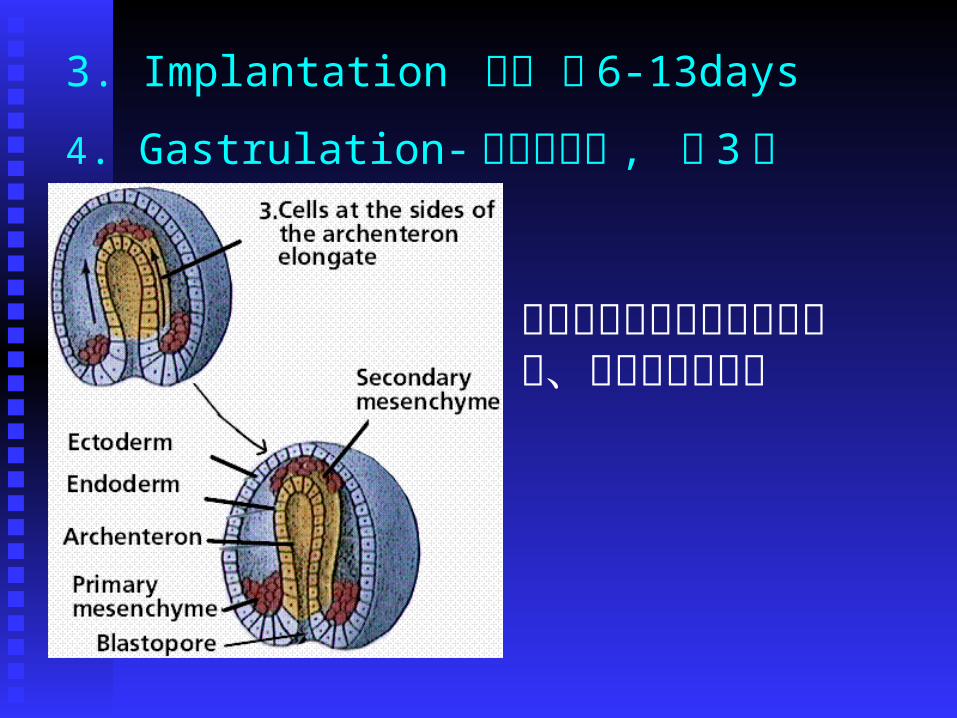
3. Implantation 著床 第 6-13days
4. Gastrulation- 三胚層形成 , 第 3 週
在此期暴露有害物質將造成眼、腦及臉部的畸形

5. Organogenesis 器官形成,第 3-8 週
為最容易受影響的時期,因為本期•Cell proliferation
•Cell migration
•Cell-cell interactions
•Morphogenetic tissue remodeling

6. Fetal period 胎兒期 第 8wk-birth
在此期暴露,影響生長和功能的成熟,需要在出生後仔細觀察才能察覺。如中樞神經的異常包括行為、智力、運動的缺失,生殖力降低,以及免疫系統、心臟、肺臟、腎臟功能受損等。
* 若有構造的改變乃是破壞原本正常的構造稱為deformation ,不同於前述 malformation


Dose-response Patterns and the threshold concept

6 Principles of Teratology1. genetic influences- 由於遺傳差異,引起個體對致畸作用
的敏感度不同2. critical periods- 不同時期對致畸作用的敏感度不同3. initiating mechanism- 致畸原以特定的機轉對細胞組織作
用引發一連串的不正常發育4. access to embryo and fetus- 致畸原的特性決定其與胚胎
接近的難易5. abnormal development- 不正常的發育的結果→ death,
malformation, growth retardation, and functional disorder
6. dose-response relationship-however, 一般致畸原存在 threshold level

Mechanisms and pathologenesis of developmental toxicology1. mutation 突變somatic mutation in the early embryo, ex.mutagen2. chromosomal abnormalities 染色體異常ex. advanced maternal age, viral infection, irradiation, and chemical agents3. mitotic interference 干擾細胞分裂slow or arrest DNA synthesis (hydroxyurea or irradiation), interfere with spindle formation (colchicine, vincristine)4. interference with nucleic acid function 干擾核酸的功能including replication , transcription, translation ex. antibiotics and antineoplastic drugs5. nutritional deficiencies 營養缺乏ex. vitamins , minerals

6. deficient or alter energy supply 缺少或改變能量的供給ex. inadequate glucose supply (hypoglycemia), interference with glycolysis (iodoacetate, 6-aminonicotinamide), inhibition of the citric acid cycle (riboflavin deficiency), blockage of the terminal electron transport (hypoxia, cyanide)7. changes in osmolarity 滲透壓的改變ex. hypoxia, trypan blue, hypertonic solutions, adrenal hormone→edema, hematoma, and blisters8. changes in cell membranes 細胞膜的改變ex. solvent, vitamin A9. enzyme inhibition 酵素的抑制抑制代謝酵素 ,DNA repairing, polymerase

Example of cyclophosphamide (CP)
Single strand DNA break
A teratogenic chemotherapeutic agent Damage to DNA inhibit cell cycle progressioncell cycle arrest too long apoptosis
Bind to protein

CP induces DNA damage(predominant occur in S phase)leading toCell cycle perturbation Cell death
Sensitivity is determined by cell cycle length and cell predisposition to apoptosis


Cell death in the neural tube by CP
Sensitivity to CP-induced cell deathNeuroepithelium >heart Cell cycle length9.5 hr vs 13.4 hr (longer Go/G1)


Advances in the Molecular basis of dysmorphogenesis
1.Using either singly or double gene knockout Retinoic acid receptor family (syndactyly)2. Antisense oligonucleotide Wnt-1, Wnt-3a (mid and hindbrain malformation)3. Reporter transgenes RA activate hoxb-1-lacZ

Pharmacokinetics and metabolism in pregnancy1.Changes in maternal physiology
hepatic metabolism, GI tract, cardiovascular system, excre
tory system, respiratory system
2.Overall decrease in hepatic xenobiotic transformation
3.Roles of placenta in influence embryonic exposure
help to regulate blood flow
-offer a transport barrier-pH gradient, weak acid
rapidly transfer
-metabolize chemicals
2-acetylaminofluorene (proteratogen)
7-hydroxyl metabolites(proximate teratogen)
4.Maternal metabolism of xenobiotics2-methoxyethanol 2-methoxyacetic acid

Maternal factors affecting development
Genetics
high incidence of cleft lip/palate in white mother
Disease-chronic hypertension
diabetes
infection-cytomegalovirus, Taoxoplasma gondii
Hyperthermia-CNS malformation
Nutrition-folate neural tube defect
Stress-noise, restraint
Placenta toxicity -46 toxicants, Cd

placental toxicity
• Metals, Cd, As, Hg, ethanol, cocaine, cigratte, so
dium salicylate
• Maternal injection vs fetal injection of Cd
• Production of metallothionein
• Interaction with Zn

Maternal toxicity-• acetazolamide inhibits carbonic anhydrase
forelimb ectrodactyly
• diflunsial results in anemia
skeleton defects in rabbits
• phenytoin affects folate metabolism and
heart rates
• metallothionein synthesis inducer-urathane,
mercaptopurine, valproic acid
Zn deficiency


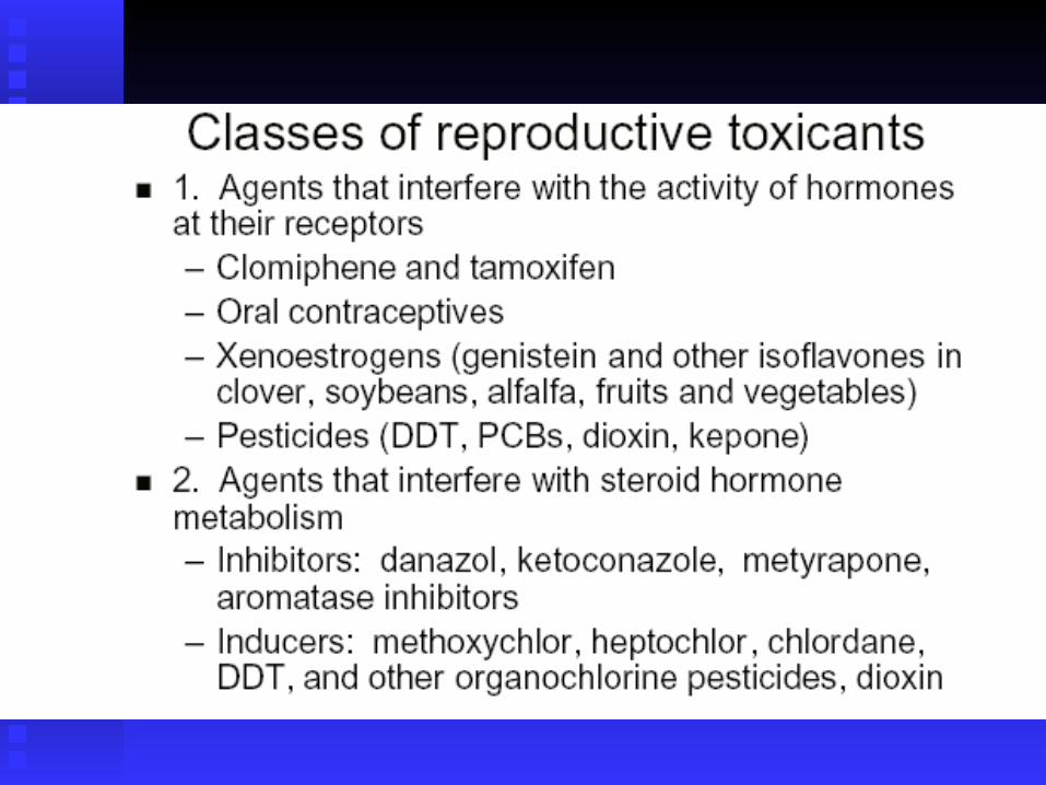
Develpmental toxicity of endocrine-disrupting chemicals
Definition of endocrine-disrupting chemicals
“Exogenous agent that interferes with the productio
n, release, transport, metabolism, binding, action, or
elimination of natural hormones responsible for the
maintenance of homeostasis and the regulation of d
evelopmental processes.”

Endocrine-disrupting chemicals
Four modes of action 1. Serving as steroid receptors ligands2. Modifying steroid hormone metabolizing enzymes3. Perturbing hypothalamic-pituitary release of
trophic hormones4. Uncharacterized proximate modes of action

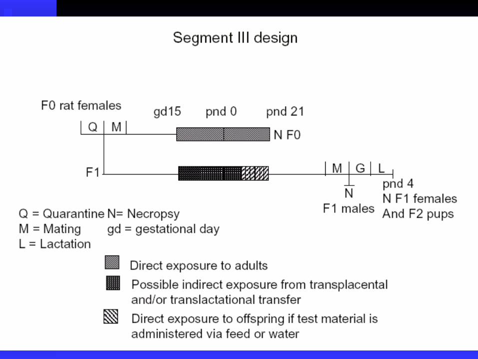
Modern safety assessmentRegulatory guidelines for in vivo testing
Multigeneration tests
Children’s health and the food quality protection act
Alternative testing strategies
Epidemiology
Concordance of data
Elements of risk assessmentuse-in pregnancy rating:A, B, C, D, X


Impact on screening and testing programs
1. Expansion of the periods of dosing from the end of organogenesis to the end of pregnancy in order to include the urogenital differentiation
2. EDSTAC recommended a high through put screening (HTPS) cell-based, receptor-mediated gene transcription assay

Tier I screening battery for EDSIn vitro:Estrogen receptor binding or transcriptional activation
assay Androgen receptor binding or transcriptional activation
assay
Steroidogenesis assay using minced testisIn vivo:Rodent urotrophic assay,
A rodent 20-day pubertal female assay for thyroid functionA male rodent 5-7 day Hershberg assay,A frog metamorphosis assay for thyroid effectsA fish partial life cycle test
T2T: more defined toxicological response would be characterized

Summary of in vivo regulatory protocol guidelines for evaluation of developmental toxicity


Alternative tests for developmental toxicity
•Mouse ovarian tumor•Human embryonic palatal mesenchyme•Micromass culture•Mouse embryonic stem cell test•Chichen embryo neural retina cell culture•Drosophila•Hydro•FETAX (Venopus embryo)•Rodent whole embryo culture•Chernoff/Kavlock assay
Sensitivity(+)/Specificity (-)

Sonic Hedge-hog signal pathway
cyclopaminejervine
Holoprosencephaly
Cholesterol synthesis inhibitor

The signalling molecule
Sonic hedgehog Shh.
Homologue of Drosophila hedgehog gene (involved in compartmentalisation of wing).
Three vertebrate homologues, Sonic hedgehog, Indian hedgehog and Desert hedgehog. Secreted molecules, recognised by transmembrane receptors (e.g. Patched1,2).
Sonic hedgehog expressed first in notochord, then floor plate.Shh misexpression in dorsal neural tube leads to ectopic floor plate and motor neurons.Inhibition of Shh, or gene knockout, leads to absence of floor plate and motor neurons.
Cells ‘read’ the Shh concentration to which they are exposed. Hi [Shh] induces floor plate. 5x lower [Shh] induces motor neurons.

Birth Defect Prevention Measures
Folate supplementationFolate supplementation
Healthy lifestyleHealthy lifestyle
Genetic counseling; diagnostic testingGenetic counseling; diagnostic testing

Consequences of Folate Deficiency
Result of low dietary intake, genetic error of folate Result of low dietary intake, genetic error of folate metabolism, lifestyle exposuresmetabolism, lifestyle exposures
1. DNA Hypomethylation− Gene overexpression, uncontrolled cell growth,
genomic instability
2. Hyperhomocysteinemia− Excessive accumulation of Hcy
3. Base Misincorporation− Decrease in thymine synthesis; replaced by uracil− DNA strands prone to nicks, breaks and vulnerable
to mutagen insertion

How Do I Get Folate?
Take a multivitamin─ 400µg of folic acid
• Eat a healthy diet─ Fruits, green leafy vegetables, beans, corn, peas, bananas, orange juice
Eat fortified cereal and grain products− Total®, Special K®, Product 19®