spontaneous andinduced color-variation of the …differ markedly in color, stability, morphology,...
TRANSCRIPT

SPONTANEOUS AND INDUCED COLOR-VARIATION OF THEHY STRAIN OF SERRATIA MARCESCENS'
EDGAR L. LABRUM2 AND MARY I. BUNTING
Department of Microbiology, Yale University, New Haven, Connecticut
Received for publication September 15, 1952
Since the elucidation of hereditary mechanismsis dependent largely on the observation of differ-ences and similarities between parents and off-spring, those species which exhibit conspicuouscolor-variations are often of great value in theperception and solution of genetic problems. Forthis reason bacteriologists have long recognizedthe usefulness of the fast growing, pigmentedSerratia, and many observations have been madeon spontaneous color changes in this species(reviewed by Bunting, 1946). The same proper-ties make it potentially valuable for the investiga-tion of induced mutations. Furthermore, sincethe organism grows readily on simple syntheticmedia and is capable of breaking down a verywide variety of organic compounds, it offersexcellent material for genetic studies with bio-chemical mutants. Although neither sexual re-combination nor transformation has yet beendemonstrated in Serratia, the organism is a gramnegative rod closely resembling the coliformbacteria morphologically and physiologically andmay well exhibit comparable genetic behavior.It appears, therefore, to be a particularly usefulspecies for the study of many of the geneticproblems which are carried out to advantage withbacteria and which promise to shed light not onlyon microbial phenomena but also on basic geneticmechanisms.
Serratia marcescens is the type species in thegenus Serratia (Breed et al., 1948) and is reportedto be the species used in most genetic studiesmade with Serratia. However, different strainsin the American Type Culture Collection andin various laboratories throughout the countrydiffer markedly in color, stability, morphology,and physiological reactions. At the present time
1 Submitted in partial fulfillment of the re-quirement for a Ph.D. degree at Yale Universityby the senior author.
2 Present address: Carnegie Institute of Wash.ington, Cold Spring Harbor, Long Island, NewYork.
the relationship between these strains, manyisolated from widely different sources, is notclear. It is hoped that further knowledge, aidedby genetic studies showing the range and patternof variation characteristic of individual strains,will provide the information needed to decidewhich could have arisen as closely related variantsand which probably had distant origins. Untilthis is evident it is essential to specify and pre-serve for later comparative studies the strainsused in any experimental series.The HY strain was selected for these studies
because it appeared to be far more stable thanstrain 274 used in previous experiments. Most ofthe data presented in this paper were securedwith the Yale culture which has been maintainedin this laboratory since the early studies ofRettger and Sherrick (1911). All essential find-ings have been confirmed with strain 8195 ob-tained from the American Type Culture Collec-tion in 1951.
Color-variation could not be detected by themicroscopic appearance of individual cells butwas determined by the color of colonies develop-ing from cells spread on the surfaces of agarplates. The cells of the HY strain are small cocco-bacilli which occur singly or in very short chainsso that most colonies represent clones developingfrom single cells and can be used to characterizethe cells.A synthetic ammonium-citrate-glycerol agar
was preferred for plating because it gave goodcolor-differentiation, was reproducible, and wasso highly buffered that the appearance of colonieswas not confused by changes in the pH whichaffect the pigment. It contained per liter of water:ammonium citrate, 5.0 g; glycerol, 10.0 ml; di-potassium phosphate, 10.0 g; magnesium sulfate,0.5 g; ferric ammonium citrate, 0.05 g. Variantswere observed also on a simple peptone (0.5 percent) glycerol (1.0 per cent) agar which intensi-fied pigmentation (Bunting et al., 1949) andpermitted the differentiation of certain paler
394

COLOR-VARIATION OF HY STRAIN OF S. MARCESCENS
types which could not be distinguished on thesynthetic medium.
Part L. Spontaneous color-variation in the HYstrain. It was not the purpose of this investiga-tion to conduct a comprehensive survey of thearray of color-variants which might be derivedfrom cultures of the HY strain but rather tostudy the pattern of variation characteristic ofthe strain under standardized conditions similarto those we anticipated using in future experi-ments with mutagenic agents. Previous workwith strain 274 had indicated (Bunting, 1946)that populations with relatively high proportionsof red cells could be obtained from colonies grownon agar or from broth cultures maintained in thelogarithmic phase. The amount of color-variationexisting in similar HY colonies and broth culturestherefore was investigated.When the HY stock cultures from the Yale
collection and from the National Type CultureCollection were plated on agar, a few pink andwhite colonies were noted in addition to the typi-cal red type. Upon subculture the pink and whitevariants proved to be very stable, but the redcolonies contained an appreciable number ofcolor-variants. A more extensive survey of spon-taneous variation as it occurred in red coloniestherefore was undertaken.Three day old red colonies from synthetic
agar plates were picked to water blanks, andaliquots from appropriate dilutions were spreadover the surfaces of fresh synthetic plates. Thenumbers of daughter colonies of each color typewere counted after the plates had been incubatedfor three days at 26 C. The results of an earlyanalysis of this kind are presented in table 1.Bright red, pink, and white variant colonies werefound on plates from many but not all of thered colonies analyzed. Speckled white-and-redcolonies occurred more frequently and withsurprising regularity. Colonies of this sort hadnot been noted in earler studies (Bunting, 1950)in which the HY strain had been plated exten-sively on a peptone-glycerol-phosphate medium.The color-characteristics and stabilities of thedifferent spontaneous variants were examinedby replating representative colonies of eachtype on synthetic and on peptone-glycerol agar.Typical results are shown in table 2. Syntheticagar plates inoculated with bright red variantscould not be distinguished from plates inoculatedwith the parent HY culture until they had been
incubated for five or six days. On old syntheticplates some of the daughter colonies from thevariants remained bright red instead of fadingto the dull lavender typical of old red typecolonies. Further selection did not stabilize thenonfading bright red variant.When suspensions of cells from the large pink
or from the large white colonies were inoculatedon synthetic plates, the resulting colonies werevery uniform in appearance. Occasional speckledpink-and-white colonies were seen on platesfrom the pink variants, and sometimes coloniesof slightly different shades of pink were observed,but reversions to more highly pigmented formswere very rare. Some of the white variants wereslightly pink when grown on peptone-glycerol
TABLE 1Spontaneous color-variation in three day old redcolonies of the HY strain of Serratia marcescens
PER
NUMBElR OP DAUGHTER COLONI CIM OF EACH COLOR-TYPm ON OR
COLONY AGE O? SYNTIC AGAR PLATES VARL-ANA- COLONY JANTSLYZED
ANTS
Red Bright Pink White SP)eck- Allred led types
days1 3 590 1 0 0 3 0.672 3 4,426 16 22 8 40 1.943 3 1,098 0 0 0 20 1.804 10 925 1 2 0 27 1.115 10 1,670 0 1 1 44 2.726 10 2,384 0 1 2 56 2.42
agar, but these were no less stable than thosewhich were incapable of producing detectablepigment even on the low-phosphate medium.When cells from the variant speckled colonieswere plated on synthetic agar, two types ofdaughter colonies were found in varying pro-portions. There were red colonies which wereindistinguishable in appearance and stabilityfrom the parent red type, and there were colonieswhich came up white but soon developed smallred areas where the unstable colorless variantsevidently had reverted to the pigmented form.No stable white or intermediate pink colonieswere seen. Very different results were obtainedwhen suspensions of cells from speckled colonieswere plated on the peptone-glycerol agar, for onthis medium all of the colonies were a uniformdark red. Quantitative platings then were made
39519531

EDGAR L. LABRUM AND MARY I. BUNTING
on synthetic agar plates and on synthetic agarwith 0.5 peptone. Comparable total counts were
found on the two media, but whereas the ma-jority of colonies were speckled on the syntheticammonium-citrate-glycerol agar, all were uni-formly red when the medium had been supple-mented with peptone. That the alteration inappearance of colonies arising from the colorlessvariants on the two media was not phenotypicbut reflected a shift in the proportions of pig-mented and colorless cells in the colonies was
demonstrated readily by picking colonies andreplating them on synthetic agar. Variants which
pink, and white variants arose far less frequentlythan had been true in colonies recently isolatedfrom stock cultures (table 1), but selectionhad had no effect on the proportion of cells givingspeckled colonies.
Observations were made then on the stabilityof the selected red line when maintained in thelogarithmic growth phase in synthetic and inpeptone-glycerol broth cultures. A modificationof the method used with strain 274 (Bunting,1946) was found to maintain cultures of strainHY in the logarithmic phase. Ten ml of culturemedium in test tubes were inoculated with ap-
TABLE 2Characterization of spontaneous color variants of the HY strain of Serratia marcescens
PER CENT 01 DAUGHE COLONOIE0ACK COLOR TYPECOLOR TYPE PICKED NO. OF COLONIS ON SYNT=TIC AG" APPEARAC 01 COL-FOR ANALYSIS COUYNTEZD ONIES ON NEPTONEC--OX GLYCEROL AGAR
Red Bright red Pink White Speckled
Bright red 827 90.6 8.6 0 0 0.8 dark redBright red 598 82.7 16.6 0 0 0.8 dark redBright red 761 78.4 20.0 0 0 1.6 dark red
Pink 2,354 0 0 99.2 0 0.8 redPink 2,053 0 0 99.5 0 0.5 redPale pink 243 0 0 99.8 0 0.2 dark pink
White ca 4,000 0 0 0 100 0 whiteWhite ca 4,000 0 0 0 100 0 whiteWhite ca 5,000 0 0 0 100 0 pink
Speckled 734 68 0 0 0 32 dark redSpeckled 700 9 0 0 0 91 dark redSpeckled 955 22 0 0 0 88 dark red
* Not evident until plates were 5 to 6 days old.
gave speckled colonies containing high propor-tions of unstable colorless cells on synthetic agargave red colonies with very few such cells whengrown in the presence of peptone, yeast extract,or casein hydrolyzate.
It was of interest from theoretical as well as
practical considerations that the incidence ofmost of the common variant types was reducedmarkedly by selecting red colonies through suc-
cessive platings and reisolations. At three dayintervals suspensions from red colonies were usedto reinoculate fresh synthetic plates; after severalweeks individual red colonies were analyzedwith the results shown in table 3. Bright red,
TABLE 3Spontaneous color-variation in three day old colonies
of a selected red line of HY strain of Serratiamarcescens
NUMBER 0 DAUGHTER COLOONE0ECH COLOR-I= 1TYP ON SYNITHETC AG PLATES
COLONYANALYZED Red Brt Pink White Speked
1 1,191 0 0 0 562 1,208 0 0 0 213 1,623 0 0 0 134 1,200 1 0 0 105 1,021 0 0 0 276 7,789 0 1 0 210
396 [VOL. 65
COLOR-VARIATION OF HY STRAIN OF S. MARCESCENS
proximately 10' cells per ml and incubated at26 C until a slight turbidity indicated approx-imately 106 cels per ml. Then one ml was trans-ferred to a 99 ml water blank from which 1.0 mlwas taken to inoculate the next tube of medium.The turbid cultures were reincubated for lateranalyses of color-variation in aging broth cultures.
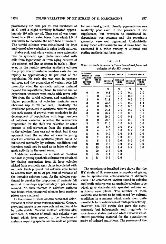
Stable pink and white variants were extremelyrare on synthetic agar plates inoculated withcells from logarithmic or from aging cultures ofthe selected red line as shown in table 4. How-ever, in the rapidly growing synthetic culturesthe proportion of unstable colorless variants roserapidly to approximately 25 per cent of thepopulation. No such rise was seen in peptonecultures, and the proportion of variant cells fellpromptly when the synthetic cultures maturedbeyond the logarithmic phase. In another similarexperiment transfers were made with fewer cells(10) from the turbid cultures, and considerablyhigher proportions of colorless variants wereobtained (up to 70 per cent). Evidently theconditions prevalent in synthetic cultures duringthe early stages of growth were conducive to thedevelopment of populations with large numbersof colorless variants. Whether the mechanismresponsible for the shift was selection or someprocess of cell-conversion from the pigmentedto the colorless form was not evident, but it wasapparent that the number of variants givingspeckled colonies on synthetic plates could beinfluenced markedly by cultural conditions andtherefore could not be used as an index of muta-genic activity in the usual sense.
Additional evidence for a burst of colorlessvariants in young synthetic cultures was obtainedby plating suspensions from 24 hour coloniespicked from synthetic agar plates inoculated withred cells. Such pinpoint red colonies were foundto contain from 10 to 20 per cent of variants ofthe unstable colorless type. As the colonies con-tinued to develop the proportion of variants felluntil at three days approximately 1 per cent re-mained. No such increase in colorless variantswas found when young red colonies from peptoneplates were analyzed.In the course of these studies occasional color-
variants of other types were encountered. Orange,gray, and purple types were isolated and found tobe quite stable. Various mottled colonies alsowere seen. A number of small, pale colonies werefound which later proved to be biochemicalmutants requiring specific amino acids or purines
for continued growth. Usually pigmentation wasrelatively poor even in the presence of thesupplement, but reversion to nutritional in-dependence was common and the revertantsgenerally were well pigmented. Undoubtedlymany other color-variants would have been en-countered if a wider variety of cultural andplating methods had been used.
TABLE 4Color variants in broth cultures inoculated from red
colonies of the HY strainDAYS YAIN- SYNTHETIC BROTH PEPTONE BROTHTAJNYID IN DAYSLOGARITH- PERIT-
GROWTHMATE Speckled Pink and Speckled! Pink andGROWTH AGZ colonies Cooine colonies whitePRASZ colonies ~~~~~~colonies% % % %
0 0 0.8 0.0 0.4 0.01 0 4.2 0.0 0.0 0.02 0 21.8 0.0 0.9 0.03 0 23.8 0.0 0.0 1.44 0 30.0 0.0 0.0 0.45 0 24.6 0.0 0.0 0.06 0 25.7 0.0 0.0 0.17 0 29.2 0.0 0.3 0.08 0 28.8 0.0 0.0 0.08 2 21.0 0.0 0.2 0.08 3 8.8 0.0 0.7 0.08 4 4.6 0.0 0.2 0.08 6 0.9 0.0 0.8 0.08 7 2.7 0.0 0.6 0.08 8 1.2 0.0 0.9 0.08 12 0.3 0.0 0.7 0.08 15 0.7 1.4 0.2 0.08 20 2.6 0.08 25 0.4 0.08 31 0.7 0.0
The experiments described have shown that theHY strain of S. marcescens is capable of givingrise to spontaneous color-variants of differentkinds. The commonest variant found in coloniesand broth cultures was an unstable colorless typewhich gave characteristic speckled colonies onsynthetic agar plates. The number of thesevariants was found to be influenced by culturalconditions in a manner which made them quiteunsuitable for the detection of mutagenic activity.However, the HY strain also gave rise spon-taneously but infrequently and irregularly toconspicuous, stable pink and white variants whichoffered promising material for the quantitativestudy of induced mutations. The presence of the
19531 397

EDGAR L. LABRUM AND MARY I. BUNTING
unstable colorless type could always be maskedby adding peptone to the plating medium. Othervariants were seen but were of rare occurrence.
Part II. The effect of mutagenic agents on color-variation in the HY strain.Experimens with ultraviolet light. Three day
old red colonies of the selected red line werepicked to water blanks to give suspensions con-taining approximately 108 viable cells. One mlof the suspension was diluted and plated to de-termine the number of spontaneous variants inthe populations. Eight ml were pipetted intoa deep petri dish and agitated at a distance of 7inches beneath a Westinghouse sterilamp with95 per cent of its output at 2537 A. The intensity
TABLE 5The effect of increasing doses of ultraviolet light on
color-variation in the HY strain of Serratiamarcescens
PER CENT 07 COLONISILETR- FRACTION COLONIESDOSE SU G COD i White S _k
0 1,385 0 0 0.910 3.0 X 10⁻¹ 555 3.2 0.9 0.715 8.0 X 10-' 1,483 4.7 2.4 1.820 2.5 X 10⁻² 1,310 8.5 3.9 0.525 7.8 X 10⁻⁴ 916 14.8 4.0 0.330 1.3 X 10⁴ 1,142 11.5 8.4 1.840 5.0 X 10-' 1,876 7.8 4.2 0.650 1.8 X 10-' 446 5.4 0.7 0.4
O0 tne ultraviolet at thns distance was approx-
imately 37 ergs/mm-2/sec-1. After exposure thesuspensions were diluted and plated in a room
illlnninated only with yellow light to avoid photo-reactivation. Synthetic agar was used for boththe control and experimental platings in order toobtain information on variations in the numbersof cells giving speckled colonies as well as othervariant types. All of the plates were incubatedin the dark for three days at 26 C before thecolonies were counted and classified as to color.The number of color-variants on the plates
inoculated with irradiated suspensions was very
much higher than on the control plates. Thecolonies varied in color from an intense redthrough different shades of pink to solid white.Since it was sometimes difficult to differentiatethe darker types from the parent red colonies,only the data for the conspicuous pink, white,
and speckled variants are presented in table 5.The proportions of pink and white colonies in-creased with the dose of ultraviolet light to amaximum at 30 seconds, but there was no signifi-cant change in the relative number of cells givingspeckled colonies. It was found later that con-siderable clumping occurred in water suspen-sions shaken under ultraviolet light for more than30 seconds, which of course would interfere withthe effectiveness of further irradiation and alsocould bring about a decrease in the apparentnumbers of variants in the suspension since allcolonies arising from clumps containing a largeproportion of red cells probably would be classi-fied as red and the presence of variants in theclumps would be masked.
Representative colonies of the different varianttypes found on the plates inoculated with ir-radiated cells were picked to water blanks andreplated on synthetic and on peptone-glycerolagar. In general the induced variants provedsimilar in stability and color characteristics tospontaneous variants of the same appearance.However, a dark red type was found which provedto be quite stable and gave colonies which werepigmented more intensely at all ages than thoseof the parent red type. Stable dark red variantshad not been noted on plates from untreatedcells. Orange, purple, gray, and other variantswere seen somewhat more frequently on platesfrom irradiated cells than on those inoculatedwith unirradiated suspensions, but they were farless common than the pink and white types.Irradiation also was observed to increase therelative numbers of biochemical mutants requir-ing nutritional supplements for good growth.In most cases the variants with nutritional de-ficiencies were less stable than those whichshowed only modifications in pigmentation.Although the proportion of color-variants on
plates from irradiated cells was very much higherthan that observed in the controls, the increasewas not sufficient to rule out entirely the pos-sibility that the results had been due to selectivekilling of the red type. In order to check thispossibility the lethal effects of irradiation weretested on various mixtures of stock red celLs andcolor-variants obtained from plates inoculatedwith irradiated celLs. The results of one suchexperiment performed with a mixture of red andstable white cells are presented in table 6. Inthis case irradiation of the mixture resulted ina large increase in the proportion of cells giving
[VOL. 65398_r L'L _ A.l:- : _ _

COLOR-VARIATION OF HY STRAIN OF S. MARCESCENS
red colonies. Evidently, the induced stable whitetype was more sensitive to ultraviolet light thanwas the original red parent. Similar results wereobtained with a mixture of HY red and inducedpink types although in this case the differentialwas not so great. These results indicated that theaction of the ultraviolet light was mutagenicrather than selective.The remarkable stability of the white variants
used in these and in previous experiments(Bunting, 1950) suggested that the cells mighthave lost permanently the ability to producepigment. Because of the theoretical importanceof this possibility considerable effort was spent inattempting to obtain reversions to the pigmentedtype. Irradiations with ultraviolet light followedby extensive platings on synthetic medium wereconsistently negative. By working with artificialmixtures containing one red cell per million whiteit was found that although the presence of redcells seldom was revealed in heavily inoculatedpour or surface-spread plates, it was demon-strated easily on streak plates. Evidently, thered type had some competitive advantage whichwas stifled under the urban conditions of anover-crowded smear or pour plate but which wasexpressed clearly at the margins of heavilypopulated streaks. Red marginal colonies de-veloped regularly on plates streaked frommixtures containing one red cell per millionwhite per ml. In spite of the use of streak plates,however, no pigmented colonies were detectedwhen suspensions of cells of a stable white typewere plated on synthetic medium before or afterirradiation with ultraviolet light.
Eventually, by substituting peptone-glycerolagar for the synthetic medium, it was possible todemonstrate occasional reversions of the stablewhite type to pigmented forms. After irradiationand plating on peptone-glycerol agar, a fewcolonies were found which showed a trace of pink.When these were treated again with ultravioletlight, well-pigmented colonies appeared on bothpeptone-glycerol and synthetic plates. It wasfound also that by plating very old peptonebroth cultures of white variants spontaneousreversions to pigmented forms sometimes wereobtained.The effects of irradiation were observed on
a dark red strain and on a spontaneous pink type.Exposure of the dark red cells resulted in anarray of red, pink, and white variants but again,no change in the proportion of cells giving
speckled colonies. When the suspension of cellsfrom the stable pink strain was irradiated, therewas an increase in both white and red types.The increase in both darker and lighter variantsprovided additional evidence that the action ofthe ultraviolet light was mutagenic rather thanselective.
Experiments then were carried out to observethe effect of photoreactivation on populationsof cells which had been treated with ultravioletlight. By comparing the total numbers and dis-tribution of color-variants on plates made beforeand after photoreactivation the ability of the
TABLE 6The effect of ultraviolet irradiation on red and whitevariants of the HY strain of Serratia marcescens
SUSPENSION ADEFROM:
Red colonies
White colonies
Red and whitecolonies
Ex-POSURE
TOULTRA-VIOLET
Sec
01530
030
030
VIABEZ
PcE ML
6,500,000148,000
240
8,000,000 0830 0
36,000,000 222,400 53
PEL CENT OJCOLONIES
Red White Pink
99.7 0.3 <188 4 779 8 12
100100
7842
00
<15
light to reverse the lethal and the mutationaleffects of the ultraviolet treatment could bedetermined. Suspensions of cells from three dayold red colonies were irradiated in the usualmanner with doses varying from 10 to 50 seconds.They then were divided into two portions; onewas kept in the dark at 37 C for one hour whilethe other was photoreactivated. For photo-reactivation 2 ml aliquots of the irradiatedsuspension were pipetted into M inch glass tubesand placed in a glass-fronted water bath at 37 C.The reactivating light source was a 500 watttungsten filament lamp in a projection lanternwith the bellows fully contracted. A filter of0.03 N aqueous CuCl, in a 3.5 cm deep cell, wasused to absorb a large part of the infrared. Thedistance from the lens to the glass tube contain-ing the suspension was 3 inches, and the periodof illumination was one hour. To avoid unwantedphotoreactivation during plating procedures the
19531 399

EDGAR L. LABRUM AND MARY I. BUNTING
work was done in a laboratory illuminated onlyby yellow light. The treated and untreated cell-ular suspensions were assayed by plating themquantitatively on synthetic agar in the usualmanner. Ten plates were inoculated for eachdilution.
TABLE 7Fraction of cells surviing with and without photo-
reactivation in ultraviolet-irradiated suspen-sions of Serratia marcescens, HY strain
ULTRAVIOLET NO. OF VIABZ TCTION SURVIVINGDOSE CRLLS/MLNONIRRADIATED Dark Light
S6C
10 3.1 X 10⁸ 3.0 X 10⁻¹ 7.1 X 10⁻²⁰ 3.1 X 10⁸ 2.3 X 10-' 1.5 X 1-130 1.7 X 10U 8.2 X 10- 3.5 X 10-240 3.8 X 10⁸ 5.0 X 10-' 5.0 X 10-50 2.7 X 10⁸ 1.8 X 10 G 2.7 X 10-'
TABLE 8Proportion of color variants among survivors of
ultraviolet-irradiated suspensions of Serratiamarcescens, HY strain, with and without
photoreactivation
DAR SURVIVORS LIGH SURVIVORsULTRAVIOLET
DOSE No. colonies Color No. colonies Colorcounted variants counted variants*
SC % %
10 555 4.1 1,310 1.320 1,310 12.4 2,872 4.430 1,142 19.9 592 19.840 1,876 12.0 1,717 18.850 446 6.1 7,480 19.1
* Total per cent pink and white variants.In these experiments no spontaneous pink or
white variants were detected among 1,385 coloniescounted on the nonirradiated control plates.
The survival data from a typical experimentare summarized in table 7. When the fractionsof cells surviving in the dark and in the lightwere plotted against the ultraviolet dose, survivalcurves similar to those described by Kelner (1949)for Escherichia coli, strain B/r, were obtained.From such curves the dose-reduction ratio forthe HY strain S. marcescn was calculated andfound to be 1.6, indicating that visible lightreduced the lethal effects of ultraviolet irradia-tion by 37 per cent. There was a decrease in therate of inactivation for populations treated with
ultraviolet doses higher than 30 seconds due,presumably, to the clumping of the bacteria.The proportions of conspicuous pink and white
color-variants among both the dark and lightsurvivors are presented in table 8. Irradiationproduced the usual increase in color-variantsamong the dark survivors with a decrease inthe apparent numbers of variants after 30seconds. Photoreactivation reduced the numberof color-variants among the light survivors up tothe critical 30 second dose associated with cellclumping. Subsequently, the proportions wereincreased by reactivation, but the increase neverexceeded the maximum frequency detectedamong unphotoreactivated survivors. Experi-ments in which suspensions of the pink and whitevariants types were irradiated and exposed tovisible light demonstrated that they were capableof being photoreactivated to the same extent asthe red parental type.
Considering only the data obtained with thelower doses of ultraviolet light it was evidentthat reactivating light reduced both the lethaland genetic effects of the irradiation. The dose-reduction ratios calculated for the mutageniceffects of ultraviolet irradiation at differentdoses were not constant, however, as they werefor lethality, but rather they decreased withincreasing ultraviolet dose. A difference betweendose-reduction ratios for lethality and muta-genesis has been reported also by Newcombe andWhitehead (1951) in their studies of ultravioletinduced color-response mutants in E. coli, strainB/r, on mannitol-tetrazolium agar.
All of the experimental results obtained withultraviolet light support the hypothesis thatthe stable variants are caused by gene mutationssimlar to those found in higher forms. The uni-formity of pattern of induced variation followingtreatment with ultraviolet light is illustratedby the collection of data presented in table 9showing the results of 30 seconds' exposure in 6different experiments performed on differentdays. No spontaneous pink or white variantswere seen in over 12,000 colonies examined onthe control plates whereas in each assay 20 to 30per cent of the colonies from the irradiatedsuspensions were conspicuously pink or white.In no experiment was there any significant shiftin the proportion of cells giving speckled colonies.Thus, the pattern of variation displayed by redpopulation of the HY strain of S. marcescensfollowing treatment with ultraviolet light was
[VOL. 65i400

15]COLOR-VARIATION OF HY STRAIN OF S. MARCESCENS
strikingly reproducible. Irradiation was capableof inducing a variety of color-mutants includingthe conspicuous pink and white types whichwere especially valuable in following the proesquantitatively. There was no evidence that it hadany effects on the production of variants of theunstable colorless type.The effect of ultraviolet light also was tested
on a few other strains of Serratia. These pre-liminary observations will not be reported indetail, but enough was done to show that dif-ferent responses may be expected from differentstrains. For example, very few stable white orpink types were obtained from strain 274 follow-ing irradiation. The pattern of induced variationproduced with a given strain seemed just ascharacteristic as its pattern of spontaneousvariation.
Experiments with chemical mutagens. The strik-ing effectiveness with which ultraviolet lightincreased the proportions of dark red, pink, andwhite color-variants in treated suspensions ofred cells of the HY strain of S. marcescens en-couraged us to hope that it might be relativelysimple to demonstrate the mutagenic action ofchemical agents with this biological system.Such, however, did not prove to be the case.The first attempts at inducing color-variation
with chemical agents were made with the surface-active compounds sodium desoxycholate andsodium lauryl sulfate. Witkin (1947) and Latarjet(1948) had reported that sodium desoxycholateinduced phage-resistant mutants in E. coli. Lowconcentrations of these agents had been foundto modify the proportions of color-variants inaging cultures of strain 274 of S. marcescens(Bunting, 1942), but this phenomenon had beenshown to be due to selection rather than anymutagenic action of the compounds (Bunting,1950). In later experiments with the HY strainthe surface-active agents were used in the sameconcentrations as those employed by Witkinand Latarjet, but although every effort was madeto follow the procedures they had used no increasewas noted in the relative numbers of color-variants following treatment with the mutagens.The only chemical agent which gave any sug-
gestion of mutagenic activity when tested forits ability to induce color-variation in the HYstrain of S. marcescens was nitrogen mustard.The methyl bis (8-chloroethyl) amino hydro-chloride (Merck) was obtained in vials contain-ing 10 mg of salt. Ten ml of sterile distilled water
were added to a vial giving an acidic aqueoussolution which was relatively stable. Suitabledilutions were made in buffer at pH 6.9, and theagent was allowed to stand only two minutesbefore it was added to the bacterial suspensionswhich were incubated for one hour in a waterbath at 37 C together with a control tube con-taining only bacterial suspension and buffer.Aliquots from the tubes were diluted and platedon synthetic agar in the usual manner.
Preliminary trials established the killing curveand showed that 0.25 mg of nitrogen mustardper ml killed 99.96 per cent of the bacteria andgave a population containing about 2.5 per centof stable pink and white variants. Multiple runswith 0.15 mg HN-2 per ml gave from 1.2 to 2.8per cent stable variants where the untreated
TABLE 9Induced color-variation in suspensions of red HY
cells treated with ultraviolet light
VTIO- PlERVVNR CENT OF COLONISVIOLET FxRACINo
DOSZ Pink White Speckled
30 4.0 X 10O 17.8 13.9 1.530 2.3 X 10'3 15.2 12.2 0.830 3.5 X 10- 17.0 6.5 1.530 1.1 X 10-' 14.5 11.3 1.230 2.1 X 10⁴ 17.0 7.5 0.630 8.6 X 10⁻³ 13.1 7.0 0.20 6 control 0 0 1. 1 a 0.6
platings
controls showed none. There was no evidence ofany shift in the number of cells giving speckledcolonies following treatment with the nitrogenmustard. However, the evidence for mutagenicactivity of the nitrogen mustard was not con-clusive since later experiments in which mixturesof red and variant types were treated showed thatin this case the red cells were appreciably moresensitive to the lethal action of the agents whichcould have accounted for the observed increasein the per cent of variants.Many other chemical agents were employed
with negative results. Three basic dyes (acri-flavine, methyl violet, and methylene blue)were tested for their possible effects on color-variation. Suspensions of Serratia were treatedboth in the dark and in the light. The dyes exerteda much greater killing effect in the light (exposedto 500 watt tungsten filament projection lantern
4011953]
EDGAR L. LABRUM AND MARY I. BUNTING
placed 2 inches from glass-fronted water bath)than in the dark but had no detectable effect onthe proportions of color-variants.
Following Demerec's report (Demerec et al.,1950) of the highly mutagenic properties ofmanganous chloride when used to produce re-versions from streptomycin-dependence to non-dependence in E. coli, this compound was testedalso for its ability to cause color-variation inSerratia. However, even though parallel experi-ments with E. coli, strain B/r/Sd4, were per-formed to make sure that the highly specificconditions essential for mutagenic activity withcoli cultures were fulfilled, and even though ahigh yield of mutants was obtained with coli,no increase in color variants was noted withSerratia.
Negative results were obtained also withurethane (ethyl carbamate) and pyrogallic acidwhich had been reported by Auerbach (1949)to have mutagenic activity. Concentrations from1 mg per ml to 0.1 mg per ml were used, and thecelLs were exposed for 1 hour at 37 C in a waterbath. A few pink variants were noted in platingsfrom two of the urethane tubes, but these werefound to revert to the red parental type whenstreaked on synthetic agar.Although disappointing, it was perhaps not
altogether surprising that the chemicals testedhere failed to induce color-variation in Serratia.As Witkin (1950) demonstrated in the case ofacriflavine and E. coli, a rather delicate relation-ship may exist between survival and the rate ofinduced mutation. Demerec et al. (1950) haveshown also with E. coli that highly specific condi-tions unrelated to survival are essential for thedemonstration of the mutagenic activity ofmanganous chloride. It appears that the condi-tions required for the demonstration of muta-genic activity for any one chemical and any oneorganism must be determined empirically andthat a different set of conditions may be requiredwhen either the chemical or the test organism isvaried. Possibly better yields of variants wouldhave been obtained if resistant strains had beendeveloped as shown by Bryson (1948).
In conclusion it may be stated that althoughit was relatively easy to induce color-variationby treatment with ultraviolet light this has notbeen the case with chemical agents.
DISCUSSION AND CONCLUSIONS
The pattern of distribution of spontaneouscolor-variants in the HY strain of S. marcescenswas found to be characteristic of the strain andstrikingly different from that previously foundwith strain 274 (Bunting, 1940). Strain 274 rarelyproduced stable variants of any color and wasnever observed to give rise to an unstable variantof the colorless type found so abundantly in HYcultures. It produced unstable bright pinkvariants which readily reverted to the dark redtype and occasionally gave rise to paler typeswhich were even more unstable. It might bedifficult to identify red cultures of the twostrains from their general appearance, but theycould be distinguished easily by the variantswhich they produced.The most conspicuous color-variants of the HY
strain were stable pink and white types whicharose infrequently and irregularly as spontaneousmutants but were found in relatively large num-bers following exposure of red cells to ultravioletirradiation. Photoreactivation with white lightreversed the mutagenic action of the ultraviolet.The data on incidence, stability, and behavior ofthese variants suggested that they arose as genemutations and that they offered promisingmaterialfortheexperimental studyof inducedmu-tations. However, all attempts to inducecolor-variation with chemical mutagens wereunsuccessful.
Color-variants also were observed whichshowed biochemical deficiencies. Presumablythese types as well as the more stable proto-trophic forms mentioned above were caused bygene mutations. It would be of interest to testthe effectiveness of chemical mutagens in in-ducing auxotrophs from the HY strain in viewof our inability to induce color variation withthese agents.The commonest color-variant found in HY cul-
tures was an unstable, colorless form whichreverted readily to the pigmented type. All pig-mented cultures were found to contain an ap-preciable number of these variants; the propor-tion varied with cultural conditions. Youngcultures in fresh synthetic medium containedlarge numbers of unstable, colorless cells whereasolder synthetic cultures or cultures grown in thepresence of peptone or yeast extract containedvery few. Colonies which developed from color-
402 [VOL. 65
COLOR-VARIATION OF HY STRAIN OF S. MARCESCENS
less variants inoculated on synthetic agar plateswere white with pigmented specks whereasthose developing from similar cells on agar sup-plemented with peptone or yeast extract soonbecame uniformly colored and were indistinguish-able from colonies arising from the pigmentedform. Ultraviolet irradiation had no detectableeffect on the production of the colorless variants.Every red HY colony analyzed contained
colorless variants, and all of these variants gavecolonies with demonstrable areas where reversionto the pigmented form had taken place. It wasevident, therefore, that both forms were highlyunstable. The data were not sufficient to de-termine whether selection or some other mecha-nism was responsible for the reproducible shiftsin the proportions of the two types found underdifferent cultural conditions. In many respectsthe system resembled that described by Spiegel-man et al. (1950, 1951) for long-term adaptationin yeasts. Pigmented cells of Serratia in theabsence of supplement rapidly lost the abilityto make pigment just as adapted yeast cells inthe absence of galactose lost the ability to fermentthat sugar. Under appropriate conditions someof the progeny from each colorless variant re-gained the ability to make pigment in much thesame manner that the yeasts readapted in thepresence of galactose.Whether the reversible instability of pig-
mented HY cells was due to a high rate of genemutation or to cytoplasmic alterations of amechanism resembling that responsible forchanges in antigenic type in paramecia (Sonne-born, 1948; Beale, 1952) was difficult to de-termine in the absence of a demonstrable sexualcycle. How many other examples of reversibleinstability affecting colonial and other charac-teristics of bacterial populations are of a similarnature remains to be investigated.The general conclusion from this study of
Serratia marcescene is that several distinct kindsof color-variants are produced, possibly as a resultof different kinds of cellular events. Knowledgeof the characteristics and behavior of specificvariant types is essential to a correct interpreta-tion of the significance of their appearance inexperiments with suspected mutagenic and otheragents. The data indicated that quantitative de-terminations of the numbers of stable pink andwhite color-variants could be used to evaluate
the action of mutagenic agents but that thenumber of unstable, colorless variants was in-fluenced greatly by cultural conditions. Themechanism responsible for the production of theseunstable variants is being investigated.
SUMMARY
Red cultures of the HY strain of Serratiamarcescens produced spontaneous color-variantsof several different kinds. Irradiation with ultra-violet light increased the proportion of stablevariant types; photoreactivation reversed themutational as well as lethal effects of the ultra-violet light. No success was attained in usingHY cultures to demonstrate mutagenic activityof chemical agents.An unstable colorless variant which gave
speckled colonies on synthetic media was foundin all cultures. Ultraviolet irradiation had noapparent effect on the production of thesevariants but cultural conditions greatly in-fluenced their numbers.
REFERENCESAuERBACH, C. 1949 Chemical mutagenesis. Biol.
Revs. Cambridge Phil. Soc., 24, 355-391.BEALE, G. H. 1952 Antigen variation in Para-
mecium aurelia, variety 1. Genetics, 37, 62-74.
BREED, R. S., MURRAY, E. G. D., AND HITCHENS,A. P. 1948 Bergey's manual of determinativebacteriology. 6th edition. The Williams &Wilkins Company, Baltimore, Maryland.
BRYSON, V. 1948 The effects of nitrogen mus-tard on Escherichia coli. J. Bact., 56, 423-433.
BUNTING, MARY I. 1940 A description of somecolor variants produced by Serratia mar-cescens strain 274. J. Bact., 40, 57-68.
BUNTING, MARY I. 1942 Factors affecting thedistribution of color variants in aging brothcultures of Serratia marcescens % 271. J.Bact., 43, 593-606.
BUNTING, MARY I. 1946 The inheritance of colorin bacteria with special reference to Serratiamarcescens. Cold Spring Harbor SymposiaQuant. Biol., 11, 25-31.
BUNTING, MARY I. 1950 The effect of surface-active agents on color variation in agingpopulations of Serratia marcescens. J. Bact.,59, 241-250.
BUNTING, MARY I., RoBINow, C. F., AND BUNT-ING, H. 1949 Factors affecting the elabora-tion of pigment and polysaccharide by Ser-ratia marcescens. J. Bact., 58, 114-115.
1953] 403
EDGAR L. LABRUM AND MARY I. BUNTING
DEMEREC, M., FLINT, J., AND DISSOWAY, C. 1950Mutations induced by chemicals. CarnegieInst. Wash. Yearbook No. 49, 145-148.
KELNER, A. 1949 Photoreactivation of ultra-violet-irradiated Escherichia coli with specialreference to the dose-reduction principle andto ultraviolet-induced mutation. J. Bact.,58, 511-522.
LATAwET, R. 1948 Production d'une mutationbacterienne par des substances cancerigenesou non. Comp. rend. soc. biol., 142, 453-455.
NEWCOMBE, H. B., AND WHITEEEAD, H. A. 1951Photoreversal of ultraviolet-induced muta-genic and lethal effects in Escherichia coli.J. Bact., 61, 243-251.
RETTGER, L. F., AND SHE1RCK, J. L. 1911 Stud-ies on bacterial variation. J. Med. Research,24, 265-284.
SONNEBORN, T. M. 1948 The determination of
hereditary differences in genically identicalParamecium cells. Proc. Natl. Acad. Sci.U. S., 34, 413 418.
SPIEGELMAN, S., DELORENZO, W. F., AND CAMP-BELL, A. M. 1951 A single cell analysis ofthetransmission of enzyme-forming capacity inyeast. Proc. Natl. Acad. Sci. U. S., 37, 513.
SPIEGELMAN, S., SUSSMAN, R. R., AND PINSKA, S.1950 On the cytoplasmic nature of "long-term adaptation" in yeasts. Proc. Natl.Acad. Sci. U. S., 36, 591-66.
WrzaN, EVELYN M. 1947 Mutations in Es-cherichia coli induced by chemical agents.Cold Spring Harbor Symposia Quant. Biol.,12, 256-269.
WIThN, EVIELYN M. 1950 The use of sodiumnucleate in the study of the mutagenic ac-
tivity of activity of acriflavine in Escherichiacoli. Proc. Natl. Acad. Sci. U. S., 36,724-731.
404 [vOL. 65