stabilization of alkaline phosphatase with au@ag o...
TRANSCRIPT

Published: August 16, 2011
r 2011 American Chemical Society 11591 dx.doi.org/10.1021/la202405t | Langmuir 2011, 27, 11591–11596
ARTICLE
pubs.acs.org/Langmuir
Stabilization of Alkaline Phosphatase with Au@Ag2O NanoparticlesBrian A. Zaccheo and Richard M. Crooks*
Department of Chemistry and Biochemistry, Texas Materials Institute, and Center for Nano- and Molecular Science and Technology,The University of Texas at Austin, 1 University Station, A5300, Austin, Texas 78712-0165, United States
bS Supporting Information
’ INTRODUCTION
Here, we report that alkaline phosphatase (AlkP) is stabilizedagainst organic inhibitors by core@shell colloidal nanoparticles(NPs). These composites are prepared by combining AlkP andan Au colloid solution with Ag+. This results in agglomeratedparticles having an Ag2O shell (AlkP-Au@Ag2O), which stabi-lizes and protects the enzyme against inhibitors that wouldotherwise greatly reduce its activity. This finding is significant,because many applications are carried out under less-than-optimal conditions for enzyme functionality.
AlkP is a common enzyme that has previously been isolatedand characterized.1,2 The wild type (WT) bovine intestinalisoform is an effective catalyst for the hydrolysis of phosphategroups present on a wide variety of substrates, has an approx-imate molecular weight of 60 kDa, and exhibits optimal activityunder moderately basic conditions (pH 9�10).2 AlkP is oftenintegrated into analytical assays and sensing schemes owing toits ease of handling and high catalytic activity.1,3 To improve thestability of AlkP and other enzymes used for applications likebiosensing, there has been interest in encapsulating themwithinmore rugged host materials. For example, Braun et al. encapsu-lated AlkP in a bulk sol�gel matrix that was subsequentlypulverized and used as column packing material.4 AlkP in thismatrix exhibited a 15% greater tolerance for incubation at 70 �Crelative to free AlkP in solution. Smith et al. examined theactivity of horseradish peroxidase (HRP) in sol�gel monoliths,coatings, and pulverized powders and reported that the powderform of HRP was active and reusable.5 These results are alsoimportant as they show sol�gel protein entrapment is notexclusive to AlkP. Sol�gels have also been used to protect proteinsagainst drying. For example, Li et al. entrapped a fluorescentprotein inside a sol�gel slab and reported that the proteinremained fluorescent after lyophilization and rehydration.6 A lossof the fluorescence signal would indicate changes in the protein
structure. The key point common to all of these examples is thatsol�gel hosts have been shown to stabilize some proteins againstcommon degradative practices including heating, aging, anddrying.
The previously discussed sol�gel materials entrap enzymeswithin a network of pores.4 Alternatively, individual enzymes canbe stabilized by other types of coatings that are directly attachedto the enzyme. For example, Kim et al. modified surface amineson chymotrypsin with alkyl silanols to yield porous silicate gelsaround the enzyme.7 This treatment was shown to extend theactive lifetime of the enzyme in solution. Thiol groups were usedin a similar way to reduce Au3+ onto R-amylase and form∼30 nm NPs that retained amylase activity. Like sol�gels, theAu shell was determined to be porous by screening for amylaseactivity using substrates with different molecular weights. HRPhas been isolated within porous, hollow nanoshells.8 In thisexperiment, AgCl-entrapped HRP colloid was prepared insidereverse micelles. HAuCl4 and a reducing agent were phase-partitioned sequentially into the micelles, and after purificationof the particles, the AgCl was removed with ammonia to leaveactive HRP inside an Au shell. Ben-Knaz and Avnir reported thatacid phosphatase could be stabilized inside a metal powder byincubating the enzyme in solutions of Ag+ or Au3+ and addingZn0 as a reducing agent.9
In the present manuscript, we report the stabilization of AlkPagainst the inhibitory chemicals urea and L-phenylalanine (Phe)using composite NPs. We characterized these AlkP-Au@Ag2ONPs using transmission electron microscopy (TEM), UV�visspectroscopy, X-ray photoelectron spectroscopy (XPS), andenergy dispersive spectroscopy (EDS). The results indicate that
Received: June 26, 2011Revised: August 1, 2011
ABSTRACT: Here, we report that a conductive Au@Ag2O nanoparticle structure significantlyenhances the stability of alkaline phosphatase (AlkP) in the presence of the inhibitors urea andL-phenylalanine (Phe). The enzyme/nanoparticle construct is prepared by associating the enzymewith citrate-capped Au particles, and then adding Ag+. UV�vis and XPS spectroscopy andtransmission electron microscopy confirm the core@shell structure. AlkP activity was quantified inthe presence and absence of the two inhibitors using a time-resolved colorimetric assay. The resultsindicate that 21% of the initial active AlkP is incorporated into the nanoparticle structure. Moreimportantly, however, the Au@Ag2O core@shell host reduces the inhibitory effect of urea and Phe byfactors ranging from 3 to 12, depending on the inhibitor and its concentration, compared to the wild-type enzyme.

11592 dx.doi.org/10.1021/la202405t |Langmuir 2011, 27, 11591–11596
Langmuir ARTICLE
AlkP strongly associates with Au colloids and that the addition ofAg+ to the enzyme-NP conjugate yields a Ag2O shell. AlkPactivity was quantified using a time-resolved colorimetric assay,and the data indicate that ∼21% of the active enzyme initiallypresent is associated with the AlkP-Au@Ag2O composite. Final-ly, the activity of free AlkP was compared to that of AlkP-Au@Ag2O in the presence of two inhibitors. Under otherwiseidentical conditions, AlkP-Au@Ag2O was at least three timesmore active, which demonstrates the stabilizing effect of theAg2O shell. Two key advantages of the method described here,compared to previous literature reports, are that this approachuses a one-pot synthesis and the time required to stabilize theenzyme is less than one day.
’EXPERIMENTAL SECTION
Chemicals and Materials. Citrate-capped Au colloids with anominal diameter of 5 nm, Silver Enhancer Solution A (henceforthreferred to as Ag+ solution); p-nitrophenol phosphate disodium salthexahydrate, enzymology grade (pNPP); and L-phenylalanine (Phe),USP, were purchased from Sigma (St. Louis, MO). Calf intestinal AlkPsolution was purchased from New England Biolabs, Inc. (Ipswich, MA).Urea, ACS grade, was purchased from Thermo Fisher Scientific(Waltham, MA). Diethanolamine, 99%, was obtained from Alfa Aesar(Ward Hill, MA). AmiconUltra-4 centrifugal filter sets having a 100 kDamolecular weight cut off (MWCO) were obtained from Millipore(Billerica, MA). Carbon-coated Cu TEM grids, 400 mesh, were pur-chased from Electron Microscopy Sciences (Hatfield, PA). DisposableUV-transmissive cuvettes with a 1.0 cm path length were purchased fromBRAND GmbH (Wertheim, Germany). All chemicals were used asreceived. Aqueous solutions were prepared using 18 MΩ 3 cm Milli-Qwater (Millipore).Synthesis of AlkP and NP Solutions. These solutions were
prepared using a 1.0 M glycine solution adjusted to pH 9.2. Solutionswere prepared in capped glass vials at an ambient temperature of21 ( 2 �C. Four species were characterized in this work, and they areidentified using the following notations: AlkP, Au@Ag2O, AlkP-Au, andAlkP-Au@Ag2O. AlkP solutions of enzyme alone were prepared bycombining 4.0 μL of AlkP with 1.35 mL of the glycine solution, and thenstirring for ∼18 h. Au@Ag2O, which is a NP species having a Au coreand a Ag2O shell (but no enzyme), was prepared by combining 1.0mL ofAu colloid solution and 0.35 mL of glycine, stirring for∼18 h, and thenadding 50.0 μL of Ag+ solution. The mixture was then stirred for anadditional 2.0 h. AlkP-Au, which is an enzyme-Au colloid conjugate, wasprepared by combining 1.0 mL of Au colloid solution, 0.35 mL ofglycine, 4.0 μL of AlkP, and then stirring for∼18 h. AlkP-Au@Ag2Owasprepared as described for AlkP-Au, with the additional steps of adding50.0 μL of Ag+ solution followed by stirring for 2.0 h. All species wereisolated by filtration in which the crude solution was transferred tothe reservoir of a MWCO tube and centrifuged for 10.0 min at 3000g.The filtrate was collected, and the pellet was resuspended with 1.0 mLof water and centrifuged again for 10.0 min at 3000g. The elutedliquid, hereafter referred to as the wash, was pooled with the initialfiltrate and retained for analysis. The pellet was resuspended with1.0 mL of water and transferred to a microcentrifuge tube forsubsequent analysis.Characterization. UV�vis spectra of filtered and desalted solu-
tions of Au colloid, Au@Ag2O, AlkP-Au, and AlkP-Au@Ag2O wereobtained using a Hewlett-Packard HP8453 spectrometer (Santa Clara,CA). TEM micrographs of AlkP-Au, Au@Ag2O, and AlkP-Au@Ag2Owere collected on a FEI TECNAI G2 F20 X-TWIN instrument(Hillsboro, OR). Grids were prepared by drop-casting 5.0 μL of eachsolution onto a TEM grid and drying overnight in a vacuum desiccator.
Element maps and scanning/transmission electron microscope (STEM)images of AlkP-Au@Ag2O were captured on a Hitachi S5500 with aBruker Quantax 4010 EDS detector operating at 30 kV in bright-fieldmode (Pleasanton, CA, and Billerica, MA, respectively). TEM grids forSTEM and EDS were prepared as for TEM, but included an extra dryingstepwhere the gridswere lyophilized for 48 h. XPS analysis was carried outusing a Kratos Axis Ultra spectrometer having an Al KR filament(Chestnut Ridge, NY). XPS spectra were collected for Au colloid,Au@Ag2O, AlkP-Au, AlkP-Au@Ag2O, and WT AlkP. Samples of Aucolloid, Au@Ag2O, AlkP-Au, and AlkP-Au@Ag2O were prepared bydrop-casting 20 μL volumes onto sections of Si wafer and drying for24 h in a vacuum desiccator. WT AlkP specimens were prepared bydrop-casting 10 μL of AlkP solution onto Au-coated glass and dryingfor 24 h in a vacuum desiccator. Spectra were collected at a pass energyof 20 eV with a step size of 0.1 eV for individual element scans. Samplecharging was compensated by referencing the observed C 1s C�Cpeak to 284.5 eV.10
AlkP Activity Assay. Time-resolved colorimetric assays were usedto measure the active quantity of AlkP in diethanolamine (DEA) units(U) for WT AlkP, AlkP-Au, Au@Ag2O, and AlkP-Au@Ag2O. One AlkPDEAU is defined as the amount of enzyme required to hydrolyze 1μmolof pNPP per minute. Activity testing was performed for each sample onthe pooled filtrate and wash fraction, and on the resuspended pelletfraction. The activity assay method was adapted from literaturereports.11,12 Assays were carried out in a buffer containing 1.0 M DEAand 1.0 mM MgSO4 adjusted to pH 9.8 with H2SO4. Briefly, 1.7 mL ofbuffer, 20.0 μL of the test solution, and 0.10 mL of 150 mM pNPP wereadded to a cuvette and mixed by repetitive pipetting. The absorbance at405 nm (A405) was recorded at 0.50 min intervals for 6.0 min. A least-squares fit was applied to the linear data to obtain the change in A405 as afunction of time (ΔA405 min�1). The activity in U was then calculatedfrom the ΔA405 min�1 value as described in the literature.11 Between3 and 5 replicates for each analyte were assayed.
Inhibition assays were performed on solutions of WT AlkP, AlkP-Au,and AlkP-Au@Ag2O. For these assays, either urea or Phe was added tothe DEA solution prior to testing. AlkP activity was measured at ureaconcentrations of 2.5 or 5.0 M and Phe concentrations of 10.0 or50.0 mM; the UV�vis blank contained DEA buffer, inhibitor at anappropriate concentration, and pNPP. The inhibitor assays were per-formed using the same procedure as the activity assays, with the addedstep of allowing the test solution and inhibitor-containing DEA solutionto incubate for 30.0 min prior to pNPP addition and UV�vis analysis.Different concentrations of enzyme were necessarily used for the WTAlkP, AlkP-Au, and AlkP-Au@Ag2O assays, and therefore, the resultswere normalized as discussed in the Supporting Information.
’RESULTS AND DISCUSSION
As discussed in detail in the Experimental Section, AlkP-Au@Ag2O colloids were prepared in three steps. First, AlkP wasmixed with citrate-capped Au colloid and glycine solutions.Glycine is compatible with AlkP activity2 without adverselyaffecting the Au colloids.13 Second, a Ag+ solution was addedto this mixture. After incubation, the resulting material waspurified and characterized. Additional information about thesynthesis of the other NP species discussed in this report isprovided in the Experimental Section.
TEM micrographs of Au@Ag2O and AlkP-Au@Ag2O areshown in Figure 1. In Figure 1a, two populations of Au@Ag2Oparticles are evident: discrete particles and small aggregates. Thecore@shell nature of these materials is suggested by the contrastdifference between the dark centers and lighter periphery. Thecontrast difference is more apparent in the higher magnificationimage provided in the inset. In Figure 1b, two populations of

11593 dx.doi.org/10.1021/la202405t |Langmuir 2011, 27, 11591–11596
Langmuir ARTICLE
AlkP-Au@Ag2O are also observed: small spherical particles andlarger agglomerates. The smaller particles resemble the Au@Ag2O NPs in Frame a. The larger clusters were observedconsistently in AlkP-Au@Ag2O preparations, but they werenot present in Au@Ag2O samples. The inset of Figure 1b showsa high magnification image of a large AlkP-Au@Ag2O agglom-erate, and it is apparent that these materials also have a core@-shell structure. TEM analysis of AlkP-Au (SupportingInformation, Figure S1) indicates well-dispersed, unagglomer-ated particles exhibiting uniform contrast. On the basis of themorphology of the large AlkP-Au@Ag2O agglomerates and theirpresence only in the AlkP-Au@Ag2O images, we draw twoconclusions. First, the large AlkP-Au@Ag2O agglomerates havea core@shell structure. Second, the formation of large agglom-erates depends on the presence of AlkP.
UV�vis spectra for desalted, aqueous solutions of Au colloids,Au@Ag2O, AlkP-Au, and AlkP-Au@Ag2O are shown in Figure 2.Each spectrum was normalized to a maximum absorbance of 1.0to facilitate comparison of the spectra. The Au colloid startingmaterial has a peak centered at 523 nm, which corresponds to the
plasmon reported for Au NPs in this size range.14 The solutioncontaining AlkP-Au is similar to that of the Au colloid solutionexcept for a slight shift in the plasmon band to 527 nm.
The spectrum of Au@Ag2O reveals the effect of Ag deposi-tion onto the Au colloids. In this case, a new peak is observed at410 nm, but the Au plasmon previously observed in the spectraof the Au colloid and AlkP-Au solutions is absent. The positionof the new peak arising from Au@Ag2O corresponds to a Agplasmon, which has previously been reported in the literaturefor Ag colloids, Ag2O thin films, and Ag NPs having a Ag2Oshell (Ag@Ag2O).15�17 The position of the peak observed forAu@Ag2O is also similar to the plasmon band reported forbimetallic Au@Ag nanoparticles, in which the Au plasmon isquenched by the presence of the Ag (or Ag2O) shell.18,19
Accordingly, this finding is consistent with the core@shellstructure revealed by the TEM images in Figure 1a.
The spectrum of AlkP-Au@Ag2O is different than the otherthree spectra in that it exhibits two overlapping peaks at 427 and505 nm. The position of the 427 nm peak is consistent withAg2O. However, the presence of the peak at 505 nm suggests thatsome fraction of the Au surface, probably in the larger agglom-erates (Figure 1b), is not covered with Ag2O. This view isconsistent with results reported by Gonzalez et al., who observeda similar spectrum for bimetallic Au@Ag nanoparticles havingonly a partial Ag shell.18 Because protein adhesion to Au NPs iswell documented,20 we speculate that AlkP is responsible forincomplete Ag2O coverage and hence the band at 505 nm.XPS Analysis. XPS spectra collected for Au colloids, Au@
Ag2O, AlkP-Au@Ag2O, and WT AlkP are provided in Figure 3.21
The high-resolution C spectra in Figure 3a exhibit bindingenergies consistent with those reported for C�C, C�N, andC�O bonds.10 For example, the C 1s spectrum for Au@Ag2Oconsists of two peaks having binding energies corresponding toC�C and C�O bonds.10 The presence of a C�O peak can beattributed to the citrate capping agent on the Au colloids. Thespectrum of AlkP-Au@Ag2O exhibits three dominant features.Two of these peaks have energies coincident with the C�C andC�O bonds in the Au@Ag2O spectrum. The binding energy ofthe center peak in the AlkP-Au@Ag2O spectrum is suggestive of
Figure 1. TEMmicrographs of (a) Au@Ag2O and (b) AlkP-Au@Ag2O. Two particle populations are present for each species. The insets show highermagnification images of individual particles and highlight the core@shell morphology.
Figure 2. UV�vis spectra of desalted, aqueous solutions of Au colloids,AlkP-Au, Au@Ag2O, and AlkP-Au@Ag2O. The absorbance has beennormalized to facilitate comparison of the spectra.

11594 dx.doi.org/10.1021/la202405t |Langmuir 2011, 27, 11591–11596
Langmuir ARTICLE
C�N bonding. These three features also appear in WT AlkP.Recalling that the AlkP-Au@Ag2O solution was washed and filteredto remove free enzyme prior to XPS analysis, we conclude that AlkPis entrapped within these Au@Ag2O agglomerates.The high-resolution Ag 3d XPS spectra of Au@Ag2O and
AlkP-Au@Ag2O are provided in Figure 3b. Dashed lines indicatethe reported binding energies for zerovalent Ag (368.3 and374.3 eV).10,22 The 3d5/2 peaks for Au@Ag2O and AlkP-Au@Ag2O appear at 368.1 and 367.7 eV, respectively, whichare somewhat lower than the corresponding literature value forzerovalent Ag.10 However, these binding energies are consistentwith values reported for Ag2O (between 367.5 and 368.2eV).10,15,23 That Ag2O has a more negative binding energy thanzerovalent Ag is attributed to the electronic structure of Ag2Oand its metal-like conductivity.24,25We conclude that Ag2O is theprincipal Ag species present in both Au@Ag2O and AlkP-Au@Ag2O. Ag2O may form via reduction of Ag+ to Ag0 bycitrate,26 which is used to stabilize the Au colloids, followed byreaction with oxygen to yield Ag2O. High-resolution XPS spectrain the Au region are provided in Supporting Information FigureS2 and indicate the presence of zerovalent Au only.
EDS Mapping. We used element mapping to carry outspatially resolved analysis of the large AlkP-Au@Ag2O agglom-erates shown in Figure 1b. Figure 4 is a drift-corrected elementmap, rendered in false color, of a single cluster surrounded byseveral smaller particles. The inset is an STEM image of the EDSfield of view. The map reveals two interesting features. First, theelemental distribution in the large cluster is consistent with thatof a Au@Ag2O core@shell morphology. Second, its Au core ismuch larger than the ∼5 nm colloids, which were the initialsource of Au. This indicates the presence of multiple Au colloidsin the core.Activity Assays and Inhibition. Colorimetric activity assays
were used to quantify the amount of AlkP present during eachstep of the synthesis of AlkP-Au@Ag2O. The DEA assaydescribed in the Experimental Section was selected to determinethe AlkP concentration in activity units, because it reports anabsolute quantity of the functional enzyme. As discussed earlier,the metal-containing solutions were purified to ensure that freeAlkP was removed. This step is important for interpreting theactivity assay, because it ensures that the measured activity arisesonly from AlkP associated with agglomerates. Filtration andwashing steps were used for purification, and they result in threefractions. The initial filtrate consists of unbound AlkP, solvent,glycine, and salts that were introduced during the synthesis. Thewash solution was used to ensure removal of free AlkP from theretained solid material comprises the second fraction. Finally, thesolid remaining after the wash step was taken up in water, and thisrepresents the third fraction. The filtrate and wash fractions werepooled and analyzed for enzyme activity alongside the resus-pended solid for each of the following samples: WT AlkP,Au@Ag2O, AlkP-Au, and AlkP-Au@Ag2O. Information aboutthe analysis of these data is provided in the Experimental Section.The results from the activity assays, in terms of catalytically
active enzyme units (U), are presented in Table 1. For WT AlkP,we find that essentially all enzyme activity arises from the pooledfiltrate and wash, which is consistent with expectations forfiltration of a 60 kDa molecular weight enzyme through a100 kDa MWCO filter. Also consistent with expectations, theenzyme-free Au@Ag2O particles registered effectively zeroactivity.The effect of NPs on the activity of the enzyme is revealed by
the results of the AlkP-Au assay. Here, there is a decrease in thetotal enzyme activity compared to free WT AlkP, which is typicalfor enzymes adsorbed to metal surfaces.27 Specifically, the totalenzyme activity for the AlkP-Au material (pellet, filtrate, and
Figure 3. (a) High-resolution C 1s XPS spectra for Au@Ag2O, AlkP-Au@Ag2O, and WT AlkP. The three features correspond to C�C (284.5 eV),C�N (286.0 eV), and C�O (287.5 eV) functional groups. (b) Ag 3d XPS spectra for Au@Ag2O and AlkP-Au@Ag2O. The dashed lines represent the3d5/2 and 3d3/2 reference energies for Ag
0.
Figure 4. EDS element map of a AlkP-Au@Ag2O agglomerate andsmaller, nearby Au@Ag2O particles. The image is rendered in false color:Ag is shown in blue and Au in red. The inset is an STEM image of theEDS field-of-view.
11595 dx.doi.org/10.1021/la202405t |Langmuir 2011, 27, 11591–11596
Langmuir ARTICLE
wash; Table 1) is ∼82% that of the total free WT enzyme.Analysis of AlkP-Au@Ag2O fractions indicates that the majorityof the enzyme is eluted in the filtrate and wash. In fact, somewhatsurprisingly, there is more free enzyme observed with AlkP-Au@Ag2O than with AlkP-Au. This could be due to displace-ment of AlkP from the Au surface during formation of the Ag2Oshell. Finally, we find that 13% of the AlkP activity is retained inAlkP-Au@Ag2O pellet compared to the total freeWT AlkP. Thisis not a high degree of activity, but as we will see in the nextsection, the 13% remaining is quite stable in the presence ofinhibitors.Inhibition Assays. The activities of WT AlkP, AlkP-Au, and
AlkP-Au@Ag2O were quantified in the presence of inhibitors toevaluate the stabilizing effect of the Ag2O shell. The inhibitionassays were performed in the same manner as the activity assaydescribed in the previous section, except for the presence of theinhibitors urea or Phe. Urea is a denaturant that disrupts thesecondary structure of AlkP.1 Phe is an uncompetitive inhibitorthat delays the release of product from AlkP.28,29
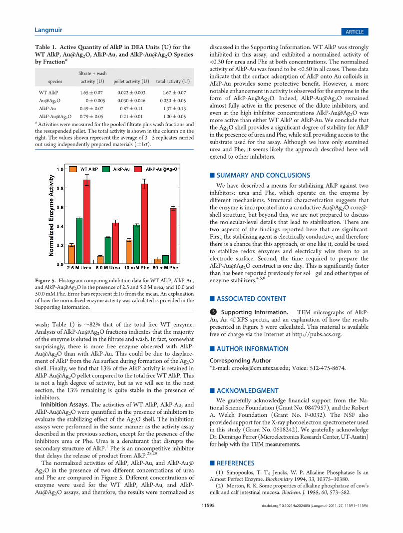
The normalized activities of AlkP, AlkP-Au, and AlkP-Au@Ag2O in the presence of two different concentrations of ureaand Phe are compared in Figure 5. Different concentrations ofenzyme were used for the WT AlkP, AlkP-Au, and AlkP-Au@Ag2O assays, and therefore, the results were normalized as
discussed in the Supporting Information. WT AlkP was stronglyinhibited in this assay, and exhibited a normalized activity of<0.30 for urea and Phe at both concentrations. The normalizedactivity of AlkP-Au was found to be <0.50 in all cases. These dataindicate that the surface adsorption of AlkP onto Au colloids inAlkP-Au provides some protective benefit. However, a morenotable enhancement in activity is observed for the enzyme in theform of AlkP-Au@Ag2O. Indeed, AlkP-Au@Ag2O remainedalmost fully active in the presence of the dilute inhibitors, andeven at the high inhibitor concentrations AlkP-Au@Ag2O wasmore active than either WT AlkP or AlkP-Au. We conclude thatthe Ag2O shell provides a significant degree of stability for AlkPin the presence of urea and Phe, while still providing access to thesubstrate used for the assay. Although we have only examinedurea and Phe, it seems likely the approach described here willextend to other inhibitors.
’SUMMARY AND CONCLUSIONS
We have described a means for stabilizing AlkP against twoinhibitors: urea and Phe, which operate on the enzyme bydifferent mechanisms. Structural characterization suggests thatthe enzyme is incorporated into a conductive Au@Ag2O core@-shell structure, but beyond this, we are not prepared to discussthe molecular-level details that lead to stabilization. There aretwo aspects of the findings reported here that are significant.First, the stabilizing agent is electrically conductive, and thereforethere is a chance that this approach, or one like it, could be usedto stabilize redox enzymes and electrically wire them to anelectrode surface. Second, the time required to prepare theAlkP-Au@Ag2O construct is one day. This is significantly fasterthan has been reported previously for sol�gel and other types ofenzyme stabilizers.4,5,8
’ASSOCIATED CONTENT
bS Supporting Information. TEM micrographs of AlkP-Au, Au 4f XPS spectra, and an explanation of how the resultspresented in Figure 5 were calculated. This material is availablefree of charge via the Internet at http://pubs.acs.org.
’AUTHOR INFORMATION
Corresponding Author*E-mail: [email protected]; Voice: 512-475-8674.
’ACKNOWLEDGMENT
We gratefully acknowledge financial support from the Na-tional Science Foundation (Grant No. 0847957), and the RobertA. Welch Foundation (Grant No. F-0032). The NSF alsoprovided support for the X-ray photoelectron spectrometer usedin this study (Grant No. 0618242). We gratefully acknowledgeDr. Domingo Ferrer (Microelectronics ResearchCenter, UT-Austin)for help with the TEM measurements.
’REFERENCES
(1) Simopoulos, T. T.; Jencks, W. P. Alkaline Phosphatase Is anAlmost Perfect Enzyme. Biochemistry 1994, 33, 10375–10380.
(2) Morton, R. K. Some properties of alkaline phosphatase of cow’smilk and calf intestinal mucosa. Biochem. J. 1955, 60, 573–582.
Table 1. Active Quantity of AlkP in DEA Units (U) for theWT AlkP, Au@Ag2O, AlkP-Au, and AlkP-Au@Ag2O Speciesby Fractiona
species
filtrate + wash
activity (U) pellet activity (U) total activity (U)
WT AlkP 1.65( 0.07 0.022( 0.003 1.67 ( 0.07
Au@Ag2O 0( 0.005 0.030( 0.046 0.030 ( 0.05
AlkP-Au 0.49( 0.07 0.87( 0.11 1.37( 0.13
AlkP-Au@Ag2O 0.79( 0.05 0.21( 0.01 1.00( 0.05aActivities were measured for the pooled filtrate plus wash fractions andthe resuspended pellet. The total activity is shown in the column on theright. The values shown represent the average of 3�5 replicates carriedout using independently prepared materials ((1σ).
Figure 5. Histogram comparing inhibition data for WT AlkP, AlkP-Au,and AlkP-Au@Ag2O in the presence of 2.5 and 5.0 M urea, and 10.0 and50.0 mM Phe. Error bars represent(1σ from the mean. An explanationof how the normalized enzyme activity was calculated is provided in theSupporting Information.

11596 dx.doi.org/10.1021/la202405t |Langmuir 2011, 27, 11591–11596
Langmuir ARTICLE
(3) Zaccheo, B. A.; Crooks, R. M. Detection of an Epstein�BarrGenome Analog at Physiological Concentrations via the Biometallizationof Interdigitated Array Electrodes. Anal. Chem. 2009, 81, 5757–5761.(4) Braun, S.; Rappoport, S.; Zusman, R.; Avnir, D.; Ottolenghi, M.
Biochemically Active Sol-Gel Glasses - the Trapping of Enzymes.Mater.Lett. 1990, 10, 1–5.(5) Smith, K.; Silvernail, N. J.; Rodgers, K. R.; Elgren, T. E.; Castro,
M.; Parker, R. M. Sol�Gel Encapsulated Horseradish Peroxidase: ACatalytic Material for Peroxidation. J. Am. Chem. Soc. 2002, 124,4247–4252.(6) Li, Y. K.; Chou, M. J.; Wu, T. Y.; Jinn, T. R.; Chen-Yang, Y. W. A
Novel method for preparing a protein-encapsulated bioaerogel: Using ared fluorescent protein as a model. Acta Biomater. 2008, 4, 725–732.(7) Kim, J.; Grate, J. W. Single-Enzyme Nanoparticles Armored by a
Nanometer-Scale Organic/Inorganic Network. Nano Lett. 2003,3, 1219–1222.(8) Kumar, R.; Maitra, A. N.; Patanjali, P. K.; Sharma, P. Hollow gold
nanoparticles encapsulating horseradish peroxidase. Biomaterials 2005,26, 6743–6753.(9) Ben-Knaz, R.; Avnir, D. Bioactive enzyme-metal composites:
The entrapment of acid phosphatase within gold and silver. Biomaterials2009, 30, 1263–1267.(10) Moulder, J. F.; Bomben, K. D.; Sobol, P. E.; Stickle, W. F.
Handbook of X-ray Photoelectron Spectroscopy; Physical Electronics USA,Inc: Chigasaki, Japan, 1995.(11) Enzymatic Assay of PHOSPHATASE, ALKALINE. January
20, 2011; http://www.sigmaaldrich.com/etc/medialib/docs/Sigma/Enzyme_Assay/phosphalkeieth.Par.0001.File.dat/phosphalkeieth.pdf.(12) Walter, K.; Schuett, C. inMethods of Enzymatic Analysis, Bergmeyer,
H. U., Ed.; Academic Press, Inc.: New York, 1974; pp 860�864.(13) Hamaguchi, K.; Kawasaki, H.; Arakawa, R. Photochemical
synthesis of glycine-stabilized gold nanoparticles and its heavy-metal-induced aggregation behavior. Colloids Surf., A 2010, 367, 167–173.(14) Perrault, S. D.; Chan, W. C. W. Synthesis and Surface Mod-
ification of Highly Monodispersed, Spherical Gold Nanoparticles of50�200 nm. J. Am. Chem. Soc. 2009, 131, 17042–17043.(15) Chiu, Y.; Rambabu, U.; Hsu, M. H.; Shieh, H. P. D.; Chen,
C. Y.; Lin, H. H. Fabrication and nonlinear optical properties ofnanoparticle silver oxide films. J. Appl. Phys. 2003, 94, 1996–2001.(16) Schinca, D. C.; Scaffardi, L. B.; Videla, F. A.; Torchia, G. A.;
Moreno, P.; Roso, L. Silver-silver oxide core-shell nanoparticles byfemtosecond laser ablation: core and shell sizing by extinction spectros-copy. J. Phys. D. 2009, 42, 215102–215111.(17) Soukupova, J.; Kvitek, L.; Kratochvilova, M.; Panacek, A.;
Prucek, R.; Zboril, R. Silver Voyage from Macro- to Nanoworld.J. Chem. Educ. 2010, 87, 1094–1097.(18) Gonzalez, C. M.; Liu, Y.; Scaiano, J. C. Photochemical Strate-
gies for the Facile Synthesis of Gold�Silver Alloy and Core�ShellBimetallic Nanoparticles. J. Phys. Chem. C 2009, 113, 11861–11867.(19) Wilson, O. M.; Scott, R. W. J.; Garcia-Martinez, J. C.; Crooks,
R. M. Synthesis, Characterization, and Structure-Selective Extraction of1�3-nm Diameter AuAg Dendrimer-Encapsulated Bimetallic Nanopar-ticles. J. Am. Chem. Soc. 2004, 127, 1015–1024.(20) Zhang, D.; Neumann,O.;Wang, H.; Yuwono, V.M.; Barhoumi,
A.; Perham, M.; Hartgerink, J. D.; Wittung-Stafshede, P.; Halas, N. J.Gold Nanoparticles Can Induce the Formation of Protein-based Ag-gregates at Physiological pH. Nano Lett. 2009, 9, 666–671.(21) Heinrich, K. F. J. Electron Beam X-Ray Microanalysis; Van
Nostrand Reinhold Company: New York, 1981.(22) Seah, M. P. Post-1989 calibration energies for X-ray photoelec-
tron spectrometers and the 1990 Josephson constant. Surf. InterfaceAnal. 1989, 14, 488.(23) Hammond, J. S.; Gaarenstroom, S. W.; Winograd, N. X-ray
photoelectron spectroscopic studies of cadmium- and silver-oxygensurfaces. Anal. Chem. 1975, 47, 2193–2199.(24) Deb, A.; Chatterjee, A. K. The electronic structure and chemical
bonding mechanism of silver oxide. J. Phys.: Condens. Matter 1998,10, 11719–11729.
(25) Raju, N. R. C.; et al. Physical properties of silver oxide thin filmsby pulsed laser deposition: effect of oxygen pressure during growth.J. Phys. D 2009, 42, 135411.
(26) Rivas, L.; Sanchez-Cortes, S.; García-Ramos, J. V.; Morcillo, G.Growth of Silver Colloidal Particles Obtained by Citrate Reduction ToIncrease the Raman Enhancement Factor. Langmuir 2001, 17, 574–577.
(27) Suckling, C. J. Immobilized enzymes. Chem. Soc. Rev. 1977, 6,215–233.
(28) Fernley, H. N.; Walker, P. G. Inhibition of alkaline phosphataseby L-phenylalanine. Biochem. J. 1970, 116, 543–544.
(29) Ghosh, N. K.; Fishman, W. H. On the Mechanism of Inhibitionof Intestinal Alkaline Phosphatase by l-Phenylalanine. J. Biol. Chem.1966, 241, 2516–2522.