stable dna formation escherichia coli reca in - pnas heteroduplex dna(3, 4); however, the...
TRANSCRIPT

Proc. Natl. Acad. Sci. USAVol. 87, pp. 21-25, January 1990Biochemistry
Stable DNA heteroduplex formation catalyzed by the Escherichiacoli RecA protein in the absence of ATP hydrolysis
(genetic recombination/DNA strand exchange/homologous DNA pairing/three-stranded DNA intermediate)
JOSEPH P. MENETSKI*t, DAVID G. BEAR*, AND STEPHEN C. KOWALCZYKOWSKI*§*Department of Molecular Biology, Northwestern University Medical School, Chicago, IL 60611; and tDepartment of Cell Biology, University of New MexicoSchool of Medicine, Albuquerque, NM 87131
Communicated by Laszlo Lorand, September 18, 1989 (received for review May 24, 1989)
ABSTRACT A question remaining to be answered aboutRecA protein function concerns the role of ATP hydrolysisduring the DNA-strand-exchange reaction. In this paper wedescribe the formation ofjoint molecules in the absence ofATPhydrolysis, using adenosine 5'-[r-thio]triphosphate (ATP[yS])as nucleotide cofactor. Upon the addition of double-strandedDNA, the ATP[ySJ-RecA protein-single-stranded DNA pre-synaptic complexes can form homologously paired moleculesthat are stable after deproteinization. Formation of these jointmolecules requires both homology and a free homologous end,suggesting that they are plectonemic in nature. This reaction isvery sensitive to magnesium ion concentration, with a maxi-mum rate and extent observed at 4-5 mM magnesium acetate.Under these conditions, the average length of heteroduplexDNA within the joint molecules is 2.4-3.4 kilobase pairs. Thus,RecA protein can form extensive regions of heteroduplex DNAin the presence of ATP[yS], suggesting that homologous pair-ing and the exchange of the DNA molecules can occur withoutATP hydrolysis. A model for the RecA protein-catalyzedDNA-strand-exchange reaction that incorporates these resultsand its relevance to the mechanisms ofeukaryotic recombinasesare presented.
The RecA protein-catalyzed DNA-strand-exchange reactionhas been divided into at least three distinct phases (1, 2). Inthe first phase of this reaction, RecA protein binds tosingle-stranded DNA (ssDNA) in the presence of ATP (pre-synapsis). During the second phase, synapsis, the ssDNAand double-stranded DNA (dsDNA) are brought together byRecA protein. Homologous pairing of the two DNA mole-cules and joint molecule formation occur within the synapticcomplex. The initial heteroduplex joint is extended through-out the DNA molecule in the final phase of the DNA-strand-exchange reaction, termed branch migration. Earlyexperiments suggested that RecA protein must bind andhydrolyze ATP to ADP and inorganic phosphate to formstable heteroduplex DNA (3, 4); however, the mechanisticrequirement for ATP hydrolysis is unknown.Experiments using the nonhydrolyzable ATP analogue,
adenosine 5'-[y-thio]triphosphate (ATP[yS]), were per-formed to examine the need for ATP hydrolysis during theDNA-strand-exchange reaction. It was shown that the firsttwo phases of this reaction can occur using this ATP analogue(4, 5). However, the joint molecules formed in the presenceof ATP['yS] were reported to require bound RecA protein forstability [i.e., they were paranemic in nature (4)]. Sinceplectonemic, as well as paranemic, joint molecules can beformed in the presence of ATP, it was suggested that con-version of paranemic to plectonemic joint molecules requiresATP hydrolysis (4). In related studies, addition ofATP[yS] toan ongoing DNA-strand-exchange reaction was shown to
immediately stop formation of heteroduplex DNA (3). Thedata were interpreted to suggest that ATP hydrolysis was alsorequired for the formation of extensive regions of heterodu-plex DNA (after joint molecules are formed). Thus, homol-ogous pairing and the formation of protein-stabilized parane-mic joint molecules are thought to occur in the absence ofATP hydrolysis, whereas the formation of plectonemic jointmolecules and extensive regions of heteroduplex DNA re-quires hydrolysis of ATP.We now report the catalysis ofDNA strand exchange in the
presence of ATP[yS]. Our data demonstrate that the jointmolecules formed in the presence ofATP[yS] are stable in theabsence of RecA protein and are, therefore, plectonemic innature. The average length of heteroduplex DNA in thesejoint molecules can be as much as 3.4 kilobase pairs (kb) withno detectable hydrolysis of ATP[yS]. The relevance of theseresults to the role of ATP hydrolysis in the mechanism of theDNA strand exchange catalyzed by the Escherichia coliRecA protein and by other recombinases is discussed.
MATERIALS AND METHODSReagents. All chemicals used were reagent grade and
solutions were made in glass-distilled H20. ATP andATP[yS] were purchased from Boehringer Mannheim; bothwere dissolved as concentrated stock solutions at pH 7.5.The concentrations of ATP and ATP[yS] were determinedspectrophotometrically using an extinction coefficient of 1.54X 104 cm-' M-1 at 260 nm.RecA protein was purified from E. coli strain JC12772 (6)
using a preparative protocol (S.C.K., unpublished protocol)based on spermidine acetate precipitation (7), Protein concen-tration was determined using an extinction coefficient of 2.7 x104 cm-' M-1 at 280 nm. Single-stranded DNA binding (SSB)protein was purified from strain RLM727 using a preparativeprotocol provided by Roger McMacken (The Johns HopkinsUniversity). The concentration of SSB protein was deter-mined using an extinction coefficient of 3 x 10 cm-1M-1 at280 nm. S1 nuclease was purchased from Pharmacia.Phage M13 ssDNA and replicative form dsDNA were iso-
lated as described (8). The replicative form was linearized usingEcoRI restriction endonuclease. The concentration of M13ssDNA and dsDNA was determined using extinction coeffi-cients of 8784 and 6500 cm-' M " at 260 nm, respectively.
Nucleotide Hydrolysis. ATP[yS] hydrolysis was determinedusing the method of Lanzetta et al. (9). Hydrolysis ofATP[yS] was examined in a solution containing 1 mM ATP-FyS], 6 ,uM RecA protein, and 10 ,uM M13 ssDNA by using
Abbreviations: ssDNA, single-stranded DNA; dsDNA, double-stranded DNA; ATP[yS], adenosine 5'-[y-thio]triphosphate; SSB,ssDNA binding.tPresent address: Laboratory of Molecular Biology, National Insti-tutes of Health, Bethesda, MD 20892.§To whom reprint requests should be addressed.
21
The publication costs of this article were defrayed in part by page chargepayment. This article must therefore be hereby marked "advertisement"in accordance with 18 U.S.C. §1734 solely to indicate this fact.
22 Biochemistry: Menetski et al.
standard DNA-strand-exchange buffer for as long as 8 hr.The background rate of ATP[yS] hydrolysis under theseconditions, in the absence of RecA protein, is 32 ± 1.9 nMATP[yS] per min (data not shown). No hydrolysis abovebackground was observed in the presence of RecA protein,in either the absence or presence of ssDNA. Thus, the rateof ATP[yS] hydrolysis by RecA protein must be less than 1.9nM/min (or kcat < 0.3 nM per min per 1.M RecA protein).This value is in good agreement with that determined previ-ously [0.7 nM per min per buM RecA protein (10)].DNA-Strand-Exchange Assay. The DNA-strand-exchange
reaction was carried out in a solution of 6 ttM RecA protein,0.9 ,uM SSB protein, 10 ILM M13 ssDNA, 10 ,uM M13 dsDNA,and 1 mM ATP[yS] in standard reaction buffer (25 mM Trisacetate, pH 7.5/4 mM magnesium acetate/1 mM dithiothrei-tol), unless stated otherwise. The procedure used to formactive presynaptic complexes in the presence of ATP[yS] isessentially the same as described (5). The M13 ssDNA waspreincubated with SSB protein for 10 min; RecA protein andATP[yS] were then added and the mixture was incubated foranother 10 min. Finally, the reaction was initiated by theaddition of M13 dsDNA. DNA samples were removed atvarious times, and the reaction was stopped with 1% SDS/50mM EDTA at 37°C for 10 min to ensure deproteinization (4).The agarose gel assay for measuring DNA-strand-ex-
change activity was conducted essentially as described (3).However, DNA samples were subjected to electrophoresis inthe absence of ethidium bromide for 7 hr at 0.88 V/cm on0.8% agarose gels in TAE (40 mM Tris acetate, pH 8.0/2mMEDTA). These gels were later stained with ethidium bromide(1 ,ug/ml) to visualize the DNA bands. Intermediate forma-tion was quantified by scanning a photographic negative witha Zeineh soft laser scanning densitometer.
S1 Nuclease Assay. DNA heteroduplex formation was de-termined by S1 nuclease assay as described (3) with thefollowing changes. Reactions were conducted exactly asdescribed for the DNA-strand-exchange reaction, except thattritiated M13 dsDNA was used. Part of a single-reactiontime-point sample was subjected to electrophoresis and asecond portion was subjected to S1 nuclease digestion.Samples were incubated at 37°C with 1% SDS for 10 min priorto the addition of S1 nuclease to disrupt ATP['ySJ-RecAprotein-DNA complexes (4). After S1 nuclease treatment,10% (wt/vol) trichloroacetic acid was added and acid-insoluble labeled DNA was pelleted by centrifugation in anEppendorf centrifuge for 10 min. The total radioactivity(cpm100) in the supernatant was determined as total cpm of anuntreated sample before centrifugation. Background radio-activity (cpmo) was determined as total cpm of an untreatedsample after centrifugation. The fraction of nuclease -
sensitive DNA was calculated as [(cpmsupet - cpmo)/(cpm1oo - cpmo)]. Since complete heteroduplex DNA forma-tion would render only 50o of the DNA sensitive to nucleasetreatment, the percent DNA heteroduplex was determined asthe fraction of nuclease-sensitive DNA x 200.
Electron Microscopy. The DNA-strand-exchange reactionwas carried out as described above. After agarose gel elec-trophoresis, the bands corresponding to the various interme-diate species were excised and electroeluted from the gel.Subsequent gel electrophoresis of the eluted DNA bandsverified that their mobility was unaltered by this procedure.
All samples were applied to the grid without prior fixation.A 5-,ul drop of sample in 10 mM Tris HCl, pH 7.5/1 mMEDTA was applied to the film side of a freshly glow-discharged 400-mesh grid containing an 8-nm carbon film.The sample was allowed to adsorb for 5 min at ambienttemperature. The grid was then placed in a spermidinesolution and processed as described (11). The grids wererotory shadowed with tungsten in an Edwards 306A vacuumcoater and imaged at 50 kV in a Hitachi H600-3 transmission
electron microscope. The size ofthe DNA heteroduplexjointwas estimated by measuring the length of the looped dsDNAthat terminates in a condensed ssDNA region and by relatingit to the size of full-length M13 dsDNA; only intermediates 1and 2 were analyzed due to the difficulty in unambiguouslyestimating the lengths of the heteroduplex DNA joints forintermediates 3 and 4.
RESULTSDNA-Strand-Exchange Intermediates Are Formed in the
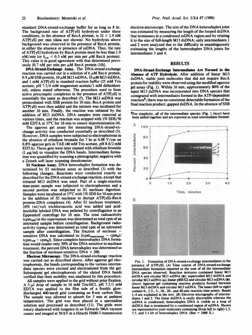
Absence of ATP Hydrolysis. After addition of linear M13dsDNA, stable joint molecules that did not require RecAprotein for stability were observed using the modified agarosegel assay (Fig. 1). Within 10 min, approximately 80%o of theinput M13 dsDNA was incorporated into DNA species thatcomigrated with intermediates formed in the ATP-dependentreactions; there was no consistent detectable formation ofthefinal reaction product, gapped dsDNA. In the absence ofSSB
VFor simplicity, all of the intermediate species (Fig. 1 Inset) havebeen added together and are reported as total intermediate formed.
A
laEa)
Ca)0a)
100
80
60
40
20
00 10 20 3 0 40
Time, minutes
B
'Gus'''~j S./
..
Cd
FIG. 1. Formation of DNA-strand-exchange intermediates in thepresence of ATP[yS]. (A) Time course of DNA-strand-exchangeintermediate formation reported as the sum of all the intermediateDNA species observed. Reaction mixtures contained linear M13dsDNA and circular M13 ssDNA (*); supercoiled M13 dsDNA andcircular M13 ssDNA (A); linear pBR322 and circular M13 ssDNA (U).(Inset) Agarose gel containing reaction products formed betweenlinear M13 dsDNA and circular M13 ssDNA. The lanes (left to right)contain 0-, 2.5-, 5-, 10-, 20-, and 40-min reaction time points. Bands1-4 are explained in the text. (B) Electron micrographs of interme-diates 1 and 2. The linear dsDNA is easily discernible whereas thessDNA is condensed; heteroduplex DNA is visible as a loop ofdsDNA that is terminated by a condensed region of ssDNA. Shownare representative joint molecules containing (from left to right) 1.3,3.5, and 5.1 kb of heteroduplex DNA. (Bar = 1000 A.)
Proc. Natl. Acad. Sci. USA 87 (1990)

Proc. Natl. Acad. Sci. USA 87 (1990) 23
protein, the yield of intermediates was 5-10% of that ob-tained in the presence of SSB protein. The structure of thejoint molecules was established by electron microscopy. Theintermediates labeled 1 and 2 are joint molecules consistingofone linear dsDNA molecule and one ssDNA molecule (Fig.1B), intermediate 3 consists of from two to four dsDNAmolecules and at least one ssDNA molecule (data notshown), and intermediate 4 is a highly entwined structureconsisting of an indeterminate number of DNA molecules(data not shown).Very little intermediate was formed when supercoiled M13
dsDNA was used as the dsDNA substrate (Fig. lA). Super-coiled M13 dsDNA could form only paranemic joint mole-cules with circular M13 ssDNA, suggesting that the DNAspecies formed with linear M13 dsDNA were plectonemic innature. (The small amount of joint molecules formed withsupercoiled dsDNA was probably due to a trace of nickeddsDNA in the DNA preparation.) No intermediate specieswere observed when heterologous linear pBR322 dsDNAwas used as a substrate, showing that formation of interme-diates required DNA sequence homology.
Effect of RecA Protein Concentration on ATP[yS]-Dependent Joint Molecule Formation. The rate of joint mol-ecule formation, in the presence ofATP[yS], was determinedas a function ofRecA protein concentration (Fig. 2). Both theobserved rate and extent of the ATP[yS]-dependent reactionincreased with increasing RecA protein concentration toapproximately 4 AM. This increase was not linear at lowprotein concentrations and appeared to be slightly sigmoidal.Saturation at 4 uM RecA protein yielded an apparent stoi-chiometry of 2.5 ± 0.5 nucleotides of ssDNA per RecAprotein monomer. This stoichiometry is similar to the valueobserved for the ATP-dependent reaction [2-4 nucleotides ofssDNA per RecA protein monomer (12)], showing that theamount of RecA protein required for either reaction issimilar.Formation of DNA-Strand-Exchange Intermediates in the
Presence of ATP[yS] Is Very Sensitive to Magnesium IonConcentration. Honigberg et al. (5) showed that the formationof paranemic joint molecules occurs at a lower magnesiumion concentration (4 mM) than required for branch migration(10 mM). Thus, we were interested in the effect ofmagnesiumion concentration on the formation of plectonemic jointmolecules in the presence ofATP[yS]. As shown in Fig. 3, therate of joint molecule formation was very sensitive to mag-nesium concentration and exhibited a maximum between 4and 5 mM magnesium acetate. The maximum percent ofintermediate species formed under these conditions followedthe same trend (Fig. 3).
gE
0
E
L._
0
140
120
100
80
60
40
20
0 2 4 6 8 10
RecA protein concentration. ,AuM
FIG. 2. Effect of RecA protein concentration on the formation ofintermediates in the presence ofATP[yS]. The relative error for eachpoint is approximately 20o.
E
0._20E0
-0
0
E0CW
30
24
1 8
12
6
0
100
080
10E60 E0c
40 Wa00
20 o
II .. . 0o0 2 4 6 8 10
[Magnesium acetate], mM
FIG. 3. Effect of magnesium acetate concentration on the for-mation of DNA-strand-exchange intermediates in the presence ofATP[yS]. The magnesium acetate concentration in the DNA-strand-exchange reaction is indicated. Results are shown for the rateof intermediate formation (o) and the maximum extent of interme-diate formation (n).
Extensive Heteroduplex DNA Can Be Formed in the Absenceof ATP Hydrolysis. The average length of the heteroduplexDNA joint can be determined by comparing the results fromthe gel assay with those from an assay that determines the totalpercent of heteroduplex DNA formed (i.e., the S1 nucleaseassay). Fig. 4 shows that the increase in amount of interme-diates formed (gel assay) was paralleled by an increase in thetotal percent of DNA heteroduplex formed (S1 nucleaseassay). Comparison of these data gave an average length forthe DNA heteroduplex region of 2.4-3.4 kb per dsDNAmolecule. The results also demonstrate that within 2.5 min theaverage length of a plectonemic joint molecule was at least2400 base pairs. Interestingly, the length of the heteroduplexregion increased from 2.4 kb to 3.4 kb over a 20-min period(Fig. 4). This observation suggests that the size of the nascentplectonemic joint molecule can grow an additional 1 kb in theabsence of detectable ATP[yS] hydrolysis.The average size of the DNA heteroduplex region was also
derived from the structures seen in the electron micrographs(Fig. 1B). The size of the DNA heteroduplex joint in inter-mediates 1 and 2 (formed after 10 min of reaction) rangedfrom 0.3 to 5.1 kb, with the mean size being 2.3 kb. This is
100
80
400
0.
60
40
2c
C0 10 20 30 4C
04000 .0co
003000 0
.0
z2000 ax0
1000 00o
00 0I)
Time, minutes
FIG. 4. Average length of heteroduplex DNA formed in theATP[yS]-dependent reaction. The DNA-strand-exchange reactionand S1 nuclease assay were conducted by using 20 ,uM M13 dsDNA.The percent intermediate as determined by the agarose gel assay (e),percent heteroduplex as determined by the S1 nuclease assay (A),and the average length ofheteroduplex DNA (m) determined from theabove two assays are shown.
Biochemistry: Menetski et al.

24 Biochemistry: Menetski et al.
in good agreement with the S1 nuclease data that show anaverage length of 3.0 kb.Under the experimental conditions used for DNA strand
exchange, ATP[yS] hydrolysis was not observed. The max-imum rate of ATP[yS] hydrolysis by RecA protein underthese conditions was 1.9 nM ATP['yS] per min. The rate ofheteroduplex DNA formation determined from the data inFig. 4 was 0.58 ,uM base pairs per min. Comparing thisapproximate rate of heteroduplex DNA formation with themaximum rate of ATP[yS] hydrolysis gave a maximum of0.003 ATP['yS] molecules hydrolyzed per base pair formed.Thus these results show that extensive regions of heterodu-plex DNA can be formed in the absence of significant ATPhydrolysis.
DISCUSSIONWe examined the ability of RecA protein to promote DNAstrand exchange in the presence of ATP[yS]. Under optimalconditions, presynaptic complexes formed in the presence ofATP[yS] can form joint molecules efficiently. Joint moleculeformation requires homology and a free end, suggesting thatthese intermediates are plectonemic in nature. The formationofjoint molecules in the presence of ATP[yS] is very sensi-tive to magnesium ion concentration and shows a maximumrate and extent at 4-5 mM magnesium acetate. The rate ofjoint molecule formation at 4 mM magnesium acetate, in thepresence of ATP[yS], is similar to the rate observed in thepresence ofATP (at optimal conditions, 8-10mM magnesiumacetate). Surprisingly, the average length of heteroduplexDNA produced by RecA protein in the ATP[IyS]-dependentreaction is 2.4-3.4 kb. Formation of this heterodup!ex DNAoccurs in the absence of observable ATP['yS] hydrolysis.These results may appear to contradict previous studies
that show that very little heteroduplex DNA is formed in thepresence of ATP[yS] (4, 12). However, the reactions weretypically carried out in buffer containing 8-12 mM magne-sium ion. This magnesium ion concentration greatly inhibitsthe ATP[yS]-dependent formation of heteroduplex DNA(Fig. 3). Therefore, our results actually support those ob-tained in previous studies. The reason for this optimum inmagnesium acetate concentration is unclear. Low magne-sium ion concentrations are probably insufficient to stimulatethe formation of an essential RecA protein-dependent struc-ture (e.g., an active presynaptic filament); the requirementfor at least 2-4 mM magnesium ion is also observed in the
..4Z.~.Z.~Z 4 /-A fnl A ) h
+
crC
CL
ATP-dependent DNA-strand-exchange reaction (12, 13). Thedecrease in ATP[yS]-dependent activity above 5 mM mag-nesium ion is not observed in the ATP-dependent reactionuntil the magnesium ion concentration exceeds 15 mM (12);the reason for this inhibition is also unknown but may berelated to extensive aggregation of RecA protein resulting innonproductive structures (7).The formation of extensive heteroduplex DNA in the
absence of ATP hydrolysis is important with regard to thekinetic mechanism of the RecA protein-catalyzed DNA-strand-exchange reaction. DNA strand exchange proceedsthrough, at least, three phases (1, 2), presynapsis, synapsis(conjunction and joint molecule formation), and branch mi-gration. In the presence ofATP, joint molecules form rapidly(<5 min) and contain about 300-500 base pairs of heterodu-plex DNA (3); extension of these joint molecules throughbranch migration then proceeds slowly and can take as longas 20 min. Here we show that RecA protein can form as muchas 2.4 kb of heteroduplex DNA in less than 2.5 min in theabsence of ATP hydrolysis (Fig. 4). Thus, either 2.4 kb ofheteroduplex DNA can be formed during the synaptic phaseof this reaction or branch migration to 2.4 kb can occurrapidly and in the absence of nucleotide hydrolysis.An important conclusion is that the pathway for RecA
protein-catalyzed DNA heteroduplex formation can proceedby a mechanism that does not require ATP hydrolysis as anobligatory step [e.g., the rotation model proposed (14)]. Themechanism by which we envision DNA heteroduplex forma-tion occurring in the presence of ATP[yS] is depicted in Fig.5. In Fig. 5 Left, homologous pairing is shown to occur alongthe length of the two DNA molecules. This is consistent withelectron microscopic studies that show the dsDNA moleculeis completely taken up into the presynaptic filament before adisplaced tail is observed (16, 17). Enzymatic studies alsosuggest that homologous pairing occurs over the entire lengthofDNA homology within 2-5 min (18). In addition, our datademonstrate that within several minutes the ATP[yS]-RecAprotein-ssDNA filament is in homologous alignment with thedsDNA molecule over at least 2.4 kb. This suggests thathomologous contacts are formed over large regions of theDNA molecule within several minutes and that the formationof these contacts does not require ATP hydrolysis (only ATPbinding).The formation of this paired complex results in an inter-
mediate complex containing RecA protein and three strandsofDNA that are homologously aligned (Fig. 5 A and B). The
Y
FIG. 5. Molecular model for the role of ATP hydrol-ysis during the DNA-strand-exchange assay. The presyn-aptic filament is formed in the presence of ATP (orATP[IyS]). RecA protein bound to ssDNA is shown as adimer based on evidence described elsewhere (ref. 15;S. D. Lauder and S.C.K., unpublished results). The
S / rD TA cross-section of each intermediate step is shown on theS/E'T' right. (A) dsDNA is bound to the presynaptic filament. (B)Homologous contacts are formed in a triple-strandedintermediate. The formation of this intermediate does notrequire ATP hydrolysis and can be formed with ATP[yS].(C) Protein dissociation is accomplished by one of twomechanisms: normally, in the presence of ATP, theformation of the low-affinity ADP-RecA protein complexallows the orderly dissociation of RecA from the dis-placed homologous strand; in the presence of ATP[yS],
/ addition of deproteinizing agents removes the RecA pro-/ tein and the complementary strand can anneal with either
homologous strand.
4N
LIA- -/%-- -/amininip ov q
,A,_,A_.
Proc. Natl. Acad. Sci. USA 87 (1990)

Proc. Natl. Acad. Sci. USA 87 (1990) 25
DNA in this three-stranded intermediate must be poised forDNA strand exchange, and we envision this intermediate asa kinetic transition state. In this protein-DNA transition statecomplex, the conformation of the dsDNA is such that thecomplementary strand is no longer stably paired with itsoriginal homologous strand while the incoming ssDNA ispositioned so as to base pair with its complement. In Fig. SB,this intermediate transition state is depicted as symmetricalwith regard to the three DNA strands, but any degree ofasymmetry could be accommodated. Resolution of this struc-ture requires only protein dissociation (Fig. 5C). This occurseither by deproteinization (i.e., SDS/EDTA treatment) whenATP[yS] is present or by ATP hydrolysis when ATP ispresent. In either case, upon protein dissociation, the freedDNA can reassociate to form either heteroduplex DNA or
parental DNA.What then is the role of ATP hydrolysis during the ATP-
dependent DNA-strand-exchange reaction? The answer maybe simply to induce dissociation of the RecA protein-DNAcomplex through the conversion of ATP to ADP and toprovide directionality by doing so. The product of ATPhydrolysis, ADP, decreases the affinity of RecA protein forDNA and increases the rate of transfer (i.e., dissociation)from one DNA molecule to another (19, 20). This suggeststhat production of an ADP-RecA protein complex allows theDNA to be released from the protein-DNA complex (Fig. 5).This dissociation event is likely to occur at the end of a
cooperative cluster of bound RecA protein (19). It is reason-
able to suggest that this dissociation from the end of a
filament could provide the polarity observed in DNA-strand-exchange reaction. This proposal differs mechanisti-cally from those proposed previously in which the polarity ofthe DNA-strand-exchange reaction derives from a polarassociation of RecA protein with DNA. These results may
suggest that the essential role of ATP hydrolysis in postsyn-aptic steps is solely to generate the release factor, ADP.ATP hydrolysis may also serve a function at the presynaptic
step; it may permit RecA protein to cycle on and off thessDNA molecule to produce a continuous presynaptic fila-ment. The presynaptic filaments formed in the presence ofATP[yS] appear to have kinks and acute bends as visualizedby electron microscopy (5). It is likely that any flaw in theformation ofthe ATP[yS]-dependent presynaptic complex willbe maintained during the time course of the DNA-strand-exchange reaction, due to the stability of the ATP~ySJ-RecA protein-ssDNA complex (19). It is possible that thesediscontinuities in the presynaptic filament impede completeextension of heteroduplex DNA joints. In this regard, we feelthat a function of SSB protein in the ATP[yS]-dependentreaction is to permit formation of a contiguous RecA proteinfilament by limiting nucleation sites (5); in the absence of SSBprotein, the binding of RecA protein-ATP[yS] complex tossDNA should be stochastic and necessarily discontinuousdue to the essentially irreversible binding of this highly stablecomplex. In contrast, in the presence of ATP, hydrolysis ofATP allows RecA protein to dissociate from the ssDNA andto repair such imperfections by reassembly.
Finally, it is possible that the ATP[yS]-dependent reactionoccurs by a different mechanism and is not related to theATP-dependent reaction. This possibility is unlikely becausemany of the characteristics of the ATP[yS]-RecA proteincomplex are very similar (if not identical) to the ATP-RecAprotein complex: (i) from electron microscopy, the structureof the ATP[yS]-RecA protein-ssDNA complex is morpho-logically very similar to the ATP-RecA protein-ssDNAcomplex (21); (ii) the high-affinity ssDNA binding state ofRecA protein possesses similar properties in the presence ofeither ATP or ATP[yS] (19, 22); and (iii) the intermediatespecies produced in the ATP[yS]-dependent and ATP-depen-dent reaction have identical structures as determined by
electron microscopy (unpublished data). Since there are greatsimilarities between the ATP-RecA protein and ATP[yS]-RecA protein complexes, we believe that the ATP[IyS]-dependent DNA-strand-exchange reaction follows the samemechanistic pathway to joint molecule formation as theATP-dependent reaction, although the rate-limiting step mayhave changed.These data are interesting in light of reports on DNA-
strand-transfer activities isolated from organisms other thanE. coli. The proteins purified from human cells (23) and fromSaccharomyces cerevisiae (24, 25) have no nucleoside tri-phosphate requirement. The human protein promotes forma-tion of approximately 200 base pairs of heteroduplex DNA(23) whereas the yeast protein forms approximately 4.1 kb(24). The latter is similar to the amount formed by RecAprotein in the presence of ATP[yS]. Thus the apparentparadox that eukaryotic proteins can promote DNA hetero-duplex formation in the absence of a nucleoside triphosphatecan be reconciled by suggesting that the eukaryotic proteinsare functionally equivalent to the ATP-bound form of RecAprotein. It is possible that in higher organisms, DNA-strand-transfer (ATP not required) and catalytic-turnover(ATP required) activities are physically separated into dif-ferent polypeptides. Thus, for the human and yeast recom-bination systems, other protein factors (presumably energyrequiring) may be necessary to promote catalytic turnover(dissociation) of the transferase activity, thereby permittingelongation of the DNA heteroduplex regions formed initially.It will be interesting to see if such activities can be isolatedin the future.
We thank Drs. Dan Camerini-Otero and Peggy Hsieh for theirmany interesting and helpful discussions about these results andLinda Roman, Polly Lavery, Angela Eggelston, Bill Rehrauer, andDan Dixon for their critical reading of this manuscript. This researchwas funded by grants from the National Institutes of Health toS.C.K. (AI-18987) and D.G.B. (RR-05583).
1. Gonda, D. K. & Radding, C. M. (1983) Cell 34, 647-654.2. Kowalczykowski, S. C. (1987) Trends Biochem. Sci. 12, 141-145.3. Cox, M. M. & Lehman, I. R. (1981) Proc. Nad. Acad. Sci. USA 78,
3433-3437.4. Riddles, P. W. & Lehman, I. R. (1985) J. Biol. Chem. 260, 170-173.5. Honigberg, S. M., Gonda, D. K., Flory, J. & Radding, C. M. (1985) J.
Biol. Chem. 260, 11845-11851.6. Uhlin, B. E. & Clark, A. J. (1981) J. Bacteriol. 148, 386-390.7. Griffith, J. & Shores, C. G. (1985) Biochemistry 24, 158-162.8. Messing, J. (1983) Methods Enzymol. 101, 20-78.9. Lanzetta, P. A., Alvarez, L. J., Reinach, P. S. & Candia, 0. A. (1979)
Anal. Biochem. 100, 95-97.10. Weinstock, G. M., McEntee, K. & Lehman, I. R. (1981) J. Biol. Chem.
256, 8850-8855.11. Griffith, J. D. & Formosa, T. (1985) J. Biol. Chem. 260, 4484-4491.12. Cox, M. M. & Lehman, I. R. (1982) J. Biol. Chem. 257, 8523-8532.13. Roman, L. J. & Kowalczykowski, S. C. (1986) Biochemistry 25, 7375-
7385.14. Cox, M. M., Pugh, B. F., Schutte, B. C., Lindsley, J. E., Lee, J. &
Morrical, S. W. (1987) DNA Replication and Recombination, eds. Mc-Macken, R. & Kelly, T. J. (Liss, New York), pp. 595-607.
15. Menetski, J. P. & Kowalczykowski, S. C. (1989) Biochemistry 28, 5871-5881.
16. Stasiak, A., Stasiak, A. Z. & Koller, T. (1984) Cold Spring Harbor Symp.Quant. Biol. 49, 561-570.
17. Register, J. C., III, Christiansen, G. & Griffith, J. (1987) J. Biol. Chem.262, 12812-12820.
18. Schutte, B. C. & Cox, M. M. (1987) Biochemistry 26, 5616-5625.19. Menetski, J. P. & Kowalczykowski, S. C. (1985) J. Mol. Biol. 181,
281-295.20. Menetski, J. P. & Kowalczykowski, S. C. (1987) J. Biol. Chem. 262,
2085-2092.21. Egelman, E. H. & Stasiak, A. (1986) J. Mol. Biol. 191, 677-697.22. Menetski, J. P., Varghese, A. & Kowalczykowski, S. C. (1988) Bio-
chemistry 27, 1205-1212.23. Hsieh, P., Meyn, M. S. & Camerii-Otero, R. D. (1986) Cell 44, 885-894.24. Kolodner, R., Evans, D. H. & Morrison, P. T. (1987) Proc. Natl. Acad.
Sci. USA 84, 5560-5564.25. Sugino, A., Nitiss, J. & Resnick, A. M. (1988) Proc. Natl. Acad. Sci.
USA 85, 3683-3687.
Biochemistry: Menetski et al.