stenotrophomonas maltophilia: a new potential biocontrol
TRANSCRIPT

FULL RESEARCH PAPER
Stenotrophomonas maltophilia: a new potential biocontrolagent of Ralstonia solanacearum, causal agent of potatobrown rot
N. A. S. Messiha Æ A. D. van Diepeningen ÆN. S. Farag Æ S. A. Abdallah Æ J. D. Janse ÆA. H. C. van Bruggen
Received: 20 September 2006 / Accepted: 19 March 2007 / Published online: 15 May 2007
� KNPV 2007
Abstract Stenotrophomonas maltophilia was
isolated from the rhizosphere of eggplant in the Nile
Delta of Egypt, and its antagonistic potential against
Ralstonia solanacearum race 3 biovar 2, the causal
agent of potato brown rot, was in vitro evaluated on
KB agar medium and in vivo on potato plants. In
vitro, four isolates of S. maltophilia (PD3531,
PD3532, PD3533, and PD3534) appeared antagonis-
tic. The isolate (PD3533) was screened as the most
promising antagonist for the in vivo tests. In the
greenhouse, the antagonist was applied directly to
soil or by bacterization of potato eyepieces. Steno-
trophomonas maltophilia significantly suppressed
potato brown rot in Egyptian clay soil but not in
Dutch clay soil. Survival of a rifampicin and
chloramphenicol-resistant S. maltophilia strain
PD4560 was investigated in two pairs of clay soils,
conventionally and organically managed, from Egypt
and the Netherlands. The survival of S. maltophilia
was significantly less in Dutch than in Egyptian soils,
while the converse occurred for R. solanacearum.
These results are in agreement with those obtained in
the in vivo biocontrol tests. In conclusion, S. malto-
philia may be useful for control of brown rot in the
area where it was originally isolated, the Nile Delta in
Egypt.
Keywords Antibiotic resistance � Biological
control � Clay soils � CMC carrier � Organic and
conventional management � Survival
Introduction
Potato brown rot is a systemic bacterial wilt disease
caused by Ralstonia solanacearum (Yabuuchi et al.
N. A. S. Messiha � A. H. C. van Bruggen
Wageningen University, Biological Farming Systems
Group, Marijkeweg 22, 6709 PG Wageningen,
The Netherlands
N. A. S. Messiha � J. D. Janse
Department of Bacteriology, Plant Protection Service,
Geertjesweg 15, 6706 EA Wageningen, The Netherlands
N. A. S. Messiha
Potato Brown Rot Project (PBRP), 3 El-Amira Fatma
Ismail Street, Dokki, Cairo, Egypt
A. D. van Diepeningen
Laboratory of Genetics, Wageningen University,
Arboretumlaan 4, 6703 BD Wageningen, The Netherlands
N. S. Farag
Plant Pathology Research Institute(PPRI),
9 Gamaa Street, Giza, Cairo, Egypt
S. A. Abdallah
Faculty of Science, Department of Botany,
Benha University, Benha, Egypt
J. D. Janse (&)
Department of Bacteriology, Plant protection service
(PD), P.O. Box 9102, 6700 HC Wageningen,
The Netherlands
e-mail: [email protected]
123
Eur J Plant Pathol (2007) 118:211–225
DOI 10.1007/s10658-007-9136-6

1995). Ralstonia solanacearum is subdivided into
five races on the basis of host range and five biovars
on the basis of catabolic properties (Schaad 1988;
Hayward 1991). The potato race 3 biovar 2, more
recently also typed on the basis of genetic sequencing
as Phylotype II, sequevar 1 (Prior and Fegan 2005), is
the dominant race in both Egypt and Europe. Potato
losses up to 75% due to the bacterial wilt have been
recorded in many countries (Cook and Sequeira
1994).
Potato is one of the most important vegetable
crops in Egypt, both for local consumption and for
export. Brown rot was first recorded in Egypt by
Briton-Jones (1925) in the Nile Delta area, based on
symptoms only. The disease became endemic after
the second world war, being serious along the river
Nile from the 1970s onwards. The pathogen has also
been found in irrigation water (Mickail et al. 1974;
Mickail et al. 1985; Farag et al. 1999). Poor drainage
and consequently high soil moisture levels in the
heavy textured soils were associated with the high
disease incidence in this area (Mickail et al. 1985).
As potato brown rot was declared a quarantine
disease of concern in the EU, Egypt moved the main
potato production area to the virgin desert adjacent to
the traditional agricultural lands, because these areas
were still free from R. solanacearum.
Since 1989, potato brown rot was also observed in
several north-western EU countries, where clear links
were established with surface water contamination in
almost all cases. It was first found in the Netherlands
in 1992 in an isolated case and again in 1995, more
widespread due to contaminations via irrigation water.
An intensive programme for eradication was started
and control measures taken according to European
legislation, EC Control Directive 98/57/EC (Janse
1996; Janse et al. 1998; Schans and Steeghs 1998).
Control of potato brown rot has proven to be a
serious, very difficult and puzzling task. Some
resistant cultivars are available but these are not
adapted to different agro-ecological zones and are not
effective against all strains of the pathogen (Hartman
and Elphinstone 1994; Mendoza 1994; Lopez and
Biosca 2004). Breeding for disease tolerance is not
desirable because of a possible correlation between
the earliness of a cultivar and low disease tolerance
(Farag 1970, 1976). Tolerant varieties could harbour
virulent bacteria in a latent form (Priou et al. 1999).
Chemical control by soil fumigants, antibiotics, and
copper compounds was tried without much success
(Farag et al. 1982; Hartman and Elphinstone 1994;
Murakoshi and Takahashi 1984). In addition, most
chemical pesticides have hazardous effects on the
environment, non-target beneficial organisms and
human health. Therefore, cultural and biological
control of bacterial diseases was tried by many
investigators as an alternative solution with some
promising success (Michel and Mew 1998; Ran et al.
2005a, b; Rhodes and Logan 1987; Xu and Gross
1986a, b).
Ralstonia solanacearum race 3, biovar 2 can
survive in latently infected potato tubers, volunteer
potatoes, tomatoes and weed hosts (such as woody
nightshade, Solanum dulcamara), but also in soil for
one to several years (Graham et al. 1979; van Elsas
et al. 2000) and a number of weeks in surface water,
depending on temperature (Janse et al. 1998; van
Elsas et al. 2001). Thus, the most commonly recom-
mended cultural practices to control potato brown rot
are: use long crop rotations with non-hosts, do not use
surface water for irrigation if possible, remove
susceptible weeds, and only use healthy, tested seed
tubers. In addition, fertilization practices can be
adjusted so that the pH is <5 or >8 (Michel and Mew
1998; Sturz et al. 2004), provided that the crop can
tolerate this. Application of certain composted animal
manures or pig slurry can also reduce R. solanacea-
rum populations and bacterial wilt disease of tomato
and potato (Gorissen et al. 2004; Islam and Toyota
2004). Similarly, amendment of organically-managed
soils with cow manure can reduce survival of
R. solanacearum in soil and suppress brown rot
development on potato plants (Messiha et al. unpub-
lished). One of the mechanisms of disease suppres-
sion by manure or slurry may be a shift in microbial
community towards greater antagonism against
R. solanacearum (Gorissen et al. 2004; Messiha
et al. unpublished). Repeated applications of organic
materials like manure or compost can ultimately
result in higher substrate availability for competitors,
reduced growth of the pathogen in the rhizosphere,
and reduced infection (Hoitink and Boehm 1999;
Satoh and Toyota 2004). Thus, soil amendment with
manure or compost may in fact be an indirect way of
stimulating biological control.
Apart from indirect biological control, limited
research has been carried out to find directly appli-
cable biocontrol agents for management of bacterial
212 Eur J Plant Pathol (2007) 118:211–225
123

wilt. Microorganisms which are able to grow in the
rhizosphere are ideal for controlling root-borne
pathogens (Bapat and Shah 2000; Ran et al. 2005b).
Brown rot infection was reduced significantly after
bacterization of healthy seed potatoes with Bacillus
cereus, B. subtilis and with an avirulent strain of
R. solanacearum under field conditions (Sunaina
et al. 1997; Wagih 1991). Pseudomonas fluorescens
was highly effective in reducing bacterial wilt in
tomato and potato under experimental conditions by
aggressively colonizing the roots of young plants and
preventing entry of R. solanacearum (Aspiras and
De-la Cruz 1986). Pseudomonas fluorescens was less
effective at controlling bacterial wilt of Eucalyptus
trees (Ran et al. 2005b). Inhibition of the pathogen by
P. fluorescens was attributed to the siderophore
pseudobactin, depriving root-colonizing microorgan-
isms, including plant pathogens, of Fe3+ and inducing
systemic resistance (Buyer and Leong 1986; Ran
et al. 2005a, b).
Stenotrophomonas maltophilia is a common
microorganism in the rhizosphere of cruciferous
plants, and has also been found in association with
corn and beets (Debette and Blondeau 1980). Excre-
tion of sulphur-containing amino acids such as
methionine by roots of cruciferous plants may favour
the growth of this species. However, S. maltophilia is
also quite dominant in the rhizosphere of cereal crops
(Lambert and Joos 1989). Stenotrophomonas malto-
philia can even colonize and persist inside tissues of
potato plants (Garbeva et al. 2001). However, S.
maltophilia has not been evaluated yet for its
potential to control potato brown rot. However, it
was proven to be an effective biocontrol agent for the
control of various fungal and oomycetous plant
pathogens (Berg et al. 2005; Dal Bello et al. 2002;
Dunne et al. 1997; Nakayama et al. 1999; Zhang and
Yuen 1999). Stenotrophomonas maltophilia inhibited
the growth of Rhizoctonia solani and Verticillium
dahliae in vitro, possibly as a result of antibiosis and
production of lytic enzymes (Berg et al. 1996). Three
antifungal compounds, designated xanthobaccins A,
B and C were isolated from the culture filtrate of a
strain of Stenotrophomonas isolated from sugar beet
that suppressed damping-off of beet seedlings
caused by Pythium spp. (Nakayama et al. 1999).
Xanthobaccins were in vitro not effective against
three bacterial species (R. solanacearum was not
included) (Nakayama et al. 1999). Stenotrophomonas
maltophilia can take up iron (to a limited extent) from
the siderophore pseudobactin (Jurkevitch et al. 1992),
but it is unknown if it produces a siderophore.
This research was undertaken to investigate the
antagonistic potential of S. maltophilia against
R. solanacearum under laboratory and greenhouse
conditions. Survival of the antagonist was tested in
two pairs of clay soils, conventional and organic from
Egyptian and Dutch origins.
Materials and methods
Isolation of antagonists and in vitro inhibition
bioassay
Selection of potential antagonists was made from
rhizosphere soils of eggplant (Solanum melongena
Black Beauty) and sweet pepper (Capsicum annuum)
at the late fruiting stage from two locations of
vegetable growing areas in Kafr El-zayat, Gharbia
governorate, Delta, Egypt. Soil was gently shaken
from the roots, and remaining soil plus roots were
considered as rhizosphere soil. About 1 g of rhizo-
sphere soil was sampled and suspended in sterile
phosphate buffer (PB 0.05 M: Na2HPO4 � 12H2O,
4.26 g; KH2PO4, 2.72 g; 1 l distilled water; pH 7.2).
Samples were shaken for 1 h. Six serial 10-fold
dilutions were made for each sample and spread on
soil extract agar media (SEA: glucose, 1 g; K2HPO4,
0.5 g; yeast extract, 1.5 g; soil extract (1 kg in 1 l
water), 500 ml; agar, 20 g; tap water, 500 ml; pH 7.2)
(Allen 1957). Colonies developed on isolation plates
were washed with sterile water (2 ml/isolate), puri-
fied, and streaked over the middle of the surface of
King’s B agar plates (KB: proteose peptone, 20 g;
K2HPO4, 1.5 g; MgSO4, 1.5 g; agar, 20 g; glycerol,
15 ml; distilled water; 1000 ml). Plates were incu-
bated at 288C for 48 h. A mixture of three previously
identified R. solanacearum strains isolated from
potato tubers in Egypt (marked as PD5239, PD5240
and PD5241 in the culture collection of the Plant
Protection Service, Wageningen, the Netherlands)
was streaked in six lines, perpendicular to the
antagonist streak, and incubated again at 288C for
48 h. The distance free from R. solanacearum, near
the antagonist line was determined. In another trial
both the antagonist and the pathogen were streaked
parallel to each other, 0.5 cm apart, then incubated
Eur J Plant Pathol (2007) 118:211–225 213
123

side by side and the plates were kept under the same
conditions. The absence of R. solanacearum growth
was scored positive for suppression by the antagonist.
In a third trial, ferric ions were incorporated in
King’s B medium in the form of ferric sulphate [Fe2
(SO4)3 � H2O] with a final concentration of 100 mM of
Fe+3. KB medium plates without supplemental iron
served as controls. One hundred ml of a S. maltophilia
suspension (109 CFU ml�1) was spread onto the
plates to determine the effect of iron availability on
the antagonistic potential of S. maltophilia. A similar
suspension of an antagonistic P. fluorescens strain
(PD3340), spread onto iron-amended and non-
amended plates, served as the control.
Identification of the antagonist
Whole cell fatty acid methyl esters (FAME) were
prepared according to standard protocols (Janse 1991;
Stead 1992), and fatty acid analysis (FAA) was
carried out using a gas chromatograph coupled to the
Microbial Identification System with a commercially
available database and software (MIDI, Newark DE
19711, USA). The method is briefly described in the
following paragraph.
Four antagonistic bacterial isolates were grown on
trypticase soy broth agar (TSBA) at 288C for 24 h and
harvested in glass tubes. For saponification, 1 ml of
reagent 1 (NaOH, 45 g; methanol, 150 ml; distilled
water 150 ml) was added to each sample and heated
in a water bath for 30 min. For methylation, 2 ml of
reagent 2 (certified 6.0N HCl, 325 ml; methanol,
275 ml) was added to the cooled tubes and heated for
10 ± 1 min at 80 ± 18C, dropping the pH below 1.5.
FAME were extracted in 1.25 ml of organic solvent
(reagent 3: 200 ml; methyl tetra-butyl ether, 200 ml)
per cooled tube. The aqueous phase was removed by
pipette and the organic phase was washed with 3 ml
of reagent 4 (NaOH, 10.8 g; distilled water 9000 ml)
to reduce contamination of the injection port liner,
the column, and of the detector of the gas chromato-
graph. A standard calibration mixture (MIDI) was
used, consisting of straight chain saturated fatty acids
from 9 to 20 carbons in length (9:0 to 20:0), with five
hydroxy fatty acids. All bacterial strains were iden-
tified based on a comparison of the fatty acid
compositions with those in the database of the MIDI
system (TSBA).
The strains were also physiologically and biochem-
ically characterized according to Bergey’s Manual of
Systematic Bacteriology (Holt et al. 1994) to confirm
the results of FAA. The following tests were carried
out: Gram-staining, motility, oxidative/fermentative
metabolism of glucose (O/F), gelatine hydrolysis,
levan formation from on 5% sucrose agar utilization of
H2S, utilization of nitrate as nitrogen source, acid
production from D-glucose, trehalose and inositol
(1%) in peptone-free basal medium with bromothy-
mol blue (Dowson 1957). The media were inoculated
at a density of 106 bacteria ml�1, and incubated at
288C for 14 days. Acid production from the three
sugars was recorded at 3-day intervals. A colour
change from green to yellow was considered positive.
Suppression of R. solanacearum and brown rot
control
Biological control potential of S. maltophilia against
R. solanacearum was evaluated under greenhouse
conditions for Egyptian and Dutch soils separately.
Conventionally managed clay soil was used in Egypt,
while both conventionally and organically managed
clay soil was used in the Netherlands. Two potato
cultivars were used: medium-early maturing disease-
susceptible cv. Nicola and medium-late maturing
moderately tolerant cv. Diamant. Potato eyepieces
were immersed for 5 min in 0.05% (aq.) NaOCl,
washed 5 times in sterile distilled water, and air dried
for 48 h. Soil was infested with a mixture of three
virulent Egyptian strains of race 3 biovar 2 of R.
solanacearum: PD5239, PD5240 and PD5241
(culture collection, Plant Protection Service, Wagen-
ingen, the Netherlands). The bacterial strains were
grown for 48 h on nutrient agar (NA) plates at 288C.
The bacterial cultures were suspended in 0.01 M
phosphate buffer (PB) and the bacterial density was
adjusted to 5 · 109 CFU ml�1 using a spectropho-
tometer. The inoculum was mixed with the soil to
have a final bacterial concentration of 107 CFU g�1
dry soils.
One strain of S. maltophilia (PD3533) that could
use nitrate as nitrogen source was selected for the
biocontrol tests, as potato brown rot is especially
severe in soils containing high nitrate concentrations
(Messiha et al. unpublished). The antagonist was
applied both by soil inoculation and bacterization of
potato eyepieces. Stenotrophomonas maltophilia was
214 Eur J Plant Pathol (2007) 118:211–225
123
propagated in trypticase soy broth (TSB) and incu-
bated for 18 h (logarithmic phase) at 288C. The
bacteria were harvested by centrifugation, washed
with sterile distilled water, centrifuged again and
re-suspended in water to the required concentration
(Campbell et al. 1986). The inoculation suspension
was adjusted to give 108 CFU g�1 soil (Ciampi-
Panno et al. 1989). For bacterization of eyepieces, the
antagonist was resuspended in sterile 1 % carboxym-
ethyl cellulose (CMC) at 1010 CFU ml�1 (Bapat and
Shah 2000). Eyepieces were immersed in the bacte-
rial suspension in CMC for 15 min and left to dry for
two days before planting. The conditions of the
greenhouse were adjusted to 258C during the day and
208C during the night, with a RH of 75 to 80% and a
total of 14 h light. Three experiments were
conducted.
Experiment 1
Non-sterilized Egyptian conventional clay soil was
used for this experiment. Stenotrophomonas malto-
philia was applied by soil inoculation or by bacter-
ization of the eyepieces of potato cv. Nicola and cv.
Diamant. Soil inoculated with the pathogen only was
added to 48 pots (5 kg each), 24 planted with non-
treated potato eye pieces and 24 planted with eye
pieces treated with S. maltophilia. Soil inoculated
with the pathogen and antagonist was added to 24
pots, and non-inoculated soil to 6 pots as negative
controls. Two eyepieces of either of the potato
cultivars (with or without S. maltophilia) were
planted in each of 12 pots (replications). The 78 pots
were placed on a greenhouse bench in a completely
randomized design. Potted plants were not fertilized
since the soil came from fertilized potato fields.
Plants were carefully irrigated overhead to prevent
splashing. The % infected plants was determined
90 days after planting. Rhizosphere samples were
collected from each pot as mentioned above. Three
ten-fold serial dilutions were made from each sample
and 100 ml was plated onto Selective Media South
Africa (SMSA) (Anon. 1998). Latent infection in the
lower stem area (crown area) of each plant was
checked by plating 5-cm long, weighed sections of
surface-sterilized and macerated plant tissues on
SMSA plates. Maceration by mortar and pestle was
done in phosphate buffer saline (PBS 0.01 M),
(Na2HPO4 � 12H2O, 2.7 g; NaH2PO4 � 2H2O, 0.4 g;
NaCl, 8 g; 1 l distilled water; pH 7.2), and the extract
was left for 1 h. About 1% of all colonies with a
morphology typical for R. solanacearum (colony with
irregular shape, diffuse white or purple centres and
luxuriant slime) were randomly selected and tested
using Immunofluorescence Antibody Staining, IFAS,
(Janse 1988). The number of R. solanacearum CFU
g�1 dry rhizosphere soil was calculated.
Experiment 2
Eyepieces of cv. Diamant were planted in non-
sterilized and sterilized Egyptian conventional clay
soil. The pathogen was mixed into soil as mentioned
before, keeping some of the soil non-inoculated. The
antagonist was applied by eyepiece bacterization
only; half of the eyepieces remained non-treated.
There were 2 · 2 = 4 treatments with 20 replications
(pots of 5 kg) each in a completely randomized
design. All 80 pots contained soil with R. solanacea-
rum; in addition, there were four negative control
pots without the pathogen. No fertilizer was added,
and overhead irrigation was carefully applied. Wilt
development was scored after 60, 75 and 85 days. A
0–5 scoring scale was used: 0 = no symptoms, 1 =
1–2 leaves wilted per plant (about 20%), 2 = 3–4
leaves wilted per plant (about 50%), 3 = most of the
leaves wilted (about 80%), 4 = all leaves wilted, and
5 = plant dead. A wilt index was calculated as the
sum of the [number of plants in each category · cat-
egory number]. Before carrying out v2-tests, plants in
the categories 1, 2 and 3 were combined and the
categories 4 and 5 were combined, so that the
following classes resulted: 0 = no symptoms, 1 = slight
to severe wilt, and 2 = completely wilted or dead.
Visible and latent infections by R. solanaceearum
were tested at the end of the experiment (after
85 days), by plating surface-sterilized and macerated
stem sections on SMSA as mentioned above. The
identity of putative R. solanacearum colonies was
confirmed by IFAS. The experiment was conducted
twice.
Experiment 3
Eyepieces of cv. Nicola were planted in non-steril-
ized Dutch conventional and organic clay soil. The
pathogen and the antagonist were applied as in
experiment 2, resulting again in 4 treatments. Instead
Eur J Plant Pathol (2007) 118:211–225 215
123
of pots, trays were used, containing 5 kg of soil. Eight
eyepieces were planted per tray. There were 4 trays
(replications) per treatment, plus 4 trays with non-
inoculated soil as negative controls. Wilt incidence
and severity were observed daily for 45 days as
described under experiment 2. Latent infection was
tested by making isolations from the crown areas of
each plant onto SMSA plates. The experiment was
carried out twice.
Survival of S. maltophilia in soil and its effect on
survival of R. solanacearum
A spontaneous rifampicin and chloramphenicol-resis-
tant mutant of S. maltophilia strain PD3533 was
selected and adapted in the laboratory for testing its
survival in different soils. The strain was selected
from the wild-type strain by growing it in TSBA
amended with increasing concentrations of rifampicin
and chloramphenicol (0, 20, 40, 60, 80 and
100 mg ml�1 for each antibiotic). The antibiotic-
resistant strain (PD4560) was subjected again to FAA
to confirm its identity as S. maltophilia, and was
tested in vitro for antagonistic potential against R.
solanacearum. The same strain mixture of R. solan-
acearum was used as in the previous experiment.
Two pairs of clay soil, half from organically
managed arable farms and the other half from
conventionally managed arable farms were used in
this study. One pair originated from Egypt, the other
was from the Netherlands (Table 1). The soils were
the same as those used in the greenhouse experi-
ments. The antibiotic resistant S. maltophilia was
grown for 48 h on trypticase soy broth agar (TSBA)
plates at 288C, and R. solanacearum was grown on
NA plates under the same conditions. The bacterial
cultures were suspended in 0.01 M phosphate buffer
(PB) separately. Suspensions of R. solanacearum,
S. maltophilia or both were mixed with 100 g of each
soil in plastic bags so that a final concentration of
5 · 107 CFU g�1 dry soil was attained for each strain.
Inoculated samples were divided over five 50-ml
Greiner tubes with loosely closed lids to allow air
exchange, and incubated at 288C. The moisture
content was 22.6% of the soil fresh weight, water
loss was checked weekly and any lost water was
replaced to keep a constant moisture level. Both the
pathogen and antagonist populations were monitored
in all treatments twice a week for the first two weeks,
starting at the day of inoculation (T0), once a week
for the next two weeks and then once a month for a
total of five months until the pathogen population was
below the detection limit (100 CFU g�1 dry soil) in
all soils.
For bacterial counts, 1-g samples of the soils (1
sample per soil replicate) were suspended in 9 ml of
sterile 0.05 M phosphate buffer. After shaking at
100 rev min�1 for 2 h at 208C, 10-fold serial dilutions
Table 1 Physical, chemical and biological composition of the different soils
Soil
codecCountry Management Location Soil compositiona Nutrients mg kg�1 Diversity
indexb
Clay(%) Silt(%) Sand(%) OM(%) TOCd N-
NO3
N-
NH4
pH-
KCl
S H0
ECC Egypt Conventional Behera, Delta
area
9.4 54.5 36.1 4.9 133.2 128 19.2 7.6 21.3 1.23
ECO Egypt Organic Kaliobia, Delta
area
13.9 65.7 20.4 6.2 079.9 62 16.3 7.6 18.3 1.19
NCC NL Conventional Ens, Northeast
polder
7.7 51.9 40.4 2.2 122.1 10 13.9 7.4 18.8 1.11
NCO NL Organic Ens, Northeast
polder
8.3 54.5 37.2 2.3 125.7 7 15.8 7.3 24.5 1.13
a Particles size: clay <2 mm, silt 2–50 mm, sand 50–2000 mm.b Average species richness S and Shannon-Wiener diversity index H0
c First letter stands for country of origin (Egypt or the Netherlands), Second for soil type (clay) and third for management
(conventional or organic)d Total soluble organic carbon
216 Eur J Plant Pathol (2007) 118:211–225
123
were made, and spread on 3 plates each of TSBA
amended with 100 mg chloramphenicol and 100 mg
rifampicin l�1 for S. maltophilia detection and on
SMSA plates for R. solanacearum detection. Steno-
trophomonas maltophilia colonies were counted after
3 days and R. solanacearum colonies after 5–7 days
of incubation at 288C. IFAS was used to confirm
putative colonies of the pathogen. The experiment
was conducted twice.
Soil analyses
For physical and chemical analysis 100-g samples of
all soils were air-dried at room temperature. Soil
textures were assessed in the Laboratory of Soil
Science and Geology of Wageningen University
(Table 1). Particle sizes of <2 mm were considered
clay, 2–50 mm was considered silt and 50–2,000 mm
was considered sand. Organic matter content was
determined using the loss on ignition method accord-
ing to Houba et al. (1997). Dissolved organic carbon
(DOC), nitrate and ammonium content and pH were
measured after Houba and Novozamsky (1998).
PCR and DGGE analysis
Duplicate soil samples used for PCR and DGGE were
stored at �208C. Total DNA was extracted from 0.5 g
(wet weight) soil samples with the Bio101 Fast-
DNA1 SPIN Kit for Soil, according to the manufac-
turer’s specifications (Bio101, Carlsbad, CA, USA); a
20 min incubation time at 658C was added to enhance
the elution. For DGGE analysis of the eubacterial soil
population, the V6 to V8 region of the 16S rRNA
gene was amplified from total soil DNA with the
primers 968 f-GC and 1401 r (Heuer and Smalla
1997). Two nanogram of DNA was added to 50 ml
PCR reactions and amplified using a touchdown
scheme (Hiddink et al. 2005).
DGGE was performed using the Dcode system
(BIOrad Laboratories, Hercules, CA, USA) according
to Hiddink et al. (2005). All samples were analyzed at
least in duplicate. Gels were stained with BIOrad’s
Silver Stain (BIOrad Laboratories, Hercules, CA,
USA), preserved in Cairn’s preservation solution of
25% ethanol (v v-1) and 10% glycerol (v v-1),
covered by a permeable cellophane sheet (Amersham
Pharmacia Biotech Ag, Uppsala, Sweden) and dried
overnight at 608C. The gels were than scanned using
ScanSoft Omnipage, programme 14 at a resolution of
300 dots inch�1 and analyzed with Phoretix 1D
(NonLinear Dynamics Ltd., Newcastle upon Tyne,
UK), including only bands with pixel intensity >1.
Statistical analyses
To evaluate the disease incidence in the first two
biocontrol experiments, v2-tests were conducted on
contingency tables with classes of number of plants
infected or wilted versus the various treatments to be
compared (soil origin, soil treatment and antagonist
amendment) using Microsoft Excel 2003 (Microsoft
Corporation, Seattle, WA, USA).
For the first experiment, ANOVA was conducted
to compare the density of R. solanacearum in the
rhizosphere for the two different cultivars and S.
maltophilia treatments (control, soil amendment with
the antagonist and bacterization of eyepieces with the
antagonist). Interaction between cultivar and antag-
onist treatment was also determined. Fisher’s LSD
test was used to compare each pair of treatments. A
t-test was conducted to compare the densities of
R. solanacearum in the rhizosphere of the two
different cultivars in the treatment without S. malto-
philia. All analyses were conducted using SPSS v 12
(SPSS Inc., Chicago, Illinois, USA).
For the third experiment, areas under the disease
progress curves (AUDPC) were calculated based on
% leaves wilted per plant over time. Non-parametric
Mann-Whitney tests were carried out on AUDPC and
% stem pieces infected for the two management types
(organic and conventional) and S. maltophilia treat-
ments (inoculated and non-inoculated eyepieces).
Survival curves of the pathogen and the antagonist
were fitted to CFUs over time as follows. Log
(CFU+1/g dry soil) was calculated for each sample.
Log-transformed data (for each replicate, soil and
treatment separately) were fitted to a logistic decay
model as described by Franz et al. (2005):
Ct ¼ am=ð1þ expð�d�ðt � cÞÞÞ
where Ct = log transformed bacterial density, am = ini-
tial density, d = decline rate (days�1), t = time (days)
and c = length of the 50%-reduction-time in days.
The estimated parameter values c and d for the
different soils were subjected to multivariate analysis
of variance (MANOVA, using SPSS v 12, SPSS Inc.,
Eur J Plant Pathol (2007) 118:211–225 217
123
Chicago, Illinois, USA) to determine the effect of soil
origin and management on the survival of S. malto-
philia and R. solanacearum, and to detect the effect
of S. maltophilia on the survival of R. solanacearum
in different soils. Effects of different treatments on c
and d separately for both the antagonist and the
pathogen were tested with ANOVA.
The bacterial diversity in the soil samples was
estimated in two ways: as species richness S and as
the Shannon–Wiener index of bacterial diversity, H0.S was determined by the number of detected DGGE
bands per soil (van Diepeningen et al. 2006), and
H0 = �PilogPi based on the relative band intensities
as formulated by Eichner et al. (1999). Pi is defined
as ni/N where ni is the area of the peak in intensity
and N the sum of all peak areas in the lane profile.
Both biodiversity indices are given as averages with
standard deviations based on multiple samples on
replicate DGGE gels. A t-test was conducted to
compare S and H0 from conventional versus organic
soil for each country.
Results
Isolation and identification of the antagonists
Total bacterial densities in rhizosphere soil from
eggplant were 3.5 · 107 cfu g�1 at the first and
1.8 · 107 cfu g�1 at the second location. Bacterial
densities from sweet pepper rhizosphere soil were
lower: 9.4 · 106 cfu g�1 and 2.4 · 107 cfu g�1,
respectively. From the rhizosphere of eggplant, three
out of sixteen (18.8%) and one out of twenty-six
(3.8%) randomly selected colonies on the countable
plates showed in vitro antagonistic properties against
R. solanacearum from the first and second location,
respectively. All four isolates showed a strong
inhibition zone of about 9 mm. The rhizosphere of
pepper did not yield any antagonistic bacteria against
the pathogen at similar dilutions.
For all four antagonistic strains, the majority of
FAs were branched and were identified as C15:0 ISO,
C15:0 ANTEISO, C16:1x7c/15 ISO 2OH, C16:0 and
C15:0 ISO 2OH/ 16: 1x7c. Three isolates (PD3531,
PD3533, and PD3534) were identified as Stenotroph-
omonas maltophilia (Hugh 1981) (previously known
as Pseudomonas maltophilia, then Xanthomonas
maltophilia), with a high degree of similarity
(>79%) to the reference strain, while PD3532 showed
only 41% of similarity to the S. maltophilia reference
strain using the TSBA database of the MIDI system.
All four strains produced a non-diffusible yellow
colour on TSBA medium, and were aerobic Gram-
negative, motile rods. All isolates were able to
liquefy gelatine, produced acid from D-glucose and
trehalose, and produced H2S. Only PD 3533 and PD
3534 were able to use nitrate as the nitrogen source.
All isolates were negative for arginine dihydrolase
production, and were unable to form levan on 5%
sucrose agar and acid from inositol. The results of the
biochemical and physiological tests were in agree-
ment with Holt et al. (1994) and with the FAA
identification.
The four S. maltophilia isolates from eggplant
rhizospheres were tested for their ability to suppress
R. solanacearum in the absence and presence of
100 mM Fe+3 in the medium. The additional iron did
not affect the antagonistic effect of S. maltophilia,
while that of P. fluorescens disappeared on iron-
amended media (data not shown).
Suppression of R. solanacearum and brown rot
control
Experiment 1
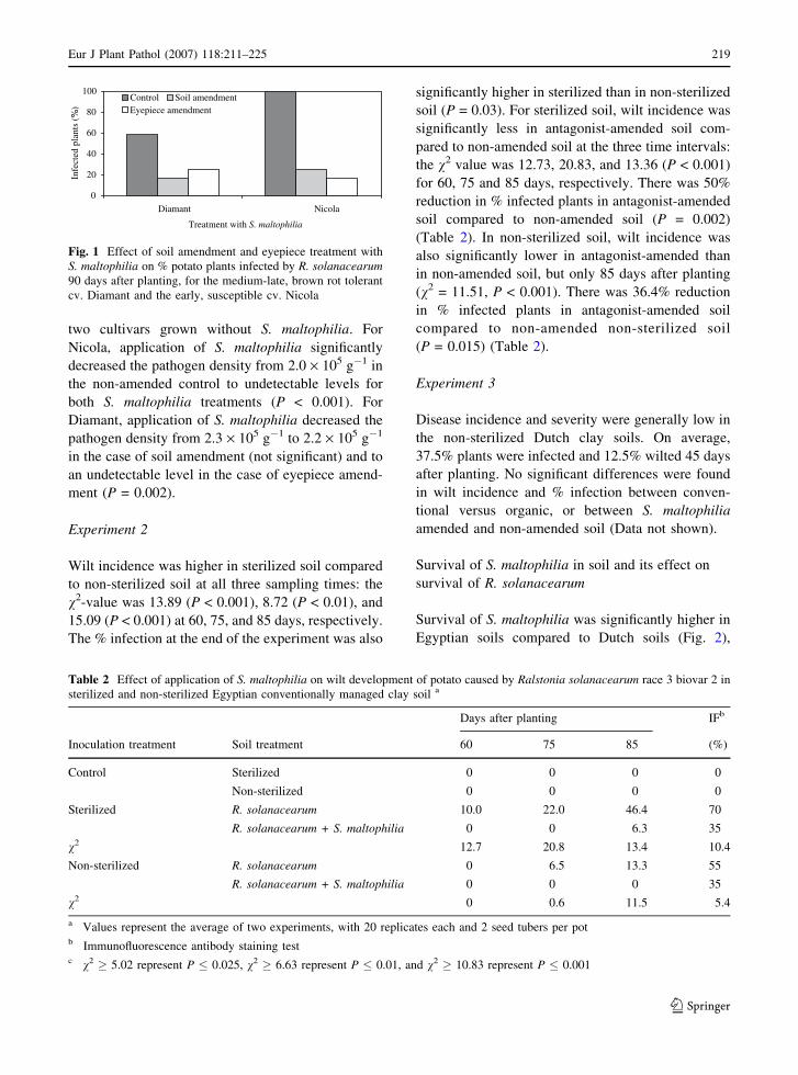
There was a significant interaction (P = 0.001)
between S. maltophilia treatment and cultivar with
respect to disease development in R. solanacearum-
inoculated soil. Potato plants in non-inoculated soil
remained healthy. The % plants infected by R. solan-
acearum was higher for Nicola compared to Diamant
in the control treatment without S. maltophilia (100%
and 58.3%, respectively; P = 0.012). For Nicola,
inoculation of soil or eyepieces with S. maltophilia
decreased % infected plants from 100% to 25% or
16.7% (P < 0.001) in the case of soil or eyepiece
amendment, respectively. For Diamant, inoculation
with S. maltophilia decreased % infected plants from
58.3% to 16.7% (P < 0.001) in the case of soil
amendment, and to 25% (P = 0.008) in the case of
eyepiece inoculation (Fig. 1).
There was also a significant interaction (P = 0.004)
in the cultivar · S. maltophilia treatment with respect
to density of R. solanacearum in the rhizosphere of
potato plants grown in inoculated soil. No significant
difference in pathogen density was found between the
218 Eur J Plant Pathol (2007) 118:211–225
123
two cultivars grown without S. maltophilia. For
Nicola, application of S. maltophilia significantly
decreased the pathogen density from 2.0 · 105 g�1 in
the non-amended control to undetectable levels for
both S. maltophilia treatments (P < 0.001). For
Diamant, application of S. maltophilia decreased the
pathogen density from 2.3 · 105 g�1 to 2.2 · 105 g�1
in the case of soil amendment (not significant) and to
an undetectable level in the case of eyepiece amend-
ment (P = 0.002).
Experiment 2
Wilt incidence was higher in sterilized soil compared
to non-sterilized soil at all three sampling times: the
v2-value was 13.89 (P < 0.001), 8.72 (P < 0.01), and
15.09 (P < 0.001) at 60, 75, and 85 days, respectively.
The % infection at the end of the experiment was also
significantly higher in sterilized than in non-sterilized
soil (P = 0.03). For sterilized soil, wilt incidence was
significantly less in antagonist-amended soil com-
pared to non-amended soil at the three time intervals:
the v2 value was 12.73, 20.83, and 13.36 (P < 0.001)
for 60, 75 and 85 days, respectively. There was 50%
reduction in % infected plants in antagonist-amended
soil compared to non-amended soil (P = 0.002)
(Table 2). In non-sterilized soil, wilt incidence was
also significantly lower in antagonist-amended than
in non-amended soil, but only 85 days after planting
(v2 = 11.51, P < 0.001). There was 36.4% reduction
in % infected plants in antagonist-amended soil
compared to non-amended non-sterilized soil
(P = 0.015) (Table 2).
Experiment 3
Disease incidence and severity were generally low in
the non-sterilized Dutch clay soils. On average,
37.5% plants were infected and 12.5% wilted 45 days
after planting. No significant differences were found
in wilt incidence and % infection between conven-
tional versus organic, or between S. maltophilia
amended and non-amended soil (Data not shown).
Survival of S. maltophilia in soil and its effect on
survival of R. solanacearum
Survival of S. maltophilia was significantly higher in
Egyptian soils compared to Dutch soils (Fig. 2),
0
20
40
60
80
100
Diamant Nicola
Treatment with S. maltophilia
)%(
stnalpdetcefnI
Control Soil amendmentEyepiece amendment
Fig. 1 Effect of soil amendment and eyepiece treatment with
S. maltophilia on % potato plants infected by R. solanacearum90 days after planting, for the medium-late, brown rot tolerant
cv. Diamant and the early, susceptible cv. Nicola
Table 2 Effect of application of S. maltophilia on wilt development of potato caused by Ralstonia solanacearum race 3 biovar 2 in
sterilized and non-sterilized Egyptian conventionally managed clay soil a
Days after planting IFb
Inoculation treatment Soil treatment 60 75 85 (%)
Control Sterilized 0 0 0 0
Non-sterilized 0 0 0 0
Sterilized R. solanacearum 10.0 22.0 46.4 70
R. solanacearum + S. maltophilia 0 0 6.3 35
v2 12.7 20.8 13.4 10.4
Non-sterilized R. solanacearum 0 6.5 13.3 55
R. solanacearum + S. maltophilia 0 0 0 35
v2 0 0.6 11.5 5.4
a Values represent the average of two experiments, with 20 replicates each and 2 seed tubers per potb Immunofluorescence antibody staining testc v2 � 5.02 represent P � 0.025, v2 � 6.63 represent P � 0.01, and v2 � 10.83 represent P � 0.001
Eur J Plant Pathol (2007) 118:211–225 219
123
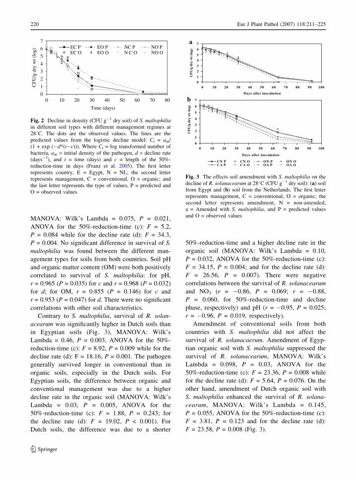
MANOVA: Wilk’s Lambda = 0.075, P = 0.021,
ANOVA for the 50%-reduction-time (c): F = 5.2,
P = 0.084 while for the decline rate (d): F = 34.3,
P = 0.004. No significant difference in survival of S.
maltophilia was found between the different man-
agement types for soils from both countries. Soil pH
and organic matter content (OM) were both positively
correlated to survival of S. maltophilia: for pH,
r = 0.965 (P = 0.035) for c and r = 0.968 (P = 0.032)
for d; for OM, r = 0.855 (P = 0.146) for c and
r = 0.953 (P = 0.047) for d. There were no significant
correlations with other soil characteristics.
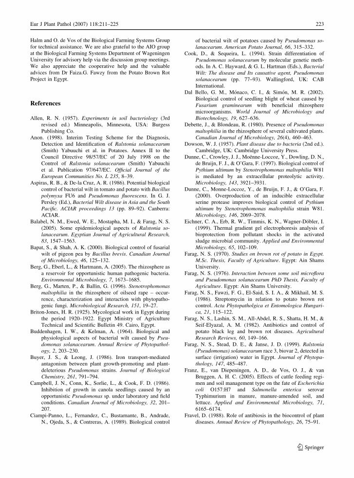
Contrary to S. maltophilia, survival of R. solan-
acearum was significantly higher in Dutch soils than
in Egyptian soils (Fig. 3), MANOVA: Wilk’s
Lambda = 0.46, P = 0.003, ANOVA for the 50%-
reduction-time (c): F = 8.92, P = 0.009 while for the
decline rate (d): F = 18.16, P = 0.001. The pathogen
generally survived longer in conventional than in
organic soils, especially in the Dutch soils. For
Egyptian soils, the difference between organic and
conventional management was due to a higher
decline rate in the organic soil (MANOVA: Wilk’s
Lambda = 0.03, P = 0.005, ANOVA for the
50%-reduction-time (c): F = 1.88, P = 0.243; for
the decline rate (d): F = 19.02, P < 0.001). For
Dutch soils, the difference was due to a shorter
50%-reduction-time and a higher decline rate in the
organic soil (MANOVA: Wilk’s Lambda = 0.10,
P = 0.032, ANOVA for the 50%-reduction-time (c):
F = 34.15, P = 0.004; and for the decline rate (d):
F = 26.56, P = 0.007). There were negative
correlations between the survival of R. solanacearum
and NO3 (r = �0.86, P = 0.069; r = �0.88,
P = 0.060, for 50%-reduction-time and decline
phase, respectively) and pH (r = �0.95, P = 0.025;
r = �0.96, P = 0.019, respectively).
Amendment of conventional soils from both
countries with S. maltophilia did not affect the
survival of R. solanacaerum. Amendment of Egyp-
tian organic soil with S. maltophilia suppressed the
survival of R. solanacearum, MANOVA: Wilk’s
Lambda = 0.098, P = 0.03, ANOVA for the
50%-reduction-time (c): F = 23.36, P = 0.008 while
for the decline rate (d): F = 5.64, P = 0.076. On the
other hand, amendment of Dutch organic soil with
S. maltophilia enhanced the survival of R. solana-
cearum, MANOVA: Wilk’s Lambda = 0.145,
P = 0.055, ANOVA for the 50%-reduction-time (c):
F = 3.81, P = 0.123 and for the decline rate (d):
F = 23.58, P = 0.008 (Fig. 3).
0
1
2
3
4
5
6
7
0 10 20 30 40 50 60 70 80
Time (days)
)gol(tw
yrdg/
UF
C
EC P EO P NC P NO PEC O EO O N C O NO O
Fig. 2 Decline in density (CFU g�1 dry soil) of S. maltophiliain different soil types with different management regimes at
288C. The dots are the observed values. The lines are the
predicted values from the logistic decline model: Ct = am/
(1 + exp (�d*(t�c))). Where Ct = log transformed number of
bacteria, am = initial density of the pathogen, d = decline rate
(days�1), and t = time (days) and c = length of the 50%-
reduction-time in days (Franz et al. 2005). The first letter
represents country, E = Egypt, N = NL; the second letter
represents management, C = conventional, O = organic; and
the last letter represents the type of values, P = predicted and
O = observed values
a
0
1
2
3
4
5
6
7
0 10 20 30 40 50 60 70 80 90 100
Days after inoculation
)gol(t
wyrd
g /U
FC
b
0
1
2
3
4
5
6
7
0 10 20 30 40 50 60 70 80 90 100
Days after inoculation
)gol(t
wyrd
g/U
FC
CN P CN O ON P ON OCA P CA O OA P OA O
Fig. 3 The effects soil amendment with S. maltophilia on the
decline of R. solanacearum at 288C (CFU g�1 dry soil): (a) soil
from Egypt and (b) soil from the Netherlands. The first letter
represents management, C = conventional, O = organic; the
second letter represents amendment, N = non-amended,
a = Amended with S. maltophilia, and P = predicted values
and O = observed values
220 Eur J Plant Pathol (2007) 118:211–225
123

Bacterial diversity in different soils
The bacterial diversity S was significantly lower
(t-test; P = 0.005) in Egyptian organic than in the
conventional soil while the Shannon-Wiener index H0
did not significantly differ (Table 1). The reverse was
true for the Dutch soils: S was significantly higher in
organic than in the conventional soil (t-test,
P = 0.031). There was no difference in H0 between
the Dutch soils. There was no significant correlation
between S and the 50%-reduction-time c or the
decline rate d of either of the decline curves for S.
maltophilia and R. solanacearum. However, there
was a significant negative correlation between H0 and
the decline rate d for R. solanacearum (r = �0.66,
P = 0.019).
Discussion
For the first time, strains of S. maltophilia were found
that inhibited R. solanacearum in vitro on agar plates
and in vivo on potato plants grown in soil. The
in vitro test revealed a possible production of
antibiotics responsible for such inhibition. Steno-
trophomonas maltophilia produces various antibiot-
ics, for example, maltophilin, a macrocyclic lactam
antibiotic, which has antifungal activity, but is
inactive against Gram-positive and Gram-negative
bacteria (Berg et al. 1996; Jakobi et al. 1996). Three
other antibiotics produced by S. maltophilia have
been characterized, namely xanthobaccin A, B, and
C, which have the same plain structure as maltophilin
(Nakayama et al. 1999). These were effective against
fungi and oomycetes, but not against the bacteria
tested (Nakayama et al. 1999). Unfortunately R.
solanacearum was not included in these tests. Apart
from antibiotics, S. maltophilia produces lytic
enzymes that have been implicated in biological
control activity against fungi (Berg et al. 1996), for
example chitinase (Zhang and Yuen 1999) and an
extracellular proteolytic enzyme (Dunne et al. 2000).
This last enzyme may also be active against
R. solanacearum, as Gram-negative bacteria contain,
next to lipopolysaccharide, Braun lipoprotein in their
cell walls.
The inhibitory effect against R. solanacearum by
the S. maltophilia strains used in this study was
proven not to be based on siderophore production like
that by P. fluorescens (Ran et al. 2005b), as the
availability of iron affected only the antagonistic
activity of P. fluorescens. The antibacterial efficiency
of P. fluorescens depends at least partially on the
production of the siderophore pseudobactin which
can efficiently form complexes with iron in soils
making it unavailable to plant pathogens, thus
inhibiting their growth (Buyer and Leong 1986;
Ciampi-Panno et al. 1989; Ran et al. 2005b).
Jurkevich et al. (1992) found one isolate of S.
maltophilia that was able to utilize Fe3+ in the
siderophore pseudobactin as the sole iron source, but
only to a limited extent. It is not known if S. malto-
philia produces its own siderophore.
The antagonistic isolates of S. maltophilia used in
this work were easily recovered from the rhizosphere
of eggplant but not from that of sweet pepper. These
two solanaceous crops have been listed among the
plant species susceptible to race 1 of R. solancearum
(Kelman 1953). Race 3 of the pathogen, used in this
work, is known to affect potatoes and tomato
(Buddenhagen and Kelman 1964). This race may
variably infect other host plants under greenhouse
conditions, especially less compatible hosts under
high soil infestation levels. Thus, S. maltophilia
probably did not evolve as a specific antagonist of
race 3 of R. solanacearum on eggplant. However, S.
maltophilia is known to be a good rhizosphere
colonizer (Juhnke et al. 1987), and is a common
member in the rhizosphere of many plants (Berg et al.
1996), especially of plants with high concentrations
of sulphur-containing compounds such as methionine
in their rhizosphere (Debette and Blondeau 1980).
Our S. maltophilia strains were able to produce H2S,
which would be beneficial in rhizospheres rich in
sulphur-containing compounds, especially under
moist, reducing conditions.
The antagonistic S. maltophilia strains were much
more effective in controlling R. solanacearum in
Egyptian clay soils than in Dutch clay soils, where
they did not have significant suppressive effects on
brown rot development. Thus, the antagonistic strain
was effective in the soil from which it was isolated,
probably because it co-evolved with the plants that
were grown in the Nile Delta for centuries. Moreover,
S. maltophilia survived significantly better in Egyp-
tian than in Dutch clay soils, while R. solanacearum
survived better in Dutch than in Egyptian soils, which
may be related to clay and organic matter content,
Eur J Plant Pathol (2007) 118:211–225 221
123

which were both higher in the Egyptian soils. A high
organic matter content was shown to be detrimental
to R. solanacearum (Balabel et al. 2005). Moreover,
S. maltophilia may not have survived long enough to
be able to control R. solanacearum in the Dutch soils.
A similar conclusion had been drawn previously
about the failure of other potential biocontrol agents
in controlling root pathogens in soil under greenhouse
conditions (Weller 1988). Biocontrol agents selected
in the laboratory often fail under field conditions
(Fravel 1988). Factors affecting direct antagonism on
agar media are usually not known, and the conditions
that allow in vitro activity may not be present in
nature (Wagih 1991). Thus, it was quite fortuitous to
find strains that were effective in field soil (at least in
Egyptian soil), although relatively few strains were
originally tested in the laboratory.
The reasons for the differences in survival of S.
maltophilia in the various soils are not clear. The
nitrate and ammonium contents and the pH of the
Egyptian soils were higher than those of the Dutch
soils. There was a positive correlation between
survival of S. maltophilia and pH, but not the
available nitrogen content. On the other hand, there
was a negative correlation between R. solanacearum
survival and pH, and also with NO3. There could
have been indirect effects, with a greater sensitivity
of R. solanacearum than S. maltophilia towards
ammonia, which could have been higher in the high-
pH Egyptian soil. The percentages of clay and
organic matter were also higher in the Egyptian
soils, especially the organically managed Egyptian
soil, than in the Dutch soils, and there was a positive
correlation between the organic matter content and
survival of S. maltophilia. Also in this case, there
might have been an indirect effect, as the availability
of organic matter can increase the activity of
biocontrol agents (Hiddink et al. 2005; Hoitink and
Boehm 1999), which may explain the better survival
of the antagonist and the suppression of the pathogen
by the antagonist in the Egyptian organic clay soil.
The bacterial diversity in the Egyptian organic clay
soil was less than in the conventional soil, which may
explain the higher efficiency of the biocontrol agent
in the Egyptian organic versus conventional soil
(Hiddink et al. 2005). On the other hand, the
antagonist enhanced the survival of the pathogen in
the Dutch organic soil by delaying its decline phase.
The biodiversity was higher in the organic than in the
conventional Dutch soil, reducing the efficacy of S.
maltophilia to suppress R. solanacearum, so that the
pathogen could survive longer. Similarly, the biocon-
trol agent P. fluorescens declined faster and was less
effective in controlling take-all disease, when added to
organically managed soil with a diverse microbial
community than in a biologically impoverished
conventionally managed soil (Hiddink et al. 2005).
Stenotrophomonas maltophilia was more efficient
at controlling the wilt disease for the early maturing
susceptible cultivar Nicola, compared to the late
maturing moderately tolerant Diamant (only tested in
Egyptian soil). The relation between susceptibility to
brown rot and maturity of the potato variety was
addressed by Farag (1976). Root exudates of early
maturing, disease susceptible potato varieties are
known to be rich in amino acid content compared to
late maturing, disease tolerant varieties, resulting in
larger microbial populations in the rhizosphere of
susceptible varieties compared to tolerant varieties
(Farag et al. 1986). The quality and quantity of
substrate available in the rhizosphere of plants greatly
affect the establishment of biocontrol agents on plant
roots (Hoitink and Boehm 1999), suggesting that
roots of Nicola may have been better colonized by S.
maltophilia than those of Diamant.
The practical implication of this work is that S.
maltophilia could be used as biological control agent
in the Nile Delta of Egypt where the disease is
endemic. This could be accomplished either by
stimulating naturally occurring populations, for
example by crop rotation or intercropping with crops
that have high concentrations of sulphur-containing
amino acids in their root exudates, or by application
of selected antagonistic S. maltophilia strains, for
example by bacterization of potato tubers. However,
the production of S. maltophilia for biological control
would need to be approved by the appropriate
regulatory agencies, as certain strains of S. malto-
philia have been associated with various illnesses in
immuno-depressed human patients (Berg et al. 2005).
Acknowledgements This project was funded by the EU
through the EU-Egypt Potato Brown Rot Project Phase II
(SEM03/220/51A / EGY 1B/1999/0192). We appreciate the
advisory and technical help from the team at the Department of
Bacteriology of the Plant Protection Service (PD),
Wageningen, namely B. Briaire, G. Willemsen, J.G.B.
Voogd, N.M. Landman, P. van de Werde, P.P.M.A. Gorkink-
Smits, and S. Somovilla-Carrasco. We are grateful to H.D.
222 Eur J Plant Pathol (2007) 118:211–225
123
Halm and O. de Vos of the Biological Farming Systems Group
for technical assistance. We are also grateful to the AIO group
at the Biological Farming Systems Department of Wageningen
University for advisory help via the discussion group meetings.
We also appreciate the cooperative help and the valuable
advices from Dr Faiza.G. Fawzy from the Potato Brown Rot
Project in Egypt.
References
Allen, R. N. (1957). Experiments in soil bacteriology (3rd
revised ed.) Minneapolis, Minnesota, USA: Burgess
Publishing Co.
Anon. (1998). Interim Testing Scheme for the Diagnosis,
Detection and Identification of Ralstonia solanacearum(Smith) Yabuuchi et al. in Potatoes. Annex II to the
Council Directive 98/57/EC of 20 July 1998 on the
Control of Ralstonia solanacearum (Smith) Yabuuchi
et al. Publication 97/647/EC. Official Journal of theEuropean Communities No. L 235, 8–39.
Aspiras, R. B., & De-la Cruz, A. R. (1986). Potential biological
control of bacterial wilt in tomato and potato with Bacilluspolymyxa FU6 and Pseudomonas fluorescens. In G. J.
Persley (Ed.), Bacterial Wilt disease in Asia and the SouthPacific. ACIAR proceedings 13 (pp. 89–92). Canberra:
ACIAR.
Balabel, N. M., Ewed, W. E., Mostapha, M. I., & Farag, N. S.
(2005). Some epidemiological aspects of Ralstonia so-lanacearum. Egyptian Journal of Agricultural Research,83, 1547–1563.
Bapat, S., & Shah, A. K. (2000). Biological control of fusarial
wilt of pigeon pea by Bacillus brevis. Canadian Journalof Microbiology, 46, 125–132.
Berg, G., Eberl, L., & Hartmann, A. (2005). The rhizosphere as
a reservoir for opportunistic human pathogenic bacteria.
Environmental Microbiology, 7, 1673–1685.
Berg, G., Marten, P., & Ballin, G. (1996). Stenotrophomonasmaltophilia in the rhizosphere of oilseed rape – occur-
rence, characterization and interaction with phytopatho-
genic fungi. Microbiological Research, 151, 19–27.
Briton-Jones, H. R. (1925). Mycological work in Egypt during
the period 1920–1922. Egypt Ministry of Agriculture
Technical and Scientific Bulletin 49. Cairo, Egypt.
Buddenhagen, I. W., & Kelman, A. (1964). Biological and
physiological aspects of bacterial wilt caused by Pseu-domonas solanacearum. Annual Review of Phytopathol-ogy, 2, 203–230.
Buyer, J. S., & Leong, J. (1986). Iron transport-mediated
antagonism between plant growth-promoting and plant-
deleterious Pseudomonas strains. Journal of BiologicalChemistry, 261, 791–794.
Campbell, J. N., Conn, K., Sorlie, L., & Cook, F. D. (1986).
Inhibition of growth in canola seedlings caused by an
opportunistic Pseudomonas sp. under laboratory and field
conditions. Canadian Journal of Microbiology, 32, 201–
207.
Ciampi-Panno, L., Fernandez, C., Bustamante, B., Andrade,
N., Ojeda, S., & Contreras, A. (1989). Biological control
of bacterial wilt of potatoes caused by Pseudomonas so-lanacearum. American Potato Journal, 66, 315–332.
Cook, D., & Sequeira, L. (1994). Strain differentiation of
Pseudomonas solanacearum by molecular genetic meth-
ods. In A. C. Hayward, & G. L. Hartman (Eds.), BacterialWilt: The disease and Its causative agent, Pseudomonassolanacearum (pp. 77–93). Wallingford, UK: CAB
International.
Dal Bello, G. M., Monaco, C. I., & Simon, M. R. (2002).
Biological control of seedling blight of wheat caused by
Fusarium graminearum with beneficial rhizosphere
microorganisms. World Journal of Microbiology andBiotechnology, 19, 627–636.
Debette, J., & Blondeau, R. (1980). Presence of Pseudomonasmaltophilia in the rhizosphere of several cultivated plants.
Canadian Journal of Microbiology, 26(4), 460–463.
Dowson, W. J. (1957). Plant disease due to bacteria (2nd ed.).
Cambridge, UK: Cambridge University Press.
Dunne, C., Crowley, J. J., Moenne-Loccoz, Y., Dowling, D. N.,
de Bruijn, F. J., & O’Gara, F. (1997). Biological control of
Pythium ultimum by Stenotrophomonas maltophilia W81
is mediated by an extracellular proteolytic activity.
Microbiology, 143, 3921–3931.
Dunne, C., Monne-Loccoz, Y., de Bruijn, F. J., & O’Gara, F.
(2000). Overproduction of an inducible extracellular
serine protease improves biological control of Pythiumultimum by Stenotrophomonas maltophilia strain W81.
Microbiology, 146, 2069–2078.
Eichner, C. A., Erb, R. W., Timmis, K. N., Wagner-Dobler, I.
(1999). Thermal gradient gel electrophoresis analysis of
bioprotection from pollutant shocks in the activated
sludge microbial community. Applied and EnvironmentalMicrobiology, 65, 102–109.
Farag, N. S. (1970). Studies on brown rot of potato in Egypt.M.Sc. Thesis, Faculty of Agriculture. Egypt: Ain Shams
University.
Farag, N. S. (1976). Interaction between some soil microfloraand Pseudomonas solanacearum PhD Thesis, Faculty ofAgriculture. Egypt: Ain Shams University.
Farag, N. S., Fawzi, F. G., El-Said, S. I. A., & Mikhail, M. S.
(1986). Streptomycin in relation to potato brown rot
control. Acta Phytopatholgica et Entomologica Hungari-ca, 21, 115–122.
Farag, N. S., Lashin, S. M., All-Abdel, R. S., Shatta, H. M., &
Seif-Elyazal, A. M. (1982). Antibiotics and control of
potato black leg and brown rot diseases. AgriculturalResearch Reviews, 60, 149–166.
Farag, N. S., Stead, D. E., & Janse, J. D. (1999). Ralstonia(Pseudomonas) solanacearum race 3, biovar 2, detected in
surface (irrigation) water in Egypt. Journal of Phytopa-thology, 147, 485–487.
Franz, E., van Diepeningen, A. D., de Vos, O. J., & van
Bruggen, A. H. C. (2005). Effects of cattle feeding regi-
men and soil management type on the fate of Escherichiacoli O157:H7 and Salmonella enterica serovar
Typhimurium in manure, manure-amended soil, and
lettuce. Applied and Environmental Microbiology, 71,
6165–6174.
Fravel, D. (1988). Role of antibiosis in the biocontrol of plant
diseases. Annual Review of Phytopathology, 26, 75–91.
Eur J Plant Pathol (2007) 118:211–225 223
123
Garbeva, P., van Overbeek, L. S., van Vuurde, J. W. L., & Van
Elsas, J. D. (2001). Analysis of endophytic bacterial
communities of potato by plating and denaturing gradient
gel electrophoresis (DGGE) of 16S rDNA based PCR
fragments. Microbial Ecology, 41, 369–383.
Graham, J., Jones, D. A., & Lloyd, A. B. (1979). Survival of
Pseudomonas solanacearum in plant debris and in latently
infected potato tubers. Phytopathology, 69, 1100–1103.
Gorissen, A., van Overbeek, L. S., & van Elsas (2004). Pig
slurry reduces the survival of Ralstonia solanacearumbiovar 2 in soil. Canadian Journal of Microbiology, 50,
587–593.
Hartman, G. L., & Elphinstone, J. G. (1994). Advances in the
control of Pseudomonas solanacearum race 1 in major
food crops. In A. C. Hayward, & G. L. Hartman (Eds.),
Bacterial Wilt: the disease and its causative agent,Pseudomonas solanacearum (pp. 157–178). Wallingford,
UK: CAB International.
Hayward, A. C. (1991). Biology and epidemiology of bacterial
wilt caused by Pseudomonas solanacearum. AnnualReview of Phytopathology, 29, 65–87.
Heuer, H., & Smalla, K. (1997). Application of denaturing
gradient gel electrophoresis for studying soil microbial
communities. In J. D. van Elsas, J. T. Trevors, & E. M. H.
Wellington (Eds.), Modern soil microbiology (pp. 353–
373). New York: Marcel Dekker Inc.
Hiddink, G. A., van Bruggen, A. H. C., Termorshuizen, A. J.,
Raaijmakers, J. M., & Semenov, A. V. (2005). Effect of
organic management of soils on suppressiveness to
Gaeumannomyces graminis var. tritici and its antagonist,
Pseudomonas fluorescens. European Journal of PlantPathology, 113, 417–435.
Hoitink, H. A. J., & Boehm, M. J. (1999). Biocontrol within the
context of soil microbial communities: A substrate-
dependent phenomenon. Annual Review of Phytopathol-ogy, 37, 427–446.
Holt J. G., Krieg N. R., Sneath P. H. A., Staley J. T., Williams
S. T. (Eds.), (1994). Bergey’s manual of determinativebacteriology (9th ed.). Baltimore, USA: Williams &
Wilkins.
Houba, V. J. G., & Novozamsky, I. (1998). Influence of storage
time and temperature of dry soils on pH and extractable
nutrients using 0.01 mol/l CaCl2. Fresenius’ Journal ofAnalytical Chemistry, 360, 362–365.
Houba, V. J. G., van der Lee, J. J., & Novozamsky, I. (1997).
Determination of organic matter content. Soil analysis
procedures, other procedures. Soil and Plant Analysis, part
5B (pp. 38–40). Wageningen Agricultural University,
Department of Soil Science and Plant Nutrition, Wagen-
ingen, the Netherlands.
Islam, T. M. D., & Toyota, K. (2004). Suppression of bacterial
wilt of tomato by Ralstonia solanacearum by incorpora-
tion of composts in soil and possible mechanisms.
Microbes and Environment, 19, 53–60.
Jakobi, M., Winkelmann, G., Kaiser, D., Kempler, C., Jung, G.,
Berg, G., & Bahl, H. (1996). Maltophilin: a new
antifungal compound produced by Stenotrophomonasmaltophilia R3089. Journal of Antibiotics (Tokyo), 49,
1101–1104.
Janse, J. D. (1988). A detection method for Pseudomonassolanacearum in symptomless potato tubers and some
data on its sensitivity and specificity. EPPO Bulletin, 18,
343–351.
Janse, J. D. (1991). Infra- and intraspecific classification of
Pseudomonas solanacearum strains, using whole cell fatty
acid analysis. Systematic and Applied Microbiology, 14,
335–345.
Janse, J. D. (1996). Potato brown rot in Western Europe –
history, present occurrence and some remarks on possible
origin, epidemiology and control strategies. EPPO Bul-letin, 26, 679–695.
Janse, J. D., Arulappan, F. A. X., Schans, J., Wenneker, M., &
Westerhuis, W. (1998). Experiences with bacterial brown
rot Ralstonia solanacearum biovar 2, race 3 in The
Netherlands. In P. Prior, C. Allen, J. G. Elphinstone (Eds.),
Bacterial Wilt disease. Molecular and ecological aspects(pp. 146–152). Heidelberg, Germany: Springer-Verlag.
Juhnke, M. E., Mathre, D. E., & Sands, D. C. (1987). Identi-
fication and characterization of rhizosphere-competent
bacteria of wheat. Applied and Environmental Microbi-ology, 53, 2793–2799.
Jurkevitch, E., Hadar, Y., & Chen, Y. (1992). Differential
siderophore utilization and iron uptake by soil and
rhizosphere bacteria. Applied and EnvironmentalMicrobiology, 58, 119–124.
Kelman, A. (1953). The bacterial wilt caused by Pseudomonassolanacearum. North Carolina Agricultural ExperimentalStation Techical Bulletin, 99, 194.
Lambert, B., & Joos, H. (1989). Fundamental aspects of rhi-
zobacterial plant growth promotion research. Trends inBiotechnology, 7, 215–219.
Lopez, M. M., & Biosca, E. G. (2004). Potato bacterial wilt
management: new prospects for an old problem. In C.
Allen, P. Prior, & A. C. Hayward (Eds.), Bacterial WiltDisease and the Ralstonia species complex (pp. 205–224).
St. Paul Minnesota, USA: APS press.
Mendoza, H. A. (1994). Development of potatoes with multiple
resistance to biotic and abiotic stresses: the International
Potato Center Approach. In G. W. Zehnder, M. L.
Powelson & R. Jansson (Eds.), Advances in Potato PestBiology and Management (pp. 627–642). St Paul, MN,
USA: APS Press.
Michel, V. V., & Mew, T. W. (1998). Effect of a soil
amendment on the survival of Ralstonia solanacearum in
different soils. Phytopathology, 88, 300–305.
Mickail, K. Y., Bishay, F., Farag, N. S., & Tawfik, A. E.
(1974). Evaluation of bacterial wilt disease in A.R.E.
during the years from 1967–1968 to 1971–1972.
Agricultural Research Review (Cairo), 52, 89–94.
Mickail, K. Y., Bishay, F., Farag, N. S., Tawfik, A. E., &
Fawzi, F. G. (1985). Status of brown rot disease of
potatoes in Egypt in the years from 1980 to 1984.
Agricultural Research Review (Cairo), 63, 165–174.
Murakoshi, S., & Takahashi, M. (1984). Trials of some control
of tomato wilt caused by Pseudomonas solanacearum.
Bulletin of the Kanagawa Horticultural Experiment Sta-tion, 31, 50–56.
Nakayama, T., Homma, Y., Hashidoko, Y., Mizutani, J., &
Tahara, S. (1999). Possible role of xanthobaccins
produced by Stenotrophomonas sp. Strain SB-K88 in
suppression of sugar beet damping-off disease. Appliedand Environmental Microbiology, 65, 4334–4339.
224 Eur J Plant Pathol (2007) 118:211–225
123

Prior, P., & Fegan, M. (2005). Recent developments in the
phylogeny and classification of Ralstonia solanacearum.
Acta Horticulturae (ISHS), 695, 127–136.
Priou, S., Gutarra, L., & Aley, P. (1999). Highly sensitive
detection of Ralstonia solanacearum in latently infected
potato tubers by post-enrichment enzyme-linked immu-
nosorobent assay on nitrocellulose membrane. EPPOBulletin, 29, 117–125.
Ran, L. X., Li, Z. N., Wu, G. J., van Loon, L. C., & Bakker, P.
A. H. M. (2005a). Induction of systemic resistance against
bacterial wilt in Eucalyptus urophylla by fluorescent
Pseudomonas spp. European Journal of Plant Pathology,113, 59–70.
Ran, L. X., Liu, C. Y., Wu, G. J., van Loon, L. C., & Bakker, P.
A. H. M. (2005b). Suppression of bacterial wilt in Euca-lyptus urophylla by fluorescent Pseudomonas spp. in
China. Biological Control, 32, 111–120.
Rhodes, D. J., & Logan, C. (1987). A method for selecting
fluorescent pseudomonads inhibitory to seed tuber decay.
Potato Research, 30, 603–611.
Satoh, K., & Toyota, K. (2004). Comparison of disease sup-
pressiveness of different soils with or without repeated
application of organic matters toward bacterial wilt of
tomato caused by Ralstonia solanacearum. Microbes andEnvironment, 19, 310–314.
Schaad, N. W. (Eds.), (1988). Laboratory Guide for Identifi-cation of Plant Pathogenic Bacteria (2nd ed.). St. Paul,
MN, USA: American Phytopathological Society.
Schans, J., & Steeghs, M. H. C. G. (1998). Strategy and results
of eradication of brown rot in The Netherlands. EPPOBulletin, 28, 121–133.
Stead, D. E. (1992). Grouping of plant-pathogenic and some
other Pseudomonas spp. by using cellular fatty acid pro-
files. International Journal of Systematic Bacteriology,42, 281–295.
Sturz, A. V., Ryan, D. A. J., Coffin, A. D., Matheson, B. G.,
Arsenault, W. J., Kimpinski, J., & Christie, B. R. (2004).
Stimulating disease suppression in soils: sulphate fertil-
izers can increase biodiversity and antibiosis of root zone
bacteria against Streptomyces scabies. Soil Biology andBiochemistry, 36, 343–352.
Sunaina, V., Kishore, V., Shekhawat, G. S., & Kumar, M.
(1997). Control of bacterial wilt of potatoes in naturally
infested soils by bacterial antagonists. Journal of PlantDisease and Protection, 104, 362–369.
van Diepeningen, A. D., de Vos, O. J., Korthals, G. W., & van
Bruggen, A. H. C. (2006). Effects of organic versus
conventional management on chemical and biological
parameters in agricultural soils. Applied Soil Ecology, 31,
120–135.
van Elsas, J. D., Kastelein, P., van Bekkum, P., van der Wolf, J.
M., de Vries, P. M., & van Overbeek, L. (2000). Survival
Ralstonia solanacearum Biovar 2, the causative agent of
potato brown rot, in field and microcosm soils in
temperate climates. Phytopathology, 90, 1358–1366.
van Elsas, J. D., Kastelein, P., de Vries, P. M., & van
Overbeek, L. (2001). Effects of ecological factors on the
survival and physiology of Ralstonia solanacearum bv. 2
in irrigation water. Canadian Journal of Microbiology,47, 842–854.
Wagih, E. (1991). Neither indole acetic acid nor bacteriocin is
apparently involved in the in vitro antagonism between
the virulent and the avirulent strains of Pseudomonassolanacearum. Journal of Phytopathology, 123, 153–160.
Weller, D. M. (1988). Biological control of soilborne patho-
gens in the rhizosphere with bacteria. Annual Review ofPhytopathology, 26, 379–407.
Xu, G. W., & Gross, D. C. (1986a). Selection of fluorescent
pseudomonads antagonistic to Erwinia carotovora and
suppressive of potato seed piece decay. Phytopathology,76, 414–422.
Xu, G. W., & Gross, D. C. (1986b). Field evaluations of the
interactions among fluorescent pseudomonads, Erwiniacarotovora and potato yield. Phytopathology, 76, 423–
430.
Yabuuchi, E., Kosako, Y., Yano, I., Hotta, H., & Nishiuchi, Y.
(1995). Transfer of two Burkholderia and an Alcaligenesspecies to Ralstonia Gen. Nov.: proposal of Ralstoniapickettii (Ralston, Palleroni and Doudoroff 1973) Comb.
Nov., Ralstonia solanacearum (Smith 1896) Comb. Nov.
and Ralstonia eutropha (Davis 1969) Comb. Nov.
Microbiology and Immunology, 39, 897–904.
Zhang, Z., & Yuen, G. Y. (1999). Biological control of Bipo-laris sorokiniana on tall fescue by Stenotrophomonasmaltophilia strain C3. Phytopathology, 89, 817–882.
Eur J Plant Pathol (2007) 118:211–225 225
123