stereophotogrammetric impression making for
TRANSCRIPT

CLINICAL REPORT
aAssociate PrbAssociate PrcProfessor, DdAssociate Pr
THE JOURNA
Stereophotogrammetric impression making forpolyoxymethylene, milled immediate partial fixed dental
prostheses
Miguel Gómez-Polo, DDS, PhD,a Cristina Gómez-Polo, DDS, PhD,b Jaime del Río, MD, PhD,c andRocío Ortega, DDS, PhDd
ABSTRACTImmediate post-extraction and same-day placement of interim prostheses have increased patientacceptance of implant-supported prostheses. However, for immediate prostheses supported bymultiple implants, meeting passive fit and esthetic standards is often challenging. In this clinicalreport, implant photogrammetry was combined with conventionally obtained digitized casts toprepare an interim, milled prosthesis from a polyoxymethylene (POM) disk, using computer-aideddesign and computer-aided manufacturing techniques. The following 2 conclusions were drawn.First, stereophotogrammetric scanning appears to be a reliable method for making impressions ofimmediate, implant-supported partial prostheses, and second, POM is suitable for preparing im-mediate interim screw-retained prosthetic implants. (J Prosthet Dent 2017;-:---)
Improvements in function andcomfort with immediately placedimplant-supported fixed dentalprostheses (FDPs) have led toclinical advantages1 and increasedpatient satisfaction.2 In addition,interim prostheses accommodatesoft tissue modifications andfurnish more information thanother options for the subsequent
fabrication of the definitive prosthesis. Although immediateprosthesis placement is regarded as a treatment with long-term predictability,3 one of the major problems posed isadaptation to implants, particularly when several areinvolved. The resulting prosthesis must fit precisely andafford the patient suitable esthetics and function.Photogrammetry is a technique that collects data andinformation on the shape and location of an objectrelative to that of others in a given space and on itsmovement or deformation.4,5
A recently introduced photogrammetric system fordigital implant impressions could increase patient con-venience while affording suitable accuracy. It is analternative imaging method for multiple implant-supported restorations,6-9 including complete archimplant-supported FDPs.10
In photogrammetry, scans are recorded by anextraoral receiver, eliminating the need for makingoverlapping images with intraoral scanners and,
ofessor, Department of Prosthetic Dentistry, School of Dentistry, Compluteofessor, Department of Surgery, School of Medicine, University of Salamaepartment of Prosthetic Dentistry, School of Dentistry, Complutense Univeofessor, Department of Prosthetic Dentistry, School of Dentistry, European
L OF PROSTHETIC DENTISTRY
theoretically, positioning the 3-dimensional (3D) im-plants more accurately than intraoral scanners. Researchis needed to confirm accuracy, however.
This clinical report describes the use of photogram-metry to record 3D implant position to prepare imme-diate multiple implant-supported interim restorations.
CLINICAL REPORT
A 53-year-old woman presented seeking predictableesthetic rehabilitation of the maxillary anterior teeth(Fig. 1). Her medical history was unremarkable, despiteher heavy smoking (>20 cigarettes/day) and poor oralhygiene.
Her treatment plan was based on diagnostic casts,periodontal charting, and radiographs. The plan con-sisted of extracting the right maxillary first premolar,lateral incisor, and central incisor and the left maxillarycentral and lateral incisors, with the immediate
nse University of Madrid, Madrid, Spain.nca, Salamanca, Spain.rsity of Madrid, Madrid, Spain.University of Madrid, Madrid, Spain.
1

Figure 1. Initial patient condition. A, Panoramic radiograph. B, Intraoral view.
Figure 2. Digitized image of diagnostic cast.
2 Volume - Issue -
placement of 3.8×12-mm implants in the right maxillaryfirst premolar, the left canine, and the left lateral incisorsites.
Digitization began with a scan of the diagnostic castto generate a standard tessellation language (STL) file(Fig. 2). After the teeth had been extracted, implants(Biohorizons Tapered Internal) were inserted to 35 Ncmto ensure adequate primary stability,11,12 and machinedtransepithelial abutments of known height were screwedinto the implants. Photogrammetric abutments, that is,flag-shaped, white-dotted elements designed to berecognized by an extraoral stereophotogrammetric cam-era (PIC camera; PIC dental, Iditec North West SL), werescrewed into the transepithelial abutments. The stereo-photogrammetric device consisted of 2 photogrammetriccameras working in unison and able to record 150 im-ages/min (Fig. 3).
The information furnished by the photogrammetricabutments and gathered by the camera was processed bysoftware (Pic Cam Soft v1.1; PIC dental, Iditec NorthWest SL) that generated a digital file showing the 3Dlocation of the implant platforms and their angulation(Fig. 4A). The STL file showed the locations of theimplant platforms relative to one another in the form ofposition vectors (Fig. 4A). The flag-shaped abutmentswere then removed, and a second conventional alginateimpression (Cavex CA 37; Cavex Hollan BV) was made ofthe soft tissues and adjacent teeth, including the platformfor the abutment of known height screwed into the im-plants to ensure best-fit alignment. The cast made fromthe impression was then digitized with a scanner (Den-talwings 5 series; Dental Wings Inc). These 2 digital files,one for the implants and the other for the soft tissue andteeth, were subsequently overlaid to a best-fit alignmentto generate the definitive digital model with informationon teeth, soft tissues, and implants (Fig. 4B).
With these files and scanning the preliminary defin-itive cast as a reference to maintain parameters such asthe midline and incisal edge, the interim screw-retained
THE JOURNAL OF PROSTHETIC DENTISTRY
implant FDP was computer-modeled (Fig. 4C, D). Theprosthesis designed was milled from a (Vita B2) poly-oxymethylene (POM) disk (Acetal resin; Delrin; DupontUSA) and screwed to the implants just a few hours afterinsertion. In the absence of machined connections and asthe Sheffield test could not be conducted, an alternativefinger pressure test was performed to ensure a proper fit.The screw access channels were subsequently filled withpolytetrafluorethylene13 and interim restoration (Telio CSInlay; Ivoclar Vivadent AG) material. An occlusaladjustment was made to prevent contact in eccentricmovements.
The 1-week (Fig. 5A) and 3-month (Fig. 5B) follow-upexaminations revealed no complications. Correcting thegingival volume deficiency on the facial side of the rightmaxillary lateral incisor with a connective tissue graftfrom the maxillary tuberosity (Fig. 5C) delayed placementof the definitive prosthesis.
The patient reported no problems during the 6months between implant placement and placement ofthe definitive restoration. Despite her smoking and poororal hygiene, gingival tissue compatibility was observedto be good when the interim prosthesis was removed
Gómez-Polo et al

Figure 3. A, Stereophotogrammetric abutment on transepithelial abutment. B, Extraoral stereophotogrammetric camera.
Figure 4. A-D, Digital files, step-by-step, from 3D locations of implants to definitive CAD design.
- 2017 3
after 6 months, although the POM materials weredistinctly stained (Fig 6).
Because their 3D location had been recorded, nofurther impressions of the implants were needed to makethe definitive prosthesis. However, an updated impres-sion of the soft tissue was required to record changessince the day of the surgery. The interim FDP was sub-sequently removed, and healing screws of known heightwere positioned. An alginate impression was made, and
Gómez-Polo et al
its resultant cast was scanned to obtain a digital modelthat was best-fit aligned to the implant scan to generatethe definitive cast, including soft tissue and adjacentteeth.
Prior to generating the definitive prosthesis, we con-ducted an esthetic test, using a 3D printer (Objet30OrthoDesk; Stratasys) for alterations. The definitiveprosthesis consisted of a sintered cobalt-chromium(Starbond CoS Powder 30+; Schseftner Dental Alloys,
THE JOURNAL OF PROSTHETIC DENTISTRY

Figure 5. Polyoxymethylene prosthesis. A, Examination at 1 week. B, Examination at 3 months after placement. C, Examination after connective tissuegraft.
Figure 6. Six-months’ view shows (A) excellent soft tissue appearance and (B) palatal staining.
Figure 7. Machine-sintered cobalt-chromium screw-retained framework.
4 Volume - Issue -
S&S Scheftner GmbH) framework machined at theimplant connections (Fig. 7). The Sheffield test and fingerpressure were applied, and periapical radiographs madeto evaluate for passive fit. The framework was then sentto the laboratory to add a resin-based veneer (Ceramage;Shofu), after which the definitive prosthesis was insertedto the manufacturer’s recommended torque (30 Ncm)(Fig. 8).
DISCUSSION
Although implant digital impression making has devel-oped, most of the existing systems do not allow adequatefit for long-span fixed prostheses or complete arch res-torations.9 In the absence of any need for overlappingand the concomitant accuracy of the implant positionsdigitized, stereophotogrammetry is a useful and effectivetool for producing immediate prostheses with a predict-ably correct passive fit (desirable to prevent complica-tions14). However, as it does not reproduce soft tissue, aconventional cast must be digitized or an additionalintraoral scan generated to supplement the informationfurnished.
POM exhibits higher wear resistance and surfacehardness than other polymers.15,16 That holds promisefor its use in immediate, interim, screw-retained, metal-free, implant-supported FDPs. POM also exhibits high
THE JOURNAL OF PROSTHETIC DENTISTRY
thermal and abrasion resistance, good biocompatibility,and reasonably good esthetics.17
SUMMARY
Stereophotogrammetric scanning seems to be a reliablemethod for making impressions of immediate, implant-supported partial FDPs. POM is suitable for preparingimmediate interim CAD-CAM screw-retained implantfixed dental prostheses. For the patient described in thisarticle, the POM material remained intact for the 6
Gómez-Polo et al
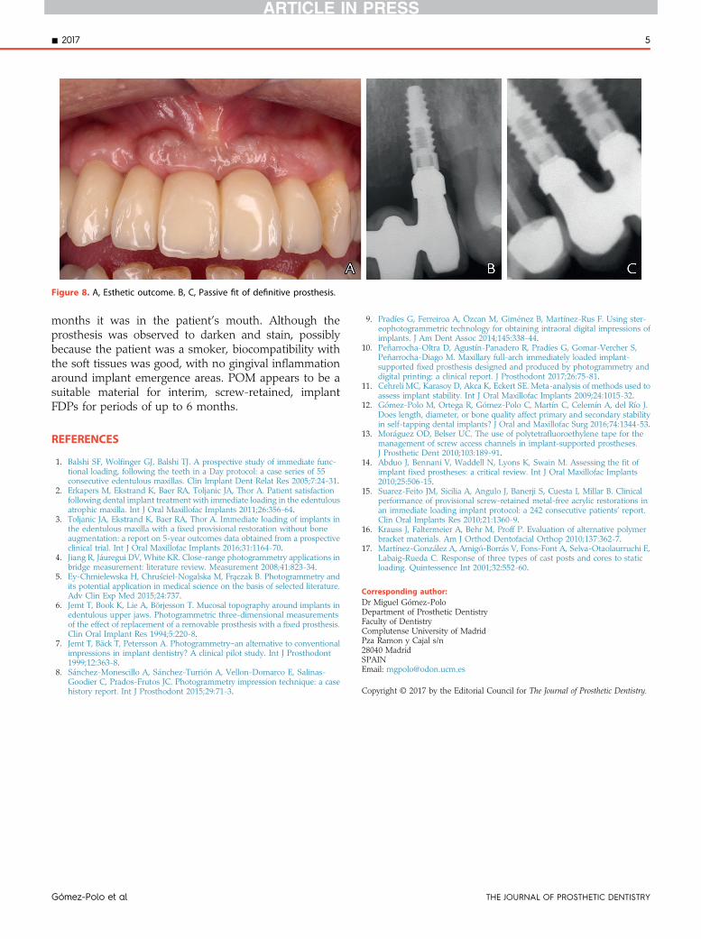
Figure 8. A, Esthetic outcome. B, C, Passive fit of definitive prosthesis.
- 2017 5
months it was in the patient’s mouth. Although theprosthesis was observed to darken and stain, possiblybecause the patient was a smoker, biocompatibility withthe soft tissues was good, with no gingival inflammationaround implant emergence areas. POM appears to be asuitable material for interim, screw-retained, implantFDPs for periods of up to 6 months.
REFERENCES
1. Balshi SF, Wolfinger GJ, Balshi TJ. A prospective study of immediate func-tional loading, following the teeth in a Day protocol: a case series of 55consecutive edentulous maxillas. Clin Implant Dent Relat Res 2005;7:24-31.
2. Erkapers M, Ekstrand K, Baer RA, Toljanic JA, Thor A. Patient satisfactionfollowing dental implant treatment with immediate loading in the edentulousatrophic maxilla. Int J Oral Maxillofac Implants 2011;26:356-64.
3. Toljanic JA, Ekstrand K, Baer RA, Thor A. Immediate loading of implants inthe edentulous maxilla with a fixed provisional restoration without boneaugmentation: a report on 5-year outcomes data obtained from a prospectiveclinical trial. Int J Oral Maxillofac Implants 2016;31:1164-70.
4. Jiang R, Jáuregui DV, White KR. Close-range photogrammetry applications inbridge measurement: literature review. Measurement 2008;41:823-34.
5. Ey-Chmielewska H, Chru�sciel-Nogalska M, Fraczak B. Photogrammetry andits potential application in medical science on the basis of selected literature.Adv Clin Exp Med 2015;24:737.
6. Jemt T, Book K, Lie A, Börjesson T. Mucosal topography around implants inedentulous upper jaws. Photogrammetric three-dimensional measurementsof the effect of replacement of a removable prosthesis with a fixed prosthesis.Clin Oral Implant Res 1994;5:220-8.
7. Jemt T, Bäck T, Petersson A. Photogrammetryean alternative to conventionalimpressions in implant dentistry? A clinical pilot study. Int J Prosthodont1999;12:363-8.
8. Sánchez-Monescillo A, Sánchez-Turrión A, Vellon-Domarco E, Salinas-Goodier C, Prados-Frutos JC. Photogrammetry impression technique: a casehistory report. Int J Prosthodont 2015;29:71-3.
Gómez-Polo et al
9. Pradíes G, Ferreiroa A, Özcan M, Giménez B, Martínez-Rus F. Using ster-eophotogrammetric technology for obtaining intraoral digital impressions ofimplants. J Am Dent Assoc 2014;145:338-44.
10. Peñarrocha-Oltra D, Agustín-Panadero R, Pradíes G, Gomar-Vercher S,Peñarrocha-Diago M. Maxillary full-arch immediately loaded implant-supported fixed prosthesis designed and produced by photogrammetry anddigital printing: a clinical report. J Prosthodont 2017;26:75-81.
11. Cehreli MC, Karasoy D, Akca K, Eckert SE. Meta-analysis of methods used toassess implant stability. Int J Oral Maxillofac Implants 2009;24:1015-32.
12. Gómez-Polo M, Ortega R, Gómez-Polo C, Martín C, Celemín A, del Río J.Does length, diameter, or bone quality affect primary and secondary stabilityin self-tapping dental implants? J Oral and Maxillofac Surg 2016;74:1344-53.
13. Moráguez OD, Belser UC. The use of polytetrafluoroethylene tape for themanagement of screw access channels in implant-supported prostheses.J Prosthetic Dent 2010;103:189-91.
14. Abduo J, Bennani V, Waddell N, Lyons K, Swain M. Assessing the fit ofimplant fixed prostheses: a critical review. Int J Oral Maxillofac Implants2010;25:506-15.
15. Suarez-Feito JM, Sicilia A, Angulo J, Banerji S, Cuesta I, Millar B. Clinicalperformance of provisional screw-retained metal-free acrylic restorations inan immediate loading implant protocol: a 242 consecutive patients’ report.Clin Oral Implants Res 2010;21:1360-9.
16. Krauss J, Faltermeier A, Behr M, Proff P. Evaluation of alternative polymerbracket materials. Am J Orthod Dentofacial Orthop 2010;137:362-7.
17. Martínez-González A, Amigó-Borrás V, Fons-Font A, Selva-Otaolaurruchi E,Labaig-Rueda C. Response of three types of cast posts and cores to staticloading. Quintessence Int 2001;32:552-60.
Corresponding author:Dr Miguel Gómez-PoloDepartment of Prosthetic DentistryFaculty of DentistryComplutense University of MadridPza Ramon y Cajal s/n28040 MadridSPAINEmail: [email protected]
Copyright © 2017 by the Editorial Council for The Journal of Prosthetic Dentistry.
THE JOURNAL OF PROSTHETIC DENTISTRY