stony brook university - suny digital repository
TRANSCRIPT

SSStttooonnnyyy BBBrrrooooookkk UUUnnniiivvveeerrrsssiiitttyyy
The official electronic file of this thesis or dissertation is maintained by the University Libraries on behalf of The Graduate School at Stony Brook University.
©©© AAAllllll RRRiiiggghhhtttsss RRReeessseeerrrvvveeeddd bbbyyy AAAuuuttthhhooorrr...

Characterization of antimicrobial activity present in the cuticle of American lobster, Homarus americanus
A Thesis Presented
by
Margaret Anne Mars
to
The Graduate School
in Partial Fulfillment of the
Requirements
for the Degree of
Master of Science
in
Marine and Atmospheric Science
Stony Brook University
December 2010

ii
Stony Brook University
The Graduate School
Margaret Anne Mars
We, the thesis committee for the above candidate for the
Master of Science degree, hereby recommend
acceptance of this thesis.
Dr. Bassem Allam – Thesis Advisor Associate Professor
School of Marine and Atmospheric Science
Dr. Anne McElroy – Thesis Advisor Associate Professor
School of Marine and Atmospheric Science
Dr. Emmanuelle Pale Espinosa Adjunct Professor
School of Marine and Atmospheric Science
This thesis is accepted by the Graduate School
Lawrence Martin Dean of the Graduate School
iii
Abstract of the Thesis Characterization of antimicrobial activity present in the cuticle of American
lobster, Homarus americanus by
Margaret Anne Mars Master of Science
in Marine and Atmospheric Science
Stony Brook University 2010
American lobster is an ecologically and socioeconomically important species. In recent years the species has been affected by disease and the catch in Southern New England has fallen dramatically. In order to fully understand how and why diseases affect lobster populations, it is imperative to fully understand lobster defense mechanisms. The cuticle, previously believed to act only as a physical barrier, has recently been shown to contain antimicrobial activity. This thesis focused on characterizing this activity and attempted to identify the molecules responsible. A sterile shell extract prepared from a pool of lobster shells was active against Vibrio parahaemolyticus, Vibrio anguillarum, Escherichia coli, and Staphylococus aureus. The activity in the extract was found to be heat stabile, as activity was not decreased after boiling (100°C, 5 min.). Activity was diminished when an extract was prepared from muffled lobster shells, indicating an organic component responsible for the observed activity. Size fractioning of the extract with centrifugal filter units decreased activity in extract unable to pass through a 10kDa filter, while extract that passed through a 10kDa and 3kDa filter retained antimicrobial activity, despite having much lower protein concentrations. Fast protein liquid chromatography (FPLC) of the shell extract revealed several protein peaks. Samples corresponding to FPLC peaks had varying antimicrobial activities. In contrast, the void volume, containing inorganic and anionic material, increased bacterial growth. Tris/tricine SDS-PAGE with the FPLC fractions revealed small peptides (<7kDa) in the fractions that displayed the highest and least variable antimicrobial activity. MALDI mass spectrometry revealed peptide peaks at 1.6, 2.8, 4.6, and 5.6 kDa. A partial sequence of the 5.6kDa peptide was determined. Manipulation of the unknown amino acids in the sequence in a search with BLAST led to a non-definitive, partial match with the wasp antimicrobial peptide mastoparan. Antimicrobial peptides have been described as being cationic peptides less than 10kDa with broad-spectrum antimicrobial activity. The small size and structure of the peptides make them very stabile and able to withstand high heat. AMPs have been isolated from a wide variety of plants and animals and are an integral part of the invertebrate defense response. This thesis demonstrated that the broad-spectrum antimicrobial activity observed in the lobster shell is due to a component that is organic, cationic, heat stabile, and less than 10kDa. Based on shared characteristics with antimicrobial peptides, it is likely that the activity observed in the shell is due to an antimicrobial peptide.
iv
In addition, antimicrobial activity in the shells of a lobster population with a high incidence of Epizootic shell disease (Eastern Long Island Sound, ELIS) was compared with the activity in shells of lobsters from a population with a low incidence of the disease (Western Long Island Sound, WLIS). Extracts from the shells of WLIS lobsters had significantly higher antimicrobial activity when compared to extracts from ELIS lobsters. The antimicrobial activity in the shell may be a factor affecting susceptibility to the disease.

v
Table of Contents
List of Figures………………………………………………….…….………………………..vii List of Tables………………………………….………………………………………….……viii Acknowledgements……………………………………………………………………………ix Introduction…………………………………………………………………………………….1 Methods………………………………………………………………………………………...4 Lobster sampling………………………………………………………………………4 Sterile shell extract…………………………………………………………………….5 Protein concentration………………………………………………………………….6 Antimicrobial activity assay…………………………………………………………...6 Detection and characterization of antimicrobial activity in the shell extract…..…7 Evaluation of antimicrobial activity in the shell against a broad spectrum of bacteria…….………………………………………………………………...7 Heat sensitivity of antimicrobial activity in American lobster shell………..7 Effect of muffling on antimicrobial activity in American lobster shell……..8 Determination of antimicrobial activity in size fractionated shell extract…8 Fast protein liquid chromatography (FPLC) of shell extract……………….9 Evaluation of antimicrobial activity in FPLC fractions………………………9 Tris/tricine SDS-PAGE with FPLC fractions…………………………………9 Evaluation of antimicrobial activity in size fractionated FPLC fractions….10 Determination of molecular weight of small peptides in shell extract…….10 Determination of the amino acid sequence of a small peptide in the shell extract…………………………………………………………………………...11 Determination of antimicrobial activity in the shells of lobsters from two populations with differing disease prevalence………………………………..…….11 Comparison of antimicrobial activity in individual lobster shell extracts from Eastern and Western Long Island Sound lobsters…………………………11 Results…………………………………………………………………………………............12 Detection and characterization of antimicrobial activity in the shell extract…......12 Evaluation of antimicrobial activity in the shell against a broad spectrum of bacteria………………………………………………………………………….12 Heat sensitivity of antimicrobial activity in American lobster shell………..13 Effect of muffling on antimicrobial activity in American lobster shell……..14 Determination of antimicrobial activity in size fractionated shell extract…15 Fast protein liquid chromatography (FPLC) of shell extract……………....16 Evaluation of antimicrobial activity in FPLC fractions……………………...17 Tris/tricine SDS-PAGE with FPLC fractions………………………………...18 Evaluation of antimicrobial activity in size fractionated FPLC fractions….19 Determination of molecular weights of small peptides in shell extract......20 Determination of the amino acid sequence of a small peptide in the shell extract…………………………………………………………………………...21 Determination of antimicrobial activity in the shells of lobsters from two populations with differing disease prevalence…………………………..………….22 Comparison of antimicrobial activity in individual lobster shell extracts from Eastern and Western Long Island Sound lobsters…………………………22

vi
Discussion………………………………………………………………………………………24 Detection and characterization of antimicrobial activity in American lobster shell …………………………………………………………………………………………...24
Evaluation of antimicrobial activity in the shells of lobsters from two populations with differing disease prevalence…………………………………………………….31
Conclusions…………………………………………………………………………………….32 Future Directions………………………………………………………....……………………32 Works Cited…………………………………………………………………………………….34

vii
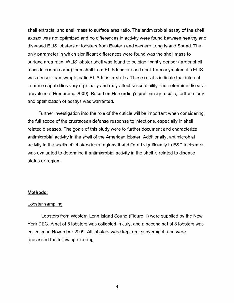
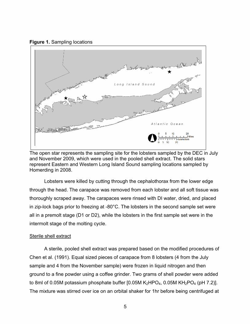
List of Figures Figure 1. Sampling locations………………………………………………………………..5
Figure 2. Percent decrease in the growth of 4 species of bacteria in cultures supplemented with sterile pooled shell extract…………………………………………13
Figure 3. Influence of boiling on antimicrobial activity of a pooled shell extract…..…...14 Figure 4. Influence of muffling lobster shells on the antimicrobial activity of a pooled shell extract…………………………………….……………………………………...……15 Figure 5. Evaluation of antimicrobial activity in size fractionated shell extract…………16 Figure 6. Chromatograph from FPLC with pooled sterile shell extract………………….17
Figure 7. Evaluation of antimicrobial activity in sterile shell extract fractionated with FPLC………………………………………………………………………………………...18 Figure 8. Digital photograph of tris/tricine SDS-PAGE with FPLC fractions..…..……....19 Figure 9. Evaluation of antimicrobial activity in size fractionated FPLC fractions..…….20 Figure 10. MALDI mass spectrometry of pooled sterile shell extract……......................21 Figure 11. Evaluation of antimicrobial activity in shell extracts from symptomatic and asymptomatic lobsters from Eastern and Western Long Island Sound……..………23 Figure 12. BLAST sequence alignment of the unknown 5.6kDa peptide with the wasp antimicrobial peptide mastoparan ……………………………………………………….31
viii
List of Tables
Table 1. Analysis of Variance table of ELIS-All and WLIS-Healthy averaged % decrease in bacterial growth with shell extracts from individual lobsters from the two regions ……………………………………………………………………………………………….23 Table 2. Selected examples of AMPs isolated from the hemolymph of large crustaceans…………………………………………………………………………………26 Table 3. Selected examples of small (<5kDa) AMPs that have been isolated from a variety of animal species………..………………………………………………………...27 Table 4. Characteristics of the unknown 5.6kDa peptide…………………………………30

Acknowledgments
I would like to acknowledge the financial support provided for this project by NOAA through Rhode Island Sea Grant and the New England Lobster Research Initiative.
I would like to thank Margaret Homerding for her preliminary work that inspired this project.
I would like to thank Don Landers of the Millstone Environmental Laboratory and Rachel Britt of the New York State Department of Environmental Conservation for providing the lobsters used in this study.
I would like to thank Sue Pawagi of The Marine Animal Disease Lab at Stony Brook University for her assistance in processing lobsters and in overseeing and maintaining the laboratory facilities.
I am grateful to Toni Koller of the Proteomics Center of Stony Brook University for performing all mass spectrometry and peptide sequencing presented in this thesis.
I am especially grateful to the members of my thesis committee, Anne McElroy, Bassem Allam, and Emmanuelle Pales Espinosa, for all of their assistance, guidance, patience, and support.
1
Introduction:
Lobsters, crabs, and other large crustaceans are ecologically important as they
act as predators, scavengers, prey, and habitat modifiers. These animals can shape
their prey’s population size and distribution as well as prey animal morphology (Castilla
& Paine 1987). Lobsters consume crabs, mussels, starfish, clams, sea urchins, shrimp,
and small fish. As scavengers, lobsters, crabs, and other crustaceans are important in
nutrient cycling as they consume a large amount of detritus (Castilla & Paine 1987).
Large crustaceans create burrows in soft bottom sediments that provide habitat
structure for other organisms once they are abandoned. Lobsters can move large rocks
and boulders to create caves that also contribute to habitat structure (McKown et al.
2009). Additionally, large crustacean adults and juveniles are included in the diets of
many demersal fish such as Atlantic cod, white hake, cunner, and shorthorn sculper
(Davis et al. 2004). Lobsters are also prey for crabs, eels, other groundfish, and seals
(Mckown et al. 2009).
Large crustaceans, especially American lobster (Homarus americanus), are
socioeconomically as well as ecologically important. American lobster is harvested on
the continental shelf and continental margin of North Eastern North America. This
fishery has historically been one of the most valuable fisheries in coastal New England
and the Canadian Maritime Provinces. From 1986-88, the U.S. landed 46 million
pounds of lobster with a net value of $129 million. During the same period, Canada
exported about 31 million pounds of lobster that was valued at approximately $102
million (Cheng & Townsend 1993). In 2008, the U.S. lobster landing was 82 million
pounds, which was worth $306 million (McKown et al. 2009).
In the last decade, the lobster landing in Southern New England has sharply
declined and has not shown signs of recovery (McKown et al. 2009). There have been
several notable events in the decline of the Southern New England lobster fishery
beginning with the 1999 Lobster mortality event in Western Long Island Sound. In August 1999, reports of lethargic, moribund, and dead animals proliferated and although most reports centered on lobsters, reports also included blue crabs, rock crabs, spider crabs, sea urchins, and starfish. By the end of the lobster season in late fall 1999, the

2
Long Island and Connecticut lobster catch had fallen 99% from previous years (Pearce & Balcom 2005). It is estimated that 11 million lobsters died during this event (Mullen et
al. 2004).
The most recent threat facing lobsters is Epizootic Shell Disease (ESD).
Epizootic shell disease causes lesions that make lobsters unmarketable and causes economic losses in the commercial lobster fishery (Smolowitz et al. 2005). These
lesions do not usually lead to mortality, but if they become severe they can penetrate
the shell, reaching the membrane and soft tissue below. Severe lesions can allow
opportunistic infection and can prevent the lobster from molting properly, which can lead
to death. ESD disproportionately affects female lobsters because they molt less
frequently. In addition, the stress of ESD can cause a female lobster to molt when it is
ovigerous and therefore lose its clutch. The larger effect on female lobsters could have
population consequences in the future (Cobb & Castro 2006). ESD is much more
prevalent in the Eastern Long Island Sound (ELIS) than the Western (WLIS) and
Central Sound (CLIS). In 2004, 30% of lobsters in commercial trawls in ELIS were
affected by ESD while only about 1% percent of lobsters in WLIS and CLIS trawls
showed signs of the disease (Howell et al. 2005). Shell diseases can also affect a
variety of other crustaceans including blue crab (Callinectes sapidus), king crab
(Paralithodes camtschatica and Paralithodes platypus), white shrimp (Penaeus
setiferus), pink shrimp (Panaeus duoraum), brown shrimp (Penaeus aztecus) (Cook &
Lofton 1973), the edible crab Cancer pagurus (Vogan et al. 2001), and European
lobster (Getchell 1989).
In order to understand diseases affecting lobsters and other crustaceans, it is
important to have a comprehensive understanding of their immune defenses.
Crustaceans do not have adaptive immune capabilities and therefore rely on their innate
defense mechanisms (Soderhall et al. 1996). The crustacean cuticle is a physical
barrier separating the external environment and the inside of the animal, thus providing
a first line of defense against invading pathogens. Crustacean cuticle consists of chitin
fibrils and inorganic salts (mostly calcium carbonate) embedded in a protein matrix
(Nousiainen et al. 1998). It is likely that the exoskeleton plays a larger role in immune

3
defense than a simple physical barrier as recent investigation has demonstrated active participation by the cuticle in defense reactions and in storing proteins (Destoumieux et al. 2000, Ashida & Brey 1995, Asano & Ashida 2001, Ferrer et al. 1989, and Homerding
2009). Internal defense mechanisms include cell mediated and humoral responses.
Cell mediated responses involve phagocytosis and encapsulation and humoral
responses involve the phenoloxidase activating system and antimicrobial peptides
(Hancock et al. 2006). The prophenoloxidase activating system recognizes small
quantities of non-self material and initiates an immune response through an enzymatic
cascade that leads to the production of melanin, which is toxic to microorganisms. The
prophenoloxidase activating system is also believed to act in the initiation of
antimicrobial peptide production (Sritunyalucksana & Soderhall 2000). Antimicrobial
peptides are small cationic peptides found in the hemolymph of crustaceans. AMPs
have broad-spectrum antibacterial activity at concentrations as low as 0.25-4µg/ml
(Powers & Hancock 2003). Their small size allows AMPs to quickly diffuse to a site of
infection and provide an immediate and rapid response to invading pathogens (Tincu &
Taylor 2004).
Homerding (2009) investigated spatial differences in defense parameters of
American lobster, as well as differences between healthy and diseased lobsters from
Eastern Long Island Sound. The internal defense parameters analyzed included
phagocytic activity, phenoloxidase activity, baseline production of reactive oxygen
species (Native ROS), induced production of reactive oxygen species (oxidative burst),
and antibacterial activity of plasma. The number of colony forming units (CFUs) present
in plasma was also evaluated. Diseased lobsters from ELIS showed elevated plasma
CFU levels and increased phagocytotic, phenoloxidase, and antibacterial activity when
compared to healthy ELIS lobsters, although only phenoloxidase activity was
significantly different. When compared to Western Long Island Sound lobsters, Eastern
Long Island Sound lobsters showed a decrease in phagocytic, phenoloxidase,
antibacterial activity, native ROS, oxidative burst, and plasma CFU levels. Of these
differences, only the phenoloxidase activity and plasma CFU level was not significant.
Homerding also conducted a preliminary evaluation of external defense parameters,
including protein concentration, phenoloxidase-like activity, and antimicrobial activity of
4
shell extracts, and shell mass to surface area ratio. The antimicrobial assay of the shell
extract was not optimized and no differences in activity were found between healthy and
diseased ELIS lobsters or lobsters from Eastern and western Long Island Sound. The
only parameter in which significant differences were found was the shell mass to
surface area ratio; WLIS lobster shell was found to be significantly denser (larger shell
mass to surface area) than shell from ELIS lobsters and shell from asymptomatic ELIS
was denser than symptomatic ELIS lobster shells. These results indicate that internal
immune capabilities vary regionally and may affect susceptibility and determine disease
prevalence (Homerding 2009). Based on Homerding’s preliminary results, further study
and optimization of assays was warranted.
Further investigation into the role of the cuticle will be important when considering
the full scope of the crustacean defense response to infections, especially in shell
related diseases. The goals of this study were to further document and characterize
antimicrobial activity in the shell of the American lobster. Additionally, antimicrobial
activity in the shells of lobsters from regions that differed significantly in ESD incidence
was evaluated to determine if antimicrobial activity in the shell is related to disease
status or region.
Methods:
Lobster sampling
Lobsters from Western Long Island Sound (Figure 1) were supplied by the New
York DEC. A set of 8 lobsters was collected in July, and a second set of 8 lobsters was
collected in November 2009. All lobsters were kept on ice overnight, and were
processed the following morning.
5
Figure 1. Sampling locations
The open star represents the sampling site for the lobsters sampled by the DEC in July and November 2009, which were used in the pooled shell extract. The solid stars represent Eastern and Western Long Island Sound sampling locations sampled by Homerding in 2008.
Lobsters were killed by cutting through the cephalothorax from the lower edge
through the head. The carapace was removed from each lobster and all soft tissue was
thoroughly scraped away. The carapaces were rinsed with DI water, dried, and placed
in zip-lock bags prior to freezing at -80°C. The lobsters in the second sample set were
all in a premolt stage (D1 or D2), while the lobsters in the first sample set were in the
intermolt stage of the molting cycle.
Sterile shell extract
A sterile, pooled shell extract was prepared based on the modified procedures of
Chen et al. (1991). Equal sized pieces of carapace from 8 lobsters (4 from the July
sample and 4 from the November sample) were frozen in liquid nitrogen and then
ground to a fine powder using a coffee grinder. Two grams of shell powder were added
to 8ml of 0.05M potassium phosphate buffer [0.05M K2HPO4, 0.05M KH2PO4 (pH 7.2)].
The mixture was stirred over ice on an orbital shaker for 1hr before being centrifuged at
6
2500g, 4oC, for 1hr. The supernatant was collected and sterilized with a 0.22µm syringe
driven filter cartridge. Aliquots of sterile shell extract were stored at -80oC.
Protein concentration
Protein concentrations of sterile shell extracts were determined using the
microplate adapted protocol of the Thermo Scientific BCA Protein Assay (Rockford, IL)
using bovine serum albumin as a standard.
Antimicrobial activity assay
Bacterial suspensions were prepared by inoculating 50ml of sterile marine broth
in a 125ml Erlenmeyer flask with one loop full of a single colony of a particular bacterial
species (grown on marine agar) and incubating overnight at 37oC on an orbital shaker
(200rpm). Bacterial cells were rinsed three times by spinning the suspensions at 300g,
28oC, for 5min, discarding the supernatant, and resuspending the pellet with sterile
PBS. After rinsing, the suspensions were diluted with sterile PBS to an O.D of 0.1 at
570nm.
Antimicrobial activity was assessed using a photometric assay based on the
methods of Noga et al. (1994) and adapted by Homerding (2009). In sterile 1.5ml
microcentrifuge tubes, 10µl of sterile shell extract was combined with 10µl of bacterial
suspension, and 30µl of sterile PBS. Negative controls included: 50µl of PBS; 40µl of
PBS added with 10µl of potassium phosphate buffer (pH 7.2); 10µl of shell homogenate
and 40µl of PBS. The growth control contained 10µl of bacterial suspension, 10µl of
0.05M potassium phosphate buffer (pH 7.2), and 30µl of PBS. The controls and
treatments were each prepared in triplicate. All treatments were incubated at 28oC for
30 minutes prior to the addition of 450µl of cold marine broth to each tube. One hundred
µl were then transferred from each treatment to a well in a sterile, clear, flat-bottomed,
96-well microtitre plate (Falcon, Franklin Lakes NJ). An initial absorbance reading was
taken using a Wallac micro-plate reader (Wallac 1420 Multilabel Counter: Perkin Elmer,
Welesley, Massachusetts). The microcentrifuge tubes containing the controls and
treatments were incubated at room temperature for 48 hours. At 24 and 48 hours, 100µl

7
of each treatment were transferred to a well in a new 96-well microtitre plate and the
absorbencies were read.
Antimicrobial activity was evaluated by calculating the percent decrease in
bacterial growth from the control for each treatment. This was done by subtracting the
final absorbance at 48h from the initial absorbance at T0 for each treatment and the
control to provide a measure of bacterial growth over 48 hours. The control growth was
an average of three replicates of the control treatment; there was very little variation in
the amount of bacterial growth in the controls. The growth in each treatment was then
subtracted from the averaged control growth, divided by the averaged control growth,
and multiplied by 100 to give a percent. The percent decrease in bacterial growth in
treatments was averaged from three replicates of each treatment and the standard
deviation of the three replicates was calculated.
€
% decrease =averaged control growth − treatment growth( )
averaged control growth( )×100
Detection and characterization of antimicrobial activity in the shell extract
Evaluation of antimicrobial activity in the shell against a broad spectrum of bacteria
Bacterial suspensions of Vibrio parahaemolyticus (gram negative, halophilic),
Vibrio anguillarum (gram negative, halophilic), Escherichia coli (gram negative), and
Staphylococcus aureus (gram positive) were prepared. The activity of the pooled sterile
shell extract against each species was measured using the antimicrobial assay
described above. All treatments and controls were prepared in quintuplet in this assay.
Heat sensitivity of antimicrobial activity in American lobster shell
To determine the effect of heat on the antimicrobial activity of the shell extract,
the sterile pooled shell extract was boiled at 100°C for 5 minutes in a heating block.
After boiling for 5 minutes, the extract was used in the antimicrobial assay with V.
parahaemolyticus. Untreated shell extracts were used as controls in the assay in
addition to the negative and positive growth controls.

8
Effect of muffling on antimicrobial activity in American lobster shell
Lobster shell material was muffled in a high heat furnace to remove all organic
matter. Four equal sized pieces of carapace from four lobsters from the July sample and
four lobsters from the November sample were frozen in liquid nitrogen and ground to a
fine powder using a coffee grinder. Two grams of the powder was placed in a crucible
and heated in a furnace at 450°C for 4.5hr. The remaining shell material was allowed to
cool overnight before being placed in 8ml of 0.05M potassium phosphate buffer (pH
7.2). The mixture was shaken over ice for 1hr on an orbital shaker before being spun at
2500g, 4°C, for 1hr. The supernatant was sterilized with a 0.22µm syringe driven filter
cartridge. Aliquots of the furnace treated sterile extract were used in the antimicrobial
assay with V. parahaemolyticus. Untreated shell extract was used as a control in the
assay in addition to the negative and positive growth controls.
Determination of antimicrobial activity in size fractionated shell extract (<3kDa, <10kDa,
>10kDa)
To establish the size range of the component of the extract with antimicrobial
activity, 3 and 10kDa microcon centrifugal size exclusion filter units (Millipore, Jaffrey,
RI) were used to filter the extract. Five hundred µl of sterile shell extract were applied to
the top of two 10kDa centrifugal filter units and the units were spun for 10min at
12000rpm and 4°C. After initial spinning, 250µl of filtered extract were taken from the
bottom of each 10kDa filter unit and were added together to the top of a 3kDa filter unit.
To the top of each 10kDa unit, an additional 500µl of sterile shell extract was added.
The units were then spun for 10min at 12000rpm and 4°C. The extract that passed
through the 10kDa filters was collected and combined. The two 10kDa filters were
turned upside down over two new microcentrifuge tubes, and along with the 3kDa unit,
were spun for 15 min at 12000rpm and 4°C. The extract from the top of the two 10kDa
filters were combined and diluted with 100µl of sterile 0.05M potassium phosphate
buffer (pH 7.2). The three filtered extracts, from the top of the 10kDa filter (10T), the
bottom of the 10kDa filter (10B), and the bottom of the 3kDa filter (3B) were sterilized
with 0.22µm syringe driven filter cartridges. Aliquots of the filtered extracts were used in
the antimicrobial assay with V. parahaemolyticus.
9
Fast protein liquid chromatography (FPLC) of shell extract
Fast protein liquid chromatography can separate proteins and other biomolecules
out of a mixture based on different parameters depending on the type of column used in
the stationary phase. A cation exchange column was used to separate cationic proteins
and biomolecules from the sterile shell extract based on charge distribution. Sterile shell
extract was applied to FPLC (BioLogic LP system, Bio-Rad Laboratories, Inc.
Philadelphia, PA) using a HiTrap Q-sepharose FF column (1.6 x 2.5cm, 5ml, GE
Healthcare, Uppsala, Sweden) equilibrated with 20 mM Tris–HCl buffer (pH 8.5). After
washing with the equilibration buffer, bound proteins were eluted with a linear gradient
of 0–1M NaCl in 20mM Tris–HCl buffer (pH 8.5) at a flow rate of 1ml/min. Elutriate was
collected for the first 4min (void volume), elutriate was discarded during minutes 5-9,
and 1ml samples were collected every minute from minutes 10 to 25. In order to provide
a control for the antimicrobial assay, 0.05M potassium phosphate buffer (pH 7.2) was
also loaded into the instrument and samples were collected as described above.
Evaluation of antimicrobial activity in FPLC fractions
The samples collected at time points corresponding to protein peaks in the
chromatograph (fractions 3, 4, 5, 6, & 10) were sterilized using 0.22µm syringe driven
filter cartridges and used in the antimicrobial assay with V. parahaemolyticus.
Potassium phosphate buffer collected at the same time point in the blank run was used
in positive controls for the corresponding FPLC fractions. Negative controls were not
changed from the assay protocol described above. Untreated shell extract were used as
a control in the assay in addition to the negative and positive growth controls.
Tris/tricine SDS-PAGE with FPLC fractions
To determine the molecular sizes of proteins eluted from the FPLC exchange
column (fractions 3, 4, 5, 6, &10), a 15% Tris/Tricine SDS PAGE polyacrylamide gel
electrophoresis adapted from Schägger & von Jagow (1987) was conducted under
reducing conditions. The molecular masses of the proteins were determined using an
unstained protein ladder (Bio-Rad, Hercules, CA) and protein bands were visualized
through silver staining.

10
Evaluation of antimicrobial activity in size fractions (<10kDa, <3kDa, >10kDa) of FPLC
fractions (3, 4)
FPLC fractions that exhibited the highest and least variable antimicrobial activity
(3 and 4) were filtered using 3 and 10kDa centrifugal size exclusion filter units as
described above. The filtered fractions of the FPLC samples (3-10T, 3-10B, 3-3B, 4-
10T, 4-10B, & 4-3B) were then used in the antimicrobial assay with V.
parahaemolyticus.
Determination of molecular weights of small peptides in sterile shell extract
Ten µl of the sterile shell extract that had passed through the 10kDa centrifugal
filter was cleaned up on a Zip Tip (Millipore, Philadelphia, PA) and the peptides eluted
onto a MALDI target with alpha cyano-4-hydroxycinnamic acid (10mg/ml in 50%
Methanol, 0.1% trifluoroacetic acid). MALDI spectra were acquired with an Applied
Biosystems Voyager DE-STR mass spectrometer (Carlsbad, CA) from m/z 1000 –
10,000. Four hundred µl of the peptide mixture was concentrated and buffer exchanged
into Buffer A (2% Acetonitrile, 0.1% formic acid) with a Microcon YM-3 3kDa NMWL
spin column (Millipore, Philadelphia, PA). Ten µl of the resulting 20µl concentrate was
pressure-loaded onto a 10cm 100 µm inner diameter (i.d.) fused-silica capillary packed
with 3µm C18 reverse phase (RP) particles (Magic, Michrom bioresources, Auburn,
CA), which had been pulled to a 5µm i.d. tip using a P-2000 CO2 laser puller (Sutter
Instruments, Novato, CA). This column was then installed in-line with a Eksigent
Nano2D HPLC pump running at 300nL/min. Peptides were eluted from the column by
applying a 30min gradient from 5% buffer B to 40% buffer B (98% acetonitrile, 0.1%
formic acid). The gradient was switched from 40% to 80% buffer B over 5min and held
constant for 3min. Finally, the gradient was changed from 80% buffer B to 100% buffer
A over 0.1min, and then held constant at 100% buffer A for 15 more minutes. The
application of a 1.8kV distal voltage electrosprayed the eluting peptides directly into an
LTQ Orbitrap XL ion trap mass spectrometer equipped with a nano-LC electrospray
ionization source (Thermo Finnigan, San Jose, CA). Full MS spectra were recorded on
the peptides over a 400 to 2000m/z range at 15,000 resolution, followed by one tandem
11
mass (MS/MS) event pair on the most intense ion, each pair containing of an HCD
(higher Energy collision Dissociation) scan and a CID (collision induced dissociation)
scan in the FT at 7,500 resolution of the same ion. Charge state dependent screening
was turned on, and peptides with a charge state of 4+ or higher charge state were taken.
Mass spectrometer scan functions and HPLC solvent gradients were controlled by the
Xcalibur data system. A second run with the remaining 10µl of concentrate was
performed where only the peptides at m/z 1123.6 (for the 5+ ion), m/z 936.5 (for the 6+
ion) and m/z 1404.2 (for the 4+ ion) were fragmented either as a HCD scan or a CID
scan in the FT at 7,500 resolution.
Determination of the amino acid sequence of a small peptide in the shell extract
As the peptide sequence is not in a protein database, manual de novo
sequencing was performed on the 5.6kDa peptide (Toni Koller, per. com.).
Evaluation of antimicrobial activity in the shells of lobsters from two populations with differing disease prevalence
Comparison of antimicrobial activity in individual shell extracts from Eastern and
Western Long Island Sound lobsters
In order to evaluate antimicrobial activity in the shells of lobsters from regions
with differing disease prevalence, the antimicrobial activity of lobster shells from Eastern
Long Island Sound (ELIS) and Western Long Island Sound (WLIS) was tested using the
antimicrobial assay described above. Eastern Long Island Sound lobsters were
collected in Waterford, Connecticut, an area with high disease prevalence and WLIS
lobsters were collected of the shores of Oyster Bay, New York, an area with a disease
prevalence of less than 5% (Homerding 2009). Sampling and processing of lobsters
was performed by M. Homerding (2009) in 2008 and specimens were stored at -80°C
until further analysis. Before use in this assay, the shell material from each lobster was
thawed over ice, scraped thoroughly, rinsed with DI water, and blotted dry with a paper
towel. The shell material from each lobster was then submerged in liquid nitrogen and
ground to fine powder in a coffee grinder. The shell powder produced from each lobster
was weighed and 0.05M potassium phosphate buffer (pH 7.2) was added in a 2:1 (v:w)
12
ratio. The mixtures were shaken over ice on an orbital shaker for 1hr and then spun at
2400g, 4°C, for 1hr. The supernatant was removed from each sample and sterilized
using a 0.22µm syringe driven filter cartridge. The shells of 5 diseased lobsters from
ELIS, 6 asymptomatic, apparently “healthy” lobsters from ELIS, and 11 healthy lobsters
from WLIS were used to make 22 individual extracts. The extracts were used in the
antimicrobial assay with V. parahaemolyticus.
Results
Detection and Characterization of Antimicrobial Activity in the Shell Extract
Evaluation of antimicrobial activity in the shell against a broad spectrum of bacteria
The pooled shell extract was found to possess antimicrobial activity against V.
parahaemolyticus (gram-negative, halophillic), V. anguillarum (gram-negative,
halophilic), E. coli (gram-negative), and S. aureus (gram-positive) (Figure 2). The
percent decrease in growth is highest in S. aureus (58±5%) and lowest in V.
anguillarum (33±5%). The overall growth of V. parahaemolyticus in positive controls
was highest and the shell extract caused a 55±3% decrease in the growth of this
species, which was the least variable percent decrease in growth. V. parahaemolyticus
was therefore used in all proceeding antimicrobial assays characterizing the
antimicrobial activity in the American lobster shell.

13
Figure 2. Percent decrease in the growth of 4 species of bacteria in cultures supplemented with sterile pooled shell extract.
The final growth measurement was made 48h after T0. Error bars illustrate one standard deviation. All assays were done in quintuplet with a shell extract prepared with lobster shell material from 8 individual lobsters. Heat sensitivity of antimicrobial activity in American lobster shell
Boiling (5 minutes at 100°C) did not significantly affect the antimicrobial activity of
the shell extract (Figure 3), indicating that the component of the shell extract
responsible for the detected antimicrobial activity is heat stabile.
% d
ecre
ase
in
ba
cte
ria
l g
row
th
!"#$
!%#$
!&#$
#$
&#$
%#$
"#$
'#$
E.
co
li
V. p
ara
ha
em
oly
ticu
s
V. a
ng
uill
aru
m
S. a
ure
us
()*+,-./0,12$(3314$556#&6#$
7822$9//2:;$:<=.1-=$>,=?$%$3@:-,:3$/A$01-=:.,1$
=.,123$;/):$,)$B8,)=8@2:=$
14
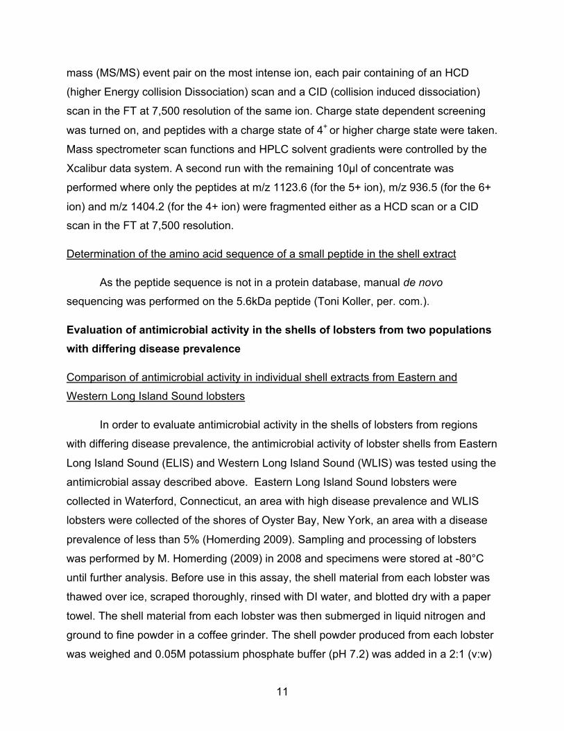
Figure 3. Influence of boiling on antimicrobial activity of a pooled shell extract
Percent decrease in V. parahaemolyticus growth in cultures supplemented with sterile pooled shell extract (Full Raw) and sterile pooled extract that had been boiled (Full Boiled). The final growth measurement was made 48h after T0. Error bars illustrate one standard deviation. All assays were done in triplicate with a shell extract prepared with lobster shell material from 8 individual lobsters. Effect of muffling on antimicrobial activity in American lobster shell
Heating the pooled shell material to 450°C for 4.5hr before preparing shell extract
significantly decreased activity, but did not eliminate it (Figure 4). The furnace treated
extract caused a 19±7% decrease in bacterial growth, which was a 56% smaller
decrease in growth than that seen with the full raw extract (44±2%). The protein
concentration of the furnace treated extract was 0.001% of the protein concentration of
raw extract, verifying that the muffling treatment destroyed virtually all organic material
in the extract. The reduced activity in the furnace treated extract indicates that the
antimicrobial activity observed in the shell extract is likely due to an organic component,
but observing residual activity in the furnace treated extract suggests that an inorganic
component may be contributing to the overall activity.
!"
#!"
$!"
%!"
&!"
'!"
(!"
)!"
*!"
+,--""
./0"
+,--""
123-45"
6"54784/94"3:";/7<483/-"=820<>"
?:@A3782;3/-"?99/B"%C##C$!#!"
D4/<"E84/<A4:<9""

15
Figure 4. Influence of muffling lobster shells on the antimicrobial activity of a pooled shell extract
Percent decrease in V. parahaemolyticus growth in cultures supplemented with sterile pooled shell extract (Full Raw), boiled extract (Full Boiled), and a sterile pooled extract prepared with muffled lobster shells (Furnace Treatment). The final growth measurement was made 48h after T0. Error bars illustrate one standard deviation. All assays were done in triplicate with a shell extract prepared with lobster shell material from 8 individual lobsters. Determination of antimicrobial activity in size fractionated shell extract (<3kDa, <10kDa,
>10kDa)
Size exclusion cartridges were used to produce three subsamples of the extract:
extract that could not pass through a 10kDa filter (10T), extract that did pass through a
10kDa filter (10B), extract that passed through a 3kDa filter (3B). Antimicrobial activity in
the extracts that passed through the 10kDa and the 3kDa filters was not significantly
different from the activity observed in the full extract (Figure 5). The antimicrobial activity
of the extract that was unable to pass through a 10kD filter was significantly lower than
!"
#!"
$!"
%!"
&!"
'!"
(!"
)!"
*!"
+,--""
./0"
+,--"
"123-45"
+,67/84""
964/:;
47:"
<"54864/=4"37">/8:463/-"?620:@"
A7B;3862>3/-"A==/C"%D##D$!#!"
E4/:"964/:;47:=""

16
that of the full extract (Figure 5). Additionally, the protein concentration of 10T
(4800µg/ml) was much higher than that of the full extract (1360µg/ml), 10B (76.8µg/ml),
or 3B (9.5µg/ml) treatments. These data indicate that the component of the extract
responsible for antimicrobial activity is less than 10kDa and is likely less than 3kDa.
Figure 5. Evaluation of antimicrobial activity in size fractionated shell extract
Percent decrease in V. parahaemolyticus growth in cultures supplemented with sterile pooled shell extract (Full), extract that did not pass through a 10kDa centrifugal filter unit (10T), extract that passed through a 10kD filter (10B), and extract that passed through a 3kD filter (3B). The final growth measurement was made 48h after T0. Error bars illustrate one standard deviation. All assays were done in triplicate with a shell extract prepared with lobster shell material from 8 individual lobsters. Fast protein liquid chromatography (FPLC) of shell extract
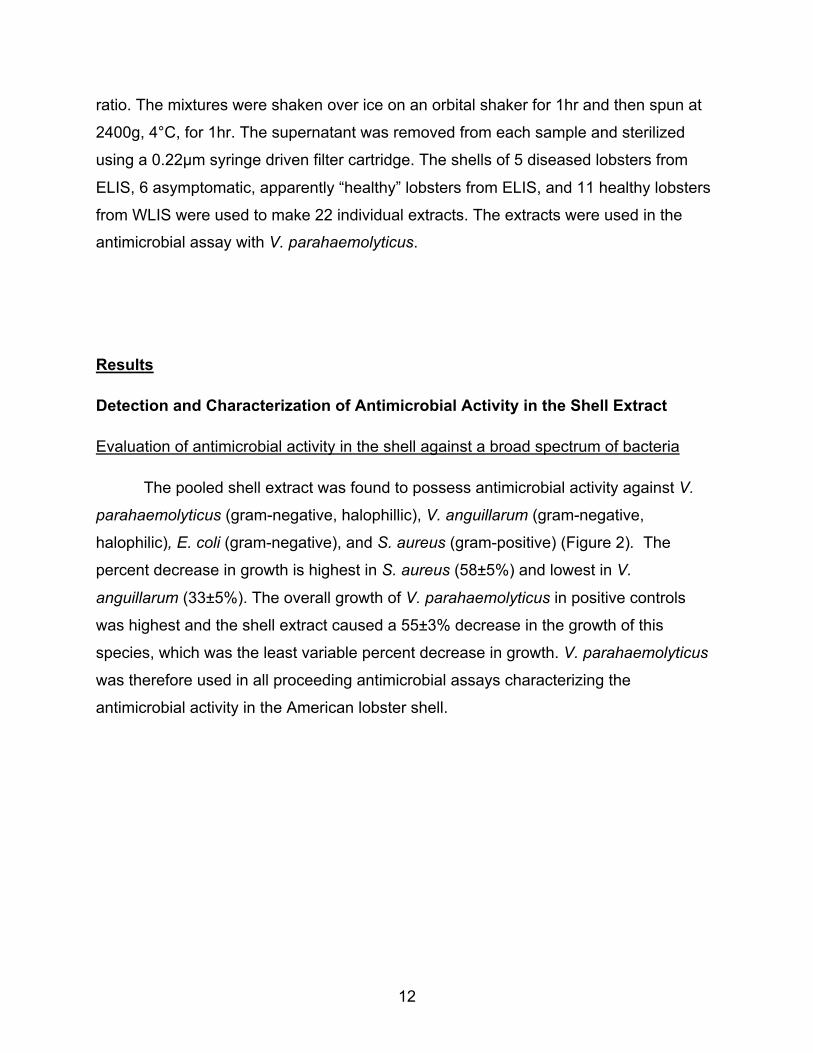
Fast protein liquid chromatography with a cation exchange column showed 5
protein peaks in the cationic fraction of the sterile shell extract (Figure 6). Samples
collected at time points 3, 4, 5, 6, and 10 correspond to protein peaks in the
chromatograph. The void volume peak (0-5min) corresponds to anionic organic
compounds in the shell fraction.
!"#$%&'()%*+,!--*.,/01201323,
4567,8'*&#("-,9%:;,-%<=,=>&+?-%(",&*':'%@A=-,
B,@=&'=*-=,%",)*&:='%*+,A'(9:;,
3,
23,
13,
C3,
/3,
D3,
E3,
F3,
G3,4?++,
23H,
23I,
CI,
17
Figure 6. Chromatograph from FPLC with pooled sterile shell extract
Full sterile shell extract was applied to an FPLC instrument with a cation exchange column and eluted with a linear gradient of 0–1M NaCl in 20mM Tris–HCl buffer (pH 8.5) at a flow rate of 1ml/min. The void volume was collected from minutes 1-4. One ml samples were collected from minutes 10-30 and labeled fractions 1-19 in the order they were collected. Fractions 3, 4, 5, 6, and 10 (underlined) correspond to protein peaks and were used in the antimicrobial assay.
Evaluation of antimicrobial activity of FPLC fractions
The FPLC fractions 3, 4, 5, 6, and 10 were chosen to be analyzed for
antimicrobial activity due to their correspondence to the protein peaks seen in the
chromatograph (Figure 6). Antimicrobial activity observed in FPLC fractions 3 and 4 was
not significantly different from that observed in the full extract (Figure 7). Fractions 5, 6,
and 10 exhibited lower antimicrobial activity and this activity was highly variable (Figure
7). The void volume, which consists of all anionic and inorganic components of the
extract, actually increased bacterial growth by 25% (Figure 7). These results indicate
!"#$%&'()*'+,-.%+/%012,3%
$),45-678/9%):%.;56),%'5<.*%=3
>?-3@%

18
that the component of the extract with antimicrobial activity is both organic and cationic
in nature and is present in FPLC fractions 3 and 4.
Figure 7. Evaluation of antimicrobial activity in sterile shell extract fractionated with FPLC
Percent decrease in V. parahaemolyticus growth in cultures supplemented with sterile pooled shell extract (Full), the void volume from the FPLC, and FPLC fractions taken at time points 3, 4, 5, 6, and 10. The final growth measurement was made 48h after T0. Error bars illustrate one standard deviation. All assays were done in triplicate with a shell extract prepared with lobster shell material from 8 individual lobsters. Tris/tricine SDS-PAGE with FPLC Fractions
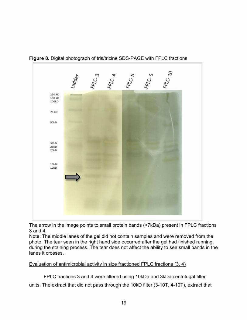
Gel electrophoresis of the FPLC fractions revealed two faint bands in the region
below the ladder’s 10kD marker in fraction 3 and 4 that were not seen in the other
fractions (Figure 8). This indicates that there are very small peptides (<10kD) present in
FPLC fractions 3 and 4, but not in FPLC fractions 5, 6, 7, and 10.
!"##$
%&'($
$%&#")*$
+$
,$
-$
.$
/0$
1$(*23*45*$'6$7428*3'4#$93&:8;$$
<6=)'23&7'4#$<554>$+?@-?@0/0$
!ABC$D&:E8;3&"9;$46($F342=&65$+G,G-G.GH$/0$
E.0$
E,0$
E@0$
0$
@0$
,0$
.0$
I0$
19
Figure 8. Digital photograph of tris/tricine SDS-PAGE with FPLC fractions
The arrow in the image points to small protein bands (<7kDa) present in FPLC fractions 3 and 4. Note: The middle lanes of the gel did not contain samples and were removed from the photo. The tear seen in the right hand side occurred after the gel had finished running, during the staining process. The tear does not affect the ability to see small bands in the lanes it crosses. Evaluation of antimicrobial activity in size fractioned FPLC fractions (3, 4)
FPLC fractions 3 and 4 were filtered using 10kDa and 3kDa centrifugal filter
units. The extract that did not pass through the 10kD filter (3-10T, 4-10T), extract that
!"#$#%&'(&)'*+',-./',"0$1*%2'

20
passed through the 10kD filter (3-10B, 4-10B), and extract that passed through a 3kD
filter (3-3B, 4-3B) were evaluated for antimicrobial activity. The antimicrobial activity of
the FPLC fraction 3 that passed through the 3kDa and 10kDa filters was not significantly
different from that of the full FPLC fraction 3 or the full raw sterile shell extract (Figure
9). The FPLC fraction 3 that was unable to pass through a 10kDa filter increased
bacterial growth (Figure 9). FPLC fraction 4 and its subsamples followed the same
pattern as FPLC fraction 3, but it was less pronounced and much more variable (Figure
9). These results confirm that the component of the extract responsible for antimicrobial
activity is less than 10kDa and is likely less than 3kDa.
Figure 9. Evaluation of antimicrobial activity in size fractionated FPLC fraction
Percent decrease in V. parahaemolyticus growth in cultures supplemented with sterile pooled shell extract (Full) and both FPLC fractions 3 & 4 that did not passed through a 10kD filter (3-10T, 4-10T), passed through a 10kD filter (3-10B, 4-10B), and passed through a 3kD filter (3-3B, 4-10B). The final growth measurement was made 48h after T0. Error bars illustrate one standard deviation. All assays were done in triplicate. The shell extract was prepared from lobster shell material from 8 individual lobsters.
!"##$$
$%$$
%$&'($$
%$&')$$
%$%)$$
*$$
*$&'($$
*$&')$
$*$%)$$
+,-./0123/4#$+5546$*78&78'&'$
!9:;$<140-2,5$=/>?$5/@A$AB0#"5/2,$041>1/CDA5$
E$CA01A45A$/,$340>A1/4#$D12=>?$
FG'$
F*'$
F8'$
'$
8'$
*'$
G'$

21
Determination of Molecular Weight of Small Peptides in Sterile Shell Extract
MALDI Mass Spectrometry shows 4 protein peaks at 1.6kDa, 2.8kDa, 4.6kDa,
and 5.6kDa, with the most intense of these peaks at 5.6kDa (Figure 10). There is a
large amount of noise in the lower mass range precluding further analysis of the smaller
peptides (1.6, 2.8, and 4.6kDa), which, based on the size filtration studies, are likely to
be involved in antimicrobial activity.
Figure 10. MALDI mass spectrometry of pooled sterile shell extract
MALDI MS spectrum obtained with sterile shell extract that passed through a 10kDa centrifugal filter unit. The most intense peak corresponding to a 5.6kDa peptide was further analyzed for amino acid sequence.
Determination of the amino acid sequence of a small peptide in the shell extract
The 5.6kDa peak was ion fractionated and a partial amino acid sequence was
determined (I/L V Y x N I/L x W N I/L V I/L x C R x G I/L I/L V N I/L W). I/L indicates
maggi mars <[email protected]>
Fw: first results1 message
[email protected] <[email protected]>
Let's see if Toni can get a better result for the MS/MS (sequencing).
It is possible that he would need something more concentrate. Let's see.
Any idea about detergent or glycerol in the sample?
Have a nice day
Emmanuelle
-----Forwarded by Emmanuelle Pales Espinosa/MSRC on 06/30/2010 03:28PM -----
To: <[email protected]>
From: "toni" <[email protected]>
Date: 06/30/2010 12:56PM
Subject: first results
Hi Emmanuelle
Here is the first experiment with your sample:
As you can see, there is a peak at ~5920, one around 5900 and one at 4589. In addition, the first experiment with my other approach gave me peaks at 5906.0, 5640.9, 5611.0, 4579.8, 3
Unfortunately, the MS/MS is pretty bad, so I will try another approach tomorrow to see if I can get a better signal/fragmentation pattern. After that we have to decide what to do. There are
Gmail - Fw: first results https://mail.google.com/mail/?ui=2&ik=4aad7ab402&view=pt...
1 of 2 7/13/10 7:37 PM
!"#$%&'
(")$%&'
*"#$%&'
+"#$%&'
!"#$%&$'(%)"
*+''",-./0"

22
amino acids that are either leucine or isoleucin and x represents an unknown amino
acid.
Evaluation of antimicrobial activity in the shells of lobsters from two populations with differing disease prevalence
Antimicrobial assay with shell extracts of lobsters from the Eastern and Western Long
Island Sound
Sterile shell extracts were made from the shells of 22 individual lobsters, which
were either apparently “healthy” from ELIS (n=5), diseased from ELIS (n=6), or healthy
from WLIS (n=11). Antimicrobial activity against V. parahaemolyticus was tested for
each individual extract and each individual extract was found to exhibit antimicrobial
activity. The average % decrease in bacterial growth for the individuals in each group
(ELIS Healthy, ELIS Diseased, ELIS All, and WLIS Healthy) was calculated. The
difference in the mean % decrease in bacterial growth with extracts from ELIS and
WLIS lobsters is significant at the 95% confidence level (Figure 11, Table 1). However,
there was no difference in antimicrobial activity between shell extracts from healthy and
diseased lobsters from ELIS (Figure 11).

23
Figure 11. Evaluation of antimicrobial activity in shell extracts from symptomatic and asymptomatic lobsters from Eastern and Western Long Island Sound
Averaged percent decrease in bacterial growth for ELIS diseased, ELIS healthy, All ELIS, and WLIS healthy lobster shell extracts .“ELIS Diseased” indicates Eastern Long Island Sound lobsters that exhibited ESD lesions on their carapace (n=5). “ELIS Healthy” (n=6) and “WLIS Healthy” (n=11) describes lobsters from Eastern and Western Long Island Sound with no visible ESD lesions. “ELIS ALL” (n=11) is an average of “ELIS Diseased” and “ELIS Healthy.” Final bacterial growth measurements were made 48h after T0. Error bars illustrate one standard deviation. All individual extracts were assessed for antimicrobial activity in triplicate.
Table 1. Analysis of Variance table of ELIS-All and WLIS-Healthy averaged % decrease in bacterial growth with shell extracts from individual lobsters from the two regions
!"#$
!%#$
!&#$
#$
&#$
%#$
"#$
'#$
()*+,-./0,12$(3314$"5&65#$
(89.1:93$/;$<=>?$@912A@4$1)B$B,39139B$,)B,8,BC123D$122$<=>?D$E$122$F=>?$
!"#$%&'()*+,-&*.&"/01&*2$-#('&-*34$('&5678&
<=>?$G$H,39139B$
<=>?$G$I912A@4$
<=>?$G$(22$
F=>?$G$I912A@4$$
J$B9-.9139$,)$01-A9.,12$:./KA@$
Df Sum Sq Mean Sq F value Pr(>F)
% decrease 1 127.04 127.039 6.9371 0.01636 *
Residuals 19 347.95 18.313
Signif. codes: 0 ‘***’ 0.001 ‘**’ 0.01 ‘*’ 0.05
24
Discussion
Detection and characterization of antimicrobial activity in American lobster shell
The sterile pooled shell extract consistently exhibited antimicrobial activity
against a broad spectrum of bacteria. Individual shell extracts from 22 lobsters each
showed a comparable activity against V. parahaemolyticus. These data indicate that
American lobster cuticle possesses an intrinsic antimicrobial activity and therefore likely
plays a larger role in the defense against invading pathogens than a simple physical
barrier. Haug et al. (2002) discovered broad-spectrum antimicrobial activity in the
exoskeletons of four marine decapods: Northern Shrimp (Pandalus borealis), Hermit
Crab (Pagurus bernhardus), Spider Crab (Hyas araneus), and King Crab (Paralithodes
camtshatica). This activity was not fully characterized, but it was found to be heat
resistant and sensitive to enzyme proteinase k (Haug et al. 2002). The antimicrobial
activity discovered here, in the shell of American lobster, is also heat resistant.
Furthermore, the antimicrobial activity in the shell of the American lobster is due to an
organic, cationic component less than 10kDa and likely less than 3kDa.
Based on the data presented, it is likely that an antimicrobial peptide (AMP) in
the shell of American lobster is responsible for the antimicrobial activity observed. AMPs
are small (<10kDa), cationic peptides that exhibit broad-spectrum antimicrobial activity.
Structural characteristics of AMPs and their small size make them stabile and heat
resistant. Although AMPs have not previously been reported to exist in crustacean
exoskeletons, they are wide spread internal defense factors in crustaceans (Table 2).
Three distinct families of crustacean AMPs have been characterized based on AMPs
isolated from shrimp: the penaeidins, antilipopolysaccharide factors (ALFs), and crustins
(Pisuttarachai et al. 2009). Because these defense molecules are ubiquitous internally,
it is possible that AMPs may be present in the crustacean cuticle as well. The chitin-
binding capabilities of shrimp penaeidins support this idea as they have been found
bound to chitin in gill cuticle surfaces of shrimp (Destoumieux et al. 2000).
Of the three families of crustacean AMPs, the penaeidins (5.48-6.62kDa) found
in shrimp are the most prominent (Bachere et al. 2000). These AMPs show antifungal
activity and specific antibacterial activity against gram-positive bacteria, but do not show
25
strong activity against Vibrio spp. (Destoumieux et al. 1999). This group of AMPs is
produced in hemocytes and they appear to be released when hemocytes lyse in
response to infection (Destoumieux et al. 2000). The penaeidins are very stable and are
resistant to proteolysis (Destoumieux et al. 1999). However, penaeidins have not been
isolated from lobsters.
AFL’s were first discovered in horseshoe crab plasma. Bacterial endotoxins
trigger a cascade in horseshoe crab hemocytes leading to rapid degranulation and
intracellular coagulation; horseshoe crab AFLs are capable of binding and neutralizing
LPS, thus inhibiting this cascade (Iwanaga 1993). AFL’s have also been isolated from
the hemolymph of many shrimp species (Somboonwiwat et al. 2005) and the American
lobster, Homarus americanus (Beale et al. 2008). AFL’s have direct antimicrobial
capabilities and have been shown to be active against gram-negative and positive
bacteria and filamentous fungi (Somboonwiwat et al. 2005).
Crustins (7-14kD) have been isolated from the hemolymph of a variety of
crustaceans and are categorized based on the crustaceans they are found in: type I
crustins are found in crabs, lobsters, and crayfish; trype II crustins are mostly found in
shrimp; types III crustins are found in decapods. All crustins, however, have a cationic
region at the carboxyl terminus with the first of twelve cysteine residues making up the
whey acidic protein (WAP) domain, which is associated with antimicrobial activity
(Pisuttharachai et al. 2009).
Specific examples of AMP’s isolated from the hemolymph of large crustaceans
(Table 2) include a proline-rich, 6.5 kD AMP (Shnapp et al. 1996) and a cysteine-rich,
11.5kD, gram-positive specific (Relf et al. 1999) AMP from the shore crab Carcinus
maenas. The 11.5kD peptide has been shown to share similarities with crustins
expressed in shrimp (Tincu & Taylor 2004). A smaller cationic antimicrobial peptide
with molecular weight of 3.7kD has been found in blue crab (Callinectus sapidus)
hemocytes (Khoo et al. 1999). Additionally, four isoforms of a type two crustin have
been characterized from the Japanese spiny lobster, Panulirus japonicus
(Pisuttharachai et al. 2009). In American lobster, cDNA encoding a 96 amino acid
crustin-like peptide has been identified and sequenced (ACN: CN853187.1). This

26
crustin is referred to as Hoa-crustin and is most similar to a crustin isoform found in the
European lobster, Homarus gammarus (Christie et al. 2007). In 2008, Battison et al.
isolated and characterized two antimicrobial peptides from the hemocytes of American
lobster. The first is approximately 12kD, expresses specific antibacterial activity against
gram-positive bacteria, and contains amino acid sequences that were predicted by the
cDNA sequenced by Christie et al. (2007). The second peptide, homarin, is composed
of four 6kD subunits and is similar to amphibian temporins, but unlike any other marine
invertebrate AMP. Homarin has been shown to exhibit antimicrobial activity against
gram-negative bacteria as well as two cultured scuticociliate parasites (Battison et al.
2008).
Table 2. Selected examples of AMPs isolated from the of hemolymoh large crustaceans Name Species Size Description Reference Penaeidins shrimp
Panaeus vannamei
2.7-8.32kD
Proline and cysteine rich, 3 disulfide bridges
Destoumieux et al. (2000)
ALFPm3 Penaeus monodon
10kD Anti-lipopolysachharide Factor Somboonwiwat et al. (2006)
broad spectrum antibacterial protein
Carcinus Maenus
6.5kD
Proline rich Schnapp et al. (1996)
Carcinin Carcinus Maenus
11.5kD
crustin, cysteine rich, disulfide bridge
Relf et al.
Callinectin
Callinectus sapidus
3.7kD
proline is most abundant AA, but is not arranged in way typical to proline rich AMPs
Khoo et al. (1999)
Hoa-crustin Homarus americanus
12kD crustin Battison et al. (2008)
Homarin Homarus americanus
4-6kD dissimilar to other crustacean AMPs, similar to amphibian temporins
Battison et al. (2008)
PJC1, 2, 3, 4 Panulirus japonicus
13.6-15.7kD
4 individual crustins more similar to shrimp than lobster crustins
Pisuttharachai et al. (2009)
ALFHa-1 & 2 Homarus americanus
13.7kD, 13.9kD
Anti-lipopolysaccharide factors Beale et al. (2008)
27
Antimicrobial peptides (AMPs) are an integral component of the invertebrate
internal defense system and have been found in every invertebrate species in which
their presence has been investigated (Hancock et al. 2006). Over 2,000 AMP’s have
been identified in a large variety of organisms (Park et al. 1998), including plants,
insects, marine invertebrates, marine vertebrates, amphibians, and mammals, including
humans (Powers & Hancock 2003). A high degree of diversity exists in AMP sequences
and the same peptide sequence has not been found in two species, but segments and
motifs of amino acid sequence can be conserved within AMP classes across species
(Zasloff 2002). AMPs are typically made up of 12-50 amino acids, 2-9 of which are
positively charged lysine or arginine residues and up to 50% of the amino acids in
AMPs are usually hydrophobic (Tincu & Taylor 2004). AMPs are usually less than
10kDa and many AMPs are less than 3kDa (table 3).
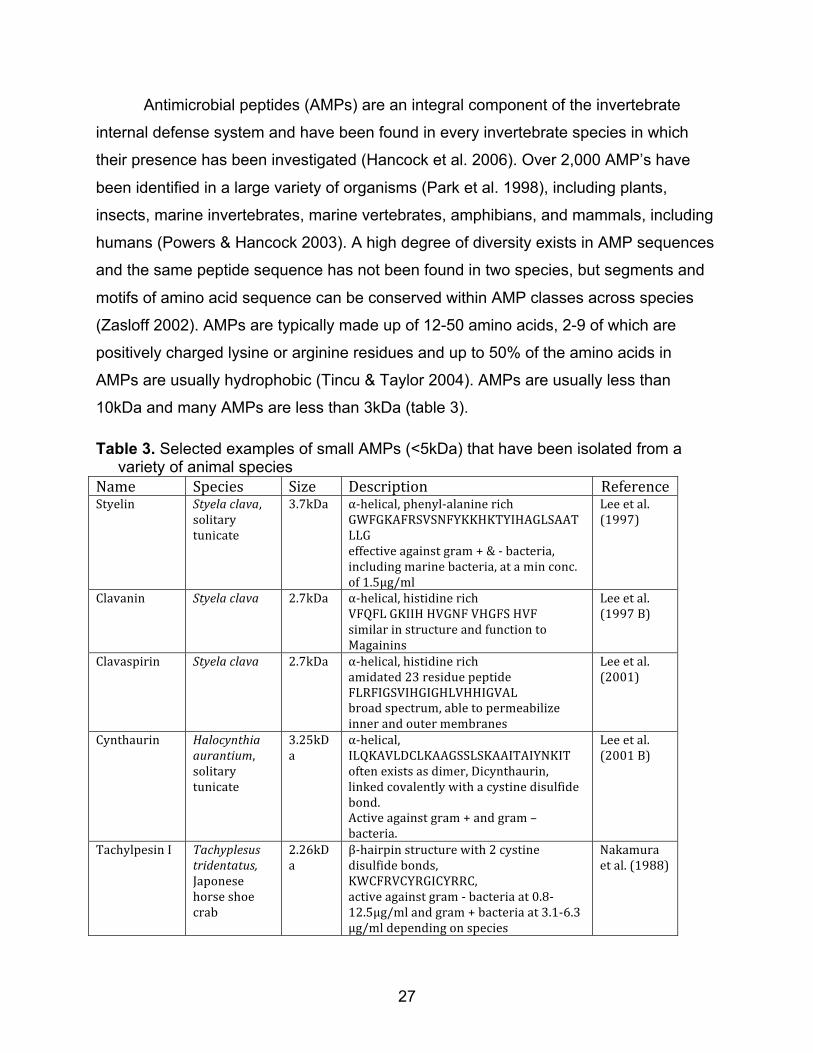
Table 3. Selected examples of small AMPs (<5kDa) that have been isolated from a variety of animal species Name Species Size Description ReferenceStyelin Styelaclava,
solitarytunicate
3.7kDa α‐helical,phenyl‐alaninerichGWFGKAFRSVSNFYKKHKTYIHAGLSAATLLGeffectiveagainstgram+&‐bacteria,includingmarinebacteria,ataminconc.of1.5μg/ml
Leeetal.(1997)
Clavanin Styelaclava 2.7kDa α‐helical,histidinerichVFQFLGKIIHHVGNFVHGFSHVFsimilarinstructureandfunctiontoMagainins
Leeetal.(1997B)
Clavaspirin Styelaclava 2.7kDa α‐helical,histidinerichamidated23residuepeptideFLRFIGSVIHGIGHLVHHIGVALbroadspectrum,abletopermeabilizeinnerandoutermembranes
Leeetal.(2001)
Cynthaurin Halocynthiaaurantium,solitarytunicate
3.25kDa
α‐helical,ILQKAVLDCLKAAGSSLSKAAITAIYNKIToftenexistsasdimer,Dicynthaurin,linkedcovalentlywithacystinedisulfidebond.Activeagainstgram+andgram–bacteria.
Leeetal.(2001B)
TachylpesinI Tachyplesustridentatus,Japonesehorseshoecrab
2.26kDa
β‐hairpinstructurewith2cystinedisulfidebonds,KWCFRVCYRGICYRRC,activeagainstgram‐bacteriaat0.8‐12.5μg/mlandgram+bacteriaat3.1‐6.3μg/mldependingonspecies
Nakamuraetal.(1988)

28
Table 3. Continued from previous page TachyplesinII Limulus
polyphemus2.5kDa β‐hairpinstructurew/twocystine
disulfidebondsRWCFRVCYRGICYRKCRactiveagainstgram+&‐bacteriaandthefunguscandidaalbicansM9
Miyataetal.(1989)
Magainin2 Xeonpuslaeuis(Africanclawedfrog)
2.5kDa α‐helical,GIGKFLHSAKKFGKAFVGEIMNS,formsporesinmembranesandtranslocatesthemembrane
Matsuzakietal.(1995)
Melittin Apismellifera,EuropeanHoneyBee
2.8kDa Interruptedα‐helicalconformation,twohelicesseparatedbybend,GIGAVLKVLTTGLPALISWIKRKRQQ,increasesmembranepermeability
Bechinger(1996)
BuforinII Bufobufogaragriozans,Asiantoad
2.0kDa α‐helical,TRSSRAGLQFPVGRVHRLLRK,strongactivityagainstgram+&‐bacteria,formsporesandinhibitscellfunctionbybindingwithDNAandRNA
Parketal.(1998)
Pyrrhocoricin Pyrrhocorisapterus,firebug
3.0kDa linear,prolinerich,VDKGSYLPRPTPPRPIYNRN,Broadspectrum,DNAbinding
Kragoletal.(2002)
Thanatin Podisusmaculiventris,SpinedSoldierBug
2.4kDa β‐hairpinstructurewithonedisulfidebridge,GSKKPVPIIYCNRRTGKCQRM,activeagainstgram+&‐bacteriaandfungi
Pascaleetal.(1996)
Apidaecin Apismellifera,HoneyBee
2.0kD linear,prolineandargininerichGNNRPVYIPQPRPPHPRIactiveagainstgram‐bacteriaonly,stereospecific,butnotporeforming,translocatesanddepolarizesinnermembrane
Castleetal.(1999)
Indolicidin Bostaurus,cow
1.5kD linear,tryptophanandprolinerich,smallestknownAMP,ILPWKWPWWPWRR,activeagainstgram+&‐bacteria,increasesmembranepermeabilityandinhibitsDNAsynthesis
Hsuetal.(2005)
Antimicrobial peptides are classified based on their structural characteristics.
They can generally be assigned to one of three structural groups: (1) linear peptides
that form α-helical structures, (2) linear peptides rich in a specific amino acid, and (3)
cysteine-rich open-ended peptides containing one or more disulfide bridges (Viziolo &
Salzet 2002). AMPs with disulphide bridges independently maintain their secondary
structure (Hancock et al 2006), typically a β-hairpin configuration. The hairpin
configuration lends stability to the peptide and allows it to maintain structural integrity at

29
high temperatures and low pH’s (Tincu & Taylor 2004). Linear peptides that form α-
helices and linear peptides that are rich in a particular amino acid exist in solution with
little or no secondary structure and fold into an amphipathic structure when they contact
a target membrane. Because these AMPs lack secondary and tertiary structure, they
are also very stabile and heat resistant (Hancock et al. 2006).
The mechanism of action has not been elucidated for all AMP’s that have been
isolated, but general structure-function relationships have been described for the major
structural groups (Viziolo & Salzet 2002). Most AMPs interact with the outer membrane
of gram-negative bacteria in a similar manner. AMPs associate with the anionic
lipopolysaccharides (LPS) of the outer membrane through electrostatic interactions that
are stronger than those between LPS and native divalent cations such as Mg2+ and
Ca2+. As a result, native divalent cations are displaced, which leads to a local
disturbance in the outer membrane allowing the AMP to cross the outer membrane and
reach the cytoplasmic membrane. Once an AMP reaches the cytoplasmic membrane,
its mode of action becomes dependent on its structure (Powers & Hancock 2003).
Linear amphipathic peptides increase bacterial membrane permeability through the
interaction of their cationic charge with anionic membrane lipids or through membrane
lipid displacement. Cysteine-rich peptides with disulfide bridges and α-helical peptides
may form ion-permeable channels in the lipid bilayer. The peptides may or may not
cross the membrane into the cell, but it is likely that this is the case for most small
AMPs. Microbial death follows either from the disturbance of membrane functioning or
damage incurred to crucial intracellular targets after an AMP enters the cell (Viziolo &
Salzet 2002).
In this study, MALDI mass spec of the shell extract revealed four small peptides
(1.6, 2.8, 4.6, and 5.6 kDa) in the shell extract. These sizes are typical of antimicrobial
peptides. Unfortunately only a partial sequence of the 5.6kDa peptide could be
determined. The sequence, I/L V Y x N I/L x W N I/L V I/L x C R x G I/L I/L V N I/L W,
has several unknowns and whether or not it is an AMP can not be determined
conclusively. The sequence includes one (known) positively charged arginine, while
most AMPs contain 2-9 arginine residues. Thirteen of the identified amino acids in the

30
22 amino acid sequence are hydrophobic; this is more than 50%, but it is close to the
percent range of hydrophobic amino acids in AMPs. The peptide is not rich in proline or
glycine as are many linear AMPs rich in a particular amino acid isolated from
invertebrates (Viziolo & Salzet 2002). Additionally, when different combinations of
unknowns in the unknown peptide were searched using the Basic Local Alignment Tool
(BLAST), a partial match was made with the α-helical peptide mastaparan, which is a
broad-spectrum AMP isolated from wasps (Figure 12) (Hisada et al. 2000). This match
is not conclusive, but the data suggest that the 5.6kDa peptide is also an α-helical AMP.
Table 4. Characteristics of the unknown 5.6kDa peptide
Abbreviation
AminoAcid
Charge
HydrophobicorphilicatpH7
%oftotalaminoacids
I/L IsoleucineorLeucine
neutral veryhydrophobic 27.3%
V Valine neutral veryhydrophobic 13.6%Y Tyrosine neutral* hydrophobic 4.5%x ‐ ‐ ‐ 18%N Asparagine neutral* hydrophilic 13.6%x W Tryptophan neutral veryhydrophobic 9.1%N Asparagine neutral* hydrophilic I/L Isoleucineor
Leucineneutral veryhydrophobic
V Valine neutral veryhydrophobic I/L Isoleucineor
Leucineneutral veryhydrophobic
x ‐ ‐ ‐ C Cysteine neutral hydrophobic 4.5%R Arginine (+)* hydrophilic 4.5%x ‐ ‐ ‐ G Glycine neutral aliphatic 4.5%I/L Isoleucineor
Leucineneutral veryhydrophobic
I/L IsoleucineorLeucine
neutral veryhydrophobic
V Valine neutral veryhydrophobic N Asparagine neutral* hydrophilic I/L Isoleucineor
Leucineneutral veryhydrophobic
W Tryptophan neutral veryhydrophobic *Polar

31
Figure 12. BLAST sequence alignment of the unknown 5.6kDa peptide with the wasp antimicrobial peptide mastoparan
Evaluation of antimicrobial activity in the shells of lobsters from two populations with differing disease prevalence
When the antimicrobial activity in shells of lobsters from ELIS and WLIS were
evaluated, extracts from WLIS lobsters showed significantly higher antimicrobial activity
than those from ELIS lobsters. There was no difference in antimicrobial activity in shell
extracts from healthy and diseased ELIS lobsters. Homerding (2009) found that
hemocytes and plasma from WLIS lobsters have significantly increased phagocytotic
rates, antimicrobial activity, native concentrations of reactive oxygen species, and
induced concentrations of reactive oxygen species when compare to ELIS lobsters. It
was also found that WLIS lobster carapaces are significantly thicker than ELIS lobster
carapaces. While lobsters are known to migrate in and off-shore, tagged lobsters from
Eastern and Western Long Island Sound are generally recaptured where they were
initially tagged, suggesting limited migration between the two populations (Landers et al.
2007). Indeed, it has been shown that WLIS lobsters are genetically isolated from ELIS
lobsters, but ELIS lobsters are not genetically isolated from off-shore lobsters (Crivello
et al 2005). It is possible that WLIS lobsters having gone through a severe die-off during
the 1999 mortality event, experienced a genetic bottleneck, and the population now has
a more robust defense system. If this is the case, then the defense capabilities of the
populations may be affecting their susceptibilities to Epizootic Shell diseased and
decreased antimicrobial activity in the shell may play a role in ELIS lobsters’ increased
susceptibility.
!"#$%&"'"())
*(+(%,()-.&/0.1)234+5")
32
Conclusions
This study has demonstrated broad-spectrum antimicrobial activity in the shell of
American lobster for the first time. Characterization of the activity has determined that it
is caused by an organic, cationic component, less than 10kDa, and likely less than
3kDa. Based on these results, it is likely that an antimicrobial peptide is responsible for
the activity observed. The presence of AMPs in crustacean cuticle has not been
previously studied. This study has therefore contributed to building a more
comprehensive understanding of crustacean defense mechanisms. Additionally, the
difference in antimicrobial activity in the shells of ELIS and WLIS lobster shells suggests
that the activity detected and characterized may play a role in susceptibility to shell
disease.
Future Directions
Continuation of this research requires the purification and full sequencing of all the
small peptides observed in the MALDI mass spec of the sterile shell extract. Evaluation
of the antimicrobial activity of purified peptides would allow for the determination of
which peptide(s) is responsible for the activity observed in the shell extract. If the
causative agent of Epizootic Shell Disease is determined, the activity of the
antimicrobial peptide(s) from the lobster shell against this pathogen should be
investigated. The activity of the peptide(s) should also be evaluated against other
pathogens affecting lobster populations. Differences in concentrations of AMPs in
lobster shells from different populations with varying disease prevalence can also be
investigated.
A public health interest exists in identifying and characterizing new AMPs.
Antimicrobial peptides often act by interrupting microbial cell membranes, an action that
is believed to be unlikely to incite microbial resistance. This property, along with their
small size, which facilitates their synthesis, and generally low toxicity to eukaryotic cells,
makes AMPs an attractive prospect for biomedical development (Tincu & Taylor 2004).
33
This is especially the case since some AMP’s are able to inhibit the replication of
enveloped viruses, such as influenza A virus and human immunodeficiency virus (HIV-
1). Some AMPs have also demonstrated anticancer capabilities and promote wound
healing (Powers & Hancock 2003). Therefore, a better characterization of AMPs in
lobster shells may also have significant biomedical applications.
34
Works Cited
Asano, T., Ashida, M. (2001) Cuticular Pro-phenoloxidas of the Silkworm, Bombyx mori: Purification and Demonstration of its Transport from Hemolymph. The Journal of Biological Chemistry. 276 (14), 11100-11112.
Ashida, M., Brey, P. T., (1995) Role of the integument in insect defense: Pro-phenol oxidase cascade in the cuticular matrix. Proceedings of the National Academy of Science USA. 92, 10698-10702.
Bachere, E., Destoumieux, D., Bulet, P. (2000) Penaids, antimicrobial peptides of shrimp: a comparison with other effectors of innate immunity. Aquaculture. 191, 71-88.
Battison, A. L., Summerfield, R., Patrzykat, A. (2008) Isolation and Characterization of two antimicrobial peptides from haemocytes of the American lobster Homarus americanus. Fish and Shellfish Immunology. 25. 181-187.
Bechinger, B. (1996) Structure and function of channel-forming peptides: magainins, cecropins, melittin, and alamethicin. Journal of Membrane Biology. 156, 197-211.
Beale, K.M., Towle, D.W., Jayasundara, N., Smith, C.M., Shields, J.D., Small, H.J., Greenwood, S.J., (2008) Anti-lipopolysaccharide factors in the American lobster Homarus americanus: molecular characterization and transcriptional response to Vibrio fluvialis challenge. Comparative Biochemistry and Physiology, Part D, 3. 263-269.
Brown, K.L., Hancock, R.E.W., (2006) Cationic host defense (antimicrobial) peptides. Current Opinion in Immunology. 18, 24-30.
Castilla, J.C., Paine, R.T. (1987) Predation and community organization on Eastern Pacific, temperate zone, rocky intertidal shores. Revista Chilena de Historia Natural. 60, 131-151.
Castle , M., Nazarian, A., Yi, S.S., Tempst, P. (1999) Lethal effects of Apidaesin on Escherichia coli involve sequential molecular interactions with diverse targets. Journal of Biological Chemistry. 274, 32555-32564.
Chen, J.S., Rolle, R.S., Marshall, M.R., Wei, C.I. (1991) Comparison of phenoloxidase activity from Florida spiny lobster and Western Australian lobster. Journal of Food Science, 56, 154-157.
Cheng, H.T., Townsend, R.E. (1993) Potential Impact of Seasonal Closures in the U.S. Lobster Fishery. Marine Resource Economics. 8, 101-117.
Christie, A. E., Rus, S., Golney, C. C., Smith, C. M., Towle, D. W., Dickinson, P. S. (2007) Identification and characterization of a cDNA encoding a crustin-like, putative antibacterial protein from the American lobster Homarus americanus. Molecular Immunology. 44 (13), 3333-3337.
Cobb, J. Stanley, Castro, Kathleen M. (2006) Sell disease in lobsters: A synthesis. New England Lobster Research Initiative.

35
Cook, D.W., Lofton, S.R., (1973) Chitinoclastic bacteria associated with shell disease in Penaeus shrimp and the blue crab (Callinectes sapidus). Journal of Wildlife Diseases. 9, 154-159.
Crivello, J.F., Landers, D., Kessler, M. Jr. (2005) The genetic stock structure of the American lobster in Long Island Sound and the Hudson Canyon. Journal of Shellfisheries Research. 24, 841-848.
Davis, A., Hoanson, J.M., Watts, H., MacPherson, H. (2004) Local ecological knowledge and marine fisheries research: the case of white hake (Urophysic tenuis) predation on juvenile Amercan lobster (Homarus americnus). Canadian Journal of Fisheries and Aquatic Science. 61, 1191-1201.
Destoumieux, D., Bulet, P., Strub, J., VanDorsselaer, A., Bachere, E. (1999) Recombinant expression and range of activity of penaeidins, antimicrobial peptides from penaeid shrimp. European Journal of Biochemistry. 266, 335-346.
Destoumieux, D., Munoz M., Cosseau, C., Rodriguez, J., Bulet, P., Comps, M., Bachere, E. (2000) Penaeidins, antimicrobial peptides with chitin-binding activity, are produced and stored in shrimp granulocytes and released after microbial challenge. Journal of Cell Science. 113, 461-469.
Ferrer, O. J., Koburger, J. A., Otwall, W. S., Gleeson, R. A., Simpson, B. K., Marshall, M. R. (1989) Phenoloxidase from the Cuticle of Florida Spiny Lobster (Panulirus argus): Mode of Activation and Characterization. Journal of Food Science, 54 (1), 63-68.
Getchell, R.G. (1989) Bacterial shell disease in crustaceans: a review. Journal of Shellfish Research. 8, 1-6.
Harvell, C.D., Kim, K., Burkholder, J.M., Colwell, R.R., Epstein, P.R., Grimes, D.J., Hofmann, E.E., Lipp, E.K., Osterhaus, A.D.M.E., Overstreet, R.M., Porter, J.W., G. W. Smith, Vasta G.R. (1999) Emerging Marine Diseases--Climate Links and Anthropogenic Factors. Science. 285, 1505 - 1510.
Hancock, R.E.W., Brown, K.L., Mookherjee, N. (2006) Host defence peptides from invertebrates - emerging antimicrobial strategies. Immunobiology. 211, 315-322.
Haug, T., Kjuul, A. K., Stensvag, K., Sandsdalen, E., Styrvold, O. B. (2002) Antibacterial activity in four marine crustacean decapods. Fish and Shellfish Immunology, 12, 371-385.
Hisada, M., Konno, K., Itagaki, Y., Naoki, H., Nakajima, T. (2000) Advantages of using nested collision induced dissociation/post-source decay with matrix-assisted laser desorption/ionization time-of-flight mass spectrometry: sequencing of novel peptides from wasp venom. Rapid communication Mass Spectrometry. 14, 1828-1834.
Homerding, M. (2009) Characterization of cuticular and hemolymph associated defense parameters in shell diseased lobsters. Master’s Thesis, State University of New York at Stony Brook.
36
Hsu, C-H., Chen, C., Jou, M-L., Lee, A. Y-L., Lin, Y-C., Yu, Y-P., Huang, W-T., Wu, S-H., (2005) Structural and DNA-binding studies on the bovine antimicrobial peptide, indolicidin: evidence for multiple conformations involved in binding to membranes and DNA. Nucleic Acids Research. 33. 4053-4064.
Iwanaga, S. (1993) The limulus clotting reaction. Current Opinions in Immunology 5, 74-82.
Khoo, L., Robinette, D. Noga, D. (1999) Callinectin, an antibacterial peptide from the blue crab, Callinectus sapidus, hemocytes. Marine Biotechnology. 1, 44-51.
Kragh, M., Molbak, L., Anderson, S. O. (1997) Cuticular proteins from the lobster, Homarus americanus.
Kragol, G., Hoffmann, R., Chattergoon, M.A., Lovas, S., Cudic, M., Bulet, P., Condie, B.A., Rosengren, K.J., Montaner, L.J., Otvos Jr., L. (2002) Identification of crucial residues for the antibacterial activity of the proline-rich peptide pyrrhocoricin. European Journal of Biochemistry. 269, 4226-4237.
Landers, D.F. (2005) Prevelance and severity of shell disease in American lobster Homarus americanusfrom Eastern Long Island Sound, Connecticut. In: Lobster Shell Disease Workshop Forum Series 05-1 (Tlusty, M.F., Halvorson, H.O., Smolowitze, R., & Sharma, U. eds.) pp. 04-97., Boston, MA: New England Aquarium.
Lee, I.H., Cho, Y., Lehrer, R.I. (1997) Styelins, broad-spectrum antimicrobial peptides from the solitary tunicate, Styela clava. Comparative Biochemistry and Physiology Part B. 119, 515-521.
Lee, I.H, Zhao, C. Nguyen, T. Menzel, L., Waring, A.J., Sherman, M.A., Lehrer (2001) Clavaspirin, an antibacterial and haemoltic peptide from Styela clava. Journal of Peptide Research. 58, 445-456.
Lee, I.H., Lee, Y.S., Kim, C.H., Kim, C.R., Hong, T. Menzel, L., Boo, L.M., Pohl, J., Sherman, M.A., Waring, A., Lehrer, R.L. (2001) Dicynthaurin: an antimicrobial peptide from hemocytes of the solitary tunicate, Halocynthia aurantium. Biochemica et Biophysica Acta (BBA)- General Subjects. 1527, 141-148.
Matsuzaki, K., Murase, O., Fujii, N., Miyajima, K. (1995) Translocation of a channel-forming antimicrobial peptide, Magainin 2, across lipid bilayers by forming a pore. Biochemistry. 34, 6521-6526.
McKown, K., Glenn, R., Howell, P., Jacobson, L., Nesslage, G., Wilson, C. (2009) American lobster stock assessment report for peer review. Atlantic States Marine Fisheries Commission, NOAA.
Miyata, T., Tokunaga, F., Yoneya, T., Yoshikawa, K., Iwanaga, S., Niwa, M., Takao, T., Shimonishi, Y. (1989) Antimicrobial peptides isolated from horshoe crab hemocytes, Tachyplesin II, and Polyphemusins I and II: chemical structures and biological activity. Journal of Biochemistry. 106, 663-668.

37
Mullen, T.E., Russell, Spencer, Tucker, Meghan, Maratea, Jennifer L., Koering, Claudia, Hinckley, Lynn, De Guise, Sylvain, Frasca, Salvatore, Jr., French, Richard, Burrage, Thomas G., Perkins, Christopher. (2004) Paramoebiasis associated with mass mortality of American lobster, Homarus americanus, in Long Island Sound, USA. Journal of Aquatic Animal Health. 16, 29-38.
Nakamura, T. Furunaka, H., Miyata, T., Tokunaga, F., Muta, T., Iwanaga, S. (1988) Tachyplesin, a class of antimicrobial peptide from the hemocytes of the horshecrab (Tachypleus tridentatus): isolation and chemical structure. The Journal of Biological Chemistry. 263. 16709-16713.
Noga, E.J., Engle, D.P., Arroll, T.W. McKenna, S., Davidian, M. (1994) Low serum antibacterial activity coincides with increased prevalence of shell disease in blue crabs Callinectes sapidus. Diseases of Aquatic Organisms. 19, 121-128.
Pascale, F., Bulet, P., Chernysh, S., Briand, J-P, Roussel, J-P, Letellier, L., Hetru, C., Hoffman, J.A. (1997) Structure-activity analysis of Thanatin, a 21-residue inducible insect defense peptide with sequence homology to frog skin antimicrobial peptides. Proceedings of the National Academy of Sciences of the United States of America. 93, 1221-1225.
Pearce, J., Balcom, N.. (2005) The 1999 Long Island Sound lobster mortality event: Findings of the comprehensive research initiative. Journal of Shellfish Research. 26, 691-697.
Pisuttharachai, D., Fagutao, F. F., Yauike, M., Aono, H., Yano, Y., Murakami, K., Kondo, H., Aoki, T., Hirono, I. (2009) Characterization of crustin antimicrobial proteins from Japanese spiny lobster Panulirus japonicus. Developmental and Comparative Immunology. 33, 1049-1054.
Park, C.B., Kim, H.S., Kim, S.C. (1998) Mechanism of action of the antimibacterial peptide buforin II: buforin II kills microorganisms by penetrating the cell membrane and inhibiting cellular functions. Biochemical and Biophysical Research Communications. 244, 253-257.
Powers, J. S., Hancock, R.E.W. (2003) The relationship between peptide structure and antibacterial activity. Peptides. 24, 1681-1691.
Relf, J. M., Chisholm, J.R.S., Kemp, G. D., Smith, V. J. (1999) Purification and characterization of a cysteine-rich 11.5kDa antibacterial protein from the granular haemocytes of the shore crab Carcinus maenus. European Journal of Biochemistry. 264 (2), 350-357.
Schnapp, D., Kemp, G. D., Smith, V. J. (1996) Purification and characterization of a proline-rich antibacterial peptide, with sequence similarity to bactenecin-7, from hemocytes of the shore crab Carcinus maenas. European Journal of Biochemistry. 240 (2), 532-539.

38
Schägger, H. & von Jagow, G. (1987) Tricine–sodium dodecyl sulfate polyacrylamide gel electrophoresis for the separation of proteins in the range from 1–100 kDalton. Analytical Biochemistry. 166, 368–379.
Somboonwiwat, K., Marcos, M., Tassanakajon, A., Klinbunga, S., Aumelas, A., Romestand, B., Gueguen, Y., Boze, H., Moulin, G., Bachere, E. (2005) Recombinant expression and anti-microbial activity of anti-lipopolysaccharide factor (ALF) from the black tiger shrimp Penaeus monodon. Developmental and Comparative Immunology. 29, 841-851.
Smolowitz, Roxanna, Chistoerdov, Andrei Y., Hsu, Andrea. (2005) A description of the pathology of epizootic shell disease in American lobster, Homarus Americanus, H. Milne Edwards 1837. Journal of Shellfish Research. 24, 749-756
Sritunyalucksans, K., Soderhall, K. (2000) The proPO and clotting system in crustaceans. Aquaculture. 191, 53-69.
Tall, B.D., Fall, S., Pereira, M.R., Ramos-Valle, M., Curtis, S.K., Kothary, M.H., Chu, D.M., Monday, S.R., Kornegay, L. Donkar, T., Prince, D., Thunberg, R.L. Shangraw, K.A., Hanes, D.E., Khambaty, F.M., Lampel, K.A., Bier, J.W., Bayer, R.C. (2003) Characterization of Vibrio fluvialis- like strains implicated in limp lobster disease. Applied Environmental Microbiology. 69, 7435, 7446.
Tincu, J. A., Taylor, S. W. (2004) Antimicrobial Peptides from Marine Invertebrates. Antimicrobial Agents and Chemotherapy, 48 (10), 3645-3654.
Viziolo, J., Salzet, M. (2002) Antimicrobial peptides from animals: focus on invertebrates. TRENDS in Pharnacological Sciences. 23, 494-496.
Vogan, C.L., Costa-Ramos, C., Rowley, A.F. (2001) A histological study of shell disease syndrome in the edible crab Cancer pagurus. Diseases of Aquatic Organisms. 47, 209-217.
Zasloff, M. (2002) Antimicrobial peptides of multicellular organisms. Nature. 415, 389-395.