stress f ower extremities none - the university of...
TRANSCRIPT
This presentation is the intellectual property of the author. Contact them for permission to reprint and/or distribute.
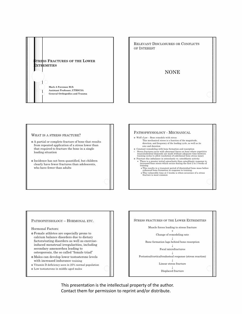
STRESS FRACTURES OF THE LOWEREXTREMITIES
Mark A Foreman M.D.
Assistant Professor, UTHSCSA
General Orthopedics and Trauma
RELEVANT DISCLOSURES OR CONFLICTS
OF INTEREST
NONE
WHAT IS A STRESS FRACTURE?
A partial or complete fracture of bone that results from repeated application of a stress lower than that required to fracture the bone in a single loading situation
Incidence has not been quantified, but children clearly have fewer fractures than adolescents, who have fewer than adults
PATHOPHYSIOLOGY - MECHANICAL
Wolf’s Law – Bone remodels with stressThis mechanical stress is a function of the magnitude, direction, and frequency of the loading cycle, as well as itsrate and duration
Constant remodeling with bone formation and resorption• Stress fractures occur with abnormal forces on bone where repetitive
musculoskeletal demands are made without adequate time between training cycles to allow resolution of subclinical bone stress injury
Fracture due imbalance in osteoclastic vs. osteoblastic activity There is a greater initial osteoclastic than osteoblastic response to
increased bone stress which occurs during the first 2 to 3 weeks of training This results in a transient period of diminished bone mass before
enhanced bone formation in response to training. This vulnerable time at 3 weeks is when occurence of a stress
fracture is most common
PATHOPHYSIOLOGY – HORMONAL ETC.
Hormonal Factors: Female athletes are especially prone to
calcium balance disorders due to dietary factors/eating disorders as well as exercise-induced menstrual irregularities, including secondary amenorrhea leading to osteoporosis, the so called “female triad”
Males can develop lower testosterone levels with increased indurance training
Vitamin D deficiency seen in 25% normal population Low testosterone in middle aged males
STRESS FRACTURES OF THE LOWER EXTREMITIES
Muscle forces leading to stress fracture+
Change of remodeling rate ↓
Bone formation lags behind bone resorption↓
Focal microfractures↓
Periosteal/cortical/endosteal response (stress reaction)↓
Linear stress fracture↓
Displaced fracture

This presentation is the intellectual property of the author. Contact them for permission to reprint and/or distribute.
DIAGNOSIS
History Insidious onset of vague, aching pain without
specific traumatic event Menstrual irregularities in female athletes
common Recent overuse or change in training
regimen/activity
DIAGNOSIS
Physical exam Discrete, point tenderness over fracture site
associated with occasional swelling Erythema and warmth may be present but are
much less likely May present with limp but rarely with muscular
atrophy Joint ROM is usually not affected Percussion and vibratory stimuli have been
reported to aid in diagnosis but are usually unnecessary
DIAGNOSIS
Laboratory workup: Vitamin D level, TSH, Serum cortisol level, PTH,
serum calcium Testosterone in males Estrogen level females plus FSH/ACTH Beware active elderly patients with “hip” or
“thigh” pain on chronic bisphosphonates for osteoporosis treatment…
DIAGNOSIS
Imaging Studies:
Plain X-rays Bone Scan MRI
IMAGING STUDIES
Plain X-rays: Are sufficient in approximately 2/3 of cases but
can be misleading. Bone response can be periosteal, endosteal, or intra-cortical
If very early in the process, may be negative initially but will usually show up radiographically within 2 to 3 weeks after initial visit
85 Y/O WITH LEFT THIGH PAIN ON FOSAMAX X 3 YEARS

This presentation is the intellectual property of the author. Contact them for permission to reprint and/or distribute.
Proximal tibial stress fracture with periosteal and endostealresponses in a 13 year old cross‐country runner
Middle aged female with 2nd metatarsal stress fracture
IMAGING STUDIES
Bone Scintigraphy (Bone Scan)
Helpful in the early detection especially if xrays are negative and the diagnosis is in question. Can be positive as early as 24 to 48 hours after injury
Very sensitive but not specific Not valuable in monitoring healing
Bone Scan IMAGING STUDIES
MRI Very sensitive and much more specific than bone
scan. Will see periosteal as well as bone marrow edema.
Characteristically low signal T1 initially, high on T2. Once out of acute phase T1 and STIR best
Better accuracy in diagnosis than bone scan in terms of definition of anatomy of the fracture site
Much more expensive modality Results if any imaging modality must be
clinically correlated before treatment is begun

This presentation is the intellectual property of the author. Contact them for permission to reprint and/or distribute.
MRI – T-2 image MRI T-1 image Tibial shaft stress fracture on MRI
Tibial stress fractureSTRESS FRACTURES OF THE LOWER
EXTREMITIES
Differential diagnosis
Tibial Shaft – Medial-Tibial Stress Syndrome (Shin Splints) Exertional Compartment syndrome Tibial Tubercle Apophysitis “Osgood Schlatter’s Disease” Calcaneus – Apophysitis “Sever’s Disease” Femur – periosteal reaction confused with osteosarcoma or
non-specific thigh pain ignored
Stress fracture vs. “Shin splints” Stress fracture vs. “Shin splints”

This presentation is the intellectual property of the author. Contact them for permission to reprint and/or distribute.
Distal femoral stress fracture in a 14 year old soccer player. The lesion was originally diagnosed as an osteosarcoma TREATMENT – “LOW RISK FRACTURES”
R/O underlying metabolic or endocrine causes Modify exercise routine Six to 12 weeks of rest with gradual resumption
of activities Occasional bracing or casting if “potentially
unstable” or at “higher risk” Correct any mal-alignment or training issues
Calcaneal tuberosity stress fracture in a 29 y/o female runner
Middle aged female with 2nd metatarsal stress fracture
TREATMENT – “HIGH RISK FRACTURES”
High risk stress fractures are in the minority and are those that have a propensity for difficult healing, persistent nonunion, and risk for fracture displacement
Surgery may be required for these and follow the same basic indications as for other fractures: nonunion, malunion, or at risk for displacement that would lead to either of the two or significant morbidity
Examples include the patella, medial malleolus, talus, tarsal navicular, and fifth metatarsal base.
These also include “tension sided” fractures such as the anterior tibial diaphysis and superior femoral neck and lateral femoral diaphysis

This presentation is the intellectual property of the author. Contact them for permission to reprint and/or distribute.
85 Y/O WITH LEFT THIGH PAIN ON FOSAMAX X 3 YEARS85 Y/O S/P PROPHYLACTIC NAILING LEFT FEMUR
85 Y/O HEALTHY ACTIVE FEMALE S/P GROUND LEVELFALL
85 Y/O ACTIVE FEMALE ON CHRONIC BISPHOSPHONATESIMMEDIATE POST OP FILMS
Femoral neck stress fracture Femoral neck tension sided stress fracture

This presentation is the intellectual property of the author. Contact them for permission to reprint and/or distribute.
44 y/o male stepped wrong on incline felt pop and immediate pain
44 y/o male. H/O previous fx 8 years ago. Metabolic work up …
44 y/o male 4 months later after starting testosterone replacement and Vitamin D
Tibial stress fracture in an 18 y/o basketball player (high risk anterior tibial cortex)
Tibial stress fracture

This presentation is the intellectual property of the author. Contact them for permission to reprint and/or distribute.
NAVICULAR STRESS FRACTURE
Seen in active athletes involved in sprinting and jumping sports
Nondescript medial arch pain worse with activity Occurs in the central 1/3 of the bone (avascular
zone) CT scan study of choice If diagnosed early, high union rate with NWB
cast immobilization for 6-8 weeks. High rate of nonunion with weight-bearing
Displaced fractures, nonunion, delayed union: ORIF
Semi-rigid molded arch support for rehab and after return to athletic activities
Tarsal navicular stress fracture
Defensive lineman with vague, persistent foot pain
Defensive lineman with vague, persistent foot pain
Tarsal navicular stress fracture
5th Metatarsal Stress Fracture
• Zone 3
– In the distal metaphysis at the meta‐diaphysealjunction
• Usually a stress fracture
• SLC for 6‐8 weeks
• WB status is controversial
• Nonunions: IM screw fixation
Stress Fracture

This presentation is the intellectual property of the author. Contact them for permission to reprint and/or distribute.
Bipartite sesamoid Fractured sesamoid
25 y/o female with
patellofemoralpain after increased
distance running
25 y/o female with mild
patellofemoralpain but no
history of injury
SUMMARY
The majority of stress fractures can be managed non-operatively with activity modification and casting/bracing if necessary
Identify the “low risk” from the “high risk” H and P should guide to correct diagnosis. Avoid
misdiagnosis… e.g. osteosarcoma DDx: Apophysitis in skeletally immature, “shin
splints” Remember can always image the unaffected side for
comparison… Don’t forget to look for dietary and hormonal causes,
especially in female athletes and vitamin D deficiency In elderly active population – bisphosphonates and
thigh pain When in doubt, refer to orthopedic colleague
BIBLIOGRAPHY
Delee and Drez’s Orthopedic Sports Medicine, 2nd
edition Arendt EA, Griffiths HJ: The use of MR imaging in the
assessment and clinical management of stress reactions of bone in high performance athletes. ClinSports Med 16:291-306, 1997
Boden BP, Osbahr DC: High risk stress fractures: Evaluation and treatment. JAAOS 8:344-352, 2000
Knapp TP, Garrett WE Jr: Stress fractures: General Concepts. Clin Sports Med: 339-356, 1997
Thank You