this presentation is the intellectual property of the...
TRANSCRIPT

This presentation is the intellectual property of the author.Contact them for permission to reprint and/or distribution.
UT Health Science Center San Antonio School of Medicine
January 23-24, 2015
Julie Barnett PT, DPT, MTCDirector of PT The Non-Surgical
Center of TexasAssistant Professor PT at UT-HSCSA
Basic walk/run biomechanics: traditional EBM for 3 diagnosis of overuse foot/ankle
injuries: plantar fasciitis, Achilles tendonitis, posterior tibialis tendonitis
Controversial discussion of minimalistic biomechanics and shoes
Future directions

This presentation is the intellectual property of the author.Contact them for permission to reprint and/or distribution.

This presentation is the intellectual property of the author.Contact them for permission to reprint and/or distribution.

This presentation is the intellectual property of the author.Contact them for permission to reprint and/or distribution.
1. Plantar Fasciitis2. Achilles Tendinitis3. Tibial Stress
Syndrome
11
Initial contact ROM Muscle Action◦ Hip @ 200 flexion hip extensors◦ Knee @ 50 flexion quadriceps◦ Ankle @ 00 tibialis anterior
Critical event◦ Heel first contact
12

This presentation is the intellectual property of the author.Contact them for permission to reprint and/or distribution.
Key ranges of motion
13
Diagnosis Tests and
Measurements Interventions for
physical therapy Outcome
Instruments
Observational Video 3-D systems Pressure plate
systems Global Positioning
System (GPS)

This presentation is the intellectual property of the author.Contact them for permission to reprint and/or distribution.
Clinical Assessment No gold-standard
Tenderness at the medial calcaneal tubercle
< 10 degrees of ankle dorsiflexion
< 65 degrees of 1st
MTP extension (weak evidence)
Decreased ankle dorsiflexion
Obesity Work-related
weight-bearing

This presentation is the intellectual property of the author.Contact them for permission to reprint and/or distribution.
• Icing• Strapping the foot
(low dye)• Calf and plantar
fascia stretches• Avoidance of flat
shoes• Avoidance of
barefoot walking• Use of over-the-
counter arch supports
• Heel cushions• Limitation of
extended activities
Supports rearfoot alignment
Reinforces plantar fascia
Lifts and supports medial longitudinal arch
McConnell Patella Kinesio Patella Ankle Sprain Low Dye

This presentation is the intellectual property of the author.Contact them for permission to reprint and/or distribution.
Custom orthotics Night splints Immobilization with
casts or other devices
Keeps plantar fascia on a stretch vs. plantar flexed and shortened
Dorsal options available. Example: Strasburg sock.
Patient walks across pressure plate barefooted to capture a dynamic foot print
Scanning the foot

This presentation is the intellectual property of the author.Contact them for permission to reprint and/or distribution.
Pressure points are differentiated with a scale of colors.
Points of higher impact are indicated in red.
Gait line is drawn over the print
Pressure Points
Cast in subtalar neutral
Mail neutral cast to lab
Positive cast made Orthotic made from
positive mold Both returned to
clinic and patient
American Academy of Orthopedic Surgeons (AAOS) Foot and Ankle questionnaire
www.aaos.org - Click on “Research”, and “Outcomes” for access to an array of outcomes assessment instruments

This presentation is the intellectual property of the author.Contact them for permission to reprint and/or distribution.
Ultrasound: 0.80 sensitivity and 0.49 specificity
MRI: 0.95 sensitivity and 0.50 specificity
Clinical assessment may provide yardstick compared to imaging
Point tenderness on the tendon
Localized swelling Crepitation during
movement
Tight heel cord Achilles contractures Hyperpronation Repetitive heel
running Change in shoes or
running surface Increase in intensity
or distance Hill climbing

This presentation is the intellectual property of the author.Contact them for permission to reprint and/or distribution.
Stretching exercises Modification of
training schedules Braces and insoles Questionable role
of eccentric versus concentric strengthening (weak evidence)
Slant board to keep foot in neutral
Obtain a negative heel for more aggressive stretch
Avoids twisting midfoot with edge of step stretches
Victorian Institute of Sport Assessment-Achilles questionnaire (VISA-A)
AAOS Foot and Ankle questionnaire not specific for Achilles tendinitis

This presentation is the intellectual property of the author.Contact them for permission to reprint and/or distribution.
Bone scan is gold standard: 84% sensitivity and 22% specificity
MRI: 79% sensitivity and 33% specificity

This presentation is the intellectual property of the author.Contact them for permission to reprint and/or distribution.
Pain along the posteromedial tibial border, usually in the distal third of tibia
Excessive and/or prolonged pronation
Recent changes in:◦ Distance◦ Speed◦ Form◦ Stretching◦ Footwear◦ Running surface
Shock-absorbing insoles (best evidence) High-Dye and low-Dye taping podiatry study
(weak evidence) Clinical experience and observational
interventions: ◦ Motion control shoes, ankle strapping OR◦ Minimalistic shoes to strengthen intrinsics and
change running biomechanics from rearfoot strike to forefoot strike with less impact forces

This presentation is the intellectual property of the author.Contact them for permission to reprint and/or distribution.
http://barefootrunning.fas.harvard.edu/

This presentation is the intellectual property of the author.Contact them for permission to reprint and/or distribution.
Alter rearfoot alignment
Decrease stretch on posterior tibialis in over-pronators
AAOS Foot and Ankle questionnaire◦ Includes Shoe
Comfort Scale◦ Population groups
are not identical
This presentation is the intellectual property of the author.Contact them for permission to reprint and/or distribution.
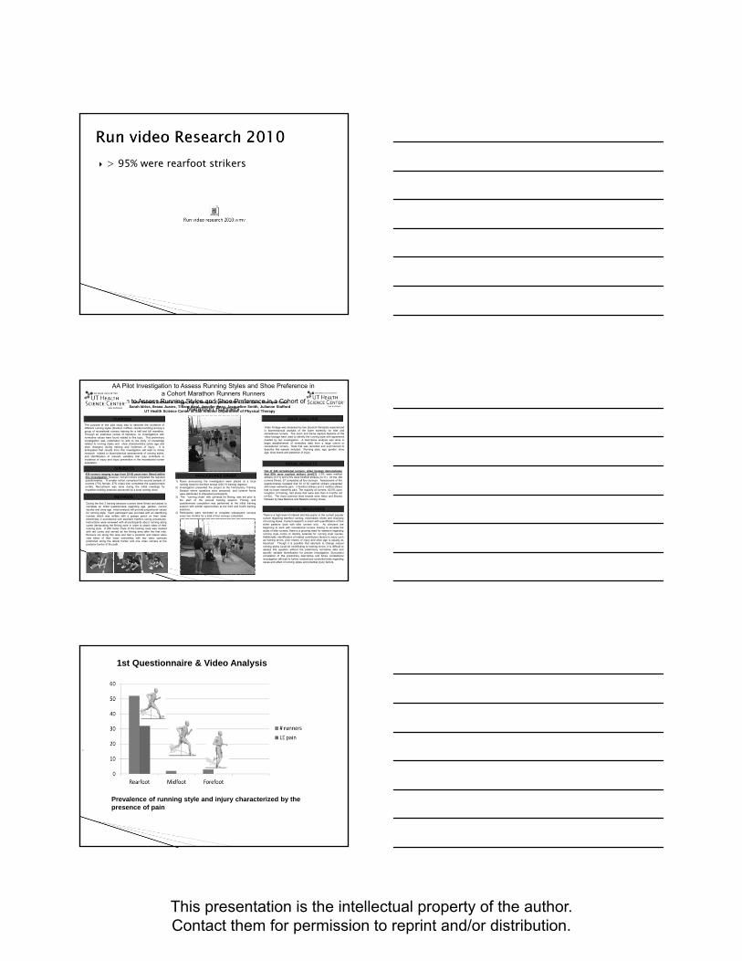
> 95% were rearfoot strikers
AA Pilot Investigation to Assess Running Styles and Shoe Preference in a Cohort Marathon Runners Runners
ion to Assess Running Styles and Shoe Preference in a Cohort of Marathon Runners
During the first 3 training sessions runners were filmed and asked tocomplete an initial questionnaire regarding age, gender, currentinjuries and shoe age. Initial analysis will provide proportional valuesfor running style. Each participant was provided with an identifyingnumber which was written with a grease pencil on their lowerextremities in accordance with standard triathlon racing procedures.Instructions were reviewed with all participants about running alongcones demarcating the filming zone in order to obtain video of theirrunning style. A 200 meter chute of the training route was markedwith red cones and served as the filming area after the first mile.Runners ran along this area and had a posterior and lateral videoview taken of their lower extremities with two video cameraspositioned along the lateral border and one video camera at theposterior border of the path.
Julie Barnett, Catherine Ortega, Barry Morgan ,Cynthia Alfaro, Lori Cano, Monique Cruz, Sarah Idriss, Ileana Juarez, Tiffany Neal, Jennifer Seay, Jacqueline Smith, Julianne Stafford
UT Health Science Center at San Antonio Department of Physical Therapy
PURPOSE DATA ANALYSIS
METHODS
The purpose of this pilot study was to describe the incidence ofdifferent running styles (forefoot, midfoot, rearfoot landing) among agroup of recreational runners training for a half and full marathon.Through an extensive review of literature, no investigations withnormative values were found related to this topic. This preliminaryinvestigation was undertaken to add to the body of knowledgerelated to running styles and shoe characteristics (shoe age andshoe changes) during training and incidence of injury. It isanticipated that results from this investigation will lead to futureresearch related to biomechanical assessments of running styles,and identification of relevant variables that may contribute toincidence of injury and injury prevention in the recreational runnerpopulation..
SUBJECTS
PROCEDURES ORDER
Video footage was assessed by two physical therapists experiencedin biomechanical analysis of the lower extremity for elite andrecreational runners. The zoom and frame capture features of thevideo footage were used to identify the running style with agreementneeded by two investigators. A descriptive analysis was done tobegin establishment of normative data from a large cohort orrecreational runners. Data that was recorded and summarized todescribe this sample included: Running style, age, gender, shoeage, shoe brand and existence of injury.
CLINICAL RELEVANCE
There is a high level of interest and discussion in the current popularculture regarding barefoot running, minimalistic shoes and changingof running styles. Current research is scant with quantification of footstrike patterns done with elite runners only. As clinicians arebeginning to work with recreational runners looking to emulate thestyles of elite runners, there is a growing need for research regardingrunning style norms to identify potential for running style injuries.Additionally, identification of related contributory factors to injury suchas training errors, prior history of injury and shoe age is equally asimportant. Though it is possible that attempts to change naturalrunning styles could be contributing to training errors, it is difficult toassess this question without the preliminary normative data andspecific variable identification for precise investigation. Successfulcompletion of this preliminary descriptive and future correlationalinvestigation will lead to further randomized controlled trials regardingcause and effect of running styles and potential injury factors.
439 runners ranging in age from 18-65 years were filmed withinthis investigation, however, not all runners completed the requiredquestionnaires. A smaller cohort comprised the second sample ofrunners (73% female, 27% male) who completed the questionnaire(n=56). Recruitment was done during the initial meetings formarathon training sessions sponsored by a local running store.
1) Flyers announcing the investigation were placed at a localrunning store for the third annual USA Fit training regimen.
2) Investigators presented the project at the Introductory TrainingSession where questions were answered, and consent formswere distributed to interested participants.
3) The “running chute” with cameras for filming, was set prior tothe start of the second training session. Filming andquestionnaire completion was performed at the initial trainingsession with similar opportunities at the third and fourth trainingsessions.
4) Participants were reminded to complete subsequent surveysevery two months for a total of four surveys completed.
INITIAL RESULTS
Out of 439 recreational runners, video footage demonstratesthat 95% were rearfoot strikers (n=417), 2.5% were midfootstrikers (n=11) and 2.5% were forefoot strikers (n=11). Of the 439runners filmed, 57 completed all four surveys. Assessment of thequestionnaires revealed that 32 of 52 rearfoot strikers presentedwith lower extremity pain. 3 forefoot strikers and 2 midfoot stikershad no lower extremity pain. The majority of runners, 62.5% uponinception of training, had shoes that were less than 6 months old(n=35). The most common shoe brands were Asics and Brooksfollowed by New Balance and Reebok running shoes.
1st Questionnaire & Video Analysis
.
Prevalence of running style and injury characterized by the presence of pain

This presentation is the intellectual property of the author.Contact them for permission to reprint and/or distribution.
Descriptive Study to Assess the Running Styles, Shoe Preference, and Injury Incidence in a Cohort of Recreational Runners.
Julie Barnett PT, DPT, MSC; Catherine Ortega PT, EdD, ATC; Barry Morgan PT; Manuel Montes SPT; Kaitlyn Warren SPT; Tricia Franks SPT; Kyle Farrell SPT et al.
Purpose
Purpose of this study was to gainnormative information in a cohort ofrecreational runners to include:● incidence of running striking
pattern● prevalence of footwear● prevalence of LE pain
Subjects
Participants were recruited from twoSolers Sports stores during initialtraining sessions for the 2013 SanAntonio Rock ‘n’ Roll marathon. Agerange 25-64 (44); females (57.1%) &males (42.9%); mileage range 6-30mi(18); runners currently experiencingpain 35.7%; shoe types(stability/neutral > cushion and motioncontrol > minimalist)
Methods
Participants were given anidentification number and marked witha grease pencil. Video cameras wereplaced along lateral and posteriorborders of the filming chute.
Data Collection
1.Paper flyers announcing the studyplaced in the stores several weeksprior to the initial data collection.2.Participants completed initialquestionnaire which was re-sentthrough email every 2 months until therace3.Participants were videotaped forfootstrike pattern and biomechanicalanalysis
Statistical Analysis
Descriptive analysis was performedfor the establishment of normativedata for age, gender, shoe type /brand, strike pattern, and injuryincidence/existence of injury.
Results
-Out of the 149 participants thatcompleted the initial survey, 14subjects completed the final survey.-90 runners attended the runningvideo chute
Clinical Relevance
Majority of individuals filmed duringthe study were still running in stabilityshoes (44%) vs cushion shoes (28%).Of those wearing minimalist shoes,the majority still ran with a RFSpattern (60%). Physical therapistsshould take into consideration BOTHfoot strike pattern and shoe typewhen performing gait analysis.
Body Mass Index (BMI): WNL. Not overweight. Neutral lower extremity, (LE) biomechanics No prior history of serious LE injuries Start with graduated training program: walk,
walk/jog, jog schedule Consider cross-training with 2 pairs of shoes:
minimalistic AND more stability shoe

This presentation is the intellectual property of the author.Contact them for permission to reprint and/or distribution.
REFERENCES
1 Levangle P, Norkin C. Joint Structure and Function: A Comprehensive Analysis.3rd ed.Canada.FA Davis Co;2001:436.2 Reischl S, De Bettignies J. A different look at the midtarsal joints. Orthopaedic Practice.1995;7;(1).11-12.3 Sackett D, Straus S, Richardson WS, Rosenberg W, Haynes RB. Evidence-based medicine. Howto practice and teach EBM. 2nd ed. Edinburgh: Churchill Livingstone; 2000:1.4 Jette DU, Bacon K, Batty C, et al. Evidence-based practice: beliefs, attitudes, knowledge, andbehaviors of physical therapist. Physical Therapy. 2003;83(9):786-805.5 Turner P, Whitfield TW. Physiotherapists’ use of evidence-based practice: a cross-nationalstudy. Physiother Res Int. 1997;2(1):17-29.6 Guyatt G, Rennie D. Users’ guides to the medical literature - Essentials of evidence-basedclinical practice. Chicago: AMA Press;2002:12.7 Yeung EW, Yeung SS. A systematic review of interventions to prevent lower limb soft tissuerunning injuries. Br J Sports Med. 2001;35:383-389.8 O’Conner AM. Walk this way:biomechanics and injuries. Sportex Health. 2003;(18):24-7.Orthopedic Gait Injuries in Runners, June 29, 2007, This information is theproperty of Barnett and should not be copied or otherwise used withoutexpress written permission of the author.9 Terrier P, Ladetto Q, Merminod B, Schutz Y. Measurement of the mechanical power of walkingby satellite positioning system (GPS). Medicine and science in sports and exercise. 2002;1912-18.10 Riddle D, Pulisic M, Pidcoe P, Johnson R. Risk factors for plantar fasciitis: a matched casecontrolstudy. J Bone Joint Surg Am. 2003;85-A(5):872-877.
11 Buchbinder R. Plantar fasciitis. N Engl J Med. 2004;350:2159-66.12 Anderson MK, Hall SJ. Fundamentals of Sports Injury Management. Williams and Wilkins.1997: 279.13 Hopson MM, McPoil RG, Cornwall MW. Motion of the first metatarsophalangeal joint.Reliability and validity of four measurement techniques. J Am Podiatr Med Assoc.1995;85(4):198-204.14 Johanson NA, Liang MH, Daltroy L, Rudicel S, Richmond J. American academy of orthopaedicsurgeons lower limb outcomes assessment instruments. J Bone Joint Surg, Inc. 2004;86-A(5):902-909.15 Khan KH, Forster BB, Robinson J, Cheong Y, Louis L, Maclean L, Taunton JE. Are ultrasoundand magnetic resonance imaging of value in assessment of Achilles tendon disorders? A two yearprospective study. Br J Sports Med 2003;37:149-153.16 Shalaby M, Almekinders LC. Patellar tendinitis: the significance of magnetic resonance imagingfindings. Am J Sports Med 1999;27:345-9.17 McLauchlan GJ, Handoll HHG. Interventions for treating acute and chronic Achilles tendinitis.Cochrane Database of Systematic reviews. 2004:2.18 Mafi N, Lorentzon R, Alfredson H. Superior short-term results with eccentric calf muscletraining compared to concentric training in a randomized prospective multicenter study on patientswith chronic Achilles tendinosis. Knee Surg, Sports Traumatol, Arthrosc. 2001;9:42-47.19 Batt M, Ugalde V, Anderson M, Shelton DK. A prospective controlled study of diagnosticimaging for acute shin splints. Med Sci Sports Exerc. 1998;30(11):1564-1571.20 Thacker S, Gilchrist J, Stroup D, Kimsey CD. The prevention of shin splints in sports: asystematic review of literature. Med Sci Sports Exerc. 2001;(3):32-40.

This presentation is the intellectual property of the author.Contact them for permission to reprint and/or distribution.
21 Kannus VP. Evaluation of abnormal biomechanics of the foot and ankle in athletes. Br J SportsMed. 1992;26(2):83-9.22 Cunningham A, Spears IR. A successful conservative approach to managing lower leg pain in auniversity sports injury clinic: a two patient case study. Br J Sports Med. 2004;38:233-234.23 Keenan A, Tanner C. The effect of high-dye and low-dye taping on rearfoot motion.2001;91(5):255-61.24 Murray, TF, Dupont JY, Fulkerson JP. Axial and lateral radiographs in evaluatingpatellofemoral malalignment. 1999;27(5):580-4.25 Shelton GL, Thigpen LK: Rehabilitation of patellofemoral dysfunction: a review of literature. JOrthop Sports Phys Ther. 1991;14:243–249.Orthopedic Gait Injuries in Runners, June 29, 2007, This information is theproperty of Barnett and should not be copied or otherwise used withoutexpress written permission of the author.26 Bizzini M, Childs J, Piva S, Delitto A. Systematic Review of the quality of RandomizedControlled Trials for Patellofemoral Pain Syndrome. 2003;33(1):4-20.27 Winter DA, Bishop PJ. Lower extremity injury. Biomechanical factors associated with chronicinjury to the lower extremity. Sports Med. 1992;14;149-56.28 Hoppenfeld S. Physical examination of the spine and extremities. Appleton and Lange.1976.167.29 Fredericson M, Cookingham CL, Chaudhari AM, Dowdell BD, Oestreicher N, Sahrmann SA.Hip abductor weakness in distance runners with iliotibial band syndrome. Clin J Sport Med.2000;10(3):169-75.30 Brosseau L, Casimiro L, Milne S Robinson V, Shea B, Tugwell P, Wells G. Deep transversefriction massage for treating tendinitis. Cochrane Database Syst R. 2002;(4):CD003528.