stretch reflex and tendon jerks - ksumscksumsc.com/download_center/2nd/1) neuropsychiatry... ·...
TRANSCRIPT
Dr Syed Shahid Habib MBBS DSDM PGDCR FCPS
Professor & Consultant Clinical Neurophysiology Dept. of Physiology
College of Medicine & KKUH King Saud University
Stretch reflex and tendon jerks
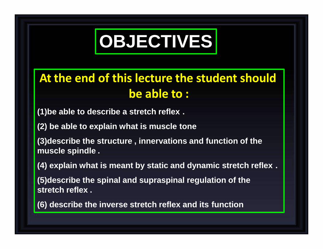
At the end of this lecture the student should be able to :
(1)be able to describe a stretch reflex .
(2) be able to explain what is muscle tone
(3)describe the structure , innervations and function of the
muscle spindle .
(4) explain what is meant by static and dynamic stretch reflex .
(5)describe the spinal and supraspinal regulation of the
stretch reflex .
(6) describe the inverse stretch reflex and its function
OBJECTIVES
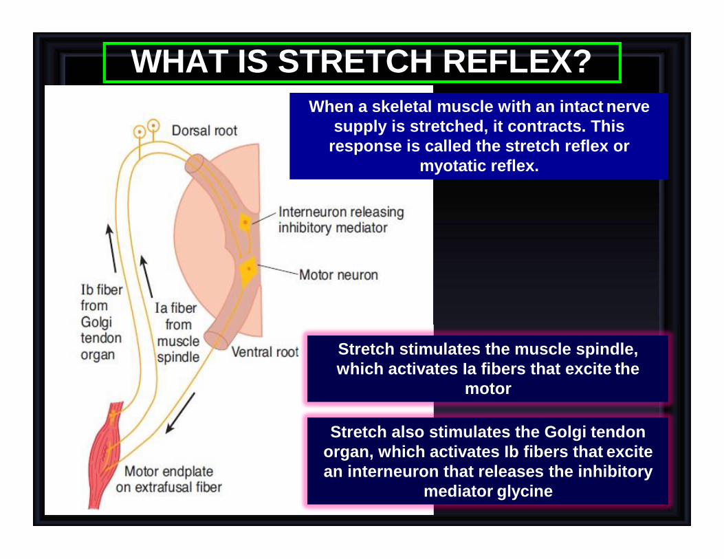
WHAT IS STRETCH REFLEX?
Stretch stimulates the muscle spindle,
which activates Ia fibers that excite the
motor
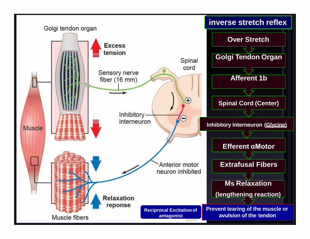
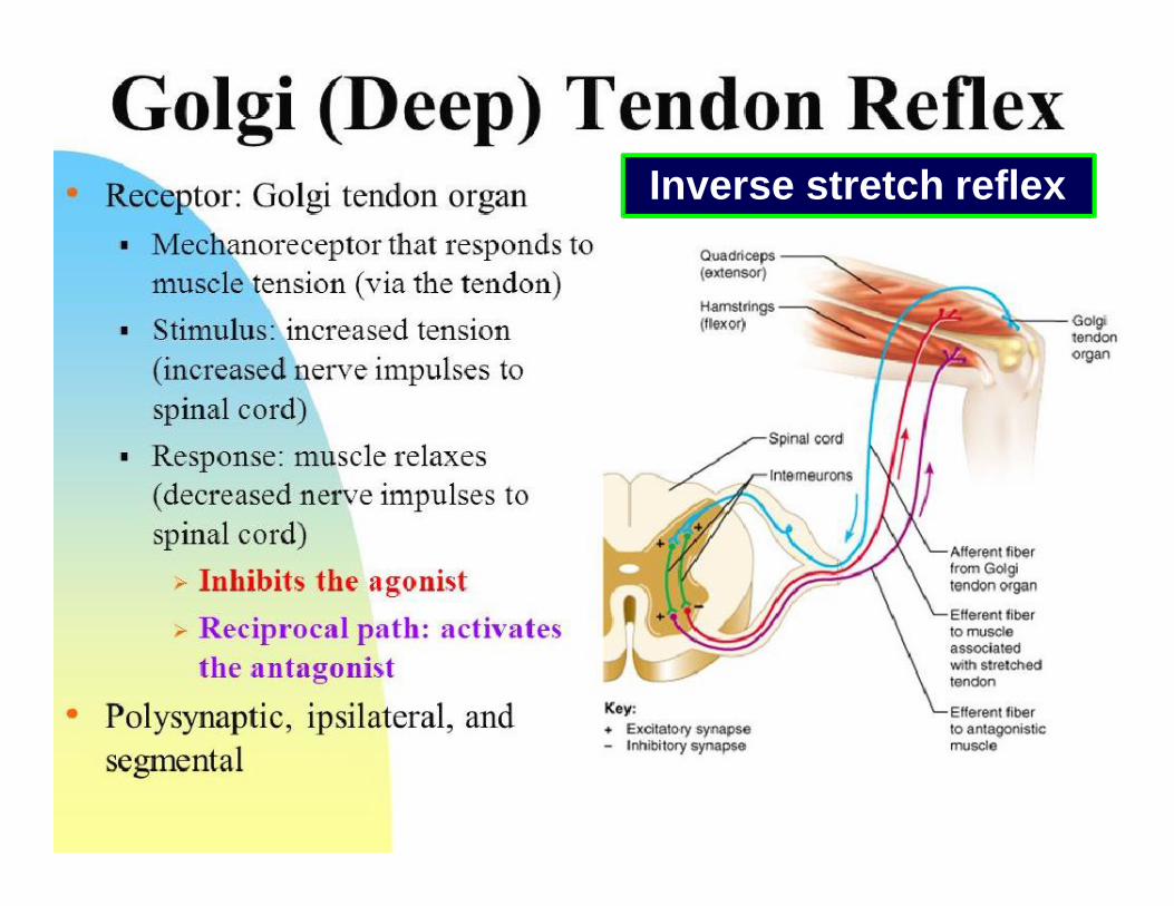
Stretch also stimulates the Golgi tendon
organ, which activates Ib fibers that excite
an interneuron that releases the inhibitory
mediator glycine
When a skeletal muscle with an intact nerve
supply is stretched, it contracts. This
response is called the stretch reflex or
myotatic reflex.
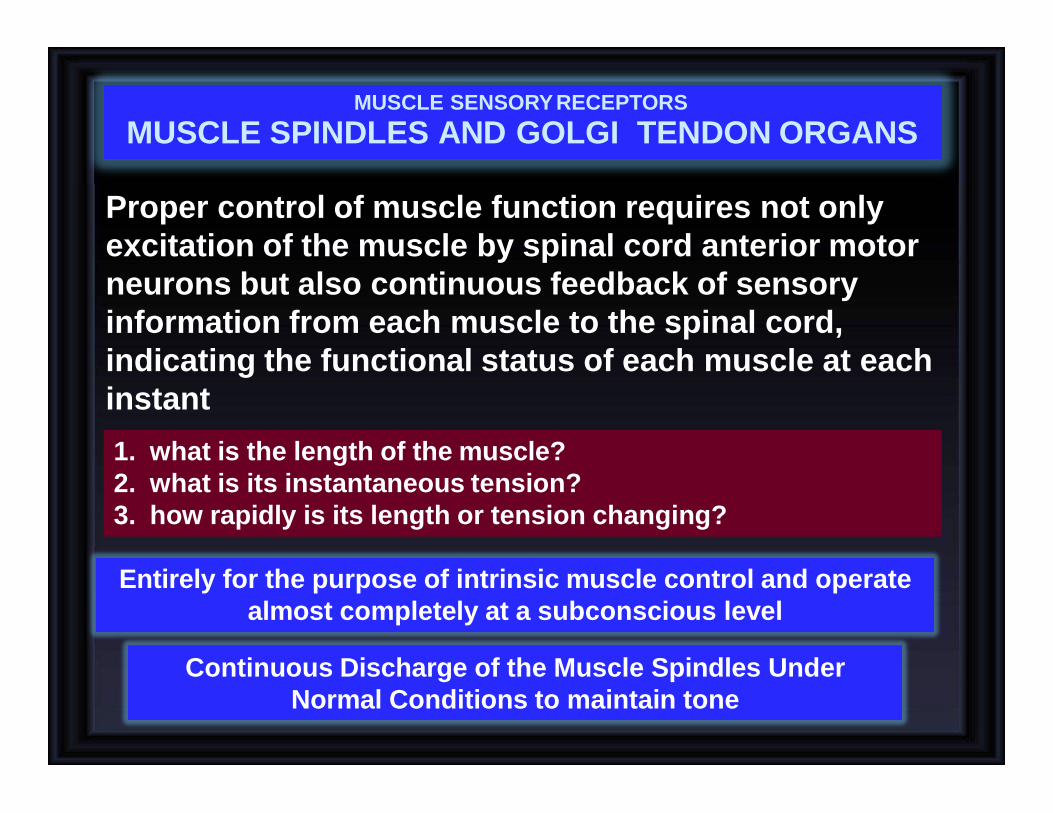
Proper control of muscle function requires not only
excitation of the muscle by spinal cord anterior motor
neurons but also continuous feedback of sensory
information from each muscle to the spinal cord,
indicating the functional status of each muscle at each
instant
1. what is the length of the muscle?
2. what is its instantaneous tension?
3. how rapidly is its length or tension changing?
Entirely for the purpose of intrinsic muscle control and operate
almost completely at a subconscious level
Continuous Discharge of the Muscle Spindles Under
Normal Conditions to maintain tone
MUSCLE SENSORY RECEPTORS
MUSCLE SPINDLES AND GOLGI TENDON ORGANS
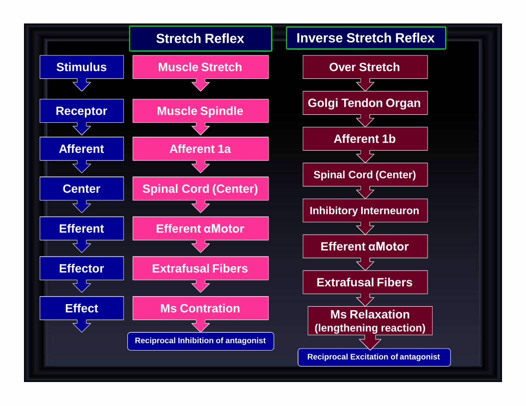
Stretch Reflex
Muscle Stretch
Muscle Spindle
Afferent 1a
Spinal Cord (Center)
Efferent αMotor
Extrafusal Fibers
Ms Contration
Over Stretch
Golgi Tendon Organ
Afferent 1b
Spinal Cord (Center)
Efferent αMotor
Extrafusal Fibers
Ms Relaxation (lengthening reaction)
Inhibitory Interneuron
Reciprocal Inhibition of antagonist
Reciprocal Excitation of antagonist
Stimulus
Receptor
Afferent
Center
Efferent
Effector
Effect
Inverse Stretch Reflex

Structure of Proprioceptors
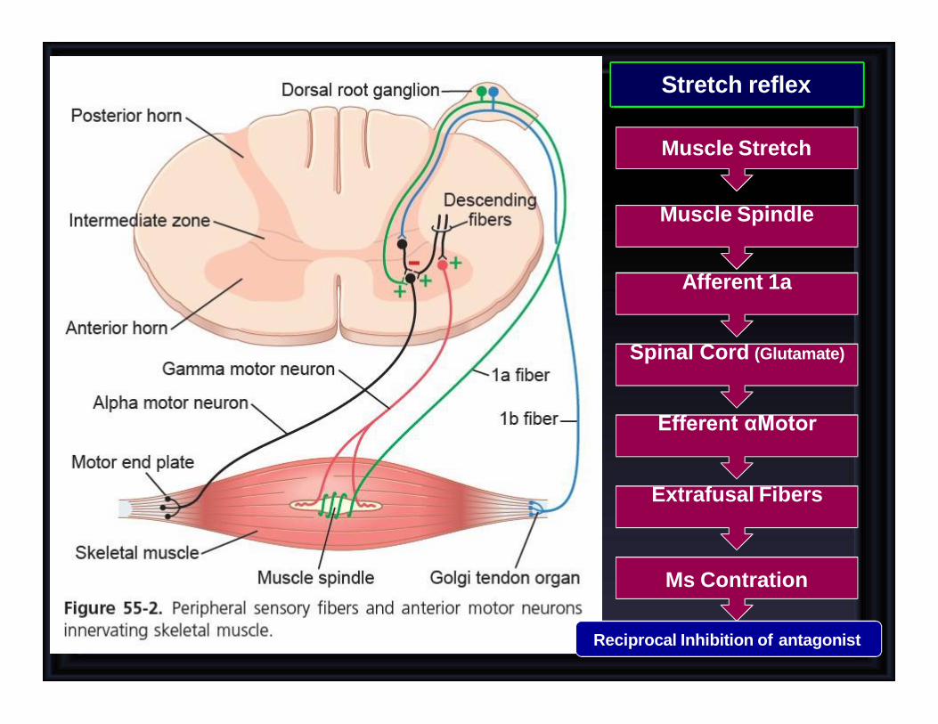
Stretch reflex
Muscle Stretch
Muscle Spindle
Afferent 1a
Spinal Cord (Glutamate)
Efferent αMotor
Extrafusal Fibers
Ms Contration
Reciprocal Inhibition of antagonist
Reciprocal Excitation of
antagonist
inverse stretch reflex
Prevent tearing of the muscle or
avulsion of the tendon
Over Stretch
Golgi Tendon Organ
Afferent 1b
Spinal Cord (Center)
Inhibitory Interneuron (Glycine)
Efferent αMotor
Extrafusal Fibers
Ms Relaxation
(lengthening reaction)
Inverse stretch reflex
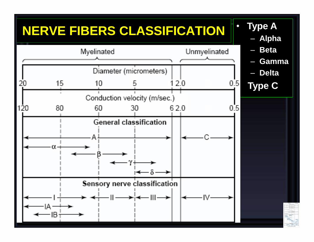
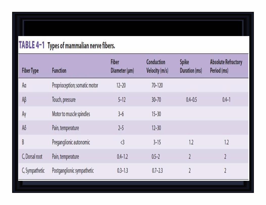
NERVE FIBERS CLASSIFICATION
•
• Type A
– Alpha
– Beta
– Gamma
– Delta
Type C
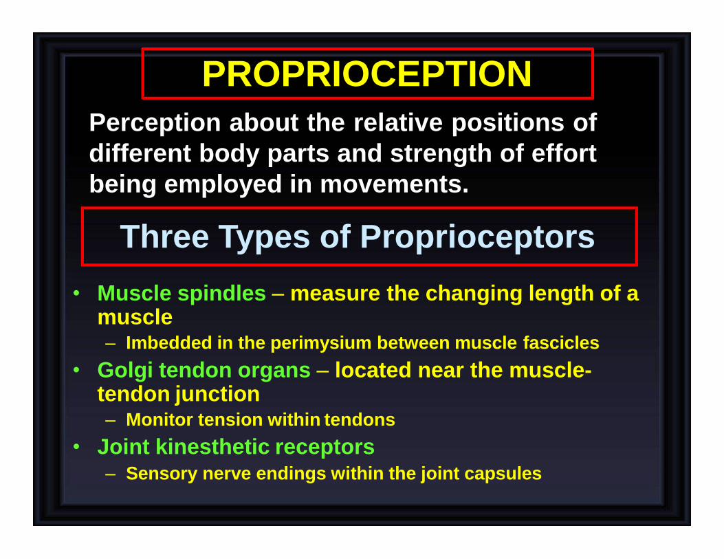
PROPRIOCEPTION
Perception about the relative positions of
different body parts and strength of effort
being employed in movements.
Three Types of Proprioceptors
• Muscle spindles – measure the changing length of a muscle – Imbedded in the perimysium between muscle fascicles
• Golgi tendon organs – located near the muscle- tendon junction – Monitor tension within tendons
• Joint kinesthetic receptors – Sensory nerve endings within the joint capsules

Three Types of Proprioceptors
• Muscle spindles – measure the changing length of a muscle
– Imbedded in the perimysium between muscle fascicles
• Golgi tendon organs – located near the muscle-tendon junction – Monitor tension within tendons
• Joint kinesthetic receptors
– Sensory nerve endings within the joint capsules
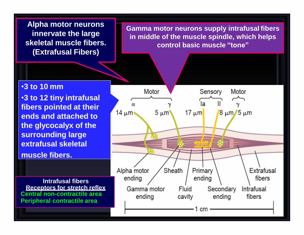
Gamma motor neurons supply intrafusal fibers
in middle of the muscle spindle, which helps
control basic muscle “tone”
Alpha motor neurons
innervate the large
skeletal muscle fibers.
(Extrafusal Fibers)
•3 to 10 mm
•3 to 12 tiny intrafusal
fibers pointed at their
ends and attached to
the glycocalyx of the
surrounding large
extrafusal skeletal
muscle fibers.
Intrafusal fibers Receptors for stretch reflex
Central non-contractile area Peripheral contractile area
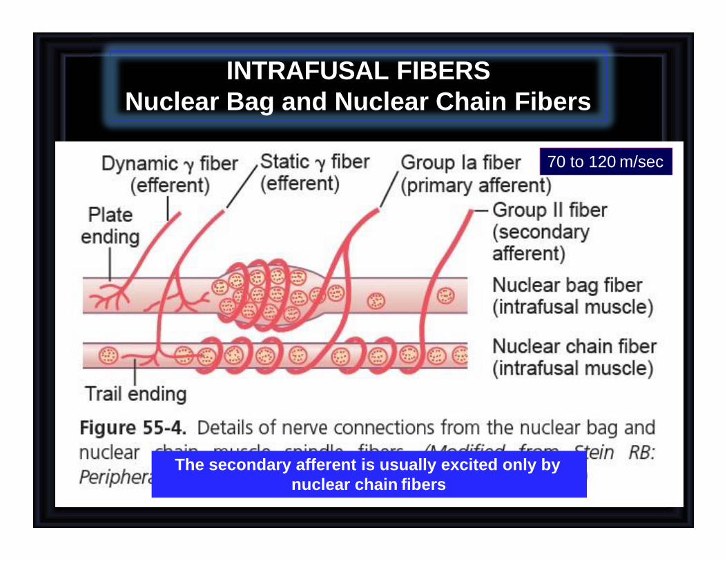
INTRAFUSAL FIBERS
Nuclear Bag and Nuclear Chain Fibers
The secondary afferent is usually excited only by
nuclear chain fibers
70 to 120 m/sec
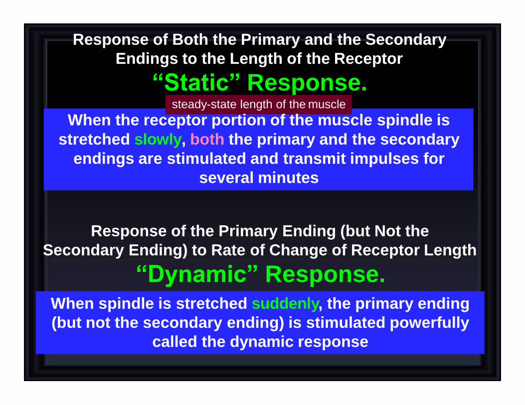
Response of Both the Primary and the Secondary
Endings to the Length of the Receptor
“Static” Response.
When spindle is stretched suddenly, the primary ending
(but not the secondary ending) is stimulated powerfully
called the dynamic response
steady-state length of the muscle
When the receptor portion of the muscle spindle is
stretched slowly, both the primary and the secondary
endings are stimulated and transmit impulses for
several minutes
Response of the Primary Ending (but Not the
Secondary Ending) to Rate of Change of Receptor Length
“Dynamic” Response.
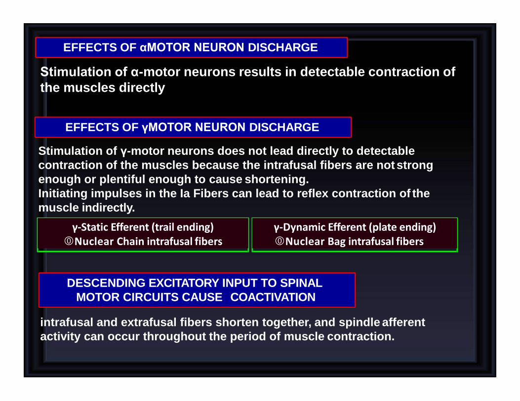
EFFECTS OF γMOTOR NEURON DISCHARGE
EFFECTS OF αMOTOR NEURON DISCHARGE
Stimulation of γ-motor neurons does not lead directly to detectable
contraction of the muscles because the intrafusal fibers are not strong
enough or plentiful enough to cause shortening.
Initiating impulses in the Ia Fibers can lead to reflex contraction of the
muscle indirectly.
Stimulation of α-motor neurons results in detectable contraction of
the muscles directly
intrafusal and extrafusal fibers shorten together, and spindle afferent
activity can occur throughout the period of muscle contraction.
DESCENDING EXCITATORY INPUT TO SPINAL
MOTOR CIRCUITS CAUSE COACTIVATION
γ-Static Efferent (trail ending) Nuclear Chain intrafusal fibers
γ-Dynamic Efferent (plate ending) Nuclear Bag intrafusal fibers
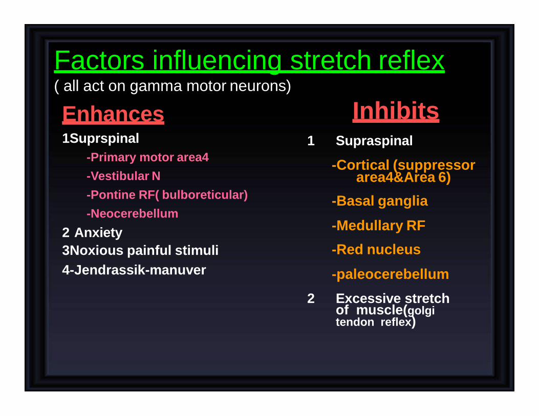
Factors influencing stretch reflex ( all act on gamma motor neurons)
Enhances 1Suprspinal
-Primary motor area4
-Vestibular N
-Pontine RF( bulboreticular)
-Neocerebellum
2 Anxiety
3Noxious painful stimuli
4-Jendrassik-manuver
Inhibits 1 Supraspinal
-Cortical (suppressor area4&Area 6)
-Basal ganglia
-Medullary RF
-Red nucleus
-paleocerebellum
2 Excessive stretch of muscle(golgi tendon reflex)
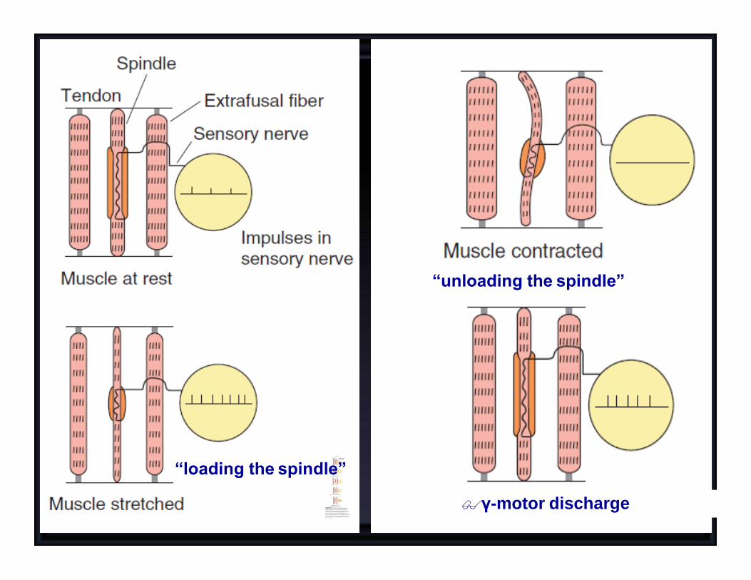
“unloading the spindle”
“loading the spindle”
γ-motor discharge

If the whole muscle
is stretched during
stimulation
of the γ-motor
neurons, the rate of
discharge in
sensory fibers is
further increased.

ROLE OF THE MUSCLE SPINDLE IN
VOLUNTARY MOTOR ACTIVITY
• It keeps the length of the receptor portion of the
muscle spindle constant. Therefore, coactivation
keeps the muscle spindle reflex from opposing the
muscle contraction.
• Second, it maintains the proper damping function of
the muscle spindle, regardless of any change in
muscle length.
With alpha motor neurons, in most instances the gamma motor
neurons (31%) are stimulated simultaneously, an effect called
COACTIVATION
Otherwise receptor portion of the spindle would sometimes be
flail and sometimes be overstretched, causing unsmooth muscle
contractions
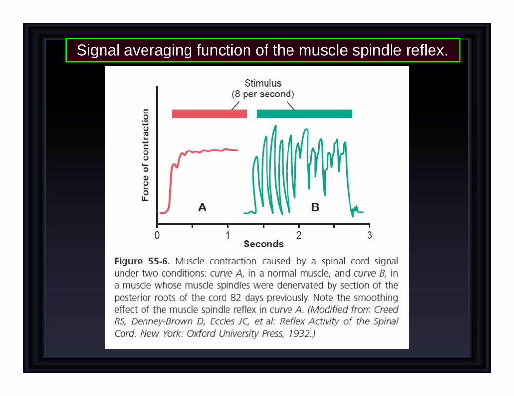
Signal averaging function of the muscle spindle reflex.