stroke cerebrovascular accident
TRANSCRIPT

Cerebrovascular Accident
DR. RAJESH. T.EAPEN


Overview of Stroke
About 85% of strokes are ischemic, and about 15% are hemorrhagic.
Approximately 795,000 strokes occur each year.
Stroke is the 3rd leading cause of death in the US, and the first cause of death worldwide.
Stroke is a leading cause of adult disability.

History of Stroke
Hippocrates-2,400 yrs ago
Names for Stroke
Most commonly known today
Brain Attack

Demographics of Stroke
Women have about 60,000 more strokes than men.
Native Americans have highest prevalence. African Americans have almost twice the
rate compared to Caucasians. Hispanics have slightly higher rates
compared to non-Hispanic whites. Modifiable risk factors must be addressed in
our aging population with the propensity to stroke.

Definition
Ischemic stroke
Caused by a blocked blood vessel in the brain.
Hemorrhagic Stroke
Caused by a ruptured blood vessel in the brain.

Nursing and Stroke
Nurses play a pivotal role in the care of stroke patients.
Nursing care directed in two phases of the acute stroke experience:
The emergent or hyper-acute phase
The acute phase

Nursing Care of the Stroke
Patient
Stroke is a complex disease requiring the efforts and skills of the multidisciplinary team.
Nurses are often responsible for the coordination of that care.
Coordinated care can result in: improved outcomes, decreased LOS, translating to decrease costs.

Etiology of Ischemic Strokes
20% caused by large vessel athero-thrombotic causes (intracranial or carotid artery)
25% caused by small vessel disease (penetrating artery disease)
20% caused by cardiac sources (cardio-embolism)
30% from unknown causes
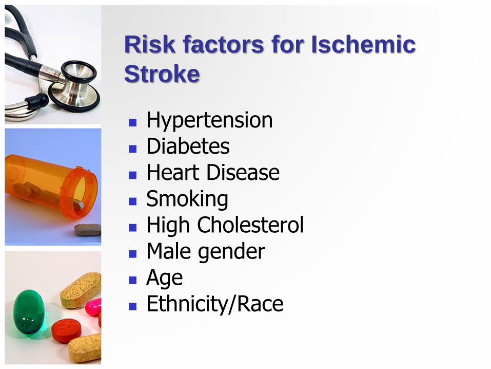
Risk factors for Ischemic
Stroke
Hypertension Diabetes Heart Disease Smoking High Cholesterol Male gender Age Ethnicity/Race

CT Scan–Right Occipital/Parietal
Infarction

Ischemic Stroke
Most patients with ischemic stroke do not have a decreased level of consciousness in the first 24 hours
May progress in the first 72 hours

Embolic stroke
Majority of emboli originate in the inside layer of the heart, with plaque breaking off from the endocardium and entering the circulation
Patient with an embolic stroke commonly has a rapid occurrence of severe clinical symptoms

Transient Ischemic Attack (TIA)
Transient ischemic attack (TIA) is a temporary focal loss of neurologic function caused by ischemia
Most TIAs resolve within 3 hours
TIAs are a warning sign of progressive cerebrovascular disease

Caused by a primary either intra-cerebral hemorrhage or subarachnoid hemorrhage.
Etiology of Hemorrhagic Stroke
SAH 3%
ICH 10%

Ischemic vs. Hemorrhagic

CT Scan Right Subcortical Intra-
cerebral Hemorrhage

Risk Factors for Hemorrhagic
Stroke
Hypertension
Bleeding disorders
African American race
Vascular malformation
Excessive alcohol use
Liver dysfunction
Risk Factors
Non Modifiable
Age
Gender
Race
Heredity

Risk Factors
Modifiable
Obesity
HTN
Smoking
Heavy alcohol consumption
Hypercoagulability
Hyperlipidemia
Asymptomatic carotid stenosis
Diabetes mellitus
Heart disease, atrial fibrillation
Oral contraceptives
Physical inactivity
Sickle cell disease

Blood supply by arteries
Blood is supplied to the brain by two major pairs of arteries
Internal carotid arteries
Vertebral arteries

Blood supply by arteries
Carotid arteries branch to supply most of the
Frontal, parietal, and temporal lobes
Basal ganglia
Part of the diencephalon
Thalamus
Hypothalamus

Blood supply by arteries
Vertebral arteries join to form the basilar artery, which supply the
Middle and lower temporal lobes
Occipital lobes
Cerebellum
Brainstem
Part of the diencephalon

Review of Cerebral Circulation

Common sites for the
development of Atherosclerosis

Clinical Manifestations
Affects many body functions Motor activity
Elimination
Intellectual function
Spatial-perceptual alterations
Personality
Affect
Sensation
Communication

The 5 Key Stroke Syndromes:
Classic Signs Referable to
Different Cerebral Areas
Left (Dominant Hemisphere)
Left gaze preference
Right visual field deficit
Right hemiparesis
Right hemisensory loss
Right (Nondominant Hemisphere)
Right gaze preference
Left visual field deficit
Left hemiparesis
Left hemisensory loss neglect (left hemi-inattention)

The 5 Key Stroke Syndromes: Classic Signs Referable to Different Cerebral Areas
Brainstem Nausea and/or vomiting Diplopia, dysconjugate
gaze, gaze palsy Dysarthria, dysphagia Vertigo, tinnitus Hemiparesis or
quadriplegia Sensory loss in hemibody
or all 4 limbs Decreased consciousness Hiccups, abnormal
respirations
Cerebellum Truncal/gait ataxia Limb ataxia neck
stiffness

Clinical Manifestations
Motor Function
Most obvious effect of stroke
Include impairment of
Mobility
Respiratory function
Swallowing and speech
Gag reflex
Self-care abilities

Clinical Manifestations
Motor Function
An initial period of flaccidity may last from days to several weeks and is related to nerve damage
Spasticity of the muscles follows the flaccid stage and is related to interruption of upper motor neuron influence

Clinical Manifestations
Communication
Patient may experience aphasia when a stroke damages the dominant hemisphere of the brain
Aphasia is a total loss of comprehension and use of language

Clinical Manifestations
Communication
Dysphasia refers to difficulty related to the comprehension or use of language and is due to partial disruption or loss
Dysphasia can be classified as nonfluent or fluent

Clinical Manifestations
Communication
Dysarthria does not affect the meaning of communication or the comprehension of language
It does affect the mechanics of speech

Clinical Manifestations
Affect
Patients who suffer a stroke may have difficulty controlling their emotions
Emotional responses may be exaggerated or unpredictable

Clinical Manifestations
Intellectual Function
Both memory and judgment may be impaired as a result of stroke
A left-brain stroke is more likely to result in memory problems related to language

Clinical Manifestations
Spatial-Perceptual Alterations
Stroke on the right side of the brain is more likely to cause problems in spatial-perceptual orientation
However, this may occur with left-brain stroke

Clinical Manifestations
Spatial-Perceptual Alterations
Spatial-perceptual problems may be divided into four categories
1. Incorrect perception of self and illness
2. Erroneous perception of self in space

Clinical Manifestations
Spatial-Perceptual Alterations
3. Inability to recognize an object by sight, touch, or hearing
4. Inability to carry out learned sequential movements on command

Clinical Manifestations
Elimination
Most problems with urinary and bowel elimination occur initially and are temporary
When a stroke affects one hemisphere of the brain, the prognosis for normal bladder function is excellent

Emergent Stroke Workup
All patients Non-contrast brain CT or brain MRI Blood glucose Serum electrolytes/renal function tests ECG Markers of cardiac ischemia Complete blood count, including platelet
count Prothrombin time/INR aPTT Oxygen saturation

Emergent Stroke Workup
Selected patients Hepatic function tests Toxicology screen Blood alcohol level Pregnancy test Arterial blood gas tests (if hypoxia is
suspected) Chest radiography (if lung disease is
suspected) Lumbar puncture (if SAH is suspected and
CT scan is negative for blood) EEG (if seizures are suspected)

Collaborative Care
Prevention
Goals of stroke prevention include
Health management for the well individual
Education and management of modifiable risk factors to prevent a stroke

Collaborative Care
Prevention
Antiplatelet drugs are usually the chosen treatment to prevent further stroke in patients who have had a TIA
Aspirin is the most frequently used anti-platelet drug

Collaborative Care
Prevention
Surgical interventions for the patient with TIAs from carotid disease include
Carotid endarterectomy
Transluminal angioplasty
Stenting
Extracranial-intracranial bypass

Once a potential stroke is suspected, EMS personnel and nurses must determine the time at which the patient was last known to be well (last known well time).
This time is the single most important determinant of treatment options during the hyperacute phase.
Collaborative Care
Hyperacute Care

From the Field to the ED:
Stroke Patient Triage and Care
EDs should establish standard operating procedures and protocols to triage stroke patients expeditiously.
Standard procedures and protocols should be established for benchmarking time to expeditiously evaluate and treat eligible stroke patients with rtPA.
Target treatment with rtPA should be within 1 hour of the patient’s arrival in the ED.
Eligible patients can be treated between the 3-4.5 hour window when carefully evaluated carefully for exclusions to treatment.

EMERGENCY NURSING INTERVENTIONS IN THE EMERGENCY/HYPERACUTE PHASE OF STROKE: The First 24 Hours
Stroke symptoms can evolve over minutes to hours.
Nurses should be aware of unusual stroke presentations.
ED assessments include: Neurological assessment, vital signs + temperature, and should be done not less than every 30 minutes.

Intensive Monitoring
30% of patients will deteriorate in the first 24 hours.
Intensive monitoring by nurses trained in stroke is very important
Trained in neurological assessment
Trained in monitoring of bleeding complications (major and minor)
Ongoing management of blood pressure, temperature, oxygenation, and blood glucose

Collaborative Care
Acute Care
Assessment findings Altered level of consciousness
Weakness, numbness, or paralysis
Speech or visual disturbances
Severe headache
↑ or ↓ heart rate
Respiratory distress
Unequal pupils

Collaborative Care
Acute Care
Interventions – Initial
Ensure patient airway
Remove dentures
Perform pulse oximetry
Maintain adequate oxygenation
IV access with normal saline
Maintain BP according to guidelines

Collaborative Care
Acute Care
Interventions – Initial
Remove clothing
Obtain CT scan immediately
Perform baseline laboratory tests
Position head midline
Elevate head of bed 30 degrees if no symptoms of shock or injury

Collaborative Care
Acute Care
Interventions – Ongoing
Monitor vital signs and neurologic status
Level of consciousness
Motor and sensory function
Pupil size and reactivity
O2 saturation
Cardiac rhythm

Collaborative Care
Acute Care
Recombinant tissue plasminogen activator (tPA) is used to
Reestablish blood flow through a blocked artery to prevent cell death in patients with acute onset of ischemic stroke symptoms

Collaborative Care
Acute Care
Thrombolytic therapy given within 3 hours of the onset of symptoms
↓ disability
But at the expense of ↑ in deaths within the first 7 to 10 days and ↑
in intracranial hemorrhage
Collaborative Care
Acute Care
Surgical interventions for stroke include immediate evacuation of
Aneurysm-induced hematomas
Cerebellar hematomas (>3 cm)

Nursing Management during the
Acute Phase of CVA Objectives of care during the acute phase:
(a) Keep the patient alive.
(b) Minimize cerebral damage by providing adequately oxygenated blood to the brain.
Support airway, breathing, and circulation.

3. Maintain neurological flow sheet with frequent observations of the following:
(a) Level of consciousness.
(b) Pupil size and reaction to light.
(c) Patient's response to commands.
(d) Movement and strength.
(e) Patient's vital signs--BP, pulse, respirations & temperature.
(f) Be aware of changes in any of the above.
Deterioration could indicate progression of the CVA.
Nursing Management during the Acute Phase of CVA

Nursing Management during the Acute Phase of CVA
4. Continually reorient patient to person, place, and time (day, month) even if patient remains in a coma. Confusion may be a result of simply regaining consciousness, or may be due to a neurological deficit.
5. Maintain proper positioning/body alignment. (a) Prevent complications of bed rest. (b) Apply foot board, sand bags, trochanter rolls,
and splints as necessary. (c) Keep head of bed elevated 30º, or as
ordered, to reduce increased intracranial pressure.
(d) Place air mattress or alternating pressure mattress on bed and turn patient every two hours to maintain skin integrity.

Nursing Management during the Acute Phase of CVA
6. Ensure adequate fluid and electrolyte balance.
(a) Fluids may be restricted in an attempt to reduce intracranial pressure (ICP).
(b) Intravenous fluids are maintained until patient's condition stabilizes, then naso-gastric tube feedings or oral feedings are begun depending upon patient's abilities.
7. Administer medications, as ordered
(a) Anti hypertensives.
(b) Antibiotics, if necessary.
(c) Seizure control medications.
(d) Anticoagulants.
(e) Sedatives and tranquilizers are not given because they depress the respiratory center and obscure neurological observations.
Nursing Management during the Acute Phase of CVA
8. Maintain adequate elimination
(a) A Foley catheter is usually inserted during the acute phase; bladder retraining is begun during rehabilitation.
(b) Provide stool softeners to prevent constipation. Straining at stool will increase intracranial pressure.
9. Include patient's family and significant others in plan of care to the maximum extent possible.
(a) Allow them to assist with care when feasible.
(b) Keep them informed and help them to understand the patient's condition.

Rehabilitation of the patient
with CVA Process of setting goals for rehabilitation must
include the patient. This increases the likelihood of the goals being met.

Rehabilitation of the patient
with CVA General rehabilitative tasks faced by the patient
include: *Learning to use strength and abilities that are
intact to compensate for impaired functions. *Learning to become independent in activities of
daily living (bathing, dressing, eating). *Developing behavior patterns that are likely to
prevent the recurrence of symptoms. *Taking prescribed medications. *Stopping smoking. *Reducing day-to-day stress. *Modifying diet.

Rehabilitation CVA
Specific teaching, encouragement, and support are needed.
Individualized exercise program involving both affected and unaffected extremities is required.
Speech therapy, as indicated by patient's condition, may be necessary.
Continuous revaluation of goals and patient's ability to meet the goals is required to maintain a realistic plan of care.
Counseling and support to family is an integral part of the rehabilitation process.
-Both family and patient need direction and support in coping with intellectual and personality impairment.
-Instruct family to expect some emotional lability such as inappropriate crying, laughing, or outbursts of temper.

