cerebrovascular disorders - courses.ucsd.educourses.ucsd.edu/frose/ps125/lectures/np_6a_cvd.pdf ·...
TRANSCRIPT
2/14/11
1
Cerebrovascular Disorders
Blood, Brain, and Energy
20% of body’s oxygen usage No oxygen/glucose reserves Hypoxia - reduced oxygen Anoxia - Absence of oxygen supply Cell death can occur in as little as 4
minutes without oxygen Hippocampus is particularly susceptible
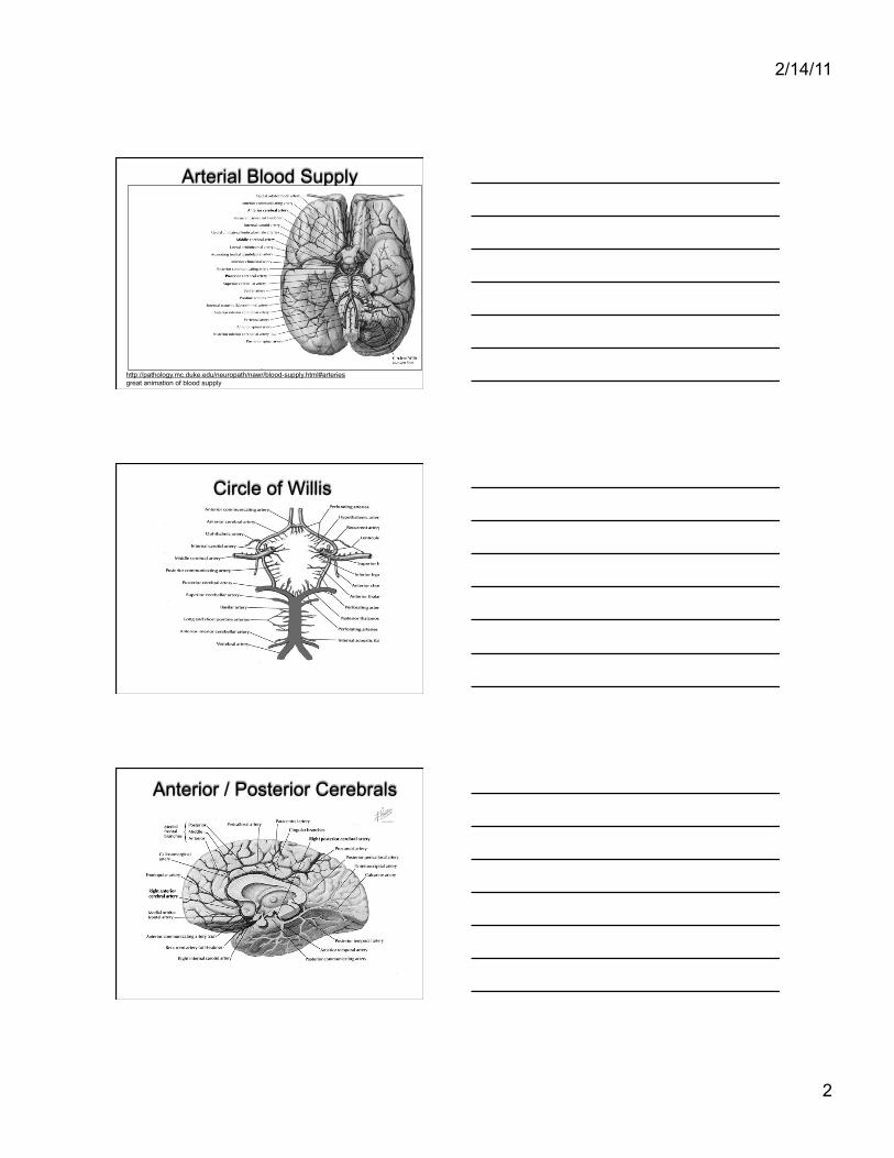
Blood Supply to the Brain
2/14/11
2
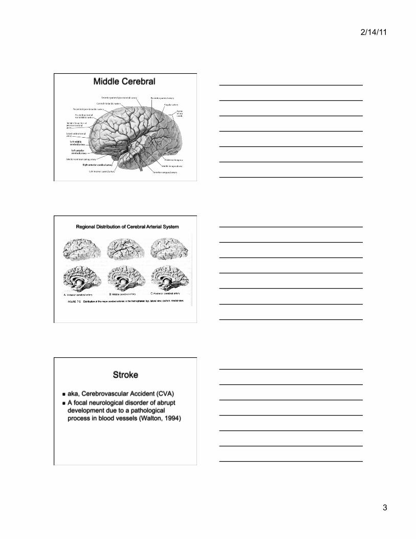
Arterial Blood Supply
http://pathology.mc.duke.edu/neuropath/nawr/blood-supply.html#arteries great animation of blood supply
Circle of Willis
Anterior / Posterior Cerebrals
2/14/11
3
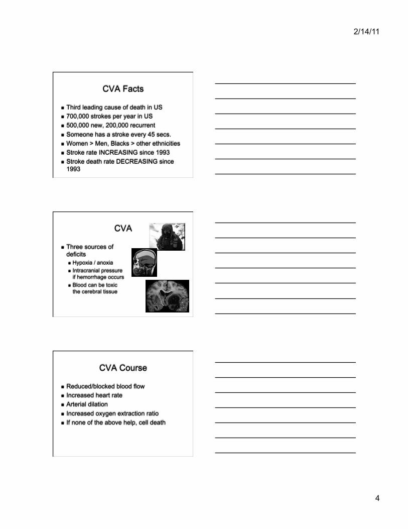
Middle Cerebral
Stroke
aka, Cerebrovascular Accident (CVA) A focal neurological disorder of abrupt
development due to a pathological process in blood vessels (Walton, 1994)
2/14/11
4
CVA Facts
Third leading cause of death in US 700,000 strokes per year in US 500,000 new, 200,000 recurrent Someone has a stroke every 45 secs. Women > Men, Blacks > other ethnicities Stroke rate INCREASING since 1993 Stroke death rate DECREASING since
1993
CVA
Three sources of deficits Hypoxia / anoxia Intracranial pressure
if hemorrhage occurs Blood can be toxic
the cerebral tissue
CVA Course
Reduced/blocked blood flow Increased heart rate Arterial dilation Increased oxygen extraction ratio If none of the above help, cell death
2/14/11
5
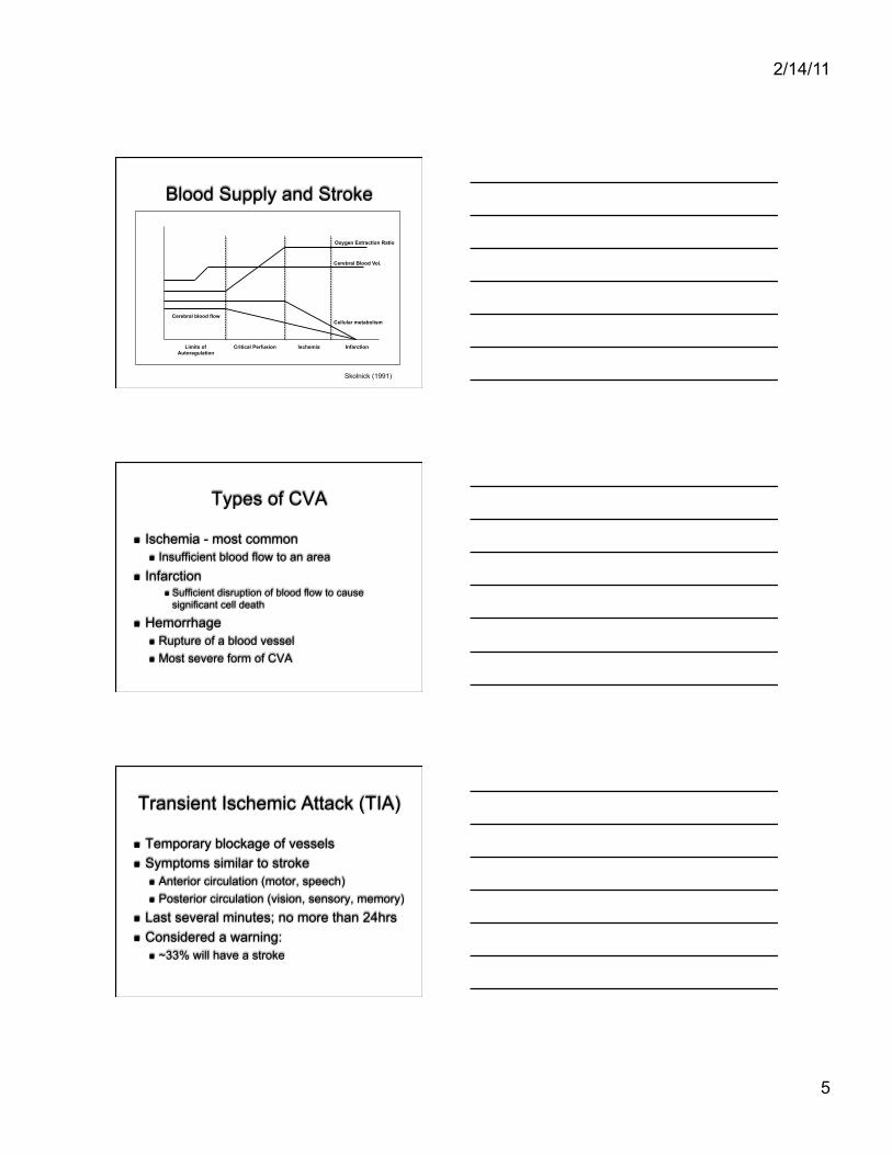
Blood Supply and Stroke
Limits of Autoregulation
Critical Perfusion Infarction
Cerebral Blood Vol.
Oxygen Extraction Ratio
Cellular metabolism Cerebral blood flow
Ischemia
Skolnick (1991)
Types of CVA
Ischemia - most common Insufficient blood flow to an area
Infarction Sufficient disruption of blood flow to cause
significant cell death
Hemorrhage Rupture of a blood vessel Most severe form of CVA
Transient Ischemic Attack (TIA)
Temporary blockage of vessels Symptoms similar to stroke
Anterior circulation (motor, speech) Posterior circulation (vision, sensory, memory)
Last several minutes; no more than 24hrs Considered a warning:
~33% will have a stroke
2/14/11
6
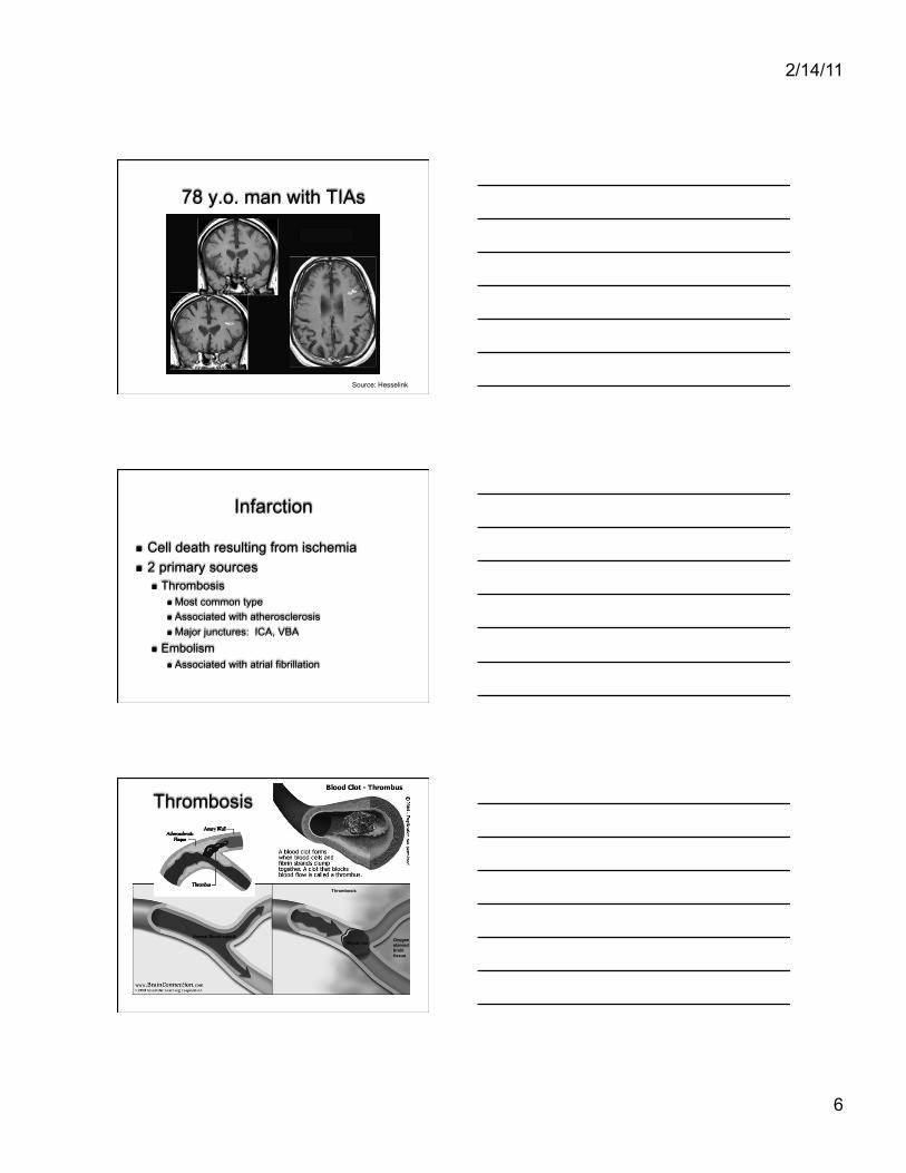
78 y.o. man with TIAs
Source: Hesselink
Infarction
Cell death resulting from ischemia 2 primary sources
Thrombosis Most common type Associated with atherosclerosis Major junctures: ICA, VBA
Embolism Associated with atrial fibrillation
Thrombosis
Normal blood flow Thrombosis
Normal Blood supply Blood clot Oxygen
starved brain tissue
2/14/11
7
Thrombosis Effects
75% of strokes Tend to be large ICA most common site Develop slowly; TIA’s common Right hemisphere lesions - greater
functional impairment Left hemisphere lesions - aphasia
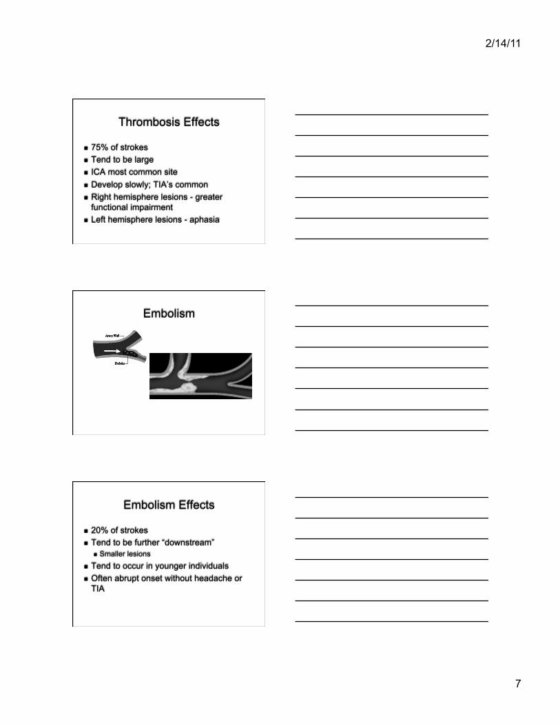
Embolism
Embolism Effects
20% of strokes Tend to be further “downstream”
Smaller lesions Tend to occur in younger individuals Often abrupt onset without headache or
TIA
2/14/11
8
Left Hemisphere Deficits
Aphasia Apraxia Agraphia / dysgraphia Right hemiplegia / hemiparesis Verbal memory problems Right visual field loss Local processing deficits Depression
Right Hemisphere Deficits
Visual-perceptual processing Left hemiplegia / hemiparesis Nonverbal communication Prosopagnosia Visual memory Left neglect Anosagnosia or euphoria
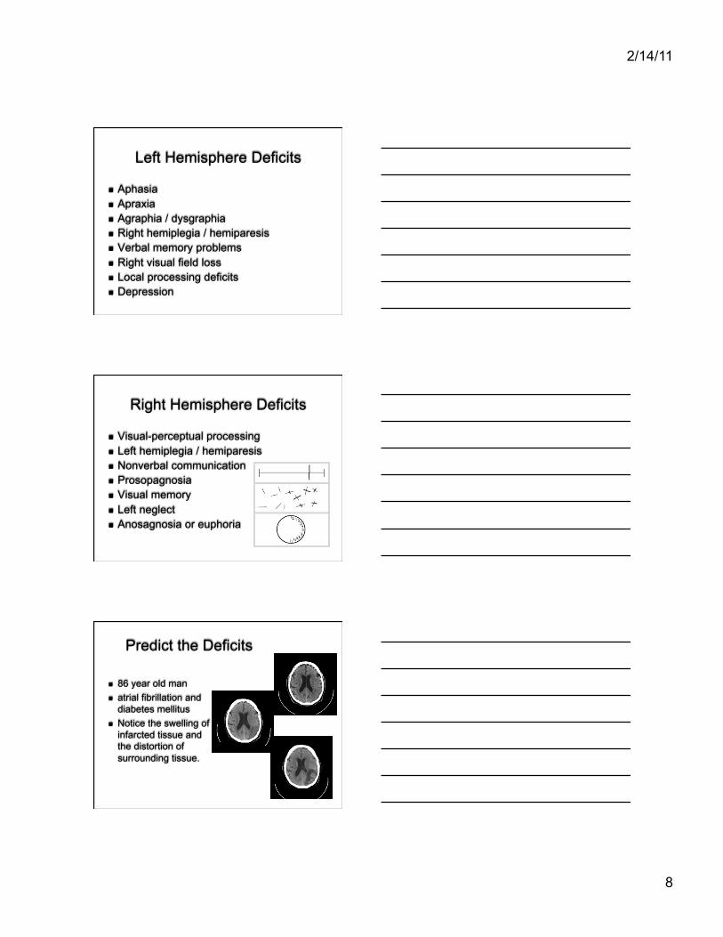
Predict the Deficits
86 year old man atrial fibrillation and
diabetes mellitus Notice the swelling of
infarcted tissue and the distortion of surrounding tissue.
2/14/11
9
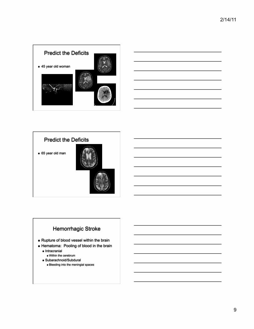
Predict the Deficits
45 year old woman
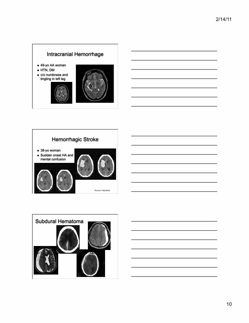
Predict the Deficits
65 year old man
Hemorrhagic Stroke
Rupture of blood vessel within the brain Hematoma: Pooling of blood in the brain
Intracranial Within the cerebrum
Subarachnoid/Subdural Bleeding into the meningial spaces
2/14/11
10
Intracranial Hemorrhage
49-yo AA woman HTN, DM c/o numbness and
tingling in left leg
Hemorrhagic Stroke
38-yo woman Sudden onset HA and
mental confusion
6 hours later
Source: Hesselink
Subdural Hematoma
2/14/11
11
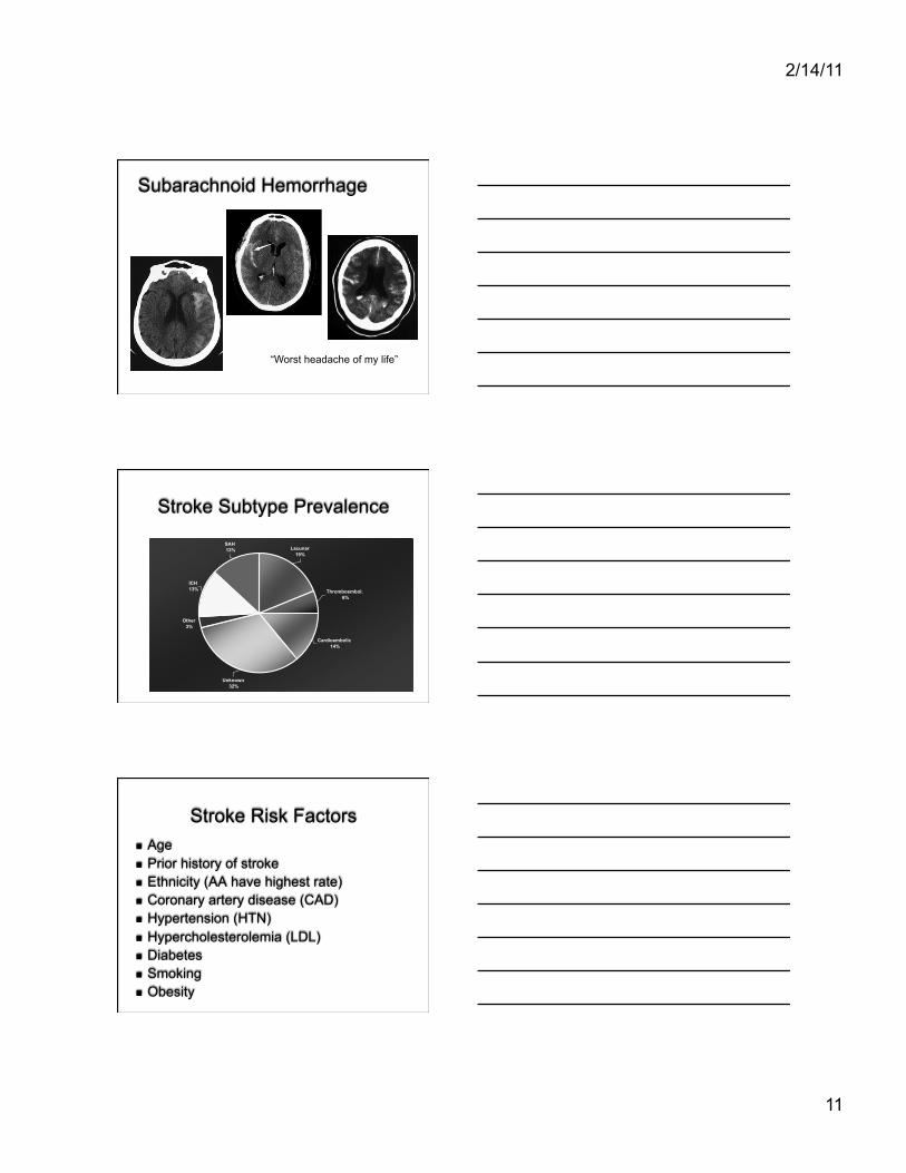
Subarachnoid Hemorrhage
“Worst headache of my life”
Stroke Subtype Prevalence
Lacunar
19%
Other
3%
ICH
13%
SAH
13%
Thromboembol.
6%
Cardioembolic
14%
Unknown
32%
Stroke Risk Factors Age Prior history of stroke Ethnicity (AA have highest rate) Coronary artery disease (CAD) Hypertension (HTN) Hypercholesterolemia (LDL) Diabetes Smoking Obesity
2/14/11
12
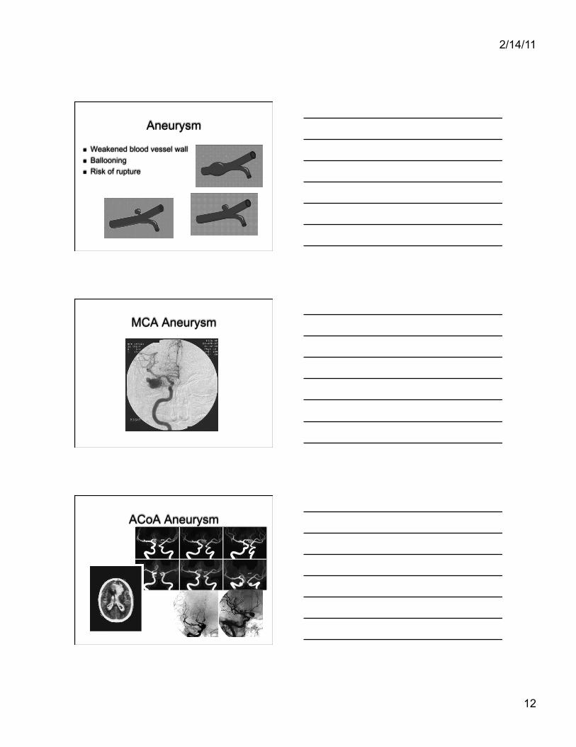
Aneurysm
Weakened blood vessel wall Ballooning Risk of rupture
MCA Aneurysm
ACoA Aneurysm
2/14/11
13
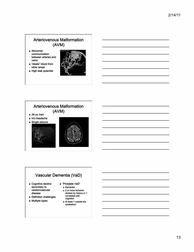
Arteriovenous Malformation (AVM)
Abnormal communication between arteries and veins
“steals” blood from other areas
High leak potential
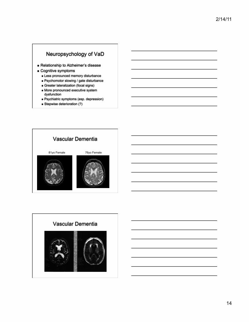
Arteriovenous Malformation (AVM)
34-yo man h/o headache Single seizure
Vascular Dementia (VaD)
Cognitive decline secondary to cerebrovascular disease
Definition challenges Multiple types
“Probable VaD” Dementia 2 or more ischemic
strokes by history or 1 correlated with cognition
At least 1 outside the cerebellum
2/14/11
14
Neuropsychology of VaD
Relationship to Alzheimer’s disease Cognitive symptoms
Less pronounced memory disturbance Psychomotor slowing / gate disturbance Greater lateralization (focal signs) More pronounced executive system
dysfunction Psychiatric symptoms (esp. depression) Stepwise deterioration (?)
81yo Female 76yo Female
Vascular Dementia
Vascular Dementia
2/14/11
15
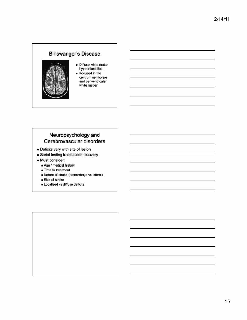
Binswanger’s Disease
Diffuse white matter hyperintensities
Focused in the centrum semiovale and periventricular white matter
Neuropsychology and Cerebrovascular disorders
Deficits vary with site of lesion Serial testing to establish recovery Must consider:
Age / medical history Time to treatment Nature of stroke (hemorrhage vs infarct) Size of stroke Localized vs diffuse deficits