structural and functional studies to investigate enzymatic
TRANSCRIPT

Structural and Functional Studies to Investigate Enzymatic Catalysis and Flavin
Transfer Mechanism of The Two-component Alkanesulfonate Monooxygenase
System from Escherichia coli
by
Jingyuan Xiong
A dissertation submitted to the Graduate Faculty of
Auburn University
in partial fulfillments for the
requirements for the Degree of
Doctor of Philosophy
Auburn, Alabama
August 4, 2012
Keywords: flavin, sulfur, enzymology,
protein dynamics, protein-protein interactions
Copyright 2012 by Jingyuan Xiong
Approved by
Dr. Holly R. Ellis, Chair, Associate Professor of Chemistry and Biochemistry
Dr. Douglas C. Goodwin, Associate Professor of Chemistry and Biochemistry
Dr. Evert C. Duin, Associate Professor of Chemistry and Biochemistry
Dr. Suanne Striegler, Associate Professor of Chemistry and Biochemistry

ii
Abstract
Several bacterial organisms rely on the two-component alkanesulfonate
monooxygenase system for the acquisition of organosulfonate compounds when
inorganic sulfur is limiting in the environment. This system is comprised of an FMN
reductase (SsuE) that supplies reduced flavin to the alkanesulfonate monooxygenase
(SsuD). Desulfonation of alkanesulfonates by SsuD is catalyzed through the activation of
dioxygen by reduced flavin. The three-dimensional structure of SsuD exists as a
TIM-barrel fold with several discrete insertion regions. An extensive insertion region
near the putative active site was unresolved in the SsuD structure, suggesting the
importance of protein dynamics in the desulfonation mechanism. Three variants
containing a partial deletion of the loop region were constructed to evaluate the
functional properties of this region. There were no overall gross changes in secondary
structure for the three SsuD deletion variants compared to wild-type SsuD, but each
variant was found to be catalytically inactive. The deletion variants were unable to
undergo the conformational changes necessary for catalysis even though they were able
to bind reduced flavin. Results from rapid kinetic analyses suggested that the SsuD
deletion variants failed to protect reduced flavin from non-enzymatic oxidation. These
studies define the importance of the unresolved loop region for the protection of reduced
flavin.

iii
A distinct feature of the alkanesulfonate monooxygenase system is that the flavin is
utilized as a substrate instead of a bound prosthetic group. The reduced flavin is
transferred from SsuE to SsuD, either through a dissociative or a channeling mechanism.
In order to kinetically discern between a dissociative and a channeling flavin transfer
mechanism, a method was designed using an SsuD variant with reduced flavin affinity to
compete with wild-type SsuD for reduced flavin. Results from this novel competition
method suggested that the flavin is transferred within a transient SsuE-SsuD complex
during catalysis. In addition, the reductive half-reaction catalyzed by SsuE is affected by
SsuD and its substrate under pre-steady-state kinetic conditions. The evidence provided
in this study support a channeling flavin transfer mechanism in the two-component
alkanesulfonate monooxygenase system.

iv
Acknowledgements
First, I would like to express my deepest appreciation and sincere thanks to my
advisor, Dr. Holly R. Ellis, for her invaluable guidance, consistent support and inspiring
encouragement throughout my studies here at Auburn University. Not only did she offer
me a great deal of knowledge in science and research, she also showed me an upstanding
attitude towards carrier and life. Her critical thinking, preciseness, diligence and fairness
will be perpetually beneficial in my life. I would also like to thank my Dr. Douglas C.
Goodwin, Dr. Evert C. Duin, Dr. Susanne Striegler, and Dr. Sang-Jin Suh for their
meaningful discussion and constructive suggestions to my dissertation. I appreciate Dr.
Goodwin for letting me use his stopped-flow spectrometer and providing me helpful
advice on my research. I wish to thank Dr. Duin for the use of his anaerobic glove box
and the -80ºC freezer. I would also like to thank Dr. Suh and Dr. Paul A. Cobine for
organizing the inter-departmental molecular biology journal club, where I greatly
improved my understanding in biological sciences.
Secondly, I would like to thank my former and current lab mates Dr. Xuanzhi Zhan,
Dr. Russell A. Carpenter, Dr. Erin M. Imsand, Mary Millwood, John M. Robbins,
Catherine Njeri, and Paritosh Dayal for their discussion and help during my studies. I
especially want to express my gratitude to my parents Siping Xiong and Min Zhang for
their everlasting unconditional love and support.
And last but not least, I wish to thank NSF and EPSCoR for their financial support.

v
Table of Contents
Abstract……………………………………………………………………………………ii
Acknowledgements………………………………………………………………………iv
List of Tables……………………………………………………………………………viii
List of Figures….................................................................................................................ix
List of Schemes…………………………………………………………………………..xii
List of Abbreviations……………………………………………………………………xiii
Chapter 1: Literature Review………………..…………………………………………….1
1.1 Sulfur metabolism in bacterial organisms……………………………………...1
1.2 Sulfur starvation and bacterial response…………………………………….…6
1.3 Regulation of sulfonate-sulfur utilization in bacterial organisms…………….15
1.4 Alkanesulfonate monooxygenase system from E. coli……………………….18
1.5 Flavins and flavoproteins……………………………………………………..18
1.6 Flavin reductases……………………………………………………………...26
1.6.1 Flavin reductase (SsuE) in the alkanesulfonate monooxygenase system
from E. coli……………………………………………………………….27
1.7 Flavin monooxygenases………………………………………………………32
1.7.1 Alkanesulfonate monooxygenase (SsuD) from E. coli…………………39
1.8 Protein dynamics and flexible loop…………………………………………...49
1.8.1 Flexible loop of triose phosphate isomerase (TIM)…………………….51
1.8.2 Flexible loop of bacterial luciferase…………………………………….54

vi
1.8.3 Flexible loop of alkanesulfonate monooxygenase SsuD….……………...59
1.9 Flavin transfer mechanism in two-component systems………………………63
1.9.1 Flavin transfer mechanism in bacterial luciferase system………………66
1.9.2 Flavin transfer mechanism the HPAH and ActVA-ActVB systems.…….68
1.9.3 Flavin transfer mechanism in alkanesulfonate monooxygenase
system…………………………………………………………………….70
1.10 Summary……………………………………………………………………...71
Chapter 2: Deletional Studies to Investigate the Functional Role of an Dynamic Loop
Region of Alkanesulfonate Monooxygenase………….…………………73
2.1 Introduction……………………………………………………………………...73
2.2 Material and methods……………………………………………………………78
2.2.1 Materials………………………………………………………………..78
2.2.2 Construction, expression and puri ficat ion of recombinant
proteins…………………………………………………………………78
2.2.3 Circular dichroism spectroscopic analysis……………………………..79
2.2.4 Proteolytic and mass spectrometric analyses…..……………..………..80
2.2.5 Affinity chromatography binding assays……………………………….80
2.2.6 Kinetic analysis…………………………………………………………81
2.3 Results…………………………………………………………………………...84
2.3.1 Functional and structural evaluation of the SsuD deletion variants……84
2.3.2 Evaluation of the SsuD deletion variants active site environment….….88
2.3.3 Proteolytic susceptibility of the SsuD deletion variants………………..90
2.3.4 Rapid reaction kinetic analysis of the SsuD deletion variants………….94
2.3.5 Protein-protein interactions between SsuE and ΔF261-N282 SsuD…..100
2.4 Discussions……………………………..……………………………………...102

vii
Chapter 3: Investigation of the Flavin Transfer Mechanism in the Two-component
Alkanesulfonate Monooxygenase System…………………………….109
3.1 Introduction…………………………………………………………………….109
3.2 Material and methods…………………………………………………………113
3.2.1 Materials………………………………………………………………113
3.2.2 Construction, expression and puri ficat ion of recombinant
proteins………………………..……………………………………….113
3.2.3 Steady-state activity assays……………………………………………114
3.2.4 Reduced flavin affinity………………………………………………..114
3.2.5 Rapid reaction kinetic analysis………………………………………115
3.3 Results………………………………………………………………………….117
3.3.1 Rapid reaction kinetics of flavin reduction and oxidation……………117
3.3.2 Kinetic parameters of Y128F/S179A SsuD……………………….......123
3.3.3 Evaluation of flavin transfer mechanism……………………………127
3.4 Discussions…………………………………………………………………….131
4 Chapter 4: Summary………………………………………………………………..141
4.1 Catalytic mechanism of alkanesulfonate monooxygenase…………………….142
4.2 Functional role of the SsuD unresolved loop in catalysis……………………143
4.3 Flavin transfer mechanism in the two-component alkanesulfonate
monooxygenase system………………………………………………………145
References………………………………………………………………………………148

viii
List of Tables
Table 1.1 Sulfate-regulated proteins in Escherichia coli………………………………9
Table 1.2 Substrate ranges and relative activities for SsuD and TauD……………….13
Table 2.1 Rate constants for FMNH2 oxidation monitored at 450 nm……………….99
Table 3.1 Kinetic parameters for Y128F/S179A and wild-type SsuD……………...118

ix
List of Figures
Figure 1.1 Sulfate assimilation pathway and cysteine biosynthesis in Escherichia
coli…………………………………………………………………………...3
Figure 1.2 The genetics and biochemistry of sulfate utilization in E. coli……………...5
Figure 1.3 Assimilation and utilization of organic sulfur sources in E. coli…………..10
Figure 1.4 Uptake and desulfonation of alkanesulfonates and taurine in Escherichia
coli………………………………………………………………………….12
Figure 1.5 Genetic organization of ssu and msu operons from Escherichia coli,
Pseudomonas putita and Pseudomonas aeruginosa……………………….14
Figure 1.6 Gene expression regulation of sulfur assimilation in Escherichia coli…….17
Figure 1.7 Overall desulfonation reaction of the flavin-dependent two-component
alkanesulfonate monooxygenase system from Escherichia coli…………..19
Figure 1.8 Numbering system for the flavin for the flavin isoalloxazine ring and the
structures of lumiflavin, riboflavin, FMN and FAD……………………….21
Figure 1.9 Redox and ionic states of flavin……………………………………………23
Figure 1.10 The spectra of flavin in different oxidation states…………………………25
Figure 1.11 Altered reaction mechanism of SsuE in the absence and presence of SsuD
and alkanesulfonate………………………………………………………...31
Figure 1.12 Reactions of reduced flavin with dioxygen………………………………..34
Figure 1.13 Catalytic mechanism of PAPAH using hydroperoxyflavin as an
electrophile…………………………………………………………………36
Figure 1.14 Proposed reaction mechanism of bacterial luciferase……………………...38
Figure 1.15 Tetrameric three dimensional structure of SsuD…………………………..41
Figure 1.16 Topology diagram showing secondary structural elements of each SsuD
monomer…………………………………………………………………...42
x
Figure 1.17 Proposed mechanism for the desulfonation reaction by SsuD……………..47
Figure 1.18 Putative active site of the alkanesulfonate monooxygenase SsuD………...48
Figure 1.19 The isomerization reaction catalyzed by TIM and the crystal structures of
the loop-open and loop-close forms of TIM from chicken muscle………..52
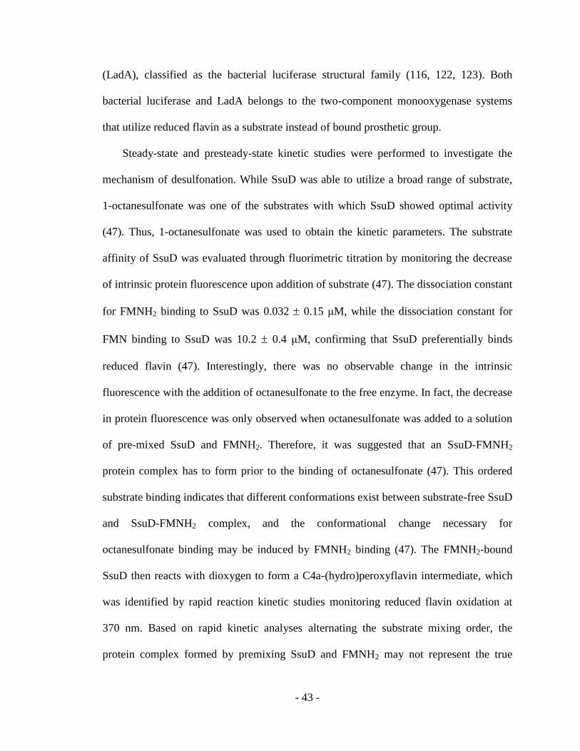
Figure 1.20 The three dimensional structure of bacterial luciferase with two observed
conformations of the mobile loop superimposed upon one another……….57
Figure 1.21 Three-dimensional structure of SsuD with the sequence of the unresolved
region indicated…………………………………………………………….61
Figure 1.22 Schematic representation of flavin transfer mechanisms in two-component
systems……………………………………………………………………..65
Figure 2.1 Three-dimensional structures of SsuD with the sequence of the dynamic
region indicated…………………………………………………………….76
Figure 2.2 Sequence alignment of SsuD homologues…………………………………77
Figure 2.3 The far-UV circular dichroism spectra of wild-type SsuD and the deletion
variants……………………………………………………………………..86
Figure2.4 Dissociation constant for reduced flavin determined by fluorimetric
titration..........................................................................................................87
Figure 2.5 Visible CD spectra of the FMNH2 with and without wild-type SsuD and the
deletion variants……………………………………………………………89
Figure 2.6 Mass spectrometry analysis of wild-type SsuD following limited tryptic
digestion……………………………………………………………………92
Figure 2.7 Limited trypsin digestion of wild-type SsuD and the deletion variants……93
Figure 2.8 Rapid reaction kinetic analyses of wild-type SsuD and deletion variants in
the presence of SsuE…………………………………………………….....97
Figure 2.9 Rapid reaction kinetic analyses of wild-type SsuD and deletion variants in
the absence of SsuE………………………………………………………..98
Figure 2.10 SDS-PAGE from the affinity chromatography experiments with SsuE and
the His-tagged ΔF261-N282 SsuD……………………………………….101
Figure 3.1 Rapid reaction kinetics of flavin reduction by SsuE in the presence of SsuD
and varying octanesulfonate concentration……………………………….120

xi
Figure 3.2 Rapid reaction kinetics of flavin reduction by SsuE in the presence of SsuD
and varying dioxygen concentration……………………………………...121
Figure 3.3 Rapid reaction kinetics of flavin reduction by SsuE and flavin oxidation by
SsuD with varying NADPH concentration……………………………….122
Figure 3.4 Three-dimensional structures of SsuD active site with FMN modeled
in………………………………………………………………………….124
Figure 3.5 Comparison of enzymatic flavin oxidation by wild-type SsuD,
Y128F/S179A SsuD, and non-enzymatic flavin oxidation……………….129
Figure 3.6 The effect of varying concentration of Y128F/S179A SsuD on the flavin
oxidation by wild-type SsuD……………………………………………..130
Figure 3.7 Schematic representation of flavin reduction and oxidation process with the
presence of SsuE, wild-type SsuD and Y128F/S179A SsuD…………….139

xii
List of Schemes
Scheme 1.1 Activation of molecular dioxygen by reduced flavin…………....22
Scheme 1.2 Order of substrates binding for SsuD………………………………………45
Scheme 2.1 Reaction mechanism of the alkanesulfonate monooxygenase system……..74
Scheme 3.1 Proposed SsuD mechanism involving a C4a-peroxyflavin intermediate...110

xiii
List of Abbreviations
DTNB 5,5-dithiobis(2-nitrobenzoic acid)
FMN flavin mononucleotide
FMNH2 reduced flavin mononucleotide
NAD(P)H nicotinamide adenine dinucleotide (phosphate)
NTA Ni-nitrilotriacetate
PMSF phenylmethylsulfonyl fluoride
SsuE alkanesulfonate flavin reductase
SsuD alkanesulfonate monooxygenase
TIM triosephosphate isomerase
LadA long-chain-alkane monooxygenase
FRP NADPH-dependent flavin reductase
Fre flavin reductase
FRase I flavin reductase

- 1 -
Chapter 1
Literature Review
1.1 Sulfur metabolism in bacterial organisms
Sulfur is an essential element for the survival and growth of all organisms. Although
sulfur is one of the basic elemental building blocks for living organisms, sulfur
metabolism is less well-understood compared to other basic elements. In bacteria, sulfur
is needed as a component of amino acids cysteine, and methionine. Sulfur is also a
crucial component in many enzyme cofactors and participates in maintaining the
appropriate redox potential in the cell through sulfur-containing redox-active compounds.
Bacterial organisms primarily assimilate inorganic sulfate for the biosynthesis of
sulfur-containing molecules (Figure 1.1) (1, 2). Inorganic sulfate is first assimilated into
the cell by sulfate permease (CysU, CysW, CysA and sulfur binding proteins) (3). The
intracellular sulfate is utilized to form adenosine phosphate (APS) and pyrophosphate by
ATP sulfurylase at the expense of ATP hydrolysis (3). The ATP sulfurylase is a tetramer
of heterodimers consisting of a GTPase subunit (CysN) and APS synthesis subunit
(CysD). Because the energy required to synthesize APS by CysD is higher than the
energy released from the hydrolysis of ATP, another reaction catalyzed by
pyrophosphatase is needed to decrease the pyrophosphate concentration in order to shift
the reaction equilibrium towards APS production (4). The GTPase subunit (CysN) of the
ATP sulfurylase catalyzes the hydrolysis reaction of GTP, providing energy for the APS

- 2 -
synthesis (4). Therefore, the activation of sulfate in the sulfur assimilation pathway is
energetically costly, and the recovery of sulfur-containing compounds from
reduced-sulfur would be more beneficial in conserving energy. The APS is then activated
by APS kinase (CysC) to form 3‟-phosphoadenosine-5‟-phosphosulfate (PAPS) with the
hydrolysis of another ATP and the release of ADP in order for the incorporated sulfur to
be reduced. This reaction is thought to further pull the sulfate incorporation reaction
towards APS formation (2). The sulfur in PAPS is then reduced to sulfite by thioredoxin
dependent PAPS reductase (CysH) in the presence of NADPH, yielding adenosine 3‟-5‟
diphosphate (PAP) as a by-product. Because PAP is also produced by the transfer of
4-phosphopantetheine from coenzyme A in fatty-acid metabolism and the synthesis of
some secondary metabolites, it is speculated that PAP might play a regulatory role in
coupling sulfur assimilation with lipid metabolism (5). In the sulfur assimilation pathway,
the sulfite is then reduced by NADPH-sulfite reductase (CysJ, CysI and CysG) to
generate sulfide by a poorly defined process (2).
The sulfide in the cell reacts with O-acetylserine to from cysteine (3). This reaction is
catalyzed by an enzyme complex cysteine synthase, which is composed of serine
transacetylase (CysE) and O-acetylserine sulfhydrylase A (CysK) (6). The serine
transacetylase catalyzes the condensation reaction of the acetyl group from acetyl CoA
onto the hydroxyl group of serine, generating O-acetylserine needed to synthesize
cysteine. The O-acetylserine sulfhydrylase A in the cysteine synthase complex then
utilizes sulfide as a nucleophile to react with O-acetylserine, forming cysteine and acetate.

- 3 -
Sulfate
(extracellular)
Sulfate permease
cysU, cysW, cysA
sulfur binding protein
Sulfate
(intracellular)
ATP
ATP sulfurylase Pyrophosphatase ppa
cysD, cysN PPi + H2O 2 Pi
APS
ATP
APS kinase Adenylate kinase adk
cysC ADP AMP
PAPS
PAP 3′
PAPS reductase phosphatase
cysH PAP
Serine
Sulfite +
Acetyl-coA
Sulfite reductase NADPH
cysJ, cysI Serine transacetylase
cysG NADP+ cysE
CoA
Sulfide O-Acetylserine
O-Acetylserine
sulfhydrylase A or B Acetate
cysK or cysM
Cysteine
Figure 1.1. Sulfate assimilation pathway and cysteine biosynthesis in Escherichia coli.
(Adapted from (2)).

- 4 -
The genes responsible for sulfate activation and reduction (cysD, cysN, cysC, cysH,
cysI and cysJ) are organized in a cluster in the bacterial chromosome, while other genes
involved in cysteine biosynthesis are isolated (Figure 1.2) (7). The operons of all cysteine
biosynthetic genes are grouped as the cysteine regulon, which is under central regulation
by a LysR-type transcriptional activator CysB (8, 9). The CysB regulator binds upstream
of the promoter region in the regulon to stimulate transcription of the cysteine
biosynthetic genes (10, 11). Furthermore, the CysB regulator is able to play an additional
role in inhibiting transcription from its own promoter (12). The O-acetylserine metabolite
serves as an inducer that stimulates the binding of CysB to the positive regulation sites of
the cys operon and facilitates the formation of the transcription initiation complex, while
inhibiting binding to the negatively autoregulated cysB promoter region (10).
O-acetylserine can spontaneously isomerize to N-acetylserine, which is speculated to be a
better inducer of the cys regulon. N-acetylserine is suggested to function as a pure
inducer because it cannot serve as a sulfur acceptor, and it is 15-fold more efficient in
inducing activation compared to O-acetylserine (13). In addition to regulation by the
central transcriptional activator CysB, the proteins in the cysteine biosynthetic pathway
are also regulated by sulfate, each sulfur intermediate in the pathway, and the end product
cysteine (7). The intracellular concentration of cysteine is found to indirectly control the
expression of cys genes by feedback inhibition of serine transacetylase (9). The positive
regulation by CysB is repressed by sulfide and thiosulfate, which act as anti-inducers that
interfere with the binding of the coinducer N-acetylserine (8). Interestingly, even though
sulfate is the starting substrate of the cysteine biosynthetic pathway, excess sulfate reduce
expression of the cys genes due to its conversion to sulfide and cysteine (9). Therefore,

- 5 -
SO42-
membrane ==== cysTWA
SO42-
GTP
cysD
ATP E. coli K-12 Chromosomal Map
cysN
PPi
Pi + GDP
APS
ATP +
cysC trans
ADP reg.
cysB
PAPS (autoregulatory)
cysH
cys gene cluster SO3
2-
AcCoA L-Serine
cysI CoA
cysJ
cysE
S2 + O-Acetylserine
(+ effector)
cysK or
cysM (feedback inhibition)
Cysteine
Figure 1.2. The genetics and biochemistry of sulfate utilization in Escherichia coli. The
genes involved in cysteine biosynthesis are indicated: cysTWA (sulfate permease), cysDN
(ATP sulfurylase), cysC (APS kinase), cysH (PAPS reductase), cysIJ (sulfite reductase),
cysE (serine transacetylase), and cysKM (O-acetylserine sulfhydrylase A or B). (Adapted
from (7)).

- 6 -
maximum expression of the cys genes requires an active CysB, coinducer O-acetylserine
or N-acetylserine, and the absence of cysteine and excess sulfate (7).
After the sulfur in inorganic sulfate is utilized to synthesize cysteine, the fate of
cysteine can be diversified in bacteria. In the methionine biosynthetic pathway cysteine is
first converted to cystathionine, which is the precursor of homocysteine. The
homocysteine is then methylized to form methionine (14). In the coenzyme A
biosynthetic pathway, the cysteine is added to the phosphorylated pantothenate, followed
by several steps of decarboxylation, adenylylation, and phosphorylation to generate
coenzyme A (15). In iron-sulfur clusters, the sulfur is suggested to be provided by
thiocysteine. The thiocysteine may be generated by the lysis of cystine, a dimeric amino
acid formed by oxidation of two cysteine residues (16). In the thiamine biosynthetic
pathway, cysteine is required to synthesize the thiazole group, which combines with a
pyrimidine to yield thiamine monophosphate. The thiamine monophosphate can then be
phosphorylated to thiamine pyrophosphate or hydrolyzed to thiamine; however, the
enzymatic mechanism of thiazole formation is not fully clear (17). It is also speculated
that lipoic acid is synthesized by inserting cysteine sulfur into the hydrocarbon chain of
octanoic acid by an enzyme containing iron-sulfur clusters (18). In biotin biosynthesis,
the incorporation of sulfur into biotin is catalyzed by iron-sulfur proteins (18, 19).
1.2 Sulfur limitation and bacterial response
Sulfur is an abundant element in the environment, and is commonly found in the
earth‟s crust, water and atmosphere (20-24). In the atmosphere the sulfur element
primarily exists as SO2 and H2S, which are eventually oxidized to sulfate (24). The sulfur
in aquatic environments is commonly found in the soluble SO32-
and SO42-
form (23).
- 7 -
While inorganic sulfate is ubiquitous in the atmosphere and aquatic system, it is relatively
poorly represented in aerobic soils, taking up less than 5% of the total sulfur content (21,
25). In addition, not all inorganic sulfur in the soil is easily accessible sulfate that can be
readily assimilated by plants and bacteria. Inorganic sulfur can also exist as absorbed
sulfate and insoluble sulfate co-precipitated with CaCO3 (25). Most of the sulfur in
aerobic soils is found to be organic sulfur such as sulfonate and sulfate esters, therefore
inorganic sulfate and cysteine that are available to bacteria become limiting in aerobic
soils (26). In order to survive, bacteria under sulfur limitation condition have developed
an alternate process to obtain sulfur from available organic sulfur sources (27-30).
Under sulfur limiting conditions where readily available inorganic sulfate and
cysteine are limiting in the environment, bacteria express a set of proteins involved in the
uptake and utilization of alternate organic sulfur sources such as alkanesulfonates and
taurine (Figure 1.3) (1, 31-34). These specific proteins expressed only in the absence of
primary sulfur sources are designated as sulfur starvation induced (ssi) proteins (Table
1.1) (28). In order to meet their sulfur requirements, bacteria employ these ssi proteins to
enzymatically liberate sulfite from organic sulfur sources. The released sulfite product
can then enter the cysteine biosynthetic pathway to provide bacteria with the necessary
precursors for the synthesis of various sulfur-containing compounds (35-36).
Even though E. coli is known to utilize organosulfur compounds for growth and
these alternate sulfur sources enter the cysteine biosynthetic pathway at the stage of
sulfite, E. coli has a preference for sulfate over sulfonate if both are available (32). It has
been shown that the presence of sulfate represses the expression of ssi proteins. When E.
coli was cultured in minimal media deprived of sulfate and cysteine, several ssi proteins

- 8 -
were found to be up-regulated (31). These ssi proteins were identified as periplasmic
sulfate binding protein (Sbp), periplasmic cysteine binding protein (FliY), O-acetylserine
sulfhydrylase A (CysK) and antioxidant alkylhydroperoxide reductase (AhpC) (33). The
Sbp and FliY proteins are involved in the initial assimilation of sulfate and cysteine. The
CysK enzyme is the last enzyme in the cysteine biosynthetic pathway. Both the substrates
(sulfide and O-acetylserine) and the product (cysteine) of CysK are reported to play
crucial regulatory roles in cysteine biosynthesis. The ssi proteins involved in the
utilization of sulfonate sulfur sources were later identified to be a taurine desulfurization
enzyme (TauD) expressed from the tauABCD gene cluster and the alkanesulfonate
desulfurization enzymes (SsuE and SsuD) expressed from the ssuEADCB gene cluster
(35-36). Both the tau and ssu operons are found to encode an oxygenase system (TauD
and SsuED) and an ABC (ATP binding cassette)-type transporter system, which includes
a periplasmic substrate binding protein, an ATP-binding protein, and a transporter
membrane component.
The oxygenase component of the tau operon is an -ketoglutarate dependent taurine
dioxygenase (TauD), which requires Fe (II) to convert taurine to aminoacetaldehyde and
sulfite (Figure 1.4) (35, 37). The oxygenase system of the ssu operon contains two
components, an NADPH-dependent FMN reductase (SsuE) and an FMNH2-dependent
alkanesulfonate monooxygenase (SsuD) (36). The SsuE enzyme catalyzes the reduction
of FMN to FMNH2 with NADPH providing reducing equivalents. The SsuD enzyme
converts alkanesulfonates to aldehyde and sulfite with the FMNH2 supplied by SsuE
(Figure 1.4). While the liberation of sulfite by the tauABCD system is performed by

- 9 -
Protein Gene locus Function
TauA, TauD tauABCD taurine desulfurization
SsuE, SsuD ssuEADCB alkanesulfonate desulfurization
Sbp Sbp periplasmic sulfate binding protein
FliY fliY periplasmic cysteine binding protein
AhpC ahpC alkylhydroperoxide reductase subunit
Table 1.1. Sulfate-regulated proteins in Escherichia coli (28).

- 10 -
Alkanesulfonates Taurine
(external) (external)
ssuABC tauABC
Alkanesulfonates Taurine
(internal) (internal)
FMN reductase ssuE
Taurine dioxygenase tauD
Alkanesulfonate monooxygenase ssuD
Sulfite
Figure 1.3. Assimilation and utilization of organic sulfur sources in Escherichia coli.
(Adapted from (32)).

- 11 -
TauD alone through the activation of dioxygen by ferrous iron, the desulfonation of
alkanesulfonates by the ssuEADCB system is dependent on SsuD, SsuE, and a flavin
cofactor.
Both the tau and ssu operons encode ABC-type transporter genes, but the proteins of
the TauABC and SsuABC transport systems are not interchangeable (Figure 1.4) (38).
Even through the substrates transported by both systems exhibit chemical similarities, the
periplasmic substrate binding protein TauA and SsuA share only 22.7% amino acid
identity (38). Some sulfonates can be transported by both TauABC and SsuEADCB
systems, while gene deletion studies demonstrated that taurine is exclusively assimilated
by the TauABC system and long-chain aliphatic sulfonates are primarily transported by
the SsuEADCB system. The substrate range for TauABC and SsuEADCB systems seems
to complement each other (Table 1.2) (36). Therefore, the expression of both systems
under sulfur limitation conditions constitutes a substrate uptake system that covers the
full spectrum of diverse aliphatic sulfonates. While similar ssi proteins found in E. coli
were also identified in Pseudomonas sp, the ssu operon from Pseudomonas sp were
found to contain one more open reading frame designated as ssuF (Figure 1.5) (39, 40).
Because E. coli majorly utilize aliphatic sulfonate whereas Pseudomonas sp can
additionally use a wide range of aromatic sulfonates, the molybdopterin protein encoded
by ssuF gene is speculated to be specifically involved in desulfonation of aromatic
sulfonates as well as alkanesulfonates in Pseudomonas sp (39). In addition to the tau and
ssu operon, an msu operon was upregulated in Pseudomonas aeruginosa during sulfur
limitation condition (Figure 1.5) (28). This msu operon codes an FMNH2-denpendent
methanesulfonate sulfonatase, which liberates sulfite from methanesulfonate (40).

- 12 -
Alkanesulfonates Taurine
External
SsuA TauA
Periplasm
SsuC SsuC TauC TauC
Cytoplasm SsuB SsuB TauB TauB
R-C-SO3- R-C-SO3
- -ketoglutarate
NAD(P)H + H+
FMNH2 O2
O2
SsuE SsuD TauD Fe2+
NAD(P)+ FMN H2O H
+ + SO3
2-
CO2
+ +
succinate
R-CHO + H+ + SO32-
R-CHO
Figure 1.4. Uptake and desulfonation of alkanesulfonates and taurine in Escherichia coli.
(Adapted from (32)).

- 13 -
Sulfonate Substrate Relative Activity %
SsuD TauD
Taurine 0 100
N-Phenyltaurine 65.5 0
4-Phenyl-1-butanesulfonaic acid 42.4 2.5
HEPES 10.6 5
MOPS 36.4 34.2
PIPES 29.2 3.1
2-(4-Pyridyl)ethanesulfonic acid 87.4 0.5
1,3-Dioxo-2-isoindolineethanesulfonic acid 100 30.1
Sulfoacetic acid 19.8 a
L-Cysteic acid 0 0
Isethionic acid 14.3 1.2
Methanesulfonic acid 0.7 0
Ethanesulfonic acid 5.2 0.8
Propanesulfonic acid 14 2.3
Butanesulfonic acid 17.8 8.4
Pentanesulfonic acid 40.4 22.5
Hexanesulfonic acid 43.8 11.3
Octanesulfonic acid 46.3
Decanesulfonic acid 43.2
Dodecanesulfonic acid 20.1 3.3
Tetradecanesulfonic acid 2.9
a, not determined
Table 1.2. Substrate ranges and relative activities for SsuD and TauD in E. coli (36).

- 14 -
E. coli
ssuE ssuA ssuD ssuC ssuB
P. putita
ssuE ssuA ssuD ssuC ssuB ssuF
P. aeruginosa
ssuE ssuA ssuD ssuC ssuB ssuF
msuE msuD msuC
Figure 1.5. Genetic organization of ssu and msu operons from Escherichia coli,
Pseudomonas putita and Pseudomonas aeruginosa. (Adapted from (28)).

- 15 -
1.3 Regulation of sulfonate-sulfur utilization in bacterial organisms
Although the tau and ssu genes are expressed under sulfur limitation as an alternate
route to synthesize sulfite and cysteine from sulfonates, the regulation of sulfonate-sulfur
utilization by bacteria is linked with the assimilatory sulfate reduction pathway (Figure
1.6) (32). The central transcriptional activator CysB not only positively regulates the cys
genes in the cysteine biosynthetic pathway, but also controls the regulation of sulfur
assimilation at a global level (3, 32).
The tau and ssu genes were found to be fully repressed when sulfate was present in
the growth environment (33). Binding sites for the global transcriptional activator CysB
were identified in both the tau and ssu promoter region (41, 42). An additional regulatory
protein, Cbl, was also found to regulate expression of the tau and ssu genes as a
LysR-type transcriptional activator (41, 42). It has been shown that Cbl binds upstream of
the -35 region of the ssu promoter. As both binding sites for CysB and Cbl are present in
the tau and ssu promoter region, these binding sites were removed separately to evaluate
the effect on gene expression. While Cbl binding was essential for the expression of both
tau and ssu, CysB binding was only needed for tau expression (42). Removal of the CysB
binding sites in ssu did not have a significant influence on protein expression. The role of
the CysB binding sites in the ssu promoter region remains unclear. Interestingly, while
CysB expression is autoregulated by self-binding to the cysB promoter region, the
expression of Cbl is also positively regulated by CysB (43). As a result, the expression of
the tau and the ssu operons are indirectly regulated by CysB and its inducers and
anti-inducers.

- 16 -
A coinducer N-acetylserine binding cavity was identified in the three-dimensional
structure of CysB located at the C-terminal portion of the protein, which is
compartmentalized by two / domains (44). Comparing the amino acid sequence of Cbl
to CysB, the Cbl protein contains 45% amino acid sequence identity to CysB and
conserved residues that constitute the inducer binding cavity in CysB (32). However,
unlike CysB, the binding of Cbl to the tau and ssu promoter regions is not affected by the
inclusion of O-acetylserine (41, 42). Further transcriptional experiments have shown that
O-acetylserine is indeed not a coinducer of transcriptional activator Cbl (32). Although
sulfate was shown to repress the expression of the tau and ssu genes, it was suggested
that sulfate does not function as an anti-inducer of Cbl because the tau and ssu genes
were not repressed by sulfate in mutants that were unable to activate sulfate to APS (32).
The repression of tau and ssu genes by sulfate becomes effective in mutants that cannot
reduce sulfite to sulfide, suggesting that the anti-inducer of Cbl is actually an
intermediate of the cysteine biosynthetic pathway (32).
Two-dimensional gel electrophoresis experiments suggested that the Cbl not only
regulates the transcription of tau and ssu operon, but also other ssi proteins such as CysK,
FliY and Sbp (41). In addition, through transcriptional analysis using DNA arrays, the cbl
gene is shown to be cotranscribed and coregulated with the nac gene, which positively
regulates the expression of operons for utilization of poor nitrogen sources (45).
Therefore, Cbl was suggested to obtain an accessory function in regulating nitrogen
assimilation apart from its original function in sulfonate utilization regulation (46).

- 17 -
cysB
N-acetylserine
CysB
N-acetylserine + N-acetylserine +
cbl cys genes
sulfide sulfide
thiosulfate thiosulfate
Cbl
N-acetylserine N-acetylserine
+ +
thiosulfate thiosulfate
+ ? ? +
tauABCD ssuEADCB
Figure 1.6. Gene expression regulation of sulfur assimilation in Escherichia coli.
(Adapted from (32)).

- 18 -
1.4 Alkanesulfonate monooxygenase system from E. coli
Under sulfur limiting conditions, proteins involved in assimilating sulfonate-sulfur
from alternate sources are expressed by the E. coli ssu operon (42). Two of these
enzymes expressed from the ssu operon are responsible for catalyzing the desulfonation
reaction of alkanesulfonates (36). The SsuE enzyme catalyzes the reduction of FMN to
FMNH2 in the presence of NADPH. The SsuD enzyme utilizes dioxygen and the FMNH2
provided by SsuE to catalyze the oxygenic cleavage of the carbon-sulfur bond in
alkanesulfonates, producing sulfite and aldehyde (Figure 1.7) (36, 47). Because the
FMNH2 substrate of SsuD is the product of SsuE, the desulfonation reaction catalyzed by
SsuD is dependent on SsuE. In addition, SsuE and SsuD are located on the same ssu
operon and are co-expressed and co-regulated by Cbl (42). Therefore, SsuE and SsuD
function as a unit classified as a two-component system. Due to the absolute requirement
for flavin by both SsuE and SsuD, this system is known as the flavin-dependent
two-component alkanesulfonate monooxygenase system. An interesting feature of this
system is that the flavin cofactor does not function as a bound prosthetic group, but
serves as a substrate for both SsuE and SsuD.
1.5 Flavins and flavoproteins
The flavin cofactors not only play a vital role in the oxidation of alkanesulfonates,
but are involved in catalyzing a wide range of reactions. Proteins associated with flavin
cofactors are classified as flavoproteins. Flavins are considered highly versatile
compounds because different flavoproteins possess distinctly different and sometimes
contradictory functions. It is shown that the flavin environment in the enzyme active site
results in the chemical versatility of flavoproteins (48). Flavins are known to participate

- 19 -
Figure 1.7. Overall desulfonation reactions of the flavin-dependent two-component
alkanesulfonate monooxygenase system from Escherichia coli. (Adapted from (47)).
- 20 -
in both one-electron and two-electron transfer processes (48-67). Some flavoproteins
catalyze oxidation and oxygenation reactions by activating dioxygen with reduced flavin
(55-62). Other flavoproteins participate in dehydrogenation reactions, flavin reduction
reactions or serve as components in the respiratory chain (63-67). Interestingly, flavins
are also involved in physiological functions that have opposing effects. For instance,
some flavins are able to contribute to oxidative stress by producing superoxide, while
others are involved in self-defense mechanism toward oxidative stress by reducing
hydroperoxides (48). Ongoing research in the flavin field is to understand the mechanism
for the wide range of reactions catalyzed by flavoproteins and how the interactions
between the flavin cofactor and the protein mediate these chemical reactions (48).
Flavin is a generic term generally referring to a group of molecules with a
heterocyclic isoalloxazine ring structure (7,8-dimethylisoalloxazine). In nature, flavins
usually exist as lumiflavin, riboflavin, flavin mononucleotide (FMN) and flavin adenine
dinucleotide (FAD) (Figure 1.8) (48). Lumiflavin contains a methyl group attached to the
isoalloxazine ring at the N-10 position. The riboflavin, FMN, and FAD differ from one
another by the functional group on the ribityl side chain at the N-10 position. The ribityl
sugar side chain is covalently attached to the isoalloxazine ring in riboflavin. FMN
contains a phosphate group attached to the terminal hydroxyl group of the ribityl side
chain, while FAD has an adenine dinucleotide group. Flavin can exist freely in the
environment or associate with an enzyme to catalyze extremely versatile chemical
reactions. Flavin was first discovered and characterized as an active component of old
yellow enzyme, the first flavoprotein ever identified in the early 1930s (68-69). Flavin is
ubiquitously found and needed in a broad range of biological systems. As the basic unit

- 21 -
Figure 1.8. Numbering system for the flavin isoalloxazine ring and the structures of
lumiflavin, riboflavin, FMN and FAD.

- 22 -
of flavin, the isoalloxazine ring cannot be synthesized by humans. Therefore, riboflavin
(vitamin B2) is required in the human diet as starting material to synthesize flavin
cofactors needed to maintain physiological functions.
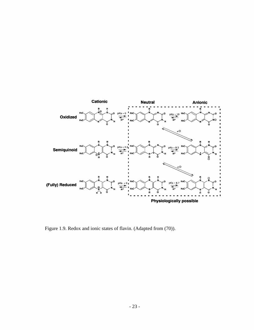
Flavins can exist in different redox states that include an oxidized, one-electron
reduced (semiquinone), and two-electron reduced (fully reduced hydroquinone) state
(Figure 1.9) (49, 70). When flavin is free in solution, a mixture of oxidized and reduced
flavin can rapidly form a certain amount of semiquinone radical through equilibrium (48).
Approximately 5% of the semiquinone radical is stabilized at pH 7 when flavin is not
bound to protein (48). However, the equilibrium can shift towards semiquinone formation
when the flavin is associated with some proteins (48, 71). Because the nitrogens in the
isoalloxazine ring are able to be protonated or deprotonated, flavin in each of the three
redox states can exist in different ionic forms. Because the theoretical cationic forms of
flavin are only possible at extremely low pH, the neutral forms and the anionic forms of
the flavin are considered physiologically relevant (70). Besides these redox/ionic forms,
flavins can adopt other electronic states known as charge-transfer states, which do not
belong to any of the three redox states but are defined as the states where partial charge is
transferred to or from one of the three redox states (70). The fully reduced hydroquinone
can rapidly react with molecular dioxygen auto-catalytically (Scheme 1.1) (48).
Scheme 1.1. Activation of molecular dioxygen by reduced flavin. (Adapted from (48)).
- 23 -
Figure 1.9. Redox and ionic states of flavin. (Adapted from (70)).
- 24 -
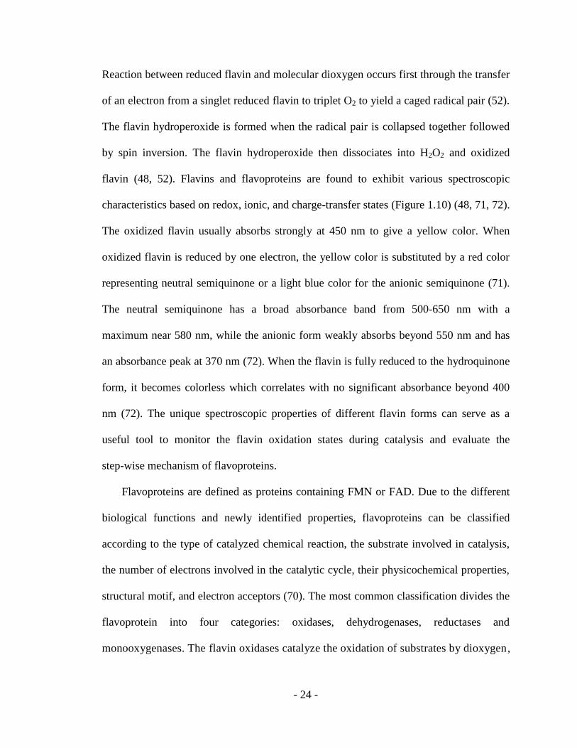
Reaction between reduced flavin and molecular dioxygen occurs first through the transfer
of an electron from a singlet reduced flavin to triplet O2 to yield a caged radical pair (52).
The flavin hydroperoxide is formed when the radical pair is collapsed together followed
by spin inversion. The flavin hydroperoxide then dissociates into H2O2 and oxidized
flavin (48, 52). Flavins and flavoproteins are found to exhibit various spectroscopic
characteristics based on redox, ionic, and charge-transfer states (Figure 1.10) (48, 71, 72).
The oxidized flavin usually absorbs strongly at 450 nm to give a yellow color. When
oxidized flavin is reduced by one electron, the yellow color is substituted by a red color
representing neutral semiquinone or a light blue color for the anionic semiquinone (71).
The neutral semiquinone has a broad absorbance band from 500-650 nm with a
maximum near 580 nm, while the anionic form weakly absorbs beyond 550 nm and has
an absorbance peak at 370 nm (72). When the flavin is fully reduced to the hydroquinone
form, it becomes colorless which correlates with no significant absorbance beyond 400
nm (72). The unique spectroscopic properties of different flavin forms can serve as a
useful tool to monitor the flavin oxidation states during catalysis and evaluate the
step-wise mechanism of flavoproteins.
Flavoproteins are defined as proteins containing FMN or FAD. Due to the different
biological functions and newly identified properties, flavoproteins can be classified
according to the type of catalyzed chemical reaction, the substrate involved in catalysis,
the number of electrons involved in the catalytic cycle, their physicochemical properties,
structural motif, and electron acceptors (70). The most common classification divides the
flavoprotein into four categories: oxidases, dehydrogenases, reductases and
monooxygenases. The flavin oxidases catalyze the oxidation of substrates by dioxygen,

- 25 -
Figure 1.10. The spectra of flavin in different oxidation states. (48, 72).

- 26 -
producing H2O2. In these reactions, the flavin cofactor is activated by the substrates, and
the reduced flavin needs to be stabilized in the oxidases before reacting with O2 (48).
Some well characterized flavin oxidases include glucose oxidase, glycine oxidase, and
amine oxidase (56-58). The flavin dehydrogenases catalyze reactions with the transfer of
a pair of electrons and a proton without the involvement of dioxygen. Examples of classic
flavin dehydrogenases include acyl-CoA dehydrogenase, succinate dehydrogenase, and
alcohol dehydrogenase (65, 73, 74). While flavoproteins are involved in diverse catalytic
reactions, this work is focused on flavin reductases and flavin monooxygenases from the
two-component systems.
1.6 Flavin reductases
Flavin reductases, also known as NAD(P)H-dependent flavin oxidoreductase,
catalyzes the reduction of the flavin cofactor through the oxidation of a pyridine
nucleotide (NADH or NADPH), producing reduced flavin for many diverse enzymatic
reactions (Equation 1.1). In the equation, E-Fl represents enzyme associated with
oxidized flavin while E-FlH2 represents enzyme bound with reduced flavin.
E-Fl + NAD(P)H + H+ ↔ E-FlH2 + NAD(P)
+ (Equation 1.1)
The pyridine nucleotide substrate specificity may vary depend on the specific flavin
reductase. Some flavin reductases preferably utilize NADPH as substrate, while others
use NADH as their preferred substrate (75-80). In addition, some flavin reductases are
able to utilize both NADPH and NADH with equal efficiency (81, 82). While the flavin
reductases have the ability to utilize FMN and/or FAD, they can differ from each other
based on the coordination of the flavin content. Some flavin reductases are standard
flavoproteins that contain a covalently bound flavin cofactor, which gives a distinct flavin
- 27 -
spectrum after the protein is purified. These flavoproteins include FRase I and FRP
associated with bacterial luciferase, EmoB in the EDTA monooxygenase system, NtaB
involved in nitrilotriacetate oxidation, C1 from hydroxyphenylacetate (Hpa) hydroxylase
(HpaH), and SnaB in pristinamycin biosynthesis (78, 82-86). Other flavin reductases do
not have a bound flavin prosthetic group and utilize flavin as a cosubstrate. These flavin
reductases include Fre-like protein and LuxG that are able to provide reduced flavin to
bacterial luciferase, SsuE in the alkanesulfonate monooxygenase system, ActVB in
actinorhodin biosynthesis, and DszD involved in degradation of dibenzothiophene (81,
87-90).
Bacteria have developed complex systems to utilize the reducing power of flavin
reductases. One function of flavin reductases is to provide reduced flavin as an efficient
reducing agent or electron transfer mediator in the reduction of iron proteins (91-93).
Another important function of flavin reductases is to supply reduced flavin as the
substrate for flavin-dependent monooxygenases and metalloproteins (75, 94). The
physiological functions of reduced flavin produced by many flavin reductases are not
established because the physiological partners of these flavin reductases have not yet
been identified. In addition, different enzymes may utilize a general flavin reductase or
require specific flavin reductase to ensure catalytic efficiency. How the organisms evolve
to benefit from the complex systems of flavin reductases cannot be fully understand until
more work is devoted to the study of flavin reductases.
1.6.1 Flavin reductase (SsuE) in the alkanesulfonate monooxygenase system from E. coli
The first gene in the E. coli ssu operon encodes a flavin reductase SsuE that is
expressed when sulfur is limiting (42). The SsuE enzyme catalyzes the reduction of FMN

- 28 -
in the presence of a pyridine nucleotide, generating FMNH2 as the product. Because the
genes of SsuE and SsuD are located on the same ssu operon, the physiological function of
SsuE is to provide reduced flavin to SsuD (42). Based on initial characterization, SsuE
exists as a homodimer with a 21.3 kDa monomeric molecular weight (36). The SsuE
enzyme does not contain a sulfur-containing amino acid residue, suggesting that the SsuE
enzyme is evolutionally efficient in sulfur conservation when sulfur is limiting. Even
though the initial characterization suggested that SsuE utilizes NADPH as the preferred
substrate, further steady-state kinetic analyses revealed similar Km value for NADPH and
NADH (36, 81). The SsuE enzyme is found to reduce both FAD and FMN as well, but
the Km value for FAD is 130-fold higher than the Km value for FMN, suggesting FMN is
the preferred flavin substrate for SsuE (81). The purified SsuE enzyme does not show a
characteristic flavin absorbance spectrum, suggesting that SsuE does not possess any
covalently or tightly bound flavin cofactor (36). Although the SsuE enzyme showed little
sequence identity to most two-component flavin reductases, it showed a 36% overall
sequence similarity compared to EmoB in the EDTA monooxygenase system (95).
Further analysis of the two sequences suggests that a highly conserved motif exists at the
N-terminal region, and this motif is speculated to assist in flavin binding and
protein-protein interactions (36).
Following the initial characterization of SsuE, detailed kinetic and mechanistic
investigations were performed (81). The affinity of SsuE for FMN was determined by
monitoring the decrease in intrinsic fluorescence emission with the titration of FMN
substrate. The dissociation constant for FMN binding was 0.015 0.004 μM with one
FMN molecule bound per monomer of SsuE. The Kd for FMNH2 binding to SsuE was

- 29 -
1000-fold higher than the Kd for FMN, suggesting that the SsuE enzyme preferentially
binds oxidized flavin. Studies to evaluate the steady-state kinetic mechanism of SsuE
suggested a sequential mechanism with an apparent kcat value of 116.0 6.3 min-1
, Km
values of 0.016 0.002 μM for FMN and 5.4 0.9 μM for NADPH. The kcat/Km values
were 5153 25 min-1
μM-1
for FMN and 5.5 0.4 min-1
μM-1
for NADPH. In addition,
inhibition studies using NADP+ as an inhibitor showed a competitive inhibition with a Ki
value of 13.6 6 μM. It is also found that saturating concentrations of substrate NADPH
can reverse the inhibiting effect by competing with the product NADP+, strongly
suggesting an ordered sequential mechanism with NADPH binding first and NADP+
dissociates last.
Because the physiological function of SsuE is to supply reduced flavin to SsuD
during sulfur limitation, the steady-state kinetics was also evaluated in the presence of
SsuD and the octanesulfonate substrate (81). The SsuD enzyme will not compete with
SsuE for FMN due to the 600-fold higher affinity of SsuE for FMN. There were no
significant changes in the kinetic parameters of SsuE in the presence of SsuD alone,
which confirmed that SsuD does not have any impact on SsuE catalysis. Interestingly,
when both SsuD and octanesulfonate were present, the steady-state kinetic mechanism of
SsuE was altered to a rapid equilibrium ordered mechanism with NADPH binding first
followed by FMN (Figure 1.11) (81). While the apparent kcat value for SsuE was not
significantly affected, rapid equilibrium between free SsuE and NADPH bound SsuE
resulted in a Km value of 0.13 0.01 μM for FMN. The change in the kinetic mechanism
of SsuE in the presence of SsuD and its substrate octanesulfonate suggested that SsuE
catalysis is only affected by the SsuD when it is actively catalyzing the desulfonation

- 30 -
reaction. The 10-fold increase in the Km value for FMN suggested that SsuE has a lower
affinity for FMN when there is a demand of reduced flavin for the desulfonation reaction.
The presence of SsuD and octanesulfonate ensures that the flavin reduction reaction is
driven forward even when the concentration of NADPH is low.
The altered mechanism and FMN affinity for SsuE is speculated to efficiently
facilitate the critical flavin transfer step in the two-component alkanesulfonate
monooxygenase system (81). If flavin reduction cannot appropriately synchronize with
desulfonation by SsuD, excess reduced flavin would be nonproductively oxidized
generating damaging hydrogen peroxide. An alternative explanation for the change in the
SsuE mechanism and decrease in FMN affinity is that there might be two FMN binding
sites, the first binding site would be involved in nonproductive flavin reduction which
would correspond to the ordered sequential mechanism and higher FMN affinity, while
the second binding site involved in flavin transfer has a low FMN affinity (81). However,
more investigations have to be performed in order to fully understand how SsuE
collaborate with SsuD during catalysis.
Microscopic steps involved in flavin reduction by SsuE were also defined by rapid
reaction kinetic analyses (96). Three distinct phases were observed after the initial
substrate binding step that forms a ternary Michaelis complex (MC-1). The first phase is
a fast interaction between NADPH and FMN with a rate of 241 s-1
representing the first
charge transfer complex (CT-1). In CT-1, the hydride is leaving NADPH and is shared
between NADPH and FMN. The second phase is a slow conversion (11 s-1
) to the second
charge transfer complex (CT-2) between NADP+ and FMNH2, which corresponds with
the deuterium-sensitive rate-limiting electron/hydride transfer step from NADPH to FMN.

- 31 -
Figure 1.11. Altered reaction mechanisms of SsuE in the absence and presence of SsuD
and alkanesulfonate (81).

- 32 -
The third phase is the decay of the charge transfer complex to product bound SsuE
(MC-2) or product release with a rate of 19 s-1
. These detailed characterizations of the
individual steps in reductive half-reaction have built the basis for understanding the
mechanism of the two-component alkanesulfonate monooxygenase system.
1.7 Flavin monooxygenases
Another class of flavoproteins, flavin monooxygenases (FMO), catalyzes the
insertion of dioxygen into a wide range of substrates. The flavin monooxygenases utilize
a reduced flavin to activate dioxygen, and then oxygenate the substrate by splitting of the
oxygen-oxygen bond, resulting in the incorporation of one oxygen atom in the substrate
and the other oxygen atom incorporated in water (Equation 1.2 and 1.3). In the equation,
E-Fl represents enzyme associated with oxidized flavin, E-FlH2 represents enzyme with
reduced flavin bound, S represents the substrate, and SO represents the oxidized substrate
(product).
E-Fl + NAD(P)H + H+ ↔ E-FlH2 + NAD(P)
+ (Equation 1.2)
E-FlH2 + S + O2 ↔ E-Fl + SO + H2O (Equation 1.3)
Flavin monooxygenases either contain a covalently or tightly bound flavin prosthetic
group, or utilize the flavin as a substrate. For the flavin monooxygenases with bound
flavin, the flavin cofactor is usually reduced by NAD(P)H in the same polypeptide that
accomplishes the oxygenation reaction. For the flavin-dependent monooxygenases, the
reduced flavin is usually supplied by a flavin reductase. Current research regarding flavin
monooxygenases is generally focused on evaluating the oxygen-activation strategies,
catalytic mechanisms, and protein conformational changes that are important for
catalysis.

- 33 -
Flavin monooxygenases activate dioxygen by reduced flavin prior to catalyzing the
oxygenation reaction (Figure 1.12) (48). The reduced flavin transfers one electron to
dioxygen, producing a flavin semiquinone radical and a superoxide radical respectively.
These two radical intermediates rapidly form a caged superoxide-semiquinone pair before
they diffuse apart. An oxygen-carbon bond between the superoxide and the semiquinone
forms at the C4a position of the flavin isoalloxazine ring, producing a flavin
C4a-peroxide (Fl-OO-) intermediate (97-104). This flavin C4a-peroxide intermediate is in
equilibrium with flavin C4a-hydoperoxide (Fl-OOH) through protonation and
deprotonation. In the absence of protein stabilization, the flavin C4a-hydroperoxide
intermediate is rapidly deprotonated at the N-5 position, and the proton is transferred to
the oxygen leaving group resulting in the elimination of H2O2 in this buffer-catalyzed
reaction. Conversely, when the flavin is associated with protein, the intermediate is found
to be stabilized by the active site environment and/or protein conformational changes
(105).
Depending on the type of reaction catalyzed, flavin monooxygenases utilize two
types of flavin intermediates (59, 98, 106-108). The C4a-hydroperoxyflavin usually
performs an electrophilic attack to hydroxylate aromatic compounds, while the
C4a-peroxyflavin often serves as a nucleophile in the oxidation of non-aromatic
compounds (47, 109-113). Alternatively, both flavin intermediates have the ability to
eliminate hydrogen peroxide to yield oxidized flavin. The flavin monooxygenases that
catalyze aromatic hydroxylations through a hydroperoxyflavin intermediate include
p-hydroxybenzoate hydroxylase (PHBH), p-hydroxyphenylacetate 3-hydroxylase
(pHPAH), kynurenine 3-monooxygenase (KMO), and a large aromatic hydroxylase

- 34 -
O2-
Figure 1.12. Reactions of reduced flavin with dioxygen (48).

- 35 -
complex called Molecule Interacting with CasL (MICAL) (98, 99, 114, 115). The flavin
monooxygenases that utilize a peroxyflavin intermediate to catalyze non-aromatic
oxidation include cyclohexanone monooxygenase, bacterial luciferase, and
alkanesulfonate monooxygenase (47, 109, 111). While some of the extensively studied
flavin monooxygenases accomplish their function as a single component monooxygenase
and use flavin as a bound prosthetic group, more two-component flavin monooxygenase
systems have been identified (80-90, 116). Based on the nature of this study, the
following review will focus on flavin monooxygenases in two-component systems.
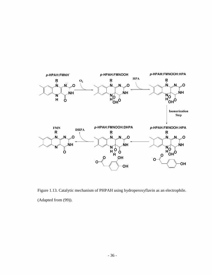
The recently identified p-hydroxyphenylacetate 3-hydroxylase (pHPAH) from a
two-component hydroxylase system catalyzes the hydroxylation reaction of
p-hydroxyphenylacetate (85). The first step of the proposed reaction mechanism of
pHPAH involves the binding of reduced flavin to the monooxygenase, and reaction of
dioxygen with reduced flavin to form a stable C4a-hydroxyflavin intermediate (Figure
1.13) (99). Following the binding of p-hydroxyphenylacetate, the bound substrate
performs an electrophilic attack on the flavin intermediate. The next step is product
release and formation of an enzyme bound hydroxyflavin, which finally decays to
oxidized flavin and H2O. Even though both pHPAH and single-component
monooxygenase PHBH catalyze hydroxylation of aromatic compound through the
hydroperoxyflavin intermediate, the overall mechanisms and substrate binding order are
different. Flavin reduction and flavin intermediate formation by pHPAH precedes the
substrate binding, supported by the observation of a stabilized hydroxyflavin intermediate
in pHPAH (99).
- 36 -
Figure 1.13. Catalytic mechanism of PHPAH using hydroperoxyflavin as an electrophile.
(Adapted from (99)).

- 37 -
A well characterized flavin-dependent monooxygenase that utilizes a
C4a-peroxyflavin intermediate as a nucleophile is bacterial luciferase (Figure 1.14) (111).
Initially the enzyme bound peroxyflavin intermediate was thought to perform a
nucleophilic attack on long-chain aldehyde substrates, followed by a Baeyer-Villiger
rearrangement to generate corresponding carboxylic acid and green blue light (102-104).
However, a general Baeyer-Villiger rearrangement would not produce the excited state of
the flavin intermediate responsible for light production. An alternative mechanism
involving a dioxirane intermediate was then proposed based on observed isotope effects
and theoretical calculations (113). An interesting feature of bacterial luciferase is the
ability to stabilize the C4a-(hydro)peroxyflavin intermediate for several hours (111). The
flavin intermediate is found to be protected by several active site residues and a flexible
loop near the active site (117-119). The gradual decay of the flavin intermediate results in
the bioluminescence of bacterial luciferase (119).
One of the challenges for flavin monooxygenases is to catalyze a diverse range of
reactions while controlling the stabilization of reaction intermediates. The highly reactive
reduced flavin can form toxic hydrogen peroxide by-products. In order to avoid wasteful
consumption of NAD(P)H and the formation of hydrogen peroxide during oxygenation
reaction, flavin monooxygenases have developed two mechanistic strategies (105). One
strategy is described as “cautious” due to the requirement of the bound substrate for
flavin reduction. Therefore, flavin intermediate formation is triggered by the binding of
the substrate to be oxygenated. Because the substrate is already present, the oxygenation
reaction out-competes the H2O2 elimination side reaction and ensures the
productive catalysis (105). Flavin monooxygenases that catalyze the hydroxylation of

- 38 -
Figure 1.14. Proposed reaction mechanism of bacterial luciferase. The intermediate
responsible for light production is indicated. (Adapted from (113)).

- 39 -
aromatic compound usually belong to this “cautious” category. In contrast, the second
strategy is described as “bold”, which does not necessarily require the presence of
substrate in order for the rapid flavin reduction and flavin intermediate formation to
proceed (105). These flavin monooxygenases are able to effectively stabilize flavin
intermediates through interactions with active site residues and/or conformational
changes that control the accessibility of the active site (97, 120). The stabilized flavin
intermediate then catalyzes the oxygen insertion reaction after a competent substrate is
encountered. This “bold” strategy is often associated with flavin monooxygenases that
oxidize the non-aromatic compounds, as well as two-component systems including
pHPAH, bacterial luciferase, and alkanesulfonate monooxygenase (47, 85, 111). Since
the active sites of these flavin monooxygenase have to be open for the substrate, the
strategies used to provide necessary protection of the labile flavin intermediate from the
bulk solvent are particularly interesting to fully understand these “simple” yet complex
flavoproteins.
1.7.1 Alkanesulfonate monooxygenase SsuD from E. coli
The SsuD enzyme is a flavin-dependent monooxygenase that catalyzes the
oxygenolytic cleavage of the carbon-sulfur bond in 1-substituted alkanesulfonates in the
presence of FMNH2 and dioxygen (36, 47). The initial characterization of purified SsuD
suggested that the desulfonation activity is absolutely dependent on the presence of SsuE,
NADPH, FMN, and dioxygen, and maximum activity was obtained for molecular ratio of
SsuE/SsuD between 2.1 and 4.2 (36). The SsuD enzyme was found to strictly use
FMNH2 as the substrate due to the absence of activity when FMN was substituted by
FAD (36). The products of the desulfonation of alkanesulfonate are confirmed to be the

- 40 -
corresponding alkanealdehyde and sulfite (36). While the E. coli SsuD enzyme cannot
utilize taurine, L-cysteic acid, methanesulfonate, and aromatic sulfonates, it is able to
desulfonate a broad range of unsubstituted 1-alkanesulfonates with chain lengths longer
than two carbons (36). In addition, substituted ethanesulfonic acid, N-phenyltaurine,
4-phenyl-1-butanesulfonic acid, and sulfonated buffers can also serve as substrates for
SsuD (36). This broad substrate range of SsuD is thought to compliment the TauD
desulfonation system to correspond to the full sulfonate sources available. However,
further structural and mechanistic characterization of SsuD is needed to illustrate how the
SsuD manages to utilize such a diverse range of substrates.
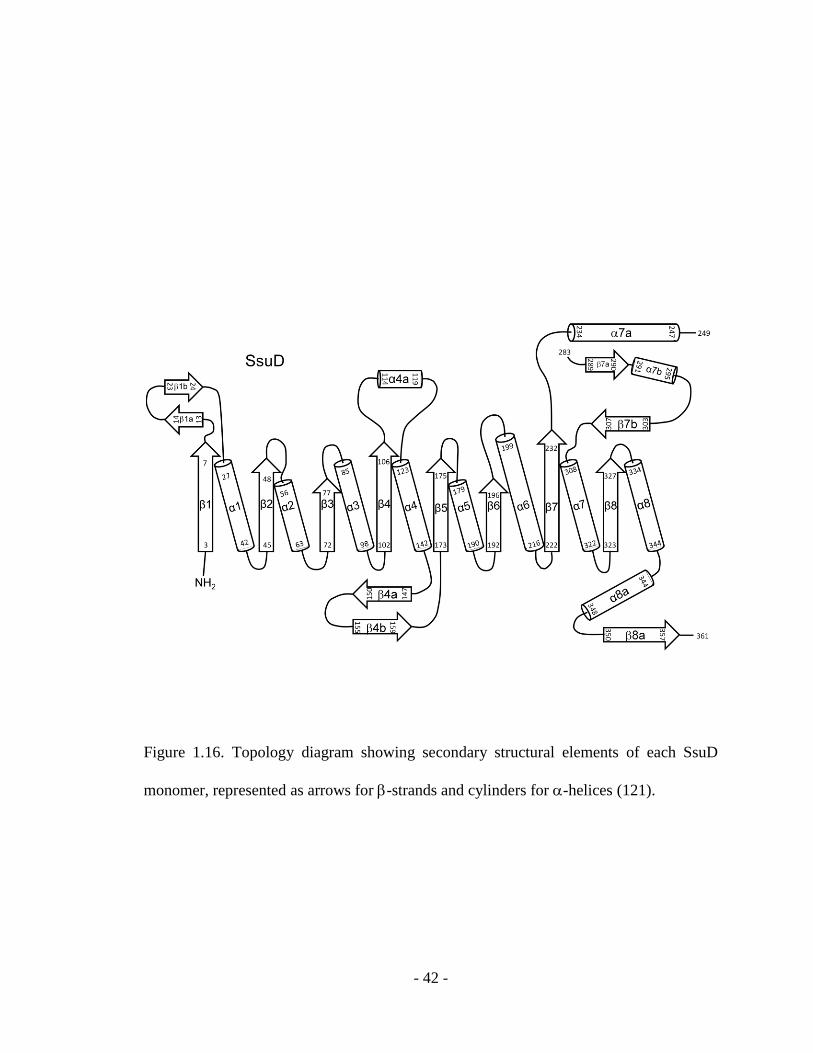
The three-dimensional structure of E. coli SsuD in the absence of substrates was
solved at a resolution of 2.3 Å (Figure 1.15) (121). The SsuD monomer was shown to
contain a single domain with an eight-parallel-stranded / barrel motif, which is
denoted as a TIM (triose phosphate isomerase) barrel structure. The TIM-barrel motif
represents a large family of proteins with distinct functions. The structure of SsuD differs
from the typical TIM-barrel proteins by four insertion regions connecting strands with
helices and an extension region at the C-terminus end of the polypeptide chain (Figure
1.16) (121). Insertion region 4 is the most extensive insertion region containing 75
residues. It is located over the C-terminal end of the TIM-barrel and covers part of the
inner core. In the crystal structure, insertion region 4 contains an unresolved loop region
from residue 250 to 282 (121). The poorly defined electron density of this region in the
crystal structure was speculated to be caused by the high mobility of this loop region.
Despite the low level of amino acid sequence identity, the SsuD enzyme is structurally
related to TIM-barrel proteins bacterial luciferase and long-chain alkane monooxygenase

- 41 -
Figure 1.15. Three-dimensional structure of SsuD (PDB entry: 1M41) (121). Two
identical subunits are shown in gray and light blue.
- 42 -
Figure 1.16. Topology diagram showing secondary structural elements of each SsuD
monomer, represented as arrows for -strands and cylinders for -helices (121).
- 43 -
(LadA), classified as the bacterial luciferase structural family (116, 122, 123). Both
bacterial luciferase and LadA belongs to the two-component monooxygenase systems
that utilize reduced flavin as a substrate instead of bound prosthetic group.
Steady-state and presteady-state kinetic studies were performed to investigate the
mechanism of desulfonation. While SsuD was able to utilize a broad range of substrate,
1-octanesulfonate was one of the substrates with which SsuD showed optimal activity
(47). Thus, 1-octanesulfonate was used to obtain the kinetic parameters. The substrate
affinity of SsuD was evaluated through fluorimetric titration by monitoring the decrease
of intrinsic protein fluorescence upon addition of substrate (47). The dissociation constant
for FMNH2 binding to SsuD was 0.032 0.15 μM, while the dissociation constant for
FMN binding to SsuD was 10.2 0.4 μM, confirming that SsuD preferentially binds
reduced flavin (47). Interestingly, there was no observable change in the intrinsic
fluorescence with the addition of octanesulfonate to the free enzyme. In fact, the decrease
in protein fluorescence was only observed when octanesulfonate was added to a solution
of pre-mixed SsuD and FMNH2. Therefore, it was suggested that an SsuD-FMNH2
protein complex has to form prior to the binding of octanesulfonate (47). This ordered
substrate binding indicates that different conformations exist between substrate-free SsuD
and SsuD-FMNH2 complex, and the conformational change necessary for
octanesulfonate binding may be induced by FMNH2 binding (47). The FMNH2-bound
SsuD then reacts with dioxygen to form a C4a-(hydro)peroxyflavin intermediate, which
was identified by rapid reaction kinetic studies monitoring reduced flavin oxidation at
370 nm. Based on rapid kinetic analyses alternating the substrate mixing order, the
protein complex formed by premixing SsuD and FMNH2 may not represent the true

- 44 -
enzyme conformation during catalysis. It is speculated that an inactive complex forms
when SsuD is premixed with FMNH2, with the observation of decreased octanal product
formation with premixed SsuD-FMNH2 (47). This may be due to the slow conversion of
the inactive complex back to the active form in the presence of octanesulfonate and
dioxygen (47). In addition, the proposed C4a-(hydro)peroxyflavin intermediate observed
without pre-mixing SsuD and FMNH2 was absent in the kinetic trace, suggesting that the
flavin intermediate cannot accumulate to a significant level due to the formation of
inactive complex (47). While it is established that FMNH2 binds to SsuD prior to
octanesulfonate, the inactive complex is only formed in the absence of dioxygen. Because
the SsuD requires dioxygen to catalyze the desulfonation reaction, the formation of this
inactive complex may be due to in vitro anaerobic experimentation and is not
physiologically relevant. Alternate mixing experiments suggested that reduced flavin
oxidation is faster if the SsuD, FMNH2 are pre-mixed with octanesulfonate instead of
dioxygen (47). Therefore, it was concluded that during catalysis FMNH2 binds to SsuD
first, followed by immediate octanesulfonate binding and dioxygen binds last to ensure
the coupling of intermediate formation and desulfonation. Therefore, an ordered substrate
binding mechanism is suggested for the alkanesulfonate monooxygenase (Scheme 1.2)
(47).
In the proposed mechanism of SsuD, a C4a-peroxyflavin intermediate is suggested to
make a nucleophilic attack on the sulfonate functional group (Figure 1.17) (47). The
alkanesulfonate peroxyflavin adduct then undergoes a Baeyer-Villiger rearrangement and
proton abstraction by an active site base, leading to the generation of hydroxyflavin,
alkanealdehyde, and sulfite. For the well studied flavin monooxygenases adopting similar

- 45 -
SsuD
FMNH2
SsuD-FMNH2
RCH2SO3-
SsuD-FMNH2-RCH2SO3-
O2
SsuD-FMNOOH-RCH2SO3-
RCHO + SO32-
+ H+
SsuD-FMNOH
H2O
SsuD-FMN
Scheme 1.2. Order of substrates binding for SsuD. (Adapted from (47)).

- 46 -
mechanistic strategies through peroxyflavin intermediate formation, the reactive flavin
peroxide intermediate is usually stabilized by the protein during catalysis. Cyclohexanone
monooxygenase can stabilize the C4a-peroxyflavin intermediate for a maximum of five
minutes with the NADP+ bound, while bacterial luciferase is able to stabilize its
intermediate for several hours at low temperature (97, 111). Although bacterial luciferase
and SsuD belong to the same structurally related FMNH2-dependent two-component
monooxygenase family, the proposed peroxyflavin intermediate is not as stable in SsuD
(47). While the flavin intermediate for bacterial luciferase can be stabilized for several
hours, the flavin intermediate for SsuD is much more susceptible to elimination than the
intermediate for bacterial luciferase. Therefore, SsuD must offer a more precise and
delicate intermediate stabilization network in order to ensure profitable and efficient
catalysis.
One of the stabilization strategies developed by SsuD is to provide hydrogen bonding
interactions through a conserved Cys54 residue (124). In the three-dimensional structure
of SsuD, Cys54 is located in the putative catalytic center at the C-terminal end of the
TIM-barrel (Figure 1.18) (121). The SsuD lost activity upon cysteine labeling with
methylmercury, suggesting that Cys54 is important for catalytic function (121). By
comparison with structurally related flavin-dependent two-component monooxygenases,
the SsuD Cys54 residue is found in a similar spatial arrangement as Cys106 from
bacterial luciferase and Cys14 from LadA (116, 122). Chemical modification of the
reactive thiol group of bacterial luciferase Cys106 abolished enzymatic activity (100).
Bioluminescence was reduced with the substitution of Cys106, indicating that Cys106
may be involved in intermediate stabilization in bacterial luciferase (103). The activity of

- 47 -
Figure 1.17. Proposed mechanism of the desulfonation reaction by SsuD (47).

- 48 -
Figure 1.18. Putative active site of the alkanesulfonate monooxygenase SsuD (PDB entry:
1M41) (121). Cys54 is shown in yellow, Ser179 is shown in orange, Tyr331 is shown in
red, Arg226 and Arg297 are shown in green, His228 and His333 are shown in blue.
Tyr128
Arg226
Arg297
Ser179 His228
His333
Cys54

- 49 -
LadA was also found to be diminished with the substitution of Cys14 (116). When Cys54
of SsuD was substituted with serine, the substrate affinity was not significantly altered
and the kcat/Km value increased 3-fold (124). In addition, an increased
C4a-(hydro)peroxyflavin intermediate formation rate was observed for the C54S SsuD
variant in the absence of octanesulfonate. The accumulation of the
C4a-(hydro)peroxyflavin intermediate was not observable even at low concentration of
octanesulfonate, suggesting that catalysis happens faster than the accumulation of the
flavin intermediate, which correlates with the increased catalytic efficiency (124). The
conservative serine substitution appears to effectively take the place of cysteine in
catalysis. However, when Cys54 was substituted with alanine, the kcat/Km value decreased
6-fold even though the affinity for both substrates was not impaired (99). No
C4a-(hydro)peroxyflavin intermediate accumulation was observed, suggesting that Cys54
of SsuD is involved in stabilizing the C4a-(hydro)peroxyflavin (124). Although the
catalytic efficiency of C54A SsuD is 6-fold lower than the wild-type SsuD, the catalytic
activity of C54A SsuD is not fully diminished, indicating that the Cys54 residue is not
directly involved in catalysis as an active site base (124). When the thiol functional group
of Cys54 was substituted with the serine hydroxyl functional group, the increase in
intermediate formation rate and overall catalytic activity suggested that the Cys54
participates directly or indirectly in intermediate stabilization through hydrogen bonding
interactions with the flavin intermediate.
1.8 Protein dynamics and flexible loop
In order for enzymes to stabilized reactive intermediates and facilitate efficient
catalysis, another commonly practiced strategy is adopting an optimal protein

- 50 -
conformation. It is generally accepted that the diverse biological functions of proteins
rely on their structural fluctuations. Different segments of protein can move in relation to
one another with only small expenditures of energy due to the intrinsic flexibility of
proteins (125). These structural motions that alter the protein conformations are the
principle type of protein dynamics. At the molecular level, protein conformational
changes are realized via the changes in polypeptide chain torsional angles and side chain
orientations (125). Small changes in a few critical placed residues can often lead to a
large dislocation of tertiary structural components, known as domain motions (125).
Domain motions mainly exist in two forms: shear motions that occur parallel to the
interface of closely packed segments of polypeptide, and hinge motions of secondary
structural components that are not constrained by tertiary packing forces (125). While
shear motions occur predominantly between -helices, hinge motions are found in
-strands, -sheets and -helices that are connected through a hinge outside of the
interface (125).
The commonly found hinge structures in many globular proteins are often located at
the surface (125). Due to their high intrinsic mobility, these surface hinges are generally
referred to as flexible loops. Since the flexible loop usually lies between the
protein/solvent interface, it is found to play a critical role in contributing to protein
function. In relation to the inner core of proteins, the flexible loops can easily assume an
open/closed conformation with the torsion of only a few residues (126). The lid-gated
movements of these flexible loops are able to create concealed compartments for precise
catalysis (126). In addition, flexible loops can also be involved in ligand binding, signal
recognition, and catalysis by providing essential catalytic residues (125). Therefore, it is

- 51 -
intriguing to understand the role of flexible loops in various proteins with distinct
functions. Protein dynamics including flexible loop movements can be induced by many
factors: temperature, pH, voltage, ion concentration, covalent labeling, hydrogen bonding,
and ligand binding etc (127). The biophysical and biochemical evaluations of the flexible
loop movements of the proteins related to the current study are reviewed in the following
subsections.
1.8.1 Flexible loop of Triose phosphate isomerase (TIM)
Triose phosphate isomerase (TIM) was the first enzyme identified to contain the
classical 8-stranded / barrel structure, a conserved motif also found in
flavin-dependent monooxygenases SsuD and bacterial luciferase (128). Triose phosphate
isomerase catalyzes the reversible isomerization of D-glyceraldehyde 3-phosphate (GAP)
to dihydroxyacetone phosphate (DHAP) through an enediolate intermediate (Figure 1.19)
(129). A substantial conformational change was observed in the three-dimensional
structure with the binding of substrate (129). In the substrate-free conformation, a loop
near the C-terminal end of the barrel extends outward from the protein inner core, leaving
the proposed active site freely accessible to solvent (130). Therefore, the substrate-free
TIM exists in the “open” conformation. When the substrate DHAP is bound, the loop
folds over the active site, resulting in the “closed” conformation (129). The loop is
referred to as a phosphate gripper loop because it interacts with the phosphate dianion of
the substrate (129). The reactions catalyzed by TIM occur at a rate approaching the
diffusion limit (131). This fast reaction rate is mainly due to the stabilization of the
transition state with the phosphate intrinsic binding energy provided by the closure of the

- 52 -
Figure 1.19. The isomerization reaction catalyzed by TIM (A) and the crystal structures
of the loop-open and loop-close forms of TIM from chicken muscle (B). The aqua
ribbons are for the open unliganded TIM [PDB entry 1TIM] and the green ribbons show
the closed TIM liganded with 2-phosphoglycolohydroxamate (PGH) [PDB entry 1TPH].
(Adapted from (128)).
GAP DHAP GAP
A.
B.
Flexible
Loop
Substrate
analog (PGH)
Glu165
Lys12

- 53 -
loop and associated conformational changes (128). Based on a series of kinetic analyses,
the substrate binding and corresponding loop closure is proposed to be rate determining
in the isomerization from GAP to DHAP, while loop opening and product release is rate
limiting in the conversion of DHAP to GAP (131-138). In addition, studies have also
shown that the interaction between the flexible loop and substrate phosphate group
activates proton transfer. Therefore, it is believed that the motion of the flexible loop in
TIM not only anchors the substrate, but also actively controls the rate of catalysis upon
interaction with substrate (128).
Another important role of the TIM flexible loop is to sequester the substrate and
intermediate from solvent by closing over the active site. In order to perform efficient
catalysis, TIM requires strong transition state stabilization activated through loop closure
(128). The TIM flexible loop not only interacts with substrate to supply binding energy, it
also provides a compartmentalized active site for optimal catalysis (128). The enediolate
intermediate was found to be sequestered from bulk solvent through studies evaluating
partitioning of the intermediate in D2O (139-141). The percentage of intramolecular
hydrogen transfer was much higher when the TIM-catalyzed isomerization of GAP
proceeded in D2O, suggesting the active site and the intermediate is less accessible to
bulk solvent. In addition, the substrate is transferred from solvent to a confined
catalytically favorable protein environment by loop closure, providing additional contacts
between the protein and the substrate shown in the three-dimensional structure (142). A
highly conserved Lys12 residue situated adjacent to the TIM flexible loop was shown to
directly interact with the substrate phosphate group (143). Substituting Lys12 to glycine
resulted in a 6×10⁵-fold decrease in activity that corresponded to a 7.8 kcal/mol

- 54 -
stabilizing energy loss, which was originally provided by the interaction between
wild-type Lys12 and the substrate (144). The same interaction of free lysine with
phosphate is much weaker in aqueous solution, suggesting that movement of the flexible
loop in the solvent-free protein active site environment enhances the interaction between
the lysine side chain and the substrate (145). Furthermore, the closed conformation
occludes bulk solvent and displaces several active site water molecules, which induces
the desolvation of the TIM catalytic base Glu165 (146-148). This ligand induced
conformational change shifts Glu165 2 Å into its catalytically active position (149). Loop
closure is also found to cause the hydrophobic side chains of an isoleucine and a leucine
to fold over Glu165, shielding the active site base from bulk solvent (150). This
loop-closure induced hydrophobic active site environment leads to an increase in the
basicity of the Glu165 side chain, which ensures efficient catalytic activity.
1.8.2 Flexible loop of Bacterial luciferase
Bacterial luciferase is a flavin dependent-monooxygenase that belongs to the family
of TIM-barrel proteins. Bacterial luciferase employs reduced flavin as a substrate to
activate dioxygen and catalyzes the oxidation of a long-chain aliphatic aldehyde to
carboxylic acid with the emission of blue-green light (102). The proposed reaction
mechanism of bacterial luciferase proceeds through several flavin intermediates (Figure
1.14) (113). The breakdown of the flavin intermediate is believed to be responsible for
light emission (119). In order to achieve maximum bioluminescence, specific
stabilization of the flavin intermediate followed by gradual decay is required. Therefore,
the mechanism for intermediate stabilization in bacterial luciferase is particularly
intriguing.

- 55 -
Bacterial luciferase is comprised of two TIM-barrel subunits designated as and
(122). The two subunits are expressed by luxA and luxB under the same lux operon and
display high sequence homology (151). Although the and subunits are similar in
overall structure and share 32% amino acid sequence similarity, the subunit contains
the active site that binds one reduced flavin substrate while the subunit is not associated
with flavin binding or product formation (122). However, studies have shown that the
subunit is essential for a high bioluminescent activity of the subunit despite its inability
to bind reduced flavin (112). The main structural component that distinguishes the
subunit from the subunit of bacterial luciferase is an unresolved loop in an insertion
region between an -helix and -sheet, indicating high mobility of this region (122).
Similar to the flexible loop of TIM and the unresolved loop of SsuD, the unresolved loop
of bacterial luciferase is located above the active site at the C-terminal end of the barrel.
It is reported that the unresolved loop of bacterial luciferase is highly susceptible to
proteolysis (152, 153). However, the presence of the FMNH2 substrate was shown to
inhibit the proteolysis of this region, indicating that the association of reduced flavin
immobilizes and protects this mobile loop region (154). In the three dimensional structure
of the bacterial luciferase/FMN complex, localized electron density was observed in the
portion of the mobile loop that is distal to the flavin binding site, suggesting this loop is
partially ordered and exists in alternative conformations with the binding of FMN (Figure
1.20) (123). Apparently, substrate binding initiates substantial conformational changes
including loop movement, which is speculated to play an important role in bacterial
luciferase bioluminescence activity.
The functional and mechanistic role of the bacterial luciferase mobile loop region

- 56 -
was first evaluated by deleting the 29-residue loop region of the subunit that was
unresolved in the original three dimensional structure without flavin bound (119). The
loop-deleted bacterial luciferase variant Δ262-290 was shown by far-UV circular
dichroism to contain a higher percentage of -helical structure than the wild-type enzyme,
which was thought to be caused by the deletion of the unresolved loop region comprising
10% of the protein (119). Based on the results from both far- and near-UV circular
dichroism spectroscopic analyses, it was believed that deletion of the unresolved loop in
bacterial luciferase did not cause significant perturbations in overall secondary structural
and the environment of the aromatic side chains. However, the Δ262-290 heterodimer
showed a subunit equilibrium dissociation constant of 1.32 1.25 μM, whereas the
wild-type heterodimer did not dissociate (119). Therefore, the unresolved loop region of
bacterial luciferase was proposed to be involved in inter-subunit interactions. Kinetic
characterization of the Δ262-290 variant showed similar substrate affinity for reduced
flavin and aldehyde as the wild-type enzyme, suggesting the unresolved loop region does
not participate in substrate binding (119). Although the Δ262-290 variant was able to
form the carboxylic acid product with similar catalytic efficiency as the wild-type
enzyme, the bioluminescence quantum yield of the reaction catalyzed by Δ262-290
variant was 100-fold lower than the wild-type enzyme (119). It was postulated that
without the unresolved loop region of the subunit the exited flavin emitter responsible
for bioluminescence either cannot efficiently populate or is quenched by the solvent that
can easily access the active site without protection from the loop region (119). Therefore,
the functional role of the bacterial luciferase unresolved loop region is believed to protect
and stabilize the excited flavin emitter intermediate.
- 57 -
Figure 1.20. Three-dimensional structures of bacterial luciferase subunit with two
observed conformations of the mobile loop (PDB entry: 1BRL and 3FGC). The mobile
loop of subunit observed in a complex with FMN is shown in red. The mobile loop of
flavin-free subunit is shaded in blue (122, 123).

- 58 -
Several conserved amino acids located on the bacterial luciferase unresolved loop
region were substituted through alanine-scanning mutagenesis to determine the role of
these residues in catalysis (117). Lys283 and Lys286 substitutions were found to result in
a loss of bioluminescent activity comparable to the loop deletion variant (117). Both
variants yielded the carboxylic acid product at the same efficiency as wild-type bacterial
luciferase. Experiments evaluating the kinetic stability of the flavin intermediate
suggested that Lys283 participates in C4a-hydroperoxyflavin stabilization (117). In order
to probe the accessibility of the flavin intermediate to the bulk solvent, a dynamic
quencher potassium iodide was used. Potassium iodide is able to quench the flavin exited
state and increase the rate constant for the decay of bioluminescence. It was reported that
potassium iodide had a greater effect on the decay of bioluminescence for the Lys286
variant (117). It seems that substituting the Lys286 to alanine increased the exposure of
the excited flavin intermediates to solvent. When residue Gly275 on the unresolved loop
region of the subunit was substituted to proline, the intrinsic torsional flexibility of
glycine was replaced by the more constrained proline ring structure (118). As a result, the
bioluminescence activity of the G275P variant was reduced by 4 orders of magnitude
compared to the wild-type enzyme (118). Because the G275P variant can bind both
substrates and form flavin intermediates at the same efficiency as the wild-type enzyme,
the decrease in bioluminescence activity was attributed to the loss of flexibility of the
unresolved loop. When residue Phe261 on the unresolved loop region of was mutated
to aspartic acid, the hydrophobic nature of the phenylalanine was substituted to a
hydrophilic positively charged side chain (118). The F261D variant showed a decrease
in bioluminescence production by 6 orders of magnitude, while maintaining similar
- 59 -
substrate affinity and catalytic efficiency toward product formation (118). It was
suggested that the bulky hydrophobic phenylalanine side chain may be involved in
providing a solvent inaccessible hydrophobic active site environment for the flavin
emitter intermediate to accumulate. These results support the role of the bacterial
luciferase unresolved loop as a mobile lid in shielding critical reaction intermediates from
exposure to the bulk solvent.
1.8.3 Flexible loop of alkanesulfonate monooxygenase SsuD
The flavin dependent monooxygenase SsuD belongs to the bacterial luciferase
structural family. Although SsuD shares a relatively low amino acid sequence identity
with bacterial luciferase, the three dimensional structure of the two enzymes show
common overall structural features including a TIM-barrel fold and an unresolved
flexible loop region above the active site (121). However, structural variations exist
between SsuD and bacterial luciferase. Bacterial luciferase is a heterodimer with one
active site per dimer, while SsuD is a homotetramer with one active site per monomer. In
addition, SsuD may differ from bacterial luciferase in the functional role of the
unresolved flexible loop region despite their similar location (121-123). The flexible loop
of bacterial luciferase was suggested to stabilize the flavin emitter responsible for
bioluminescence (117-119). Deletion of this region does not affect product formation,
suggesting the flexible loop may not be directly involved in bacterial luciferase catalysis
(119). On the other hand, a random mutation of SsuD Arg297 to cysteine in the initial
cloning process resulted in the loss of SsuD activity (Figure 1.21) (36). It was speculated
that the Arg297Cys may form a disulfide bond with Cys54, affecting the flexibility of the
loop region. Therefore, it is suggested that the SsuD unresolved loop and its motions may

- 60 -
be required for efficient catalysis (155). In the structure of SsuD without substrate bound,
the conserved Arg297 resides near the unresolved loop that protrudes over the putative
active site, but the functional group of this arginine is found to point away from the active
site (121). Therefore, conformational changes of Arg297 would be necessary for catalytic
involvement of this residue.
In order to understand the role of the conserved Arg297, various biochemical and
mechanistic evaluations were performed on two SsuD variants with the Arg297
substituted by alanine or lysine (155). Although the reduced flavin substrate affinity was
not altered significantly, the kcat/Km value was decreased 30-fold by the substitution of
Arg297 with lysine. The R297A SsuD variant showed no detectable activity with a 5-fold
increase in the Kd value for FMNH2 compared to wild-type SsuD (155). In addition, both
arginine variants had similar affinity for octanesulfonate as wild-type SsuD. The results
suggested that Arg297 may interact with another amino acid or substrate functional group
to stabilize conformational changes induced by reduced flavin binding that correlates
with flexible loop closure (155). Rapid reaction kinetic analyses of the R297A and
R297K SsuD variants showed no accumulation of C4a-(hydro)peroxyflavin intermediate,
and there was no observable dependence on the kobs with increasing concentrations of
octanesulfonate as previously observed for wild-type (47). These results suggested that
conformational changes of the flexible loop cannot be realized without the crucial
Arg297 residue, and the disrupted conformational changes would leave the active flavin
intermediate accessible to solvent and susceptible to non-enzymatic
oxidation (155). The requirement of Arg297 to undergo essential conformational changes

- 61 -
Figure 1.21. Three-dimensional structures of SsuD with the sequence of the unresolved
region indicated [PDB entry 1M41]. The conserved Arg297 is shown in green (121).
Arg297

- 62 -
was further supported by studies evaluating proteolytic susceptibility of both wild-type
and arginine variants (155). The SsuD wild-type and Arg297 variants were rapidly (<15 s)
digested in the absence of substrates, while there was 20% of the wild-type SsuD
remaining after extended proteolysis (30 s) in the presence of reduced flavin substrate
(155). It was speculated that substrate binding provided some level of protection towards
proteolysis. Based on results from mass spectrometric analysis of the digested wild-type
SsuD, the susceptible proteolytic sites were located at Arg263 and Arg271 on the
insertion region containing the unresolved flexible loop (155). Therefore, the binding of
reduced flavin substrate initiated conformational changes that immobilized the flexible
loop. As a result, the proteolytic susceptible sites Arg263 and Arg271 on the ordered loop
region were less accessible to tryptic digestion upon FMNH2 binding. The same level of
protection was not observed with R297A or R297K SsuD in the presence of reduced
flavin substrate (155). In fact, the R297A and R297K SsuD variant exhibited a similar
susceptibility toward proteolysis regardless of the appearance of the substrates FMNH2
and octanesulfonate. It was concluded that Arg297 variants are not able to undergo
conformational changes observed with wild-type SsuD, and that Arg297 is critical in
stabilizing the conformational changes associated with loop movement (155).
Substitution of Arg297 results in a more solvent accessible active site due to the inability
of the loop to undergo the requisite conformational changes. The flavin intermediates are
not adequately stabilized in the less protected active site in the Arg297 variants, leading
to deleterious effects on catalysis. These kinetic and structural studies of the conserved
Arg297 residue indicate that the SsuD unresolved loop region may be involved in
stabilizing and protecting the reaction intermediates from bulk solvent, which is similar

- 63 -
to the functional role of the bacterial luciferase flexible loop. However, direct evaluation
of the structural and functional role of the SsuD flexible loop was not performed. Since
deviations exist between the flexible loops of bacterial luciferase and SsuD, the SsuD
flexible loop may play additional functional roles in the desulfonation reaction.
Because the SsuD flexible loop is located above the active site and functions as a
mobile lid, another possible role of this loop could be assisting in flavin transfer between
SsuE and SsuD. The reduced flavin needs to be transferred from the SsuE active site to
SsuD active site. In order for FMNH2 to access the active site, the SsuD flexible loop
would need to assume an extended conformation before flavin transfer. Following the
flavin transfer events, the SsuD flexible loop would close over the active site for effective
catalysis. Since the phosphate gripper loop of TIM is known to bind the substrate
phosphate group, the SsuD flexible loop may also interact with the phosphate group of
flavin and assist in flavin transfer.
1.9 Flavin transfer mechanism in two-component systems
The SsuD enzyme belongs to a family of two-component monooxygenase systems
that utilize reduced flavin as a substrate. Although identified only in bacterial organisms,
these flavin dependent two-component monooxygenase systems are capable of catalyzing
a wide range of reactions such as oxidation of aromatic and polycyclic compounds,
biosynthesis of antibiotics, bioluminescence, oxidation of long-chain alkanes, and
desulfurization of sulfonate compounds (36, 80-90, 116). In these two-component
systems, the monooxygenase activity relies on a flavin reductase that supplies reduced
flavin to activate dioxygen. Because the reduced flavin is extremely labile, a controlled
flavin transfer mechanism is necessary for these monooxygenase to efficiently utilize the

- 64 -
reduced flavin substrate.
Reduced flavin transfer could either occur through a dissociative or channeling
mechanism (Figure 1.22). In the dissociative mechanism, the reduced flavin is released
by the reductase into solution, followed by passive diffusion to the monooxygenase. In
the dissociative flavin transfer process, no protein-protein interactions are required and
the diffusion is driven by the affinity of the monooxygenase enzyme for reduced flavin.
In the channeling mechanism, a transient reductase/monooxygenase complex is formed
prior to flavin transfer, and the flavin reduced by the reductase is actively shuffled to the
monooxygenase within the reductase/monooxygenase complex. Typically, the flavin
reductases have a higher affinity for oxidized flavin while the monooxygenases
preferentially bind reduced flavin (47, 81, 94). This difference in flavin affinity facilitates
flavin transfer from the reductase to the monooxygenase. However, reduced flavin can
also be oxidized rapidly in solution, generating toxic reactive oxygen species and
damaging cellular components. Therefore, it still remains to be explored whether
protein-protein interactions are critical to avoid in vivo non-enzymatic flavin oxidation in
bacterial organisms. Among the characterized flavin-dependent monooxygenases, many
are capable of utilizing reduced flavin supplied by flavin reductases from other
two-component systems, supporting a dissociative flavin transfer mechanism (78, 82, 87,
88). However, the flavin reductases from different systems could share similar structural
motifs. Although structural information is limiting, if flavin reductases contain similar
structural motifs, it is possible that protein-protein interactions could be formed between
these flavin reductases and monooxygenases. Then, the ability of some monooxygenases
to couple with different flavin reductase in vitro is not a convincing argument to support

- 65 -
Figure 1.22. Schematic representation of flavin transfer mechanisms in two-component
systems. The blue square represents flavin reductase, the yellow triangle represents
reduced flavin, and the green circle represents the flavin monooxygenase.

- 66 -
a dissociative flavin transfer mechanism. Both dissociative and channeling flavin transfer
mechanisms have been reported in two-component systems depending on the specific
enzymes under investigation.
1.9.1 Flavin transfer mechanism in bacterial luciferase system
Even though bacterial luciferase is the most extensively studied FMN-dependent
two-component monooxygenase system, the physiological relevant flavin reductase
component was suggested to be LuxG in Photobacterium leiognathi (88). In vitro,
bacterial luciferase can utilize reduced flavin supplied by various flavin reductases
including FRP, FRase I, Fre-like reductase, and LuxG (78, 82, 87, 88). Alternative results
describing the flavin transfer mechanism of bacterial luciferase have been reported
depending on the flavin reductase and the techniques used for evaluation. Therefore, the
in vivo flavin transfer mechanism in bacterial luciferase system is still under debate.
There are two major arguments against a channeling flavin transfer mechanism in the
bacterial luciferase system. One is that the flavin reductase component is not specific in
the system. When bacterial luciferase was assayed with various flavin reductases, it was
able to accept reduced flavin provided by different flavin reductases and still maintain
full activity (78, 82, 87, 88). Based on the assumption that these flavin reductases have no
structural similarities, it was suggested that no protein-protein complex is formed
between the two components, and a dissociative flavin transfer mechanism is highly
likely (156-160). The other argument against a channeling mechanism is that studies
designed specifically to identify protein-protein interactions between bacterial luciferase
and some flavin reductases showed no evidence for complex formation (156-160). The
gene responsible for expressing flavin reductase LuxG is located on the same operon with
- 67 -
luciferase lux genes in Photobacterium leiognathi TH1 (160). In this specific organism,
LuxG is believed to be the functioning flavin reductase for bacterial luminescence based
on in vitro and in vivo studies (160). In the in vitro coupled assays, LuxG was able to
supply reduced flavin to bacterial luciferase for bioluminescence activity. When the luxG
gene was knocked out from Photobacterium leiognathi TH1, the organism showed much
dimmer bioluminescence, indicating that LuxG provides the most significant source of
reduced flavin for the bioluminescence reaction in vivo. When gel filtration
chromatography was utilized to evaluate if LuxG interacts with bacterial luciferase there
were no protein-protein interactions identified, suggesting a dissociative flavin transfer
mechanism (160). Similarly, investigation of interactions between bacterial luciferase and
flavin reductase Fre or FRP via pull-down assays have also shown that there is no
evidence for protein complex formation (161). However, based on the author‟s
speculation, the possibility of transient protein-protein interactions cannot be ruled out
simply by the negative results from one or two techniques. More aspects need to be
investigated before a final conclusion can be made.
Evidence demonstrating a channeling flavin transfer mechanism in bacterial
luciferase system has also been identified (75, 162-164). A transient protein complex
between bacterial luciferase and monomeric FRP was identified by fluorescence
anisotropy (164). This study also suggested that dimeric FRP cannot form a complex with
bacterial luciferase as efficiently as monomeric FRP. Therefore, the negative results in
previous attempts to identify protein-protein interactions could have been due to the
formation of dimeric FRP, which is not effective in complexing with luciferase. It was
established that 94% of FRP in the cell is in complex with luciferase when V. harveyi

- 68 -
exhibits maximum in vivo bioluminescence (164). Direct channeling of reduced flavin
within the complex of FRP and luciferase would save energy, avoid toxifications, and
maintain efficient bioluminescence at low levels of FMN and NADPH (162). A
channeling flavin transfer mechanism between FRP and bacterial luciferase was also
supported by kinetic evidence (162). In the absence of bacterial luciferase, flavin
reduction reaction catalyzed by FRP proceeds through a ping-pong mechanism with a Km
value of 8 μM for FMN and a Km value of 20 μM for NADPH (162). However, in the
presence of bacterial luciferase, the FRP mechanism was altered to a sequential
mechanism with a Km value of 0.3 μM for FMN and a Km value of 0.02 μM for NADPH
(162). The change in kinetic mechanism and Km values strongly supported complex
formation between FRP and bacterial luciferase, suggesting a channeling flavin transfer
mechanism. In addition, an engineered fusion protein of FRP and luciferase closely
mimics a single-component bifunctional monooxygenase, exhibiting efficient activities
with minimum FMN supply (165). The fusion protein does not need exogenously added
flavin because the enzyme-bound flavin is shuffled and recycled within the fusion protein.
Thus, in order for the two-component bacterial luciferase system to maintain efficient
activity without consuming excess flavin, a direct transfer of flavin would be more
favorable.
1.9.2 Flavin transfer mechanism in the HPAH and ActVA-ActVB systems
The p-hydroxyphenylacetate hydroxylase (HPAH) system from Acinetobacter
baumannii is a flavin-dependent two component system containing an FMN reductase
(C1) and an oxygenase (C2) (85, 99). A unique feature of the HPAH system is that the
HPA not only serves as the substrate of C2, it also stimulates flavin reduction and release
- 69 -
by C1 (99). A dissociative flavin transfer mechanism was proposed in the HPAH system
based on kinetic evidence (166). It was suggested that in a mixture of C1 and C2, the
reduced flavin substrate bound more tightly to C2 and the reduced flavin associated with
C1 was rapidly transferred to C2 before it can react with dioxygen in solution. It was
shown that the dissociation of reduced flavin from C1 was the rate-limiting step in the
intermolecular transfer of flavin from C1 to C2. The flavin transfer event was found to be
regulated by the presence of HPA. It was speculated that the absence of HPA, production
of H2O2 was avoided by reducing the rate of flavin release. Therefore, no protein-protein
interaction between C1 and C2 was necessary for efficient flavin transfer and a
dissociative flavin transfer mechanism was possible.
The ActVA-ActVB system from Streptomyces coelicolor involved in antibiotic
actinorhodin biosynthesis is also an FMN-dependent two-component system that
transfers reduced flavin between the reductase ActVB and monooxygenase ActVA (159).
It was demonstrated by kinetic analysis that the reduced flavin can be transferred
efficiently without the formation of any protein-protein complexes (159). The higher
reduced flavin affinity of ActVA ensured that the reduced flavin was rapidly acquired
upon release by ActVB and was tightly bound to ActVA. It was found that the rate of
flavin transfer was controlled by NAD+ product release from ActVB and the
concentration of NAD+, which was associated with the formation of a strong charge
transfer complex between NAD+ and reduced flavin in the ActVB active site. It was
speculated that the reductase component ActVB serves as a regulatory component of the
monooxygenase activity by controlling the flavin transfer process. When NADH
concentration was low, the reduced flavin would remain in the reductase preventing
- 70 -
further flavin reduction and H2O2 production. Therefore, the studies on the
ActVA-ActVB system demonstrate a dissociative flavin transfer mechanism and the
modulation of flavin transfer by the kinetics of the reductase component.
1.9.3 Flavin transfer mechanism in the alkanesulfonate monooxygenase system
Although different flavin transfer mechanisms were suggested for bacterial luciferase
from different organisms, the flavin transfer event for the alkanesulfonate
monooxygenase system in E. coli has always been speculated to occur through a substrate
channeling mechanism. The ordered sequential mechanism of SsuE in the absence of
SsuD was altered to a rapid equilibrium ordered mechanism in the presence of SsuD and
octanesulfonate (81). It would be difficult for the SsuD enzyme to affect the flavin
reductive reaction catalyzed by SsuE without protein-protein interactions. Therefore, a
channeling flavin transfer mechanism has been proposed. The static SsuE-SsuD
interaction was later identified through several experimental approaches (167). Results
from pull-down assays using a Ni-NTA column showed coelution of SsuE and
His-tagged SsuD. This result suggested that the SsuE forms a stable complex with
His-tagged SsuD. The possibility of random protein aggregation was excluded by a
control experiment with His-tagged SsuD and an unrelated protein (167). Since the
His-tagged does not significantly alter the structure and activity of SsuD, the interactions
between SsuE and His-tagged SsuD should presumably resemble the interactions
between SsuE and wild-type SsuD (167). The identification of static interactions between
SsuE and His-tagged SsuD provide evidence for a channeling flavin transfer mechanism.
In addition, the direct interactions between SsuE and wild-type SsuD were identified
through chemical crossing-linking and visible circular dichroism (CD) spectroscopy. It

- 71 -
was established that perturbations in the flavin environment within the active site could
be monitored through visible CD spectroscopy (167). Because SsuE binds FMN 100-fold
tighter than SsuD, the change of the FMN-bound SsuE active site flavin environment
with the addition of SsuD was attributed to specific SsuE/SsuD protein-protein
interactions. Furthermore, the dissociation constant for SsuD binding to SsuE was
determined to be 0.0022 0.0010 μM by fluorescence spectroscopy (167). These static
interactions between SsuE and SsuD highly support a channeling flavin transfer
mechanism in the alkanesulfonate monooxygenase system. However, whether the
SsuE/SsuD complex is formed during catalysis still remains unclear. A channeling flavin
transfer mechanism would be more convincing if direct kinetic evidence were obtained
during the course of catalysis.
1.10 Summary
Flavin is a vital cofactor for the survival of all organisms. Flavin can be easily
reduced to transport electrons or to catalyze a variety of reactions. The reduced flavin is
volatile under aerobic conditions because it is rapidly oxidized by dioxygen, generating
reactive oxygen species that could be toxic to cellular components. In order to avoid
production of these unwanted toxic molecules, the concentration of free reduced flavin
must be cautiously controlled.
The reduced flavin is considered a key catalytic force in the two-component
alkanesulfonate monooxygenase system that catalyzes the desulfonation of 1-substituted
alkanesulfonates. The SsuE enzyme reduces FMN to FMNH2 in the presence of NADPH.
The reduced flavin is supplied by SsuE to SsuD for the desulfonation reaction. The SsuD
enzyme utilizes reduced flavin as substrate to activate dioxygen to form an enzyme

- 72 -
bound C4a-flavin peroxide intermediate, which serves as an oxygenating agent to directly
insert an oxygen atom into the alkanesulfonate substrates (47). The oxygenolytic
cleavage of the sulfur carbon bond results in the liberation of inorganic sulfite, which is a
crucial sulfur source for bacteria when inorganic sulfate is not readily available.
In the desulfonation process, both the reduced flavin transfer from SsuE to SsuD and
formation of the flavin peroxide intermediate have the potential to generate toxic H2O2 by
undergoing non-enzymatic flavin oxidation. How the alkanesulfonate monooxygenase
system is designed to prevent non-enzymatic flavin oxidation during catalysis becomes a
rather intriguing question. Even through previous studies have suggested that an
unresolved loop region closes over the SsuD active site when substrates bind, the detailed
mechanistic role of the SsuD unresolved loop region was not evaluated (155). In the
following chapter, the functional roles of this unresolved loop region in the desulfonation
reaction will be fully discussed. Another common way for the flavin-dependent
two-component systems to avoid non-enzymatic flavin oxidation is by providing a
cautiously controlled flavin transfer mechanism. The identification of static SsuE-SsuD
interactions have indicated that the flavin might be transferred through a channeling
mechanism in the alkanesulfonate monooxygenase system to reduced non-enzymatic
flavin oxidation by limiting the accessibility of reduce flavin to bulk solvent (167).
Nevertheless, static SsuE-SsuD complex does not fully convey a channeling mechanism
during catalysis. In chapter three, the flavin transfer mechanism in alkanesulfonate
monooxygenase will be evaluated in detail through direct kinetic analyses.

- 73 -
Chapter 2
Deletional Studies to Investigate the Functional Role of a Dynamic Loop Region of
Alkanesulfonate Monooxygenase
2.1 Introduction
Sulfur is an essential element for the survival and growth of all living organisms.
Being a primary component of several essential biological molecules, sulfur plays an
important role in various metabolic events. The principal sulfur source for many bacterial
organisms is cysteine or inorganic sulfate. Sulfur exists in various forms in the
environment and the majority of sulfur-containing compounds in aerobic soils are organic
sulfonates and sulfate esters that are not readily assimilated by bacteria (28). These
organic sulfonate compounds are an alternate sulfur source metabolized by bacteria that
are able to express sulfonate-sulfur-utilization (ssu) proteins (31). In E. coli, induction of
the ssuEADCB operon during sulfur limitation leads to the expression of ABC-type
transport proteins as well as an NAD(P)H-dependent flavin mononucleotide (FMN)
reductase (SsuE) and FMNH2-dependent alkanesulfonate monooxygenase (SsuD) (36).
The SsuE enzyme catalyzes the reduction of FMN, and SsuD catalyzes the desulfonation
of alkanesulfonates. The alkanesulfonate monooxygenase system is capable of utilizing a
diverse range of alkanesulfonates with varying carbon lengths as substrates including
sulfonated buffers (36). The ssu operon provides bacteria with a mechanism for obtaining
sulfur from a broad range of organosulfonate compounds during periods of sulfur
limitation.

- 74 -
The alkanesulfonate monooxygenase system belongs to a family of two-component
enzymes that utilize flavin as a substrate (32). The flavin is not a tightly bound prosthetic
group, but is released and transferred to SsuD following reduction by SsuE. Once the
reduced flavin is transferred, the SsuD enzyme activates dioxygen with the reduced flavin
cofactor. Similar to many other two-component flavin-dependent monooxygenases, the
SsuD enzyme catalyzes the desulfonation reaction through a C4a-(hydro)peroxyflavin
intermediate (47, 102, 113, 168, 169). The C4a-(hydro)peroxyflavin intermediate
catalyzes the oxygenolytic cleavage of the carbon-sulfonate bond of alkanesulfonate to
generate the corresponding aldehyde and sulfite product (Scheme 2.1) (47). The sulfite
product of the alkanesulfonate monooxygenase system is then assimilated into various
sulfur-containing compounds for the survival and growth of the organism (36).
Evaluation of the SsuD desulfonation reaction through steady-state and rapid reaction
kinetics analysis suggests that the reduced flavin binds to SsuD before the
octanesulfonate substrate can bind, indicating a possible conformational change induced
by the binding of reduced flavin (47).
Scheme 2.1. Reaction mechanism of the alkanesulfonate monooxygenase system (47).

- 75 -
The three-dimensional structure of the SsuD enzyme is composed of a triose
phosphate isomerase (TIM)-barrel fold, with a dynamic loop region located in an
insertion sequence that deviates from the (/)8 barrel core (Figure 2.1) (121). In several
TIM-barrel proteins a mobile loop located over the active site protects reactive
intermediates from bulk solvent and prevents the premature release of catalytic
intermediates (170, 171). The SsuD enzyme shares similar structural properties with the
flavin-dependent monooxygenase enzymes bacterial luciferase and LadA. As observed
with other TIM-barrel proteins, members of the bacterial luciferase family contain a loop
region located over the active site. The loop region of bacterial luciferase was shown to
display time-dependent proteolytic resistance with the binding of substrates that
correlated with conformational changes (172). Conserved amino acid residues on the
mobile loop are crucial to bacterial luciferase activity suggesting that the loop contributes
to the active site architecture (117-119). Amino acid sequence alignment of E. coli SsuD
with homologues from different organisms show that the dynamic loop region is highly
conserved (Figure 2.2). Studies suggest a conformational change of the SsuD dynamic
loop is initiated by the binding of substrates and protects the peroxyflavin intermediate
generated in the desulfonation mechanism (155). Three variants containing partial
deletions of the dynamic loop of SsuD were constructed to evaluate how the loop region
of SsuD correlates with catalytic function. The reported results define the mechanistic
roles of the dynamic loop region of SsuD in the desulfonation reaction. Based on these
results, the roles of the loop region in SsuD appear more diverse than in bacterial
luciferase, suggesting mechanistic differences exist between members of the bacterial
luciferase structural family.

- 76 -
Figure 2.1. Three-dimensional structures of SsuD with the sequence of the dynamic
region indicated (PDB entry 1M41) (121). The insertion region 4 is shaded in blue.
Arg297

- 77 -
Figure 2.2. Sequence alignment of SsuD homologues. Sequence containing the dynamic
region of E. coli SsuD (SSUD_ECOLI) is aligned with its homologues from
Pseudomonas putida (SSUD_PSEPU), Pseudomonas sp. (SSUD_PSESP), Pseudomonas
aeruginosa (SSUD_PSEAE), Yersinia pestis (SSUD_YERPE), Shigella flexneri
(SSUD_SHIFL) and Erwinia tasmaniensis (SSUD_ERWT9). The underlined amino acid
residues are conserved in all seven proteins. Dashed line indicates the dynamic region in
the E. coli SsuD three-dimensional structure. The figure was prepared with CLUSTALW.
SSUD_ECOLI 241 QAAERLISHLDDETIAKAQAAFARTDSVGQQRMAALHNGKRDNLEISPNLWAGVGLVRGG 300
SSUD_PSEPU 241 AAADKLISHLDDDTIARAQASLARFDSVGQQRMAALHNGNRDKLEVSPNLWAGVGLVRGG 300
SSUD_PSESP 241 AAADRLISHLDDDTIARAQASLARFDSVGQQRMAALHGGNRDNLEVSPNLWAGVGLVRGG 300
SSUD_PSEAE 241 QAADRLIAHLDDDTIARAQASLARFDSVGQQRMAALHGGSRDNLEVSPNLWAGVGLVRGG 300
SSUD_YERPE 241 RAANRLIANLDDKTIADAQQAFAGFDSVGQQRMAALHGGKKDNLEISPNLWAGVGLVRGG 300
SSUD_SHIFL 241 QAAERLISHLDDETIAKAQAAFARTDSVGQQRMAALHNGKRDNLEISPNLWAGVGLVRGG 300
SSUD_ERWT9 241 QAADRLISHLDDETIAKAQEALARTDSVGQQRMAALHGGKRDRLEISPNLWAGVGLVRGG 300

- 78 -
2.2 Material and Methods
2.2.1 Materials
Potassium phosphate (monobasic and dibasic), flavin mononucleotide phosphate
(FMN), nicotinamide adenine dinucleotide phosphate (NADPH),
5,5-dithiobis-(2-nitrobenzoic acid) (DTNB), glucose, glucose oxidase, ammonium sulfate,
ammonium bicarbonate, calcium chloride, phenylmethylsulfonyl fluoride (PMSF),
ampicillin, streptomycin sulfate, lysozyme, trypsin, EDTA, and imidazole were
purchased from Sigma (St. Louis, MO). Glycerol, calcium chloride, isopropanol, and
sodium chloride were obtained from Fisher (Pittsburgh, PA). Octanesulfonate was from
Fluka (Milwaukee, WI). QIAprep Spin Miniprep Kit, QIAquick PCR Purification Kit and
Ni-nitrilotriacetate (NTA) Superflow resin were from Qiagen (Valencia, CA).
Super-competent E. coli BL21 (DE3) cells were from Stratagene (La Jolla, CA).
Oligonucleotide primers were from Invitrogen (Carlsbad, CA).
2.2.2 Construction, expression, and purification of recombinant proteins
The SsuD deletion variants were generated using the pET21a plasmid containing
the ssuD gene as the template for PCR-amplification. The primers used to construct the
SsuD deletion variants were (5‟
ATAACGGAGGAGGCGACAATCTGGAGATCAGCCCCAAT) and (5‟
CTACGGGAGGAGTCCAGTCGAGTGCGGCCTGTGCT) for ΔF261-N282 SsuD,
( 5 ‟ A T A A C G G A G G A G G C G A C A A T C T G G A G A T C A G C C C C A A T ) a n d

- 79 -
(5‟GAGTGTGAGGAGTGGAGCTCAGTAACGCCGCCATTCGC) for ΔH276-N282
SsuD, (5‟GCAACAGAGGAGGGCGGCGTCACATAACGGCAAGCGC) and
(5‟ACGAAGGAGGAGCGGCGAATGCGGCCTGTGCTTT) for ΔF261-L275 SsuD that
includes a BseRI recognition site. The PCR products were purified with the QIAquick
PCR Purification Kit, and digested with BseRI for 1 hr at 37 °C. Following digestion, the
truncated vectors were ligated generating the specified deletions. The C-terminal
His-tagged ΔF261-N282 SsuD variant was constructed with the Stratagene QuikChange
site-directed mutagenesis kit. The stop codon (TAA) of ΔF261-N282 ssuD was replaced
with the alanine codon (GCG). All deletion and 6His-tagged variants were confirmed by
sequence analysis at Davis Sequencing (Davis, CA). The vectors containing the correct
gene sequence for the SsuD variant was transformed into chemically-competent E. coli
BL21 (DE3) cells for protein expression. The expression and purification of the variants
and wild-type SsuD proteins were performed as previously described (81).
2.2.3 Circular dichroism spectroscopic analysis
The far-UV circular dichroism (CD) spectra of the deletion variants and wild-type
SsuD enzymes were obtained from 300 nm to 190 nm with an enzyme concentration of
1.2 μM in 25 mM potassium phosphate buffer, pH 7.5, at 25 °C as previously described
(167). The visible CD spectrum of reduced flavin was obtained under anaerobic
conditions with 20 μM FMNH2 in 25 mM potassium phosphate buffer, pH 7.5, at 25 °C.
The reduced flavin solution was prepared as previously described (47). The visible CD
spectra of the deletion variants and wild-type SsuD in the presence of reduced flavin were
obtained with 20 μM enzyme and 20 μM FMNH2 in 25 mM potassium phosphate buffer,
pH 7.5, at 25 °C. Measurements were taken in 1.0-nm increments from 550 to 300 nm in

- 80 -
a 1-cm-path-length cuvette with a bandwidth of 1.0 nm and a scanning speed of 50
nm/min. All CD spectra were obtained on a Jasco J-810 Spectropolarimeter (Easton, MD).
Each spectrum is the average of eight scans.
2.2.4 Proteolytic and mass spectrometric analyses
The proteolytic digestion of the deletion variants and wild-type SsuD was performed
at room temperature with trypsin (10 μg/ml) and SsuD (24 μM) in 200 mM ammonium
bicarbonate/1 mM calcium chloride reaction buffer adjusted to pH 8.4. Trypsin stock
solution (2 mg/mL) was prepared in 1 mM HCl/1 mM calcium chloride, pH 8.4. After the
addition of trypsin, 10 μl aliquots of the reaction mixture were taken out at 30 s, 60 s, and
120 s, and immediately added to 2 μl PMSF in isopropanol (6 mg/ml) to quench the
digestion reaction. The degree of proteolysis of each sample was analyzed by SDS-PAGE.
For analysis of the tryptic digests of wild-type SsuD by mass spectrometry, 2% formic
acid (final concentration) was added to the sample prior to direct injection into a Waters
Q-TOF Premier mass spectrometer. The multiply charged protein was deconvoluted
using MaxEnt 1 to obtain the protein molecular mass. Peptide mapping was performed
through database analysis with GPMAW software (Lighthouse, Odense, Denmark).
2.2.5 Affinity chromatography binding assays
The native SsuE and His-tagged ΔF261-N282 SsuD were expressed as previously
described (81). A 1.0 liter cell culture containing 6His-tagged ΔF261-N282 SsuD was
harvested by centrifugation at 5000 g for 15 min and resuspended in 100 ml 25 mM
potassium phosphate, pH 7.5, and 10% glycerol containing 4 μg/ml lysozyme. Cell lysis
was performed by sonication followed by centrifugation (20 min, 15,000 g). Solid
streptomycin sulfate (1.5% w/v) was added to the cell supernatant and stirred slowly for

- 81 -
one hour at 4 °C to precipitate nucleic acids. The treated supernatant was centrifuged (20
min, 15,000 g), and the clarified supernatant was loaded onto a Ni-NTA column and
washed with 100 ml of 25 mM potassium phosphate, pH 7.5, and 10% glycerol. A 1.0
liter cell culture of expressed SsuE was treated similarly to ΔF261-N282 SsuD and the
resulting clarified supernatant loaded onto the Ni-NTA column, followed by an additional
wash with 100 ml of 25 mM potassium phosphate, pH 7.5, and 10% glycerol to remove
unbound proteins. Adventitiously bound proteins were removed from the column with
200 ml of 25 mM potassium phosphate, pH 7.5, 10% glycerol, and 125 mM imidazole.
His-tagged ΔF261-N282 SsuD and interacting proteins were eluted with 100 ml 25 mM
potassium phosphate, pH 7.5, 10% glycerol, and 300 mM imidazole. The collected
fractions from the 125 and 300 mM imidazole wash were collected and analyzed by
SDS-PAGE.
2.2.6 Kinetic analysis
The kinetic parameters for wild-type SsuD and the deletion variants were determined
based on previously reported methods (47). Reactions were initiated by the addition of
NADPH (500 μM) into a solution containing SsuD (0.2 μM), SsuE (0.6 μM), FMN (2.0
μM), and varying concentrations of octanesulfonate (5-1000 μM) in 25 mM potassium
phosphate buffer, pH 7.5, 10% glycerol at 25 °C. Because of undetectable activity, a final
protein concentration of 1.0 μM was used in assays with the SsuD deletion variants.
A previously described spectrofluorimetric titration method was used to determine
the Kd values of wild-type SsuD and the deletion variants for reduced flavin with slight
modifications (47, 81). Protein solutions in the absence of SsuE were prepared in a glass
titration cuvette by at least 15 cycles of evacuation followed by equilibration with

- 82 -
ultrahigh purity argon gas. Flavin prepared in 25 mM potassium phosphate, pH 7.5, and
10% glycerol was bubbled with ultrahigh purity argon gas for 15 min. Both protein and
flavin solution were then incubated in an anaerobic glovebox for 20 min after adding
glucose (20 mM) and glucose oxidase (10 Units) to remove trace amounts of dioxygen.
The anaerobic FMN solution was photoreduced by two consecutive 15 min irradiations in
the presence of EDTA (10 mM). For the titration of ΔF261-N282 SsuD with reduced
flavin, aliquots of reduced flavin (0.2-9.1 μM) were added to a 1 mL solution of
ΔF261-N282 SsuD (0.5 μM) in 25 mM potassium phosphate, pH 7.5, and 10% glycerol
and the fluorescence spectra were recorded following a 5 min incubation after each
addition. The addition of reduced flavin was performed in an anearobic glovebox to
ensure anaerobiosis. Protein samples were exited at 280 nm and the emission intensity
recorded at 344 nm on a Perkin-Elmer LS 55 luminescence spectrometer (Palo Alto, CA).
The Kd value was determined as previously described (47). Corrections for inner filter
effects were not included because the absorbance contribution of reduced flavin at the
excitation and emission wavelengths was negligible at the flavin concentrations used in
these studies (181).
Rapid reaction kinetic analyses monitoring flavin reduction and oxidation were
performed by mixing SsuE (35 μM), wild-type SsuD or the deletion variants (35 μM),
and FMN (25 μM) in 25 mM potassium phosphate, pH 7.5, and 10% glycerol against
NADPH (250 μM) and octanesulfonate (50 μM) in air-equilibrated 10 mM Tris-HCl
buffer, pH 8.5, and 10% glycerol. Control experiments were also performed as described
above in the absence of the deletion variants or wild-type SsuD in order to monitor flavin
reduction by SsuE and subsequent non-enzymatic flavin oxidation.

- 83 -
For the FMNH2 oxidation experiments, FMNH2 (25 μM) in 25 mM potassium
phosphate, pH 7.5, and 10% glycerol was mixed against deletion variants or wild-type
SsuD (35 μM) and octanesulfonate (25 μM) in air-equilibrated 25 mM potassium
phosphate, pH 7.5, 10% glycerol, and 100 mM NaCl. The single wavelength traces of
each deletion variant at 450 nm were fitted to the following equations using
KaleidaGraph software (Abelbeck Software, Reading, PA):
(1)
where k1 and k2 are the apparent rate constants for the different phases, A is the
absorbance at time t, A1 and A2 are amplitudes of each phases, and C is the absorbance at
the end of the reaction.
A = A1e(-k1t ) + A2e
(-k2t ) +C
- 84 -
2.3 Results
2.3.1 Functional and structural evaluation of the SsuD deletion variants
In order to evaluate the structural and functional role of the SsuD dynamic loop
region, three partial loop deletion variants were constructed. The shorter ΔH276-N282
deletion region was generated to evaluate the effect of populated charges or polar
residues on enzyme function. The ΔF261-L275 SsuD variant was constructed to evaluate
the effect of the 12-amino acid conserved sequence on enzyme activity. The ΔF261-N282
SsuD variant contains a 22-amino acid deletion of the dynamic loop region that included
the 12-amino acid conserved sequence of the dynamic loop as well as the charged and
polar amino acid region. The individual CD spectra of the SsuD deletion variants were
obtained in the far UV region to examine if there were any observable variations in
secondary structure (Figure 2.3). The CD spectra of the SsuD deletion variants showed
no significant changes in the negative ellipticity in the 200-240 nm region and each
spectrum essentially overlapped with wild-type. Interestingly, the positive ellipticity in
the 190-200 nm region showed some small perturbations. However, the overall
similarities between the far-UV CD spectra of the deletion variants and wild-type SsuD
suggest that partial deletion of the dynamic loop region did not affect the gross secondary
structural fold of SsuD.
Steady-state kinetic studies were performed to evaluate the kinetic properties of the
deletion variants. The SsuE-coupled steady-state kinetic assay monitors the reaction of
DTNB with the sulfite product at 412 nm (47). All three SsuD deletion variants showed
no observable sulfite production at standard protein concentrations. Increasing the
concentration of each variant 5-fold (1 μM) with increased octanesulfonate

- 85 -
concentrations (5 to 5000 μM), while still maintaining steady-state conditions, did not
result in detectable activity indicating that the absence of activity was not due to an
increase in the Km value. The absence of the loop region may result in the inability of the
flavin to bind. Fluorescence titration experiments were performed to determine if reduced
flavin binding was affected with the longer ΔF261-N282 SsuD deletion variant. The Kd
value for reduced flavin binding to the ΔF261-N282 SsuD was 0.31 ± 0.90 µM similar to
the value obtained for wild-type SsuD (0.32 ± 0.15 µM) (Figure 2.4). These results
indicate that although there was no general disruption of the overall secondary structure,
partial deletion of the SsuD dynamic loop region abolished the ability of the enzyme to
catalyze the desulfonation reaction. However, the resulting lack of activity was not due to
a decrease in the reduced flavin binding affinity.

- 86 -
Figure 2.3. The far-UV circular dichroism spectra of wild-type SsuD and the deletion
variants. Wild-type SsuD (), ΔF261-N282 SsuD (), ΔH276-N282 SsuD () and
ΔF261-L275 SsuD (△ ) were obtained with 0.05 mg/ml of SsuD in 25 mM potassium
phosphate buffer, pH 7.5, at 25 °C . Measurements were taken in 1 nm increments from
300 nm to 190 nm in a 0.1 cm path length cuvette. Each spectrum is the average of eight
accumulated scans.
-4 106
-2 106
0
2 106
4 106
6 106
8 106
1 107
1.2 107
200 220 240 260 280 300
d
eg
.cm
2/d
mo
l
Wavelength (nm)

- 87 -
Figure 2.4. Fluorimetric titration experiments determining the Kd value of FMNH2
binding to ΔF261-N282 SsuD. The ΔF261-N282SsuD (0.5 μM) in anaerobic buffer was
titrated with photoreduced FMNH2 (2.63-56.24 μM). Emission spectra were obtained
with an excitation wavelength of 280 nm. The fluorescence intensity maxima at 344 nm
of each spectrum were used to obtain the Kd value for reduced flavin binding. The Kd
value was obtained through the average of three separate experiments.
0
0.1
0.2
0.3
0.4
0.5
0 1 2 3 4 5 6 7 8
Bo
un
d [
FM
NH
2]
(M
)
Free [FMNH2] (M)

- 88 -
2.3.2 Evaluation of the SsuD deletion variants active site environment
It was previously reported that in the visible region, a change in the flavin CD spectra
could be observed due to changes in the flavin environment following binding to
wild-type SsuD (167). Flavin binding has been proposed to induce a conformational
change that modifies the active site environment (47). Visible CD experiments were
performed with the deletion variants to determine if the integrity of the flavin
environment was affected by the loop deletion. The individual CD spectra were obtained
by scanning reduced flavin incubated with standard buffer alone, wild-type SsuD, or the
SsuD deletion variants from 300 to 550 nm. A characteristic weak CD spectrum was
observed for free reduced flavin (Figure 2.5, ). When wild-type SsuD was incubated
with reduced flavin (Figure 2.5, ), an increase in the negative (300-350 nm) and
positive (350-500 nm) ellipticity was observed that correlated with flavin binding.
Conversely, the spectra with reduced flavin and the SsuD deletion variants showed only
minimal changes in ellipticity compared to wild-type SsuD (Figure 2.5). These results
suggest that the presence of the intact loop region is essential to maintain the integrity of
the active site flavin environment. Although the SsuD deletion variants were still able to
bind reduced flavin, the flavin environment was altered compared to wild-type SsuD.

- 89 -
Figure 2.5. Visible CD spectra of the FMNH2 with and without wild-type SsuD and the
deletion variants. FMNH2 in the absence of SsuD (), in the presence of wild-type SsuD
(), ΔF261-N282 SsuD (), ΔH276-N282 SsuD () and ΔF261-L275 SsuD (△ ) were
obtained with 20 µM FMNH2 in 25 mM potassium phosphate buffer, pH 7.5, at 25 °C.
Measurements were taken in 1 nm increments from 550 nm to 300 nm in a 1 cm path
length cuvette. Each spectrum is the average of eight accumulated scans.
-8 104
-6 104
-4 104
-2 104
0
2 104
4 104
6 104
8 104
300 350 400 450 500 550
d
eg
.cm
2/d
mo
l
Wavelength (nm)

- 90 -
2.3.3 Proteolytic susceptibility of the SsuD deletion variants
The wild-type SsuD enzyme was susceptible to time-dependent limited trypsin
digestion in the absence of FMNH2 and octanesulfonate. There was a decrease in
proteolytic susceptibility with the addition of substrates suggesting SsuD undergoes
conformational changes with the binding of substrates (155). Digestion of wild-type
SsuD (42 kDa) resulted in the appearance of two bands with nearly equal intensity
resolving at approximately 30 kDa. Results from mass spectrometric analysis of partially
digested wild-type SsuD identified two peptide fragments with a high relative abundance
at 29.3 and 30.1 kDa (Figure 2.6 A). These peptide fragments contained the tryptic
digestion site on the loop region following Arg263 (29.3 kDa peptide fragment) and
Arg271 (30.1 kDa peptide fragment) (Figure 2.6 B). The presence of a tryptic target on
the loop region indicates the loop region is the accessible proteolytic site. The tryptic
sites become partially protected during substrate binding suggesting conformational
changes associated with loop movement are important in catalysis (155). Time-dependent
limited trypsin digestion experiments were performed with the deletion variants to
evaluate the role of the loop in conferring proteolytic resistance (Figure 2.7). The
ΔF261-N282 and ΔF261-L275 SsuD variants that lacked the primary tryptic digestion
sites at Arg263 and Arg271 showed minimal digestion even with longer incubation times.
Interestingly, while wild-type enzyme (41.7 kDa) in lane 2, 6 and 10 underwent gradual
proteolysis over time, the ΔH276-N282 SsuD variant that still contained the two primary
trypsin recognition sites were resistant to proteolysis (lane 4, 8, and 12). The shortened
loop could alter the accessibility of trypsin to the Arg263 and Arg271 recognition sites.

- 91 -
With the shortened unresolved loop, the flexibility of the loop would likely be decreased
and the motion constrained leading to an overall decrease in proteolytic susceptibility.

- 92 -
A.
B.
Mass Peptide
29251.3 1 SLNMFWFLPTHG....ETIAKAQAAFAR 263
30123.8 1 SLNMFWFLPTHG....AFARTDSVGQQR 271
Figure 2.6. Mass spectrometric analysis of wild-type SsuD following limited tryptic
digestion. A. Peptide fragments with the highest relative abundance from limited trypsin
proteolysis. B. Peptide mapping of the peptides peaks identified by mass spectrometry
with the highest relative abundance using the known SsuD amino acid sequence.
0
5000
1 104
1.5 104
2 104
2.5 104
29,000 29,500 30,000 30,500 31,000
Rela
tiv
e A
bu
nd
an
ce
Mass, amu
29,251.3
30,123.8

- 93 -
Figure 2.7. Limited trypsin digestion of wild-type SsuD and the deletion variants.
Wild-type SsuD (lane 2, 6 and 10), ΔF261-N282 SsuD (lane 3, 7 and 11), ΔH276-N282
SsuD (lane 4, 8 and 12) and ΔF261-L275 SsuD (lane 5, 9 and 13) were incubated with
10.0 µg/ml TPCK-treated trypsin in 200 mM ammonium bicarbonate, 1 mM CaCl2, pH
8.4, at 25 °C. At various times (30-120 s) 0.01 ml aliquots were removed and added to 2
µl of 6 mg/ml phenylmethylsulfonyl fluoride in isopropyl alcohol to quench the reaction.
The resolved proteins near 45 kDa marker represent intact wild-type SsuD or deletion
variants. Lane 1 is protein standard.
kDa 1 2 3 4 5 6 7 8 9 10 11 12 13
97.4
66.2
45.0
31.0
21.5
30 seconds 60 seconds 120 seconds

- 94 -
2.3.4 Rapid reaction kinetic analysis of the SsuD deletion variants
In the alkanesulfonate monooxygenase system, the flavin reductive and oxidative
half-reactions occur on separate enzymes, and there is a defined separation between these
two events (155). Following transfer from SsuE, the binding of reduced flavin to SsuD
induces a conformational change that facilitates the binding of octanesulfonate (47). The
loop region of SsuD may assist in flavin transfer or trigger the conformational changes
necessary for the desulfonation reaction. Rapid reaction kinetic studies were performed
with the SsuD deletion variants in the presence of SsuE to investigate if flavin transfer or
protein dynamics were affected by the loop deletion. Under the experimental conditions,
higher concentrations of SsuE ensured that the majority of FMN was associated with
SsuE and subsequently reduced before being transferred to SsuD. The kinetic traces of
the oxidative half-reaction by the deletion variants were altered from wild-type SsuD.
There was a distinct lag phase in the kinetic trace for wild-type SsuE and SsuD, where
the flavin remains reduced between the reductive and oxidative half-reaction (Figure 2.8,
). This lag phase likely represents the transfer of reduced flavin from SsuE to SsuD or a
conformational change that occurs with the binding of substrates to SsuD. Conversely,
the kinetic traces for the SsuD deletion variants showed no apparent lag phase, and the
reduced flavin was oxidized immediately after the reductive half-reaction (Figure 2.8).
While reduced flavin oxidation by wild-type SsuD was previously shown to be coupled
with sulfite product formation, reduced flavin oxidation for the SsuD deletion variants
did not result in detectable sulfite production (47). The immediate oxidation of the
reduced flavin observed with the SsuD deletion variants was comparable to the kinetic
trace for the oxidation of free flavin obtained in the absence of SsuD (Figure 2.8, ).

- 95 -
Therefore, the reduced flavin was not protected from unproductive flavin oxidation.
Although the rates for flavin reduction were similar between the SsuD deletion variants
and wild-type SsuD (27 s-1
for wild-type SsuD), the kinetic trace for flavin reduction in
the presence of the SsuD deletion variants were delayed by 0.004 s compared to the
kinetic trace for flavin reduction in the presence of wild-type SsuD. When SsuE was
coupled with wild-type SsuD, the kinetic trace for flavin reduction started at 0.002 s and
was complete at 0.14 s (Figure 2.8, ). When SsuE was coupled with the SsuD deletion
variants, the kinetic trace for flavin reduction started at 0.006 s and was complete by 0.3 s
(Figure 2.8). The kinetic traces for flavin reduction in the presence of the deletion
variants were similar to the kinetic trace for flavin reduction without SsuD. In addition to
affording protection of the reduced flavin, the loop region of SsuD may also play a role in
enhancing the SsuE reaction through protein-protein contacts.
The dynamic loop region of SsuD may function as a gated lid to protect reduced
flavin from bulk solvent following the transfer event. In order to firmly establish the
accessibility of the SsuD active site in the SsuD loop deletion variants, stopped-flow
kinetic experiments monitoring only flavin oxidation were performed (Figure 2.9). The
kinetic traces obtained at 450 nm for the SsuD deletion variants and wild-type SsuD were
best fit to a double exponential equation (eq 1). The rate constants for flavin oxidation for
the shorter deletion variants (ΔF261-L275 and ΔF276-L282 SsuD) were similar to
wild-type (Table 2.1). While the longer deletion (ΔF261-N282 SsuD) showed a similar k1
value as wild-type SsuD, there was a 4-fold increase in the k2 value. Although the rate
constants were similar between the SsuD deletion variants and wild-type, the amplitudes
for the two phases showed distinct differences. In fact, the amplitudes for k1 and k2

- 96 -
obtained for the SsuD deletion variants were similar to the amplitudes obtained with
non-enzymatic oxidation of free flavin (182-184). The comparable amplitudes obtained
with the SsuD deletion variants and free flavin suggests that the reduced flavin substrate
is accessible to dioxygen. As a result, the deletion variants may not be able to fully
protect the reduced flavin from oxidation uncoupled from sulfite production.

- 97 -
Figure 2.8. Rapid reaction kinetic analysis of wild-type SsuD and deletion variants.
FMNH2 (25 µM) in anaerobic buffer potassium phosphate buffer, pH 7.5 and 10%
glycerol was mixed against NADPH (250 µM), octanesulfonate (50 µM), SsuE (35 µM),
FMN (25 µM), in the absence of SsuD (●), wild-type SsuD (○), ΔF261-N282 SsuD (□),
ΔH276-N282 SsuD (◇), and ΔF261-L275 SsuD (△) (35 µM). Kinetic traces were
obtained by following flavin reduction and oxidation at 450 nm. All the reactions were
carried out in 25 mM phosphate buffer, pH 7.5, 10% glycerol at 4 °C.
0
20
40
60
80
100
0.001 0.01 0.1 1 10
% C
han
ge i
n A
bso
rban
ce
Time (s)

- 98 -
Figure 2.9. Rapid reaction kinetic analysis of wild-type SsuD and deletion variants.
FMNH2 (25 µM) in anaerobic buffer potassium phosphate buffer, pH 7.5 and 10%
glycerol was mixed against octanesulfonate (50 µM), in the absence of SsuD (●),
wild-type SsuD (○), ΔF261-N282 SsuD (□), ΔH276-N282 SsuD (◇) and ΔF261-L275
SsuD (△) (35 µM). Kinetic traces were obtained by following flavin reduction and
oxidation at 450 nm. All the reactions were carried out in 25 mM phosphate buffer, pH
7.5, 10% glycerol at 4 °C.
0
20
40
60
80
100
0.001 0.01 0.1 1 10
% C
han
ge
in
Ab
so
rban
ce
Time (s)
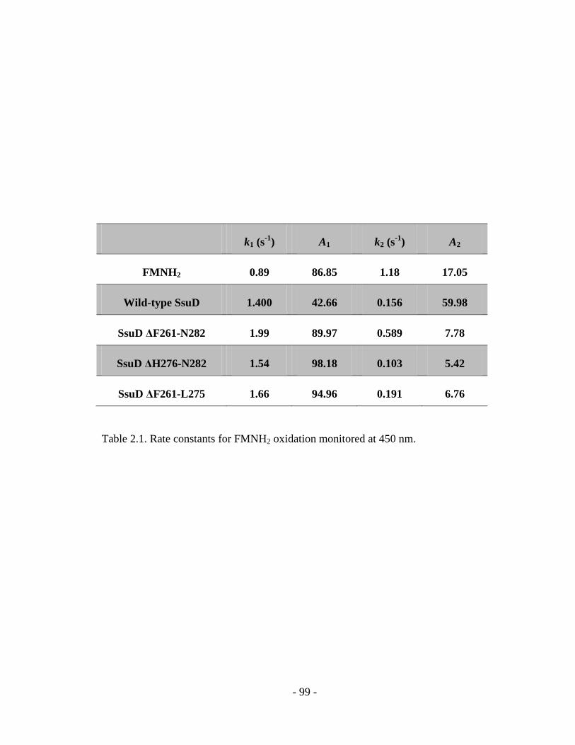
- 99 -
k1 (s-1
) A1 k2 (s-1
) A2
FMNH2 0.89 86.85 1.18 17.05
Wild-type SsuD 1.400 42.66 0.156 59.98
SsuD ΔF261-N282 1.99 89.97 0.589 7.78
SsuD ΔH276-N282 1.54 98.18 0.103 5.42
SsuD ΔF261-L275 1.66 94.96 0.191 6.76
Table 2.1. Rate constants for FMNH2 oxidation monitored at 450 nm.

- 100 -
2.3.5 Protein-protein interactions between SsuE and ΔF261-N282 SsuD
For the flavin-dependent two-component systems, it has been postulated that reduced
flavin can be transferred through a channeling or dissociative mechanism (156-164).
Previous studies have identified static interactions between SsuE and SsuD, supporting a
channeling mechanism for flavin transfer (167). The rapid reaction kinetic evaluation of
the SsuD deletion variants with SsuE suggested that flavin reduction could be altered due
to the disruption of protein-protein interactions. In order to examine if the dynamic loop
region of SsuD is involved in static protein-protein interactions, affinity chromatography
experiments with His-tagged ΔF261-N282 SsuD and native SsuE were performed.
Analysis of the eluted protein by SDS-PAGE showed that the unbound native SsuE was
removed following the 125 mM imidazole wash (Figure 2.10 A, lane 3-10). With the 300
mM imidazole wash, native SsuE coeluted with His-tagged ΔF261-N282 SsuD (Figure
2.10 B, lane 8-15), suggesting that ΔF261-N282 SsuD was able to form static interaction
with native SsuE. Because ΔF261-N282 SsuD contains the largest deletion of the
dynamic loop region, the protein-protein interactions between ΔH276-N282 and
ΔF261-L275 SsuD with SsuE would likely still be intact. Therefore, the apparent flavin
oxidation and altered kinetic traces for SsuE flavin reduction observed in rapid reaction
kinetic experiments with the SsuD deletion variants were not due to the disruption of
protein-protein interactions.

- 101 -
kDa 1 2 3 4 5 6 7 8 9 10 11 12 13 14 15
97.4
66.2
45.0
31.0
21.5
kDa 1 2 3 4 5 6 7 8 9 10 11 12 13 14 15
97.4
66.2
45.0
31.0
21.5
A.
B.
Figure 2.10. SDS-PAGE (12% acrylamide) from the affinity chromatography
experiments with SsuE and the 6His-tagged ΔF261-N282 SsuD. 50 ml cell lysate
containing 6His-tagged ΔF261-N282 SsuD was loaded onto Ni-NTA column followed
by loading 50 ml of native SsuE cell lysate. A: SDS-PAGE of fractions after 125 mM
imidazole wash, lane 1 is protein standard, lane 2 is wild-type SsuD (41.7 kDa) and SsuE
(21.3 kDa), lane 3-15 represent fractions after 125 mM imidazole wash over time. B:
SDS-PAGE of fractions after 300 mM imidazole wash, lane 1 is protein standard, lane 2
is wild-type SsuD (41.7 kDa) and SsuE (21.3 kDa), and 3-15 represent fractions after 300
mM imidazole wash over time.

- 102 -
2.4 Discussion
The SsuD enzyme belongs to the bacterial luciferase family that includes bacterial
luciferase and long-chain alkane monooxygenase (LadA). Enzymes in this family are
defined as flavin-dependent two-component monooxygenases that rely on a flavin
reductase to provide reduced flavin to the monooxygenase enzyme (94). A mechanistic
challenge for the two-component monooxygenase systems is the transfer of reduced
flavin from the flavin reductase to the monooxygenase (94, 156). The monooxygenase
active site would need to be in an open conformation for flavin transfer, leaving the
reduced flavin susceptible to unproductive oxidation (173). Protection of the active site
from bulk solvent would be essential in order to prevent flavin intermediates from
unproductive oxidations. Members of the bacterial luciferase structural family have the
characteristic TIM-barrel conformation with several extended regions that deviate from
the typical (/)8 barrel fold (116, 121-123). The subunit of the heterodimeric structure
of bacterial luciferase contains a loop region located in a conserved insertion sequence
that was unresolved in the initial three-dimensional crystal structure of the enzyme (122).
A partial ordering of this region was observed with flavin bound (123). Detailed
mechanistic studies of the bacterial luciferase loop region demonstrate the vital
importance of this region in maintaining catalytic function (117-119). In the
three-dimensional structure of LadA with FMN bound, a similar insertion sequence
protrudes over the proposed active site (116). This region was proposed to be involved in
providing functionality to LadA by assisting in FMN binding (116). Unlike bacterial
luciferase, there were no obvious conformational changes observed between the
flavin-bound and apoenzyme in LadA (116). The three-dimensional structure of SsuD

- 103 -
also contains a dynamic insertion region located over the putative active site that is
distinct from the barrel (121). This region is highly conserved among the SsuD enzymes
from different organisms suggesting a potential role of this region in maintaining
catalytic function. A conserved arginine residue (Arg297) located near the dynamic loop
region in SsuD was shown to be involved in facilitating loop closure over the active site
(115). These results established the dynamic movement of the loop with the binding of
substrates.
In order to investigate the structural and functional role of the loop region of SsuD,
three deletion variants were constructed based on the grouping of amino acid properties
of this region. The gross secondary structure of the SsuD deletion variants was not
markedly altered by partial deletion of the dynamic loop region. Similarly the deletion of
the mobile loop in bacterial luciferase and triosephosphate isomerase did not disrupt the
overall fold of the TIM barrel core (119, 174). All three of the SsuD deletion variants
were unable to generate the sulfite product in steady-state kinetic assays, suggesting the
loop region is essential for effective catalytic activity. A variant of bacterial luciferase
with a loop deletion in the subunit showed a decrease in bioluminescence by 2-orders of
magnitude (119). Active site hydrophobicity was suggested to be critical to the
bioluminescence activity (175). Although bioluminescence was decreased, the loop
deletion variant of bacterial luciferase was still able to form the carboxylic acid product
suggesting the overall catalytic function was not considerably affected (119). The loss of
activity observed with the SsuD deletion variants indicates that the loop region likely
plays an important role in vital catalytic steps related to desulfonation. Although SsuD

- 104 -
and bacterial luciferase are classified within the same structural family, the SsuD
dynamic loop appears to play a critical role in maintaining catalytic function.
The interaction between the loop region and substrate phosphate group is necessary for
the isomerization reaction of several TIM-barrel enzymes, even though the phosphate is
not directly involved in catalysis (135, 144, 146, 171, 174, 176-178). A change in the
conformation of the dynamic loop region of bacterial luciferase was observed with the
addition of phosphate and with the binding of FMNH2 (123). An insertion region
containing the loop region was also shown to coordinate the phosphate group of FMN in
the three-dimensional structure of LadA with flavin bound (116). The SsuD deletion
variants were still able to bind FMNH2 with similar Kd values as wild-type SsuD. Further
studies to evaluate the reduced flavin environment in the active site by visible CD
spectroscopy demonstrated that the flavin environment of the SsuD deletion variants
were similar to the reduced flavin spectrum in the absence of enzyme indicating the loop
region assists in maintaining the integrity of the active site architecture. Although, the
reduced flavin was able to bind to the active site of the SsuD deletion variants and form
loose interactions with active site residues, the loop was required to close over the active
site to for a stabilized flavin environment in order to promote catalysis for wild-type
SsuD.
The proteolytic resistance of the bacteria luciferase mobile loop with the binding of
reduced flavin was correlated with a closed conformation where the mobile loop was
positioned over the active site and protected from proteolysis by the main enzyme
structure (154). Interestingly, similar resistance to time-dependent limited trypsin
digestion was observed for wild-type SsuD when substrates were present, indicating

- 105 -
possible conformational changes were induced by substrate binding (155). In the absence
of substrates, the susceptible proteolytic sites were located at Arg263 and Arg271 on the
loop region (155). Proteolytic susceptibility experiments on the SsuD deletion variants in
the absence of substrates showed limited digestion compared to wild-type SsuD. The
ΔF261-N282 and ΔF261-L275 SsuD variants were not susceptible to proteolytic trypsin
digestion because both variants lacked the primary digestion sites. Although
ΔH276-N282 SsuD contained both cleavage sites, this deletion variant showed minimal
proteolytic digestion in the absence of substrates compared to wild-type SsuD.
Decreasing the length of the loop likely decreases the mobility of this region altering
protease accessibility. The conformational changes necessary for catalysis that
corresponds with loop closure requires the full-length loop region, and a shortened region
would not induce the requisite conformational changes.
In the alkanesulfonate monooxygenase system the reduced flavin must be transferred
from SsuE to SsuD. Therefore, the oxidative and reductive half-reactions are partitioned
between the two enzymes, which allows for the evaluation of the reduced flavin transfer
step followed by binding to SsuD. Rapid reaction kinetic traces obtained at 450 nm with
both SsuE and wild-type SsuD in the reaction show a lag phase between the reductive
and oxidative half-reactions (155). The observed lag phase may be associated with
reduced flavin transfer and/or a conformational change associated with reduced flavin
binding to SsuD. The kinetic traces representing reduced flavin oxidation by the SsuD
deletion variants were essentially the same as the kinetic traces for free flavin oxidation.
Additionally, flavin reduction by SsuE was initiated 0.004 s later in the presence of the
SsuD deletion variants compared to the wild-type enzyme. These studies suggest that the

- 106 -
SsuD loop region may be involved in promoting flavin transfer to SsuD. Previous studies
have demonstrated protein-protein interactions between SsuE and SsuD, suggesting that
flavin transfer occurs through a channeling mechanism in the alkanesulfonate
monooxygenase system (167). If the loop region were involved in promoting the required
contacts between SsuE and SsuD, then deletion of the loop would ultimately disrupt
flavin transfer. However, protein-protein interactions were observed between the
6His-tagged ΔF261-N282 SsuD variant and native SsuE in pull-down experiments. The
possibility of SsuE coeluting with His-tagged ΔF261-N282 SsuD due to non-specific
protein-protein interaction was excluded since all non-specifically bound native SsuE was
removed with lower imidazole concentrations. Because the deletion regions of
ΔH276-N282 SsuD and ΔF261-L275 SsuD were located within the deletion region of
ΔF261-N282 SsuD, it was assumed that the ability of these variants to interact with SsuE
was still intact. The preservation of protein-protein interactions between SsuE and the
SsuD deletion variant suggest that the altered flavin reduction may be due to a disruption
in the synchronization of activities between the SsuE and SsuD active sites.
Synchronization of active sites has been observed in enzymes with multiple active sites
that tunnel intermediates between active sites (185-187). In these enzymes, the reaction at
a catalytic site is often enhanced by chemical or substrate binding events occurring at a
distant catalytic site. While the loop was not shown to play a major role in protein-protein
interactions, the loop may be involved in subtle contacts that promote active site
synchronization between SsuE and SsuD.
In the coupled rapid reaction kinetic studies, evaluation of reduced flavin oxidation
was complicated by the flavin transfer and binding event. Rapid reaction kinetic analyses

- 107 -
in the absence of SsuE provided additional support regarding the importance of the loop
in protecting the reduced flavin substrate. The shorter SsuD deletion variants
(ΔF261-N282 and ΔF261-L275 SsuD) had similar rate constants for flavin oxidation at
450 nm as wild-type SsuD, while the longer deletion showed a 4-fold decrease in the k2
value. However, the amplitudes obtained from a fit of the kinetic traces for the deletion
variants were in fact very similar to those for non-enzymatic flavin oxidation. The
reduced flavin may be able to bind to the active site in the SsuD deletion variants, but
cannot trigger the conformational changes needed for catalysis leading to unproductive
flavin oxidation. Therefore flavin oxidation is no longer effectively coupled to the
formation of the sulfite product.
Many TIM barrel enzymes are not dependent on the transfer of substrates between
two proteins; rather loop movement is associated with the initial binding of substrates
(128, 135, 170, 171, 174). In contrast, the alkanesulfonate monooxygenase enzymes are
expressed from the same operon and appear to be tightly associated with each other (31).
The reduced flavin must be transferred from the reductase to the alkanesulfonate
monooxygenase enzyme, and the loop may assist with flavin binding following the flavin
transfer event. Deletion of the bacterial luciferase loop region resulted in the flavin being
effectively transferred in in vivo studies, and there was no disruption in flavin binding or
generation of the carboxylic acid product (119). The decrease in bacterial luciferase
bioluminescence was attributed to the flavin emitter being exposed to solvent (119).
Conversely, deletion of the dynamic loop of SsuD resulted in a complete loss of activity
due to the altered active site environment that was not able to protect reactive reduced

- 108 -
flavin from bulk solvent. These distinct differences suggest different mechanistic roles of
the loop region between members of the bacterial luciferase family.

- 109 -
Chapter 3
Investigation of the Flavin Transfer Mechanism in the Two-component Alkanesulfonate
Monooxygenase System
3.1 Introduction
Sulfur is an essential element for the survival and growth of all organisms. Bacteria
require inorganic sulfur sources to synthesize sulfur-containing metabolites and cofactors.
Over 95% of the sulfur in the aerobic soils exists as sulfonate and sulfate esters, therefore
bacteria must have an alternative mechanism to obtain sulfur when inorganic sulfur is
poorly represented (28). During sulfur limitation when inorganic sulfur in the
environment is limiting, bacteria express a set of proteins in order to utilize alternate
sulfur sources (31). Alkanesulfonates are an alternate sulfur source that can be
metabolized by bacteria that are capable of expressing sulfonate-sulfur-utilization (ssu)
proteins. Induction of the ssu operon during sulfur limiting conditions leads to the
expression of the two-component alkanesulfonate monooxygenase system, that includes
an NAD(P)H-dependent flavin mononucleotide (FMN) reductase (SsuE) and
FMNH2-dependent alkanesulfonate monooxygenase (SsuD). The SsuE enzyme catalyzes
the reduction of FMN to FMNH2 with NAD(P)H providing the reducing equivalents. The
reduced flavin is transferred from SsuE to SsuD for the desulfonation of alkanesulfonate.
Once the reduced flavin is transferred, the SsuD enzyme activates dioxygen with the
reduced flavin cofactor. Many two-component flavin-dependent monooxygenases
catalyze the enzymatic oxidation reaction via a C4a-(hydro)peroxyflavin intermediate

- 110 -
(47, 102, 113, 168, 169). Similarly, the SsuD enzyme is proposed to utilize the
C4a-(hydro)peroxyflavin intermediate to catalyze the oxygenolytic cleavage of the
carbon-sulfonate bond of alkanesulfonate to generate the corresponding aldehyde and
sulfite product (Scheme 3.1) (47). This highly reactive flavin peroxide intermediate is
thought to be stabilized and protected by an active site cysteine and the closure of a
flexible loop over the active site (124, 155). The sulfite product of the alkanesulfonate
monooxygenase system is then assimilated into various sulfur-containing compounds for
the survival and growth of the organism (36). Sulfonate buffers and alkanesulfonates with
varying carbon lengths can be utilized by the alkanesulfonate monooxygenase system as
substrates (36). The ability of SsuD to utilize a broad range of substrates ensures bacteria
enough alternate sulfur sources during sulfur limitation.
Scheme 3.1. Proposed SsuD mechanism involving a C4a-peroxyflavin intermediate (37).

- 111 -
The flavin-dependent two-component systems are considered atypical flavoproteins
because the flavin cofactors are not tightly bound to proteins through covalent or
non-covalent interactions. Therefore, a distinctive feature of these flavin-dependent
two-component systems is the transfer of the reduced flavin from one enzyme component
to the other. Although an increasing number of bacterial flavin-dependent
two-component systems have been identified, the mechanism of flavin transfer has not
been fully determined (60, 75, 78, 82, 87, 88, 94, 156-159, 160-164). Reduced flavin
transfer could either occur through a dissociative mechanism from the reductase to the
monooxygenase, or by a coupled channeling mechanism involving protein-protein
interactions between the enzyme components.
It is postulated that flavin transfer occurs through a channeling mechanism in the
alkanesulfonate monooxygenase system. Previous studies reported an altered kinetic
mechanism for SsuE in the presence and absence of SsuD and the alkanesulfonate
substrate, which was attributed to the interactions between SsuE and SsuD (81). Direct
protein-protein interaction between SsuE and SsuD with the formation of a stable
complex has been identified, suggesting flavin transfer occurs through a channeling
mechanism (167). In order for the reduced flavin to access the active site of SsuD without
undergoing non-enzymatic oxidation, the SsuD flexible loop region was thought to
participate in conformational changes that assist in flavin transfer events (179).
Nonetheless, there has been no direct kinetic evidence to support the channeling flavin
transfer mechanism in the alkanesulfonate monooxygenase system. A method has been
described to directly evaluate substrate transfer mechanism utilizing an inactive donor
enzyme variant (180). Because the donor enzyme SsuE is highly unstable in the

- 112 -
alkanesulfonate monooxygenase system, a modified method was designed using an SsuD
variant with reduced flavin affinity to kinetically discern between a dissociative or
channeling mechanism for reduced flavin transfer. In addition, rapid kinetic studies were
performed to investigate if the kinetics of each enzyme is affected in the presence of the
other enzyme component. The reported results provide insights into the flavin transfer
mechanism in the two-component alkanesulfonate monooxygenase system.
- 113 -
3.2 Material and Methods
3.2.1 Materials
Potassium phosphate (monobasic and dibasic), flavin mononucleotide phosphate
(FMN), nicotinamide adenine dinucleotide phosphate (NADPH),
5,5-dithiobis-(2-nitrobenzoic acid) (DTNB), glucose, glucose oxidase, ammonium sulfate,
ampicillin, streptomycin sulfate, lysozyme, ethylenediaminetetraacetic acid (EDTA) and
dimethyl sulfoxide (DMSO) were purchased from Sigma (St. Louis, MO). Glycerol,
isopropyl-β-D-thiogalactoside (IPTG) and sodium chloride were obtained from Fisher
(Pittsburgh, PA). Octanesulfonate was from Fluka (Milwaukee, WI). QIAprep Spin
Miniprep Kit and QIAquick PCR Purification Kit were from Qiagen (Valencia, CA).
Super-competent cells E. coli BL21 (DE3) were from Stratagene (La Jolla, CA).
Oligonucleotide primers were from Invitrogen (Carlsbad, CA).
3.2.2 Construction, expression, and purification of recombinant proteins
The Y128F/S179A SsuD variant was generated using the pET21a plasmid containing
the ssuD gene as the template for PCR-amplification. The TAC codon for Tyr128 was
replaced with TTC for Phe, and the TCA codon for Ser179 was replaced with GCA for
Ala. The PCR products were purified with the QIAquick PCR Purification Kit
(Stratagene, La Jolla, CA), and digested with DpnI to remove parental plasmids. The
SsuD variant was confirmed by sequence analysis at Davis Sequencing (Davis, CA). The
vector containing the correct gene sequence for the SsuD variant was transformed into

- 114 -
super-competent cells E. coli BL21 (DE3) for protein expression. The expression and
purification of the variant and wild-type SsuD proteins were performed as previously
described (81).
3.2.3 Steady-state activity assays
The steady-state kinetic parameters for wild-type and Y128F/S179A SsuD were
determined based on previously reported methods (47). Reactions were initiated by the
addition of NADPH (500 μM) into a solution containing SsuD (0.2 μM), SsuE (0.6 μM),
FMN (2.0 μM), and varying concentrations of octanesulfonate (5-1000 μM) in 25 mM
potassium phosphate buffer, pH 7.5, 10% glycerol at 25 °C. Because of decreased
enzymatic activity with the SsuD variant, assays contained a final concentration of 3.0
μM Y128F/S179A SsuD, 9.0 μM SsuE, 500 μM NADPH and 5.0 μM FMN. The sulfite
product was quantified as previously described (47).
3.2.4 Reduced flavin affinity
A previously described spectrofluorimetric titration method was used to determine
the Kd values of wild-type and Y128F/S179A SsuD for reduced flavin with slight
modifications (47, 81). Protein solutions were prepared in a glass titration cuvette by at
least 15 cycles of evacuation followed by equilibration with ultrahigh purity argon gas.
Flavin was prepared in 25 mM potassium phosphate, pH 7.5, and 10% glycerol was
bubbled with ultrahigh purity argon gas for 15 min. Both protein and flavin solution were
then incubated in an anaerobic glovebox for 20 min after adding glucose (20 mM) and
glucose oxidase (10 Units) to remove trace amounts of dioxygen. The anaerobic FMN
solution was photoreduced by two consecutive 15 min irradiations in the presence of
EDTA (10 mM). For the titration of Y128F/S179A SsuD with reduced flavin, aliquots of

- 115 -
reduced flavin (2.63-56.42 μM) were added to a 1 mL solution of Y128F/S179A SsuD
(0.5 μM) in 25 mM potassium phosphate, pH 7.5, and 10% glycerol and the fluorescence
spectra were recorded following a 5-minute-incubation after each addition. The addition
of reduced flavin was performed in an anearobic glovebox to ensure anaerobiosis. Protein
samples were exited at 280 nm and the emission intensity recorded at 344 nm on a
Perkin-Elmer LS 55 luminescence spectrometer (Palo Alto, CA). The Kd value was
determined as previously described (47).
3.2.5 Rapid reaction kinetic analysis
Stopped-flow kinetic analyses were carried out using an Applied Photophysics SX.
18 MV stopped-flow spectrophotometer (47). All stopped-flow experiments were carried
out in single-mixing mode at 4 °C by mixing equal volumes of the solutions, and
monitoring the reactions by single wavelength analyses at 450 nm.
To investigate if the kinetics of each enzyme is affected in the presence of the other
enzyme component, stopped-flow experiments with varying concentrations of substrates
were performed. For the evaluation of the effect of SsuD and octanesulfonate on SsuE
catalysis, wild-type SsuD (35 μM), SsuE (35 μM), and FMN (25 μM) in 25 mM
potassium phosphate, pH 7.5, 10% glycerol were mixed against NADPH (250 μM) and
octanesulfonate (0-1000 μM) in air-equilibrated 10 mM Tris-HCl buffer, pH 8.5, 10%
glycerol. For the evaluation of the effect of SsuD and dioxygen on SsuE catalysis,
wild-type SsuD (35 μM), SsuE (35 μM), and FMN (25 μM) in anaerobic 25 mM
potassium phosphate, pH 7.5, 10% glycerol were mixed against NADPH (250 μM),
octanesulfonate (250 μM) and dioxygen (0-60%) in anaerobic 10 mM Tris-HCl buffer,
pH 8.5, 10% glycerol buffer. Control experiments were performed by mixing SsuE (35

- 116 -
μM) and FMN (25 μM) in 25 mM potassium phosphate, pH 7.5, 10% glycerol with
NADPH (60-250 μM) and octanesulfonate (250 M) in air-equilibrated 10 mM Tris-HCl
buffer, pH 8.5, 10% glycerol. The single wavelength traces of flavin reduction by SsuE at
450 nm were fitted to the following equations using KaleidaGraph software (Abelbeck
Software, Reading, PA):
where k1 and k2 are the apparent rate constants for the different phases, A is the
absorbance at time t, A1 and A2 are amplitudes of each phases, and C is the absorbance at
the end of the reaction.
Stopped-flow experiments were performed to investigate the rapid reaction kinetics
of Y128F/S179A SsuD. SsuE (30 μM), Y128F/S179A SsuD (30 μM), and FMN (25 μM)
in 25 mM potassium phosphate, pH 7.5, 10% glycerol were mixed against NADPH (250
μM) and octanesulfonate (250 μM) in air-equilibrated 10 mM Tris-HCl buffer, pH 8.5, 10%
glycerol. In order to evaluate the flavin transfer mechanism between SsuE and SsuD,
stopped-flow experiments were performed by mixing SsuE (30 μM), wild-type SsuD (30
μM), Y128F/S179A SsuD (0-30 μM) and FMN (25 μM) in 25 mM potassium phosphate,
pH 7.5, 10% glycerol against NADPH (250 μM) and octanesulfonate (250 μM) in
air-equilibrated 10 mM Tris-HCl buffer, pH 8.5, 10% glycerol. The kinetic traces of
flavin oxidation in the presence of both wild-type and varying concentration of variant
SsuD were compared to the kinetic trace of flavin oxidation in the presence of wild-type
SsuD alone.
A = A1e(-k1t ) + A2e
(-k2t ) +C

- 117 -
3.3 Results
3.3.1 Rapid reaction kinetics of flavin reduction and oxidation
In the alkanesulfonate monooxygenase system, the flavin reductive and oxidative
half-reactions occur on separate enzymes, and there is a defined separation between these
two events (155). In steady-state kinetic analysis, the SsuE mechanism was altered in the
presence of SsuD and octanesulfonate, suggesting that SsuD affects flavin reduction
possibly by forming protein-protein interactions with SsuE (81). Although reductive
half-reaction is linked with the oxidative half-reaction under steady-state conditions,
whether the flavin reduction by SsuE and oxidation by SsuD affect each other under
pre-steady-state conditions have never been investigated.
In order to evaluate if SsuE is affected by SsuD in catalysis under single-turnover
conditions, stopped-flow kinetic analyses were performed monitoring flavin reduction by
SsuE in the presence of SsuD with varying octanesulfonate and dioxygen substrates. In
the presence of SsuD and octanesulfonate, the onset of flavin reduction by SsuE was
initiated earlier with increasing concentrations of octanesulfonate (Figure 3.1). This
change was shown to be dependent on the octanesulfonate concentration. When these
kinetic traces of flavin reduction were fitted, the rates were not significantly altered. The
rate of flavin reduction was 8.8 s-1
for the kinetic trace without octanesulfonate, while the
rate of flavin reduction was 12.1 s-1
for the kinetic trace with 1000 μM octanesulfonate. A
control experiment in the absence of SsuD showed that kinetic traces monitoring flavin

- 118 -
reduction were unchanged with varying concentrations of octanesulfonate, confirming
that octanesulfonate alone has no influence on SsuE catalysis (data not shown). These
results suggest that the initiation of flavin reduction by SsuE starts faster in the presence
SsuD and octanesulfonate. Interestingly, in the presence of SsuD, dioxygen had similar
effect on flavin reduction by SsuE compared to octanesulfonate, further suggesting that
the oxidative half-reaction by SsuD has an impact on the reductive half-reaction by SsuE
(Figure 3.2). The ability of the SsuD enzyme to affect flavin reduction by SsuE under
single turnover conditions suggests physical contacts may exist between the two
components during catalysis.
In order to evaluate if SsuD is affected by SsuE in catalysis under single-turnover
conditions, stopped-flow kinetic analyses were performed monitoring flavin oxidation by
SsuD in the presence of SsuE with varying concentrations of NADPH. The flavin
oxidation started earlier with lower concentrations of NADPH, and the flavin stayed
reduced for a longer period of time with the increasing concentration of NADPH (Figure
3.3). The lag phase separating the reductive and oxidative half-reactions was not apparent
with low concentration of NADPH. This lag phase was previously thought to represent
flavin transfer/conformational changes events where the flavin remains reduced (155).
The requirement of high NADPH concentration for the apparent lag phase suggests that
flavin is being constantly reduced by SsuE and equilibrium between flavin reduction and
flavin oxidation is reached in the lag phase with an excess of NADPH in the reaction.
Due to the absence of the apparent lag phase, it is hard to fit the kinetic traces and
difficult to distinguish flavin oxidation by SsuD and flavin transfer/conformational

- 119 -
changes events. As a result, it is unclear whether the oxidative half-reaction catalyzed by
SsuD catalysis is affected by the catalysis of SsuE or not.

- 120 -
Figure 3.1. Rapid reaction kinetics of flavin reduction by SsuE in the presence of SsuD
and varying octanesulfonate concentration. Stopped-flow kinetic traces monitoring
reductive half reaction were followed at 450 nm and 4 ˚C after mixing wild-type SsuD
(35 μM), SsuE (35 μM), and FMN (25 μM) against NADPH (250 μM) and
octanesulfonate (0 μM , 50 μM ■, 200 μM ◆ and 1000 μM ▲) in aerobic buffer.
0
20
40
60
80
100
0.01 0.1
% A
bs
orb
an
ce
Ch
an
ge
at
45
0 n
m
Time (s)
B

- 121 -
Figure 3.2. Rapid reaction kinetics of flavin reduction by SsuE in the presence of SsuD
and varying dioxygen concentration. Stopped-flow kinetic traces were followed at 450
nm and 4 ˚C after mixing wild-type SsuD (35 μM), SsuE (35 μM), and FMN (25 μM)
against NADPH (250 μM), octanesulfonate (250 μM) and dioxygen (0% , 30% ■ and
60% ▲) in anaerobic buffer.
0
20
40
60
80
100
0.01 0.1
% A
bs
rob
an
ce
Ch
an
ge
at
45
0 n
m
Time (s)

- 122 -
Figure 3.3. Rapid reaction kinetics of flavin reduction by SsuE and flavin oxidation by
SsuD with varying NADPH concentration. Stopped-flow kinetic traces were followed at
450 nm and 4 ˚C after mixing wild-type SsuD (35 μM), SsuE (35 μM), and FMN (25 μM)
against NADPH (60 μM , 90 μM , 120 μM ■, 150 μM , 180 μM ◆, 210 μM ◇
and 250 μM ▲) and octanesulfonate (250 μM) in aerobic buffer.
0
20
40
60
80
100
0.01 0.1 1 10
% A
bs
orb
an
ce
Ch
an
ge
at
45
0 n
m
Time (s)

- 123 -
3.3.2 Kinetic parameters of Y128F/S179A SsuD
In order to discern between a dissociative and a channeling flavin transfer
mechanism in the alkanesulfonate monooxygenase system, a method was developed
requiring an SsuD variant with decreased activity and reduced flavin affinity. If the flavin
transfer occurs through a dissociative mechanism, the SsuD variant will not compete with
wild-type enzyme for reduced flavin. Due to the reduced FMNH2 affinity of the SsuD
variant, the FMNH2 will only diffuse into wild-type SsuD when both wild-type and
variant SsuD are present. When comparing flavin oxidation in the presence of wild-type
SsuD with or without the variant SsuD, the kinetic trace representing flavin oxidation
would not change if the flavin is transferred dissociatively. On the other hand, if the
flavin transfer occurs through a channeling mechanism, the reduced flavin will be
actively transferred within a transient SsuE-SsuD complex. The SsuD variant is still able
to compete with wild-type SsuD for the interaction site(s) on SsuE despite its low affinity
for reduced flavin. Therefore, a portion of reduced flavin will not be directly transferred
to wild-type enzyme in the presence of both wild-type and variant SsuD. A channeling
flavin transfer mechanism would results in an altered flavin oxidation kinetic trace when
the SsuD variant is included in the reaction mixture containing wild-type SsuD. In
conclusion, this variant competition method is able to demonstrate the flavin transfer
mechanism by comparison of the flavin oxidation in the presence of wild-type SsuD with
or without the variant.
The crystal structures of bacterial luciferase and LadA with FMN bound revealed a
tyrosine and serine residue within hydrogen bonding distance with the FMN phosphate
oxygens (116, 123). Based on the three dimensional structure, the SsuD active site

- 124 -
architecture is similar to the active site of bacterial luciferase and LadA (121). The
phosphate oxygens of LadA FMN modeled in the SsuD active site were shown to be
within hydrogen bonding distance to Tyr128 and Ser179 (Figure 3.4). Therefore active
site residues Tyr128 and Ser179 from SsuD are speculated to be involved in flavin
binding. The ability of these amino acid residues to coordinate the reduced flavin
substrate was examined by substituting Tyr128 with phenylalanine and Ser179 with
alanine by site-directed mutagenesis. The dissociation constant (Kd) for the binding of
reduced flavin to Y128F/S179A SsuD was determined by fluorimetric titrations (Table
3.1). Compared to wild-type SsuD, the Kd value for reduced flavin binding to
Y128F/S179A SsuD was found to be approximately 20-fold higher (Table 3.1),
suggesting that Tyr128 and Ser179 plays a role in coordinating the reduced flavin
substrate. The steady-state kinetic parameters of Y128F/S179A SsuD showed a 5-fold
decrease kcat value. The Km value for octanesulfonate was increased nearly 7-fold. These
changes in the kinetic parameters resulted in a 40-fold decrease in the kcat/Km value for
the Y128F/S179A SsuD variant compared to wild-type (Table 3.1). These combined
results suggest that the Y128F/S179A SsuD variant cannot catalyze the desulfonation
reaction efficiently due to its reduced FMNH2 affinity.
In order to evaluate ability of Y128F/S179A SsuD to oxidize reduced flavin under
pre-steady-state conditions in the presence of SsuE, stopped-flow analyses monitoring
flavin reduction and oxidation at 450 nm were performed. Distinctive differences were
observed between kinetic traces representing flavin oxidation by Y128F/S179A and
wild-type SsuD (Figure 3.5). In the kinetic trace obtained with wild-type SsuD, the
apparent lag phase was observed, suggesting the rate of flavin reduction and oxidation

- 125 -
Figure 3.4. Three-dimensional structures of SsuD active site with FMN modeled in. The
tyrosine 128 and serine 179 are shown in hydrogen bonding distance with the phosphate
group of FMN. The structure was generated and rendered with PyMOL (PDB ID:1M41)
(121).

- 126 -
kcat
(min-1
)
Km
(μM)
kcat/ kcat
(μM min-1
)
kd(FMNH2) a
(μM)
Wild-type SsuD b 51.7 2.1 44.0 8.3 1.2 0.2 0.30 0.15
Y128F/S179A SsuD 8.6 1.6 294.8 55.9 0.03 0.006 5.43 0.60
a Determined under anaerobic conditions as described in Material and Methods
b Previously reported (47)
Table 3.1. Kinetic parameters for Y128F/S179A and wild-type SsuD.

- 127 -
reached equilibrium with excess NADPH. In the control experiment without SsuD, the
flavin reduced by SsuE was auto-oxidized by the dioxygen in solution immediately
following flavin reduction (Figure 3.5, ◆). It is reported that the rate of flavin
auto-oxidation is 1.43 S-1
while the rate of enzymatic flavin oxidation by SsuD is 0.16 S-1
(47). Therefore, no apparent lag phase was observed in the control experiment without
SsuD because the rate of non-enzymatic flavin auto-oxidation is faster than the rate of
flavin reduction by SsuE and the equilibrium cannot be reached. Similar to the control
experiment in the absence of SsuD, the kinetic trace obtained with Y128F/S179A SsuD
did not contain the apparent lag phase (Figure 3.5, ). The kinetic trace of flavin
oxidation by Y128F/S179A SsuD lies in between the kinetic trace obtained with
wild-type SsuD and the kinetic trace without SsuD, resembling a mixture of enzymatic
flavin oxidation and non-enzymatic flavin oxidation. These results suggest that
Y128F/S179A SsuD cannot utilize the reduced flavin efficiently due to decreased affinity,
which interrupts the flavin transfer event to SsuD.
3.3.3 Evaluation of flavin transfer mechanism
Flavin transfer can occur either through a dissociation mechanism or a channeling
mechanism in the alkanesulfonate monooxygenase system. In order to kinetically discern
between the two mechanisms, a variant competition method was developed by including
Y128F/S179A SsuD in single turnover reactions monitoring flavin reduction by SsuE and
the subsequent oxidation of the flavin by wild-type SsuD. When varying concentrations
of Y128F/S179A SsuD (10-30 μM) were added in the reaction mixture of SsuE (30 μM),
wild-type SsuD (30 μM), and FMN (25 μM) under single turnover condition, altered
flavin oxidation traces were observed (Figure 3.6 A). The flavin oxidation was delayed in
- 128 -
the presence of both wild-type and Y128F/S179A SsuD compared to flavin oxidation by
wild-type SsuD alone. The observed delay in flavin oxidation corresponds to a longer lag
phase between flavin reduction and oxidation, which increased with increasing
concentration of Y128F/S179A SsuD (Figure 3.6 B). Because this lag phase is thought to
represent the length of time in which the rate of flavin reduction is in equilibrium with the
rate of flavin oxidation during flavin transfer, a longer lag phase would suggest that the
flavin transfer event is prolonged with the addition of Y128F/S179A SsuD. Based on the
results from the completion method, the Y128F/S179A SsuD is able to effectively
compete with wild-type SsuD during catalysis, strongly supporting a channeling flavin
transfer mechanism in alkanesulfonate monooxygenase system.

- 129 -
Figure 3.5. Comparison of enzymatic flavin oxidation by wild-type SsuD, Y128F/S179A
SsuD, and non-enzymatic flavin oxidation. Kinetics traces following the reduced flavin
oxidation at 450 nm and 4 ˚C after mixing SsuE (30 μM), FMN (25 μM), and wild-type
SsuD (◆ 30 μM) or Y128F/S179A SsuD (■ 30 μM) or buffer (● ) against
octanesulfonate (250 μM) and NADPH (250 μM). The kinetic traces shown represent an
average of three separate experiments.
0
20
40
60
80
100
1 10
% A
bso
rba
nc
e C
han
ge
at
450 n
m
Time (s)

- 130 -
0
20
40
60
80
100
0.01 0.1 1 10
% A
bs
orb
an
ce
Ch
an
ge
at
45
0 n
m
Time (s)
A
0
20
40
60
80
100
3 4 5 6 7 8 9 10
% A
bs
orb
an
ce
Ch
an
ge
at
45
0 n
m
Time (s)
B
A.
B.
Figure 3.6. The effect of varying concentration of Y128F/S179A SsuD on the flavin
oxidation by wild-type SsuD. A: Kinetics traces following the flavin reduction and
oxidation at 450 nm and 4 ˚C after mixing SsuE (30 μM), FMN (25 μM), wild-type SsuD
(30 μM) and Y128F/S179A SsuD (0 μM●, 10 μM■, 20 μM◆ and 30 μM▲) against
octanesulfonate (250 μM) and NADPH (250 μM). The kinetic traces shown represent an
average of three separate experiments. B: Kinetic traces of flavin oxidation from A.

- 131 -
3.4 Discussion
The alkanesulfonate monooxygenase system is a two-component flavin-dependent
enzyme system that utilizes the flavin cofactor as a substrate instead of a covalently or
tightly bound prosthetic group. The two-component flavin-dependent systems rely on a
flavin reductase to provide reduced flavin to the monooxygenase enzyme for activity. In
addition, the genes expressing the two enzyme components are often located in the same
operon, indicating a close cooperative relationship between the two enzymes (42). A
mechanistic challenge for these two-component monooxygenase systems is the transfer
of reduced flavin from the flavin reductase to the monooxygenase (94, 165).
In the flavin-dependent two-component systems that have been studied, the flavin
transfer event can either occur through a dissociative mechanism or a channeling
mechanism. In the dissociative mechanism, the reduced flavin is released from the
reductase and passively diffuses into the cellular environment, followed by binding to the
monooxygenase. This entire free diffusion flavin transfer process is performed without
protein-protein interaction between the two components. Typically, the flavin reductases
have a higher affinity for the oxidized flavin, while the monooxygenases have a higher
affinity for the reduced flavin (47, 94). The preference of the reduced flavin binding to
the monooxygenase ensures that the flavin once reduced will be rapidly transferred. The
dissociative flavin transfer mechanism has been reported in several two-component
systems (156-159, 160, 163). In these systems, direct protein-protein interactions were

- 132 -
not observed by kinetic, fluorescence and affinity chromatography studies (156-159, 160,
161). Free diffusion has been proposed as the mechanism of flavin transfer in bacterial
luciferase system because in vitro analyses have demonstrated that bacterial luciferase
can utilize reduced flavin supplied by different flavin reductases (78, 82, 87, 88, 160).
However, this argument is based on the hypothesis that these flavin reductases are not
structurally related. The possibility of these flavin reductases containing similar common
structural motifs cannot be eliminated without evaluating the three dimensional structures
of different flavin reductases. If these flavin reductases contain similar structural motifs
that are able to interact with bacterial luciferase, a channeling flavin transfer mechanism
becomes possible between bacterial luciferase and these flavin reductases. Even though
the reduced flavin is able to diffuse rapidly from the reductases to the monooxygenases, it
can also be oxidized rapidly in solution generating reactive dioxygen species leading to
the damage of cellular components. Therefore, it is speculated by a few groups that the
channeling flavin transfer mechanism would be favored in cellular systems (75, 81,
162-164, 167). The channeling mechanism is defined as the reduced flavin being actively
transferred through a molecular channel within a protein complex formed between the
reductase and monooxygenase. A substrate channeling mechanism for flavin transfer has
also been reported in bacterial luciferase system through biochemical and biophysical
analysis (75, 162-164). Stable protein-protein complex formation was manifested
between monomeric bacterial luciferase and FRP through fluorescence anisotropy studies
in Vibrio harveyi (164). Results from kinetic analyses also demonstrated that FRP
exhibited a ping-pong kinetic pattern in a single-enzyme spectrophotometric assay
monitoring the NADPH oxidation, while the kinetic mechanism changed to a sequential

- 133 -
pattern in a luciferase-coupled assay measuring light emission (162). As a result, a
channeling flavin transfer mechanism between bacterial luciferase and FRP was proposed
in Vibrio harveyi. In addition, a novel fusion enzyme of FRP and bacterial luciferase was
generated and characterized (165). This fusion enzyme closely mimics a
single-component bifunctional monooxygenase. In the coupled bioluminescence reaction,
the total quantum outputs observed with the fusion enzyme without exogenously added
FMN was higher than the case with 25 μM FMN added, suggesting that the
bioluminescence activity of the fusion enzyme relies on a shuffling of the
FRP-bound-FMN between the reductase and luciferase within the fusion protein.
Therefore, it is speculated that a channeling flavin transfer mechanism would be favored
in vivo in order for the two-component bacterial luciferase systems to achieve maximum
efficiency.
A channeling flavin transfer mechanism has also been proposed in the
two-component alkanesulfonate monooxygenase system. Flavin reduction by SsuE has
been reported to switch from an ordered sequential mechanism to a rapid equilibrium
ordered mechanism in the presence of SsuD and octanesulfonate (81). In addition, SsuE
also showed a 10-fold increase in Km value for FMN with the addition of SsuD and
octanesulfonate. The altered mechanism for SsuE suggested that the SsuE catalysis is
influenced by the SsuD under steady-state conditions. The increased Km value for FMN
may indicate that the flavin reduction by SsuE is synchronized with SsuD catalysis to
avoid the production of excess reduced flavin. Static protein-protein interaction was also
identified between SsuE and SsuD by chemical cross-linking, visible CD spectroscopy
and affinity chromatography (167). In addition, the dissociation constant for SsuD

- 134 -
binding to SsuE was determined to be 0.0022 0.0010 μM by fluorescence spectroscopy
(167). If these physical contacts between SsuE and SsuD exist during catalysis, it is
consistent with the observation of an altered SsuE mechanism in the presence of SsuD
and a proposed channeling flavin transfer mechanism in the two-component
alkanesulfonate monooxygenase system.
Even though the flavin transfer mechanism for bacterial luciferase system is still
under debate and a channeling mechanism may occur in both bacterial luciferase system
and alkanesulfonate monooxygenase system, how the flavin is actively delivered within
the protein complex may differ in the these two-component systems. An unresolved
mobile loop was observed above the active site of both bacterial luciferase and SsuD (121,
122). Deletion of the bacterial luciferase loop region resulted in the flavin being
effectively transferred in in vivo studies, suggesting the mobile loop of bacterial
luciferase does not participate in flavin transfer (119). However, the SsuD mobile loop of
bacterial luciferase was suggested to not only induce essential conformational changes
important for catalysis but also play a dual role in assisting in flavin transfer (179).
In order to further evaluate the flavin transfer mechanism in alkanesulfonate
monooxygenase system, rapid reaction kinetic studies were performed to investigate if
the kinetics of each enzyme was affected in the presence of the other enzyme under
pre-steady-state conditions. In the presence of SsuD and octanesulfonate substrate, the
onset of flavin reduction by SsuE was initiated earlier with higher octanesulfonate
concentration. It was previously established that octanesulfonate is not a substrate of
SsuE and does not form any interactions with SsuE (81). It is speculated that higher
octanesulfonate concentrations increase the rate of conformational changes of SsuD and

- 135 -
promotes a more rapid and effective communication between SsuE and SsuD, which
enhances the onset flavin reduction by SsuE. This change was shown to be dependent on
octanesulfonate concentration. A control experiment without SsuD showed that the flavin
reduction was not affected by octanesulfonate alone. These results suggest that the
dynamics of SsuD affect the catalysis of SsuE. Rapid reaction kinetic analyses evaluating
the effect of dioxygen on the catalysis of SsuE and SsuD also suggested that the onset of
flavin reduction by SsuE starts earlier with higher concentration of dioxygen, further
suggesting that dioxygen-induced conformational changes of SsuD has an impact on the
reductive half-reaction by SsuE. Interestingly, this impact by dioxygen was not as
significant as octanesulfonate, indicating the SsuD conformational change induced by
dioxygen may not be as substantial as the one induced by octanesulfonate. When the
effect of SsuE catalysis on flavin oxidation by SsuD was evaluated, flavin oxidation was
initiated earlier with lower concentration of NADPH. Because the lag phase between the
reductive and oxidative half-reactions was not apparent in the kinetic traces with lower
concentrations of NADPH, it is hard to distinguish and describe the flavin transfer and
oxidation. Therefore, it is uncertain if oxidative half reaction by SsuD is influenced by
SsuE. Nevertheless, the overall results of these studies were sufficient to conclude that
the conformational changes of SsuD induced by its substrate influence the flavin
reduction by SsuE, indicating protein-protein interaction and communication exist
between SsuE and SsuD during catalysis. The structural evidences of static
protein-protein interactions and the kinetic evidences under both steady-state and
pre-steady-state conditions strongly suggest a channelling flavin transfer mechanism in
the alkanesulfonate monooxygenase system.

- 136 -
A novel method has been described to directly evaluate the substrate transfer
mechanism between the donor enzyme and the acceptor enzyme (180). In the original
method, an inactive variant of the donor enzyme was generated to compete with the
wild-type donor enzyme in coupled activity assays. If the activity of the acceptor enzyme
is decreased with increasing concentrations of the inactive donor variant, less substrate is
transferred from the active donor enzyme to the acceptor enzyme because more complex
is formed between the inactive donor variant and the acceptor enzyme, indicating a
channeling substrate transfer mechanism. If the activity of the acceptor enzyme is not
affected by the addition of the inactive donor variant, all substrate produced by the active
donor enzyme can efficiently diffuse into the acceptor enzyme, indicating a dissociative
substrate transfer mechanism.
Even though a channeling flavin transfer mechanism in the alkanesulfonate
monooxygenase system is indicated by analyzing flavin reduction and static
protein-protein interaction, a direct evaluation of the flavin transfer event during catalysis
has not been performed. Therefore, a variant completion method was utilized to directly
study the flavin transfer during catalysis. Because the donor SsuE enzyme in the
alkanesulfonate monooxygenase system is highly unstable, the inactive SsuE variants
generated were susceptible to aggregation and precipitation during the purification
process (data not shown). Thus, a modified method was developed by using an SsuD
variant with a decreased affinity for reduced flavin. Modeling the flavin coordinates in
the flavin-bound LadA three-dimensional structure into SsuD places Tyr128 and Ser179
in a position that coordinates the phosphoryl group of flavin. The Y128F/S179A SsuD
variant was generated as the candidate with decreased flavin affinity for the evaluation of

- 137 -
the flavin transfer mechanism. The perturbed flavin binding ability and the decreased
catalytic efficiency of Y128F/S179A SsuD suggested the ability of this variant to
catalyze the desulfonation reaction was greatly weakened due to its decreased reduced
flavin affinity. In the rapid reaction kinetic analysis, distinct differences were observed
between the flavin oxidation by Y128F/S179A or wild-type SsuD. Similar to the control
of non-enzymatic flavin oxidation, the kinetic trace obtained with Y128F/S179A SsuD
did not show an apparent lag phase. The kinetic trace of flavin oxidation by
Y128F/S179A SsuD resembled a mixture of enzymatic flavin oxidation and
non-enzymatic flavin oxidation, suggesting that some reduced flavin is oxidized
non-enzymatically. This non-enzymatic flavin oxidation could be due to the reduced
flavin not being properly transferred between the SsuE and Y128F/S179A SsuD variant.
The flavin reduced by SsuE is likely to be oxidized by dioxygen in SsuE or in solution
because of the impaired flavin binding ability of Y128F/S179A SsuD. Therefore,
Y128F/S179A SsuD could serve as a proper inactive variant to investigate flavin transfer
through designed competition methods.
In order to kinetically discern between the two mechanisms, reduced flavin oxidation
by wild-type SsuD was monitored under single turnover conditions in the presence of
different concentrations of Y128F/S179A SsuD. If the flavin transfer occurs through a
dissociation mechanism, the flavin reduced by SsuE should dissociate into solution.
Because wild-type SsuD has a 20-fold higher reduced flavin affinity than Y128F/S179A
SsuD, the reduced flavin in solution should only be diffused into wild-type SsuD when
both enzymes are present. As a result, flavin oxidation by wild-type SsuD should not be
affected in the presence of Y128F/S179A SsuD. If flavin transfer occurs through a

- 138 -
channeling mechanism, the reduced flavin should be actively transferred within a
transient SsuE-SsuD complex. Because Y128F/S179A SsuD will compete with wild-type
SsuD for the interaction site(s) on SsuE, a portion of the reduced flavin will not be
directly transferred to wild-type SsuD. It was demonstrated that the kinetic trace
monitoring reduced flavin oxidation by Y128F/S179A SsuD does not contain the lag
phase observed in flavin oxidation by wild-type SsuD. Therefore, the overall kinetic trace
of flavin oxidation should be altered with both wild-type and Y128F/S179A SsuD
included in the reaction if the flavin transfer occurs through a channeling mechanism.
When varying concentrations of Y128F/S179A SsuD were included in the reaction
mixture of SsuE, wild-type SsuD, and FMN under single turnover condition, the flavin
oxidation trace was altered. The change in flavin oxidation induced by Y128F/S179A
SsuD was also shown to be dependent on the variant concentration. Interesting, a longer
lag phase was observed for the altered flavin oxidation in the presence of Y128F/S179A
SsuD. When the transient SsuE-Y128F/S179A SsuD complex forms, the reduced flavin
may not be transferred properly and is retained in SsuE after the dissociation of the
SsuE-Y128F/S179A SsuD complex due to the decreased flavin affinity of the variant.
Because there was unoccupied free wild-type SsuD in solution, the SsuE-reduced flavin
could reassociates with wild-type SsuD to transfer reduced flavin to wild-type SsuD for
oxidation (Figure 3.7). The longer lag phase is believed to be caused by this
protein-protein equilibrium complex. These results suggest that a transient SsuE-SsuD
complex is formed during catalysis, strongly supporting a channeling mechanism for
flavin transfer.

- 139 -
Figure 3.7. Schematic representation of flavin reduction and oxidation process with the
presence of SsuE, wild-type SsuD and Y128F/S179A SsuD. In the presence of both
wild-type (green circle) and Y128F/S179A SsuD (grey circle), the SsuE (blue square) is
able to form protein-protein complex with both SsuD after reducing FMN (yellow
triangle). In the SsuE/wild-type SsuD complex, the FMNH2 is transferred directly to
wild-type SsuD for enzymatic oxidation. In the SsuE/Y128F/S179A SsuD complex, the
FMNH2 is not transferred and retained in SsuE due to the reduced affinity of
Y128F/S179A SsuD for FMNH2. The FMNH2 bound SsuE then dissociates
Y128F/S179A SsuD and re-associates with unoccupied wild-type SsuD in solution,
followed by enzymatic oxidation. The resulted flavin oxidation in the presence of both
wild-type and Y128F/S179A SsuD is a mixture of a normal and a slowed enzymatic
flavin oxidation.
- 140 -
The flavin cofactors play an important role in biological systems due to their ability
to assume versatile redox states. A large number of flavoproteins fulfill their functions by
relying on the flavin cofactor to transfer electrons. The reduced flavin is extremely
reactive toward molecular dioxygen, generating H2O2 and reactive oxygen species that
are damaging to cellular components. In order to eliminate the production of these
unwanted toxic compounds, the intracellular concentration of free reduced flavin should
be cautiously controlled. In the flavin-dependent two-component systems, the reduced
flavin is transferred from the reductase to the monooxygenase. Alternating flavin transfer
mechanisms have been reported depending on the specific system. For the
two-component systems that utilize a dissociative flavin transfer mechanism, the reduced
flavin diffusion occurs faster than the reaction with free dioxygen in solution. Therefore,
the production of damaging reactive oxygen species can be avoided. For the
two-component systems that utilizing a channeling flavin transfer mechanism, transient
complex is formed between the reductase and the monooxygenase. As a result, the
reduced flavin is protected from the bulk solvent in the complex. The evidences provided
in this study thoroughly supported a channeling flavin transfer mechanism in the
alkanesulfonate monooxygenase system, and explains how the reduced flavin is protected
from non-enzymatic oxidation during the desulfonation reaction.

- 141 -
Chapter 4
Summary
The flavin-dependent two-component alkanesulfonate monooxygenase system from
E. coli is composed of a NAD(P)H-dependent FMN reductase (SsuE) and an
FMNH2-dependent alkanesulfonate monooxygenase (SsuD). In the presence of
NAD(P)H, the SsuE enzyme catalyzes the reduction of FMN to FMNH2. In the presence
of O2 and FMNH2, the SsuD enzyme is capable of cleaving the C-S bond of a wide range
of 1-susbtitued alkanesulfonates, resulting in the liberation of sulfite and production of a
corresponding aldehyde (36, 47). The SsuD enzyme is proposed to catalyze the
desulfonation reaction through a C4a-(hydro)peroxyflavin intermediate. Although an
unresolved loop region above the SsuD active site was suggested to protect the flavin
peroxide intermediate, direct characterization of this unresolved loop region has not been
performed (155). In addition, how SsuD mechanistically protect and stabilize the reactive
intermediate has not been fully addressed. A distinct feature of the two-component
alkanesulfonate monooxygenase system is that the reduced flavin supplied by SsuE is
transferred to SsuD for the desulfonation reaction. Although a channeling flavin transfer
mechanism has been suggested with the identification of static SsuE-SsuD interaction,
the evidence was not enough to conclude that the flavin transfer occurs though a
channeling mechanism during catalysis. This research has focused on
elucidating the functional role of the SsuD unresolved loop region, as well as the flavin

- 142 -
transfer mechanism during catalysis with a combination of biophysical, biochemical and
kinetic approaches.
4.1 Catalytic mechanism of the alkanesulfonate monooxygenase
In the two-component systems, the flavin-dependent monooxygenase usually
catalyze a variety of reactions in three steps: 1) utilizing reduced flavin supplied by a
flavin reductase; 2) activating dioxygen to form a C4a-(hydro)peroxyflavin intermediate;
3) oxygenating the substrate. The initial characterization of SsuD has suggested an
ordered substrate binding mechanism with the reduced flavin binding first and
octanesulfonate binding last (47). In addition, each substrate binding event is likely to be
associated with conformational changes (47, 155). In the proposed mechanism of SsuD, a
C4a-peroxyflavin intermediate performs a nucleophilic attack on the sulfonate functional
group. The alkanesulfonate peroxyflavin adduct then undergoes Baeyer-Villiger
rearrangement and proton abstraction from an active site base, leading to the generation
of hydroxyflavin, alkanealdehyde and sulfite. This mechanism is supported by the
identification of a C4a-(hydro)peroxyflavin intermediate through rapid reaction kinetic
analysis. The active site Cys54 residue was suggested to directly or indirectly stabilize
the flavin peroxide intermediate by hydrogen bonding (124). Based on site-directed
mutagenesis studies and kinetic characterization, the active site Cys54 was considered to
not directly catalyze the desulfonation reaction as an active site base. In addition, the
evaluation of active site His228 residue also suggested that it is not the catalytic base
(data not shown). Although pH profile of SsuD suggest that an amino acid must be
deprotonated in order to achieve the optimal activity, the identity of the catalytic base still
remains unclear (data not shown).
- 143 -
4.2 Functional role of the SsuD unresolved loop region in catalysis
In the three-dimensional structure of SsuD, a conserved loop region located above
the putative active site was unresolved in an insertion region that deviates from
TIM-barrel core, suggesting mobility of this loop region (121). In the initial
characterization, a highly conserved Arg297 on this insertion region was speculated to
involve in catalysis due to the diminishing enzymatic activity with an aberrant mutation
of Arg297→Cys (121). It was speculated that the Cys mutation at position 279 would
form a disulfide bond with the Cys54 located in the active site, sacrificing the mobility of
the unresolved loop region and resulting in the abolished activity (121). Further
site-directed mutagenesis studies, biochemical and kinetic analysis of the Arg297 residue
suggested that this conserved residue plays an important role in the desulfonation reaction,
and confirmed that the mobility of the unresolved loop is crucial to the catalysis (155).
The substitution of Arg297 with Lys or Ala did not significantly alter the secondary
structural components or the substrate affinity. The R297A and R297K SsuD showed
impaired catalytic activity and were unable to accumulate critical
C4a-(hydro)peroxyflavin intermediate, indicating that Arg297 is required for the
protection and stabilization of the reactive flavin peroxide intermediate (155). Studies
evaluating proteolytic susceptibility of the unresolved loop region of wild-type SsuD and
the Arg297 variants suggested that the SsuD Arg297 facilitates necessary conformational
changes induced by reduced flavin binding (155). These conformational changes
correlate with loop closure and are suspected to protect the intermediate and promote
catalysis.

- 144 -
Three variants containing a partial deletion of the unresolved loop region were
constructed to directly evaluate the functional properties of this region. Although the
secondary structures and the substrate affinity of all three loop-deleted SsuD variants
were not significantly altered, the catalytic activity toward sulfite production was
completely abolished, suggesting an absolute requirement of this loop region for catalysis.
Further near-UV CD spectroscopic studies evaluating the reduced flavin binding
environment in the active site of loop-deleted SsuD variants demonstrated that the SsuD
unresolved loop region is important in stabilizing the flavin environment of SsuD in order
to facilitate catalysis. In addition, proteolytic susceptibility experiments on the
loop-deleted SsuD variants suggested that the loop closure requires the full-length loop
region, and a shortened loop region would not induce the requisite conformational
changes necessary for catalysis. The rapid reaction kinetic analysis in the presence of
both SsuE and loop-deleted SsuD variants suggested that the reduced flavin was oxidized
non-enzymatically due the lack of protection from the unresolved loop. Even though the
static protein-protein interactions were not compromised between SsuE and loop-deleted
SsuD variants, the slower onset of flavin reduction and immediate flavin oxidation in the
presence of loop-deleted SsuD variants implied that the unresolved loop region may be
involved in promoting flavin reduction by SsuE and flavin transfer from SsuE to SsuD
during catalysis. Therefore, dual roles of inducing essential conformational changes and
assisting flavin transfer were proposed for the SsuD unresolved loop region. These
studies provided better understanding of the mechanistic roles of the SsuD unresolved
loop region in the desulfonation reaction.
- 145 -
4.3 Flavin transfer mechanism in the alkanesulfonate monooxygenase system
The flavin transfer event for two-component alkanesulfonate monooxygenase system
in E. coli has been speculated to occur through a substrate channeling mechanism. The
altered reaction mechanism of SsuE in the presence of SsuD and octanesulfonate
suggested that protein-protein interactions exist between the two enzyme components of
alkanesulfonate monooxygenase system (81, 167). The static SsuE-SsuD interactions
were later identified through several approaches including pull-down assays, chemical
cross-linking and near-UV CD spectroscopy (167). Although a channeling flavin transfer
mechanism has been proposed for two-component the alkanesulfonate monooxygenase
system based on the static protein-protein interactions, there has been no direct kinetic
evidence to support the channeling flavin transfer mechanism during the desulfonation
reaction.
Therefore, rapid reaction kinetic studies were performed to investigate if the kinetics
of each enzyme is affected in the presence of the other enzyme component. The onset of
the flavin reduction by SsuE was initiated earlier in the presence of SsuD and substrates,
suggesting that the dynamics of SsuD affect the catalysis of SsuE. It is also implied that
the higher substrate concentration increases the rate of conformational changes of SsuD
and promotes a more rapid and effective communication between SsuE and SsuD,
resulting in an earlier onset of flavin reduction by SsuE. In order to kinetically discern
between the channeling and dissociative flavin transfer in the alkanesulfonate
monooxygenase system, a competition method was developed by utilizing an SsuD
variant with decreased reduced flavin affinity. Based on the thee-dimensional structure
similarities with related protein, SsuD active site residues Tyr128 and Ser179 were

- 146 -
speculated to be involved in reduced flavin binding. In order to generate an SsuD variant
with reduced flavin affinity, the Y128F/S179A SsuD was generated via site-directed
mutagenesis. Steady-state kinetic analysis confirms that ability of Y128F/S179A SsuD to
catalyze the desulfonation reaction was weakened due to its decreased reduced flavin
affinity. Based on the results from rapid reaction kinetic analysis, it is concluded that the
reduced flavin can be transferred from SsuE to Y128F/S179A SsuD, while a portion of
the transferred flavin is oxidized non-enzymatically due to the impaired flavin binding
ability of Y128F/S179A SsuD. Therefore, Y128F/S179A SsuD could serve as a proper
implement to investigate flavin transfer mechanism through the designed competition
method. To directly evaluate the flavin transfer mechanism, the flavin reduction and
oxidation was monitored when varying concentrations of Y128F/S179A SsuD were
added to a reaction mixture containing SsuE and wild-type SsuD. The kinetic trace
representing flavin oxidation was significantly altered when Y128F/S179A SsuD was
added, suggesting that a transient SsuE-SsuD complex was formed and Y128F/S179A
SsuD actively competed with wild-type SsuD for the interaction site(s) on SsuE. As a
result, a portion of the reduced flavin was not directly transferred from SsuE to wild-type
SsuD, resulting in altered flavin oxidation traces. This change in flavin oxidation induced
by Y128F/S179A SsuD was also shown to be dependent on the variant concentrations,
highly supporting a channeling flavin transfer mechanism. Based the altered kinetic trace
of flavin oxidation trace with the addition of Y128F/S179A SsuD, a complex
dissociation/re-association model was proposed. When the transient SsuE-Y128F/S179A
SsuD complex forms, the reduced flavin may not be transferred properly and is retained
in SsuE after the dissociation of the SsuE-Y128F/S179A SsuD complex due to the

- 147 -
decreased flavin affinity of the variant. Because there was unoccupied free wild-type
SsuD in solution, the reduced flavin bound SsuE re-associates with wild-type SsuD and
transfers the reduced flavin to wild-type SsuD for enzymatic oxidation, resulting an
altered flavin oxidation trace with a prolonged lag phase. This model explains the
observation of a prolonged lag phase in the flavin oxidation kinetic trace when
Y128F/S179A SsuD was added to compete with wild-type SsuD, supporting complex
formation and a channeling flavin transfer mechanism in the two-component
alkanesulfonate monooxygenase system.,
In summary, the functional role of the SsuD unresolved loop region and the flavin
transfer mechanism of the two-component alkanesulfonate monooxygenase system has
been evaluated in depth. Conformational changes involving the closure of the unresolved
loop region play critical roles to ensure catalysis: controlling substrate binding order,
protecting the reduced flavin from bulk solvent, stabilizing the reactive
C4a-(hydro)peroxyflavin intermediate, and facilitating flavin transfer from SsuE to SsuD.
In the two-component alkanesulfonate monooxygenase system, the flavin transfer occurs
through a channeling mechanism, where the reduced flavin is actively shuffled within a
transient SsuE-SsuD complex. Therefore, the system is able to strictly control the
oxidation of the reactive reduced flavin and avoid production of damaging reactive
oxygen species. These studies on the structural dynamics of SsuD and flavin transfer
mechanism are essential to understanding the mechanism of the desulfonation reaction
catalyzed by the two-component alkanesulfonate monooxygenase system.

- 148 -
References
1. Van Der Ploeg, J.R., Weiss, M.A., Saller, E., Nashimoto, H., Saito, N., Kertesz, M.A.,
and Leisinger, T. (1996) Identification of sulfate starvation-regulated genes in
Escherichia coli: a gene cluster involved in the utilization of taurine as a sulfur source.
J. Bacterial. 178, 5438-5446.
2. Sekowska, A., Kung, H-F., and Danchin, A. (2000) Sulfur metabolism in Escherichia
coli and related bacteria: facts and fiction. J. Mol. Microbiol. Biotechnol. 2, 145-177.
3. Kredich, N.M. (1996) Biosynthesis of cysteine. In: Neidhardt, F.C., Curtiss, R.,
Ingraham, J.L., Lin, E.C.C., et al (eds) Escherichia coli and Salmonella, 2nd
edn.
ASM, Washington DC, pp 514-527.
4. Liu, C., Wang, R., Varlamova, O., and Leyh, T.S. (1998) Regulating energy transfer
in the ATP sulfurylase-GTPase system. Biochemistry 37, 3886-3892.
5. Reuter, K., Mofid, M.R., Marahiel, M.A., and Ficner, R. (1999) Crystal structure of
the surfactin synthetase-activating enzyme Sfp: a prototype of the
4‟-phosphopantetheinyl transferase superfamily. EMBO J. 18, 6823-6831.
6. Levy, S., and Danchin, A. (1988) Phylogeny of metabolic pathways: O-acetylserine
sulphydrylase A is homologous to the tryptophan synthase beta subunit. Mol.
Microbiol. 2, 777-783.
7. Leyh, T.S. (1993) The physical biochemistry and molecular genetics of sulfate
activation. Crit. Rev. Biochem. Mol. Biol. 28: 515-542.

- 149 -
8. Jones-Mortimer, M.C. (1968) Positive control of sulphate reduction in Escherichia
coli. Biochem. J. 110, 589-602.
9. Kredich, N.M. (1971) Regulation of L-cysteine biosynthesis in Salmonella
typhimurium. J. Biol. Chem. 246, 3474-3484.
10. Ostrowski, J., and Kredich, N.M. (1989) Molecular characterization of the cysJIH
promoters of Salmonella typhimurium and Escherichia coli: regulation by CysB
protein and N-acetyl-L-serine. J. Bacteriol. 171, 130-140.
11. Ostrowski, J., and Kredich, N.M. (1990) In vitro interactions of CysB protein with the
cysJIH promoters of Salmonella typhimurium: inhibitory effects of sulfide. J.
Bacteriol. 172, 779-785.
12. Ostrowski, J., and Kredich, N.M. (1991) Negative autoregulation of cysB in
Salmonella typhimurium: in vitro interactions of CysB protein with the cysB promoter.
J. Bacteriol. 173, 2212-2218.
13. Kredich, N.M. (1992) The molecular basis for positive regulation of cys promoters in
Salmonella typhimurium and Escherichia coli. Mol. Microbiol. 6, 2747-2753.
14. Greene, R.C. (1996) Biosynthesis of methionine. In: Neidhardt, F.C., Curtiss, R.,
Ingraham, J.L., Lin, E.C.C., et al (eds) Escherichia coli and Salmonella, 2nd
edn.
ASM, Washington DC, pp 542-560.
15. Izard, T., and Geerlof, A. (1999) The crystal structure of a novel bacterial
adenylyltransferase reveals half of sites reactivity. EMBO J. 18, 2021-2030.
16. Zheng, L., Cash, V.L., Flint, D.H., and Dean, D.R. (1998a) Assembly of iron-sulfur
clusters. Identification of an iscSUA-hscBA-fdx gene cluster from Azotobacter
vinelandii. J. Biol. Chem. 273, 13264-13272.

- 150 -
17. Webb, E., Claas, K., and Downs, D.M. (1997) Characterization of thil, a new gene
involved in thiazole biosynthesis in Salmonella typhimurium. J. Bacteriol. 179,
4399-4402.
18. Michal, G. (1999) Biochemical pathways: An atlas of biochemistry and molecular
biology. New York Heidelberg: A John Wiley & Sons, Spectrum Akademischer
Verlag.
19. Bui, B.T., Florentin, D., Fournier, F., Ploux, O., Mejean, A., and Marquet, A. (1998)
Biotin synthase mechanism: on the origin of sulphur. FEBS Lett. 440, 226-230.
20. Ehrlich, H.L. (1996) Geomicrobiology. New York: Marcel Dekker.
21. Brown, K.A. (1982) Sulphur in the environment: a review. Environ. Pollot. Ser. B. 3,
47-80.
22. Brasted, R.C. (1961) Sulfur, selenium, tellurium, polonium and oxygen. In
Comprehensive inorganic chemistry. 8, 7. New York, D. Van Nostrand.
23. Berner, R.A. (1971) Worldwide sulphur pollution of rivers. J. geophys. Res. 76,
6597-6600.
24. Granat, L., Rodhe, H., and Hallberg, R. O. (1976) Nitrogen, phosphorous and
sulfur-global cycles. In SCOPE Report, 7 ed. by Svensson, B.H., and Soderlund, R.
Ecol. Bull. 22, 89-134.
25. Halstead, R.L, and Rennie, P.J. (1977) The effects of sulphur on soils in Canada. In
Sulphur and its inorganic derivatives in the Canadian environment, Nat. Res. Counc.
Can. Rep. No. 15015, 181-220.
26. Freney, J.R. (1967) Sulphur-containing organics. In Soil biochemistry, Vol. 1, ed. by
Rasool, S.I., 177-201. New York, Marcel Dekker.

- 151 -
27. Roberts, R.B., Abelson, P.H., Cowie, D.B., Bolton, E.T., and Britten, R.J. (1955)
Studies of biosynthesis in Escherichia coli. Carnegie Institution, Washington DC.
28. Kertesz, M.A. (1999) Riding the sulfur cycle-metabolism of sulfonates and sulfate
esters in gram-negative bacteria. FEMS Microbiol. Rev. 24, 135-175.
29. Seitz, A.P., and Leadbetter, E.R. (1995) Microbial assimilation and dissimilation of
sulfonate sulfur. In: Vairavamurthy, M.A., Schoonen, M.A.A. (eds) Geochemical
transformations of sedimentary sulfur. ACS Symposium Series 612. ACS,
Washington DC, pp 365-376.
30. Cook, A.M., Laue, H., and Junker, F. (1998) Microbial desulfonation. FEMS
Microbiol. Rev. 22, 399-419.
31. Kertesz, M., Leisinger, T., and Cook, A.M. (1993) Proteins induced by sulfate
limitation in Escherichia coli, Pseudomonas putida, or Staphylococcus aureus. J.
Bacteriol. 175, 1187-1190.
32. Van der Ploeg, J.R., Eichhorn, E., and Leisinger, T. (2001) Sulfonate-sulfur
metabolism and its regulation in Escherichia coli. Arch. Microbiol. 176, 1-8.
33. Quadroni, M., Staudenmann, W., Kertesz, M., and James, P. (1996) Analysis of
global responses by protein and peptide fingerprinting of proteins isolated by
two-dimensional gel electrophoresis. Application to the sulfate-starvation response of
Escherichia coli. Eur. J. Biochem. 239, 773-781.
34. Hummerjohann, J., Kuttel, E., Quadroni, M., Ragaller, J., Leisinger, T., and Kertesz,
M.A. (1998) Regulation of the sulfate starvation response in Pseudomonas
aeruginosa: role of cysteine biosynthetic intermediates. Microbiology 144,
1375-1386.

- 152 -
35. Eichhorn, E., van der Ploeg, J.R., Kertesz, M.A., and Leisinger, T. (1997)
Characterization of the -ketoglutarate-dependent taurine dioxygenase from
Escherichia coli. J. Biol. Chem. 272, 23031-23036.
36. Eichhorn, E., van der Ploeg, J.R., and Leisinger, T. (1999) Characterization of a
two-component alkanesulfonate monooxygenase from Escherichia coli. J. Biol. Chem.
274, 26639-26646.
37. Ryle, M. J., Padmakumar, R., and Hausinger, R.P. (1999) Stopped-flow kinetic
analysis of Escherichia coli taurine/α-ketoglutarate dioxygenase: interactions with
α-ketoglutarate, taurine, and oxygen. Biochemistry 38, 15278–15286.
38. Eichhorn, E., Van der Ploeg, J.R., and Leisinger, T. (2000) Deletion analysis of the
Escherichia coli taurine and alkanesulfonate transport systems. J. Bacteriol. 182,
2687-2795.
39. Kahnert, A., Vermeij, P., Wietek, C., James, P., Leisinger, T., and Kertesz, M.A.
(2000) The ssu locus plays a key role in organosulfur metabolism in Pseudomonas
putida. J. Bacteriol. 182, 2869-2878.
40. Kertesz, M.A., Schmidt, K., and Wuest, T. (1999) A novel reduced flavin
mononucleotide-dependent methanesulfonate sulfonatase encoded by the
sulfur-regulated msu operon of Pseudomonas aeruginosa. J. Bacteriol. 181,
1464-1473.
41. Van der Ploeg J.R., Iwanicka-Nowicka R., Kertesz M.A., Leisinger T., and
Hryniewicz M.M. (1997) Involvement of CysB and Cbl regulatory proteins in
expression of the tauABCD operon and other sulfate starvation-inducible genes in
Escherichia coli. J Bacteriol 179, 7671–7678.
- 153 -
42. Van der Ploeg, J.R., Iwanicka-Nowicka, R., Bykowski, T., Hryniewicz, M.M., and
Leisinger, T. (1999) The Escherichia coli ssuEADCB gene cluster is required for the
utilization of sulfur from aliphatic sulfonates and is regulated by the transcriptional
activator Cbl. J. Biol. Chem. 274, 29358-29365.
43. Iwanicka-Nowicka, R. and Hryniewicz, M.M. (1995) A new gene, cbl, encoding a
member of the LysR family of transcriptional regulators belongs to Escherichia coli
cys regulon, Gene 166, 11-17.
44. Tyrrell, R., Verschueren, K.H.G., Dondson, E.J., Murshudov, G.N., Addy, C., and
Wilkinson, A.J. (1997) The structure of the cofactor-binding fragment of the LysR
family member, CysB: a familiar fold with a surprising subunit arrangement.
Structure 5, 1017-1032.
45. Bender, R. (1991) The role of the NAC protein in the nitrogen regulation of
Klebsiella aerogenes. Mol. Microbiol. 5, 2575-2580.
46. Zimmer, D.P., Soupene, E., Lee, H.L., Wendisch, V.F., Khodursky, A.B., Peter, B.J.,
Bender, R. A., and Kustu, S. (2000) Nitrogen regulatory protein C-controlled genes of
Escherichia coli: scavenging as a defense against nitrogen limitation. Proc. Natl.
Acad. Sci. USA 97, 14674-14679.
47. Zhan, X., Carpenter, R. A. and Ellis, H. R. (2008) Catalytic importance of substrate
binding order for the FMNH2-dependent alkanesulfonate monooxygenase enzyme.
Biochemistry 47, 2221-2230.
48. Massey, V. (1999) The Chemical and Biological Versatility of Riboflavin,
Biochemical Society Transactions 28, 283-296.
- 154 -
49. Müller, F. (1987) Flavin radicals: chemistry and biochemistry. Free Radical Biology
& Medicine 3, 215-230.
50. Ghisla, S., and Massey, V. (1989) Mechanisms of flavoproteins-catalyzed reactions, J.
Biol. Chem. 181, 1-17.
51. Hemmerich, P., and Müller, F. (1973) Flavin-O2 interaction mechanisms and the
function of flavin in hydroxylation reactions Annals New York Academy of Sciences
212, 13-26.
52. Gibson, Q.H., and Hastings, J.W. (1962) The oxidation of reduced flavin mononucleotide
by molecular oxygen. Biochem. J. 83, 368-377.
53. Anderson, R.F. (1982) in Flavins and Flavoproteins (Massey, V., and Williams, C.H.,
Jr., eds) pp. 278-283, Elsevier/North-Holland, New York.
54. Mewies, M., Mcintire, W.S., and Scrutton, N.S. (1998) Covalent attachment of flavin
adenine dinucleotide (FAD) and flavin mononucleotide (FMN) to enzymes: The
current state of affairs. Protein Sci. 7, 7-20.
55. Job, V., Marcone, G.L., Pilone, M.S., and Pollegioni, L. (2002) Glycine oxidase from
Bacillus subtilis. Characterization of a new flavoprotein. J. Biol. Chem. 277,
6985-6993.
56. Swoboda, B.E.P., and Massey, V. (1965) Purification and properties of the glucose
oxidase from Aspergillus niger. J. Biol. Chem. 240, 2209-2215.
57. Nishiya, Y. and Imanaka, T. (1998) Purification and characterization of a novel
glycine oxidase from Bacillus subtilis. FEBS Lett. 438, 263-266.
58. Binda, C., Mattevi, A., and Edmondson, D.E. (2002) Structure-function relationships
in flavoenzyme-dependent amine oxidations. J. Biol. Chem. 277, 23973-23976.

- 155 -
59. Entsch, B., Ballou, D.P. and Massey, V. (1976) Flavin-oxygen derivatives involved in
hydroxylation by p-hydroxybenzoate hydroxylase. J. Biol. Chem. 251, 2550-2563.
60. Duane, W. and Hastings, J.W. (1975) Flavin mononucleotide reductase of luminous
bacteria. Mol. Cell. Biochem. 6, 53-64.
61. Macheroux, P., Ghisla, S., and Hastings, J.W. (1993) Spectral detection of an
intermediate preceding the excited state in the bacterial luciferase reaction.
Biochemistry 32, 14183-12186.
62. Ryerson, C.C., Ballou, D.P., and Walsh, C.T. (1982) Mechanistic studies on
cyclohexanone oxygenase. Biochemistry 21, 2644-2655.
63. Vas, A.D.N., Chakraborty, S., and Massey, V. (1995) Old yellow enzyme:
aromatization of cyclic enones and the mechanism of a novel dismutation reaction.
Biochemistry 34, 4246-4256.
64. Iyanagi, T., Makino, N., and Mason, H.S. (1974) Redox properties of the reduced
nicotinamide adenine dinucleotide phosphate-cytochrome P-450 and reduced
nicotinamide adenine dinucleotide-cytochrome b5 reductases. Biochemistry 13,
1701-1710.
65. Thorpe, C., and Kim, J-J.P. (1995) Structure and mechanism of action of the
acyl-CoA dehydrogenases. FASEB J. 9, 718-725.
66. Venkataram, U.V. and Bruice, T.C. (1984) On the mechanism of flavin-catalyzed
dehydrogenation α, β to an acyl function. The mechanism of 1,5-dihydroflavin
reduction of maleimides. J. Am. Chem. Soc. 106, 5703-5709.

- 156 -
67. Jablonski, E. and DeLuca, M. (1977) Purification and properties of the NADH and
NADPH specific FMN oxidoreductases from Beneckea harveyi. Biochemistry 16,
2932-2936.
68. Kuhn, R., Reinemund, K., and Weygand, F. (1934) Synthese des lumi-lactoflavins.
Ber. 67, 1460-1463.
69. Karrer, P., Schopp, K., and Benz, F. (1935) Synthesen von flavinen IV. Helv. Chim.
Acta. 18, 426-429.
70. Miura, R. (2001) Versatility and specificity in flavoenzymes: control mechanisms of
flavin reactivity. Chem. Rec. 1, 183-194.
71. Massey, V., and Palmer, G. (1966) On the existence of spectrally distinct classes of
flavoprotein semiquinones. A new method for the quantitative production of
flavoprotein semiquinones. Biochemistry 5, 3181-3189.
72. Murataliev, M.B. (1999) Application of electron spin resonance (ESR) for detection
and characterization of flavoprotein. In: Flavoprotein protocols (Chapman, S.K., and
Reid. G.A. eds.) 131, 97-101. Humana Press Inc. Totowa, New Jersey.
73. Ackrell, B.A.C., Johnson, M.K., Gunsalus, P.R., and Cecchini, G. (1992) In
Chemistry and Biochemistry of Flavoenzymes (Muller, F., ed) Vol. III, pp. 229–297,
CRC Press, Boca Raton, FL.
74. Sund, H., and Theorell, H. (1963) Alcohol dehydrogenase. The Enzymes 7, 25-83.
75. Tu, S-C. (2001) Reduced flavin: donor and acceptor enzymes and mechanisms of
channeling. Antioxid. Redox Signal. 3, 881-897.

- 157 -
76. Jablonski, E., and DeLuca, M. (1978) Studies of the control of luminescence in
Beneckea harveyi: properties of the NADH and NADPH:FMN oxidoreductases.
Biochemistry 17, 672-678.
77. Gerlo, E., and Charlier, J. (1975) Identification of NADH-specific and
NADPH-specific FMN reductases in Beneckea harveyi. Eur. J. Biochem. 57, 461-467.
78. Lei, B., Liu, M., Huang, S., and Tu, S-C. (1994) Vibrio harveyi NADPH-flavin
oxidoreductase: cloning, sequencing, and overexpression of the gene and purification
of the cloned enzyme. J. Bacteriol. 176, 3552-3558.
79. Michaliszyn, G.A., Wing, S.S., and Meighen, E.A. (1977) Purification and properties
of a NAD(P)H: flavin oxidoreductase from the luminous bacterium, Beneckea
harveyi. J. Biol. Chem. 252, 7495-7499.
80. Watanable, H., and Hastings, J.W. (1982) Specificity and properties of three pyridine
nucleotide flavin mononucleotide reductases coupling to bacterial luciferase. Mol.
Cell. Biochem. 44, 181-187.
81. Gao, B. and Ellis, H.R. (2005) Altered mechanism of the alkanesulfonate FMN
reductase with the monooxygenase enzyme. Biochem. Biophys, Res. Comm. 331,
1137-1145.
82. Zenno, S., Saigo, K., Kanoh, H., and Inouye, S. Identification of the gene encoding
the major NAD(P)H-flavin oxidoreductase of the bioluminescent bacterium Vibrio
fischeri ATCC 7744. J. Bacteriol. 176, 3536-3543.
83. Payne, J.W., Bolton, H.J., Campbell, J.A., and Xun, L. (1980) Purification and
characterization of EDTA monooxygenase from the EDTA-degrading bacterium
BNC1. J. Bacteriol. 180, 3823-3827.
- 158 -
84. Uetz, T., Schneider, R., Snozzi, M., and Egli, T. (1992) Purification and
characterization of a two-component monooxygenase that hydroxylates
nitrilotriacetate from "Chelatobacter" strain ATCC 29600. J. Bacteriol. 174,
1179-1188.
85. Arunachalam, U., Massey, V., and Vaidyanathan, C.S. (1992)
p-Hydroxyphenylacetate-3-hydroxylase. A two-protein component enzyme. J. Biol.
Chem. 267, 25848-25855.
86. Thibaut, D., Ratet, N., Bisch, D., Faucher, D., Debussche, L, and Blanche, F. (1995)
Purification of the two-enzyme system catalyzing the oxidation of the D-proline
residue of pristinamycin IIB during the last step of pristinamycin IIA biosynthesis. J.
Bacteriol. 177, 5199-5205.
87. Spyrou, G., Haggard-Ljungquist, E., Krook, M., Jornvall, H., Nilsson, E., and
Reichard, P. (1991) Characterization of the flavin reductase gene (fre) of Escherichia
coli and construction of a plasmid for overproduction of the enzyme. J. Bacteriol. 173,
3673-3679.
88. Lin, J. W., Chao, Y. F., and Weng, S. F. (1998) Characteristic analysis of the luxG
gene encoding the probable flavin reductase that resides in the lux operon of
Photobacterium leiognathi. Biochem. Biophys. Res. Commun. 246, 446-452.
89. Kendrew, S.G., Harding, S.E., Hopwood, D.A., and Marsh, E.N. (1995) Identification
of a flavin: NADH oxidoreductase involved in the biosynthesis of actinorhodin
purification and characterization of the recombinant enzyme. J. Biol. Chem. 270,
17339-17343.
90. Oldfield, C., Pogrebinsky, O., Simmonds, J., Olson, E.S., and Kulpa, C.F. (1997)
- 159 -
Elucidation of the metabolic pathway for dibenzothiophene desulphurization by
Rhodococcus sp. strain IGTS8 (ATCC 53968). Microbiology 143, 2961-2973.
91. Fontecave, M., Coves, L. and Pierre, J.L. (1994) Ferric reductases or flavin reductases?
Biometals 7, 3-8.
92. Coves, J., Delon, B., Climent, I., Sjoberg, B-M., and Fontecave, M. (1995) Enzymic
and chemical reduction of the iron center of the Escherichia coli ribonucleotide
reductase protein R2. The role of the C-terminus. Eur. J. Biochem. 233, 357-363.
93. Fontecave, M., Eliasson, R., and Reichard, P. (1989) Enzymatic regulation of the
radical content of the small subunit of Escherichia coli ribonucleotide reductase
involving reduction of its redox centers. J. Biol. Chem. 264, 9164-9170.
94. Ellis, H.R. (2010) The FMN-dependent two-component monooxygenase systems.
Arch. Biochem. Biophys. 497, 1–12.
95. Driggers, C.M., Ellis H.R., and Karplus, P.A. (2010) Crystal structure of Escherichia
coli NADPH FMN reductase SsuE with and without bound FMN. 24th Protein
Society Symposium.
96. Gao, B., and Ellis, H.R. (2007) Mechanism of flavin reduction in the alkanesulfonate
monooxygenase system. Biochim. Biophys. Acta. 3, 359-367.
97. Sheng, D., Ballou, D.P. and Massey, V. (2001) Mechanistic studies of cyclohexanone
monooxygenase: chemical properties of intermediates involved in catalysis.
Biochemistry 40, 11156-11167.
98. Nakamura, S., Ogura, Y., Yano, K., Higashi, N., and Arima, K. (1970) Kinetic
studies on the reaction mechanism of p-hydroxybenzoate hydroxylase. Biochemistry 9,
3235-3242.

- 160 -
99. Sucharitakul, J., Chaiyen, P., Entsch, B., and Ballou, D.P. (2006) Kinetic mechanisms
of the oxygenase from a two-component Enzyme, p-hydroxyphenylacetate
3-hydroxylase from Acinetobacter baumannii. J. Biol. Chem. 281, 17044-17053.
100. Baldwin, T.O., Chen, L.H., Chlumsky, L.J., Devine, J.H., and Ziegler, M.M. (1989)
Site-directed mutagenesis of bacterial luciferase: analysis of the „essential‟ thiol. J.
Biolumin., Chemilumin. 4, 40-48.
101. Xin, X., Xi, L., and Tu, S-C. (1991) Functional consequences of site-directed
mutagenesis of conserved histidyl residues of the bacterial luciferase α Subunit.
Biochemistry 30, 11255-11262.
102. Abu-Soud, H., Mullins, L.S., Baldwin, T.O., and Raushel, F. M. (1992) Stopped-flow
kinetic analysis of the bacterial luciferase reaction. Biochemistry 31, 3807-3813.
103. Abu-Soud, H.M., Clark, A.C., Francisco, W.A., Baldwin, T.O., and Raushel, F.M.
(1993) Kinetic destabilization of the hydroperoxy flavin intermediate by site-directed
modification of the reactive thiol in bacterial luciferase, J. Biol. Chem. 268,
7699-7706
104. Francisco, W.A., Abu-Soud, H.M, Topgi, R., Baldwin, T. ., and Raushel, F.M. (1996)
Interaction of bacterial luciferase with 8-substituted flavin mononucleotide
derivatives. J. Biol. Chem. 271, 104-110.
105. Palfey, B.A., and McDonald, C.A. (2009) Control of catalysis in flavin-dependent
monooxygenase. Arch. Biochem. Biophys. 493, 26-36.
106. Massey, V. (1994) Activation of molecular oxygen by flavin and flavoproteins. J.
Biol. Chem. 269, 22459-22464.

- 161 -
107. Entsch, B., Palfey, P.A., Ballou, D.P., and Massey, V. (1991) Catalytic function of
tyrosine residues in para-hydroxybenzoate hydroxylase as determined by the study of
site-directed mutants. J. Biol. Chem. 266, 17341-17349.
108. Palfey B.A., Entsch B., Ballou D.P., and Massey V. (1994) Changes in the catalytic
properties of p-hydroxybenzoate hydroxylase caused by the mutation Asn300Asp.
Biochemistry 33, 1545-1554.
109. Ryerson, C.C., Ballou, D.P., and Walsh, C.T. (1982) Mechanistic studies on
cyclohexanone oxygenase. Biochemistry 21, 2644-2655.
110. Schwab, J.M., Li, W.B., and Thomas, L.P. (1983) Cyclohexanone oxygenase:
stereochemistry, enantioselectivity, and regioselectivity of an enzyme-catalyzed
Baeyer-Villiger reaction. J. Am. Chem. Soc. 105, 4800-4808.
111. Hastings, J.W., Balny, C., Christian Le. P., and Pierre, D. (1973) Spectral properties
of an oxygenated luciferase-flavin intermediate isolated by low-temperature
chromatography. Proc. Natl. Acad. Sci. 70, 3468-72.
112. Baldwin, T.O., and Ziegler, M.M. (1992) in Chemistry and biochemistry of
flavoenzymes (Müller, F., ed) vol. III, pp. 467-530, CRC Press, Boca Raton, FL.
113. Francisco, W.A., Abu-Soud, H.M., DelMonte, A.J., Singleton, D.A., Baldwin, T.O.,
and Raushel, F.M. (1998) Deuterium kinetic isotope effects and the mechanism of the
bacterial luciferase reaction. Biochemistry 37, 2596-2606.
114. Crozier-Reabe, K.R., Phillips, R.S., and Moran, G.R. (2008) Kynurenine
3-monooxygenase from Pseudomonas fluorescens: Substrate-like inhibitors both
stimulate flavin reduction and stabilize the flavin−peroxo intermediate yet result in
the production of hydrogen peroxide. Biochemistry 47, 12420-12433.
- 162 -
115. Terman, J.R., Mao, T., Pasterkamp, R.J., Yu, H., and Kolodkin, A.L. (2002) MICALs,
a family of conserved flavoprotein oxidoreductases, function in plexin-mediated
axonal repulsion. Cell 109, 887-900.
116. Li, L., Liu, X., Yang, W., Xu, F., Wang, W., Feng, L., Bartlam, M., Wang, L., and
Rao, Z. (2008) Crystal structure of long-chain alkane monooxygenase (LadA) in
complex with coenzyme FMN: unveiling the long-chain alkane hydroxylase. J. Mol.
Biol. 376, 453-465.
117. Campbell, Z.T., and Baldwin, T.O. (2009) Two lysine residues in the bacterial
luciferase mobile loop stabilize reaction intermediates. J. Biol. Chem. 284,
32827-32834.
118. Low, J.C., and Tu, S-C. (2002) Functional roles of conserved residues in the
unresolved loop of Vibrio harveyi bacterial luciferase. Biochemistry 41, 1724-1731.
119. Sparks, J.M., and Baldwin, T.O. (2001) Functional implications of the unresolved
loop in the (α/β)8 barrel structure of the bacterial luciferase α subunit. Biochemistry
40, 5436-5443.
120. Mirza, I.A., Yachnin, B.J., Wang, S., Grosse, S., Bergeron, H., Imura, A., Iwaki, H.,
Hasegawa, Y., Lau, P.C.K., and Berghuis, A.M. (2009) Crystal structures of
cyclohexanone monooxygenase reveal complex domain movements and a sliding
cofactor. J. Am. Chem. Soc. 131, 8848-8854.
121. Eichhorn, E., Davey, C.A., Sargent, D.F., Leisinger, T., and Richmond, T.J. (2002)
Crystal structure of Escherichia coli alkanesulfonate monooxygenase SsuD. J. Mol.
Biol. 324, 457-468.
122. Fisher, A.J., Thompson, T.B., Thoden, J.B., Baldwin, T.O., and Rayment, I. (1996)

- 163 -
The 1.5-Å resolution crystal structure of bacterial luciferase in low salt conditions. J.
Biol. Chem. 271, 21956-21968
123. Campbell, Z.T., Weichsel, A., Montfort, W.R., and Baldwin, T.O. (2009) Crystal
structure of the bacteria luciferase/flavin complex provides insight into the function
of the β subunit. Biochemistry 48, 6085-6094.
124. Carpenter, R.A., Zhan, X., and Ellis, H.R. (2010) Catalytic role of a conserved
cysteine residue in the desulfonation reaction by the alkanesulfonate monooxygenase
enzyme. Biochim. Biophys. Acta. 1804, 97-105.
125. Gerstein, M., Lesk, A.M., and Chothia, C. (1994) Structural mechanisms for domain
movements in proteins. Biochemistry 33, 6739-6749.
126. Gerstein, M., and Chothia, C. (1991) Analysis of protein loop closure: two types of
hinges produce one motion in lactate dehydrogenase. J. Mol. Biol. 230, 133-149.
127. Janin, J., and Wodak, S.J. (1983) Structural domains in proteins and their role in the
dynamics of protein function. Prog. Biophys. Mol. Biol. 42, 21-78.
128. Malabanan, M.M., Amyes, T.L., and Richard, J.P. (2010) A role for flexible loops in
enzyme catalysis. Current Opinion in Structural Biology 20, 702-710.
129. Zhang, Z., Sugio, S., Komives, E.A., Liu, K.D., Knowles, J.R., Petsko, G.A., and
Ringe, D. (1994) Crystal structure of recombinant chicken triosephosphate
isomerase-phosphoglycolohydroxamate complex at 1.8-A° resolution. Biochemistry
33, 2830-2837.
130. Banner, D.W., Bloomer, A.C., Petsko, G.A., Phillips, D.C., and Wilson, I.A. (1976)
Atomic coordinates for triose phosphate isomerase from chicken muscle. Biochem.
Biophys. Res. Commun. 72, 146-155.

- 164 -
131. Blacklow, S.C., Raines, R.T., Lim, W.A., Zamore, P.D., and Knowles, J.R. (1988)
Triosephosphate isomerase catalysis is diffusion controlled. Biochemistry 27,
1158-1165.
132. Knowles, J.R., and Albery, W.J. (1977) Perfection in enzyme catalysis: the energetics
of triosephosphate isomerase. Acc. Chem. Res. 10, 105-111.
133. Williams, J.C., and McDermott, A.E. (1995) Dynamics of the flexible loop of
triose-phosphate isomerase: the loop motion is not ligand gated. Biochemistry 34,
8309-8319.
134. Rozovsky, S., and McDermott, A.E. (2001) The time scale of the catalytic loop
motion in triosephosphate isomerase. J. Mol. Biol. 310, 259-270.
135. Xu, Y., Lorieau, J., and McDermott, A.E. (2010) Triosephosphate isomerase: 15
N and
13C chemical shift assignments and conformational change upon ligand binding by
magic-angle spinning solid-state NMR spectroscopy. J. Mol. Biol. 397, 233-248.
136. Rozovsky, S., Jogl, G., Tong, L., and McDermott, A.E. (2001) Solution-state NMR
investigations of triosephosphate isomerase active site loop motion: ligand release in
relation to active site loop dynamics. J. Mol. Biol. 310, 271-280.
137. Massi, F., Wang, C., and Palmer, A.G. (2006) Solution NMR and computer
simulation studies of active site loop motion in triosephosphate isomerase.
Biochemistry 45, 10787-10794.
138. Desamero, R., Rozovsky, S., Zhadin, N., McDermott, E.A., and Callender, R. (2003)
Active site loop motion in triosephosphate isomerase: T-jump relaxation spectroscopy
of thermal activation. Biochemistry 42, 2941-2951.
139. O‟Donoghue, A.C., Amyes, T.L., and Richard, J.P. (2005) Hydron transfer catalyzed

- 165 -
by triosephosphate isomerase. Products of isomerization of (R)-glyceraldehyde
3-phosphate in D2O. Biochemistry 44, 2610-2621.
140. O‟Donoghue, A.C., Amyes, T.L., and Richard, J.P. (2008) Slow proton transfer from
the hydrogen-labeled carboxylic acid side chain (Glu-165) of triosephosphate
isomerase to imidazole buffer in D2O. Org. Biomol. Chem. 6, 391-396.
141. O‟Donoghue, A.C., Amyes, T.L., and Richard, J.P. (2005) Hydron transfer catalyzed
by triosephosphate isomerase. Products of isomerization of dihydroxyacetone
phosphate in D2O. Biochemistry 44, 2622-2631.
142. Joseph-McCarthy, D., Lolis, E., Komives, E.A., and Petsko, G.A. (1994) Crystal
structure of the K12M/G15A triosephosphate isomerase double mutant and
electrostatic analysis of the active site. Biochemistry 33, 2815-2823.
143. Lodi, P.J., Chang, L.C., Knowles, J.R., and Komives, E.A. (1994) Triosephosphate
isomerase requires a positively charged active site: the role of lysine-12. Biochemistry
33, 2809-2814.
144. Go, M.K., Koudelka, A., Amyes, T.L., and Richard, J.P. (2010) The role of Lys-12 in
catalysis by triosephosphate isomerase: a two-part substrate approach. Biochemistry
49, 5377-5389.
145. Haake, P., and Prigodich, R.V. (1984) Method for determination of phosphate
anion-cation association constants from phosphorus-31 chemical shifts. Inorg. Chem.
23, 457-462.
146. Alahuhta, M., and Wierenga, R.K. (2010) Atomic Resolution crystallography of a
complex of triosephosphate isomerase with a reaction intermediate analog: new
insight in the proton transfer reaction mechanism. Proteins: Struct., Funct., Bioinf. 78,

- 166 -
1878-1888.
147. Wierenga, R.K., Noble, M.E.M., Vriend, G., Nauche, S., and Hol, W.G.J. (1991)
Refined 1.83 Å structure of trypanosomal triosephosphate isomerase crystallized in
the presence of 2.4 M-ammonium sulfate. A comparison with the structure of the
trypanosomal triosephosphate isomerase-glycerol-3-phosphate complex. J. Mol. Biol.
220, 995-1015.
148. Richard, J.P. (1998) The enhancement of enzymatic rate accelerations by Bronsted
acid-base catalysis. Biochemistry 37, 4305-4309.
149. Lolis, E., and Petsko, G.A. (1990) Crystallographic analysis of the complex between
triosephosphate isomerase and 2-phosphoglycolate at 2.5-Å resolution: implications
for catalysis. Biochemistry 29, 6619-6625.
150. Kursula, I., and Wierenga, R.K. (2003) Crystal structure of triosephosphate isomerase
complexed with 2-phosphoglycolate at 0.83-Å resolution. J. Biol. Chem. 278,
9544-9551.
151. Baldwin, T.O., Ziegler, M.M., and Powers, D.A. (1979) Covalent structure of
subunits of bacterial luciferase: NH2-terminal sequence demonstrates subunit
homology. Proc. Natl. Acad. Sci. 76, 4887-4889.
152. Baldwin T.O., Hastings J.W., and Riley P.L. (1978) Proteolytic inactivation of the
luciferase from the luminous marine bacterium Beneckea harveyi. J. Biol. Chem. 253,
5551-5554.
153. Njus D., Baldwin, T.O., and Hastings, J.W. (1974) A sensitive assay for proteolytic
enzymes using bacterial luciferase as a substrate, Anal. Biochem. 61, 280-287.
154. Holzman, T.F., Riley, P.L., and Baldwin, T.O. (1980) Inactivation of luciferase from

- 167 -
the luminous marine bacterium Beneckea harveyi by proteases: evidence for a
protease labile region and properties of the protein following inactivation, Arch.
Biochem. Biophys. 205, 554-563.
155. Carpenter, R.A., Xiong, J., Robbins, J.M., and Ellis, H.R. (2011) Functional role of a
conserved arginine residue located on a mobile loop of alkanesulfonate
monooxygenase. Biochemistry. 50, 6469–6477.
156. Sucharitakul, J., Phongsak, T., Entsch, B., Svasti, J., Chaiyen, P., and Ballou, D.P.
(2007) Kinetics of a two-component p-hydroxyphenylacetate hydroxylase explain
how reduced flavin is transferred from the reductase to the dioxygenase. Biochemistry
46, 8611-8623.
157. Louie, T.M., Xie, X.S., and Xun, L. (2003) Coordinate production and utilization of
FADH2 by NAD(P)H-flavin oxidoreductase and 4-hydroxyphenylacetate
3-monooxygenase. Biochemistry 42, 7509-7517.
158. Kantz, A., Chin, F., Nallamothu, N., Nguyen, T., and Gassner, G.T. (2005)
Mechanism of flavin transfer and oxygen activation by the two-component
flavoenzyme styrene monooxygenase. Arch. Biochem. Biophys. 442, 102-106.
159. Valton, J., Mathevon, C., Fontecave, M., Nivie`re, V., and Ballou, D.P. (2008)
Mechanism and regulation of the two-component FMN-dependent monooxygenase
ActVA-ActVB from Streptomyces coelicolor. J. Biol. Chem. 283, 10287–10296.
160. Nijvipakul, S., Wongratana, J., Suadee, C., Entsch, B., Ballou, D.P., and Chaiyen, P.
(2008) LuxG is a functioning flavin reductase for bacterial luminescence. J. Bacteriol.
190, 1531-1538.
161. Campbell, Z.T., and Baldwin, T.O. (2009) Fre is the major flavin reductase
- 168 -
supporting bioluminescence from Vibrio harveyi luciferase in Escherichia coli. J.
Biol. Chem. 284, 8322-8328.
162. Lei, B., and Tu, S-C. (1998) Mechanism of reduced flavin transfer from Vibrio
harveyi NADPH-FMN oxidoreductase to luciferase. Biochemistry 37, 14623-14629.
163. Jeffers, C.E., and Tu, S-C. (2001) Differential transfers of reduced flavin cofactor and
product by bacterial flavin reductase to luciferase. Biochemistry 40, 1749-1754.
164. Jeffers, C.E., Nichols, J.C., and Tu, S-C. (2003) Complex formation between Vibrio
harveyi luciferase and monomeric NADPH:FMN oxidoreductase. Biochemistry 42,
529-534.
165. Jawanda, N., Ahmed, K., and Tu, S-C. (2008) Vibrio harveyi flavin
reductase-luciferase fusion protein mimics a single-component bifunctional
monooxygenase. Biochemistry. 47, 368-377.
166. Sucharitakul, J. Chaiyen, P., Entsch, B., and Ballou, D.P. (2005) The reductase of
p-hydroxyphenylacetate 3-hydroxylase from Acinetobacter baumannii requires
p-hydroxyphenylacetate for effective catalysis. Biochemistry 44, 10434-10442.
167. Abdurachim, K., and Ellis, H.R. (2006) Detection of protein-protein interactions in
the alkanesulfonate Monooxygenase system from Escherichia coli. J. Bacteriol. 188,
8153-8159.
168. Hastings, J.W., and Gibson, Q.H. (1963) Intermediates in the bioluminescent
oxidation of reduced flavin mononucleotide. J. Biol. Chem. 238, 2537-2554.
169. Tu, S-C. (1979) Isolation and properties of bacterial luciferase-oxygenated flavin
intermediate complexed with long-chain alcohols. Biochemistry 18, 5940-5945.

- 169 -
170. Farber, G.K., and Petsko, G.A. (1990) The evolution of α/β barrel enzymes. Trends
Biochem. Sci. 15, 228-234.
171. Wierenga, R.K. (2001) The TIM-barrel fold: a versatile framework for efficient
enzymes. FEBS Lett. 492, 193-198.
172. AbouKhair, N.K., Ziegler, M.M., and Baldwin, T.O. (1985) Bacterial luciferase:
demonstration of a catalytically competent altered conformational state following a
single turnover. Biochemistry 24, 3942-3947.
173. Bruice, T.C. (1982) A progress report on studies of the activation of molecular
oxygen by dihydroflavins. In: Massey, V., Williams C.H. (ed.), Flavins and
flavoproteins. Elsevier North-Holland, Inc., New York, pp. 265-277.
174. Norledge, B.V., Lambeir, A.M., Abagyan, R.A., Rottmann, A., Fernandez, A.M.,
Filimonov, V.V., Peter, M.G., and Wierenga, R.K. (2001) Modeling, mutagenesis,
and structural studies on the fully conserved phosphate-binding loop (Loop 8) of
triosephosphate isomerase: Toward a new substrate specificity. Proteins 42, 383-389.
175. Li, C-H., and Tu, S-C. (2005) Active site hydrophobicity is critical to the
bioluminescence activity of Vibrio harveyi luciferase. Biochemistry 44, 12970-12977.
176. Copley, R.R., and Bork, P. (2000) Homology among (βα)8 barrels: implications for
the evolution of metabolic pathways. J. Mol. Biol. 303, 627-640.
177. Larsen, T.M., Laughlin, L.T., Holden, H.M., Rayment, I., and Reed, G.H. (1994)
Structure of rabbit muscle pyruvate kinase complexed with Mn2+
, K+, and pyruvate.
Biochemistry 33, 6301-6309.
178. Pompliano, D.L., Peyman, A., and Knowles, J.R. (1990) Stabilization of a reaction
intermediate as a catalytic device: definition of the functional role of the flexible loop

- 170 -
in triosephosphate isomerase. Biochemistry 29, 3186-3194.
179. Xiong, J., and Ellis, H.R. (2012) Deletional studies to investigate the functional role
of a dynamic loop region of alkanesulfonate monooxygenase. Biochim. Biophys. Acta.
Accepted.
180. Geck, M.K., and Kirsch, J.F. (1999) A novel, definitive test for substrate channeling
illustrated with the aspartate aminotransferase/malate dehydrogenase system.
Biochemistry 38, 8032-8037.
181. Tu, S-C., and McCormick, D.B. (1974) Conformation of porcine D-amino acid
oxidase as studied by protein fluorescence and optical rotatory dispersion.
Biochemistry 13, 893-899.
182. Kemal, C., Chan, T.W., and Bruice, T.C. (1977) Reaction of 3O2 with dihydroflavins.
1. N3,5-dimethyl-1,5-dihydrolumiflavin and 1,5-dihydroisoalloxazines. J. Am. Chem.
Soc. 99, 7272-7286.
183. Bruice, T.C. (1984) Oxygen-flavin chemistry. Israel J. Chem. 24, 54-61.
184. Massey, V. (1994) Activation of molecular oxygen by flavins and flavoproteins. J.
Biol. Chem. 269, 22459-22462.
185. Leja, C.A., Woehl, E.U., and Dunn, M.F. (1995) Allosteric linkage between beta-site
covalent transformations and alpha-site activation and deactivation in the tryptophan
synthase bienzyme complex. Biochemistry 34, 6552-6561.
186. Anderson, K.S., Miles, E.W., and Johnson, K.A. (1991) Serine modulates substrate
channeling in tryptophan synthase: a novel intersubunit triggering mechanism. J. Biol.
Chem. 266, 8020-8033.

- 171 -
187. Miles, B.W., and Raushel, F.M. (2000) Synchronization of the three reaction center
within carbamoyl phosphate synthetase. Biochemistry 39, 5051-5056.