structural characteristics and mechanisms of fluorapatite … · 76 jokanović v. et al. structural...
TRANSCRIPT

74 Serbian Dental Journal, vol. 63, No 2, 2016
INFORMATIVE ARTICLE DOI: 10.1515/sdj-2016-0008INFORMATIVNI RAD UDC: 616.314-7
Address for correspondence: Vukoman JOKANOVIĆ, Institute for Nuclear Sciences “Vinča”, Mike Petrovića Alasa 12-14, 11001 Belgrade, Serbia; [email protected]
Structural characteristics and mechanisms of fluorapatite mechanochemical synthesis
Vukoman Jokanović1, Božana Čolović1, Marija Sandić-Živković2, Marijana Popović Bajić2, Slavoljub Živković2
1University of Belgrade, Institute of Nuclear Sciences ”Vinca”, Belgrade, Serbia;2University of Belgrade, Faculty of Dental Medicine, Belgrade, Serbia
SUMMARYThis paper analyzes mechanisms of fluorapatite mechanochemical synthesis and its structural characteristics. Several studies of Jokanovic et al. published in appropriate journals and the book“Nanomedicine, the biggest challenge of the 21st century” are the base for this article. Characteristics of obtained materials show numerous biological advantages associated with the specific structural design of material during the process of synthesis.X-ray diffraction (XRD) and infrared spectroscopy with Fourier transform (FTIR) were used for studying the processes of fluorapatite synthesis.Keywords: fluoroapatite; mechanochemical synthesis; X-ray diffraction; infrared spectroscopy; low-temperature treat-ment
INTRODUCTION
Fluorapatite (FA), chemical formula Ca5(PO4)3F, or Ca10(PO4)6F2 is the most stable, least soluble, and the hard-est calcium orthophos mineral (Mohs hardness scale 5). Such characteristics of fluoroapatite are associated to the specific position of F- ions in the center of the Ca2 trian-gle and its crystal structure. The synthesis techniques are similar to those of hydroxyapatite, but it should be noted that synthesis of fluoroapatite involves the presence of F- ions, which are transmitted into synthesis through CaF, NaF or NH4F. Compared to hydroxyapatite (HA), which is Ca-deficient, there are no data to suggest Ca-deficiency of fluoroapatite. The chemical formula of fluorapatite is Ca10(PO4)6(F,OH)2 as the most frequent modification of OH-ions by F-ions is not complete. Among all human cal-cified tissues, the greatest concentration of fluorapatite is found in bones, and the lowest in enamel. However, even where there is the largest concentration of fluorapatite, the amount of fluoride is usually reduced related to stoichio-metric quantities. Due to its low solubility (degradation rate), it is rarely used as a bone substitute.
Due to mechanical stability, its solubility is reduced and proliferation of bone tissue is improved. Hydroxy-apatite / fluorapatite (FHA / FA) has been used as clinical restorative material in the recent years [1, 2]. In addition, FHA and HA / FA, are used in biomedicine as carriers of drugs and catalysts or absorbents [3, 4].
Compared to HA [1], FHA / FA has better thermal and chemical stability [5, 6]. When a certain number of OH groups in the HA matrix is replaced by F-ions, thermal and chemical stability of FHA / FA ceramics increases signifi-
cantly. Theoretically, the ratio F: OH ≥ 1 within the chain OH (in the FHA structure) would be sufficient to arrange HA crystals, stabilizing their structure due to alternating schedule of F-ions among OH-ions.
In practice, materials that contain F-ions are widely used for dental restorations as they prevent tooth decay and reduce bacterial activity in an acidic environment. In addition, F-ions themselves favour mineralization and crystallization of calcium phosphate during bone forma-tion [7]. Furthermore, in vitro studies FHA / FA have in-dicated its slow dissolution, better deposition layer as with hydroxyapatite, better adsorption of the protein [6-8], and similar or better cell attachment compared to pure HA [7, 9] as well as improved activity of alkaline phosphatase [6].
It has also ben shown that the presence of fluoride af-fects the increase in quantity and quality of bone in body [5]. Fluoride ion is used to treat osteoporosis because bone mass increases with the application of F- ions [9]. F-ions also stimulate the activity of osteoblasts, both in vitro and in vivo. In addition, the mineral phase of enamel consists of HA (95 - 97%) with from 0.04 to 0.07 wt. % Fluoride. A dose of about 1.5-4 mg of fluoride per day significantly reduces the risk of tooth decay [5]. In addition to FHA and FHA phase, materials like CaF2 are also important in dentistry, because they can be used as reservoir of labile fluoride in caries prevention [10-14].
Some studies have shown that dual delivery system of (F-and Ca2+ ions) is necessary to allow homogeneous nucleation and formation of very small crystals of CaF2 in the mouth. These amounts are very efficient in increasing the deposition and retention of labile F- ions in the mouth, while at the same time remineralization effect increases

75Stomatološki glasnik Srbije. 2016;63(2):76-86
without consequent increasing of F-content [15-23]. Ac-cording to the research of Jokanovic et al. [24] it was de-scribed for the first time not only a specific method of synthesis of fluorapatite, but also a synthesis of combined system encapsulated in surface-active substance polyeth-ylene vinyl acetate / versatate, which is a potential source of labile CaF2 phase. This is very important in order to maintain a balance of F ions content and to improve chemical and mechanical stability of the tooth.
For the synthesis of FHA / FA using precipitation, dif-ferent methods are used like sol-gel, hydrolysis, hydro-thermal method and solid phase reactions. They include appropriate ion exchange between the reactants that are used in the synthesis of FA [23-26]. Most chemical methods require very precise control of parameters of the synthesis process, product composition control and con-trol of its characteristics, which is not so easy to achieve. Therefore, those methods are not suitable for the synthesis of FHA / FA on an industrial scale [27].
On the other hand, mechanochemical process is simple method that takes place in the solid state, allows synthe-sis of materials through the extremely efficient process of mixing different ion types due to shear forces, which using reduction of particle size and their alternating lay-ers positioning improve thermodynamics and kinetic reactions between different solid substance precursors. In addition, compared to other above-mentioned pro-cesses, this method is more suitable regarding economic and technical sides because it enables mass production of nanocrystalline powders and high flexibility of process parameters [13].
The aim of this study was to present the method of synthesis of Nano powders fluorhydroxyapatite / fluor-apatite using the method of mechanical alloying. Milling parameters such as speed of rotation, diameter, number of spheres, and weight ratio of the dust-spheres were con-stant, while the influence of milling time on phase compo-sition was carefully defined. The kinetics and mechanism to obtain FHA / FA and other transitional phases were examined using XRD and FTIR spectroscopy.
MECHANISMS OF FLUORAPATITE SYNTHESIS
The mechanisms of fluorapatite synthesis are shown in the case of the most commonly used precursors such as calcium hydroxide Ca(OH)2, phosphorus pentoxide P2O5, and calcium fluoride CaF2 (synthesis 1) and calcium hy-droxide Ca(OH)2, phosphorus pentoxide P2O5 and ammo-nium fluoride NH4F with the addition of surface-active substance vinyl acetate / versatate (EVA / AVV) (synthesis 2). Both mechanisms are carried out through the series of processing steps that can be analyzed using IR spectrosco-py and X-ray diffraction [24]. It was noted that each phase is followed by certain degree of transformation of starting reactants in fluorhydroxyapatite with smaller segments of OH-groups and larger segments of F-ions instead of OH-groups, until complete transformation of fluorapatite is finished. Generally, the reaction is carried out with an incomplete stoichiometry where x is thevalue that defines
deviation from complete symmetry and can be found in the interval x1<x<xk
Ca10(PO4)1-y(CO3)y(PO4)5 (OH)2-2x1 (F)2x1 +(2-2x1)HF → Ca10(PO4)1-y(CO3)y (PO4)5(OH)2-xkFxk + (2-xk) HF + H2O;
where xk maximum value is 1 and x1 minimum value is 0. So, the summary reaction in the reaction with total
stoichiometry would be:9 Ca(OH)2-2y(CO3)y + 3P2O5 + CaF2 → Ca10(PO4)1-y(CO3)y (PO4)5F2 + 9H2O ; x=1
Based on X-ray diffraction, it was found that after only 1h of mechanochemical treatment, amorphization had occurred.
Due to extremely high concentration of mechanical strain on a very small contact surface (the contact that is realized in mutual globe collision or in globe collision with the surface of the inner lining) conditions are gen-erated for the emergence of high shear stresses in a rela-tively small contact surface. Thus, the size of tension strain depends primarily on the diameter of spheres used in mechanochemical treatment (the size of the contact por-tion of a sphere indentation deformation in a crash) and the speed collision. Simultaneously, strain transfer leads to mechanical activation of the system and highly resilient flow that follows intense chemical and phase changes in the material (reaction shift, mixing ionic types, creating new phase, etc.). These changes can be such that mate-rial during the relaxation time partly suffers reversible deformation (highly resilient flow) or can be entirely ir-reversible when creeping material mechanism dominates.
The tension of critical deformation depends on the sys-tem exposure time to deformation (the number of sphere blows), in other words the number of pressure cycles, so that with time, the tension which provokes critical defor-mation demolition/formation of fissures and new areas, has less and less value, leading to larger amorphousness of the system. The process of mechanical activation in which water appears as a reaction product, is additionally accelerated by facilitating the transport of adequate ion types to places that correspond to the minimum of free energy of the system.
In this case, because of the exceptional hydrophilicity of P2O5, immediately upon its adding to other reactive substances, the process creates phosphorous acid (P2O5+ 3H2O→2H3PO4), which then reacts with Ca(OH)2-2y(CO3)y and creates Ca(HPO4)1-y(CO3)y(carbonate calcium hydro-gen phosphate).
After 4h of milling, the distinctive HPO42- start to
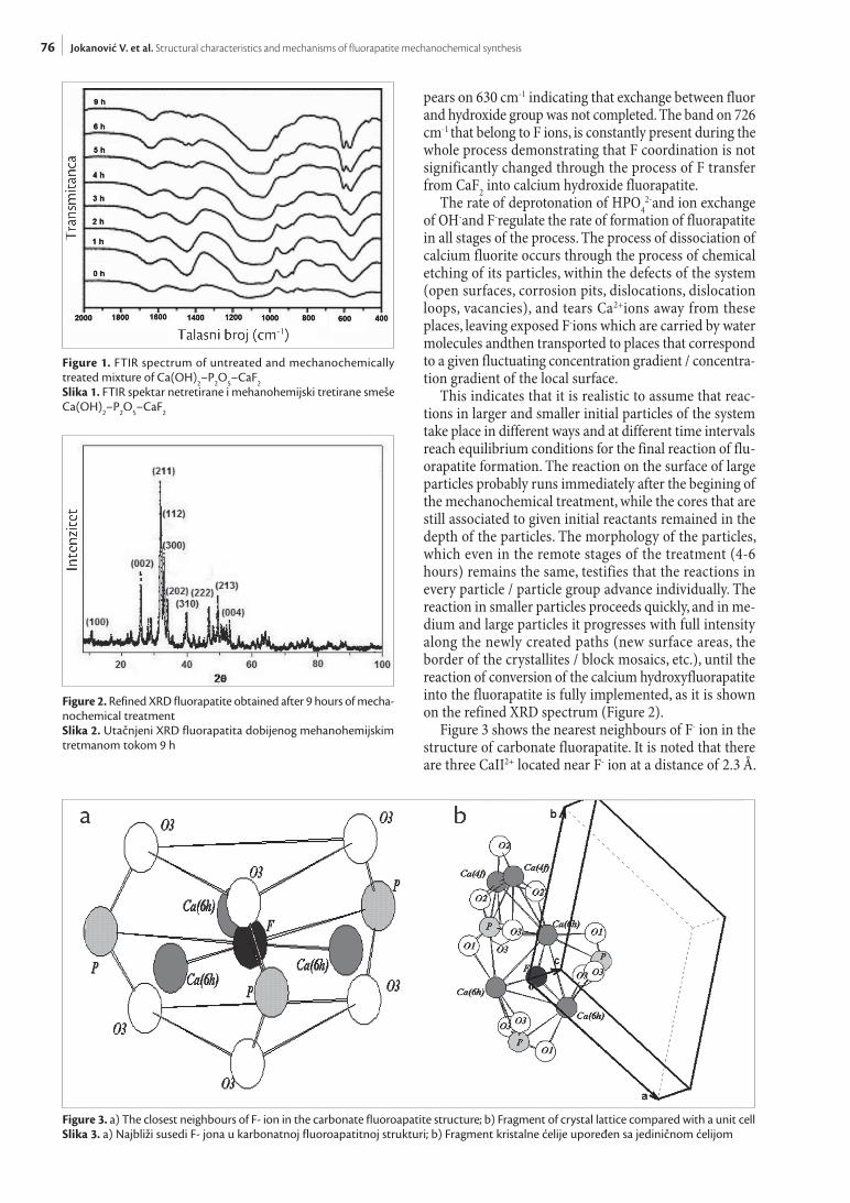
vanish intensively and 5h after completely disappears, while the band on 963 cm-1, appears (carbonate calcium efficienthydroxide fluorapatite) (Figure 1). Simultane-ously, during the whole process, CaF2 dissociates and F ions that enter into reactionare created with calcium de-ficient hydroxide fluorapatite until the formation of its final chemical form. Finally, on previously mechanically treated samples during the period of 6 and 9 hours, and their afterwards thermic treatment on 1100°C, the bands belonging to CO3
2-disappear on 1420 and 1455 cm-1. In samples mechanically treated for 6 hours one band ap-
76 Jokanović V. et al. Structural characteristics and mechanisms of fluorapatite mechanochemical synthesis
pears on 630 cm-1 indicating that exchange between fluor and hydroxide group was not completed. The band on 726 cm-1 that belong to F ions, is constantly present during the whole process demonstrating that F coordination is not significantly changed through the process of F transfer from CaF2 into calcium hydroxide fluorapatite.
The rate of deprotonation of HPO42-and ion exchange
of OH-and F-regulate the rate of formation of fluorapatite in all stages of the process. The process of dissociation of calcium fluorite occurs through the process of chemical etching of its particles, within the defects of the system (open surfaces, corrosion pits, dislocations, dislocation loops, vacancies), and tears Ca2+ions away from these places, leaving exposed F-ions which are carried by water molecules andthen transported to places that correspond to a given fluctuating concentration gradient / concentra-tion gradient of the local surface.
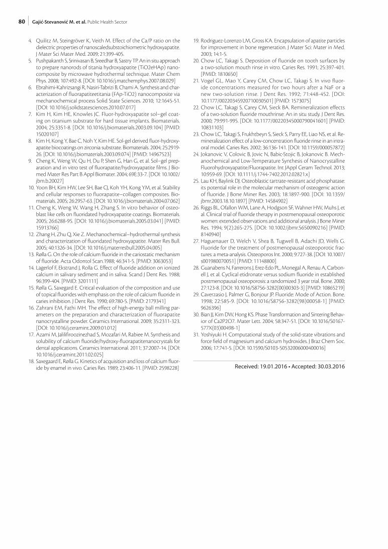
This indicates that it is realistic to assume that reac-tions in larger and smaller initial particles of the system take place in different ways and at different time intervals reach equilibrium conditions for the final reaction of flu-orapatite formation. The reaction on the surface of large particles probably runs immediately after the begining of the mechanochemical treatment, while the cores that are still associated to given initial reactants remained in the depth of the particles. The morphology of the particles, which even in the remote stages of the treatment (4-6 hours) remains the same, testifies that the reactions in every particle / particle group advance individually. The reaction in smaller particles proceeds quickly, and in me-dium and large particles it progresses with full intensity along the newly created paths (new surface areas, the border of the crystallites / block mosaics, etc.), until the reaction of conversion of the calcium hydroxyfluorapatite into the fluorapatite is fully implemented, as it is shown on the refined XRD spectrum (Figure 2).
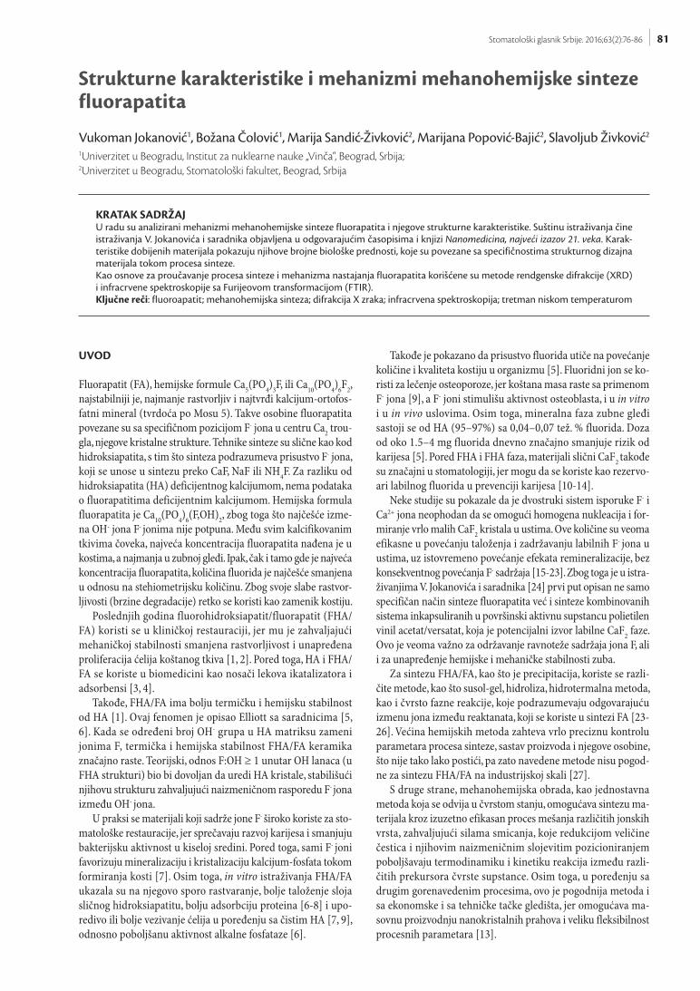
Figure 3 shows the nearest neighbours of F- ion in the structure of carbonate fluorapatite. It is noted that there are three CaII2+ located near F- ion at a distance of 2.3 Å.
Figure 1. FTIR spectrum of untreated and mechanochemically treated mixture of Ca(OH)2–P2O5–CaF2Slika 1. FTIR spektar netretirane i mehanohemijski tretirane smeše Ca(OH)2–P2O5–CaF2
Figure 2. Refined XRD fluorapatite obtained after 9 hours of mecha-nochemical treatmentSlika 2. Utačnjeni XRD fluorapatita dobijenog mehanohemijskim tretmanom tokom 9 h
Figure 3. a) The closest neighbours of F- ion in the carbonate fluoroapatite structure; b) Fragment of crystal lattice compared with a unit cellSlika 3. a) Najbliži susedi F- jona u karbonatnoj fluoroapatitnoj strukturi; b) Fragment kristalne ćelije upoređen sa jediničnom ćelijom

77Stomatološki glasnik Srbije. 2016;63(2):76-86
These CaII2+ ions form the vertices of an equilateral tri-angle with F- ion in the center. There are three CaII2+, P5+, and O2- ions in the second coordination sphere, which mu-tually form the vertices of triangles. The distance between F- and P5+ is 3.6 Å, and between P5+ and O2-(1) is 3.9 Å. There are O2-(3) ions above and below the plane contain-ing F- and CaII2+. The oxygen ions occupy the vertices of a dodecahedron. The distance between F- and O2-(3) ions is 3.1 Å, whereas between P5+ and O2-(3) ions it is 1.5 Å. Figure 2 shows a fragment of the crystal lattice compared with the unit cell. According to the research of Pandaet al.CaII2+ has larger atomic size compared toCaI2+. When OH- ions are substituted with F-, there is greater distortion in the structure due to the larger size of the ionic radius of F-. At the end of the mechanochemical synthesis process, F- ion occupies large space in the center of the latticeform-ing a stable fluorapatite structure.
According to the research of Jokanovicet al. [24] (OH-, F-), in addition to the three types of [OH-], the chain of apa-tite also contains the fourth type with different vibrational energies. It is observed in this study that if the criteria for displacement of free vibration OH- is taken as the criteria for quantifying the changes of OH- with F-, then it is in-dicated that about 50% of OH- groups are modified with F-, while the system, with almost completely changed OH- groups with F- (for pure fluorapatite), provides the value of the wave number of 758 cm-1 (Figure 4 and Table 1).
THE METHOD OF SYNTHESIZING WITH ADDITIONAL LOW-TEMPERATURE THERMAL TREATMENT
Another method of mechanochemical synthesis using the precursorsCa(OH)2, P2O5, NH4F and surfactant of vinyl acetate / versatate, shows that mechanochemical process only can not form fluorapatite. That is why it is necessary to carry out an additional low-temperature treatment.
FTIR method (Figure 5) proved to be the most suitable method for monitoring the synthesis. In order to obtain complete picture of phase transitions that occur in materi-als during mechanochemical and low-temperature treat-ment, the method of X-ray diffraction [23] can be used in addition to FTIR method.
XRD spectra of samples (Figure 6) show very intense peaks of portlandite (P), while β-Ca2P2O7(CP) peaks are also visible. In addition, CaCO3 (C) and CaF2 (CF) peaks are strongly emphasized. All the characteristic diffraction peaks for FA are almost negligible, and some of them ab-sent [29].
After 5 minutes of milling, the most intense peaks be-long to Ca(OH)2, and typical peaks for β-Ca2P2O7, CaF2 and CaCO3 are clearly visible. The peak corresponding to Ca(OH)2 is clearly visible and shows low rate of reac-tion for forming fluorapatite. Consequently, the amount of synthesized FA in the mixture is negligible.
Table 1. Assignment of IR absorption bandsTabela 1. Asignacija IR apsorpcionih traka
Mod
SampleUzorak
FA (t = 6 h) FA (t = 9 h) FA (t = 6 h, 1100 C)
FA (t = 9 h, 1100 C)
ν3(O-H) 3430 3430 3430 3430
2926 2926 2926 2926
δ(O-H) 1626 1626 1626 1626
ν3(CO32-) 1455 1455 - -
ν3(CO32-) 1420 1420 - -
ν3(PO43-) 1092 1092 1093 1093
ν3(PO43-) 1046 1042 1043 1043
ν1(PO43-) 963 962 963 963
ν(F-) 726 726 - 726
νL(O-H) - - 630 -
ν4(PO43-) 603 603 603 603
ν4(PO43-) 571 571 571 571
ν2(PO43-) 473 473 473 473
Figure 4. FTIR spectrum of mechanochemically treated mixture of Ca(OH)2–P2O5–CaF2 for various milling times: a) 6 hours, and b) 9 hours after and before thermal treatmentSlika 4. FTIR spektar mehanohemijski tretirane smeše Ca(OH)2-P2O5-CaF2 za različita vremena mlevenja: a) 6 časova i b) 9 časova posle i pre termičkog tretmana

78
There were identified changes in the structure from amorphous to crystalline, for all thermally treated sam-ples. The typical peaks of FA confirmed transformation that took place in almost all samples (Figure 7). The amount of residual CaCO3 and CaF2 was still significant only in the sample milled for 5 minutes. The emphasized peaks of these phases indicate that parts of the samples remained unchanged, despite the high energy involved in the mechanochemical treatment during the preparation of the precursors mixture (Figure 7a).
As shown in Figure 7 a-c, clearly emphasized and sharp peaks typical for FA are present as a result of an adequate mixing of samples for 2 hours and particularly milling for 8 hours. The sample milled for 5 minutes shows that the transformation of the reaction mixture in flourapatite was only partial, despite the thermal treatment at 550°C for 3 hours. In addition to FA peaks, there are also CaCO3 and CaF2peaks. This proves that even though the inside of the micelle of a surfactant contains the components of the building blocks of precursors phase that can be easily transformed into the pure fluorapatite, they still remain unchanged.
On the contrary, the mixture that was milled for at least 2 hours and additionally thermally treated for 3 hours is
completely transformed into the pure fluorapatite. Similar was for a sample that was milled for 8 hours. The peak cor-responding to FA only moves towards the greater angles of diffraction. This means that the content of OH-groups was reduced during the milling of the sample and that FA finally became predominant phase (possibly mixed with a small amount of hydroxyapatite).
REACTION MECHANISM
During 5 minutes and 2 hours of milling, the reactions in which β- Ca2P2O7 and CaF2 are formed are dominant, while Ca(OH)2part remains unchanged. This has a strong influence on the synthesis rate of FA, which is very slow and cannot be completed only by milling (even after 8 hours of milling). This low rate comes from a very slow diffusion and rearrangement of certain ions that are nec-essary for FA formation. The exchange and incorpora-tion of Ca2+ ions in β- Ca2 (1-x)P2(1-x)O7(1-x) and Ca(1-x)F2(1-x) pre-formed cells is strongly inhibited by the presence of EVA/EVV. Therefore, in order to provide bigger reaction rate of formation of FA, crystal structure of β- Ca2 (1-x)P2(1-x)O7(1-x) and Ca(1-x)F2(1-x) must firstly be transformed to amor-phous by additional milling (2 – 8 hours). This procedure provides good mixing, which reduces the diffusion paths of different ions.
The second stage of the formation of FA started after low temperature treatment of the samples at 550°C for 3 hours. In this step, according to certain researches, the reactions can be initiated on the surface of the dominant phase β- Ca2P2O7 through the surface diffusion of addi-tional Ca2+ and F-ions in its volume. According to the dif-fraction peaks, it is evident that the process of formation of FA during these thermal treatments is very intense. The only exception was a sample milled for 5 minutes. Despite conducted thermal treatment, short milling time was not
Jokanović V. et al. Structural characteristics and mechanisms of fluorapatite mechanochemical synthesis
Figure 5. FTIR spectrum of Ca(OH)2-P2O5-NH4F mechanically treat-ed mixture: 1 – 5 minutes, 2 – 20 minutes, 3 – 35 minutes, 4 – 1 hours, 5 – 2 hours, 6 – 3 hours, 7 – 4 hours, 8 – 6 hours, 9 – 8 hours, A-C – thermally treatedSlika 5. FTIR spektar Ca(OH)2-P2O5-NH4F smeše mehanički treti-rane: 1 – 5 min., 2 – 20 min., 3 – 35 min., 4 – 1 h, 5 – 2 h, 6 – 3 h, 7 – 4 h, 8 – 6 h, 9 – 8 h, A-C – termički tretirane
Figure 6. XRD spectrum of Ca(OH)2-P2O5-NH4F mechanically treat-ed mixture: 1 – 5 minutes, 2 – 20 minutes, 3 – 35 minutes, 4 – 1 hours, 5 – 2 hours, 6 – 3 hours, 7 – 4 hours, 8 – 6 hours, 9 – 8 hoursSlika 6. XRD spektar Ca(OH)2-P2O5-NH4F smeše mehanički treti-rane: 1 – 5 min., 2 – 20 min., 3 – 35 min., 4 – 1 h, 5 – 2 h, 6 – 3 h, 7 – 4 h, 8 – 6 h, 9 – 8 h

79Stomatološki glasnik Srbije. 2016;63(2):76-86
enough to obtain a single-phase system (FA). However, the milling of at least 2 hours led to the formation of pure FA phase. The presence of almost negligible peaktypical for β- Ca2P2O7, proves that this milling time is the border time for obtainingthe single-phase FA.
It is important to emphasize that, no matter how long the samples were milled; this reaction wouldn’t be pos-sible without the thermal treatment. With the extended milling time (up to 3 hours or more), the system became amorphous. The reaction did not progress even during the longest milling time (8 hours), which was confirmed by XRD. The progress was observed only in the amorphous samples (milled for at least 2 hours), which were subjected to subsequent thermal treatment. The mechanism of the process, which took place during milling, was possibly significantly activated by the water present in Ca(OH)2. The explanation provided by some researchers shows that smaller Ca(OH)2 particles, during milling under the influence of shearing forces, tend to grow, causing further disintegration [29-31], so that the exchange of different ion species becomes more efficient.
During milling, the distortion of Ca2+-O polyhedra is much more prominent in comparison with P5+-O tetra-hedra. The distortion led to displacement of cations from the center of its coordination sphere. It has a strong im-pact on the diffusion rate of the remaining amount of Ca2+ and F- ions and consequent destruction of β-Ca2P2O7 during milling. Thus, the “empty” space in β-Ca2P2O7is increasinglyfilled with these ions, untilβ-Ca2P2O7 cell is completely destroyed. It should be noted that the capacity of dissolution of Ca2+ and F- ions within β-Ca2P2O7 is very high. Accordingly, the smaller size of CaF2, and especially Ca(OH)2 crystallites is suitable for further propagation reaction of the formation of FA. Although crystallization of amorphous phase cannot be achieved through milling, the distribution of ions provides very rapid crystallization of the samples in FA in the next step of a very low thermal treatment (at 550 °C for 3 hours).
Therefore, it seems that there is enough space within β-Ca2P2O7 structure for F- anions to be placed in large gaps, and within the network of calcium and phosphate ions. In addition to F- ions, very small CaF2 nanoparticles are placed randomly in the gaps within β-Ca2P2O7 lattices,
which produce significant changes in its symmetry, caus-ing the corresponding chemical changes responsible for the transformation of mixture into fluorapatite during the next thermal treatment. This treatment can significantly accelerate the processes of diffusion, causing degradation of EVA/EVV micellar cages and supporting small ion re-distribution by distortion in β-Ca2P2O7 structure induced by shear forces, until the final transformation of the mix-ture into fluorapatite.
CONCLUSION
Mechanochemical process of fluorapatite synthesis is based on the use of two kinds of precursors: calcium hydroxide, phosphorus pentoxide and calcium fluoride, or calcium hydroxide, phosphorus pentoxide and am-monium fluoride with the addition of surfactant vinyl acetate/verstat. On the basis of XRD and FTIR analysis it was observed that fluorapatite has significant advantages in comparison with hydroxyapatite. These benefits are re-lated to its greater stability, lower solubility and especially better protection against cavities.
APPRECIATION
This work was supported by the Ministry of Education, Science and Technological Development of the Republic of Serbia (Project no. 172026).
REFERENCES
1. Jokanović V. Monografija: Nanomedicina, najveći izazov 21. veka, DATA STATUS, 2012.
2. Barinov SM, Shvornevab LI, Ferroc D, Fadeevaa IV, Tumanov SV. Solid solution formation at the sintering of hydroxyapatite/fluorapatite ceramics. Sci Technol Adv Mater. 2004; 5:537-41. [DOI: 10.1016/j.stam.2004.02.012]
3. Nikcevic I, Jokanovic V, Mitric M, Nedic Z, Makovec D, Uskokovic D. Mechanochemical synthesis of nanostructured fluorapatite/fluorhy-droxyapatite and carbonated fluorapatite/fluorhydroxyapatite. J Solid State Chem. 2004; 177:2565-74. [DOI: 10.1016/j.jssc.2004.03.024]
Figure 7. XRD spectra of Ca(OH)2-P2O5-NH4F mixture after different milling times: a) 5 minutes, b) 2 hours, c) 8 hoursSlika 7. XRD spektri Ca(OH)2-P2O5-NH4F smeše nakon različitih vremena mlevenja: a) 5 min., b) 2 h, c) 8 h
80 Gajić-Stevanović M. et al. Public Health Sector
4. Quilitz M, Steingröver K, Veith M. Effect of the Ca/P ratio on the dielectric properties of nanoscaledsubstoichiometric hydroxyapatite. J Mater Sci Mater Med. 2009; 21:399-405.
5. Pushpakanth S, Srinivasan B, Sreedhar B, Sastry TP. An in situ approach to prepare nanorods of titania hydroxyapatite (TiO2eHAp) nano-composite by microwave hydrothermal technique. Mater Chem Phys. 2008; 107:492-8. [DOI: 10.1016/j.matchemphys.2007.08.029]
6. Ebrahimi-Kahrizsangi R, Nasiri-Tabrizi B, Chami A. Synthesis and char-acterization of fluorapatiteetitania (FAp-TiO2) nanocomposite via mechanochemical process Solid State Sciences. 2010; 12:1645-51. [DOI: 10.1016/j.solidstatesciences.2010.07.017]
7. Kim H, Kim HE, Knowles JC. Fluor-hydroxyapatite sol–gel coat-ing on titanium substrate for hard tissue implants. Biomaterials. 2004; 25:3351-8. [DOI: 10.1016/j.biomaterials.2003.09.104] [PMID: 15020107]
8. Kim H, Kong Y, Bae C, Noh Y, Kim HE. Sol-gel derived fluor-hydroxy-apatite biocoatings on zirconia substrate. Biomaterials. 2004; 25:2919-26. [DOI: 10.1016/j.biomaterials.2003.09.074] [PMID: 14967523]
9. Cheng K, Weng W, Qu H, Du P, Shen G, Han G, et al. Sol–gel prep-aration and in vitro test of fluorapatite/hydroxyapatite films. J Bio-med Mater Res Part B Appl Biomater. 2004; 69E:33-7. [DOI: 10.1002/jbm.b.20027]
10. Yoon BH, Kim HW, Lee SH, Bae CJ, Koh YH, Kong YM, et al. Stability and cellular responses to fluorapatite–collagen composites. Bio-materials. 2005; 26:2957-63. [DOI: 10.1016/j.biomaterials.2004.07.062]
11. Cheng K, Weng W, Wang H, Zhang S. In vitro behavior of osteo-blast like cells on fluoridated hydroxyapatite coatings. Biomaterials. 2005; 26:6288-95. [DOI: 10.1016/j.biomaterials.2005.03.041] [PMID: 15913766]
12. Zhang H, Zhu Q, Xie Z. Mechanochemical–hydrothermal synthesis and characterization of fluoridated hydroxyapatite. Mater Res Bull. 2005; 40:1326-34. [DOI: 10.1016/j.materresbull.2005.04.005]
13. Rølla G. On the role of calcium fluoride in the cariostatic mechanism of fluoride. Acta Odontol Scan.1988; 46:341-5. [PMID: 3063053]
14. Lagerlof F, Ekstrand J, Rolla G. Effect of fluoride addition on ionized calcium in salivary sediment and in saliva. Scand J Dent Res. 1988; 96:399-404. [PMID: 3201111]
15. Rølla G, Saxegard E. Critical evaluation of the composition and use of topical fluorides with emphasis on the role of calcium fluoride in caries inhibition. J Dent Res. 1990; 69:780-5. [PMID: 2179341]
16. Zahrani EM, Fathi MH. The effect of high-energy ball milling par-ameters on the preparation and characterization of fluorapatite nanocrystalline powder. Ceramics International. 2009; 35:2311-323. [DOI: 10.1016/j.ceramint.2009.01.012]
17. Azami M, Jalilifiroozinezhad S, Mozafari M, Rabiee M. Synthesis and solubility of calcium fluoride/hydroxy-fluorapatitenanocrystals for dental applications. Ceramics International. 2011; 37:2007-14. [DOI: 10.1016/j.ceramint.2011.02.025]
18. Saxegaard E, Rølla G. Kinetics of acquisition and loss of calcium fluor-ide by enamel in vivo. Caries Res. 1989; 23:406-11. [PMID: 2598228]
19. Rodriguez-Lorenzo LM, Gross KA. Encapsulation of apatite particles for improvement in bone regeneration. J Mater Sci: Mater in Med. 2003; 14:1-5.
20. Chow LC, Takagi S. Deposition of fluoride on tooth surfaces by a two-solution mouth rinse in vitro. Caries Res. 1991; 25:397-401. [PMID: 1810650]
21. Vogel GL, Mao Y, Carey CM, Chow LC, Takagi S. In vivo fluor-ide concentrations measured for two hours after a NaF or a new two-solution rinse. J Dent Res. 1992; 71:448-452. [DOI: 10.1177/00220345920710030501] [PMID: 1573075]
22. Chow LC, Takagi S, Carey CM, Sieck BA. Remineralization effects of a two-solution fluoride mouthrinse: An in situ study. J Dent Res. 2000; 79:991-995. [DOI: 10.1177/00220345000790041601] [PMID: 10831103]
23. Chow LC, Takagi S, Frukhtbeyn S, Sieck S, Parry EE, Liao NS, et al. Re-mineralization effect of a low-concentration fluoride rinse in an intra-oral model. Caries Res. 2002; 36:136-141. [DOI: 10.1159/000057872]
24. Jokanovic V, Colovic B, Jovic N, Babic-Stojic B, Jokanovic B. Mech-anochemical and Low-Temperature Synthesis of Nanocrystalline Fluorohydroxyapatite/Fluorapatite. Int JAppl Ceram Technol. 2013; 10:959-69. [DOI: 10.1111/j.1744-7402.2012.02821.x]
25. Lau KH, Baylink DJ. Osteoblastic tartrate-resistant acid phosphatase: its potential role in the molecular mechanism of osteogenic action of fluoride. J Bone Miner Res. 2003; 18:1897-900. [DOI: 10.1359/jbmr.2003.18.10.1897] [PMID: 14584902]
26. Riggs BL, Ofallon WM, Lane A, Hodgson SF, Wahner HW, Muhs J, et al. Clinical trial of fluoride therapy in postmenopausal osteoporotic women: extended observations and additional analysis. J Bone Miner Res. 1994; 9(2):265-275. [DOI: 10.1002/jbmr.5650090216] [PMID: 8140940]
27. Haguenauer D, Welch V, Shea B, Tugwell B, Adachi JD, Wells G. Fluoride for the treatment of postmenopausal osteoporotic frac-tures: a meta-analysis. Osteoporos Int. 2000; 9:727-38. [DOI: 10.1007/s001980070051] [PMID: 11148800]
28. Guanabens N, Farrerons J, Erez-Edo PL, Monegal A, Renau A, Carbon-ell J, et al. Cyclical etidronate versus sodium fluoride in established postmenopausal osteoporosis: a randomized 3 year trial. Bone. 2000; 27:123-8. [DOI: 10.1016/S8756-3282(00)00303-3] [PMID: 10865219]
29. Caverzasio J, Palmer G, Bonjour JP. Fluoride: Mode of Action. Bone. 1998; 22:585-9. [DOI: 10.1016/S8756-3282(98)00058-1] [PMID: 9626396]
30. Bian JJ, Kim DW, Hong KS. Phase Transformation and Sintering Behav-ior of Ca2P2O7. Mater Lett. 2004; 58:347-51. [DOI: 10.1016/S0167-577X(03)00498-1]
31. Yoshiyuki H. Computational study of the solid-state vibrations and force field of magnesium and calcium hydroxides. J Braz Chem Soc. 2006; 17:741-5. [DOI: 10.1590/S0103-50532006000400016]
Received: 19.01.2016 • Accepted: 30.03.2016
81Stomatološki glasnik Srbije. 2016;63(2):76-86
Strukturne karakteristike i mehanizmi mehanohemijske sinteze fluorapatita
Vukoman Jokanović1, Božana Čolović1, Marija Sandić-Živković2, Marijana Popović-Bajić2, Slavoljub Živković2
1Univerzitet u Beogradu, Institut za nuklearne nauke „Vinča“, Beograd, Srbija;2Univerzitet u Beogradu, Stomatološki fakultet, Beograd, Srbija
KRATAK SADRŽAJU radu su analizirani mehanizmi mehanohemijske sinteze fluorapatita i njegove strukturne karakteristike. Suštinu istraživanja čine istraživanja V. Jokanovića i saradnika objavljena u odgovarajućim časopisima i knjizi Nanomedicina, najveći izazov 21. veka. Karak-teristike dobijenih materijala pokazuju njihove brojne biološke prednosti, koje su povezane sa specifičnostima strukturnog dizajna materijala tokom procesa sinteze.Kao osnove za proučavanje procesa sinteze i mehanizma nastajanja fluorapatita korišćene su metode rendgenske difrakcije (XRD) i infracrvene spektroskopije sa Furijeovom transformacijom (FTIR). Ključne reči: fluoroapatit; mehanohemijska sinteza; difrakcija X zraka; infracrvena spektroskopija; tretman niskom temperaturom
UVOD
Fluorapatit (FA), hemijske formule Ca5(PO4)3F, ili Ca10(PO4)6F2, najstabilniji je, najmanje rastvorljiv i najtvrđi kalcijum-ortofos-fatni mineral (tvrdoća po Mosu 5). Takve osobine fluorapatita povezane su sa specifičnom pozicijom F- jona u centru Ca2 trou-gla, njegove kristalne strukture. Tehnike sinteze su slične kao kod hidroksiapatita, s tim što sinteza podrazumeva prisustvo F- jona, koji se unose u sintezu preko CaF, NaF ili NH4F. Za razliku od hidroksiapatita (HA) deficijentnog kalcijumom, nema podataka o fluorapatitima deficijentnim kalcijumom. Hemijska formula fluorapatita je Ca10(PO4)6(F,OH)2, zbog toga što najčešće izme-na OH- jona F- jonima nije potpuna. Među svim kalcifikovanim tkivima čoveka, najveća koncentracija fluorapatita nađena je u kostima, a najmanja u zubnoj gleđi. Ipak, čak i tamo gde je najveća koncentracija fluorapatita, količina fluorida je najčešće smanjena u odnosu na stehiometrijsku količinu. Zbog svoje slabe rastvor-ljivosti (brzine degradacije) retko se koristi kao zamenik kostiju.
Poslednjih godina fluorohidroksiapatit/fluorapatit (FHA/FA) koristi se u kliničkoj restauraciji, jer mu je zahvaljajući mehaničkoj stabilnosti smanjena rastvorljivost i unapređena proliferacija ćelija koštanog tkiva [1, 2]. Pored toga, HA i FHA/FA se koriste u biomedicini kao nosači lekova ikatalizatora i adsorbensi [3, 4].
Takođe, FHA/FA ima bolju termičku i hemijsku stabilnost od HA [1]. Ovaj fenomen je opisao Elliott sa saradnicima [5, 6]. Kada se određeni broj OH- grupa u HA matriksu zameni jonima F, termička i hemijska stabilnost FHA/FA keramika značajno raste. Teorijski, odnos F:OH ≥ 1 unutar OH lanaca (u FHA strukturi) bio bi dovoljan da uredi HA kristale, stabilišući njihovu strukturu zahvaljujući naizmeničnom rasporedu F- jona između OH- jona.
U praksi se materijali koji sadrže jone F- široko koriste za sto-matološke restauracije, jer sprečavaju razvoj karijesa i smanjuju bakterijsku aktivnost u kiseloj sredini. Pored toga, sami F- joni favorizuju mineralizaciju i kristalizaciju kalcijum-fosfata tokom formiranja kosti [7]. Osim toga, in vitro istraživanja FHA/FA ukazala su na njegovo sporo rastvaranje, bolje taloženje sloja sličnog hidroksiapatitu, bolju adsorbciju proteina [6-8] i upo-redivo ili bolje vezivanje ćelija u poređenju sa čistim HA [7, 9], odnosno poboljšanu aktivnost alkalne fosfataze [6].
Takođe je pokazano da prisustvo fluorida utiče na povećanje količine i kvaliteta kostiju u organizmu [5]. Fluoridni jon se ko-risti za lečenje osteoporoze, jer koštana masa raste sa primenom F- jona [9], a F- joni stimulišu aktivnost osteoblasta, i u in vitro i u in vivo uslovima. Osim toga, mineralna faza zubne gleđi sastoji se od HA (95–97%) sa 0,04–0,07 tež. % fluorida. Doza od oko 1.5–4 mg fluorida dnevno značajno smanjuje rizik od karijesa [5]. Pored FHA i FHA faza, materijali slični CaF2 takođe su značajni u stomatologiji, jer mogu da se koriste kao rezervo-ari labilnog fluorida u prevenciji karijesa [10-14].
Neke studije su pokazale da je dvostruki sistem isporuke F- i Ca2+ jona neophodan da se omogući homogena nukleacija i for-miranje vrlo malih CaF2 kristala u ustima. Ove količine su veoma efikasne u povećanju taloženja i zadržavanju labilnih F- jona u ustima, uz istovremeno povećanje efekata remineralizacije, bez konsekventnog povećanja F- sadržaja [15-23]. Zbog toga je u istra-živanjima V. Jokanovića i saradnika [24] prvi put opisan ne samo specifičan način sinteze fluorapatita već i sinteze kombinovanih sistema inkapsuliranih u površinski aktivnu supstancu polietilen vinil acetat/versatat, koja je potencijalni izvor labilne CaF2 faze. Ovo je veoma važno za održavanje ravnoteže sadržaja jona F, ali i za unapređenje hemijske i mehaničke stabilnosti zuba.
Za sintezu FHA/FA, kao što je precipitacija, koriste se razli-čite metode, kao što susol-gel, hidroliza, hidrotermalna metoda, kao i čvrsto fazne reakcije, koje podrazumevaju odgovarajuću izmenu jona između reaktanata, koji se koriste u sintezi FA [23-26]. Većina hemijskih metoda zahteva vrlo preciznu kontrolu parametara procesa sinteze, sastav proizvoda i njegove osobine, što nije tako lako postići, pa zato navedene metode nisu pogod-ne za sintezu FHA/FA na industrijskoj skali [27].
S druge strane, mehanohemijska obrada, kao jednostavna metoda koja se odvija u čvrstom stanju, omogućava sintezu ma-terijala kroz izuzetno efikasan proces mešanja različitih jonskih vrsta, zahvaljujući silama smicanja, koje redukcijom veličine čestica i njihovim naizmeničnim slojevitim pozicioniranjem poboljšavaju termodinamiku i kinetiku reakcija između razli-čitih prekursora čvrste supstance. Osim toga, u poređenju sa drugim gorenavedenim procesima, ovo je pogodnija metoda i sa ekonomske i sa tehničke tačke gledišta, jer omogućava ma-sovnu proizvodnju nanokristalnih prahova i veliku fleksibilnost procesnih parametara [13].

82 Jokanović V. et al. Structural characteristics and mechanisms of fluorapatite mechanochemical synthesis
Zbog toga, cilj ovog rada je bio da se predstavi suština me-tode sinteze nanopraha fluorhidroksiapatita/fluorapatita me-todom mehaničkog legiranja.
Parametri mlevenja, kao što su brzina rotacije, prečnik i broj kugli, maseni odnos kugle–prah, bili su konstantni, dok je uti-caj vremena mlevenja na sastav faze veoma pažljivo određen. Kinetika i mehanizam reakcije za dobijanje FHA/FA i druge prelazne faze su ispitivani koristeći XRD i FTIR spektroskopiju.
MEHANIZMI SINTEZE FLUORAPATITA
Tipični mehanizmi sinteze fluorapatita prikazani su na primeru najčešće primenjivanih postupaka mehanohemijske sinteze iz najčešće korišćenih prekursora kao što su kalcijum-hidroksid – Ca(OH)2, fosfor-pentoksid – P2O5 i kalcijum-fluorid – CaF2 (sinteza 1) i kalcijum-hidroksid – Ca(OH)2, fosfor-pen-toksid – P2O5, i amonijum-fluorid – NH4F, uz dodatak površin-ski aktivne supstance vinil acetat/versatata (EVA/AVV) (sinteza 2). Oba mehanizma odvijaju se kroz niz procesnih faza, koje je moguće pratiti na adekvatan način prvenstveno primenom IR spektroskopije i rendgenske difrakcije [24]. Na osnovu rezultata izvedenih analiza uočeno je da svaku fazu prati određeni stepen transformacije polaznih reaktanata u fluorhidroksiapatit, sa sve manjim i manjim udelima OH- grupe i sve većim i većim ude-lima jona F- umesto OH- grupe, sve do potpune transformacije sistema u fluorpatit.
Reakcija se u osnovi odvija sa nepotpunom stehiometrijom, pri čemu se vrednost x, koja definiše odstupanje od potpune simetrije, nalazi u intervalu x1<x<xk,
Ca10(PO4)1-y(CO3)y(PO4)5 (OH)2-2x1 (F)2x1 +(2-2x1)HF →Ca10(PO4)1-y(CO3)y (PO4)5(OH)2-xkFxk + (2-xk) HF + H2O;
pri čemu xk može imati maksimalnu vrednost 1, a x1 minimalnu vrednost 0.
Dakle, sumarna reakcija, kod reakcije sa potpunom stehio-metrijom, imala bi oblik:
9 Ca(OH)2-2y(CO3)y + 3P2O5 + CaF2 → Ca10(PO4)1-y(CO3)y (PO4)5F2 + 9H2O; x=1
Na osnovu rendgenske difrakcije utvrđeno je da je već posle 1 h mehanohemijskog tretmana došlo do amorfizacije sistema/mešavine.
Naime, zbog izuzetno visoke koncentracije mehaničkih napre-zanja na veoma maloj površini kontakta (kontakt koji se ostvaru-je pri međusobnom sudaru kugli ili sudaru kugli sa površinom unutrašnje obloge), ostvaruju se uslovi za nastajanje visokih smicajnih naprezanja na relativno maloj površini kontakta. Pri tome veličina napona deformacije zavisi prvenstveno od prečnika kugli koje se koriste kod mehanohemijskog tretmana (veličina kontaktne kalote utiskivanja deformacije pri sudaru) i brzine sudara. Istovremeno sa procesom prenosa deformacije dolazi do mehaničke aktivacije sistema i visokoelastičnog toka, koji prati intenzivne hemijske i fazne promene unutar materijala (reakcije izmene, mešanja jonskih vrsta, nastajanja novih faza itd.). Ove promene mogu biti takve da materijal tokom relaksacije delimič-no trpi povratnu deformaciju (viskoelastični tok), ili su pak u ce-losti nepovratne, kada dominira mehanizam puzanja materijala.
Napon kritične deformacije zavisi od vremena izlaganja si-stema deformaciji (broja udara kugli), odnosno broja ciklusa
opterećenja, tako da vremenom napon koji izaziva kritičnu deformaciju razaranja/formiranja pukotina i novih površina ima sve manju i manju vrednost, što vodi sve većoj amorfizaciji sistema. Proces mehaničke aktivacije u sistemima u kojima kao reakcioni produkt nastaje voda dodatno je ubrzan time što je olakšan transport odgovarajućih jonskih vrsta na mesta koja odgovaraju minimumu slobodne energije sistema.
U ovom slučaju, zbog izuzetne hidrofilnosti P2O5 odmah po njegovom dodavanju ostatku reakcione smeše dolazi do nastan-ka fosforne kiseline (P2O5+ 3H2O→2H3PO4), koja potom reagu-je sa Ca(OH)2-2y(CO3)y i nastaje Ca(HPO4)1-y(CO3)y (karbonatni kalcijum-hidrogenfosfat).
Nakon 4 h mlevenja karakteristične trake za HPO42- počinju
intenzivno da iščezavaju, a nakon 5 h potpuno iščezavaju, a po-javljuje se traka na 963 cm-1 (karbonatni kalcijum-deficijentni hidroksifluorapatit) (Slika 1). Istovremeno tokom celog procesa dolazi i do disocijacije CaF2, pri čemu nastaju joni F- koji tokom celog procesa ulaze u reakciju sa deficijentnim kalcijum-hidrok-sifluorapatitom sve do formiranja njegove konačne hemijske forme. Napokon, na uzorcima koji su termički tretirani na 1100°C, a prethodno mehanički tretirani 6 i 9 h iščezavaju tra-ke na 1420 i 1455 cm-1, koje pripadaju CO3
2-, a kod uzorka koji je mehanički tretiran 6 h pojavljuje se traka na 630 cm-1, koja govori da nije došlo do kraja reakcije izmene fluora i hidrok-silne grupe. Traka na 726 cm-1 pripada jonima F i konstantno je prisutna tokom celokupnog procesa, što govori o tome da se kordinacija F- ne menja bitno kroz sam proces transfera F- iz CaF2 u kalcijum-hidroksifluorapatit.
Brzina deprotonizacije HPO42- i jonske izmene OH- i F- re-
gulišu brzinu nastajanja fluorapatita u svim fazama procesa. Proces disocijacije kalcijum-fluorita odvija se kroz proces he-mijskog nagrizanja njegovih čestica, u okviru defekata sistema (otvorene površine, jamice nagrizanja, dislokacije, dislokacij-ske petlje, vakancije), čime se otkidaju joni Ca2+ sa tih mesta ostavljajući ogoljene jone F-
, koji se tek u tom trenutku nošeni molekulima vode transportuju na mesta koja odgovaraju datom fluktuacionom koncentracionom gradijentu / koncentracionom gradijentu lokalne površine.
To ukazuje da je realno pretpostaviti da se reakcija u većim i manjim polaznim česticama sistema odvija na različit način i da u različitim vremenskim intervalima doseže ravnotežne uslove za krajnju reakciju nastajanja fluorapatita. U velikim če-sticama verovatno reakcija na površini teče odmah po početku mehanohemijskog tretmana, dok u dubini čestica ostaju jezgra koja su još uvek pripadajuća datim polaznim reaktantima. Sama morfologija čestica, koja i u udaljenim fazama tretmana (4–6 h) ostaje ista, svedoči o tome da reakcija u svakoj čestici / grupi čestica napreduje na individualan način. U manjim česticama reakcija se odvija brzo, u srednjim česticama i posebno u ve-likim ona napreduje punim intenzitetom duž novootvorenih puteva (novih površina, granica kristalita / blok mozaika itd.), sve dok se u celosti ne realizuje reakcija transformacije kalci-jum-hidroksifluorapatita u fluorapatit, kao što je pokazano na utačnjenom XRD spektru (Slika 2).
Slika 3. pokazuje najbliže susede F- jona u strukturi karbo-natnog fluorapatita. Uočava se da se tri CaII2+ nalaze u nepo-srednoj blizini F- jona na rastojanju od 2,3 Å. Ovi CaII2+ joni for-miraju temena jednakostraničnog trougla sa F- jonom u centru. U drugoj koordinacionoj sferi nalaze se po tri CaII2+, P5+ i O2- jona koja međusobno formiraju temena trouglova. Rastojanje

83Stomatološki glasnik Srbije. 2016;63(2):76-86
između F- i P5+ je 3,6 Å, a između P5+ i O2-(1) je 3,9 Å. O2-(3) jona nalaze se ispod i iznad ravni koja sadrži F- i CaII2+. Kiseonični joni zauzimaju temena dodekaedrona. Rastojanje između F- i O2-(3) jona je 3,1 Å, dok je između P5+ i O2-(3) jona 1,5 Å. Slika 2b pokazuje fragment kristalne rešetke upoređen sa jediničnom ćelijom. Saglasno istraživanjima Pande i njegovih saradnika, CaII2+ ima veću atomsku veličinu nego CaI2+. Kada su OH- joni supstituisani sa F-
, dolazi do veće distorzije u strukturi zbog veće veličine jonskog radijusa F-. Na kraju mehanohemijskog procesa sinteze F- jon zauzima veliki prostor u centru rešetke, formirajući na taj način stabilnu fluorapatitnu strukturu.
Saglasno istraživanjima V. Jokanovića i saradnika [24] (OH-, F-), lanac apatita sadrži pored tri tipa [OH-] i četvrti tip sa razli-čitim vibracionim energijama. U ovim istraživanjima uočeno je da, ako se kao kriterijum kvantifikacije izmene OH- sa F- uzme kriterijum pomeranja slobodne vibracije OH-, sledi da je oko 50% OH- grupa izmenjeno sa F-, dok je kod sistema sa skoro potpuno izmenjenim OH- grupama sa F- (kod čistog fluorapa-tita) vrednost talasnog broja od 758 cm-1 (Slika 4 i tabela 1).
POSTUPAK SINTEZE SA DODATNIM NISKOTEMPERATURNIM TERMIČKIM TRETMANOM
Drugi postupak mehanohemijske sinteze, koji je izveden pri-menom prekursora Ca(OH)2, P2O5, NH4F i površinski aktivne supstance vinil acetata/versatata, pokazuje da samo u meha-nohemijskom postupku nije moguće dobiti fluorapatit, nego da je za tu svrhu potrebno izvesti dodatni niskotemperaturni tretman.
Kao najadekvatnija metoda za praćenje same sinteze pokaza-la se kao i u prethodnom primeru FTIR metoda (Slika 5).Radi dobijanja kompletne slike o faznim prelazima koji se događaju u materijalu tokom mehanohemijskog i niskotemperaturnog tretmana, pored FTIR metode korišćena je i metoda rendgenske difrakcije, koje su detaljno opisane u referenci 23.
XRD spektri uzoraka (Slika 6) pokazuju vrlo intenzivne pi-kove portlandita (P), dok su pikovi β-Ca2P2O7(CP) takođe vidlji-vi. Osim toga, veoma su naglašeni i pikovi CaCO3(C) i CaF2(CF). Svi karakteristični difrakcioni pikovi za FA su gotovo zanemar-ljivi, a neki od njih su odsutni [29].
Posle pet minuta mlevenja, najintenzivniji vrhovi pripadaju Ca(OH)2, a pikovi karakteristični za β-Ca2P2O7, CaF2 i CaCO3 jasno su vidljivi. Pik koji odgovara Ca(OH)2 je jasno vidljiv i pokazuje nisku brzinu reakcije formiranja fluorapatita. Usled toga, količina sintetisanog FA u smeši je zanemarljiva.
Za sve termički tretirane uzorke identifikovane su promene u strukturi od amorfne do kristalinične. Karakteristični pikovi FA potvrđuju transformacije koji su se desile u gotovo svim uzorcima (Slika 7). Samo u uzorku mlevenom pet minuta količi-na rezidualnog CaCO3 i CaF2 je bila još značajna. Izraženi pikovi ovih faza ukazuju da su delovi uzoraka ostali nepromenjeni, pored visoke energije uključene u mehanohemijski tretman prilikom pripreme smeše prekursora (Slika 7a).
Kao što je prikazano na slici 7a–c, vrlo izraženi i oštri vrhovi karakteristični za FA prisutni su kao posledica adekvatnog me-šanja uzorka u toku 2 h i posebno 8 h mlevenja. Uzorak mleven pet minuta pokazuje da je transformacija reakcione smeše u fluorapatit samo delimična, uprkos termičkom tretmanu na 550°C u toku 3 h. Osim pikova FA, javljaju se i pikovi CaCO3 i
CaF2. Ovo je dokaz da iako se unutar micela površinski aktivne supstance delovi gradivnih blokova faza prekursora mogu lako transformisati u čisti fluorapatit, oni ipak ostaju nepromenjeni.
Naprotiv, smeša mlevena najmanje 2 h i dodatno termički tretirana 3 h potpuno je transformisana u čist fluorapatit. Slično je primećeno za uzorak mleven 8 h. Pik koji odgovara FA samo se pomera ka većim uglovima difrakcije. To znači da je sadržaj OH-grupa smanjen tokom mlevenja uzorka i da je FA postao konačno prevladavajuća faza (pomešan verovatno sa malom količinom hidroksiapatita).
MEHANIZAM REKACIJE
Tokom mlevenja od pet minuta i 2 h, reakcije u kojima na-staju β- Ca2P2O7 i CaF2 su dominante, a deo Ca(OH)2 ostaje ne-promenjen. Ovo ima snažan uticaj na brzinu sinteze FA, koja se odvija veoma sporo i ne može biti završena samo mlevenjem (čak i posle 8 h mlevenja). Ova niska brzina potiče od jako spore difuzije i preuređenja određenih jonskih vrsta, koje su neophod-ne za formiranje FA. Razmena i inkorporacija Ca2+ jona u pret-hodno formirane ćelije β- Ca2 (1-x)P2(1-x)O7(1-x) iCa(1-x)F2(1-x) snažno je inhibirana prisustvom EVA/EVV. Zbog toga, za veću brzinu reakcije formiranja FA, kristalna struktura β- Ca2 (1-x)P2(1-x)O7(1-x) iCa(1-x)F2(1-x) mora biti prvo transformisana u amorfnu dodatnim mlevenjem (2–8 h). Ova procedura omogućava dobro mešanje, čime se umanjuju difuzioni putevi različitih jonskih vrsta.
Druga faza formiranja FA počela je nakon niskotemperatur-nog tretmana uzoraka na 550°C tokom 3 h. Tokom ovog koraka, prema nekim istraživanjima, reakcije mogu biti pokrenute na površini dominantne faze β- Ca2P2O7 kroz površinsku difuziju dodatnih Ca2+ i F- jona u njenu zapreminu. Prema difrakcionim pikovima, evidentno je da je proces formiranja FA tokom tih termičkih tretmana veoma intenzivan. Jedini izuzetak bio je uzorak mleven pet minuta. Uprkos primenjenom termičkom tretmanu, tako kratko vreme mlevenja nije bilo dovoljno da se dobije monofazni sistem (FA). Međutim, mlevenje od najma-nje 2 h dovelo je do formiranja čiste FA faze. Prisustvo gotovo zanemarljivog pika karakteristnog za β- Ca2P2O7 dokazuje da je ovo vreme mlevenja granično vreme za dobijanje mono faze FA.
Veoma je važno naglasiti da bez obzira na to koliko su uzorci mleveni, bez termičkog tretmana ne bi došlo do ove reakcije. Sa produženim vremenom mlevenja (do 3 h ili duže), sistem je postao amorfniji. Reakcija nije napredovala čak i tokom naj-dužeg vremena mlevenja (8 h), što je potvrđeno pomoću XRD. Napredak je primećen samo u amorfnijim uzorcima (mlevenim najmanje 2 h) izloženim naknadnom termičkom tretmanu. Me-hanizam procesa koji se dogodio tokom mlevenja verovatno je značajno aktiviran vodom prisutnom u Ca(OH)2. Objašnje-nje Hedina i nekih drugih istraživača jeste to da manje čestice Ca(OH)2 tokom mlevenja pod dejstvom sila smicanja pokazuju tendenciju da rastu, izazivajući dalju dezintegraciju [29-31], pa razmena različitih jonskih vrsta postaje efikasnija.
Tokom mlevenja distorzija Ca2+-O poliedara je mnogo izra-ženija nego P5+-O tetraedara. Distorzija je dovela do pomera-nja katjona od centra njegove koordinacione sfere. Ona ima jak uticaj na brzinu difuzije preostalih količina Ca2+i F- jona i konsekventno razaranje β-Ca2P2O7 tokom mlevenja. Dakle, „prazan“ prostor u β-Ca2P2O7 ispunjen је sve više ovim jon-skim vrstama, dok ćelija β-Ca2P2O7 ne bude potpuno razorena.

84 Jokanović V. et al. Structural characteristics and mechanisms of fluorapatite mechanochemical synthesis
Treba napomenuti to da je kapacitet rastvaranja Ca2+i F- jona unutar β-Ca2P2O7 veoma visok. Shodno tome, manje veličine CaF2, a naročito Ca(OH)2 kristaliti, prikladni su za dalju propa-gaciju reakcije formiranja FA. Iako se kristalizacija amorfnih faza ne može postići kroz mlevenje, raspodela jona omogućava vrlo brzu kristalizaciju uzoraka u FA u sledećem koraku veoma slabog termičkog tretmana (na 550°C tokom 3 h).
Stoga, čini se da unutar β-Ca2P2O7 strukture ima dovoljno prostora za F- anjone da se postave u velike praznine, unutar mreže kalcijumovih i fosfatnih jona. Pored F- jona, veoma male nanočestice CaF2 smeštaju se nasumice u praznine u β-Ca2P2O7 rešetki, koje proizvode značajne promene u njenoj simetriji, iza-zivajući odgovarajuće hemijske promene koje su odgovorne za transformaciju smeše u fluorapatit tokom naredne termičke obrade. Ovaj tretman, kako je dokazano u ovim istraživanjima, može značajno ubrzati procese difuzije, uzrokujući degradaciju EVA/EVV micelarnih kaveza i podrške malim jonskim preras-podelama distorzijom u strukturi β-Ca2P2O7 izazvanoj silama smicanja, do konačne transformacije mešavine u fluorapatit.
ZAKLJUČAK
Mehanohemijski postupak sinteze fluorapatita bazira se na primeni dve vrste prekursora: kalcijum-hidroksida, fosfor-pentoksida i kalcijum-fluorida, odnosno kalcijum-hidroksida, fosfor-pentoksida i amonijum-fluorida uz dodatak površinski aktivne supstance vinil acetat/verstata. Na osnovu XRD i FTIR analize, kao mehanizma formiranja FA, uočeno je da fluorapatit ima značajne prednosti u poređenju sa hidroksiapatitom. Te prednosti su vezane za njegovu bolju stabilnost, nižu rastvor-ljivost i pre svega bolju zaštitu od karijesa.
ZAHVALNOST
Ovaj rad je podržan od strane Ministarstva prosvete, nauke i tehnološkog razvoja Republike Srbije (projekat br. 172026).