structural identification of gas biomolecules using
TRANSCRIPT
STRUCTURAL IDENTIFICATION OF GAS-PHASE
BIOMOLECULES USING INFRARED SPECTROSCOPY
JOOSTBAKKER

Structural identification of gas-phase biomolecules using infrared spectroscopyJ.M. BakkerThesis Radboud Universiteit Nijmegen - IllustratedWith references - With summary in DutchISBN 90-9018659-XNUR 926Subject headings: physical chemistry / infrared spectroscopy/ biomolecules / conformational structure
Cover design by Louise Thomas.
STRUCTURAL IDENTIFICATION OF GAS-PHASE
BIOMOLECULES USING INFRARED SPECTROSCOPY
EEN WETENSCHAPPELIJKE PROEVE OP HET GEBIED VAN DE
NATUURWETENSCHAPPEN, WISKUNDE EN INFORMATICA
PROEFSCHRIFT
TER VERKRIJGING VAN DE GRAAD VAN DOCTOR
AAN DE RADBOUD UNIVERSITEIT NIJMEGEN
OP GEZAG VAN DERECTORMAGNIFICUS PROF. DR. C.W.P.M. BLOM
VOLGENS BESLUIT VAN HET COLLEGE VAN DECANEN
IN HET OPENBAAR TE VERDEDIGEN
OP VRIJDAG10 DECEMBER2004,DES NAMIDDAGS OM 1:30 UUR PRECIES
DOOR
JOOSTM IENTE BAKKER
GEBOREN OP8 OKTOBER 1975TE UITGEEST
PROMOTOR : PROF. DR. G.J.M. MEIJER
CO-PROMOTOR : G. VON HELDEN, PH. D.FRITZ-HABER-INSTITUT DER MAX -PLANCK -GESELLSCHAFT,BERLIJN, DUITSLAND
MANUSCRIPTCOMMISSIE : DR. L.C. SNOEK
OXFORD UNIVERSITY, OXFORD, GROOT-BRITTANNI E
: PROF. DR. M.S. DE VRIES
UNIVERSITY OF CALIFORNIA , SANTA BARBARA , VERENIGDESTATEN
: PROF. DR. W.J.VAN DER ZANDE
The work described in this thesis was performed as part of the research program of the “Stichting voorFundamenteel Onderzoek der Materie” (FOM), which is financially supported by the “Nederlandseorganisatie voor Wetenschappelijk Onderzoek” (NWO). The research was also financed by the NWOcouncil for Chemical Sciences (CW).
Contents
1 Introduction 11.1 Motivation . . . . . . . . . . . . . . . . . . . . . . . . . . . . . . . . . . . . . . . . 21.2 Instrumentation . . . . . . . . . . . . . . . . . . . . . . . . . . . . . . . . . . . . . 4
1.2.1 The Free-Electron Laser FELIX . . . . . . . . . . . . . . . . . . . . . . . . 41.2.2 The molecular beam machine . . . . . . . . . . . . . . . . . . . . . . . . . 71.2.3 Sources . . . . . . . . . . . . . . . . . . . . . . . . . . . . . . . . . . . . . 8
1.3 Spectroscopic techniques . . . . . . . . . . . . . . . . . . . . . . . . . . . . . . . . 101.3.1 UV spectroscopy . . . . . . . . . . . . . . . . . . . . . . . . . . . . . . . . 111.3.2 Infrared ion-dip spectroscopy . . . . . . . . . . . . . . . . . . . . . . . . . 121.3.3 Infrared photodissociation spectroscopy . . . . . . . . . . . . . . . . . . . . 13
1.4 Signal interpretation . . . . . . . . . . . . . . . . . . . . . . . . . . . . . . . . . . . 131.4.1 A two- and three-level approximation . . . . . . . . . . . . . . . . . . . . . 131.4.2 Application to an experimental ion-dip spectrum . . . . . . . . . . . . . . . 15
1.5 Theoretical methods . . . . . . . . . . . . . . . . . . . . . . . . . . . . . . . . . . . 171.5.1 Conformational search . . . . . . . . . . . . . . . . . . . . . . . . . . . . . 171.5.2 Electronic structure calculations . . . . . . . . . . . . . . . . . . . . . . . . 181.5.3 Harmonic vibration frequency calculations . . . . . . . . . . . . . . . . . . 20
2 The benzene–Ne and –Ar complex cations 232.1 Introduction . . . . . . . . . . . . . . . . . . . . . . . . . . . . . . . . . . . . . . . 242.2 Spectroscopic details . . . . . . . . . . . . . . . . . . . . . . . . . . . . . . . . . . 252.3 Results and Discussion . . . . . . . . . . . . . . . . . . . . . . . . . . . . . . . . . 29
2.3.1 Benzene-Ne . . . . . . . . . . . . . . . . . . . . . . . . . . . . . . . . . . . 292.3.2 Van der Waals sidebands . . . . . . . . . . . . . . . . . . . . . . . . . . . . 332.3.3 Symmetry breaking effects . . . . . . . . . . . . . . . . . . . . . . . . . . . 352.3.4 Deuterated benzene . . . . . . . . . . . . . . . . . . . . . . . . . . . . . . . 382.3.5 Hot band spectroscopy of benzene-Ar . . . . . . . . . . . . . . . . . . . . . 40
2.4 Conclusions . . . . . . . . . . . . . . . . . . . . . . . . . . . . . . . . . . . . . . . 42
3 The benzene dimer 493.1 Introduction . . . . . . . . . . . . . . . . . . . . . . . . . . . . . . . . . . . . . . . 503.2 Experiment . . . . . . . . . . . . . . . . . . . . . . . . . . . . . . . . . . . . . . . 513.3 Results . . . . . . . . . . . . . . . . . . . . . . . . . . . . . . . . . . . . . . . . . . 523.4 Conclusions . . . . . . . . . . . . . . . . . . . . . . . . . . . . . . . . . . . . . . . 58
4 The benzoic acid monomer and dimer 614.1 Introduction . . . . . . . . . . . . . . . . . . . . . . . . . . . . . . . . . . . . . . . 624.2 Experiment . . . . . . . . . . . . . . . . . . . . . . . . . . . . . . . . . . . . . . . 624.3 Results . . . . . . . . . . . . . . . . . . . . . . . . . . . . . . . . . . . . . . . . . . 63
4.3.1 The benzoic acid monomer . . . . . . . . . . . . . . . . . . . . . . . . . . . 634.3.2 The benzoic acid dimer . . . . . . . . . . . . . . . . . . . . . . . . . . . . . 65
4.4 Conclusions . . . . . . . . . . . . . . . . . . . . . . . . . . . . . . . . . . . . . . . 68
5 Conformations of tryptophan 715.1 Introduction . . . . . . . . . . . . . . . . . . . . . . . . . . . . . . . . . . . . . . . 725.2 Experiment . . . . . . . . . . . . . . . . . . . . . . . . . . . . . . . . . . . . . . . 735.3 Results and Discussion . . . . . . . . . . . . . . . . . . . . . . . . . . . . . . . . . 74
5.3.1 Calculations . . . . . . . . . . . . . . . . . . . . . . . . . . . . . . . . . . 745.3.2 IR spectra . . . . . . . . . . . . . . . . . . . . . . . . . . . . . . . . . . . . 745.3.3 Discussion . . . . . . . . . . . . . . . . . . . . . . . . . . . . . . . . . . . 78
5.4 Conclusions . . . . . . . . . . . . . . . . . . . . . . . . . . . . . . . . . . . . . . . 78
6 The nucleobase pair guanine-cytosine 816.1 Introduction . . . . . . . . . . . . . . . . . . . . . . . . . . . . . . . . . . . . . . . 826.2 Experimental . . . . . . . . . . . . . . . . . . . . . . . . . . . . . . . . . . . . . . 846.3 Theoretical methods . . . . . . . . . . . . . . . . . . . . . . . . . . . . . . . . . . . 846.4 Results and discussion . . . . . . . . . . . . . . . . . . . . . . . . . . . . . . . . . 856.5 Conclusions . . . . . . . . . . . . . . . . . . . . . . . . . . . . . . . . . . . . . . . 89
7 Benzyl-β-lactoside 937.1 Introduction . . . . . . . . . . . . . . . . . . . . . . . . . . . . . . . . . . . . . . . 947.2 Results and discussion . . . . . . . . . . . . . . . . . . . . . . . . . . . . . . . . . 957.3 Conclusions . . . . . . . . . . . . . . . . . . . . . . . . . . . . . . . . . . . . . . . 101
8 Summary and outlook 105
9 Samenvatting 107
10 Dankwoord 113
11 Curriculum Vitae 117
12 Publicatielijst 119

CHAPTER 1
INTRODUCTION
A motivation of the experimental work in this thesis is presented, followed by an overview of theemployed experimental and theoretical techniques.
1
2
1.1 Motivation
Bridging such scientific areas as biology, chemistry and physics, the field of biochemistry studies themicroscopic interactions that determine macroscopic processes in living matter. By relating macro-scopic processes to those on the inter- and intramolecular scale a better fundamental understanding ofthe mechanisms of life can be gained.
To understand processes that take place on the molecular scale it is imperative to have knowledgeof the three-dimensional structure of the individual molecules. Generally, biomolecules are ratherlarge. A typical protein, for instance, consists of hundreds or thousands of atoms that are arrangedin a chainlike fashion. Such an arrangement brings about a great flexibility of the macromolecule,which allows the molecule to arrange itself in a vast variety of folded higher order structures. Thespecific higher order structure of a biomolecule determines the type of interactions it can engage in,simply by the fact that only functional groups that are exposed to the surface of the folded moleculecan interact with external bodies. Usually, the interaction then takes place based on a match of thepatterns of the molecules’ conformation. This type of molecular pattern recognition is well illustratedin the interaction between the nucleic acid molecules of DNA and RNA, where the unique interactionsbetween the nucleobase molecules allow for the inheritance of genetic code. The functions of proteins,saccharides and lipids are also determined by the three-dimensional structures. Interestingly, despitethe numerous degrees of freedom biomolecules possess, they are almost always observed in a singleconformation. For a fundamental understanding of the properties and function of biomolecules itis thus of great interest to study the intermolecular forces that govern the formation and stability ofconformational structure.
To allow for a description of the three-dimensional structure of biomolecules a classification ofthe structure into increasingly larger ordering exists: first, the primary structure describes the waya molecule is built from individual atoms using covalent bonds. This description also includes theway in which longer molecules consist of molecular subunits or building blocks. A sequence of thesebuilding blocks in the molecular chain is then sufficient to characterize the primary structure of abiomolecule. The secondary structure describes the local orientation of molecular side groups withrespect to each other. The secondary structure is mostly determined by weaker interatomic inter-actions, such as electrostatic and dispersive interactions. Typically, they are an order of magnitudeweaker than the covalent interactions that determine the primary structure. Since these weaker in-teractions between side groups determine the (local) folding structure, they are responsible for theformation of well-known structural organizations, such as theα-helix andβ-pleated sheet structuresand the various turns that characterize a biomolecule. The macromolecules are further characterizedby the tertiary structure, describing the larger substructures and longer-range interactions. Finally,the quaternary structure describes conformations of multiple entangled biomolecules. The conforma-tional structure of biomolecules is thus described by the secondary, tertiary and quaternary structures.
Several techniques are employed to study the conformational structure of biomolecules. From theearly 1950s X-ray crystallography emerged as a first reliable technique. In this method biomoleculesare crystallized and studied using X-ray diffraction, from which structural information can be ex-tracted. A drawback for this technique is the difficulty to crystallize biomolecules, and the uncertaintywhether the crystalline form of the molecule is the same as the one in solution.
In the late 1980s, the development of Matrix-Assisted Laser Desorption Ionization (MALDI) andElectroSpray Ionization (ESI) allowed mass-spectrometrists to bring large molecules (of masses upto typically 106 amu) intact into the gas phase [1, 2]. This led to a dramatic increase in the number
3
of biomolecules that can be studied, and has become a routine tool in analytical biochemistry todetermine mass and sequences of biomolecules. However, little information on the conformationalstructure is obtained. Some mass-spectrometric techniques have been developed for evaluation ofthe conformational structure. Among these are the development of ion chromatography, in whichthe conformation is deduced from the measured collision cross-section of the molecule with a buffergas of inert atoms [3–5]. In other gas-phase mass-spectrometric experiments, the H-D exchange rateof a molecule is measured, which is a measure of the surface area of the molecule, and thus of itsconformation. The drawback that all mass-spectrometric techniques have in common is that theyinvolve (often multiply) charged molecules, which could influence the conformational structure ofthe system under study.
Recently, a different approach has been introduced. This approach involves the determination ofthe conformational structure of biomolecules by measuring their spectroscopic properties [6, 7]. Theaim of this method is to study the effects of inter- and intramolecular interactions on the spectralproperties of biomolecules on a basic level,i.e., starting from the building blocks. To isolate inherentproperties of the molecules from solvent effects, the molecules are studied in the gas-phase. Later, theeffects of the addition of solvent molecules can then be studied by adding them in a controled fashionin so-called microsolvation experiments. Initially, the spectral properties of gas-phase biomoleculeswere studied solely in the UV, where information of the nuclear structure of the molecules is mixedwith information on the electronic structure. Recently, the infrared (IR) properties of biomoleculeshave drawn more interest, as they are a more direct probe of the (nuclear) structure.
The general strategy for such experiments is the following: first, the UV spectrum of a biomoleculebrought into the gas phase is obtained. This UV spectrum consists of contributions of several stableconformational isomers, or conformers, of the studied molecule. These contributions can then be dis-entangled by the performance of UV-UV hole burning spectroscopy [8]. The IR spectral properties ofindividually selected conformers can then be probed by IR–UV hole burning experiments. Finally, thecombination of these experimental data with high-level ab-initio calculations can lead to a structuralassignment.
The scope of systems that are studied using these techniques undergoes a constant evolution intolarger and more complex systems. Initially, single amino acid molecules or mimic systems were stud-ied, nowadays more involved systems are studied. The systems under study range from nucleobasemolecules and the pairing of them in the gas phase, to di-, tri- and larger peptides (where the firstsigns of model secondary structure formation are observed), to the complexation in the gas-phase ofthe molecules with water molecules to study microsolvation effects. The results from these studies ingeneral terms allow for an evaluation of the importance of several intramolecular interactions in theformation of higher order structure. They also form a large set of experimental data that theoreticalchemists can use to improve quantum chemical calculations and to parametrize force fields used insemi-classical trajectory calculations.
While the IR spectroscopic studies have proven to be highly successful at determining the stablegas-phase structures of biomolecules, they have so-far been limited to a rather narrow spectral regime.Almost all work has been performed using table-top laser systems that produce light in the near-IRspectral region up to∼ 7 µm. In this wavelength region the stretching vibrations of C=O, O–H, N–Hand C–H groups are probed. Due to the importance of the intramolecular hydrogens bonds in the for-mation of higher order structure, and due to the sensitivity of the stretching vibrations to the presenceof these hydrogen bonds, these studies have had great success. Consequently, the benchmarking of

4
quantum chemical methods has mainly been performed using the stretching vibrations. However, themain body of molecular vibrations is found at lower energies, and it is in the mid-IR spectral regionthat a wealth of information lies hidden that is still to be uncovered.
The main limitation for studies in the mid-IR regime has been the lack of a suitable laser sourcethat produces widely tunable, pulsed IR radiation. In previous studies, it has been shown that a Free-Electron Laser (FEL) is a highly suitable apparatus for performing molecular spectroscopy through-out the mid-IR spectral region [9]. The aim of this thesis is to investigate whether information onthe conformational structure of gas-phase biomolecules can be obtained using FEL-based moleculardouble-resonance spectroscopic methods. The central questions in this work are thus the following:
• can differences in conformational structure be observed in the mid-IR spectral range with FEL-based molecular spectroscopy?
• do the current theoretical methods accurately describe the vibrations to allow structural identi-fication from the mid-IR spectra?
In the present work these questions are investigated by evaluating several molecular systems,which are prototypes for the interactions that play a role in conformational structure formation andstabilization. In Chapter 2 a classical chemistry problem, that of the Jahn-Teller effect in the benzenecation, is studied to investigate the capabilities of the present experimental setup in conjunction withthe Free-Electron Laser for Infrared eXperiments (FELIX). Next, the effects of dispersive forces andhydrogen bonding on the IR spectral properties are probed by studying the benzene dimer (Chapter3) and the benzoic acid monomer and dimer (Chapter 4), respectively. Then the applicability of IRspectroscopy in the mid- and far-IR to probe the conformational structure of highly flexible moleculesis explored by studying the amino acid tryptophan (Chapter 5). Finally, in Chapters 6 and 7, IR spec-troscopy is applied to more exotic species: to the isolated nucleobase pair of guanine and cytosine,and to a model disaccharide system benzyl-β-lactoside.
First, however, it is imperative to describe the experimental and theoretical methods that are usedin this thesis. This introductory chapter starts out with a description of the unique and unrivaled capa-bilities that the free-electron laser FELIX possesses in the mid- and far-IR. After that, the techniquesthat are used to prepare the molecules under study are described, followed by a discussion on thespectroscopic techniques and on the interpretation of the obtained data.
1.2 Instrumentation
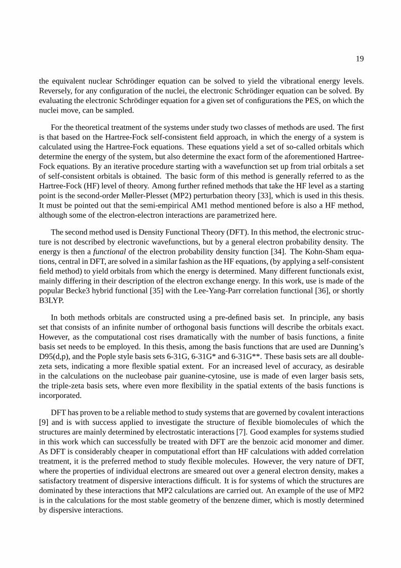
1.2.1 The Free-Electron Laser FELIX
Central in this work is the possibility to perform mid- and far-infrared (IR) spectroscopic studies onmolecular species. Although there exist several table-top sources that can produce radiation in thewavelength range up to 10µm and even longer wavelengths, only few are able to produce a highenough fluence and a narrow enough bandwidth to effectively study absorptions in dilute samples ofgas-phase molecules. A common approach to produce tunable IR radiation is to combine the outputsof pulsed dye lasers and of a Nd:YAG laser at 1064 nm in a nonlinear crystal and produce radiationwith the difference frequency of the two input frequencies. The resulting difference frequency cantypically reach wavelengths of up to 3-4µm. For longer wavelengths, usually an optical parametricoscillator (OPO) system is employed where wavelengths of up to 7µm can be reached.
5
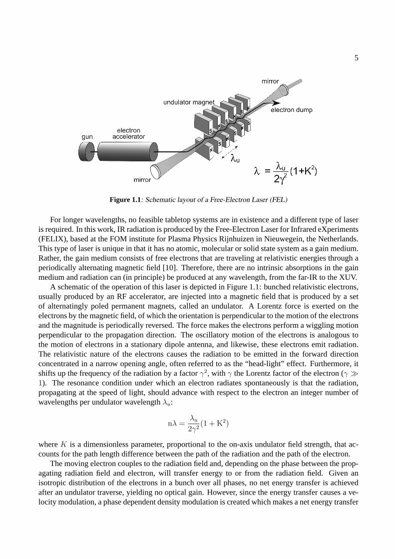
Figure 1.1: Schematic layout of a Free-Electron Laser (FEL)
For longer wavelengths, no feasible tabletop systems are in existence and a different type of laseris required. In this work, IR radiation is produced by the Free-Electron Laser for Infrared eXperiments(FELIX), based at the FOM institute for Plasma Physics Rijnhuizen in Nieuwegein, the Netherlands.This type of laser is unique in that it has no atomic, molecular or solid state system as a gain medium.Rather, the gain medium consists of free electrons that are traveling at relativistic energies through aperiodically alternating magnetic field [10]. Therefore, there are no intrinsic absorptions in the gainmedium and radiation can (in principle) be produced at any wavelength, from the far-IR to the XUV.
A schematic of the operation of this laser is depicted in Figure 1.1: bunched relativistic electrons,usually produced by an RF accelerator, are injected into a magnetic field that is produced by a setof alternatingly poled permanent magnets, called an undulator. A Lorentz force is exerted on theelectrons by the magnetic field, of which the orientation is perpendicular to the motion of the electronsand the magnitude is periodically reversed. The force makes the electrons perform a wiggling motionperpendicular to the propagation direction. The oscillatory motion of the electrons is analogous tothe motion of electrons in a stationary dipole antenna, and likewise, these electrons emit radiation.The relativistic nature of the electrons causes the radiation to be emitted in the forward directionconcentrated in a narrow opening angle, often referred to as the “head-light” effect. Furthermore, itshifts up the frequency of the radiation by a factorγ2, with γ the Lorentz factor of the electron (γ �1). The resonance condition under which an electron radiates spontaneously is that the radiation,propagating at the speed of light, should advance with respect to the electron an integer number ofwavelengths per undulator wavelengthλu:
nλ =λu
2γ2(1 + K2)
whereK is a dimensionless parameter, proportional to the on-axis undulator field strength, that ac-counts for the path length difference between the path of the radiation and the path of the electron.
The moving electron couples to the radiation field and, depending on the phase between the prop-agating radiation field and electron, will transfer energy to or from the radiation field. Given anisotropic distribution of the electrons in a bunch over all phases, no net energy transfer is achievedafter an undulator traverse, yielding no optical gain. However, since the energy transfer causes a ve-locity modulation, a phase dependent density modulation is created which makes a net energy transfer
6
µ
µ
FEL-1
FEL-2
Linac 1 Linac 2
5 - 40 m
25 - 250 m
15-25 MeV 25-45 MeV
injector
6 m
22 m
FELIX layout pulse structure
7 µs
MACROPULSE
100 ms
MICROPULSE
0.3 - 5 ps 1 ns 0.3 - 5 ps
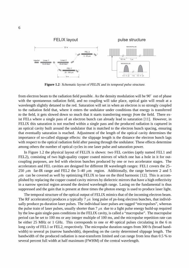
Figure 1.2: Schematic layout of FELIX and its temporal pulse structure.
from electron beam to the radiation field possible. As the density modulation will be 90˚ out of phasewith the spontaneous radiation field, and no coupling will take place, optical gain will result at awavelength slightly detuned to the red. Saturation will set in when an electron is so strongly coupledto the radiation field that, where it enters the undulator under conditions that energy is transferredto the field, it gets slowed down so much that it starts transferring energyfrom the field. There ex-ist FELs where a single pass of an electron bunch can already lead to saturation [11]. However, inFELIX this saturation is not reached within a single pass and the produced radiation is captured inan optical cavity built around the undulator that is matched to the electron bunch spacing, ensuringthat eventually saturation is reached. Adjustment of the length of the optical cavity determines theimportance of so-called slippage effects: the slippage length is the distance the electron bunch lagswith respect to the optical radiation field after passing through the undulator. These effects determineamong others the number of optical cycles in one laser pulse and saturation power.
In Figure 1.2 the physical layout of FELIX is shown: two FEL cavities (aptly named FEL1 andFEL2), consisting of two high-quality copper coated mirrors of which one has a hole in it for out-coupling purposes, are fed with electron bunches produced by one or two accelerator stages. Theaccelerators and FEL cavities are designed for different IR wavelength ranges: FEL1 covers the 25–250 µm far-IR range and FEL2 the 5–40µm region. Additionally, the range between 2 and 5µm can be covered as well by optimizing FELIX to lase on the third harmonic [12]. This is accom-plished by replacing the copper coated cavity mirrors by dielectric mirrors that have a high reflectivityin a narrow spectral region around the desired wavelength range. Lasing on the fundamental is thussuppressed and the gain that is present at three times the photon energy is used to produce laser light.
The temporal structure of the optical output of FELIX mimics that of the incoming electron beam.The RF accelerator(s) produces a typically 7µs long pulse of ps-long electron bunches, that individ-ually produce ps-duration laser pulses. The individual laser pulses are tagged “micropulses”, whereasthe pulse train of laser pulses, usually shorter than 7µs due to a light pulse energy build-up requiredby the low-gain single-pass conditions in the FELIX cavity, is called a “macropulse”. The macropulseperiod can be set to 100 ms or any integer multiple of 100 ms, and the micropulse repetition rate canbe either 25 MHz or 1 GHz. This corresponds to one or 40 optical pulses circulating in the 6 mlong cavity of FEL1 or FEL2, respectively. The micropulse duration ranges from 300 fs (broad band-width) to several ps (narrow bandwidth), depending on the cavity determined slippage length. Thebandwidth of the produced radiation is near-transform limited and can range from less than 0.5 % toseveral percent full width at half maximum (FWHM) of the central wavelength.
7
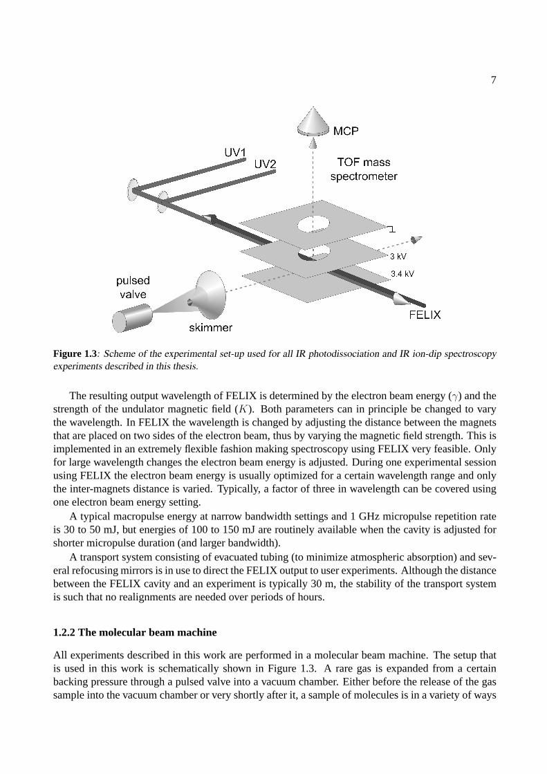
Figure 1.3: Scheme of the experimental set-up used for all IR photodissociation and IR ion-dip spectroscopyexperiments described in this thesis.
The resulting output wavelength of FELIX is determined by the electron beam energy (γ) and thestrength of the undulator magnetic field (K). Both parameters can in principle be changed to varythe wavelength. In FELIX the wavelength is changed by adjusting the distance between the magnetsthat are placed on two sides of the electron beam, thus by varying the magnetic field strength. This isimplemented in an extremely flexible fashion making spectroscopy using FELIX very feasible. Onlyfor large wavelength changes the electron beam energy is adjusted. During one experimental sessionusing FELIX the electron beam energy is usually optimized for a certain wavelength range and onlythe inter-magnets distance is varied. Typically, a factor of three in wavelength can be covered usingone electron beam energy setting.
A typical macropulse energy at narrow bandwidth settings and 1 GHz micropulse repetition rateis 30 to 50 mJ, but energies of 100 to 150 mJ are routinely available when the cavity is adjusted forshorter micropulse duration (and larger bandwidth).
A transport system consisting of evacuated tubing (to minimize atmospheric absorption) and sev-eral refocusing mirrors is in use to direct the FELIX output to user experiments. Although the distancebetween the FELIX cavity and an experiment is typically 30 m, the stability of the transport systemis such that no realignments are needed over periods of hours.
1.2.2 The molecular beam machine
All experiments described in this work are performed in a molecular beam machine. The setup thatis used in this work is schematically shown in Figure 1.3. A rare gas is expanded from a certainbacking pressure through a pulsed valve into a vacuum chamber. Either before the release of the gassample into the vacuum chamber or very shortly after it, a sample of molecules is in a variety of ways
8
vaporized and mixed with the rare gas. The vaporization sources are described in section 1.2.3. Inthe expansion process, the atom-molecule gas mixture is cooled adiabatically,i.e., internal energy ofthe gas is transferred into kinetic energy of the atoms. On a microscopic level, the molecules arecooled through inelastic collisions with the carrier gas atoms. Several inert gases are used of whichAr, Ne and He are the most frequently used. The cooling properties of the so-called carrier gas aredetermined by its mass (and thus its ability for momentum transfer). Typically, the molecules arevibrationally and rotationally cooled to some 5 K. The expansion conditions enable the formation ofweakly bonded molecular Van der Waals complexes, complexes of molecules with rare gas atoms,and hydrogen bonded complexes and these complexes are internally cooled as well.
After the expansion, all particles travel at rather high speed (typically 500 ms−1) and with a rathernarrow velocity distribution, which is a measure of the translational temperature of the sample. Themolecules then enter an interaction region after being skimmed by a conically shaped skimmer. Thedistance from the nozzle of the pulsed valve to the skimmer, some 50 mm, and the skimmer diameter,1 mm, determine the geometry of the beam, that interacts with various laser sources in the sourceregion of a Wiley-McLaren type linear Time-Of-Flight (TOF) mass spectrometer. The molecules inthe beam interact with incoming UV laser beams as well as with the IR laser beam at the crossingpoint of the mutually perpendicular molecular beam axis, laser beam axis and TOF tube axis. Ionsare produced in this region and subsequently pulse extracted and accelerated toward a Micro ChannelPlate (MCP) detector, yielding mass spectra with a resolution ofM/∆M ≈ 200. The signal fromthe MCP detector is amplified and fed into a 10 bit, 100 Ms/s digital oscilloscope that is read out bya PC. The experiment is running at a 10 Hz repetition rate; digital delay/pulse generators are used tosynchronize the molecular beam to the various laser sources.
1.2.3 Sources
The experiments described in this thesis are all studies of gas-phase species. For crystalline or liquidsamples with a substantial vapor pressure at room temperature the sample can directly be mixed withthe carrier gas. Other molecules can be heated to produce a reasonable vapor pressure and are thenseeded into the carrier gas before the expansion into the vacuum chamber. Temperatures that can beemployed are limited by the properties of the pulsed valve; the Jordan type valve can be heated to amaximum of 75˚C. For experiments with benzene (Chapters 2 and 3) and the benzoic acid molecule(see Chapter 4), no higher temperatures are required and the standard equipment available is used.When higher temperatures are needed, an oven that is placedafter the nozzle and where thermalcontact between valve body and oven is minimized, can be used. It is less trivial to study systemsthat have both a low vapor pressure and are thermally labile. For these molecules the method oflaser-desorption is employed.
A sublimation oven
To heat samples of low vapor pressure that are thermally stable an oven is constructed, based on adesign from Dr. L.C. Snoek [13]. Schematically, the oven is shown in Figure 1.4, part A. The ovenis placed behind the nozzle of a General Valve (series 9), a pulsed valve that can be heated to 200 ˚C.The oven body is heated by heating coils, while the temperatures of both the valve and oven bodies aremonitored using thermocouple gauges. Typically, the oven can be heated to 250 ˚C without exceedingthe temperature limit of the valve, due to the poor thermal contact between oven and valve, both of
9
0 500 1000 1500 2000
0
1
Ion s
ignal (
arb
. units
)Time (µs)
initial shockwave:
warm tryptophan expansion:
cooled tryptophan
M6 bolt
insulating spacers
thermocouple
heating coils
pulsed valve
crystalline
sample
thermocouple
A B
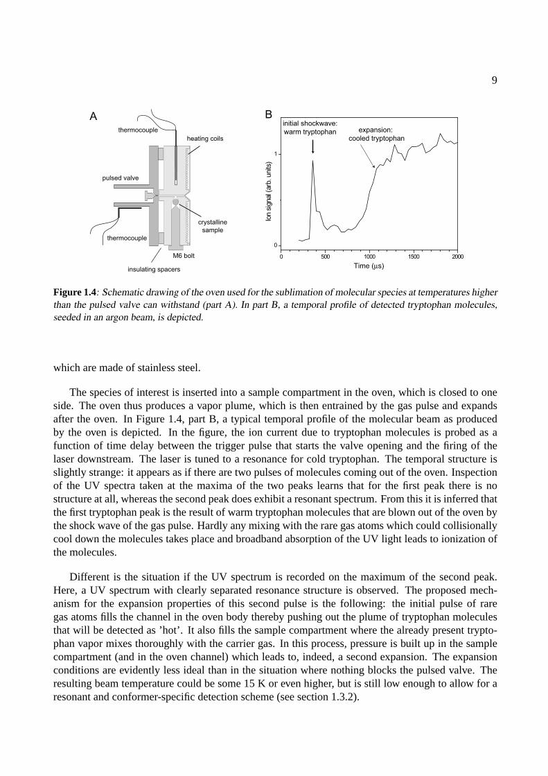
Figure 1.4: Schematic drawing of the oven used for the sublimation of molecular species at temperatures higherthan the pulsed valve can withstand (part A). In part B, a temporal profile of detected tryptophan molecules,seeded in an argon beam, is depicted.
which are made of stainless steel.
The species of interest is inserted into a sample compartment in the oven, which is closed to oneside. The oven thus produces a vapor plume, which is then entrained by the gas pulse and expandsafter the oven. In Figure 1.4, part B, a typical temporal profile of the molecular beam as producedby the oven is depicted. In the figure, the ion current due to tryptophan molecules is probed as afunction of time delay between the trigger pulse that starts the valve opening and the firing of thelaser downstream. The laser is tuned to a resonance for cold tryptophan. The temporal structure isslightly strange: it appears as if there are two pulses of molecules coming out of the oven. Inspectionof the UV spectra taken at the maxima of the two peaks learns that for the first peak there is nostructure at all, whereas the second peak does exhibit a resonant spectrum. From this it is inferred thatthe first tryptophan peak is the result of warm tryptophan molecules that are blown out of the oven bythe shock wave of the gas pulse. Hardly any mixing with the rare gas atoms which could collisionallycool down the molecules takes place and broadband absorption of the UV light leads to ionization ofthe molecules.
Different is the situation if the UV spectrum is recorded on the maximum of the second peak.Here, a UV spectrum with clearly separated resonance structure is observed. The proposed mech-anism for the expansion properties of this second pulse is the following: the initial pulse of raregas atoms fills the channel in the oven body thereby pushing out the plume of tryptophan moleculesthat will be detected as ’hot’. It also fills the sample compartment where the already present trypto-phan vapor mixes thoroughly with the carrier gas. In this process, pressure is built up in the samplecompartment (and in the oven channel) which leads to, indeed, a second expansion. The expansionconditions are evidently less ideal than in the situation where nothing blocks the pulsed valve. Theresulting beam temperature could be some 15 K or even higher, but is still low enough to allow for aresonant and conformer-specific detection scheme (see section 1.3.2).

10
valve body
graphite
bar
Nd
:YA
G la
se
r pu
lse
sample molecules
embedded in carbon
matrix
expanding
gas pulse
Laser desorption
60 80 100 120 140 160 180 200
0
1
ion
sig
na
l (a
rb.
un
its)
time delay (µs)
tryptophan anilineA B
Figure 1.5: Schematic overview of laser desorption (top): a pulsed Nd:YAG laser (1064 nm) desorbs moleculesthat are embedded in a graphite matrix. The – intact – molecules are then entrained by a passing gas pulse andare cooled adiabatically. Next to the schematic a time dependence of a gas pulse with desorbed molecules isshown for optimum cooling conditions. Plotted is the yield of ions produced downstream by a 1+1 REMPIprocess as a function of the delay of the probe pulse with respect to the opening of the valve.
Laser desorption
A second method to bring molecules that have a low vapor pressure at room temperature into the gasphase is laser desorption [14]. The method is schematically described in Figure 1.5, part A. In thelaser desorption source a crystalline sample of the molecules under study is mixed with fine graphitepowder and applied onto a surface of a bar of solid graphite (50x15x2 mm) that is placed directlyunder the orifice of a pulsed valve. The 10 Hz pulsed Jordan valve releases gas pulses of argon(typically 50µs long) through a 0.5 mm diameter nozzle into vacuum at a backing pressure of 3.5 bar.Directly after opening the nozzle a pulsed Nd:YAG laser (Thales Diva-2, 1064 nm, 5 ns,< 1 mJ perpulse) desorbs sample molecules from the graphite matrix. The desorbed molecules are entrained inthe supersonically expanding carrier gas. In the adiabatic expansion the internal degrees of freedomin the nucleobase molecules are cooled to some 10 K.
In part B of Figure 1.5 a time dependence of the gas pulse is shown under optimal cooling condi-tions. The yield of ions produced downstream by a 1+1 REMPI process is plotted as a function of thedelay of the probe pulse with respect to the opening of the valve. The squared, open symbols repre-sent the yield of ionized test molecules (aniline) that are seeded into the beam prior to the expansionto detect the gas pulse. The round, full symbols represent the tryptophan molecules that are desorbedfrom the sample bar and seeded into the beam. It is experimentally observed that the optimum probelaser delay is shorter for desorbed material than for molecules seeded into the beam before expansion.
1.3 Spectroscopic techniques
The techniques that are used in this thesis to record IR absorption spectra of dilute samples of gas-phase molecules are double-resonance techniques,i.e., they are based on the presence of two well-synchronized lasers that interact with the same sample. Mostly, these double-resonance techniquesare based on already existing techniques that probe the electronic structure of the molecules under
11
study. Therefore, these techniques are introduced first, and after that the natural extension to usage inthe IR is discussed.
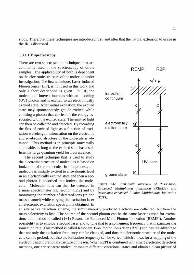
1.3.1 UV spectroscopy
REMPI
electronically
excited state
ground state
ionization
continuum
M++ e-
M*
M
UV laser
R2PI
ν1
ν1
ν2
ν1
Figure 1.6: Schematic overview of Resonance-Enhanced Multiphoton Ionization (REMPI) andResonance-enhanced 2-color Multiphoton Ionization(R2PI).
There are two spectroscopic techniques that arecommonly used in the spectroscopy of dilutesamples. The applicability of both is dependenton the electronic structure of the molecule underinvestigation. The first technique, Laser-InducedFluorescence (LIF), is not used in this work andonly a short description is given. In LIF, themolecule of interest interacts with an incoming(UV) photon and is excited to an electronicallyexcited state. After initial excitation, the excitedstate may spontaneously get de-excited whileemitting a photon that carries off the energy as-sociated with the excited state. The emitted lightcan then be collected and detected. By recordingthe flux of emitted light as a function of exci-tation wavelength, information on the electronicand rovibronic structure of the molecule is ob-tained. This method is in principle universallyapplicable, as long as the excited state has a suf-ficiently large quantum yield for fluorescence.
The second technique that is used to studythe electronic structure of molecules is based onionization of the molecule. In this process, themolecule is initially excited to a rovibronic levelin an electronically excited state and then a sec-ond photon is absorbed that ionizes the mole-cule. Molecular ions can then be detected ina mass spectrometer (cf. section 1.2.2) and bymonitoring the number of detected ions (in onemass channel) while varying the excitation laseran electronic excitation spectrum is obtained. Inan alternative detection scheme, the simultaneously produced electrons are collected, but here themass-selectivity is lost. The source of the second photon can be the same laser as used for excita-tion; this method is called (1+1)-Resonance-Enhanced Multi-Photon Ionization (REMPI). Anotherpossibility is to employ a second laser and to tune that to a convenient frequency that maximizes theionization rate. This method is called Resonant Two-Photon Ionization (R2PI) and has the advantagethat not only the excitation frequency can be changed, and thus the electronic structure of the mole-cule can be probed, but also the ionization frequency can be varied, which allows for a mapping of theelectronic and vibrational structure of the ion. When R2PI is combined with smart electronic detectionmethods, one can separate molecular ions in different vibrational states and obtain a clean picture of
12
0 1
IR-IDS
IR laser
Detected ions
+ion
time delay
Detection
UV laser
vibrationally
excited
states
electronically
excited state
electronic
ground state
ionization
continuum
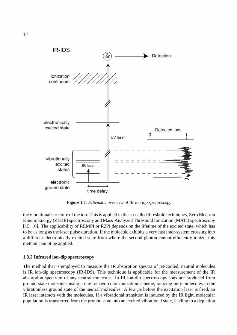
Figure 1.7: Schematic overview of IR-ion-dip spectroscopy
the vibrational structure of the ion. This is applied in the so-called threshold techniques, Zero ElectronKinetic Energy (ZEKE) spectroscopy and Mass-Analyzed Threshold Ionization (MATI) spectroscopy[15, 16]. The applicability of REMPI or R2PI depends on the lifetime of the excited state, which hasto be as long as the laser pulse duration. If the molecule exhibits a very fast inter-system crossing intoa different electronically excited state from where the second photon cannot efficiently ionize, thismethod cannot be applied.
1.3.2 Infrared ion-dip spectroscopy
The method that is employed to measure the IR absorption spectra of jet-cooled, neutral moleculesis IR ion-dip spectroscopy (IR-IDS). This technique is applicable for the measurement of the IRabsorption spectrum of any neutral molecule. In IR ion-dip spectroscopy ions are produced fromground state molecules using a one- or two-color ionization scheme, ionizing only molecules in thevibrationless ground state of the neutral molecules. A fewµs before the excitation laser is fired, anIR laser interacts with the molecules. If a vibrational transition is induced by the IR light, molecularpopulation is transferred from the ground state into an excited vibrational state, leading to a depletion

13
of ground state molecules. This results in a dip in the number of produced ions. By measuring theion yield, while varying the wavelength of the IR laser, the ion-dip spectrum is obtained. Similar toIR-IDS, IR fluorescence-dip spectroscopy is often applied [6].
1.3.3 Infrared photodissociation spectroscopy
To record the IR absorption spectrum of gas-phase ions, IR photodissociation of a complex of thespecies of interest with a rare gas atom is a very suitable technique [17]. The technique makesuse of the fact that the interaction between the inert rare gas atom and the molecule is of little orno influence on the structure of the molecule. IR photodissociation requires a well-defined initialion distribution of molecular complex ions. Such a population could for instance be prepared in anelectron-impact (EI)-source [18] but a more controlled way is to make use of a complex specific R2PIscheme. The transition frequencies for the complex in general are shifted from those of the isolatedmolecule, enabling a complex-specific excitation. The ionizing photon is then chosen such that themolecular complex is ionized and all ionic population is in the vibrational ground state. Directly aftercreation of the ionic population, an IR laser interacts with the molecules. The IR laser can induce atransition of the molecules into a vibrationally excited state. If the vibrational energy is larger thanthe dissociation of the complex there is a probability that the complex will dissociate. In this case thecharge will remain localized on the molecule and a bare molecular ion will be detected. If the numberof bare molecular ions is recorded while varying the IR frequency the IR photodissociation spectrumis obtained. Because there is initially no population of the bare ion, this method is background free,which makes it very sensitive. Additionally, contrary to threshold techniques it has the advantage ofbeing subject to the selection rules that govern IR absorption processes. Using FELIX, this methodhas been successfully demonstrated to study the IR absorption spectra of aniline-Ar+, naphthalene-Ar+, phenanthrene-Ar+ and benzene-Ar+ [9, 19–22].
1.4 Signal interpretation
1.4.1 A two- and three-level approximation
ground
state
excited
state2
1
σρΝ1 σρΝ2
Figure 1.8: Schematic two-level system, neglectingspontaneous emission.
In a first-order approximation, IR absorptionspectroscopy is governed by the rate equationsfor a closed two-level system (see Figure 1.8).Since the FELIX macropulse duration is longcompared to decoherence times of excited vi-brational states, of which the full rotational en-velopes can be covered with the FELIX band-width, coherent effects such as Rabi oscillationscan be neglected. In the rate equations, the popu-lationsN1(ν, t) of the initial state1, andN2(t) ofthe final state2 are coupled by the radiation field,represented by a time-dependent fluenceρ(ν, t),and an absorption cross-sectionσ(ν), both func-tions of frequencyν. Spontaneous emission is neglected as, in general, vibrational lifetimes are on theorder of milliseconds, which is well out of the experimental time window. Additionally, in the cold
14
ground
state
excited
state2
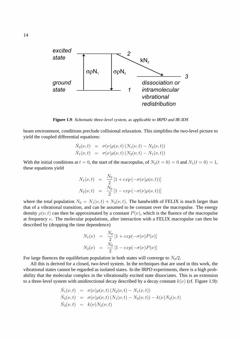
1
σρN1 σρN2
3
kN2
dissociation or
intramolecular
vibrational
redistribution
Figure 1.9: Schematic three-level system, as applicable to IRPD and IR-IDS
beam environment, conditions preclude collisional relaxation. This simplifies the two-level picture toyield the coupled differential equations:
N2(ν, t) = σ(ν)ρ(ν, t) (N1(ν, t)−N2(ν, t))
N1(ν, t) = σ(ν)ρ(ν, t) (N2(ν, t)−N1(ν, t))
With the initial conditions att = 0, the start of the macropulse, ofN2(t = 0) = 0 andN1(t = 0) = 1,these equations yield
N1(ν, t) =N0
2[1 + exp (−σ(ν)ρ(ν, t))]
N2(ν, t) =N0
2[1− exp (−σ(ν)ρ(ν, t))]
where the total populationN0 = N1(ν, t) + N2(ν, t). The bandwidth of FELIX is much larger thanthat of a vibrational transition, and can be assumed to be constant over the macropulse. The energydensityρ(ν, t) can then be approximated by a constantP (ν), which is the fluence of the macropulseat frequencyν. The molecular populations, after interaction with a FELIX macropulse can then bedescribed by (dropping the time dependence)
N1(ν) =N0
2[1 + exp(−σ(ν)P (ν)]
N2(ν) =N0
2[1− exp(−σ(ν)P (ν)]
For large fluences the equilibrium population in both states will converge toN0/2.All this is derived for a closed, two-level system. In the techniques that are used in this work, the
vibrational states cannot be regarded as isolated states. In the IRPD experiments, there is a high prob-ability that the molecular complex in the vibrationally excited state dissociates. This is an extensionto a three-level system with unidirectional decay described by a decay constantk(ν) (cf. Figure 1.9):
N1(ν, t) = σ(ν)ρ(ν, t) (N2(ν, t)−N1(ν, t))
N2(ν, t) = σ(ν)ρ(ν, t) (N1(ν, t)−N2(ν, t))− k(ν)N2(ν, t)
N3(ν, t) = k(ν)N2(ν, t)
15
The solutions of this three level system can be solved analytically. As the decay process in IRPD forall ν takes place well within the experimental timescale [23], the approximationk(ν) � σ(ν)ρ(ν, t)is introduced. With this approximation one gets:
N1(ν) = N0(exp(−σ(ν)P (ν)))
N2(ν) = 0
N3(ν) = N0 (1− exp(−σ(ν)P (ν)))
Again,N0 = N1(ν) +N2(ν) +N3(ν) is the total population. From this it is clear that the populationin the intermediate, excited state is negligible and is directly transferred into the dissociated state. Therelative cross-section can then be retrieved by:
σ(ν) = − 1
P (ν)ln(
N1(ν)
N1(ν) +N3(ν))
The relative cross-section can of course also be obtained from the population in state1 only, butinclusion of the dissociated state3 will reduce the experimental noise substantially since the shot-to-shot noise in the initially created total populationN0 is factored out.
For IR-IDS a similar picture can be drawn: there is no decay channel for dissociation or fragmen-tation, but there is Intramolecular Vibrational Redistribution (IVR). The density of states for largermolecules becomes rapidly so large that the probability of accidental degeneracies between differ-ent vibrational states becomes rather high. It is then possible to redistribute the population that istransferred from the initially excited state to isoenergetic vibrational states [24]. Now the three-levelapproximation is technically no longer valid, since redistribution does not necessarily completely de-populate the initially excited state. If there are many levels over which population is redistributed,however, the states over which this is done can be described as abath to which the population is de-caying. In this approach, the description of the population in states1, 2 and3 is valid, and the relativecross-section is retrieved by
σ(ν) = − 1
P (ν)ln(N1(ν))
In this approach, the fluctuations in the total population are not factored out, and care should be takenthat the ion signal for state1 is normalized to a constant background of ions. Experimentally this isdone by running FELIX at half the repetition rate of the UV laser(s) and recording alternating IR-onand IR-off signals so that the former can be normalized to the latter.
1.4.2 Application to an experimental ion-dip spectrum
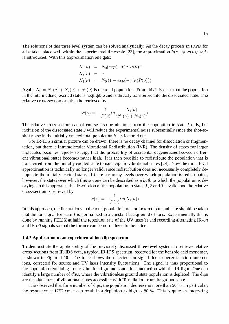
To demonstrate the applicability of the previously discussed three-level system to retrieve relativecross-sections from IR-IDS data, a typical IR-IDS spectrum, recorded for the benzoic acid monomer,is shown in Figure 1.10. The trace shows the detected ion signal due to benzoic acid monomerions, corrected for source and UV laser intensity fluctuations. The signal is thus proportional tothe population remaining in the vibrational ground state after interaction with the IR light. One canidentify a large number ofdips, where the vibrationless ground state population is depleted. The dipsare the signatures of vibrational states accessible with IR radiation from the ground state.
It is observed that for a number of dips, the population decrease is more than 50 %. In particular,the resonance at 1752 cm−1 can result in a depletion as high as 80 %. This is quite an interesting
16
600 800 1000 1200 1400 1600 1800
0
1
Ion s
ignal (a
rb. units)
Wavenumber (cm-1)
0 1
0
1
Laser fluence (arb.units)
Io
n s
ign
al (a
rb.
un
its)
631 cm-1
710 cm-1
1752 cm-1
Figure 1.10: Observed IR ion-dip spectrum of the benzoic acid monomer. In the inset the laser fluence depen-dence of the ion depletion on the resonances marked with symbols is shown.
observation, as in the interaction of an isolated, non-degenerate two-level system with electromagneticradiation population transfer is limited to 50 %, as discussed above. Apparently, there is an extrachannel into which population is transferred after excitation to this vibrational state. One possibilityis the occurrence of IVR, by which vibrational energy is redistributed over ”dark” states that are iso-energetic with the excited vibrational state. Through this coupling, a more than 50 % depletion of theground state population is possible. Depletions of over 50 % are even reached at wavenumbers below1000 cm−1 , although it appears to be quite unlikely that the BA monomer will exhibit IVR here, asmolecules of this (still rather limited) size simply lack the required density of states that allows forIVR to take place.
To be able to convert the observed IR-IDS spectra to IR absorption spectra, one needs to know themaximum possible depletion signal at each frequency,i.e., one needs to know where the baseline ofthe spectrum is. The formulas for signal interpretation, as derived in the previous section, representthe ideal case. Here, experimental imperfections, such as a slight misalignment, cannot be ruled out.They can be taken into account by introducing an experimental parametera(ν) describing the signalbaseline. The remaining population in the ground state is then described by:
N1(ν) = N0(a(ν) + (1− a(ν))exp(−σ(ν)P (ν)))
= N0exp(−σ(ν)P (ν)) +N0a(ν)[1− exp(−σ(ν)P (ν))]
In addition, one needs to verify that the observed depletion signals are due to single-photon absorp-tions. In order to test this, the population decrease as a function of laser fluence is measured for a few
17
selected resonances. This is done by varying the laser fluence with a set of fixed-value attenuators.The results of these measurements are displayed in the inset in Figure 1.10, where the populationdecreases for the resonances at 631 cm−1, 710 cm−1 and 1752 cm−1 are plotted as a function of laserfluence. As the observed curves are well described by the functional dependence it it concluded thatwe indeed deal with single-photon absorptions. In the high laser power limit, the population for allresonances converges to about 0.1, setting a base line valuea(ν) = a = 0.1.
The IR-IDS data can thus directly be interpreted as a measurement of the (relative) IR absorptioncross-section. However, since the IR laser has a finite bandwidth that is larger than the line width ofthe vibrational transitions, the cross-section is smeared out over the full laser bandwidth and an IRabsorption intensity is measured, of which the integral equals the absorption cross-section. Further-more, although the obtained data could be corrected for variation of the FEL bandwidth, it is chosento present them as recorded in the experiment, and correct the theoretically calculated spectra insteadto facilitate direct comparison.
1.5 Theoretical methods
The work presented in this thesis consists largely of results from experimental studies. However, forthe interpretation of experimental data several theoretical studies have been performed. As numericalmethods available in quantum chemistry can become rather demanding on computational resourcesone usually has to compromise between accuracy and computational cost. In this work, the calcu-lations necessary for the structural identification of gas-phase biomolecules can be divided into twoparts: conformational search and electronic structure calculations.
Most of the studies performed here are done using commercial quantum chemistry packages suchas Gaussian 98 [25], the Turbomole suite [26] and force-field based molecular mechanics programbundles like Tinker [27] and Sybyl [28]. Although these programs contain computational tools thatnowadays can be considered standard for both experimental as well as for theoretical chemists, onecannot deny that a certain air of “black box” methods hangs around them. It therefore seems appro-priate to shortly discuss the fundamental principles behind the used methods.
1.5.1 Conformational search
As biomolecules possess numerous bonds over which subgroups can rather freely be rotated, a largenumber of possible geometries results. It is the aim of the conformational search to find all these pos-sible geometries and to define criteria based upon which a found geometry is further investigated, andeventually submitted to the more costly quantum chemical methods. The results of those calculationsare then compared to the experimental data.
The conformational search is performed using two methods. The first method involves the useof classical molecular dynamics (MD) simulations. Here, the interactions between different atoms isdescribed in a parametrized force field. The classical Newtonian equations of motion are solved fora set of atoms with pre-defined interaction parameters. The procedure used is the following: witha given temperature the MD simulation is run for some time. After a suitable time the structure’stemperature is slowly lowered to zero. The thus obtained structure is then optimized to obtain thelowest potential energy of the system. It is then stored for later evaluation, and the MD simulationis restarted to generate a next structure. With sensibly chosen MD run times, temperatures and dura-
18
tion of the cool-down period, the phase space can effectively be sampled. This method yields largenumbers of different geometries, the stabilities of these structures are mainly determined by the forcefield parameters, which are not known accurately. As a result, the most stable geometries found withMD simulations are thus not guaranteed to be the lowest energy conformers found at higher levels oftheory or even a potential energy minimum at all. Added to that, there is still the possibility that thelowest energy conformer is not that which is observed in the experiment. Force fields that are used inthe calculations in this work are MM3 [29] and Sybyl [28].
The second method to probe the multidimensional phase space is by performing a systematic scanover all freely rotatable bonds. Geometries that exhibit major steric hindrance are rejected. The struc-ture of all other resulting geometries are optimized at a theoretical level which is both acceptable inpredicting structure and stability and at the same time cheap enough to optimize multiple geome-tries. As is the case with the force field search, not all geometries optimized at lower theoretical levelare guaranteed to be stable minima on the potential energy surface (PES). Additionally, the cost ofsuch systematic scans is growing enormously when more rotatable bonds are included. In this work,conformers for tryptophan were investigated using the semi-empirical method AM1 [30].
1.5.2 Electronic structure calculations
For the interpretation of experimentally obtained IR absorption spectra electronic structure calcula-tions are performed. Geometries obtained from the conformational search are submitted to a fullstructural optimization byab initio methods. These methods evaluate for the geometry the electronicSchrodinger equation to find equilibrium structures.
The Hamiltonian for a many body system, ignoring relativistic and spin-orbit interactions is
H = − h2
2
∑a
1
ma
∇2a −
h2
me
∑i
∇2i +
∑a
∑b>a
ZaZbe2
rab
−∑
a
∑i
Zae2
rai
+∑
i
∑j>i
e2
rij
wherea andb are the nuclear indices andi and j are the electron indices,me the electron mass,ma
the nuclear masses,r the distances between the various particles andZ the nuclear charges. The firsttwo terms describe the kinetic energy of the nuclei and electrons, respectively, the fourth term theCoulombic electron-nucleus attraction and the third and fifth terms the Coulombic repulsion of thenuclei and electrons, respectively.
To solve the molecular Schrodinger equation anAnsatzin the form of the Born-Oppenheimerapproximation is made. The Born-Oppenheimer approximation implies that the masses of the nucleimake them move much slower than the electrons and as a result the electrons instantaneously followthe nuclear motion. This results in a separation between the wavefunctions and Hamiltonian operatorsof the nuclei and of the electrons:
ψ(R, r) = ψe(R, r)ψn(R)
H(R, r) = He(R, r) +Hn(R)
whereR are the coordinates of the nuclei andr the coordinates of the electrons. The implication ofthis separation is that for a given solution for the electronic Schrodinger equation
He(R, r)ψe(R, r) = Eeψe(R, r)
Consulted literature includes refs [31] and [32]
19
the equivalent nuclear Schrodinger equation can be solved to yield the vibrational energy levels.Reversely, for any configuration of the nuclei, the electronic Schrodinger equation can be solved. Byevaluating the electronic Schrodinger equation for a given set of configurations the PES, on which thenuclei move, can be sampled.
For the theoretical treatment of the systems under study two classes of methods are used. The firstis that based on the Hartree-Fock self-consistent field approach, in which the energy of a system iscalculated using the Hartree-Fock equations. These equations yield a set of so-called orbitals whichdetermine the energy of the system, but also determine the exact form of the aforementioned Hartree-Fock equations. By an iterative procedure starting with a wavefunction set up from trial orbitals a setof self-consistent orbitals is obtained. The basic form of this method is generally referred to as theHartree-Fock (HF) level of theory. Among further refined methods that take the HF level as a startingpoint is the second-order Møller-Plesset (MP2) perturbation theory [33], which is used in this thesis.It must be pointed out that the semi-empirical AM1 method mentioned before is also a HF method,although some of the electron-electron interactions are parametrized here.
The second method used is Density Functional Theory (DFT). In this method, the electronic struc-ture is not described by electronic wavefunctions, but by a general electron probability density. Theenergy is then afunctionalof the electron probability density function [34]. The Kohn-Sham equa-tions, central in DFT, are solved in a similar fashion as the HF equations, (by applying a self-consistentfield method) to yield orbitals from which the energy is determined. Many different functionals exist,mainly differing in their description of the electron exchange energy. In this work, use is made of thepopular Becke3 hybrid functional [35] with the Lee-Yang-Parr correlation functional [36], or shortlyB3LYP.
In both methods orbitals are constructed using a pre-defined basis set. In principle, any basisset that consists of an infinite number of orthogonal basis functions will describe the orbitals exact.However, as the computational cost rises dramatically with the number of basis functions, a finitebasis set needs to be employed. In this thesis, among the basis functions that are used are Dunning’sD95(d,p), and the Pople style basis sets 6-31G, 6-31G* and 6-31G**. These basis sets are all double-zeta sets, indicating a more flexible spatial extent. For an increased level of accuracy, as desirablein the calculations on the nucleobase pair guanine-cytosine, use is made of even larger basis sets,the triple-zeta basis sets, where even more flexibility in the spatial extents of the basis functions isincorporated.
DFT has proven to be a reliable method to study systems that are governed by covalent interactions[9] and is with success applied to investigate the structure of flexible biomolecules of which thestructures are mainly determined by electrostatic interactions [7]. Good examples for systems studiedin this work which can successfully be treated with DFT are the benzoic acid monomer and dimer.As DFT is considerably cheaper in computational effort than HF calculations with added correlationtreatment, it is the preferred method to study flexible molecules. However, the very nature of DFT,where the properties of individual electrons are smeared out over a general electron density, makes asatisfactory treatment of dispersive interactions difficult. It is for systems of which the structures aredominated by these interactions that MP2 calculations are carried out. An example of the use of MP2is in the calculations for the most stable geometry of the benzene dimer, which is mostly determinedby dispersive interactions.
20
1.5.3 Harmonic vibration frequency calculations
When the electronic structure calculations have determined a stable structure, the harmonic energiesof the normal mode vibrations are calculated by solving the nuclear Schrodinger equation. The po-tential energy of the molecule is approximated by a second-order Taylor expansion as
V (R) = V (R0) +
(dV
dR
)T
0
(R−R0) +1
2(R−R0)
T
(d2V
dR2
)0
(R−R0)
whereR is a 3N dimensional vector containing all N nuclear coordinates and(
dVdR
)0
and(
d2VdR2
)0
are
3N × 3N matrices, evaluated at the equilibrium position. (The superscriptT denotes the transposedmatrix.) Since a stable structure is found at a minimum of the PES, the first derivative vanishes.Translating the equilibrium position to the origin and introducing the displacementx = R−R0 onegets the harmonic approximation of the vibrational potential (in matrix notation):
V (x) = xTFx
with F the 3N × 3N force constant orHessianmatrix. Now, the nuclear Schrodinger equationcan be solved, by first applying a coordinate transformationyi =
√mixi, which makes the nuclear
Schrodinger equation mass-independent. By diagonalizing the mass-weighted Hessian the vibrationaleigenfrequencies and eigenvectors are obtained, constituting the normal modes. The IR intensitiesassociated with the normal modes are calculated as the second derivative of the dipole momentµ withrespect to the normal coordinates.
In the calculations two important approximations are introduced. First, the Born-Oppenheimerapproximation assumes separation between electronic and nuclear coordinates. However, when twoPES’s come close together energetically, vibrational energies become comparable to inter-PES energydifferences and couplings between nuclear motion and electronic structure can occur. At that point theBorn-Oppenheimer breaks down. This is the case for the benzene cation, which has two iso-energeticPES’s and electronic-vibrational coupling distorts both (Chapter 2). The second approximation is thatthe PES can be considered harmonic along every normal coordinate. Although this approximationis often good, there exist regions on the PES where the anharmonicity is so large that one or morevibrational modes are not well described in the harmonic approximation. A good example of this isthe NH2 inversion mode in aniline, for which the potential is strongly anharmonic [19].
21
References
[1] F. Hillenkamp, M. Karas, R. Beavis, and B. Chait, Anal. Chem.63, A1193 (1991).
[2] J. Fenn, M. Mann, C. Meng, S. Wong, and C. Whitehouse, Science246, 64 (1989).
[3] G. von Helden, T. Wyttenbach, and M. T. Bowers, Science267, 1483 (1995).
[4] D. E. Clemmer, R. Hudgins, and M. F. Jarrold, J. Am. Chem. Soc.117, 10141 (1995).
[5] C. S. Hoaglund-Hyzer, Y. J. Lee, A. E. Counterman, and D. E. Clemmer, Anal. Chem.74, 992 (2002).
[6] T. S. Zwier, J. Phys. Chem. A105, 8827 (2001).
[7] E. G. Robertson and J. P. Simons, Phys. Chem. Chem. Phys.3, 1 (2001).
[8] F. Huisken, A. Kulcke, C. Laush, and J. M. Lisy, J. Chem. Phys.95, 3924 (1991).
[9] H. Piest, Ph.D. thesis, Katholieke Universiteit Nijmegen (2002).
[10] L. Elias, W. Fairbank, J. Madey, H. Schwettman, and T. Smith, Phys. Rev. Lett.36, 717 (1976).
[11] J. Andruszkow andet al., Phys. Rev. Lett.85, 3825 (2000).
[12] S. V. Benson and J. M. J. Madey, Phys. Rev. A39, 1579 (1989).
[13] L. C. Snoek, private communication.
[14] G. Meijer, M. de Vries, H. Hunziker, and H. R. Wendt, Appl Phys. B51, 395 (1990).
[15] K. Muller-Dethlefs, M. Sander, and E. Schlag, Chem. Phys. Lett.112, 291 (1984).
[16] L. Zhu and P. Johnson, J. Chem. Phys.94, 5769 (1991).
[17] M. Okumura, L. Yeh, J. Myers, and Y. Lee, J. Phys. Chem.94, 3416 (1990).
[18] O. Dopfer, R. V. Olkhov, and J. P. Maier, J. Chem. Phys.111, 10754 (1999).
[19] H. Piest, G. von Helden, and G. Meijer, J. Chem. Phys.110, 2010 (1999).
[20] H. Piest, G. von Helden, and G. Meijer, Ap. J.520, L75 (1999).
[21] J. H. Piest, J. Oomens, J. Bakker, G. von Helden, and G. Meijer, Spec. Acta A57, 717 (2001).
[22] R. G. Satink, H. Piest, G. von Helden, and G. Meijer, J. Chem. Phys.111, 10750 (1999).
[23] R. G. Satink, J. M. Bakker, G. Meijer, and G. von Helden, Chem. Phys. Lett.359, 163 (2002).
[24] A. Beil, D. Luckhaus, M. Quack, and J. Stohner, Ber. Bunsenges. Phys. Chem.101, 311 (1997).
[25] M. J. Frisch, G. W. Trucks, H. B. Schlegel, G. E. Scuseria, M. A. Robb, J. R. Cheeseman, V. G. Za-krzewski, J. J. A. Montgomery, R. E. Stratmann, J. C. Burant, et al.,Gaussian 98, Revision A.7, Pittsburgh,PA (1998).
[26] R. Ahlrichs, M. Bar, M. Haser, H. Horn, and C. Kolmel, Chem. Phys. Lett.162, 165 (1989).
22
[27] URL http://dasher.wustl.edu/tinker/ .
[28] M. Clark, R. Cramer, and N. Opdenbosch, J. Comput. Chem.10, 982 (1989).
[29] N. L. Allinger, Y. H. Yuh, and J.-H. Lii, J. Am. Chem. Soc.111, 8551 (1989).
[30] M. Dewar, E. Zoebisch, E. Healy, and J. Stewart, J. Am. Chem. Soc.107, 3902 (1985).
[31] P. Atkins and R. Friedman,Molecular quantum mechanics(Oxford University Press, Oxford, GreatBritain, 1997), 3rd ed.
[32] F. Jensen,Introduction to computational chemistry(John Wiley & Sons, Chichester, Great Britain, 1999).
[33] C. Møller and M. Plesset, Phys. Rev.46, 618 (1934).
[34] P. Hohenberg and W. Kohn, Phys. Rev.136, B864 (1964).
[35] A. Becke, J. Chem. Phys.107, 8554 (1997).
[36] C. Lee, W. Yang, and R. Parr, Phys. Rev. B37, 785 (1988).
CHAPTER 2
INFRARED PHOTODISSOCIATION SPECTROSCOPY OF
BENZENE–NE AND BENZENE–AR COMPLEX CATIONS
The infrared (IR) absorption spectrum of the jet-cooled benzene cation complexed with Ne has beenrecorded throughout the 275–1900 cm−1 and the 2800–3200 cm−1 range via IR-laser induced vibra-tional dissociation spectroscopy. Measuring the spectrum of the complex ion rather than the spectrumof the bare benzene cation, leads to the appearance of weak Van der Waals sidebands. In addition,subtle effects of the symmetry lowering in the complex, giving rise to additional lines in the spec-trum, have been observed for the benzene-Ar complex. The recorded IR absorption spectrum of thedeuterated benzene-Ar complex as well as the IR absorption spectra of benzene complex cations pre-pared in selected low-lying vibrationally excited levels yield a more detailed picture of the vibrationalstructure of the benzene cation.
Adapted from: J. M. Bakker, R. G. Satink, G. von Helden, and G. Meijer,Phys. Chem. Chem. Phys.4, 24–33(2002)
23
24
2.1 Introduction
A detailed understanding of the vibrational structure of the benzene cation is both interesting andrelevant for a variety of reasons. The benzene cation, with its doubly degenerate electronic groundstate, is of fundamental interest as it is the prototypical system for studies of the Jahn-Teller interaction[1, 2]. In the vibrational spectrum the Jahn-Teller effect manifests itself as a splitting of certainvibrational modes. This splitting can be rather large, on the order of the vibrational energy, andit is often difficult to discern a clear vibrational energy level pattern, complicating assignment ofvibrational modes. The benzene cation might also be of astrophysical interest. Recently, neutralbenzene has been identified as an emitter of infrared (IR) radiation in a proto-planetary nebula [3]. Itis therefore not unlikely that the benzene cation can be identified in astrophysical objects via its IRemission spectrum as well. After all, the polycyclic aromatic hydrocarbons (PAHs), and specificallytheir cations, have been proposed as carriers of the Unidentified Infrared Bands (UIBs), a series ofdistinct IR emission bands observed from the interstellar medium [4, 5].
The use of direct IR absorption techniques to obtain information on the vibrational structure ofthe benzene cation, for instance on benzene cations deposited in rare gas matrices, has thus far notbeen successful. Another commonly used method to study vibrational structure, Laser Induced Flu-orescence (LIF) on ionic species either in the gas phase or in matrices, has not proven to be viablefor the benzene cation either, due to an unfavorably low quantum yield for fluorescence [6]. Asa consequence, much of the early experimental work on benzenoid systems consisted of dispersedfluorescence studies of partly or completely halogenated benzene ions, for which the fluorescencequantum yield is close to one. For an overview, the reader is referred to the work by Miller andBondybey [7].
With the advent of the dye laser and with the development of supersonic molecular beam coolingtechniques, other methods for studying benzene cations became available. By recording the gasphase one color (1+1) photoelectron spectrum, Long, Meek and Reilly identified several low-lyingfundamental vibrational frequencies in the benzene cation [8]. The spectral resolution in this originalwork was limited by the resolution of the electron energy analyzer to about 3 meV. In later studies,in which a second tunable dye laser photon was used to induce ionization, the resolution was largelyimproved, making a more detailed analysis possible. These studies include accurate determination ofphotoionization thresholds via Resonance Enhanced Multi Photon Ionization (REMPI) spectroscopy[9, 10], Zero Electron Kinetic Energy (ZEKE) spectroscopy [11], and Mass Analyzed ThresholdIonization (MATI) spectroscopy [12, 13]. The measurement of rotationally resolved high-n Rydbergspectra of neutral benzene allowed for the extrapolation to the various ionization thresholds, leadingto a very precise determination of some vibrational frequencies in the ion [14]. The most recentthreshold technique studies on the benzene cation include MATI studies of the benzene cation usingeither vacuum UV to directly probe the ion from the neutral ground state [15] or by using the3B1u
triplet state of benzene (populated in a discharge source) as the initial state [16].Although the methods mentioned above yield accurate vibrational frequencies, definite assign-
ment of the modes involved might nevertheless be complicated. This is because in the electronictransitions involved in the detection process, the relative line intensities are mainly governed byFranck-Condon overlap and there are no strict vibrational selection rules. Additional informationcan therefore be obtained when the true vibrational absorption spectrum of the cation is measuredinstead. For this, a sensitive IR absorption detection scheme is required, for instance a scheme basedon the IR induced photodissociation of weakly bonded complexes of the benzene cations with rare
25
gas atoms. Although the IR absorption spectrum of the complex ions, rather than of the bare ions,is measured in this case, it is expected that these spectra will closely resemble each other; this issubstantiated by the good agreement between the observed MATI spectra for bare benzene and thoseof its argon complexes [17]. To be able to efficiently induce photodissociation of these complexes,an intense (pulsed) IR source, tunable throughout the relevant portion of the spectrum, is required.Using a pulsed laser system tunable in the 3µm region, Dopferet al. measured the IR active C–Hstretch mode via IR laser induced photodissociation of the benzene-Ar cation [18]. In a complemen-tary study, using a Free Electron Laser (FEL) as a source of widely tunable IR radiation, Satinket al.reported on the IR absorption spectrum of the same species in the 450–1500 cm−1 range [19].
The experimental activity in the recent years has led to a refinement in the theoretical models [20,21]. While most spectroscopic information in the spectral region below 1000 cm−1 can satisfactorilybe explained by invoking only the Jahn-Teller activity of two low energy modes (see section 2.2), newstudies have attempted to assign all observed resonances making use of spectral information availableat that time [22, 23].
In this chapter the results of a study on the IR spectrum of the benzene cation complexed witheither Ne or Ar are presented. There are two main advantages of using Ne over Ar as a messengeratom. First, the binding energy of the benzene-Ne complex is significantly lower than that of benzene-Ar, enabling the detection of IR absorption lines at lower frequencies. Second, the lower polarizabilityof Ne is expected to yield a spectrum that is more closely resembling the spectrum of the bare benzenecation. In the spectra of the complex ions Van der Waals sidebands have been observed, yieldinginformation on the intermolecular potential energy surface. Finally, the IR spectrum of the deuteratedbenzene cation, complexed with argon, and IR absorption spectra of benzene-Ar cations prepared inselected low-lying vibrationally excited levels have been recorded to obtain a more detailed pictureof the complex vibrational structure of the benzene cation in the region up to 1800 cm−1.
2.2 Spectroscopic details
The neutral benzene molecule in its electronic ground state belongs to theD6h molecular symmetrygroup. Its 30 vibrational modes are of the species
Γν = 2a1g ⊕ 1a2g ⊕ 2b2g ⊕ 1e1g ⊕ 4e2g ⊕ 1a2u ⊕ 2b1u ⊕ 2b2u ⊕ 3e1u ⊕ 2e2u. (2.1)
Throughout this chapter the commonly used Wilson notation [24] is adopted. In the neutral groundstate there are four IR active modes, one ofa2u and three ofe1u symmetries. The fundamental fre-quencies of these modes are experimentally found at 671 cm−1 for the a2u ν11 mode [out-of-planeC–H bending], at 1037 cm−1 for thee1u ν18 mode [in-plane C–H bending], at 1485 cm−1 for thee1u
ν19 mode [C–C stretching] and at 3099 cm−1 for thee1u ν20 mode [C–H stretching] [25]. The elec-tronic configuration of neutral benzene is(a2u)
2(e2g)4(e1g)
4 and upon removal of one electron fromone of thee1g orbitals, the cation is left in a degenerateE1g state. When the vibrational symmetry iscombined with theE1g electronic symmetry of the ground state of the cation, the number of vibratio-nal states is greatly increased. In particular, six ungerade states of electronic-vibrational A symmetryand ten of E symmetry result; all are expected to lie in the spectral range covered in this study.
In a classic paper published in 1937, Jahn and Teller pointed out that for any nonlinear moleculewith a threefold (or higher) rotational symmetry axis in an orbitally degenerate electronic state therewill be at least one normal coordinate for which the potential energy minimum does not coincide with
26
the symmetrical position, thus effectively distorting the nuclear framework, lowering the symmetryand lifting the degeneracy [1]. The benzene cation in its electronic ground is a prototypical systemfor this. For molecules ofD6h symmetry, vibrations ofe2g symmetry will cause a distortion thatis linear in the corresponding normal coordinate whereas vibrations ofe1g, e1u and e2u symmetrywill cause a quadratical distortion. In the benzene cation, there are foure2g modes that are linearlyJahn-Teller active. Of course, none of these four modes can be directly observed in IR absorptionspectroscopy of the bare cation. Using photoelectron spectroscopy and the more sophisticated ZEKEand MATI methods, only the frequency and Jahn-Teller activity of the lowest energye2g mode, theν6
in-plane ring-bending mode, have been identified thus far. The benzene molecule is distorted alongthis coordinate and two possible structures ofD2h symmetry result. No theoretical agreement hasbeen reached so far as to which of the two structures has the lowest energy [2, 26]. Experimentally,however, ZEKE spectroscopy studies revealed that the barriers between these structures are so lowthat ν6 acts as a pseudorotational coordinate linking the two. The resulting system as a whole isthen necessarily viewed asD6h [11], a conclusion that has recently been confirmed by rotationallyresolved ZEKE spectra of the ionic vibrational ground state [27]. Of the quadratically Jahn-Telleractive modes, only the lowest frequencye2u mode, theν16 out-of-plane ring bending mode around300 cm−1, has been identified. It is split up into two non-degenerate and one doubly degeneratemode, of which only the latter is IR active [11].
The low frequencies of the linear and quadratical Jahn-Teller activeν6 andν16 modes give riseto a rich energy level structure in the ion, even when only overtones and combinations of these twomodes are included. Due to the possibility of interaction of such combination modes with IR activefundamental modes via Fermi resonances, this might lead to unexpected features in the IR spectrumof the ion [19]. In Figure 2.1, a schematic representation of the energy level structure originating fromtheν6 andν16 modes of the benzene ion is given. The two-mode interaction model we have used toproduce this figure is the same as used by Goodeet al. [13]. For interpretation of their spectroscopicdata on halogenated benzenoid systems, Miller and Bondybey developed a theoretical framework,based on a treatment by Longuet-Higgins [28], to explain the observed energy level structure [7]. Thiswork was used and refined for the benzene cation by various groups [8, 11, 13]. The method consistsof diagonalization of a Hamiltonian matrix representing the two degenerate electronic states coupledthrough the Jahn-Teller active vibrational states. For a complete treatment the reader is referred tothe original work [7]. In Figure 2.1, the linear and quadratic Jahn-Teller coupling parameters aresimultaneously increased from left to right, going from the situation of no Jahn-Teller interaction (leftaxis) to the situation that is in best agreement with experimental observations (right axis) [13]. Fromthis figure, level assignments can be deduced.
Based on the experimental work that was available at the time, Applegate and Miller proposeda full assignment for the data observed via threshold techniques and via IR photodissociation [22].For completeness, all presently known vibrational frequencies are listed in Tables 2.1 and 2.2, bothfor C6H
+6 and forC6D
+6 . The numbers in these tables are taken from MATI studies by Krauseet al.
[12, 29] and Goodeet al. [13], ZEKE spectroscopy by Lindneret al. [11, 30], Rydberg spectroscopyby Siglowet al. [14, 31] and recent vacuum UV MATI work by Kwonet al. [15] as well as from thepresent work. The photoelectron studies by Longet al. [8] are among the landmark works on benzene,but the values obtained from these low-resolution studies have not been included. Mode assignmentsare given in the table, both based on assignments previously made in the literature and on the presentwork, as will be discussed below. Although the assignment put forward by Applegate and Miller

27
0.0 0.1 0.2 0.3
300
400
500
600
700
800
900
1000
161
61
162
6116
1
163
6116
26
2
Jahn-Teller interaction parameters
Vib
ratio
na
l e
ne
rgy (
cm
-1)
Figure 2.1: Correlation diagram, linking the energy levels in the benzene cation with Jahn-Teller interaction(right axis) to those expected in the absence of Jahn-Teller interaction (left axis). In the two-mode interactionmodel that is used, only the ν6 and ν16 vibrations have been included. From left to right the Jahn-Tellerinteraction parameters are linearly increased to their experimentally determined values [13].
is still open for debate, most of the assignments in the tables are based upon their interpretation.Additionally, in the table it has been recognized that all linearly Jahn-Teller active modes are coupledto each other through the ground state. This is reflected in the notation, where all resonances dueto linearly Jahn-Teller active modes are labeled by a vibronic angular momentum quantum numberj = lv + 1
2Λ wherelv is the vibrational angular quantum number for Jahn-Teller active modeνv; Λ =
±1 the electronic angular momentum quantum number associated with the two degenerate electronic
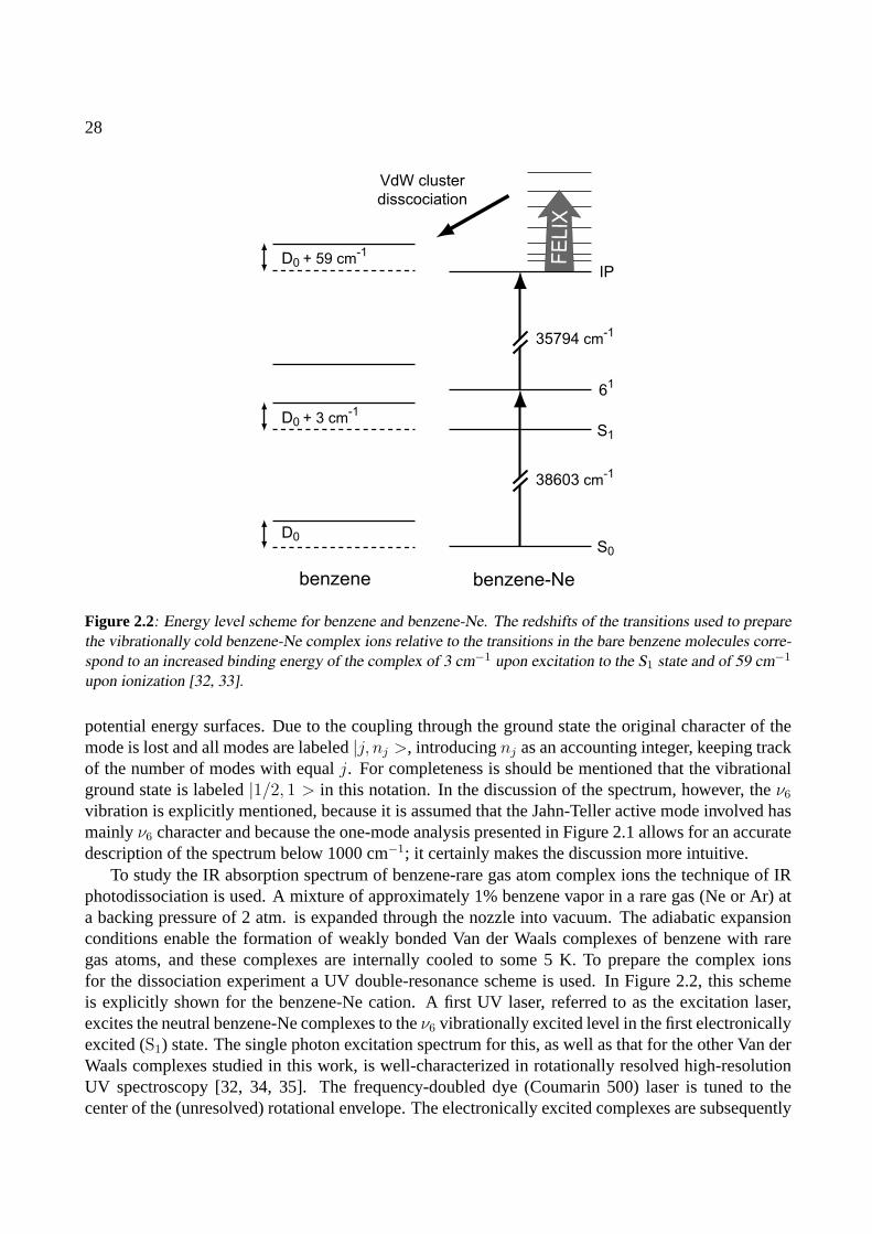
28
benzene benzene-Ne
VdW cluster
disscociation
D0
D0 + 3 cm-1
D0 + 59 cm-1
S0
S1
61
IP
38603 cm-1
35794 cm-1
FE
LIX
Figure 2.2: Energy level scheme for benzene and benzene-Ne. The redshifts of the transitions used to preparethe vibrationally cold benzene-Ne complex ions relative to the transitions in the bare benzene molecules corre-spond to an increased binding energy of the complex of 3 cm−1 upon excitation to the S1 state and of 59 cm−1
upon ionization [32, 33].
potential energy surfaces. Due to the coupling through the ground state the original character of themode is lost and all modes are labeled|j, nj >, introducingnj as an accounting integer, keeping trackof the number of modes with equalj. For completeness is should be mentioned that the vibrationalground state is labeled|1/2, 1 > in this notation. In the discussion of the spectrum, however, theν6
vibration is explicitly mentioned, because it is assumed that the Jahn-Teller active mode involved hasmainlyν6 character and because the one-mode analysis presented in Figure 2.1 allows for an accuratedescription of the spectrum below 1000 cm−1; it certainly makes the discussion more intuitive.
To study the IR absorption spectrum of benzene-rare gas atom complex ions the technique of IRphotodissociation is used. A mixture of approximately 1% benzene vapor in a rare gas (Ne or Ar) ata backing pressure of 2 atm. is expanded through the nozzle into vacuum. The adiabatic expansionconditions enable the formation of weakly bonded Van der Waals complexes of benzene with raregas atoms, and these complexes are internally cooled to some 5 K. To prepare the complex ionsfor the dissociation experiment a UV double-resonance scheme is used. In Figure 2.2, this schemeis explicitly shown for the benzene-Ne cation. A first UV laser, referred to as the excitation laser,excites the neutral benzene-Ne complexes to theν6 vibrationally excited level in the first electronicallyexcited (S1) state. The single photon excitation spectrum for this, as well as that for the other Van derWaals complexes studied in this work, is well-characterized in rotationally resolved high-resolutionUV spectroscopy [32, 34, 35]. The frequency-doubled dye (Coumarin 500) laser is tuned to thecenter of the (unresolved) rotational envelope. The electronically excited complexes are subsequently
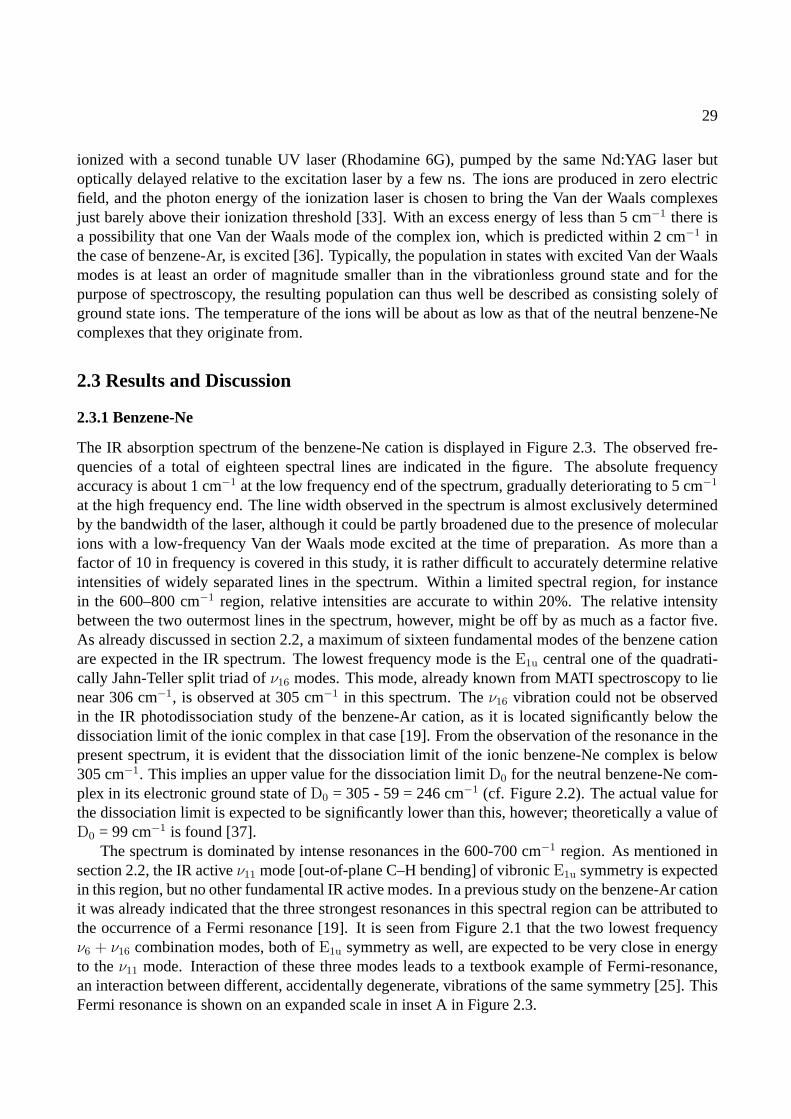
29
ionized with a second tunable UV laser (Rhodamine 6G), pumped by the same Nd:YAG laser butoptically delayed relative to the excitation laser by a few ns. The ions are produced in zero electricfield, and the photon energy of the ionization laser is chosen to bring the Van der Waals complexesjust barely above their ionization threshold [33]. With an excess energy of less than 5 cm−1 there isa possibility that one Van der Waals mode of the complex ion, which is predicted within 2 cm−1 inthe case of benzene-Ar, is excited [36]. Typically, the population in states with excited Van der Waalsmodes is at least an order of magnitude smaller than in the vibrationless ground state and for thepurpose of spectroscopy, the resulting population can thus well be described as consisting solely ofground state ions. The temperature of the ions will be about as low as that of the neutral benzene-Necomplexes that they originate from.
2.3 Results and Discussion
2.3.1 Benzene-Ne
The IR absorption spectrum of the benzene-Ne cation is displayed in Figure 2.3. The observed fre-quencies of a total of eighteen spectral lines are indicated in the figure. The absolute frequencyaccuracy is about 1 cm−1 at the low frequency end of the spectrum, gradually deteriorating to 5 cm−1
at the high frequency end. The line width observed in the spectrum is almost exclusively determinedby the bandwidth of the laser, although it could be partly broadened due to the presence of molecularions with a low-frequency Van der Waals mode excited at the time of preparation. As more than afactor of 10 in frequency is covered in this study, it is rather difficult to accurately determine relativeintensities of widely separated lines in the spectrum. Within a limited spectral region, for instancein the 600–800 cm−1 region, relative intensities are accurate to within 20%. The relative intensitybetween the two outermost lines in the spectrum, however, might be off by as much as a factor five.As already discussed in section 2.2, a maximum of sixteen fundamental modes of the benzene cationare expected in the IR spectrum. The lowest frequency mode is theE1u central one of the quadrati-cally Jahn-Teller split triad ofν16 modes. This mode, already known from MATI spectroscopy to lienear 306 cm−1, is observed at 305 cm−1 in this spectrum. Theν16 vibration could not be observedin the IR photodissociation study of the benzene-Ar cation, as it is located significantly below thedissociation limit of the ionic complex in that case [19]. From the observation of the resonance in thepresent spectrum, it is evident that the dissociation limit of the ionic benzene-Ne complex is below305 cm−1. This implies an upper value for the dissociation limitD0 for the neutral benzene-Ne com-plex in its electronic ground state ofD0 = 305 - 59 = 246 cm−1 (cf. Figure 2.2). The actual value forthe dissociation limit is expected to be significantly lower than this, however; theoretically a value ofD0 = 99 cm−1 is found [37].
The spectrum is dominated by intense resonances in the 600-700 cm−1 region. As mentioned insection 2.2, the IR activeν11 mode [out-of-plane C–H bending] of vibronicE1u symmetry is expectedin this region, but no other fundamental IR active modes. In a previous study on the benzene-Ar cationit was already indicated that the three strongest resonances in this spectral region can be attributed tothe occurrence of a Fermi resonance [19]. It is seen from Figure 2.1 that the two lowest frequencyν6 + ν16 combination modes, both ofE1u symmetry as well, are expected to be very close in energyto theν11 mode. Interaction of these three modes leads to a textbook example of Fermi-resonance,an interaction between different, accidentally degenerate, vibrations of the same symmetry [25]. ThisFermi resonance is shown on an expanded scale in inset A in Figure 2.3.
30
Wavenum
ber (c
m-1)
IR intensity (arb. units)A
B
30
95
11
80
1399
1481
1433
1067
990
1361
30
5
63
2
661
69
7
948
1295
18
15
16
29
15
02
172
1
IR intensity (arb. units)
Wavenum
ber (c
m-1)
IR intensity (arb. units)
200
400
600
800
1000
1200
1400
1600
1800
2800
3000
3200
0 13000
3050
3100
3150
0 1
600
650
700
750
0 1
Wavenum
ber (c
m-1)
Figure
2.3:IRabsorption
spectrumofthe
benzene-Ne
complex
cation.IninsetA
thespectralregion
from600–750
cm−
1is
shown
onan
expandedscale
togetherw
itha
simulation
(dashedcurve)
ofthe
Fermi-resonance
between
theIR
activeν11
mode
andthe
ν6
+ν16
combination
modes.
IninsetB
thehighestfrequency
resonanceis
shown
onan
expandedscale,together
with
adecom
positioninto
two
11cm
−1
wide
resonances(dashed
curves).
31
In the most simplified model of the Fermi resonance, it is assumed that bothν6 + ν16 combinationmodes couple only to theν11 mode with the same coupling parameter, and that only theν11 modecarries IR intensity. These assumptions result in four parameters (three unperturbed line positionsand one coupling parameterα) to fit five observables from the experimental spectrum (three linepositions and two relative line intensities). The best fit to the experimentally observed spectrum isshown as the lower (dashed) trace in inset A of Figure 2.3; the calculated stick spectrum is convolutedwith a line shape function representing the laser line width. As can be seen, fairly good agreementis obtained, and a coupling parameterα of 22 cm−1 is found. The unperturbed line position for theν11 mode is 668 cm−1, whereas the twoν6 + ν16 combination modes are found at 650 cm−1 and 669cm−1. The latter values are in good agreement with the expected unperturbed frequencies as depictedin Figure 2.1.
In the high-frequency C–H stretching region thee1u ν20 vibration, resulting inA1u, A2u andE2u
modes, as well as theb1u ν13 vibration, resulting in an IR active mode ofE2u vibronic symmetry,are expected. In Figure 2.3 a single absorption feature centered at 3097 cm−1 is observed. The fullwidth at half maximum (FWHM) of this absorption is approximately 14 cm−1, mainly determined bythe spectral profile of the IR radiation. As shown more clearly in inset B, a slight asymmetry can berecognized in the line shape, hinting at a possible overlap of more absorption lines. The absorptioncan be deconvoluted into two 11 cm−1 wide components centered at 3089 cm−1 and 3098 cm−1
with a 1:3 intensity ratio, as shown by the dashed curves in the inset. For the benzene-Ar cation, asignificantly wider absorption feature centered at 3094 cm−1 was recently reported by Dopferet al.[18]. In their experiment, Van der Waals complex ions are produced after electron impact ionization ofneutral benzene in the expansion region of a pulsed gas jet. The complex ions that are thus producedcan be at relatively high internal temperatures, leading to a broadening of the absorption lines.
Based on the known vibrational frequencies in the ground state of neutral benzene [25], the tenremaining IR active fundamental modes in the cation are all expected to lie in the 800–1600 cm−1
spectral region. In this region of the spectrum, the peak absorption frequency of ten recognizableabsorption lines is indicated in Figure 2.3. To obtain a better understanding of the observed spectralstructure in this region, density functional theory calculations are performed using the B3LYP methodwith the 6-31G* basis set, as implemented in GAUSSIAN 98 [38]. Starting with the twoD2h benzenecation structures that were calculated by Raghavachariet al. [2] the results obtained earlier by Muller-Dethlefs and Peel were reproduced [26]; for a more detailed discussion on the methods used and theinterpretation of theD2h calculations the reader is referred to ref. [26].
The calculated IR spectra for these two structures incorporate a convolution of the stick spectrumwith a Gaussian function representing the FELIX spectral profile and are displayed in Figure 2.4. Itis clearly visible that the four resonances in the 900–1200 cm−1 region are well reproduced in bothcalculated spectra and could thus be attributed to four fundamental vibrational modes of the benzenecation. A visual inspection of the calculated normal modes unfortunately does not allow for a fullassignment to specific fundamentals. The character of the calculated mode around 1045 cm−1 isthat of theν18 vibration, which seems a perfectly reasonable assignment as the neutral benzeneν18
vibration is found at 970 cm−1. On the other hand it is expected thatν18, the only IR active mode inthis region for the neutral species, would be the strongest resonance. Since none of the other threecalculated modes in this region could be assigned based on their motions, they are included in Table2.1 as unspecified fundamental modes.
In the region between 1300 cm−1 and 1600 cm−1, only two benzene fundamental modes are

32
Wavenumber (cm-1)
IR inte
nsity (
arb
. units)
A
B
C
400 600 800 1000 1200 1400 1600 1800
0
1
2
0
1
0
1
Figure 2.4: Density functional calculations of the IR absorption spectrum of the benzene cation. The ex-perimentally obtained spectrum is given in trace A. Traces B and C represent the calculated spectra for theelongated benzene structure of electronic B2g symmetry and for the compressed structure of B3g symmetry,respectively.
expected based on their frequencies in neutral benzene. One of these, the strong neutral modeν19,could be split up due to the quadratic Jahn-Teller interaction into three IR active modes, leading to atotal of four resonances. However, no symmetric splitting signature (as for the quadratically splitν16)is observed. The calculated spectrum in Figure 2.4 exhibits only three resonances in this region, noneof which can be assigned based on the molecular motion, further complicating a full assignment. The
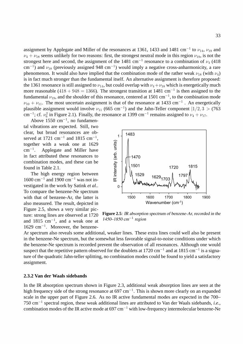
33
assignment by Applegate and Miller of the resonances at 1361, 1433 and 1481 cm−1 to ν14, ν19 andν4 + ν18 seems unlikely for two reasons: first, the strongest neutral mode in this regionν19, is not thestrongest here and second, the assignment of the 1481 cm−1 resonance to a combination ofν4 (418cm−1) andν18 (previously assigned 948 cm−1) would imply a negative cross-anharmonicity, a rarephenomenon. It would also have implied that the combination mode of the rather weakν18 (with ν4)is in fact much stronger than the fundamental itself. An alternative assignment is therefore proposed:the 1361 resonance is still assigned toν14, but could overlap withν4+ν18 which is energetically muchmore reasonable (418 + 948 = 1366). The strongest transition at 1481 cm−1 is then assigned to thefundamentalν19, and the shoulder of this resonance, centered at 1501 cm−1, to the combination modeν10 + ν11. The most uncertain assignment is that of the resonance at 1433 cm−1 . An energeticallyplausible assignment would involveν11 (665 cm−1) and the Jahn-Teller component|1/2, 3 > (763cm−1; cf. ν2
6 in Figure 2.1). Finally, the resonance at 1399 cm−1 remains assigned toν4 + ν17.
1500 1600 1700 1800 1900
0
1
Wavenumber (cm-1)
IR in
ten
sity (
arb
. u
nits)
1483
1720 1815
17971703
16291529
1501
1470
Figure 2.5: IR absorption spectrum of benzene-Ar, recorded in the1450–1850 cm−1 region
Above 1550 cm−1, no fundamen-tal vibrations are expected. Still, twoclear, but broad resonances are ob-served at 1721 cm−1 and 1815 cm−1,together with a weak one at 1629cm−1. Applegate and Miller havein fact attributed these resonances tocombination modes, and these can befound in Table 2.1.
The high energy region between1600 cm−1 and 1900 cm−1 was not in-vestigated in the work by Satinket al..To compare the benzene-Ne spectrumwith that of benzene-Ar, the latter isalso measured. The result, depicted inFigure 2.5, shows a very similar pic-ture: strong lines are observed at 1720and 1815 cm−1, and a weak one at1629 cm−1. Moreover, the benzene-Ar spectrum also reveals some additional, weaker lines. These extra lines could well also be presentin the benzene-Ne spectrum, but the somewhat less favorable signal-to-noise conditions under whichthe benzene-Ne spectrum is recorded prevent the observation of all resonances. Although one wouldsuspect that the repetitive pattern observed for the doublets at 1720 cm−1 and at 1815 cm−1 is a signa-ture of the quadratic Jahn-teller splitting, no combination modes could be found to yield a satisfactoryassignment.
2.3.2 Van der Waals sidebands
In the IR absorption spectrum shown in Figure 2.3, additional weak absorption lines are seen at thehigh frequency side of the strong resonance at 697 cm−1. This is shown more clearly on an expandedscale in the upper part of Figure 2.6. As no IR active fundamental modes are expected in the 700–750 cm−1 spectral region, these weak additional lines are attributed to Van der Waals sidebands,i.e.,combination modes of the IR active mode at 697 cm−1 with low-frequency intermolecular benzene-Ne
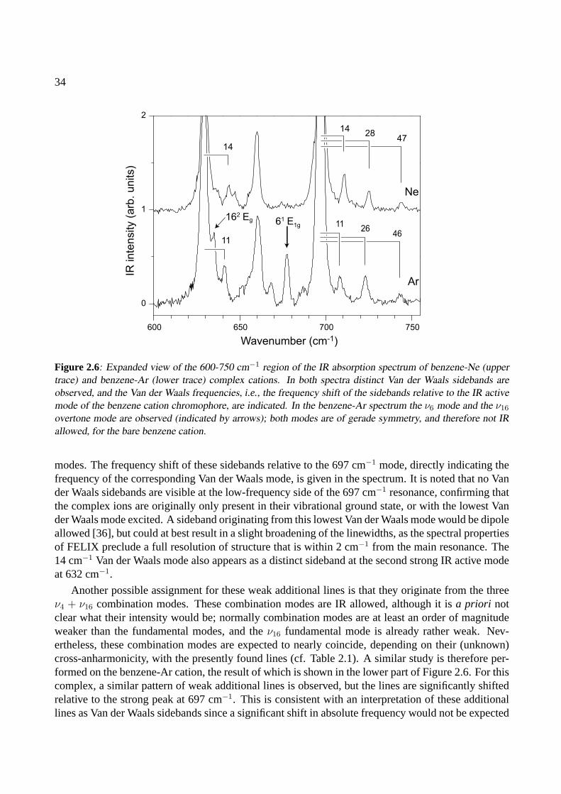
34
600 650 700 750
0
1
2
Ar
Ne
Wavenumber (cm-1)
IR in
ten
sity (
arb
. u
nits)
1428
47
1126
46
14
11
162 Eg 61 E1g
Figure 2.6: Expanded view of the 600-750 cm−1 region of the IR absorption spectrum of benzene-Ne (uppertrace) and benzene-Ar (lower trace) complex cations. In both spectra distinct Van der Waals sidebands areobserved, and the Van der Waals frequencies, i.e., the frequency shift of the sidebands relative to the IR activemode of the benzene cation chromophore, are indicated. In the benzene-Ar spectrum the ν6 mode and the ν16
overtone mode are observed (indicated by arrows); both modes are of gerade symmetry, and therefore not IRallowed, for the bare benzene cation.
modes. The frequency shift of these sidebands relative to the 697 cm−1 mode, directly indicating thefrequency of the corresponding Van der Waals mode, is given in the spectrum. It is noted that no Vander Waals sidebands are visible at the low-frequency side of the 697 cm−1 resonance, confirming thatthe complex ions are originally only present in their vibrational ground state, or with the lowest Vander Waals mode excited. A sideband originating from this lowest Van der Waals mode would be dipoleallowed [36], but could at best result in a slight broadening of the linewidths, as the spectral propertiesof FELIX preclude a full resolution of structure that is within 2 cm−1 from the main resonance. The14 cm−1 Van der Waals mode also appears as a distinct sideband at the second strong IR active modeat 632 cm−1.
Another possible assignment for these weak additional lines is that they originate from the threeν4 + ν16 combination modes. These combination modes are IR allowed, although it isa priori notclear what their intensity would be; normally combination modes are at least an order of magnitudeweaker than the fundamental modes, and theν16 fundamental mode is already rather weak. Nev-ertheless, these combination modes are expected to nearly coincide, depending on their (unknown)cross-anharmonicity, with the presently found lines (cf. Table 2.1). A similar study is therefore per-formed on the benzene-Ar cation, the result of which is shown in the lower part of Figure 2.6. For thiscomplex, a similar pattern of weak additional lines is observed, but the lines are significantly shiftedrelative to the strong peak at 697 cm−1. This is consistent with an interpretation of these additionallines as Van der Waals sidebands since a significant shift in absolute frequency would not be expected
35
if these lines originated from theν4 + ν16 combination modes. Also for the benzene-Ar complexcation, the lowest frequency Van der Waals mode can be recognized on the high frequency side of the632 cm−1 mode. The corresponding Van der Waals frequencies are therefore again indicated in thefigure. The frequencies of the benzene-Ar cation are slightly smaller than those of the Ne complexand are – with frequencies of 11 cm−1, 26 cm−1 and 46 cm−1 – considerably lower than the Van derWaals frequencies in the neutral benzene-Ar complex [39].
IR selection rules are not very helpful in trying to identify the observed Van der Waals modes.In the complex, the mirror symmetry in the plane of the benzene molecule is no longer present,lowering the symmetry fromD6h to C6v. The IR activeν11 mode ofa2u symmetry inD6h is of a1
symmetry inC6v. Motion of the noble gas atom in the plane of the benzene chromophore is ofe1
symmetry, while motion perpendicular to the plane is ofa1 symmetry. Character multiplication thenreveals that both bending and stretching modes are IR allowed as Van der Waals sidebands. Recentcalculations, performed by Van der Avoird and Lotrich, predict a bending mode at 10.1 cm−1 and astretching mode at 47.9 cm−1 [36]. These computed values, surprisingly low due to the flatness of theintermolecular potential, are in excellent agreement with the frequencies observed here. Moreover,they compare well to an earlier experimentally determined value of 48 cm−1 [40]. Van der Avoird andLotrich also predict an intense intermolecular mode at 1.7 cm−1. Although an IR induced transitionto a combination mode involving this low-frequency mode would be dipole allowed, it has not beenobserved. This could of course be due to the fact that the combination band could be indiscerniblefrom such strong resonances as the Fermi-triplet components. Additionally, Van der Avoird andLotrich predict a dissociation energy E0 = 5.74 kJ/mole (480 cm−1) for deuterated benzene-Ar, whichis in good agreement with the experimentally found upper limit of E0 = 5.80 kJ/mole (485 cm−1) [19].
2.3.3 Symmetry breaking effects
As already mentioned above, the mirror symmetry in the plane of the benzene molecule is no longerpresent in the benzene-Ne and benzene-Ar complex, lowering the symmetry in the complex fromD6h
to C6v. The most important consequence of this is that all modes of vibronic A and E symmetry inD6h, both gerade and ungerade, in principle become IR allowed in the complex cation. Interestingly,this clearly manifests itself in the spectrum of the benzene-Ar cation as shown by the appearance ofthe two peaks indicated with an arrow in Figure 2.6. The relatively strong resonance at 678 cm−1 isassigned to the high-frequency mode of the Jahn-Teller activeν6 vibration (see Figure 2.1). In thebare benzene cation, this mode hasE1g vibronic symmetry and is not IR allowed. This mode has,however, unambiguously been assigned in other experimental studies on bare benzene and its variouscomplexes [11–14]. The second manifestation of the symmetry breaking in the benzene-Ar complexis the shoulder on the high-energy side of the strong resonance at 632 cm−1. One could suspect thisto be the predicted Van der Waals band at 1.7 cm−1, but as it is only observed as a shoulder on thisparticular resonance, and as it is shifted by∼ 6 cm−1 this possibility is unlikely [36]. From Figure 2.1it can be seen, however, that in this region theν16 overtone modes, obviously of gerade character inD6h, are expected. The shoulder is assigned to the highest frequency, doubly degenerate,ν16 overtonemode, which is thus found at 635 cm−1. In the ZEKE spectrum shown by Lindneret al. [11], threesmall features are discernible at 586, 611 and 638 cm−1, which have not been assigned thus far. It ishere proposed to assign these three features to theν16 overtone modes, as indicated in Table 2.1.
Both resonances that appear due to symmetry breaking in the benzene-Ar complex, are almostabsent in the case of benzene-Ne, even though the formal breaking of symmetry is, of course, the
36
same. The effect of the symmetry breaking on the IR intensity of lines that only become allowedin the complex is expected to be significantly less for the neon complex, however, due to its loweratomic mass and in particular due to its substantially lower polarizability (0.3956A3 for Ne versus1.6411A3 for Ar [41]). Therefore, the clear difference in the spectrum of benzene-Ar compared tobenzene-Ne with respect to these two lines is the best evidence for their assignments.
To study these symmetry breaking effects somewhat further, measurements are also performed onthe benzene-Kr complex (not shown). Here one would expect an even more pronounced appearanceof the twoEg modes of the benzene cation in the IR spectrum of the Van der Waals complex. Theinterpretation of relative line-intensities in the spectrum of the benzene-Kr complex in the 600–700cm−1 spectral region is complicated, however, as the vibrational levels in this region are very close tothe dissociation limit of the benzene-Kr cation [17].
It is now also interesting to study a benzene-Arn Van der Waals complex in which the originalD6h
symmetry is retained. The obvious candidate for such a study is the benzene-Ar2 complex, for whichtwo different conformers exist [35, 42]. In one conformer, denoted as the(1|1) complex, the argonatoms are centered on the axis above and below the benzene plane. In the other conformer, denotedas the(2|0) complex, both argon atoms are located on the same side of the benzene plane. The(1|1)complex hasD6h symmetry, and it is expected that modes of gerade symmetry are rigorously absentfrom its IR absorption spectrum. To selectively prepare the(1|1) complex cation, the excitation laseris tuned to 38564.2 cm−1, which is the center of theS1(6
1)← S0 transition in the neutral benzene-Ar2
complex [35]. From the rotationally resolved spectrum as recorded and analyzed by Weberet al. ithas been concluded that this transition is that of a symmetric top molecule, and that the rotationalconstants obtained are consistent with the(1|1) structure of the complex [35]. Moreover, this tran-sition is red-shifted by some 20 cm−1 with respect to the corresponding transition in benzene-Ar, ashift that is almost identical to the 21 cm−1 red-shift observed in going from the benzene monomerto the benzene-Ar complex. The latter is strong evidence that both argon atoms are in identical posi-tions relative to the benzene plane, and that there is relatively little interaction between the two argonatoms. Therefore, it is believed that the(1|1) complex is selectively excited in this transition. Forcompleteness it should be mentioned that there is another transition of the benzene-Ar2 complex thatis found to be blue-shifted by 4 cm−1 with respect to the same transition in benzene-Ar, which hasconsequently been assigned to theS1(6
1)← S0 transition in the(2|0) conformer [42].
After preparing the(1|1) complex in the electronically excited state, ionization is performed bytuning the second UV laser just slightly above the ionization threshold, which is some 160 cm−1 lowerthan that of the benzene-Ar complex [43]; an almost identical lowering of the ionization threshold isfound in going from the benzene monomer to the benzene-Ar complex, again supporting the selectivepreparation of the(1|1) complex cation.
The IR absorption spectrum of the benzene-Ar2 (1|1) complex cation throughout the 600-750cm−1 region is depicted in Figure 2.7, together with the spectrum of the benzene-Ar cation alreadyshown earlier. The IR spectrum of the benzene-Ar2 cation is deduced through recording the appear-ance of the benzene-Ar ionic dissociation product as a function of IR wavelength; single photondissociation of the benzene-Ar2 cation into a bare benzene ion and two argon atoms is energeticallynot possible, and indeed not observed, in this frequency range.
In the spectrum of the benzene-Ar2 complex the Fermi-resonance structure of theν11 andν6 +ν16 modes discussed earlier can still be recognized. The two gerade modes of the benzene cation,indicated by arrows in the spectrum of the benzene-Ar cation, appear to be absent in the higher
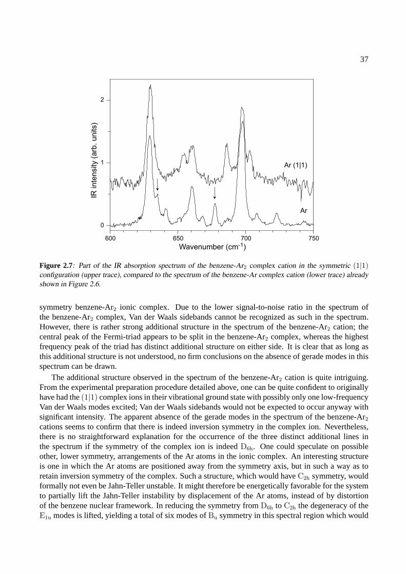
37
600 650 700 750
0
1
2
Wavenumber (cm-1)
IR in
ten
sity (
arb
. u
nits)
Ar (1|1)
Ar
Figure 2.7: Part of the IR absorption spectrum of the benzene-Ar2 complex cation in the symmetric (1|1)configuration (upper trace), compared to the spectrum of the benzene-Ar complex cation (lower trace) alreadyshown in Figure 2.6.
symmetry benzene-Ar2 ionic complex. Due to the lower signal-to-noise ratio in the spectrum ofthe benzene-Ar2 complex, Van der Waals sidebands cannot be recognized as such in the spectrum.However, there is rather strong additional structure in the spectrum of the benzene-Ar2 cation; thecentral peak of the Fermi-triad appears to be split in the benzene-Ar2 complex, whereas the highestfrequency peak of the triad has distinct additional structure on either side. It is clear that as long asthis additional structure is not understood, no firm conclusions on the absence of gerade modes in thisspectrum can be drawn.
The additional structure observed in the spectrum of the benzene-Ar2 cation is quite intriguing.From the experimental preparation procedure detailed above, one can be quite confident to originallyhave had the(1|1) complex ions in their vibrational ground state with possibly only one low-frequencyVan der Waals modes excited; Van der Waals sidebands would not be expected to occur anyway withsignificant intensity. The apparent absence of the gerade modes in the spectrum of the benzene-Ar2
cations seems to confirm that there is indeed inversion symmetry in the complex ion. Nevertheless,there is no straightforward explanation for the occurrence of the three distinct additional lines inthe spectrum if the symmetry of the complex ion is indeedD6h. One could speculate on possibleother, lower symmetry, arrangements of the Ar atoms in the ionic complex. An interesting structureis one in which the Ar atoms are positioned away from the symmetry axis, but in such a way as toretain inversion symmetry of the complex. Such a structure, which would haveC2h symmetry, wouldformally not even be Jahn-Teller unstable. It might therefore be energetically favorable for the systemto partially lift the Jahn-Teller instability by displacement of the Ar atoms, instead of by distortionof the benzene nuclear framework. In reducing the symmetry fromD6h to C2h the degeneracy of theE1u modes is lifted, yielding a total of six modes ofBu symmetry in this spectral region which would

38
400 600 800 1000 1200 1400 1600 1800
0
1 x 12
500 550 600 650
13
505
34
538
621605
590
626
1266
1229
1164
1154
1132
1721
1361
1282
1401
1437
15351637
1742
485
Wavenumber (cm-1)
IR in
ten
sity (
arb
. u
nits)
Figure 2.8: Parts of the IR absorption spectrum of the C6D6-Ar cation. For reference, the spectrum that wasmeasured by Satink et al. [19] is added in gray. In the inset, weak additional resonances on the high frequencyside of the strong IR active ν11 mode at 485 cm−1 are shown, and the frequencies of two Van der Waals modesare indicated.
couple in an, IR active, Fermi-multiplet, and which might explain the observed spectra.
2.3.4 Deuterated benzene
To get more insight into the vibrational structure of the benzene cation and to confirm some of thevibrational assignments made, IR photodissociation studies on complexes of fully deuterated benzene(C6D6) with Ar have been performed as well. Ground stateC6D6-Ar cations are produced after laserexcitation (38765 cm−1) and subsequent ionization (35650 cm−1) of the jet-cooled neutral complexes[34, 44]. A preliminary version of the IR spectrum covering the 450–1500 cm−1 range has beenpublished before [19]. In the present study, a particular focus is laid on the low frequency rangefrom 450–650 cm−1, (which is investigated with an improved spectral resolution) and on the highfrequency range, between 1000 cm−1 and 1850 cm−1 . It would also have been interesting to studythe C–D stretching vibrations, but unfortunately, at the time of the experiment the region around 2200cm−1 was not yet accessible with the IR source.
The resulting IR absorption spectrum is shown in Figure 2.8. To present a complete overview ofthe deuterated benzene-Ar spectrum, the spectrum that is recorded by Satinket al. [19] is included in
39
gray. In the low-frequency range of the spectrum, the dominant IR absorption peak observed at 485cm−1 can unambiguously be assigned to theν11 [out-of-plane C–H bending] mode, found in neutralC6D6 at 496 cm−1 [25]. The peak positions of four resonances in the 600 cm−1 region are indicatedin the figure as well. The resonances at 621 cm−1 and 626 cm−1 could not be resolved as separateones in an earlier study [19]. In addition, the line shape of the 605 cm−1 resonance suggests that itmight be composed of more than one mode. In trying to assign all these observed modes, it has tobe realized that the two lowestν6 + ν16 combination modes are expected in this region, borrowingintensity from theν11 mode via a Fermi-resonance, just as in the protonated benzene cation. Apartfrom these modes, the threeν4 + ν16 combination modes fall in this region. A consistent picture isobtained when the modes at 605 cm−1 and 621 cm−1 are assigned asν6 + ν16 combination modes.In the Fermi-resonance model described earlier, now with a slightly increased coupling parameterαof 23 cm−1, unperturbed values of 494 cm−1 for theν11 mode and 601 cm−1 and 616 cm−1 for theν6 + ν16 combination modes are obtained. The latter values are in good agreement with the valuesexpected from a two-mode Jahn-Teller simulation [13]. The absorption at 590 cm−1, the (blended)absorption at 605 cm−1 and the absorption at 626 cm−1 are then assigned to theν4 + ν16 combinationmodes, havingA2u, E2u andA1u symmetry, respectively. All these assignments are indicated in Table2.2.
On the high-frequency side of the 485 cm−1 resonance, additional structure is observed as shownon an expanded scale in the inset to Figure 2.8. This structure could be the Van der Waals sidebandsof the strongν11 mode. However, the resonances at 506 cm−1 and 538 cm−1 have also been reportedin ZEKE studies on the bare deuterated benzene cation [30]. There, the only vibrational modes thatcan occur in this spectral region are theν16 overtone modes. Although these modes are of geradesymmetry in the bare benzene cation, we already know from our work on protonated benzene-Arcomplex cations, that such modes can appear in the IR spectrum due to symmetry breaking in thecomplex. We therefore attribute these two absorptions to the two lowestν16 overtone modes; an addi-tional mode observed by Lindneret al. at 562 cm−1 is most likely the highest frequencyν16 overtonemode (see Table 2.2). The remaining two resonances in the low-frequency part of the spectrum areattributed to intermolecular Van der Waals vibrations, and their frequency offset relative to the 485cm−1 peak is given in the figure, indicating Van der Waals modes at 13 cm−1 and at 34 cm−1. Thesevalues are higher than those of protonated benzene, which is contrary to the expectations as the re-duced mass is increased [36]. It is noted, however, that the character of the intra-molecular mode(pure out-of-plane C–D bending) to which the Van der Waals modes appear as sidebands is rather dif-ferent than the character of the intra-molecular modes for which sidebands are observed in protonatedbenzene (theν6 + ν16–ν11 Fermi triad). This difference may lead to the appearance of different Vander Waals modes as sidebands. It may prove necessary to perform a similar experiment on deuteratedbenzene-Ne to ascertain the present (conflicting) assignment.
The high frequency range is dominated by two strong resonances at 1229 cm−1 and 1401 cm−1.It is interesting to note that, although they are also the strongest observed resonances in the work ofSatinket al., the relative intensities are different. One reason for this could be that the settings ofFELIX in the present study are optimized for a higher frequency range than in the previous work; thebandwidth in the high-frequency range is then narrower and the peak intensities observed in that rangeare higher. It can be seen in Figure 2.8 that the integrated intensities for the two recorded spectra aresimilar, supporting this hypothesis.
For the assignment some major changes are proposed to the previous work by Applegate and
40
Miller. First, it seems somewhat awkward that in the protonated species bothν14 andν19 are assignedto strong resonances, whereas forC6D6 ν14 has not been identified. Second, there are two reasonablyalike pairs of resonances, one at 1266 and 1282 cm−1, and one at 1721 and 1742 cm−1, respectively.The spacings and relative intensities in these sets are quite similar and are reminiscent of the quadraticsplittings that are observed for the linearly Jahn-Teller active modes. Indeed, Applegate and Millerassign two sets of ZEKE resonances, (338,357 cm−1) and (786,797 cm−1) to quadratically split Jahn-Teller modes. In combination with an IR active modes at 485 cm−1 (ν11) the latter set yields 1271 and1282 cm−1; the former set gives rise to the high-frequency resonances in combination with a strongIR active mode at 1385 cm−1. Taking into account a slight anharmonicity in the combination modefrequency, the resonance at 1401 cm−1 is a perfectly reasonable partner. It is, however, unlikely that acombination band (as this is what Applegate and Miller assigned the 1401 cm−1 resonance to) is thesource of yet another combination. It is therefore proposed to assign the resonance at 1401 cm−1 toν19 and that at 1229 cm−1 to ν14.
2.3.5 Hot band spectroscopy of benzene-Ar
The combined experimental work on the C6H6 cation has resulted in the assignment of all modesbelow 800 cm−1, as well as in the assignment of some additional strong resonances like theν1 modeof E1g symmetry at 969 cm−1 (see Table 2.1). Many more resonances have been observed, but afurther unambiguous identification has proven to be difficult. IR absorption spectroscopy has theobvious advantage that it has rigorous selection rules, as opposed to ZEKE and MATI spectroscopy,where the appearance of lines in the spectra is governed by Franck-Condon overlap of the wavefunctions of the corresponding vibronic levels.
To further exploit the possibilities offered by IR photodissociation spectroscopy, vibronic levels ofdifferent symmetry can be used as initial states in the IR absorption process. Obviously, the initiallyexcited vibrational state for such an experiment should lie below the dissociation limit of the complexto prevent dissociation before the actual IR transition can be induced. The preparation of a puresample of ions in one selected vibrationally excited state,i.e., without any population in the vibrationalground state of the ion, is experimentally difficult. It is relatively straightforward, however, to producea sample of ions with a known distribution over a selected set of vibrational levels. By measuring theIR absorption spectrum of this sample, a type of ‘hot-band’ spectroscopy is performed.
In this experiment, neutral benzene-Ar complexes are resonantly excited as described earlier, butnow the ionization laser is tuned to a frequency several hundred cm−1 above the ionization threshold.Specifically, the frequency of the ionization laser is tuned slightly above that required to produceionic complexes in a selected vibrationally excited state. In this way, ions are produced in a mixtureof vibrational states; ground state ions are produced when the electron carries away the maximumpossible excess energy, whereas vibrationally excited ions in the targeted state are prepared whenelectrons with near-zero kinetic energy are produced. The vibrational population distribution that isthus created is completely governed by Franck-Condon factors, which are well known from, e.g.,ZEKE spectroscopy measurements [11].
In the first experiment of this type, the ionization laser is tuned some 350 cm−1 above the ion-ization threshold for the benzene-Ar complex. As can be seen from Table 2.1, ions can then beprepared in the lowest energyν6 mode ofB1g symmetry. Obviously, it is energetically also possibleto produce ions in any of the lower lying vibrational levels, e.g., in the vibrational ground state as wellas in the threeν16 modes around 300 cm−1. Based on the results of ZEKE-measurements [11] it is
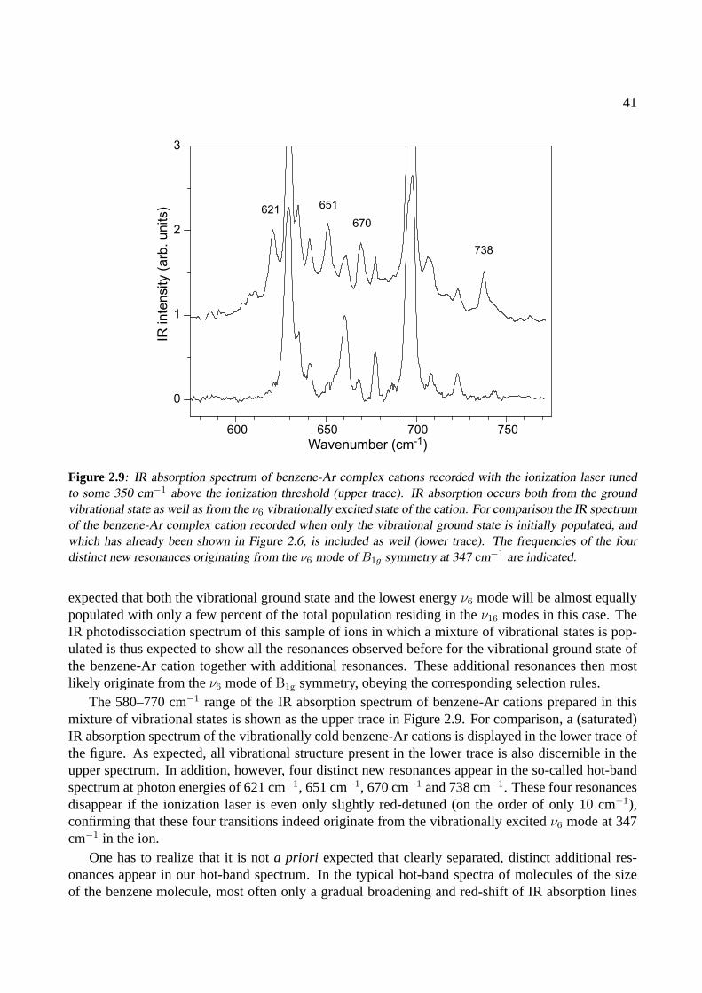
41
600 650 700 750
0
1
2
3
Wavenumber (cm-1)
IR inte
nsity (
arb
. units) 621 651
670
738
Figure 2.9: IR absorption spectrum of benzene-Ar complex cations recorded with the ionization laser tunedto some 350 cm−1 above the ionization threshold (upper trace). IR absorption occurs both from the groundvibrational state as well as from the ν6 vibrationally excited state of the cation. For comparison the IR spectrumof the benzene-Ar complex cation recorded when only the vibrational ground state is initially populated, andwhich has already been shown in Figure 2.6, is included as well (lower trace). The frequencies of the fourdistinct new resonances originating from the ν6 mode of B1g symmetry at 347 cm−1 are indicated.
expected that both the vibrational ground state and the lowest energyν6 mode will be almost equallypopulated with only a few percent of the total population residing in theν16 modes in this case. TheIR photodissociation spectrum of this sample of ions in which a mixture of vibrational states is pop-ulated is thus expected to show all the resonances observed before for the vibrational ground state ofthe benzene-Ar cation together with additional resonances. These additional resonances then mostlikely originate from theν6 mode ofB1g symmetry, obeying the corresponding selection rules.
The 580–770 cm−1 range of the IR absorption spectrum of benzene-Ar cations prepared in thismixture of vibrational states is shown as the upper trace in Figure 2.9. For comparison, a (saturated)IR absorption spectrum of the vibrationally cold benzene-Ar cations is displayed in the lower trace ofthe figure. As expected, all vibrational structure present in the lower trace is also discernible in theupper spectrum. In addition, however, four distinct new resonances appear in the so-called hot-bandspectrum at photon energies of 621 cm−1, 651 cm−1, 670 cm−1 and 738 cm−1. These four resonancesdisappear if the ionization laser is even only slightly red-detuned (on the order of only 10 cm−1),confirming that these four transitions indeed originate from the vibrationally excitedν6 mode at 347cm−1 in the ion.
One has to realize that it is nota priori expected that clearly separated, distinct additional res-onances appear in our hot-band spectrum. In the typical hot-band spectra of molecules of the sizeof the benzene molecule, most often only a gradual broadening and red-shift of IR absorption lines
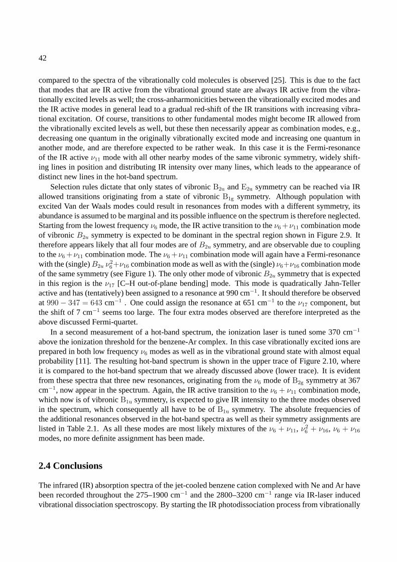
42
compared to the spectra of the vibrationally cold molecules is observed [25]. This is due to the factthat modes that are IR active from the vibrational ground state are always IR active from the vibra-tionally excited levels as well; the cross-anharmonicities between the vibrationally excited modes andthe IR active modes in general lead to a gradual red-shift of the IR transitions with increasing vibra-tional excitation. Of course, transitions to other fundamental modes might become IR allowed fromthe vibrationally excited levels as well, but these then necessarily appear as combination modes, e.g.,decreasing one quantum in the originally vibrationally excited mode and increasing one quantum inanother mode, and are therefore expected to be rather weak. In this case it is the Fermi-resonanceof the IR activeν11 mode with all other nearby modes of the same vibronic symmetry, widely shift-ing lines in position and distributing IR intensity over many lines, which leads to the appearance ofdistinct new lines in the hot-band spectrum.
Selection rules dictate that only states of vibronicB2u andE2u symmetry can be reached via IRallowed transitions originating from a state of vibronicB1g symmetry. Although population withexcited Van der Waals modes could result in resonances from modes with a different symmetry, itsabundance is assumed to be marginal and its possible influence on the spectrum is therefore neglected.Starting from the lowest frequencyν6 mode, the IR active transition to theν6 +ν11 combination modeof vibronicB2u symmetry is expected to be dominant in the spectral region shown in Figure 2.9. Ittherefore appears likely that all four modes are ofB2u symmetry, and are observable due to couplingto theν6 +ν11 combination mode. Theν6 +ν11 combination mode will again have a Fermi-resonancewith the (single)B2u ν
26+ν16 combination mode as well as with the (single)ν6+ν16 combination mode
of the same symmetry (see Figure 1). The only other mode of vibronicB2u symmetry that is expectedin this region is theν17 [C–H out-of-plane bending] mode. This mode is quadratically Jahn-Telleractive and has (tentatively) been assigned to a resonance at 990 cm−1. It should therefore be observedat 990− 347 = 643 cm−1 . One could assign the resonance at 651 cm−1 to theν17 component, butthe shift of 7 cm−1 seems too large. The four extra modes observed are therefore interpreted as theabove discussed Fermi-quartet.
In a second measurement of a hot-band spectrum, the ionization laser is tuned some 370 cm−1
above the ionization threshold for the benzene-Ar complex. In this case vibrationally excited ions areprepared in both low frequencyν6 modes as well as in the vibrational ground state with almost equalprobability [11]. The resulting hot-band spectrum is shown in the upper trace of Figure 2.10, whereit is compared to the hot-band spectrum that we already discussed above (lower trace). It is evidentfrom these spectra that three new resonances, originating from theν6 mode ofB2g symmetry at 367cm−1, now appear in the spectrum. Again, the IR active transition to theν6 + ν11 combination mode,which now is of vibronicB1u symmetry, is expected to give IR intensity to the three modes observedin the spectrum, which consequently all have to be ofB1u symmetry. The absolute frequencies ofthe additional resonances observed in the hot-band spectra as well as their symmetry assignments arelisted in Table 2.1. As all these modes are most likely mixtures of theν6 + ν11, ν2
6 + ν16, ν6 + ν16
modes, no more definite assignment has been made.
2.4 Conclusions
The infrared (IR) absorption spectra of the jet-cooled benzene cation complexed with Ne and Ar havebeen recorded throughout the 275–1900 cm−1 and the 2800–3200 cm−1 range via IR-laser inducedvibrational dissociation spectroscopy. By starting the IR photodissociation process from vibrationally
43
600 650 700 750
0
1
2
3
Wavenumber (cm-1)
IR inte
nsity (
arb
. units)
591
651
724
Figure 2.10: IR absorption spectrum of benzene-Ar complex cations recorded with the ionization laser tunedto some 370 cm−1 above the ionization threshold (upper trace). IR absorption occurs both from the groundvibrational state as well as from the two ν6 vibrationally excited states of the cation. For comparison the IRspectrum of the benzene-Ar complex cation recorded when only the vibrational ground state and the lowestvibrationally excited ν6 state are initially populated (see Figure 2.9), is included as well (lower trace). Thefrequencies of the three distinct new resonances originating from the ν6 mode of B2g symmetry at 367 cm−1
are indicated.
excited complex cations, additional information on the vibrational structure has been obtained. Thecombined results from the work presented here and previous ZEKE and MATI studies on benzenecations have made a complete assignment of all observed vibrational modes below 800 cm−1 possi-ble. It has also enabled a tentative first assignment of the benzene cation spectrum up to 1900 cm−1.A detailed comparison between the IR absorption spectra of benzene-Ne and benzene-Ar in selectedspectral regions convincingly demonstrates that the spectrum of the benzene-Ne cation indeed repre-sents the IR absorption spectrum of the bare benzene cation. In using Ne as a messenger atom in theIR photodissociation technique, Van der Waals sidebands can appear in the spectrum, but no effectsof the formal symmetry breaking in the complex due to the presence of the messenger atom have beenobserved. We therefore conclude that, to within our present spectral resolution, the spectrum of thebenzene-Ne cation as shown in Figure 2.3 is thetrue and complete IR spectrum of the benzene cat-ion, and should serve as a benchmark for future analyses of the vibrational properties of this importantspecies.
44
References
[1] H. Jahn and E. Teller, Proc. Royal Soc. London A161, 220 (1937).
[2] K. Raghavachari, R. Haddon, T. A. Miller, and V. Bondybey, J. Chem. Phys.79, 1387 (1983).
[3] J. Cernicharo, A. M. Heras, A. G. G. M. Tielens, J. R. Pardo, F. Herpin, M. Guelin, and L. B. F. M. Waters,Ap. J.546, L123 (2001).
[4] A. Leger and J. L. Puget, Astron. Astrophys.137, L5 (1984).
[5] L. J. Allamandola, A. G. G. M. Tielens, and J. R. Barker, Ap. J.290, L25 (1985).
[6] O. Braltbart, E. Castellucci, G. Dujardin, and S. Leach, J. Phys. Chem.87, 4799 (1983).
[7] T. A. Miller and V. Bondybey,Molecular Ions: Spectroscopy, Structure and Chemistry(North-HollandPublishing Company, 1983), chap. The Jahn-Teller Effect in Benzenoid Cations: Theory and Experiment,p. 201.
[8] S. R. Long, J. T. Meek, and J. P. Reilly, J. Chem. Phys.79, 3206 (1983).
[9] G. Muller, J. Y. Fan, J. L. Lyman, W. E. Schmidt, and K. L. Kompa, J. Chem. Phys.90, 3490 (1989).
[10] J. Le Calve, M. Schmidt, and M. Mons, J. Phys. Chem.96, 4131 (1992).
[11] R. Lindner, K. Muller-Dethlefs, E. Wedum, K. Haber, and E. Grant, Science271, 1698 (1996).
[12] H. Krause and H. J. Neusser, J. Chem. Phys.97, 5923 (1992).
[13] J. G. Goode, J. D. Hofstein, and P. M. Johnson, J. Chem. Phys.107, 1703 (1997).
[14] K. Siglow and H. J. Neusser, J. El. Spect. Relat. Phenom.112, 199 (2000).
[15] C. H. Kwon, H. L. Kim, and M. S. Kim, J. Chem. Phys.119, 215 (2003).
[16] A. B. Burrill, J. T. Zhou, and P. M. Johnson, J. Phys. Chem. A107, 4601 (2003).
[17] H. Krause and H. J. Neusser, J. Chem. Phys.99, 6278 (1993).
[18] O. Dopfer, R. V. Olkhov, and J. P. Maier, J. Chem. Phys.111, 10754 (1999).
[19] R. G. Satink, H. Piest, G. von Helden, and G. Meijer, J. Chem. Phys.111, 10750 (1999).
[20] T. A. Barckholtz and T. A. Miller, Int. Rev. of Phys. Chem.17, 435 (1998).
[21] B. E. Applegate, T. A. Barckholtz, and T. A. Miller, Chem. Soc. Rev.32, 38 (2003).
[22] B. E. Applegate and T. A. Miller, J. Chem. Phys.117, 10654 (2002).
[23] P. M. Johnson, J. Chem. Phys.117, 9991 (2002).
[24] E. B. Wilson, Jr., Phys. Rev.45, 706 (1934).
[25] G. Herzberg,Molecular Spectra and Molecular Structure(Krieger Publishing company, Malabar, FLA,1991), 2nd ed.
45
[26] K. Muller-Dethlefs and J. B. Peel, J. Chem. Phys.111, 10550 (1999).
[27] M. Ford, R. Lindner, and K. Muller-Dethlefs, Mol. Phys.101, 705 (2003).
[28] H. Longuet-Higgins, Adv. Spectrosc.2, 429 (1961).
[29] H. Krause, Ph.D. thesis, Technische Universitat Munchen (1993).
[30] R. Lindner, Ph.D. thesis, Technische Universitat Munchen (1996).
[31] K. Siglow, Ph.D. thesis, Technische Universitat Munchen (2000).
[32] T. Weber, E. Riedle, H. J. Neusser, and E. Schlag, J. Mol. Struct.249, 69 (1991).
[33] K. Siglow, R. Neuhauser, and H. J. Neusser, Chem. Phys. Lett.293, 19 (1998).
[34] T. Weber, A. V. Bargen, E. Riedle, and H. J. Neusser, J. Chem. Phys.92, 90 (1990).
[35] T. Weber and H. J. Neusser, J. Chem. Phys.94, 7689 (1991).
[36] A. van der Avoird and V. F. Lotrich, J. Chem. Phys.120, 10069 (2004).
[37] P. Hobza, O. Bludsky, K. Selzle, and E. Schlag, J. Chem. Phys.97, 335 (1992).
[38] M. J. Frisch, G. W. Trucks, H. B. Schlegel, G. E. Scuseria, M. A. Robb, J. R. Cheeseman, V. G. Za-krzewski, J. J. A. Montgomery, R. E. Stratmann, J. C. Burant, et al.,Gaussian 98, Revision A.7, Pittsburgh,PA (1998).
[39] R. Neuhauser, J. Braun, H. J. Neusser, and A. van der Avoird, J. Chem. Phys.108, 8408 (1998).
[40] R. Neuhauser, K. Siglow, and H. J. Neusser, Phys. Rev. Lett.80, 5089 (1998).
[41] Handbook of Chemistry and Physics(CRC press, Boca Raton, FL, 1993-1994), 74th ed.
[42] M. Schmidt, M. Mons, and J. L. Calve, Chem. Phys. Lett.177, 371 (1991).
[43] H. J. Neusser and H. Krause, Chem. Rev.94, 1829 (1994).
[44] K. Siglow, R. Neuhauser, and H. J. Neusser, J. Chem. Phys.110, 5589 (1999).
46
Assignment Symmetry Krause Lindner Goode Siglow This work Kwon& Satink
[12, 29] [11, 30] [13] [14, 31] [19] [15]
ν16 B1u 285 289 292 296ν16 E1u 303 306 306 305 305ν16 B2u 328 327|3/2, 1 > B1g 343 347 350 350|3/2, 2 > B2g 363 367 370 363ν4 E2g 416 418 422 420ν216 586 596
ν216 608
ν16 + |3/2, 1 >, |3/2, 2 > /ν11 ‡ E1u 626 630 632 632ν216 638
ν16 + |3/2, 1 >, |3/2, 2 > /ν11‡ E1u 659 660 663 661
|1/2, 1 > E1g 674 677 677 678ν16 + |3/2, 1 >, |3/2, 2 > /ν11
‡ E1u 696 700 697 697ν10 A1g 724 727 724|1/2, 3 > E1g 763 761ν10 E2g 843 843ν24 E2g 847
853ν12 E2u 883 878ν5 E2g 934ν∗ A1u, A2u, E1u 948
B1u 958†
ν1 E1g 967 969 969 967B2u 968†
ν10 A2g 983 986ν∗ 994 990
B2u 998†
B2u 1017†
B1u 1018†
|3/2, 3 > 1018|3/2, 4 > 1031ν∗ 1067|1/2, 4 > 1069 1073 1073 1072
B2u 1089 1085†
B1u 1091†
ν∗ 1180 1183|3/2, 5 > 1228 1230|3/2, 6 > 1245|1/2, 6 > 1242 1257 1255ν16 + ν17 B1g, B2g, E1g 1286 1283ν18 + |3/2, 1 > E2u 1297 1295ν1 + |3/2, 1 > B1g 1303 1305ν1 + |3/2, 2 > B2g 1326 1328 1322ν14, ν4 + ν18 E2u, E1u 1356 1361 1357ν4 + ν17 A1u, A2u, E2u 1399 1393|3/2, 7 > 1408ν11 + |1/2, 3 > 1433 1420|3/2, 8 > 1435
1440ν19 A1u, A2u, E2u 1481 1475ν10 + ν11 A1u, A2u, E2u 1501ν4 + ν11 E2u 1522 1529ν1 + ν11 E1u 1629
1648ν11 + |3/2, 3 > or ν14 + |3/2, 1 > 1704ν11 + |3/2, 4 > or ν14 + |3/2, 2 > 1721
1797 1786ν17 + ν2
18 1815 1825ν20 A2u 3089ν13 E2u 3098
Table 2.1: Assignment of observed vibrational frequencies (in cm−1) for the benzene cation in different ex-periments. The resonances labeled with ν∗ are assigned to unspecified fundamental modes (cf. section 2.3.1).Frequencies labeled with † are deduced from the hot-band spectroscopy described in section 2.3.5.
47
Assignment Symmetry Krause Lindner Goode This work Kwon& Satink
[12, 29] [11, 30] [13] [19] [15]
ν16 B1u 245 249 245 252ν16 E1u 262 265 262 263ν16 B2u 286 289 285 289|3/2, 1 > B1g 336 338 335 334ν4 E2g 340 344 343|3/2, 2 > B2g 353 357ν11 E1u 485 491ν216 Eg 505 506 509
ν216 Ag 540 538 550
ν216 Eg 562
ν10 A1g 562 550ν4 + ν16 A2u 588 590ν4 + ν16, ν16 + |3/2, 1 > E2u, E1u 601 604 603 605ν16 + |3/2, 1 > E1u 622 620 621ν4 + ν16 A1u 626|1/2, 2 > A2u 633 636 634ν10 E2g 673 673 658ν24 E1g 697 680
|1/2, 3 > 739ν5 E2g 754 750ν18 E2u 773 766|3/2, 3 > 786|3/2, 4 > 797ν17 E1u 813ν4 + ν11 E2u 823ν15 E2u 835 840|1/2, 4 > 868ν12 E2u 877ν16 + |1/2, 2 > 887ν16 + |1/2, 2 > 900ν16 + |1/2, 2 > 922 924ν1 E1g 926 926|1/2, 5 > 1058|3/2, 5 > 1119ν18 + |3/2, 1 > E2u 1132|3/2, 6 > 1143ν18 + |3/2, 2 > E2u 1154ν4 + ν17 A1u, A2u, E2u 1164ν14, ν11 + |1/2, 3 > E2u 1229ν11 + |3/2, 3 > 1266|1/2, 8 > 1266ν11 + |3/2, 4 > 1282|3/2, 7 > 1297|3/2, 8 > 1316ν19, ν18 + |1/2, 2 > A1u, A2u, E2u 1401ν17 + |1/2, 2 > 1437|3/2, 11 > 1545|3/2, 12 > 1563ν5 + ν12, ν4 + ν14, ν18 + |1/2, 4 > 1637ν19 + |3/2, 1 > E2u 1722ν19 + |3/2, 2 > E2u 1738
Table 2.2: Assignment of observed vibrational frequencies for the deuterated benzene cation in different ex-periments.

48
CHAPTER 3
THE IR ABSORPTION SPECTRUM OF THE GAS PHASE
NEUTRAL BENZENE DIMER
The infrared (IR) absorption spectrum of the jet-cooled benzene dimer has been recorded throughoutthe 300–1700 cm−1 range via ion-dip spectroscopy. The spectrum for (C6H6)2 closely resembles thatof the neutral benzene molecule and that of C6H6–C6D6 can be described as a combination of twomonomer spectra. On close inspection several resonances appear in spectra for both species that areIR forbidden in the monomer. The spectra can be favorably compared to calculated spectra for adistorted T-shaped geometry. Evidence has been found for one intermolecular vibration for the fullyprotonated dimer with an energy of 51 cm−1.
49
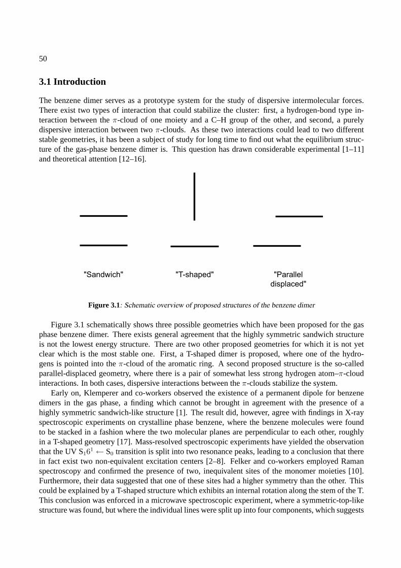
50
3.1 Introduction
The benzene dimer serves as a prototype system for the study of dispersive intermolecular forces.There exist two types of interaction that could stabilize the cluster: first, a hydrogen-bond type in-teraction between theπ-cloud of one moiety and a C–H group of the other, and second, a purelydispersive interaction between twoπ-clouds. As these two interactions could lead to two differentstable geometries, it has been a subject of study for long time to find out what the equilibrium struc-ture of the gas-phase benzene dimer is. This question has drawn considerable experimental [1–11]and theoretical attention [12–16].
"Sandwich" "T-shaped" "Parallel
displaced"
Figure 3.1: Schematic overview of proposed structures of the benzene dimer
Figure 3.1 schematically shows three possible geometries which have been proposed for the gasphase benzene dimer. There exists general agreement that the highly symmetric sandwich structureis not the lowest energy structure. There are two other proposed geometries for which it is not yetclear which is the most stable one. First, a T-shaped dimer is proposed, where one of the hydro-gens is pointed into theπ-cloud of the aromatic ring. A second proposed structure is the so-calledparallel-displaced geometry, where there is a pair of somewhat less strong hydrogen atom–π-cloudinteractions. In both cases, dispersive interactions between theπ-clouds stabilize the system.
Early on, Klemperer and co-workers observed the existence of a permanent dipole for benzenedimers in the gas phase, a finding which cannot be brought in agreement with the presence of ahighly symmetric sandwich-like structure [1]. The result did, however, agree with findings in X-rayspectroscopic experiments on crystalline phase benzene, where the benzene molecules were foundto be stacked in a fashion where the two molecular planes are perpendicular to each other, roughlyin a T-shaped geometry [17]. Mass-resolved spectroscopic experiments have yielded the observationthat the UV S161 ← S0 transition is split into two resonance peaks, leading to a conclusion that therein fact exist two non-equivalent excitation centers [2–8]. Felker and co-workers employed Ramanspectroscopy and confirmed the presence of two, inequivalent sites of the monomer moieties [10].Furthermore, their data suggested that one of these sites had a higher symmetry than the other. Thiscould be explained by a T-shaped structure which exhibits an internal rotation along the stem of the T.This conclusion was enforced in a microwave spectroscopic experiment, where a symmetric-top-likestructure was found, but where the individual lines were split up into four components, which suggests
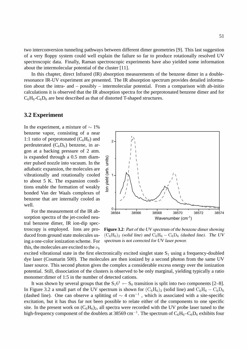
51
two interconversion tunneling pathways between different dimer geometries [9]. This last suggestionof a very floppy system could well explain the failure so far to produce rotationally resolved UVspectroscopic data. Finally, Raman spectroscopic experiments have also yielded some informationabout the intermolecular potential of the cluster [11].
In this chapter, direct Infrared (IR) absorption measurements of the benzene dimer in a double-resonance IR-UV experiment are presented. The IR absorption spectrum provides detailed informa-tion about the intra- and – possibly – intermolecular potential. From a comparison with ab-initiocalculations it is observed that the IR absorption spectra for the perprotonated benzene dimer and forC6H6-C6D6 are best described as that of distorted T-shaped structures.
3.2 Experiment
38564 38566 38568 38570 38572 38574
0
1
2
Wavenumber (cm-1)
Ion
yie
ld (
arb
. u
nits)
Figure 3.2: Part of the UV spectrum of the benzene dimer showing(C6H6)2 (solid line) and C6H6 − C6D6 (dashed line). The UVspectrum is not corrected for UV laser power.
In the experiment, a mixture of∼ 1%benzene vapor, consisting of a near1:1 ratio of perprotonated (C6H6) andperdeuterated (C6D6) benzene, in ar-gon at a backing pressure of 2 atm.is expanded through a 0.5 mm diam-eter pulsed nozzle into vacuum. In theadiabatic expansion, the molecules arevibrationally and rotationally cooledto about 5 K. The expansion condi-tions enable the formation of weaklybonded Van der Waals complexes ofbenzene that are internally cooled aswell.
For the measurement of the IR ab-sorption spectra of the jet-cooled neu-tral benzene dimer, IR ion-dip spec-troscopy is employed. Ions are pro-duced from ground state molecules us-ing a one-color ionization scheme. Forthis, the molecules are excited to theν6
excited vibrational state in the first electronically excited singlet state S1 using a frequency-doubleddye laser (Coumarin 500). The molecules are then ionized by a second photon from the same UVlaser source. This second photon gives the complex a considerable excess energy over the ionizationpotential. Still, dissociation of the clusters is observed to be only marginal, yielding typically a ratiomonomer:dimer of 1:5 in the number of detected cations.
It was shown by several groups that the S161 ← S0 transition is split into two components [2–8].
In Figure 3.2 a small part of the UV spectrum is shown for(C6H6)2 (solid line) andC6H6 − C6D6
(dashed line). One can observe a splitting of∼ 4 cm−1 , which is associated with a site-specificexcitation, but it has thus far not been possible to relate either of the components to one specificsite. In the present work on (C6H6)2, all spectra were recorded with the UV probe laser tuned to thehigh-frequency component of the doublets at 38569 cm−1. The spectrum of C6H6–C6D6 exhibits four
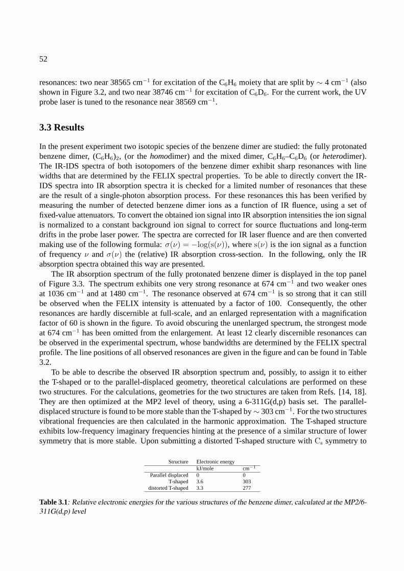
52
resonances: two near 38565 cm−1 for excitation of the C6H6 moiety that are split by∼ 4 cm−1 (alsoshown in Figure 3.2, and two near 38746 cm−1 for excitation of C6D6. For the current work, the UVprobe laser is tuned to the resonance near 38569 cm−1.
3.3 Results
In the present experiment two isotopic species of the benzene dimer are studied: the fully protonatedbenzene dimer, (C6H6)2, (or thehomodimer) and the mixed dimer, C6H6–C6D6 (or heterodimer).The IR-IDS spectra of both isotopomers of the benzene dimer exhibit sharp resonances with linewidths that are determined by the FELIX spectral properties. To be able to directly convert the IR-IDS spectra into IR absorption spectra it is checked for a limited number of resonances that theseare the result of a single-photon absorption process. For these resonances this has been verified bymeasuring the number of detected benzene dimer ions as a function of IR fluence, using a set offixed-value attenuators. To convert the obtained ion signal into IR absorption intensities the ion signalis normalized to a constant background ion signal to correct for source fluctuations and long-termdrifts in the probe laser power. The spectra are corrected for IR laser fluence and are then convertedmaking use of the following formula:σ(ν) = −log(s(ν)), wheres(ν) is the ion signal as a functionof frequencyν andσ(ν) the (relative) IR absorption cross-section. In the following, only the IRabsorption spectra obtained this way are presented.
The IR absorption spectrum of the fully protonated benzene dimer is displayed in the top panelof Figure 3.3. The spectrum exhibits one very strong resonance at 674 cm−1 and two weaker onesat 1036 cm−1 and at 1480 cm−1. The resonance observed at 674 cm−1 is so strong that it can stillbe observed when the FELIX intensity is attenuated by a factor of 100. Consequently, the otherresonances are hardly discernible at full-scale, and an enlarged representation with a magnificationfactor of 60 is shown in the figure. To avoid obscuring the unenlarged spectrum, the strongest modeat 674 cm−1 has been omitted from the enlargement. At least 12 clearly discernible resonances canbe observed in the experimental spectrum, whose bandwidths are determined by the FELIX spectralprofile. The line positions of all observed resonances are given in the figure and can be found in Table3.2.
To be able to describe the observed IR absorption spectrum and, possibly, to assign it to eitherthe T-shaped or to the parallel-displaced geometry, theoretical calculations are performed on thesetwo structures. For the calculations, geometries for the two structures are taken from Refs. [14, 18].They are then optimized at the MP2 level of theory, using a 6-311G(d,p) basis set. The parallel-displaced structure is found to be more stable than the T-shaped by∼ 303 cm−1. For the two structuresvibrational frequencies are then calculated in the harmonic approximation. The T-shaped structureexhibits low-frequency imaginary frequencies hinting at the presence of a similar structure of lowersymmetry that is more stable. Upon submitting a distorted T-shaped structure withCs symmetry to
Structure Electronic energykJ/mole cm−1
Parallel displaced 0 0T-shaped 3.6 303
distorted T-shaped 3.3 277
Table 3.1: Relative electronic energies for the various structures of the benzene dimer, calculated at the MP2/6-311G(d,p) level
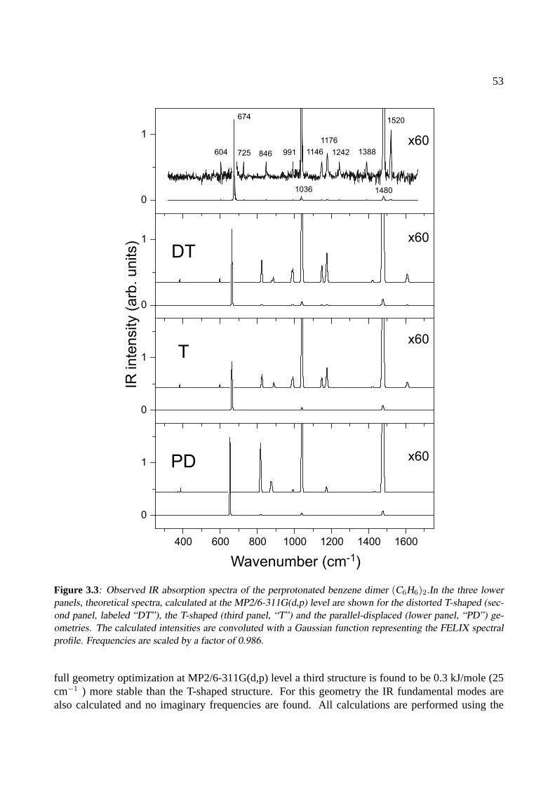
53
400 600 800 1000 1200 1400 1600
DT
T
PD
Wavenumber (cm-1)
604
674
725 846 991
1036
1146
1176
1242 1388
1480
1520
IR in
ten
sity (
arb
. u
nits) x60
x60
x60
x60
0
1
0
1
0
1
0
1
Figure 3.3: Observed IR absorption spectra of the perprotonated benzene dimer (C6H6)2.In the three lowerpanels, theoretical spectra, calculated at the MP2/6-311G(d,p) level are shown for the distorted T-shaped (sec-ond panel, labeled “DT”), the T-shaped (third panel, “T”) and the parallel-displaced (lower panel, “PD”) ge-ometries. The calculated intensities are convoluted with a Gaussian function representing the FELIX spectralprofile. Frequencies are scaled by a factor of 0.986.
full geometry optimization at MP2/6-311G(d,p) level a third structure is found to be 0.3 kJ/mole (25cm−1 ) more stable than the T-shaped structure. For this geometry the IR fundamental modes arealso calculated and no imaginary frequencies are found. All calculations are performed using the

54
(C6H6)2 C6H6
Observed Theory Observed Theory Assignment604 601 606 601 ν6
674 667 671 666 ν11
725846 829 849 827 ν10
991 997 992 999 ν1
1036 1047 1037 1047 ν18
1146 1154 1156 ν15
1176 1182 1178 1182 ν9
1242 1242 ν10 + ν16
1388 1388 ν5 + ν16
1480 1484 1485 1487 ν19
1520 1522 ν10 + ν11
Table 3.2: Observed and theoretical values for the IR active modes of the benzene dimer (C6H6)2 (in cm−1).For reference, observed and calculated frequencies for the monomer are also given. The assignments are givenin the Wilson notation for the monomer modes.
Gaussian98 package [19].The resulting theoretical vibrations can best be described by vibrations of the monomer units
that are mostly localized in one subunit. The vibrations that are associated with the same modesin different monomers are found at frequencies that are spaced apart by typically less than 1 cm−1,which agrees with previous experimental findings [10]. The bandwidth of FELIX precludes detectionof any such small shifts and therefore, in all assignments that are made, modes will be referred to asif they are strictly monomeric vibrations. Furthermore, throughout this chapter, the Wilson notationwill be used to identify the vibrations [20].
To compare the obtained theoretical spectra to the experimental spectrum, the IR spectrum of thebenzene monomer is also calculated and its theoretical line positions are scaled by a factor of 0.986 tomatch observed frequencies best [21]. The dimer frequencies are then scaled with the same factor andthe stick spectra are convoluted by a Gaussian function representing the FELIX spectral profile. Thewidth of the Gaussian function was chosen to be 0.5 % of the FELIX central frequency. The theoret-ical spectra are displayed in Figure 3.3 for the distorted T-shaped dimer, the T-shaped dimer and theparallel-displaced dimer in the second, third and lowest panel, respectively. As in the panel with theexperimental spectrum, both panels with calculated spectra include a 60x zoom-in of the spectrum.It can immediately be observed that in all theoretical spectra the three strongest resonances that canbe discerned are equally well reproduced. This is not too surprising as these are the three IR activemodes that are also observed in the benzene monomer,ν11, ν18, andν19, respectively. The main dif-ferences lie in the intensities that are found in the dimer for the modes that are symmetry-forbidden inthe monomer. Here one can see that the theoretical spectra of the distorted T-shaped and symmetricalT-shaped geometries match the experimental spectrum much better than that of the parallel displacedgeometry. Specifically, five out of the remaining nine observed resonances, observed at 604, 846,991, 1146 and 1176 cm−1, are predicted with an excellent agreement between experiment and theoryfor the (distorted) T-shaped geometries. Most experimental resonances can directly be assigned tomonomer vibrations, both by their close match to monomer line position, but also from inspectionusing visualization software of the motions associated with theoretical resonances [22]. The reso-nance at 604 cm−1 for instance is easily attributed toν6, an in-plane ring deformation mode, and thestrongest resonance, at 674 cm−1, can easily be assigned to theν11 benzene umbrella mode. For allexperimental resonances, the line positions can be found in Table 3.2, together with theoretical linepositions found for the distorted T-shaped geometry and the assigned benzene fundamental modes.

55
Figure 3.4: Structure of the assigned, distorted T-shaped benzene dimer.
For reference, calculated and observed vibrational modes for the benzene monomer are also included.
Not all observed resonances are predicted by the calculations. The resonances found at 725 cm−1,1242 cm−1, 1388 cm−1 and 1520 cm−1 cannot be attributed to benzene fundamental modes. Theyare therefore most probably the result of combination modes,i.e., two normal modes that are excitedsimultaneously. The resonance observed at 1520 cm−1, for instance, is most likely a combinationmode between the intenseν11 mode and theν10 mode, as fundamental modes, both observed inbenzene and calculated for the dimer, are too far away. The other two resonances that are assignedto combination bands are found at 1242 cm−1 and 1388 cm−1 and are attributed to the combinationbandsν5 + ν10 andν5 + ν16, respectively. All these combination modes have also been observed inthe neutral monomer, making their assignment much safer [21]. The resonance at 725 cm−1 cannotbe assigned to a combination band or to a benzene fundamental mode. The most likely origin ofthis resonance is a combination of anintramolecular vibration,ν11, and anintermolecular vibration.Low energy vibrations have been observed for the benzene dimer in Raman experiments around 50cm−1 [11]. The separation betweenν11 and the combination band of 51 cm−1 found here would bein perfect agreement with the earlier observations. It is unclear, however, what the nature of thisintermolecular mode is. The present calculations yield intermolecular vibrational frequencies of 9cm−1, 15 cm−1, 30 cm−1, 66 cm−1, 78 cm−1 and 79 cm−1. However, it is not clear whether harmonicfrequencies are meaningful for these modes. From parametrized force field calculations Hobza andco-workers predict stretching vibrations at 41 cm−1 [14].
The calculated geometry of the distorted T-shaped benzene dimer is depicted in Figure 3.4. The
56
distance between the centers of mass is ca. 4.76A, which agrees fairly well with the experimentalvalue of 4.96A[9]. The angle over which the monomer forming the stem of the T is rotated fromthe symmetric T position is∼ 21o. The C–H bond of the stem monomer’s hydrogen atom pointinginto the second monomer’sπ-cloud is 1.081A, only slightly shorter than the bond lengths of theother C–H groups, roughly 1.083A. This C–H bond shortening has in a recent paper been labeled’anti-hydrogen bond’, a somewhat misleading term, since the shortening is most likely explained bya profitable larger overlap for theπ clouds that is achieved by a steric compression of the C–H bond[18]. Unfortunately, the signature of this shortening, a blue-shifted C–H stretching vibration, lies outof the frequency range that is probed in the present experiment.
The spectrum of the isotopically mixed dimer,i.e., the dimer of C6H6 and C6D6, is recorded withthe UV probe laser tuned to the 38569 cm−1 transition that is associated with the excitation of theC6H6 moiety. The mixed dimer exhibits an even richer IR absorption spectrum than the perproto-nated dimer and it is depicted in Figure 3.5. As in the case of the perprotonated dimer, theoreticalspectra for all three calculated geometries for the benzene dimer are displayed below the experimentalspectrum. They are scaled with the same factor as for the homodimer, 0.986. For the two T-shapedstructures, the spectra are shown for the system in which the cross-bar of the T is deuterated and thestem is protonated. The experimental spectrum is dominated by two resonances at 498 cm−1 and at674 cm−1 . A similar enlargement as in Figure 3.3 reveals a wealth of other resonances. One res-onance is observed very close to the 674 cm−1 resonance and is only seen unobscured in the insetin the experimental spectrum. As in the case of the perprotonated dimer, the assignment of mostmodes is rather straightforward, and can be done to vibrations that are localized in a single monomer.The strong resonances in the spectrum, found at 674 cm−1, 1037 cm−1, and at 1481 cm−1 can againbe attributed to dipole allowed fundamental modes in perprotonated benzene. The equivalent fun-damental normal modesν11, ν18, andν19 in perdeuterated benzene are observed at 498 cm−1, 816cm−1 and 1334 cm−1, respectively. Most other resonances can easily be assigned to benzene fun-damental modes and all but one resonance observed for the perprotonated dimer are observed here.It is interesting to note that no combination mode is observed where the two component vibrationsare localized in different monomer units: the characters of all observed combination modes are eitherpurely C6H6 or purely C6D6. The most interesting resonance found in the perprotonated dimer, thesuspected combination band ofν11 and the intermolecular vibration, observed at 725 cm−1, is notobserved. Although one can argue that there is some structure around the same frequency for theisotopically mixed dimer, it is certainly not as prominent as in the perprotonated dimer.
All 22 observed resonances, their assignments and calculated frequencies have been included inTable 3.3. One, albeit weak, resonance at 966 cm−1 could unfortunately not be assigned.
All IR absorption spectra for the benzene heterodimer are recorded with the UV probe laser tunedto the high-frequency component of the S16
1 ← S0 transition doublet at 38569 cm−1. From thecalculated spectra it has been inferred that this is a transition associated with a (distorted) T-shapedgeometry. It would be of interest to also study the IR absorption properties of the benzene dimerwhen the UV probe laser is tuned to the low-frequency component. Calculations show that the IRspectra for distorted T-shaped structures are different when the monomer units on different sites areperdeuterated. This is clearly observed in Figure 3.6, where the calculated spectra are shown forthe distorted T-shaped structure where either the cross-bar (top trace) or the stem of the T (bottomtrace) are deuterated. One can see that several low-intensity modes have substantially different IRintensities when different monomer sites are deuterated. Specifically the absence of the modes around
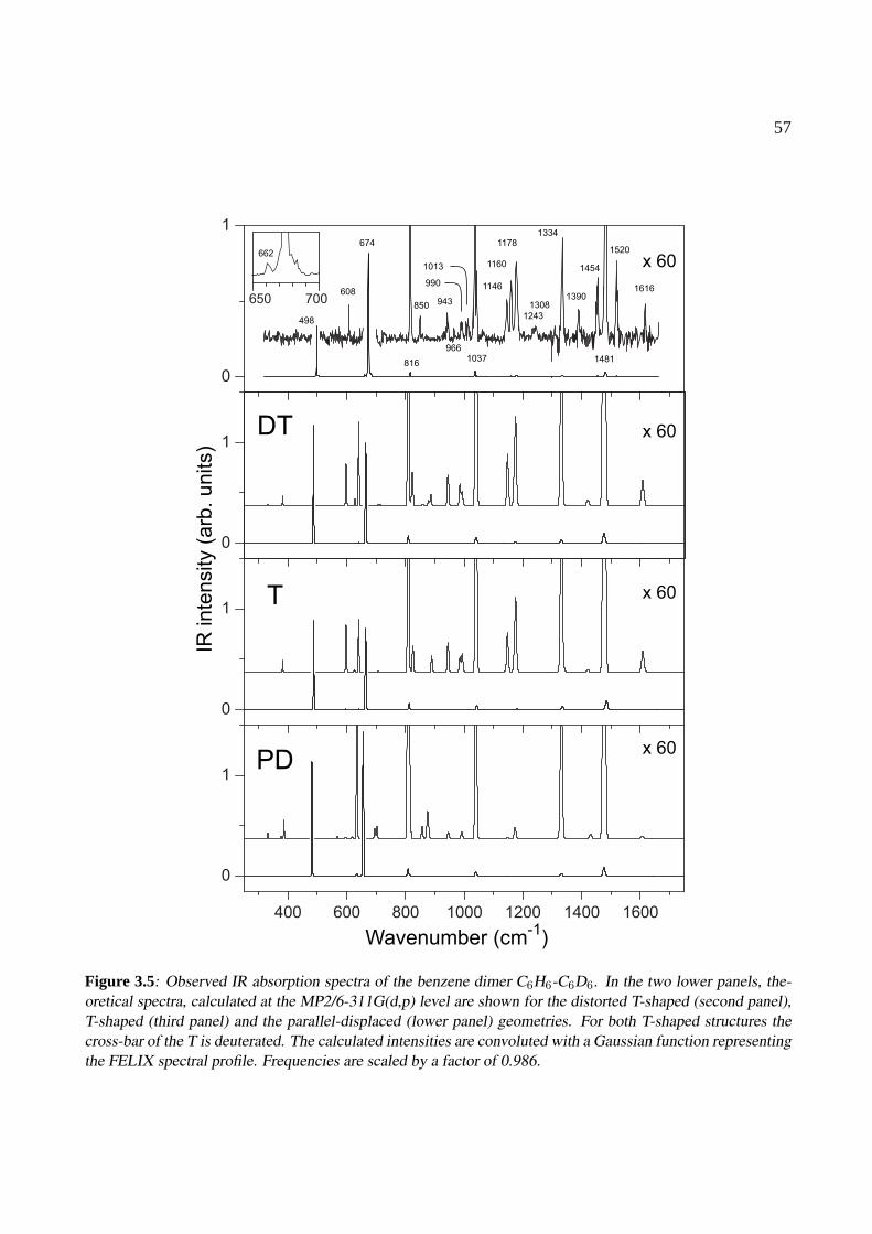
57
650 700
400 600 800 1000 1200 1400 1600
0
1
0
1
0
1
0
1
Wavenumber (cm-1
)
IR inte
nsity (
arb
. units)
T
PD
DT
x 60
x 60
x 60
x 60
674
816
850 943
1146
1160
1178
1243
1308
1334
1390
1454
1520
1481
608
498
662
990
1013
9661037
1616
Figure 3.5: Observed IR absorption spectra of the benzene dimer C6H6-C6D6. In the two lower panels, the-oretical spectra, calculated at the MP2/6-311G(d,p) level are shown for the distorted T-shaped (second panel),T-shaped (third panel) and the parallel-displaced (lower panel) geometries. For both T-shaped structures thecross-bar of the T is deuterated. The calculated intensities are convoluted with a Gaussian function representingthe FELIX spectral profile. Frequencies are scaled by a factor of 0.986.

58
IR inte
nsity (
arb
. units)
Wavenumber (cm-1)
400 600 800 1000 1200 1400 1600 1800
0
1
Figure 3.6: Calculated IR absorption spectra for distorted T-shaped structures that are deuterated in two differ-ent sites: in the top spectrum the cross bar of the T is perdeuterated, while in the bottom spectrum the stem ofthe T is perdeuterated.
1170 cm−1 in the stem-deuterated species suggests that a different spectrum may be obtained whenprobing the other S161 ← S0 transition.
The distorted T-shaped structure shown in Figure 3.4 is a very floppy structure. The stem mole-cule can occupy one of six symmetry equivalent positions above the cross-bar molecule. The barriersseparating those minima are expected to be low. There are two principal pathways that connect thoseminima. One is a precession around the sixfold symmetry axis of the cross-bar molecule. The otherone is a pathway that connects two opposite sides via the symmetric transition state. The relativeheight of this transition state is rather low: the present calculations indicate an energy difference of25 cm−1 between minimum and transition state. Exchange between the minima might be responsiblefor the observed splitting is the microwave spectra. This data indicates the presence of two exchangepathways, which might be the ones described above. The floppiness of the benzene dimer systemis similar to that what has been found earlier for the ammonia dimer: initially several experimentalfindings seemed to contradict each other but they could be brought into accordance with each otherand with theoretical calculations by assuming a very dynamical system [23]. For the benzene dimerthe last word, obviously, has not been said and still more theoretical work may be needed. The presentobservations can then serve as useful additional experimental information for further theoretical cal-culations.
3.4 Conclusions
For the fully protonated benzene dimer and for a dimer of one fully protonated and one fully deuter-ated benzene the IR absorption spectra have been measured in the range of 400 cm−1 to 1700 cm−1.
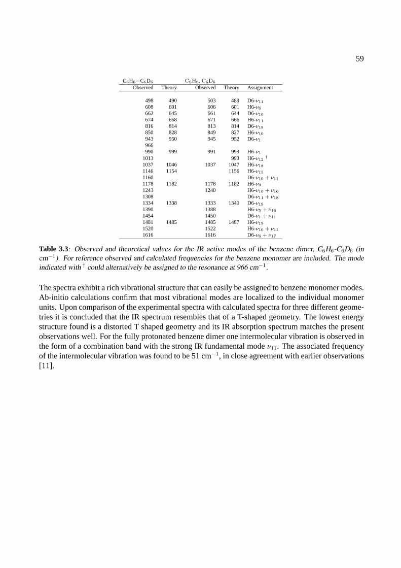
59
C6H6−C6D6 C6H6, C6D6
Observed Theory Observed Theory Assignment
498 490 503 489 D6-ν11
608 601 606 601 H6-ν6
662 645 661 644 D6-ν10
674 668 671 666 H6-ν11
816 814 813 814 D6-ν18
850 828 849 827 H6-ν10
943 950 945 952 D6-ν1
966990 999 991 999 H6-ν1
1013 993 H6-ν12†
1037 1046 1037 1047 H6-ν18
1146 1154 1156 H6-ν15
1160 D6-ν10 + ν11
1178 1182 1178 1182 H6-ν9
1243 1240 H6-ν10 + ν16
1308 D6-ν11 + ν18
1334 1338 1333 1340 D6-ν19
1390 1388 H6-ν5 + ν16
1454 1450 D6-ν1 + ν11
1481 1485 1485 1487 H6-ν19
1520 1522 H6-ν10 + ν11
1616 1616 D6-ν6 + ν17
Table 3.3: Observed and theoretical values for the IR active modes of the benzene dimer, C6H6-C6D6 (incm−1). For reference observed and calculated frequencies for the benzene monomer are included. The modeindicated with † could alternatively be assigned to the resonance at 966 cm−1.
The spectra exhibit a rich vibrational structure that can easily be assigned to benzene monomer modes.Ab-initio calculations confirm that most vibrational modes are localized to the individual monomerunits. Upon comparison of the experimental spectra with calculated spectra for three different geome-tries it is concluded that the IR spectrum resembles that of a T-shaped geometry. The lowest energystructure found is a distorted T shaped geometry and its IR absorption spectrum matches the presentobservations well. For the fully protonated benzene dimer one intermolecular vibration is observed inthe form of a combination band with the strong IR fundamental modeν11. The associated frequencyof the intermolecular vibration was found to be 51 cm−1, in close agreement with earlier observations[11].

60
References
[1] K. C. Janda, J. C. Hemminger, J. S. Winn, S. E. Novick, S. J. Harris, and W. Klemperer, J. Chem. Phys.63, 1419 (1975).
[2] J. Hopkins, D. Powers, and R. Smalley, J. Phys. Chem.85, 3739 (1981).
[3] P. R. Langridge-Smith, D. V. Brumbaugh, C. A. Haynam, and D. H. Levy, J. Phys. Chem.85, 3742 (1981).
[4] K. Fung, H. L. Selzle, and E. W. Schlag, J. Phys. Chem.87, 5113 (1983).
[5] K. Bornsen, H. L. Selzle, and E. W. Schlag, J. Chem. Phys.85, 1726 (1986).
[6] K. Bornsen, H. L. Selzle, and E. W. Schlag, Z. Naturforsch.39a, 1255 (1984).
[7] O. Kratzschmar, H. L. Selzle, and E. W. Schlag, J. Phys. Chem.98, 3501 (1994).
[8] K. Law, M. Schauer, and E. Bernstein, J. Chem. Phys.81, 4871 (1984).
[9] E. Arunan and H. Gutowsky, J. Chem. Phys.98, 4294 (1993).
[10] B. F. Henson, G. V. Hartland, V. A. Venturo, and P. M. Felker, J. Chem. Phys.97, 2189 (1992).
[11] V. Venturo and P. Felker, J. Chem. Phys.99, 748 (1993).
[12] S. Tsuzuki, T. Uchimaru, K. Matsumura, and M. Mikami, Chem. Phys. Lett.319, 547 (2000).
[13] P. Hobza, H. L. Selzle, and E. W. Schlag, J. Phys. Chem.100, 18790 (1996).
[14] V. Spirko, O. Engkvist, P. Soldan, H. L. Selzle, E. W. Schlag, and P. Hobza, J. Chem. Phys.111, 572(1999).
[15] C. Gonzalez, T. C. Allison, and E. C. Lim, J. Phys. Chem. A104, 2953 (2000).
[16] C. Gonzalez, T. C. Allison, and E. C. Lim, J. Phys. Chem. A105, 10583 (2001).
[17] E. Cox, D. Cruickshank, and J. Smith, Proc. Royal Soc. London A247, 1 (1958).
[18] P. Hobza, V.Spirko, H. L. Selzle, and E. W. Schlag, J. Phys. Chem. A102, 2501 (1998).
[19] M. J. Frisch, G. W. Trucks, H. B. Schlegel, G. E. Scuseria, M. A. Robb, J. R. Cheeseman, V. G. Za-krzewski, J. J. A. Montgomery, R. E. Stratmann, J. C. Burant, et al.,Gaussian 98, Revision A.7, Pittsburgh,PA (1998).
[20] E. B. Wilson, Jr., Phys. Rev.45, 706 (1934).
[21] G. Di Lonardo, L. Fusina, G. Masciarelli, and F. Tullini, Spectrochim. Acta, Part A55, 1535 (1999), andreferences therein.
[22] J.-R. Hill, Program Viewmol, version 2.4(2003).
[23] G. Cotti, H. Linnartz, W. L. Meerts, A. van der Avoird, and E. H. T. Olthof, J. Chem. Phys.104, 3898(1996).
CHAPTER 4
THE IR ABSORPTION SPECTRUM OF THE GAS PHASE
NEUTRAL BENZOIC ACID MONOMER AND DIMER
The infrared (IR) absorption spectrum of the jet-cooled benzoic acid monomer and dimer have beenrecorded throughout the 500–1900 cm−1 range via ion-dip spectroscopy. Both spectra show a wealthof vibrational modes and the monomer spectrum is remarkably different from that of the dimer. Den-sity functional theory calculations show quantitative agreement with the experimental data. The C–O–H out-of-plane bending vibration in the dimer is poorly reproduced in the theoretical calculationsand a more accurate description of the doubly hydrogen bonded structure is therefore still needed.
Adapted from: J. M. Bakker, L. Mac Aleese, G. von Helden, and G. Meijer,J. Chem. Phys.119, 11180 (2003)
61
62
4.1 Introduction
Over the last decade a substantial number of experiments have been performed to investigate the struc-ture of isolated molecules of biological interest. These studies include the pioneering spectroscopicwork of the Levy and Simons groups on, among others, amino acids in the gas phase [1–4] and ofDe Vries on gas phase nucleobases [5]. Although these studies are highly relevant, they only addressisolated molecules. In nature, most biologically relevant molecules are attached to other species byhydrogen and/or Van der Waals bonding. The most notable example of this is the double strandedhelix of DNA. This structure is bonded through numerous hydrogen bonds and it is of interest tostudy the effects of bonding on the structure of individual sub-units. It has been concluded that bond-ing solely by dispersive forces can result in almost no structural changes to the different constituents[6]. For hydrogen bonded molecular systems, however, the structure of the individual monomer unitsmight be significantly altered.
Studies involving (doubly) hydrogen bonded systems include investigations of the pairing con-figurations of nucleic acid base pairs [7–10], of their analogues [11–14], small acid dimer systems[15–18] and hydrated amino-acids and their analogues [19–23].
Some of these involve IR spectroscopy in the spectral region where the X–H (X=C,O,N,...) stretch-ing fundamentals are found. A remarkable observation is the, commonly found, broad, structuredabsorption around 3000 cm−1, which is attributed to strong mixing between the X–H stretching fun-damental and bending overtone modes [11, 15, 16]. Finally, Gerhardset al. probed the C=O stretchingvibrations of a protected amino acid dimer system [24].
To study other vibrational modes a tunable source of IR radiation that allows one to probe thelower vibrational energy region is required. Here, the IR absorption spectroscopy of the smallestaromatic acid, benzoic acid (BA), and its dimer are presented using FELIX as an IR source. TheBA dimer is doubly hydrogen bonded through the carboxylic acid group; it has a double minimumpotential in the O–H stretching coordinate in which the hydrogen atoms of the OH group can switchsynchronously between monomer units. Its high symmetry makes the BA dimer a very interestingsystem that can serve as a prototype for doubly hydrogen bonding, and it has been studied quiteextensively using various techniques [25–28]. From a direct measurement of the IR spectra, onecan conclude on possible changes in the vibrational structure upon dimerization of individual BAmonomer units. In an earlier study, Stepanianet al. recorded the matrix isolation spectrum of BA andfound substantial differences between the IR spectra of the monomer and dimer [29]. It is unclear,however, what the influence of the argon matrix is in such studies and gas phase experiments arehighly desirable.
4.2 Experiment
A sample of benzoic acid is heated to a temperature of 75oC and seeded into argon (stagnationpressure approx. 1 bar). This mixture is then expanded through a 0.5 mm diameter nozzle intovacuum. In the adiabatic expansion the internal degrees of freedom in the molecules are cooled androtational temperatures of about 5 K are reached. The expansion conditions enable the formation ofbenzoic acid dimer clusters, which are internally cooled as well.
For the measurement of the IR absorption spectra of the jet-cooled neutral benzoic acid monomerand dimer, IR ion-dip spectroscopy (IR-IDS) is employed. Ions are produced from ground state mo-
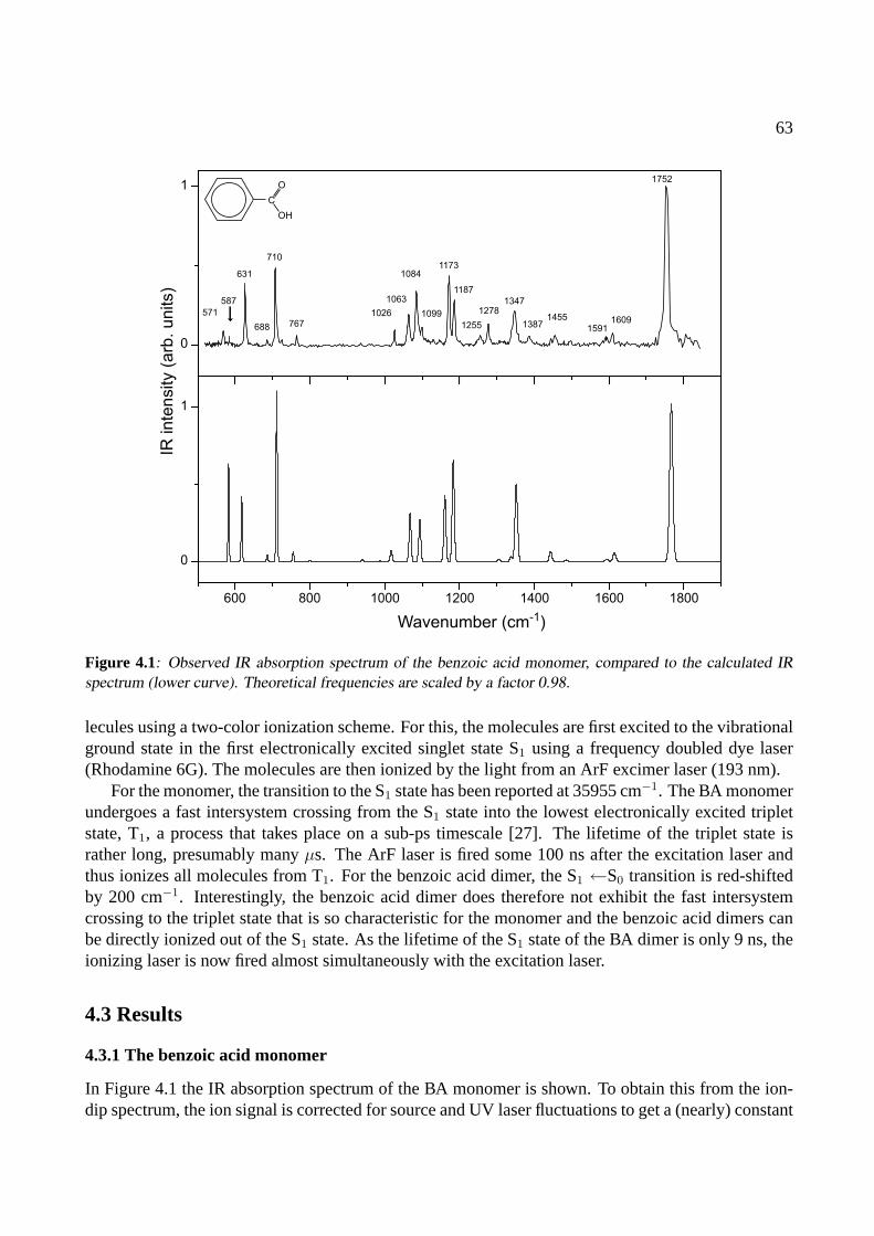
63
571
631
688
710
7671026
1063
1084
1099
1173
1187
1255
12781347
13871455
15911609
1752
587
Wavenumber (cm-1)
IR in
ten
sity (
arb
. u
nits)
600 800 1000 1200 1400 1600 1800
0
1
0
1
C
O
OH
Figure 4.1: Observed IR absorption spectrum of the benzoic acid monomer, compared to the calculated IRspectrum (lower curve). Theoretical frequencies are scaled by a factor 0.98.
lecules using a two-color ionization scheme. For this, the molecules are first excited to the vibrationalground state in the first electronically excited singlet state S1 using a frequency doubled dye laser(Rhodamine 6G). The molecules are then ionized by the light from an ArF excimer laser (193 nm).
For the monomer, the transition to the S1 state has been reported at 35955 cm−1. The BA monomerundergoes a fast intersystem crossing from the S1 state into the lowest electronically excited tripletstate, T1, a process that takes place on a sub-ps timescale [27]. The lifetime of the triplet state israther long, presumably manyµs. The ArF laser is fired some 100 ns after the excitation laser andthus ionizes all molecules from T1. For the benzoic acid dimer, the S1 ←S0 transition is red-shiftedby 200 cm−1. Interestingly, the benzoic acid dimer does therefore not exhibit the fast intersystemcrossing to the triplet state that is so characteristic for the monomer and the benzoic acid dimers canbe directly ionized out of the S1 state. As the lifetime of the S1 state of the BA dimer is only 9 ns, theionizing laser is now fired almost simultaneously with the excitation laser.
4.3 Results
4.3.1 The benzoic acid monomer
In Figure 4.1 the IR absorption spectrum of the BA monomer is shown. To obtain this from the ion-dip spectrum, the ion signal is corrected for source and UV laser fluctuations to get a (nearly) constant
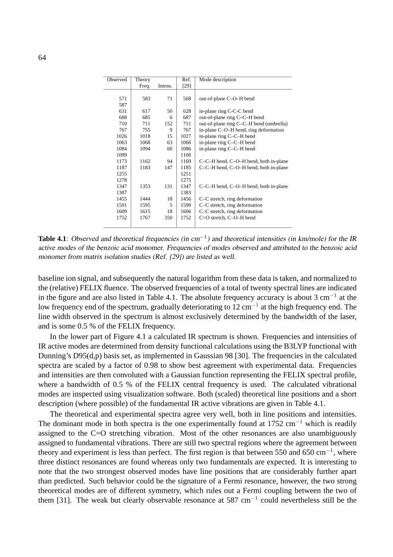
64
Observed Theory Ref. Mode descriptionFreq. Intens. [29]
571 583 71 568 out-of-plane C–O–H bend587631 617 50 628 in-plane ring C-C-C bend688 685 6 687 out-of-plane ring C–C–H bend710 711 152 711 out-of-plane ring C–C–H bend (umbrella)767 755 9 767 in-plane C–O–H bend, ring deformation
1026 1018 15 1027 in-plane ring C–C–H bend1063 1068 63 1066 in-plane ring C–C–H bend1084 1094 60 1086 in-plane ring C–C–H bend1099 11001173 1162 94 1169 C–C–H bend, C–O–H bend, both in-plane1187 1183 147 1185 C–C–H bend, C–O–H bend, both in-plane1255 12511278 12751347 1353 131 1347 C–C–H bend, C–O–H bend, both in-plane1387 13831455 1444 18 1456 C–C stretch, ring deformation1591 1595 5 1590 C–C stretch, ring deformation1609 1615 18 1606 C–C stretch, ring deformation1752 1767 350 1752 C=O stretch, C–O–H bend
Table 4.1: Observed and theoretical frequencies (in cm−1) and theoretical intensities (in km/mole) for the IRactive modes of the benzoic acid monomer. Frequencies of modes observed and attributed to the benzoic acidmonomer from matrix isolation studies (Ref. [29]) are listed as well.
baseline ion signal, and subsequently the natural logarithm from these data is taken, and normalized tothe (relative) FELIX fluence. The observed frequencies of a total of twenty spectral lines are indicatedin the figure and are also listed in Table 4.1. The absolute frequency accuracy is about 3 cm−1 at thelow frequency end of the spectrum, gradually deteriorating to 12 cm−1 at the high frequency end. Theline width observed in the spectrum is almost exclusively determined by the bandwidth of the laser,and is some 0.5 % of the FELIX frequency.
In the lower part of Figure 4.1 a calculated IR spectrum is shown. Frequencies and intensities ofIR active modes are determined from density functional calculations using the B3LYP functional withDunning’s D95(d,p) basis set, as implemented in Gaussian 98 [30]. The frequencies in the calculatedspectra are scaled by a factor of 0.98 to show best agreement with experimental data. Frequenciesand intensities are then convoluted with a Gaussian function representing the FELIX spectral profile,where a bandwidth of 0.5 % of the FELIX central frequency is used. The calculated vibrationalmodes are inspected using visualization software. Both (scaled) theoretical line positions and a shortdescription (where possible) of the fundamental IR active vibrations are given in Table 4.1.
The theoretical and experimental spectra agree very well, both in line positions and intensities.The dominant mode in both spectra is the one experimentally found at 1752 cm−1 which is readilyassigned to the C=O stretching vibration. Most of the other resonances are also unambiguouslyassigned to fundamental vibrations. There are still two spectral regions where the agreement betweentheory and experiment is less than perfect. The first region is that between 550 and 650 cm−1, wherethree distinct resonances are found whereas only two fundamentals are expected. It is interesting tonote that the two strongest observed modes have line positions that are considerably further apartthan predicted. Such behavior could be the signature of a Fermi resonance, however, the two strongtheoretical modes are of different symmetry, which rules out a Fermi coupling between the two ofthem [31]. The weak but clearly observable resonance at 587 cm−1 could nevertheless still be the

65
600 800 1000 1200 1400 1600 1800
0
1
0
1
1709
510
540
664
682
708
797810
962
1022
10661126
1176
1243
1269
12971322
1432
1453
14981591
1618
1824
Wavenumber (cm-1
)
IR inte
nsity (
arb
. u
nits)
C
O
OH
C
O
OH
Figure 4.2: Observed IR absorption spectrum of the benzoic acid dimer, together with the calculated IR spec-trum (lower curve). Theoretical frequencies are scaled by a factor 0.98.
result of a Fermi resonance with one of the fundamentals. A possible set of fundamentals that maycombine to serve as a partner in the Fermi interaction are calculated fundamentals at 157 and 424cm−1. The second discrepancy between theory and experiment is found between 1200 and 1300 cm−1.As no fundamentals are expected in this region it is very likely that the resonances observed hereare combination modes. A possible assignment for the resonance at 1278 cm−1 is the combinationbetween the fundamental at 710 cm−1 and one of the modes participating in the (possible) Fermiresonance around 600 cm−1. For the resonance at 1255 cm−1 various combinations between strongIR active modes and calculated low-lying vibrations could be responsible. As the latter have neverbeen observed, a definite assignment is not possible.
It is noted that most resonances that are found in this work, have been identified by Stepanianet al. in a matrix isolation (MI) spectroscopy experiment as well [29]. The MI intensities differsomewhat from the present data, but in general good agreement is found. For completeness, thevalues determined via MI spectroscopy have been included in Table 4.1.
4.3.2 The benzoic acid dimer
In Figure 4.2 the IR absorption spectrum of the BA dimer is displayed. It is clear from lookingat the experimental data that the signal-to-noise ratio for the dimer spectrum is somewhat worsethan for the monomer spectrum, which is solely due to the lower abundance of the dimer in the
66
beam (∼ 10% relative to the monomer abundance). Below the experimental data, the correspondingtheoretical spectrum is shown. The line positions are scaled with the scaling factor that gave the bestmatch between theoretical and experimental spectra for the monomer,i.e., a factor of 0.98. Like forthe monomer, calculated frequencies and intensities are then convoluted with a Gaussian functionrepresenting the FELIX spectral profile.
The agreement between experimental and theoretical spectra is rather good. From a visual inspec-tion of the modes, it is clear that most vibrations in the dimer are best described as either a symmetricor an anti-symmetric combination of two monomer vibrations. Of these, only the latter have anysignificant IR intensity. As in the IR absorption spectrum of the BA monomer, most resonances arereadily assigned to fundamental vibrations. The spectrum is dominated by a strong mode at 1709cm−1 which very well matches the theoretical calculations, and which is assigned to the asymmetricC=O stretching vibration. Here, it is interesting to note that Gerhardset al. observed a considerablysmaller shift of a C=O stretching vibration frequency upon dimerization, in an experiment involving aprotected amino acid dimer system [24]. In the low energy region the agreement between theory andexperiment is striking: with a single exception – the resonance at 962 cm−1 – all resonances between500 and 1200 cm−1 are very well predicted, both in line positions and intensities. At higher energies,in the region between 1200 and 1700 cm−1, line positions still match quite well, but intensities seemsomewhat off. In the whole spectrum, two modes show clear discrepancies. One of these is the fairlystrong resonance at 962 cm−1. It seems quite likely that this resonance has to be assigned to the strongmode in the theoretical spectrum at 1028 cm−1. This mode is the asymmetric out-of-plane bendingvibration of the O–H groups. The hydrogen atoms are directly involved in the double hydrogen bond,and this thus forms a test for the description of out-of-plane vibrations hindered by a hydrogen bond.
The mismatch between theory and experiment could result from either a deficient theoretical de-scription of the interaction or from a breakdown in the harmonic approach to calculate the vibrationalfrequency. To check the last hypothesis the potential energy surface was evaluated along the normalcoordinate involved in the out-of-plane bending vibration∗. Over a range between -0.5A and +0.5A (total displacement) single-point energies are calculated every 0.01A. In Figure 4.3, this inter-section of the potential energy surface is shown where the calculated values are depicted with solidcircles. In the inset, a close-up is shown of the equilibrium position of the molecule. Included inthe graphs are two fits of quadratic functions to calculated values: the first fit takes into account allcalculated values (dashed line), whereas the second only takes into account the calculated values inthe range of -0.1A and 0.1A (solid line). It can be observed in the inset that the full-range fit de-viates slightly from the calculated values in the equilibrium region, where the calculated potential issomewhat shallower. Also note the slightly higher energy (< 1 cm−1) for the equilibrium position, anoffset which is well within the error margins for DFT calculations. For both fitted quadratic functionsthe vibrational eigenstates are analytically evaluated using the harmonic formulaω =
√k/µ where k
is the force constant from the potential functionV (x) = 12kx2, andµ the reduced mass of the atoms
involved in the normal mode. For the full-range fit a (scaled) vibrational energy of 1117 cm−1 isfound, and for the equilibrium position range fit a value of 1024 cm−1. The latter value is rather closeto the (scaled) value of 1028 cm−1 found in the initial frequency calculation, for which only the sec-ond derivative of the potential energy in the equilibrium position is evaluated. Since the potential is
∗A word of care is in its place right here as the evaluation of the potential here isstricto sensunot correct: Thepotential is evaluated alonga Cartesian vector describingthe normal mode. A more rigorous test can be done when theactual internal coordinate is scanned. The evaluation along the Cartesian vector is expected to yield a good indication ofthe shape of the potential, though.
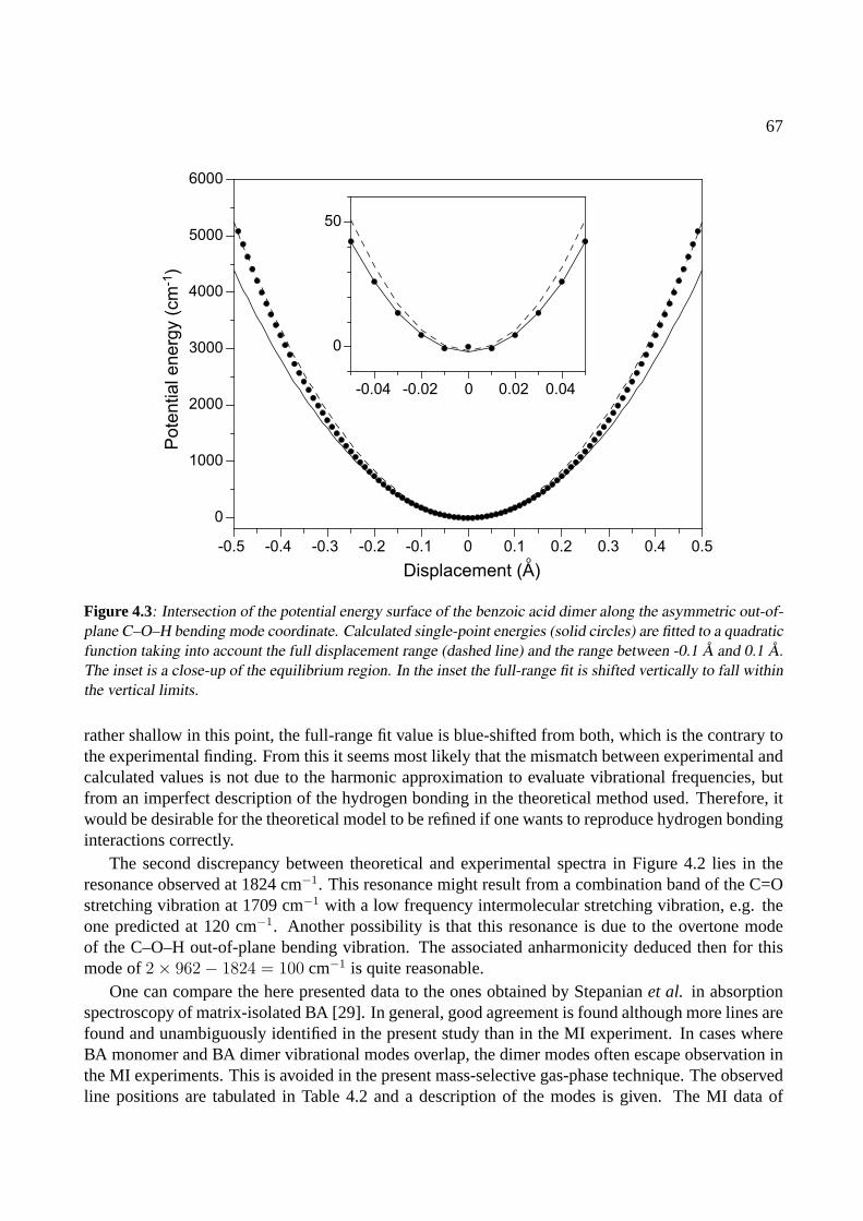
67
Pote
ntial energ
y (
cm
-1)
-0.5 -0.4 -0.3 -0.2 -0.1 0 0.1 0.2 0.3 0.4 0.5
0
1000
2000
3000
4000
5000
6000
Displacement (A)o
-0.04 -0.02 0 0.02 0.04
0
50
Figure 4.3: Intersection of the potential energy surface of the benzoic acid dimer along the asymmetric out-of-plane C–O–H bending mode coordinate. Calculated single-point energies (solid circles) are fitted to a quadraticfunction taking into account the full displacement range (dashed line) and the range between -0.1 A and 0.1 A.The inset is a close-up of the equilibrium region. In the inset the full-range fit is shifted vertically to fall withinthe vertical limits.
rather shallow in this point, the full-range fit value is blue-shifted from both, which is the contrary tothe experimental finding. From this it seems most likely that the mismatch between experimental andcalculated values is not due to the harmonic approximation to evaluate vibrational frequencies, butfrom an imperfect description of the hydrogen bonding in the theoretical method used. Therefore, itwould be desirable for the theoretical model to be refined if one wants to reproduce hydrogen bondinginteractions correctly.
The second discrepancy between theoretical and experimental spectra in Figure 4.2 lies in theresonance observed at 1824 cm−1. This resonance might result from a combination band of the C=Ostretching vibration at 1709 cm−1 with a low frequency intermolecular stretching vibration, e.g. theone predicted at 120 cm−1. Another possibility is that this resonance is due to the overtone modeof the C–O–H out-of-plane bending vibration. The associated anharmonicity deduced then for thismode of2× 962− 1824 = 100 cm−1 is quite reasonable.
One can compare the here presented data to the ones obtained by Stepanianet al. in absorptionspectroscopy of matrix-isolated BA [29]. In general, good agreement is found although more lines arefound and unambiguously identified in the present study than in the MI experiment. In cases whereBA monomer and BA dimer vibrational modes overlap, the dimer modes often escape observation inthe MI experiments. This is avoided in the present mass-selective gas-phase technique. The observedline positions are tabulated in Table 4.2 and a description of the modes is given. The MI data of
68
Observed Theory Ref. Mode descriptionFreq. Intens. [29]
510540 543 130 547 acid groups asymmetric rocking664 659 45 670 acid groups asymmetric scissor682 681 15 out-of-plane ring C–C–H bend708 706 163 out-of-plane ring C–C–H bend797 799 22 acid groups scissor, ring deformation810 803 17 out-of-plane ring C–C–H bend962 1028 221 960 out-of-plane acid group C–O–H bend1022 1018 24 in-plane ring C–C–H bend1066 1073 24 in-plane ring C–C–H bend1126 1126 8 in-plane ring C–C–H bend1176 1166 48 in-plane ring C–C–H bend124312691297 1300 140 1297 C–C–H bend, C–O–H bend, both in-plane1322 1324 687 1322 C–C–H bend, C–O–H bend, both in-plane1432 1440 76 1430 C–C–H bend, C–O–H bend, both in-plane1453 1446 199 C–C–H bend, C–O–H bend, both in-plane1498 1490 50 C–C–H bend, C–O–H bend, both in-plane1591 1593 51 C–C stretch, ring deformation1618 1615 60 C–C stretch, ring deformation1709 1710 956 1699 C=O stretch, in-plane C–O–H bend1824
Table 4.2: Observed and theoretical frequencies (in cm−1) and theoretical intensities (in km/mole) for the IRactive modes of the benzoic acid dimer. Frequencies of modes observed and attributed to the benzoic aciddimer from matrix isolation studies (Ref. [29]) are listed as well.
Stepanianet al. are added for completeness [29].
4.4 Conclusions
The IR absorption spectra of the jet-cooled benzoic acid monomer and dimer have been recorded inthe 500–1900 cm−1 region. Due to the mass-selected detection scheme, one can directly comparethe vibrational structure of the monomer to that of the dimer, and it is seen that dimerization leadsto a dramatic change in the vibrational structure. The theoretical methods used to calculate the IRabsorption spectrum proved to be very accurate in this mid-IR spectral region, in contrast to earlierstudies in the O–H stretching region. The fact that theoretical calculations clearly fail in the predictionof hydrogen bond affected vibrations, in this study demonstrated for the O–H out-of-plane bendingvibration in the dimer, leads to conclude that some fine-tuning in the methods to describe hydrogen-bonded systems is imperative.

69
References
[1] T. R. Rizzo, Y. D. Park, L. A. Peteanu, and D. H. Levy, J. Chem. Phys.84, 2534 (1986).
[2] T. R. Rizzo, Y. D. Park, and D. H. Levy, J. Chem. Phys.85, 6945 (1986).
[3] L. C. Snoek, E. G. Robertson, R. T. Kroemer, and J. P. Simons, Chem. Phys. Lett.321, 49 (2000).
[4] L. C. Snoek, R. T. Kroemer, M. Hockridge, and J. P. Simons, Phys. Chem. Chem. Phys.3, 1819 (2001).
[5] E. Nir, L. Grace, B. Brauer, and M. S. de Vries, J. Am. Chem. Soc.121, 4896 (1999).
[6] B. F. Henson, G. V. Hartland, V. A. Venturo, and P. M. Felker, J. Chem. Phys.97, 2189 (1992).
[7] E. Nir, K. Kleinermanns, and M. S. de Vries, Nature (London)408, 949 (2000).
[8] E. Nir, C. Janzen, P. Imhof, K. Kleinermanns, and M. S. de Vries, Phys. Chem. Chem. Phys.4, 740(2002).
[9] E. Nir, C. Janzen, P. Imhof, K. Kleinermanns, and M. S. de Vries, Phys. Chem. Chem. Phys.4, 732(2002).
[10] C. Plutzer, I. Huning, and K. Kleinermanns, Phys. Chem. Chem. Phys.5, 1158 (2003).
[11] Y. Matsuda, T. Ebata, and N. Mikami, J. Chem. Phys.110, 8397 (1999).
[12] A. Muller, F. Talbot, and S. Leutwyler, J. Chem. Phys.112, 3717 (2000).
[13] A. Muller, F. Talbot, and S. Leutwyler, J. Chem. Phys.115, 5192 (2001).
[14] A. Muller, F. Talbot, and S. Leutwyler, J. Am. Chem. Soc.124, 14486 (2002).
[15] G. M. Florio, E. L. Sibert, and T. S. Zwier, Faraday Discuss.118, 315 (2001).
[16] T. Yahagi, A. Fujii, T. Ebata, and N. Mikami, J. Phys. Chem. A105, 10673 (2001).
[17] F. Madeja and M. Havenith, J. Chem. Phys.117, 7162 (2002).
[18] C. A. Southern, D. H. Levy, G. M. Florio, A. Longarte, and T. S. Zwier, J. Phys. Chem. A107, 4032(2003).
[19] J. A. Dickinson, P. W. Joireman, R. W. Randall, E. G. Robertson, and J. P. Simons, J. Phys. Chem. A101,513 (1997).
[20] J. A. Dickinson, M. R. Hockridge, E. G. Robertson, and J. P. Simons, J. Phys. Chem. A103, 6938 (1999).
[21] E. G. Robertson, Chem. Phys. Lett.325, 299 (2000).
[22] E. G. Robertson, M. R. Hockridge, P. D. Jelfs, and J. P. Simons, Phys. Chem. Chem. Phys.3, 786 (2001).
[23] M. Mons, I. Dimicoli, B. Tardivel, F. Piuzzi, E. G. Robertson, and J. P. Simons, J. Phys. Chem. A105,969 (2001).
[24] M. Gerhards, C. Unterberg, and A. Gerlach, Phys. Chem. Chem. Phys.4, 5563 (2002).
70
[25] D. E. Poeltl and J. K. McVey, J. Chem. Phys.78, 4349 (1983).
[26] S. Kamei, H. Abe, N. Mikami, and M. Ito, J. Phys. Chem.89, 3636 (1985).
[27] G. Meijer, M. S. de Vries, H. E. Hunziker, and H. R. Wendt, J. Phys. Chem.94, 4394 (1990).
[28] K. Remmers, W. L. Meerts, and I. Ozier, J. Chem. Phys.112, 10890 (2000).
[29] S. Stepanian, I. Reva, E. Radchenko, and G. Sheina, Vib. Spectrosc.11, 123 (1996).
[30] M. J. Frisch, G. W. Trucks, H. B. Schlegel, G. E. Scuseria, M. A. Robb, J. R. Cheeseman, V. G. Za-krzewski, J. J. A. Montgomery, R. E. Stratmann, J. C. Burant, et al.,Gaussian 98, Revision A.7, Pittsburgh,PA (1998).
[31] G. Herzberg,Molecular Spectra and Molecular Structure(Krieger Publishing company, Malabar, FLA,1991), 2nd ed.
CHAPTER 5
FINGERPRINT IR SPECTROSCOPY TO PROBE AMINO
ACID CONFORMATIONS IN THE GAS PHASE
The infrared (IR) absorption spectra of different conformational isomers of gas phase amino acidmolecules are measured in the molecular fingerprint region of 330–1500 cm−1. The IR absorptionspectra for three conformers of the amino acid tryptophan show absorption bands that uniquely iden-tify the conformational structure of the molecule and that are well matched by Density FunctionalTheory (DFT) calculations. The present observations hold great promise for future identification ofconformational folding of larger molecules by means of their IR absorption characteristics.
Adapted from: J. M. Bakker, L. Mac Aleese, G. Meijer, and G. von Helden,Phys. Rev. Lett.91, 203003 (2003)
71
72
5.1 Introduction
Over the last twenty years sophisticated techniques have been developed to bring intact large biomoleculesinto the gas phase. The techniques most widely used today are Matrix-Assisted Laser Desorption Ion-ization (MALDI) [1] and ElectroSpray Ionization (ESI) [2]. Due to their ability to provide mass andsequence information of biological samples, both techniques have revolutionized analytical biochem-istry. However, inferring structural information on gas phase ions and molecules is often a difficulttask. The knowledge of the gas phase structure can provide important insight into fundamental intra-molecular interactions and can serve as calibration point for theoretical models. While elaborate toolsfor solid and liquid samples exist, most of them cannot be applied to gas phase samples. One of thefew techniques that can provide direct structural information on gas phase species is ion mobility, inwhich large ionized molecules are mass-selected and pass through a drift cell filled with a buffer gas[3, 4]. The molecular ions will arrive at the end of the drift cell after a period of time which dependson their collisional cross-section. Information can also be obtained using spectroscopic methods, ei-ther in the Ultraviolet (UV) [5] or in the Infrared (IR)[6, 7]. Especially the IR absorption spectrumis a unique identifier for the structure: line intensities and frequencies give direct information on theforces that hold the molecule together. Additional information can be obtained by the direct measure-ment of the direction of the vibrational transition dipole moment of isolated species [8]. SelectiveIR induced isomerization in the gas phase is another interesting technique that provides structuraland dynamical information [9]. Recently, conformational isomerization has been demonstrated fora flexible molecule using UV-UV stimulated emission pumping spectroscopy. This method not onlyallows for the determination of lower and upper values for isomerization barriers between differentconformers, it also enables a determination of relative energies of the zero-point levels of the variousconformers [10].
IR absorption spectra of gas phase species can be obtained by ion- or fluorescence-dip spec-troscopy [6, 7, 11], a technique that has been applied to various interesting systems, such as isolated[12] or paired nucleobases [13], to the amino acids phenylalanine and tryptophan [14, 15] and toβ-sheet model systems [16, 17]. These experiments have all been performed using laser systems thatcover the near- and mid-IR and are limited to wavelengths shorter than about 7µm. In this rangeX–H (X=C,O,N) and C=O stretching vibrations are probed and the experiments provide insight intothe conformational arrangement, as transitions exhibit shifts in absorption frequency and changes inintensity in the presence of, for instance, intra-molecular hydrogen bonds or solvating molecules.However, most vibrational modes have characteristic frequencies that lie further in the IR and are notaccessible using standard laser systems.
The amino acid tryptophan has been studied extensively by various groups as it absorbs stronglyin the near-UV and as it is an important probe in fluorescence spectroscopy [18]. Even though tryp-tophan is a relatively simple molecule, in the gas phase it is observed to fold into several differentconformers. The various conformers have different electronic transitions and can be selectively ex-cited using UV lasers [19]. A detailed structural assignment, however, requires more information. IRspectra in the X–H spectral region have recently been obtained for different conformers and, com-bined with Density Functional Theory (DFT), a first assignment to specific geometries is obtained[15].
Here the IR absorption spectra are presented for three conformers of tryptophan in the gas phaseover a wide frequency range, from 330 cm−1 to 1500 cm−1. It is observed that the conformation hasa strong influence on the spectra. The experimental results are combined with DFT calculations, and

73
34850 34900 34950 35000 35050
0
1
Wavenumber (cm-1)
Ion s
ignal (a
rb. units)
α'
α''
α'''
β
γ
δ
ε
κ
λ
A
B
C
D
E
F
Figure 5.1: REMPI spectrum of tryptophan. The 0–0 transitions for each conformer (A–F) are labeled with acapital. The other resonances labeled by Greek letters are all excited vibrations of conformer A. Labeling hasbeen adopted from Refs. [15, 19].
unique information on the structures of the individual conformers is obtained.
5.2 Experiment
The present IR absorption spectra of tryptophan conformers in the region of 330–1500 cm−1 aremeasured in a pulsed molecular beam experiment with mass and conformer specific detection. Gasphase tryptophan molecules are evaporated in a custom-made oven that is placed behind the nozzle ofa pulsed valve (General Valve). Gas-phase tryptophan molecules are entrained by an argon gas pulseof typically 200µs duration and in a supersonic expansion into vacuum the molecules are internallycooled. After passing through a skimmer, they interact with pulsed UV and IR laser beams.
The IR absorption spectra are measured making use of IR Ion-Dip Spectroscopy (IR-IDS). In thistechnique the internally cooled molecules are resonantly ionized by a pulsed UV laser to produce aconstant ion signal. A fewµs before the UV laser fires, the IR laser interacts with the molecules. Ifa vibrational transition in the molecules is induced by the IR light, population is transferred into anexcited vibrational state, yielding a depletion of ground state molecules. This results in a reduction ofthe number of ions produced. By measuring the ion signal while tuning the IR wavelength, an ion-dipspectrum is recorded.
Conformer specificity is achieved by selecting the frequency of the UV laser. In Figure 5.1 the
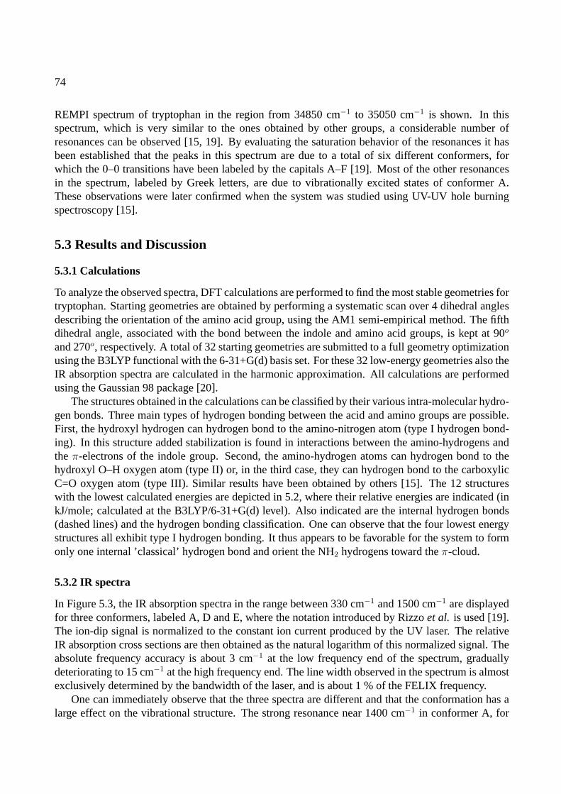
74
REMPI spectrum of tryptophan in the region from 34850 cm−1 to 35050 cm−1 is shown. In thisspectrum, which is very similar to the ones obtained by other groups, a considerable number ofresonances can be observed [15, 19]. By evaluating the saturation behavior of the resonances it hasbeen established that the peaks in this spectrum are due to a total of six different conformers, forwhich the 0–0 transitions have been labeled by the capitals A–F [19]. Most of the other resonancesin the spectrum, labeled by Greek letters, are due to vibrationally excited states of conformer A.These observations were later confirmed when the system was studied using UV-UV hole burningspectroscopy [15].
5.3 Results and Discussion
5.3.1 Calculations
To analyze the observed spectra, DFT calculations are performed to find the most stable geometries fortryptophan. Starting geometries are obtained by performing a systematic scan over 4 dihedral anglesdescribing the orientation of the amino acid group, using the AM1 semi-empirical method. The fifthdihedral angle, associated with the bond between the indole and amino acid groups, is kept at 90o
and 270o, respectively. A total of 32 starting geometries are submitted to a full geometry optimizationusing the B3LYP functional with the 6-31+G(d) basis set. For these 32 low-energy geometries also theIR absorption spectra are calculated in the harmonic approximation. All calculations are performedusing the Gaussian 98 package [20].
The structures obtained in the calculations can be classified by their various intra-molecular hydro-gen bonds. Three main types of hydrogen bonding between the acid and amino groups are possible.First, the hydroxyl hydrogen can hydrogen bond to the amino-nitrogen atom (type I hydrogen bond-ing). In this structure added stabilization is found in interactions between the amino-hydrogens andtheπ-electrons of the indole group. Second, the amino-hydrogen atoms can hydrogen bond to thehydroxyl O–H oxygen atom (type II) or, in the third case, they can hydrogen bond to the carboxylicC=O oxygen atom (type III). Similar results have been obtained by others [15]. The 12 structureswith the lowest calculated energies are depicted in 5.2, where their relative energies are indicated (inkJ/mole; calculated at the B3LYP/6-31+G(d) level). Also indicated are the internal hydrogen bonds(dashed lines) and the hydrogen bonding classification. One can observe that the four lowest energystructures all exhibit type I hydrogen bonding. It thus appears to be favorable for the system to formonly one internal ’classical’ hydrogen bond and orient the NH2 hydrogens toward theπ-cloud.
5.3.2 IR spectra
In Figure 5.3, the IR absorption spectra in the range between 330 cm−1 and 1500 cm−1 are displayedfor three conformers, labeled A, D and E, where the notation introduced by Rizzoet al. is used [19].The ion-dip signal is normalized to the constant ion current produced by the UV laser. The relativeIR absorption cross sections are then obtained as the natural logarithm of this normalized signal. Theabsolute frequency accuracy is about 3 cm−1 at the low frequency end of the spectrum, graduallydeteriorating to 15 cm−1 at the high frequency end. The line width observed in the spectrum is almostexclusively determined by the bandwidth of the laser, and is about 1 % of the FELIX frequency.
One can immediately observe that the three spectra are different and that the conformation has alarge effect on the vibrational structure. The strong resonance near 1400 cm−1 in conformer A, for
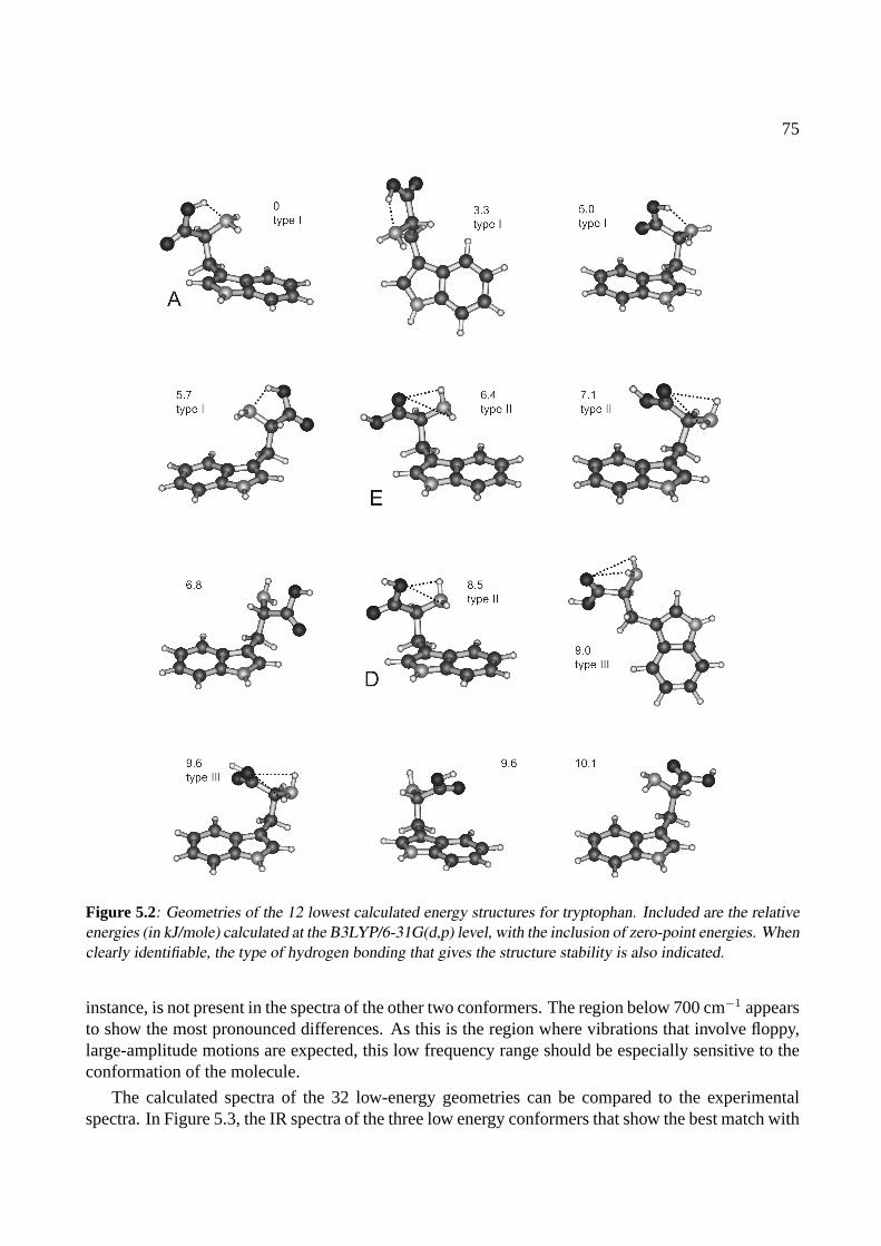
75
Figure 5.2: Geometries of the 12 lowest calculated energy structures for tryptophan. Included are the relativeenergies (in kJ/mole) calculated at the B3LYP/6-31G(d,p) level, with the inclusion of zero-point energies. Whenclearly identifiable, the type of hydrogen bonding that gives the structure stability is also indicated.
instance, is not present in the spectra of the other two conformers. The region below 700 cm−1 appearsto show the most pronounced differences. As this is the region where vibrations that involve floppy,large-amplitude motions are expected, this low frequency range should be especially sensitive to theconformation of the molecule.
The calculated spectra of the 32 low-energy geometries can be compared to the experimentalspectra. In Figure 5.3, the IR spectra of the three low energy conformers that show the best match with
76
Figure 5.3: Infrared absorption spectra of three conformers of tryptophan. Below each experimental spectruma theoretical spectrum is displayed. The corresponding geometries are displayed in the figure as well.

77
the experiment are presented as simulated spectra where the theoretical IR intensities are convolutedwith the (frequency dependent) spectral profile of FELIX. No scaling of the calculated frequencies isperformed.
The experimental spectrum of conformer A (top trace) matches best with the calculated spec-trum of the lowest energy conformer, in which a hydrogen bond between the hydroxyl hydrogen andthe amino-nitrogen is present. Computer animation of the calculated normal modes reveals that thestrongest mode in the spectrum, at 1434 cm−1 is the carboxylic C–O–H bend which is perturbed bythe hydrogen bond. The second strongest mode is the free C=O stretch at 1806 cm−1. Other uniquefeatures are the four lowest energy modes below 600 cm−1, of which the strongest, around 400 cm−1,results from the (very much local-mode) out-of-plane motion of the N–H in the indole group. Thestrong resonance at 738 cm−1, which is observed for all conformers close to this frequency, is asso-ciated with the indole “umbrella mode”. The largest difference between theoretical and experimentalspectra is the predicted strength of a fundamental mode at 869 cm−1. The motion associated with thiscalculated normal mode is essentially an inversion of the amino hydrogens through the (amino acidgroup) N–C–H plane. This mode is predicted around 900 cm−1 for all calculated structures, but isexperimentally not found for any of the three conformers investigated. From earlier experiments, forinstance on aniline, it is known, however, that such an inversion mode is rather poorly described inthe harmonic approximation [21].
The strong mode around 1400 cm−1, observed for conformer A and assigned to result from hin-dered C–O–H bending, is not observed in the spectra of conformer D and E. This suggests that theC–O–H groups in these two conformers are not involved in hydrogen bonding. IR active modes thatinvolve bending of the C–O–H group are calculated for those conformers as well, but the intensitiesare predicted to be substantially lower.
A definite assignment of conformer D (middle trace) is more difficult than for conformer A. Theresulting assignment is based on a good overall agreement between the experimental and the calcu-lated spectra shown in the figure. A key feature in this calculated geometry is the presence of thehydrogen bonds between the amino-hydrogens and the hydroxyl oxygen. In addition, the geometryshown is the lowest energy structure of type III hydrogen bonding.
The spectrum of conformer E (bottom trace) allows for a more solid assignment. The experimen-tal spectrum is observed to have four sharp resonances in the spectral range between 300 cm−1 and500 cm−1. These modes are extremely well reproduced in the calculated spectrum. The three lowest-frequency modes represent the indole N–H out-of-plane bending vibration, but in this structure theyare rather strongly coupled to concerted deformations of the amino-acid group. This coupling leadsto a distribution of IR intensity over all three modes, which explains the lower intensity relative tothat of the localized N–H out-of-plane bending vibration in conformer A. In the proposed structurefor conformer E, hydrogen bonds are present between the amino-hydrogens and the carbonyl C=Ooxygen. A unique feature in the experimental and calculated spectra, that is not found in the spectraof the other calculated structures, is a resonance at 480 cm−1. Although the agreement between cal-culated and experimental spectra is remarkable and warrants the present assignment, it is unfortunatethat there is no data in the spectral region of the C=O stretch. The hydrogen bond could shift thisfrequency substantially (cf. Chapter 4 or the work by Gerhardset al. [16]).
78
5.3.3 Discussion
The structures that are here assigned to conformers A, D and E are the lowest energy structurescalculated for each of the above mentioned types of hydrogen bonding. For A and E, this assignmentis the same as that obtained previously from IR experiments in the hydrogen stretching region [15].For D, a slightly different structure, having the same type of hydrogen bonding, is found to be lower inenergy. The differences in energy, structure and IR spectrum between the conformer D proposed hereand the one proposed previously [15] are small, however. Since neither of the two spectra calculatedfor D agree very well with the experiment, we cannot exclude that this conformer has another not yetconsidered structure.
It can be seen from the present data that for a molecule of the size of tryptophan many local minimain the Potential Energy Surface (PES) can be found in ab-initio calculations. For the assignment of IRspectra of larger molecules, such as di-, tri- and higher order peptides, the computational efforts for afull geometry optimization become more and more expensive. It is thus of the utmost importance tofind a method to generate good starting structures where the full PES is sampled. The use of low-levelapproximative techniques to obtain starting structures does reduce the computational effort, but canby no means guarantee obtaining all minima. In the present method the semi-empirical AM1 methodis used to optimize structures that were generated in a systematic scan over the dihedral angles thatdetermine the orientation of the amino acid group with respect to the indole residue. In an alternativemethod, structures were generated in a simulated-annealing procedure using Allinger’s MM3 forcefield [22]. A full geometry optimization of these structures at the B3LYP/6-31+G(d) level yieldedagain the lowest energy structure that conformer A is assigned to, together with some, but not allof the low-energy structures obtained with AM1. Interesting is the comparison with the previouscalculations of Snoeket al.: in their calculations the first evaluation of a structure was not done atthe AM1 level, but using the Hartree-Fock method. Snoek and co-workers obtained several low-energy structures that were not found in this work, which would suggest that their method is a betterone [15]. On the other hand, in the present work several other stable structures are obtained thatwere not found by Snoek and co-workers. [23]. As the Hartree-Fock optimization is, in comparisonwith AM1, still rather time-consuming it might not be feasible to use this procedure for much largersystems. On the bright side, most low energy structures that were ’missing’ in the present calculationscould be guessed from visually inspecting the obtained geometries and making small changes in theorientations of the amino acid side group using chemical intuition, which makes the semi-empiricalor force field approach a useful instrument.
5.4 Conclusions
The data presented here constitute the first IR measurement of a gas phase biomolecule in the molecu-lar fingerprint region. The combination of a widely tunable IR laser with the ion-dip technique makesit possible to obtain IR spectra that are both mass and conformer specific over a broad spectral range,thereby dramatically increasing the number of vibrations that can be probed. The technique can beextended to larger species as well as toward longer wavelength. However, when the molecules be-come very large, difficulties arise in bringing them into the gas phase, as well as in selectively ionizingthem. For large species, complementary IR spectral information could be obtained from experimentsperformed on ionic species in the gas phase, generated by MALDI or ESI. In such experiments, amass change after resonant multi-photon dissociation could be monitored using mass spectrometry or

79
an IR induced conformational change could be detected by ion mobility measurements.
80
References
[1] F. Hillenkamp, M. Karas, R. Beavis, and B. Chait, Anal. Chem.63, A1193 (1991).
[2] J. Fenn, M. Mann, C. Meng, S. Wong, and C. Whitehouse, Science246, 64 (1989).
[3] G. von Helden, T. Wyttenbach, and M. T. Bowers, Science267, 1483 (1995).
[4] D. E. Clemmer, R. Hudgins, and M. F. Jarrold, J. Am. Chem. Soc.117, 10141 (1995).
[5] D. W. Pratt, Annu. Rev. Phys. Chem.49, 481 (1998).
[6] T. S. Zwier, Annu. Rev. Phys. Chem.47, 205 (1996).
[7] T. Ebata, A. Fujii, and N. Mikami, Int. Rev. Phys. Chem17, 331 (1998).
[8] F. Dong and R. E. Miller, Science298, 1227 (2002).
[9] B. C. Dian, A. Longarte, and T. S. Zwier, Science296, 2369 (2002).
[10] B. C. Dian, J. R. Clarkson, and T. S. Zwier, Science303, 1169 (2004).
[11] R. H. Page, Y. Shen, and Y. T. Lee, J. Chem. Phys.88, 4621 (1988).
[12] E. Nir, C. Janzen, P. Imhof, K. Kleinermanns, and M. S. de Vries, J. Chem. Phys.115, 4604 (2001).
[13] E. Nir, C. Janzen, P. Imhof, K. Kleinermanns, and M. S. de Vries, Phys. Chem. Chem. Phys.4, 732(2002).
[14] L. C. Snoek, E. G. Robertson, R. T. Kroemer, and J. P. Simons, Chem. Phys. Lett.321, 49 (2000).
[15] L. C. Snoek, R. T. Kroemer, M. Hockridge, and J. P. Simons, Phys. Chem. Chem. Phys.3, 1819 (2001).
[16] M. Gerhards, C. Unterberg, and A. Gerlach, Phys. Chem. Chem. Phys.4, 5563 (2002).
[17] C. Unterberg, A. Gerlach, T. Schrader, and M. Gerhards, J. Chem. Phys.118, 8296 (2003).
[18] J. M. Beechem and L. Brand, Ann. Rev. Biochem.54, 43 (1985).
[19] T. R. Rizzo, Y. D. Park, L. A. Peteanu, and D. H. Levy, J. Chem. Phys.84, 2534 (1986).
[20] M. J. Frisch, G. W. Trucks, H. B. Schlegel, G. E. Scuseria, M. A. Robb, J. R. Cheeseman, V. G. Za-krzewski, J. J. A. Montgomery, R. E. Stratmann, J. C. Burant, et al.,Gaussian 98, Revision A.7, Pittsburgh,PA (1998).
[21] H. Piest, G. von Helden, and G. Meijer, J. Chem. Phys.110, 2010 (1999).
[22] N. L. Allinger, Y. H. Yuh, and J.-H. Lii, J. Am. Chem. Soc.111, 8551 (1989).
[23] R. T. Kroemer, private communication.
CHAPTER 6
THE MID-IR ABSORPTION SPECTRUM OF GAS-PHASE
COMPLEXES OF THE NUCLEOBASES GUANINE AND
CYTOSINE
The mid-infrared (IR) absorption spectrum of jet-cooled complexes of the nucleobases guanine andcytosine has been recorded in the 500-1800 cm−1 range by ion-dip spectroscopy. Some 20 clearlyseparated and sharp resonances are observed. The combination of the experimental data with newhigh-level ab-initio calculations is consistent with a previous structural assignment and a tentativerefined assignment is made. These data can serve as a test of the method in the mid-IR regime.
Adapted from: J. M. Bakker, I. Compagnon, G. Meijer, G. von Helden, M. Kabelac, P. Hobza, and M. S. de Vries,Phys. Chem. Chem. Phys.6, 2810-2815 (2004)
81

82
Figure 6.1: Structures of the four lowest-energy G-C pairs. The stabilization energies calculated at RI-MP2/TZVPP level are given for each structure. Also included is the atom labeling convention which is usedthroughout this chapter.
6.1 Introduction
One of the questions yet to be answered for the DNA system is what determines the ultimate stabi-lization of the classical Watson-Crick structure [1]. Several factors play a role and it is unclear whatpart in the total stabilization they play: (i) the multiple hydrogen bonds between the base pairs thatcross-link the two strands, (ii) the interaction between the nucleobases on one strand, the so-calledstacking mechanism, and (iii) the stabilization by the deoxyribosoic backbone and other environmen-tal effects. This question has caused a considerable theoretical effort to evaluate the various factors[2–4]. To investigate this matter experimentally one can adopt the approach of trying to separatethe factors that play a role, which can be achieved by studying isolated molecules in the gas phase.Spectroscopic studies of gas phase biological molecules have enabled the elucidation of fundamentalinteractions governing biological processes unobscured by such effects as the broadening of spectrallines by sample-solvent interactions [5, 6]. The obvious start of gas phase studies involving the bio-logical heredity system lies in interrogating the fundamental building blocks, the isolated purine andpyrimidine molecules. By extending the study to complexes of these building blocks, one can extractvaluable information on the multiple hydrogen-bonded interactions, stabilizing the two strands.
Considerable spectroscopic knowledge on individual gas-phase nucleobases has been obtained inrecent years [7–12]. One of the striking findings is the presence of many tautomers that are differentfrom the form in which they are found in DNA in living matter. Using spectroscopic techniques,also complexes of nucleobases have been investigated [13–15]. In those studies the Watson-Crickgeometries of the paired nucleobases have not been observed so-far.
83
Figure 6.1 shows the four lowest energy structures of the guanine-cytosine (G-C) complex, out ofmore than 23 possible hydrogen bonded structures [16]. Structure A represents the triply hydrogenbonded Watson-Crick structure. Structure C is very similar, but it has the cytosine as an enol ratherthan a keto tautomer. Structure D is again very similar: it is another Watson-Crick type bondedstructure, now with an enol cytosine tautomer and a guanine tautomer where one hydrogen is attachedat the N7 position (as opposed to N9 for structure C). Finally, in structure B the tautomers as found inDNA are hydrogen bonded in a way which would be impossible if the sugar backbone were present.To discern the structures, a nomenclature was introduced by Nir et al. [14]. In this notation, structureC is called K9-1E, indicating that guanine is found in the keto form, with a hydrogen attached toN9; cytosine is in its enol form, and the OH group is oriented toward N1 (cf. Figure 6.1). UsingREMPI spectroscopy and UV-UV hole burning so-far two structures of G-C complexes have beendistinguished [14]. With the help of IR-UV hole burning in the near IR between 3300 cm−1 and 3700cm−1 one of these could tentatively be identified as structure C [14] and the other one as structure B[17]. The identification of structure C is not unambiguous: the X-H (X=N,C) stretching vibrations ofstructures C and D are very similar, and assignment was done based on the relative energies of thestructures. Both tautomers of guanine have been observed in a jet experiment, and assignment of thespectrum to structure D can not be ruled out [12]. No spectrum is observed that can be assigned tostructure A, however. This could be caused by a considerable difference in the UV spectral propertiesor by a dramatically reduced lifetime of the G-C complex in structure A. Broad REMPI spectra havebeen observed for complexes of 1-methyl cytosine with 9-methyl guanine and of cytidine with guaninethat can tentatively be assigned to structure A [18].
The general strategy to elucidate the structure of a molecular system in the gas phase is to combineUV spectroscopy and UV-UV / IR-UV hole-burning spectroscopy with high-level theoretical calcula-tions. The UV-UV hole burning spectroscopy ascertains that a specific UV transition is resulting fromone single structure, whereas the IR-UV hole burning spectroscopy is used to identify this structure.
Here, the IR absorption spectrum of the gas-phase complex of guanine and cytosine, recorded inthe 500–1800 cm−1 region, is presented. At the same time, a set of new high-level calculations tointerpret the recorded data are presented. These data serve the dual purpose of testing the originalassignment of this complex and testing the comparison of the highest level of the high-level compu-tations with the experiment in this region of the IR spectrum.
The calculations represent a new step in theoretical studies of DNA bases [16]. All calculations onDNA bases and base pairs should be performed at a single, common level that accurately describes allinteractions in the system. Hartree-Fock (HF) and Density Functional Theory (DFT) calculations, forinstance, only correctly describe hydrogen bonded interactions but are not able to describe the equallyimportant stacking interactions. Surprisingly, up to now no DFT functional exists that describes thestacking of DNA bases. The first acceptable level for a correct description of both hydrogen bondingand stacking interactions is the MP2 level using at least a DZ+P basis set. This level provides accu-rate characteristics (structure, stabilization energies, and vibrational frequencies) of hydrogen bondedpairs but underestimates stabilization energies for stacking. Reliable values for all characteristics ofany type of DNA base pair are generated, however, if the MP2 calculations are performed with theTZ+P basis set, i.e. a triple-zeta basis set with two sets of first polarization functions and one setof second polarization functions on all the atoms [19]. Such a treatment is feasible for obtainingstructure and stabilization energy of a pair but computationally too costly for generating vibrationalfrequencies. Therefore the following computational strategy is adopted: structure and stabilization
84
energy are calculated at the MP2/TZ+P level, allowing for a comparison between the stabilizationenergies of the studied hydrogen bonded complexes with those of stacked complexes. The vibrationalfrequencies are then determined at a cheaper MP2/DZ+P level.
6.2 Experimental
Since the vapor pressure of the nucleobases is very low at room temperature and they are thermallylabile the experiment is performed using the laser-desorption source. For the measurement of theIR absorption spectra of the jet-cooled guanine-cytosine (GC) complexes IR ion-dip spectroscopy isemployed. Ions are produced from ground state molecules using a two-photon ionization scheme.For this, the molecules are first excited to the lowest vibrational level in the first electronically excitedsinglet state (S1) using the frequency doubled dye laser (Rhodamine 640). A second photon fromthe excitation laser pulse ionizes the molecules. In the present experiment, the UV detection laser isrunning at a 10 Hz repetition rate, while FELIX is set to run at 5 Hz. By independently recordingboth the IR-on signal and the IR-off signal a normalized ion-dip spectrum can be obtained, that isinsensitive to long-term drifts in UV laser power or source conditions.
The IR ion-dip spectrum for GC is recorded with the UV probe laser at 33314 cm−1. This fre-quency corresponds to theS1 ← S0(0− 0) transition of the complex structure that was earlier as-signed to structure C, based on IR-UV hole burning spectroscopy in the near IR region between 3300and 3700 cm−1 [14]. That earlier spectrum contained the frequency of the OH stretching vibration ofthe enol-cytosine as a distinctive feature. As the OH stretching vibration of structure D was calculatedto be almost identical, no definite assignment was made. The present data in the mid-IR may assistin accomplishing a final assignment as the current spectral range contains the CO stretching vibratio-nal frequency as well as a large number of skeletal vibrational frequencies that can serve for furtherstructural identification.
6.3 Theoretical methods
The use of DZ+P or even TZ+P basis sets in a combination with the exact MP2 treatment for DNAbase pairs is difficult if not impractical. Recently the applicability of the approximative resolution ofidentity (RI) MP2 methods [20, 21] for nucleic acid base pairs and larger DNA fragments [22] hasbeen explored and it has been shown that this method is capable of an accurate description of bothhydrogen bonded and stacked DNA base interactions. The results obtained with the RI-MP2 methoddiffer only marginally from those evaluated with the exact MP2 method, while the efficiency is largerby up to one order of magnitude.
The potential energy surface (PES) of the GC pair is investigated using the molecular dynam-ics/quenching (MD/Q) method with the Cornellet al. empirical force field [23]. MD/Q simulationsare performed in the NVE microcanonical ensemble (N, V and E refer to the number of particles,volume and energy, respectively) within the quaternion formalism. The MD simulations are carriedout at a constant total energy corresponding to an average temperature of 298 K which is high enoughto allow crossing over relatively high energy barriers and thus to sample the whole potential energysurface. Every 1 ps the MD run is interrupted, the kinetic energy is removed and the structure of thecomplex of guanine and cytosine is optimized using the conjugate gradient method. The geometry
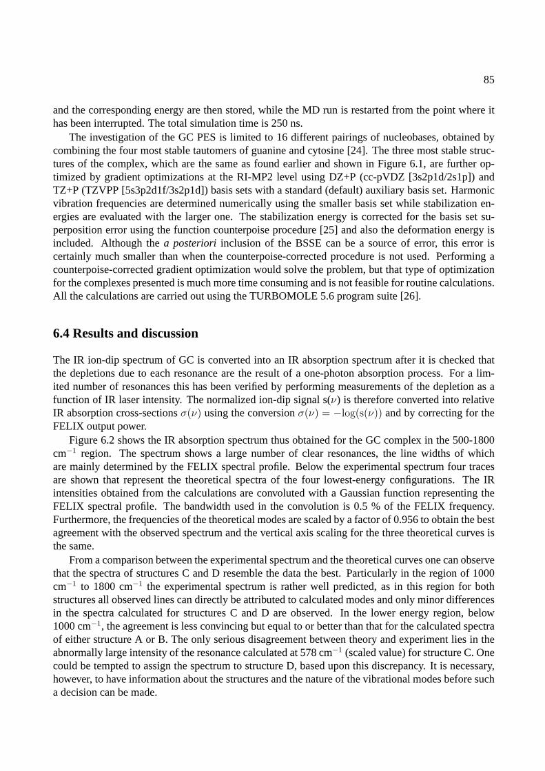
85
and the corresponding energy are then stored, while the MD run is restarted from the point where ithas been interrupted. The total simulation time is 250 ns.
The investigation of the GC PES is limited to 16 different pairings of nucleobases, obtained bycombining the four most stable tautomers of guanine and cytosine [24]. The three most stable struc-tures of the complex, which are the same as found earlier and shown in Figure 6.1, are further op-timized by gradient optimizations at the RI-MP2 level using DZ+P (cc-pVDZ [3s2p1d/2s1p]) andTZ+P (TZVPP [5s3p2d1f/3s2p1d]) basis sets with a standard (default) auxiliary basis set. Harmonicvibration frequencies are determined numerically using the smaller basis set while stabilization en-ergies are evaluated with the larger one. The stabilization energy is corrected for the basis set su-perposition error using the function counterpoise procedure [25] and also the deformation energy isincluded. Although thea posteriori inclusion of the BSSE can be a source of error, this error iscertainly much smaller than when the counterpoise-corrected procedure is not used. Performing acounterpoise-corrected gradient optimization would solve the problem, but that type of optimizationfor the complexes presented is much more time consuming and is not feasible for routine calculations.All the calculations are carried out using the TURBOMOLE 5.6 program suite [26].
6.4 Results and discussion
The IR ion-dip spectrum of GC is converted into an IR absorption spectrum after it is checked thatthe depletions due to each resonance are the result of a one-photon absorption process. For a lim-ited number of resonances this has been verified by performing measurements of the depletion as afunction of IR laser intensity. The normalized ion-dip signal s(ν) is therefore converted into relativeIR absorption cross-sectionsσ(ν) using the conversionσ(ν) = −log(s(ν)) and by correcting for theFELIX output power.
Figure 6.2 shows the IR absorption spectrum thus obtained for the GC complex in the 500-1800cm−1 region. The spectrum shows a large number of clear resonances, the line widths of whichare mainly determined by the FELIX spectral profile. Below the experimental spectrum four tracesare shown that represent the theoretical spectra of the four lowest-energy configurations. The IRintensities obtained from the calculations are convoluted with a Gaussian function representing theFELIX spectral profile. The bandwidth used in the convolution is 0.5 % of the FELIX frequency.Furthermore, the frequencies of the theoretical modes are scaled by a factor of 0.956 to obtain the bestagreement with the observed spectrum and the vertical axis scaling for the three theoretical curves isthe same.
From a comparison between the experimental spectrum and the theoretical curves one can observethat the spectra of structures C and D resemble the data the best. Particularly in the region of 1000cm−1 to 1800 cm−1 the experimental spectrum is rather well predicted, as in this region for bothstructures all observed lines can directly be attributed to calculated modes and only minor differencesin the spectra calculated for structures C and D are observed. In the lower energy region, below1000 cm−1, the agreement is less convincing but equal to or better than that for the calculated spectraof either structure A or B. The only serious disagreement between theory and experiment lies in theabnormally large intensity of the resonance calculated at 578 cm−1 (scaled value) for structure C. Onecould be tempted to assign the spectrum to structure D, based upon this discrepancy. It is necessary,however, to have information about the structures and the nature of the vibrational modes before sucha decision can be made.
86
Wavenumber (cm-1)
IR inte
nsity (
arb
. units)
C
D
A
B
NH
NN
H
N
O
NH2
N NH
H2N
O
NH
NN
H
N
O
NH2
NN
H2N
OH
NH
NN
H
N
O
NH2
N HN
NH2
O
NH
NN
HN
O
NH2
NN
H2N
OH
500 1000 1500
0
1
0
1
0
1
0
1
0
1
Figure 6.2: IR absorption spectrum of the guanine-cytosine complex. Below the experimental spectrum aretheoretical spectra of the four lowest-energy configurations of guanine-cytosine complexes. Theoretical fre-quencies for all structures are scaled by a factor of 0.956 and convoluted with a Gaussian function representingthe FELIX spectral profile.
87
Structure C is a slightly non-planar (deviation of planes of the bases is about 20o), Watson-Crick-type structure of canonical guanine and a tautomer of cytosine in which a hydrogen atom is detachedfrom the N1-H group and attached to the C2=O group, forming an enol side group. The presence ofthe OH group as well as the change of the cytosine electronic structure leads to a weaker interactionbetween G and C than in structure A. The distances between the heavy atoms in the hydrogen bonds(cyt N-H-gua O), (cyt N3-gua H-N1) and (cyt O- gua H-N) in structure C are 2.84, 2.97 and 3.07A, respectively (cf. Figure 6.1). Here, all geometric parameters refer to the calculations done at theRI-MP2/cc-pVDZ level. Both amino groups in structure C are non-planar with respect to the baseplane (deviation in both subsystems is approximately 20o). Structure D is very similar to structure Cbut a hydrogen atom is detached from the N9-H group and attached to the N7. The base planes aretilted with respect to each other by about 23o, the hydrogen bond distances are 2.85, 2.95 and 3.10A and the amino hydrogens are oriented out-of-plane by about 25o.
Structures C and D correspond to the second and third local minima at the G-C potential energysurface (PES). Their (absolute) stabilization energies (including deformation energy and difference intautomerization energies, a sum of relative energies of isolated tautomers with respect to the canonicalform) are 93 kJ/mole and 88 kJ/mole, respectively. This is about 17 kJ/mole and 23 kJ/mole less stablethan the global minimum structure, the canonical Watson-Crick structure A. Structure A is perfectlyplanar with only negligible deviation (less than 1o) of amino hydrogens from the plane of the base.The distances between the heavy atoms participating in the hydrogen bonds are 2.78, 2.92 and 2.92A, respectively and they are slightly shorter than in the cases of structures C and D.
Structure B, possessing a different hydrogen bond pattern between canonical forms of guanineand cytosine, is about 4 kJ/mole more stable than structure C (97 kJ/mole) and this structure thuscorresponds to the first local minimum at the PES. Structure B is slightly non-planar and it containsonly 2 hydrogen bonds with the following distances between heavy atoms : O(G)... N1H(cyt)=2.77A, and N1H(gua)...O(cyt)=2.71A.
The present RI-MP2/TZVPP stabilization energies are systematically larger than the respectiveRI-MP2/cc-pVDZ ones by about 6-8 kJ/mole. More detailed information on all 50 complexes ob-tained at the G-C PES by considering the four most stable tautomers of guanine and of cytosine willbe published later [24].
To discuss the nature of the resonances in the experimental spectrum the calculated vibrationalmodes are inspected using visualization software to identify the associated motions of the atoms.Line positions of all observed resonances and (scaled) frequencies of the assigned theoretical vibra-tions, including short descriptions of the modes, can be found in Table 6.1. Most of the assignedmodes are identical for both structure C and D; only when they differ an explicit mode descriptionis given for structure D. Throughout the rest of the discussion scaled values will be given for theo-retical frequencies to facilitate comparison to Figure 6.1. The dominating feature in the experimentalspectrum, the highest frequency resonance found at 1706 cm−1, is associated with the guanine C=Ostretching mode, with a slight coupling to a bending motion of the guanine N1-H group. As thisis the only C=O group in structures C and D, its assignment is unambiguous. For structure A thetwo C=O stretching modes are coupled, and result in one symmetric and one asymmetric stretchingmode of which the latter (calculated at 1727 cm−1) has large IR intensity, and the former (1699 cm−1)only little. The two C=O stretching vibrations in structure B are not coupled to each other, but toan N-H in-plane bending mode. This interaction is less favorable for the cytosine stretching mode(1674 cm−1), which has considerably less IR intensity than its guanine counterpart (1718 cm−1). The
88
modes lying at somewhat lower energy, between 1000 cm−1and 1700 cm−1are all associated withskeletal motions in the plane of the molecules, coupled in varying strength with in-plane NH2 or NHbending vibrations. Most of the modes here are rather well reproduced, except for a somewhat strongIR intensity for the resonance experimentally found at 1374 cm−1, which might be due to overlappingresonances. The region below 1000 cm−1 contains a number of clear resonances that cannot so easilybe assigned to a specific theoretical mode. Most vibrations here are out-of-plane bending motions ofthe various hydrogen atoms.
There is one mode with appreciable IR activity that could make an assignment to either structure Cor D possible. The N9-H and N7-H out-of-plane bending vibrations are calculated to be very localizedin structures C and D, respectively, and should be sensitive to its environment: in structure C the N9-Hgroup is rather isolated, whereas in structure D the N7-H group could be influenced by the presence ofthe C6=O group. For both structures the respective N-H out-of-plane bending vibration is indicatedwith an arrow in the calculated spectrum. In structure C, around 500 cm−1 two vibrational modesare expected: the C-O-H in-plane bending mode at 506 cm−1 and the N9-H out-of-plane bendingmode at 515 cm−1. In structure D, only the C-O-H in-plane bending mode is expected here, at 508cm−1 while the N7-H out-of-plane bending mode is calculated to lie at 477 cm−1. From the factthat there is only one observed resonance, at 514 cm−1, we are tempted to assign the experimentalspectrum to structure D. Unfortunately, the data do not cover the range where the N7-H bending modeis expected in structure D, and a definite assignment can still not be made.
It must be pointed out that the calculated vibration at 578 cm−1 in the spectrum of structure C,which is predicted with a larger IR intensity than what is observed cannot be invoked to finalizethe assignment: the motions associated with this mode are those of the two hydrogens of the guanineamino group moving in-phase through the plane of the molecule. It has been observed earlier that suchinversion-like modes that have a double minimum in the PES are poorly reproduced in the harmonicapproximation [27, 28]. It therefore is better not to use this resonance to assign the structure basedon the comparison between calculated and experimental spectra. The cytosine NH2 group does notexhibit a simultaneous bending vibration or tunneling mode in this structure nor in the three others;in structure C one localized N-H out-of-plane bending mode is predicted at 401 cm−1.
Structure D shows the same pattern: one tunneling-like mode is found at 598 cm−1 (although withsubstantially less IR intensity than its equivalent in structure C) and a localized N-H out-of-planebending mode at 392 cm−1. The guanine NH2 group in structure A does not exhibit the tunnelingmode. Rather, its out-of-plane bending vibration is symmetric and yields almost no IR intensity. Thisdifferent behavior is most likely caused by the fact that in this structure the cytosine is found as a ketotautomer, and the hydrogen bonding is substantially different.
Although the guanine tautomer in structure D is energetically not favored, it has been observedin a jet-experiment [12]. Moreover, the GC origin is found to be much closer to the 7H keto form ofguanine than the 9H keto tautomer [12]. Differently phrased: GC complexes with G in the K9 formmight have resonances that are found at higher frequencies in the UV spectrum, and hence may betoo difficult to observe, because they could be hidden by spectral congestion due to resonances of theK7-C complex. Alternatively, their higher-lying excited state could favor fast dynamical processesthat reduce the efficiency of the R2PI process.
89
6.5 Conclusions
The IR absorption spectrum is measured for the complex of the nucleobases guanine and cytosineisolated in the gas phase in the mid-IR fingerprint region, between 500 cm−1 and 1800 cm−1. TheIR spectrum contains a large number of sharp resonances that can be used to assign the spectrum.A comparison with theoretical spectra of a set of possible geometries is consistent with a previousassignment to a Watson-Crick type hydrogen bonded structure that contains an enol cytosine tautomer.A tentative assignment is made to a structure with a guanine tautomer in which a hydrogen atom isdetached from the N9-H group and attached to N7, but a final assignment can only be made whenfurther experimental work in the frequency regime below 500 cm−1 is done.
The comparison between experimental and calculated spectra for the benzoic acid dimer in Chap-ter 4 appears more favorable than the present one, but one has to keep in mind that the IR absorptionspectrum of the benzoic acid dimer is much simpler than that of the guanine-cytosine basepair. Notonly does the benzoic acid dimer have a substantially smaller number of degrees of freedom, but itshigher symmetry rules out IR transitions to a large fraction of its vibrational modes. Given the highercomplexity of the basepair system it can be concluded that the presently used MP2 method gives an al-together fairly accurate description of the vibrational structure of the basepair, allowing for an almostunique assignment. The MP2 method does not exhibit errors in the description of hydrogen bondedbending modes, as seen for the B3LYP treatment of the benzoic acid dimer. From these observationsit is concluded that the MP2 method can be used with confidence for the theoretical treatment of largerhydrogen bonded systems and for structural identification based on IR spectroscopic measurements.
90
References
[1] J. Watson and F. Crick, Nature (London)171, 737 (1953).
[2] C. F. Guerra, F. M. Bickelhaupt, J. G. Snijders, and E. J. Baerends, J. Am. Chem. Soc.122, 4117 (2000).
[3] P. Hobza and J. Sponer, J. Am. Chem. Soc.124, 11802 (2002).
[4] D. Sivanesan, I. Sumathi, and W. J. Welsh, Chem. Phys. Lett.367, 351 (2003).
[5] T. S. Zwier, J. Phys. Chem. A105, 8827 (2001).
[6] E. G. Robertson and J. P. Simons, Phys. Chem. Chem. Phys.3, 1 (2001).
[7] E. Nir, L. Grace, B. Brauer, and M. S. de Vries, J. Am. Chem. Soc.121, 4896 (1999).
[8] E. Nir, M. Muller, L. I. Grace, and M. S. de Vries, Chem. Phys. Lett.355, 59 (2002).
[9] N. J. Kim, G. Jeong, Y. S. Kim, J. Sung, S. K. Kim, and Y. D. Park, J. Chem. Phys.113, 10051 (2000).
[10] C. Plutzer, E. Nir, M. de Vries, and K. Kleinermanns, Phys. Chem. Chem. Phys.3, 5466 (2001).
[11] D. Luhrs, V. J., and I. Fischer, Phys. Chem. Chem. Phys.3, 1827 (2001).
[12] W. Chin, M. Mons, I. Dimicoli, F. Piuzzi, B. Tardivel, and M. Elhanine, Eur. J. Phys. D20, 347 (2002).
[13] E. Nir, K. Kleinermanns, and M. S. de Vries, Nature (London)408, 949 (2000).
[14] E. Nir, C. Janzen, P. Imhof, K. Kleinermanns, and M. S. de Vries, Phys. Chem. Chem. Phys.4, 732(2002).
[15] C. Plutzer and K. Kleinermanns, Phys. Chem. Chem. Phys.4, 4877 (2002).
[16] Theoretical calculations for this work are performed by Pavel Hobza and Martin Kabelac.
[17] M. S. de Vries, private communication.
[18] A. Abu-Riziq, L. S. Grace, and M. S. de Vries, to be published.
[19] P. Jurecka and P. Hobza, J. Am. Chem. Soc.125, 15608 (2003).
[20] M. Feyereisen, G. Fitzgerald, and A. Komornicki, Chem. Phys. Lett.208, 359 (1993).
[21] F. Weigend and M. Haser, Theor. Chem. Acc.97, 331 (1997).
[22] P. Jurecka, P. Nachtigall, and P. Hobza, Phys. Chem. Chem. Phys.3, 4578 (2001).
[23] W. D. Cornell, P. Cieplak, C. I. Bayly, I. R. Gould, K. M. Merz, D. M. Ferguson, D. C. Spellmeyer, T. Fox,J. W. Caldwell, and P. A. Kollman, J. Am. Chem. Soc.117, 5179 (1995).
[24] M. Kabelac, K. Kleinermanns, M. de Vries, and P. Hobza, to be published.
[25] S. Boys and F. Bernardi, Mol. Phys.19, 553 (1970).
[26] R. Ahlrichs, M. Bar, M. Haser, H. Horn, and C. Kolmel, Chem. Phys. Lett.162, 165 (1989).

91
[27] J. M. Bakker, L. Mac Aleese, G. Meijer, and G. von Helden, Phys. Rev. Lett.91, 203003 (2003).
[28] H. Piest, G. von Helden, and G. Meijer, J. Chem. Phys.110, 2010 (1999).
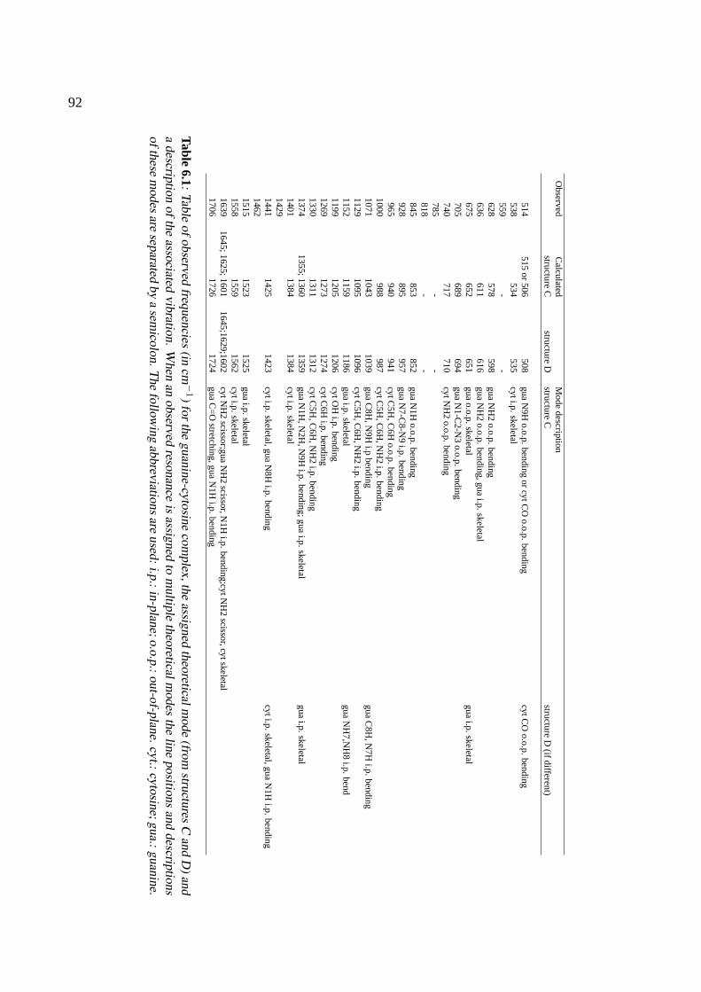
92
Observed
Calculated
Mode
descriptionstructure
Cstructure
Dstructure
Cstructure
D(ifdifferent)
514515
or506
508gua
N9H
o.o.p.bending
orcytC
Oo.o.p.
bendingcytC
Oo.o.p.
bending538
534535
cyti.p.skeletal
559-
-628
578598
guaN
H2
o.o.p.bending
636611
616gua
NH
2o.o.p.
bending,guai.p.
skeletal675
652651
guao.o.p.
skeletalgua
i.p.skeletal
705689
694gua
N1-C
2-N3
o.o.p.bending
740717
710cytN
H2
o.o.p.bending
785-
-818
--
845853
852gua
N1H
o.o.p.bending
928895
957gua
N7-C
8-N9
i.p.bending
965940
941cytC
5H,C
6Ho.o.p.
bending1000
988987
cytC5H
,C6H
,NH
2i.p.
bending1071
10431039
guaC
8H,N
9Hi.p
bendinggua
C8H
,N7H
i.p.bending
11291095
1096cytC
5H,C
6H,N
H2
i.p.bending
11521159
1186gua
i.p.skeletal
guaN
H7,N
H8
i.p.bend
11991205
1206cytO
Hi.p.
bending1269
12731274
cytC6H
i.p.bending
13301311
1312cytC
5H,C
6H,N
H2
i.p.bending
13741355;1360
1359gua
N1H
,N2H
,N9H
i.p.bending;gua
i.p.skeletal
guai.p.
skeletal1401
13841384
cyti.p.skeletal
14291441
14251423
cyti.p.skeletal,gua
N8H
i.p.bending
cyti.p.skeletal,gua
N1H
i.p.bending
14621515
15231525
guai.p.
skeletal1558
15591562
cyti.p.skeletal
16391645;1625;1601
1645;1629;1602cytN
H2
scissor;guaN
H2
scissor,N1H
i.p.bending;cytN
H2
scissor,cytskeletal1706
17261724
guaC
=O
stretching,guaN
1Hi.p.
bending
Table6.1:Table
ofobservedfrequencies
(incm
−1)forthe
guanine-cytosinecom
plex,theassigned
theoreticalmode
(fromstructures
Cand
D)and
adescription
ofthe
associatedvibration.
When
anobserved
resonanceis
assignedto
multiple
theoreticalmodes
theline
positionsand
descriptionsofthese
modes
areseparated
bya
semicolon.T
hefollow
ingabbreviations
areused:i.p.:in-plane;o.o.p.:out-of-plane.cyt.:cytosine;gua.:guanine.
CHAPTER 7
THE IR ABSORPTION SPECTRUM OF
BENZYL-β-LACTOSIDE
The IR absorption spectrum of the gas-phase disaccharide benzylβ-lactoside is recorded in the 330–1650 cm−1 region. The spectrum of the largest system investigated in this thesis using IR-UV doubleresonance methods still exhibits a very clear and fingerprint-like structure in this spectral region. Thecombination of the present data with IR spectroscopic data recorded in the O–H stretching vibrationregion leads to an assignment of the IR spectrum to that of a structure with the lowest calculatedenergy. This structure has glycosidic torsion angles ofφ (H1-C1-O-C4’)∼ 180o and ψ (C1-O-C4’-H4’) ∼ 0o which correspond to a rotation of∼ 15o about the glycosidic bond compared tothe accepted solution-phase conformation. Strong inter-ring hydrogen bonds rigidify the gas-phasestructure.
Adapted from: R. A. Jockusch, R. T. Kroemer, F. O. Talbot, J. P. Simons, L. C. Snoek, P. Carcabal, M. Havenith,J. M. Bakker, I. Compagnon, G. Meijer, and G. von Helden,J. Am. Chem. Soc.126, 5709–5714 (2004).
93

94
7.1 Introduction
The biological activities of oligosaccharide chains bound in glycoproteins or glycolipids are inti-mately linked to their structures and conformations [1, 2]. Many selective cell-cell interactions andmolecular recognition processes involve glycoconjugates. Specificity is often already present evenat the monosaccharide level, and specificity for disaccharides may be a thousandfold higher. Explo-ration and characterization of the hydrogen-bonded conformational landscapes of oligosaccharides isan essential first step in understanding the origins of their molecular specificity. The component sugarring units in oligosaccharide chains are generally taken to be rigid, but the same assumption cannot bemade about the glycosidic links between them - the types of linkages and torsion angles about theselinks are the primary determinants of their overall conformation(s). Unfortunately, the assignment ofoligosaccharide structure and conformation is a notoriously demanding activity due to the chemicalsimilarity of component monosaccharides, which all contain multiple hydroxyl groups, capable of ex-tensive intra- and intermolecular hydrogen bonding networks. In some circumstances the glycosidiclinkages may be ’stiffened’ by hydrogen-bonded interactions across them, thereby influencing boththeir shape and their structural and biological specificity [3, 4]. In others, the glycosidic linkages maybe highly flexible. This flexibility hinders crystallographic structural determinations; it also compli-cates or limits NMR (solution phase) structural assignments based upon time-averaged measurements[5]. Computational studies of (gas-phase) oligosaccharide structure are handicapped by uncertaintiesin the parameters employed in molecular mechanics calculations, andab initio calculations are re-stricted by molecular size and complexity constraints [6–9] - though with the current rate of increasein computer power, these restrictions are diminishing rapidly. Calculations of oligosaccharide confor-mations in solution are constrained by the additional complexity introduced by interactions with thesolvent [4, 6, 10]. Taken together, these limitations represent a very significant challenge to experi-ment and to reliable molecular modeling.
Recently, IR spectroscopy in the near-IR probing the O–H stretching vibrations has been ap-plied to study the structure of two monosaccharides, phenyl-β-D-galactopyranoside and phenyl-β-D-glucopyranoside, and to hydrated clusters of the latter [11–13]. In all cases the combination of IRspectral data with ab-initio data proved sufficient for a detailed structural assignment, especially asthe O–H stretching vibrations turned out to be very sensitive to local variations. This sensitivity givesrise to optimism about the possibility to identify the structures of even larger systems. For saccha-ride systems so far no information in the fingerprint region is available, and the interesting questionwhether other vibrational modes are as sensitive to the environment as the O–H stretching vibrationshas not yet been answered.A priori one could suspect that the C–O–H twisting or bending vibrationscould also be a good identifier of the structure of the system.
Lactose, a very common disaccharide consisting of a glucose and a galactose unit, presents a goodchoice for an investigation of both the possibility to apply the combination of IR spectroscopy andab-initio calculations to identify the gas phase structure of larger systems, and to the sensitivity ofthe mid-IR fingerprint region to probe larger hydrogen bonding networks. NMR studies of lactosein solution indicate the dominance (> 85%) of a single conformation in which the hydroxymethylgroups of the two rings adopt atrans orientation about the glycosidic linkage [with the glycosidictorsion angles,φ (H1-C1-O-C4’)∼35o, ψ (C1-O-C4’-H4’)∼-21o [8, 14, 15]. X-ray diffraction andNMR studies of lactose bound in proteins also indicate the adoption of atransconformation [16]. Incontrast, empirical force field andab initio computational studies of lactose and of its near relative,cellobiose (Glc-β-D–Glc), indicate at least three regions of torsional stability about the glycosidic
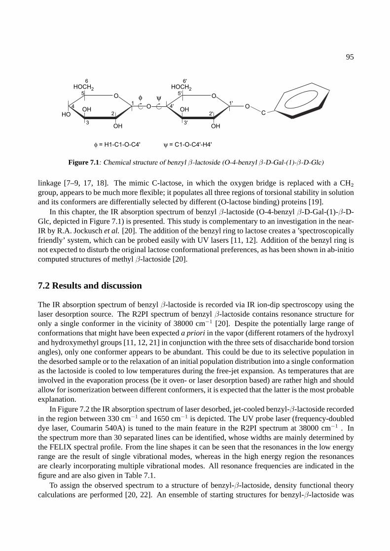
95
O
HOCH2
OH
HOOH
1
2
3
4
5
6
O
φ = H1-C1-O-C4'
φ O
HOCH2
OH
OH1'
2'
3'
4'
5'
6'
ψ
OC
ψ = C1-O-C4'-H4'
Figure 7.1: Chemical structure of benzyl β-lactoside (O-4-benzyl β-D-Gal-(1)-β-D-Glc)
linkage [7–9, 17, 18]. The mimic C-lactose, in which the oxygen bridge is replaced with a CH2
group, appears to be much more flexible; it populates all three regions of torsional stability in solutionand its conformers are differentially selected by different (O-lactose binding) proteins [19].
In this chapter, the IR absorption spectrum of benzylβ-lactoside (O-4-benzylβ-D-Gal-(1)-β-D-Glc, depicted in Figure 7.1) is presented. This study is complementary to an investigation in the near-IR by R.A. Jockuschet al. [20]. The addition of the benzyl ring to lactose creates a ’spectroscopicallyfriendly’ system, which can be probed easily with UV lasers [11, 12]. Addition of the benzyl ring isnot expected to disturb the original lactose conformational preferences, as has been shown in ab-initiocomputed structures of methylβ-lactoside [20].
7.2 Results and discussion
The IR absorption spectrum of benzylβ-lactoside is recorded via IR ion-dip spectroscopy using thelaser desorption source. The R2PI spectrum of benzylβ-lactoside contains resonance structure foronly a single conformer in the vicinity of 38000 cm−1 [20]. Despite the potentially large range ofconformations that might have been expecteda priori in the vapor (different rotamers of the hydroxyland hydroxymethyl groups [11, 12, 21] in conjunction with the three sets of disaccharide bond torsionangles), only one conformer appears to be abundant. This could be due to its selective population inthe desorbed sample or to the relaxation of an initial population distribution into a single conformationas the lactoside is cooled to low temperatures during the free-jet expansion. As temperatures that areinvolved in the evaporation process (be it oven- or laser desorption based) are rather high and shouldallow for isomerization between different conformers, it is expected that the latter is the most probableexplanation.
In Figure 7.2 the IR absorption spectrum of laser desorbed, jet-cooled benzyl-β-lactoside recordedin the region between 330 cm−1 and 1650 cm−1 is depicted. The UV probe laser (frequency-doubleddye laser, Coumarin 540A) is tuned to the main feature in the R2PI spectrum at 38000 cm−1 . Inthe spectrum more than 30 separated lines can be identified, whose widths are mainly determined bythe FELIX spectral profile. From the line shapes it can be seen that the resonances in the low energyrange are the result of single vibrational modes, whereas in the high energy region the resonancesare clearly incorporating multiple vibrational modes. All resonance frequencies are indicated in thefigure and are also given in Table 7.1.
To assign the observed spectrum to a structure of benzyl-β-lactoside, density functional theorycalculations are performed [20, 22]. An ensemble of starting structures for benzyl-β-lactoside was
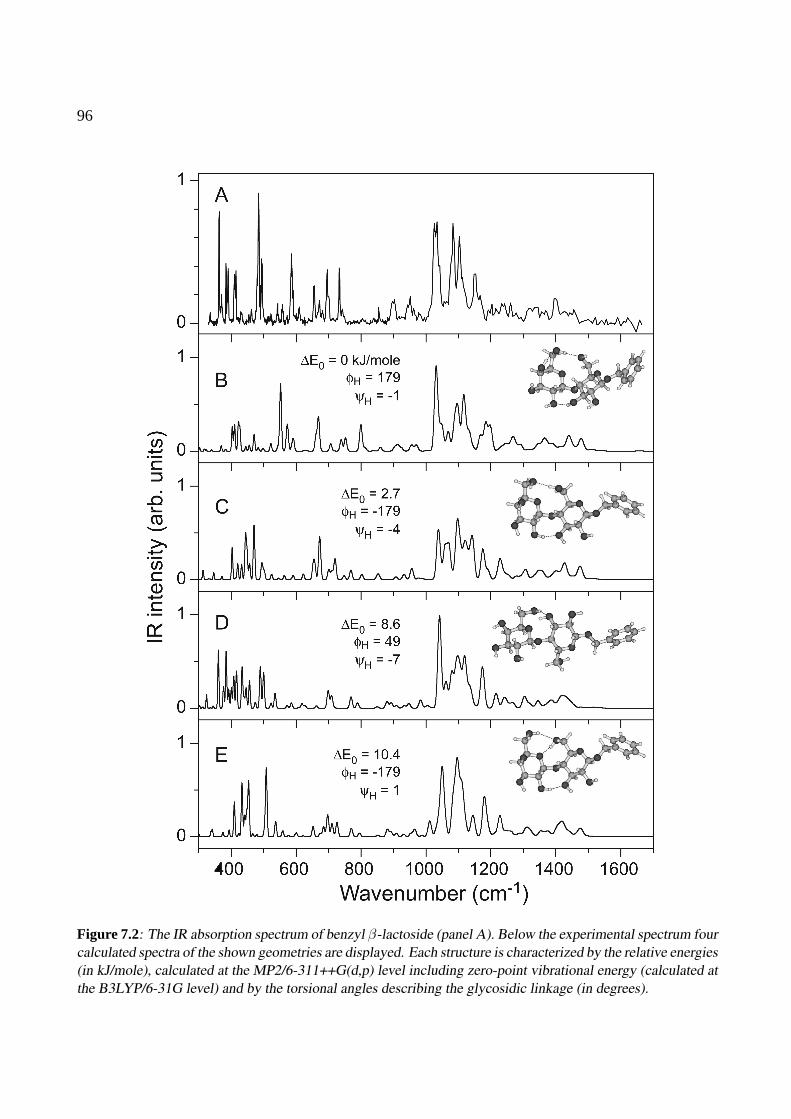
96
Figure 7.2: The IR absorption spectrum of benzyl β-lactoside (panel A). Below the experimental spectrum fourcalculated spectra of the shown geometries are displayed. Each structure is characterized by the relative energies(in kJ/mole), calculated at the MP2/6-311++G(d,p) level including zero-point vibrational energy (calculated atthe B3LYP/6-31G level) and by the torsional angles describing the glycosidic linkage (in degrees).
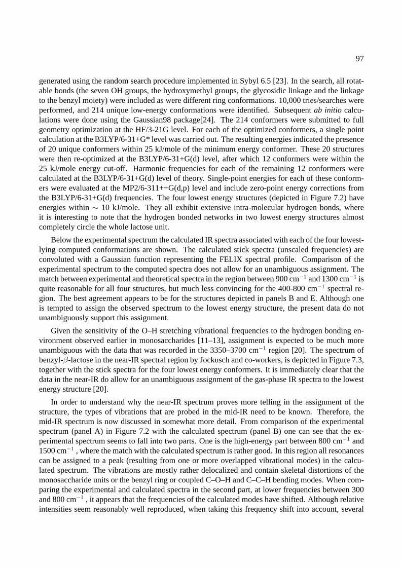
97
generated using the random search procedure implemented in Sybyl 6.5 [23]. In the search, all rotat-able bonds (the seven OH groups, the hydroxymethyl groups, the glycosidic linkage and the linkageto the benzyl moiety) were included as were different ring conformations. 10,000 tries/searches wereperformed, and 214 unique low-energy conformations were identified. Subsequentab initio calcu-lations were done using the Gaussian98 package[24]. The 214 conformers were submitted to fullgeometry optimization at the HF/3-21G level. For each of the optimized conformers, a single pointcalculation at the B3LYP/6-31+G* level was carried out. The resulting energies indicated the presenceof 20 unique conformers within 25 kJ/mole of the minimum energy conformer. These 20 structureswere then re-optimized at the B3LYP/6-31+G(d) level, after which 12 conformers were within the25 kJ/mole energy cut-off. Harmonic frequencies for each of the remaining 12 conformers werecalculated at the B3LYP/6-31+G(d) level of theory. Single-point energies for each of these conform-ers were evaluated at the MP2/6-311++G(d,p) level and include zero-point energy corrections fromthe B3LYP/6-31+G(d) frequencies. The four lowest energy structures (depicted in Figure 7.2) haveenergies within∼ 10 kJ/mole. They all exhibit extensive intra-molecular hydrogen bonds, whereit is interesting to note that the hydrogen bonded networks in two lowest energy structures almostcompletely circle the whole lactose unit.
Below the experimental spectrum the calculated IR spectra associated with each of the four lowest-lying computed conformations are shown. The calculated stick spectra (unscaled frequencies) areconvoluted with a Gaussian function representing the FELIX spectral profile. Comparison of theexperimental spectrum to the computed spectra does not allow for an unambiguous assignment. Thematch between experimental and theoretical spectra in the region between 900 cm−1 and 1300 cm−1 isquite reasonable for all four structures, but much less convincing for the 400-800 cm−1 spectral re-gion. The best agreement appears to be for the structures depicted in panels B and E. Although oneis tempted to assign the observed spectrum to the lowest energy structure, the present data do notunambiguously support this assignment.
Given the sensitivity of the O–H stretching vibrational frequencies to the hydrogen bonding en-vironment observed earlier in monosaccharides [11–13], assignment is expected to be much moreunambiguous with the data that was recorded in the 3350–3700 cm−1 region [20]. The spectrum ofbenzyl-β-lactose in the near-IR spectral region by Jockusch and co-workers, is depicted in Figure 7.3,together with the stick spectra for the four lowest energy conformers. It is immediately clear that thedata in the near-IR do allow for an unambiguous assignment of the gas-phase IR spectra to the lowestenergy structure [20].
In order to understand why the near-IR spectrum proves more telling in the assignment of thestructure, the types of vibrations that are probed in the mid-IR need to be known. Therefore, themid-IR spectrum is now discussed in somewhat more detail. From comparison of the experimentalspectrum (panel A) in Figure 7.2 with the calculated spectrum (panel B) one can see that the ex-perimental spectrum seems to fall into two parts. One is the high-energy part between 800 cm−1 and1500 cm−1 , where the match with the calculated spectrum is rather good. In this region all resonancescan be assigned to a peak (resulting from one or more overlapped vibrational modes) in the calcu-lated spectrum. The vibrations are mostly rather delocalized and contain skeletal distortions of themonosaccharide units or the benzyl ring or coupled C–O–H and C–C–H bending modes. When com-paring the experimental and calculated spectra in the second part, at lower frequencies between 300and 800 cm−1 , it appears that the frequencies of the calculated modes have shifted. Although relativeintensities seem reasonably well reproduced, when taking this frequency shift into account, several
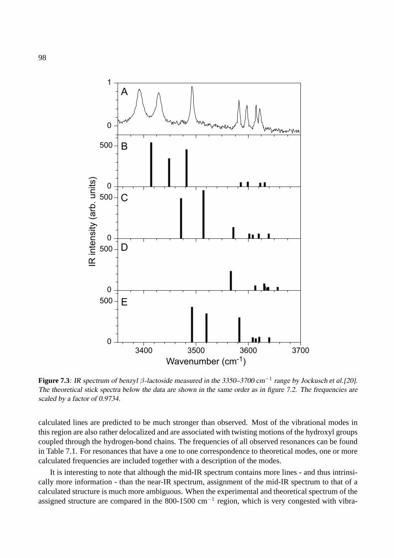
98
3400 3500 3600 3700
0
1
0
500
0
500
0
500
0
500
Wavenumber (cm-1)
IR inte
nsity (
arb
. units)
A
B
C
D
E
Figure 7.3: IR spectrum of benzyl β-lactoside measured in the 3350–3700 cm−1 range by Jockusch et al.[20].The theoretical stick spectra below the data are shown in the same order as in figure 7.2. The frequencies arescaled by a factor of 0.9734.
calculated lines are predicted to be much stronger than observed. Most of the vibrational modes inthis region are also rather delocalized and are associated with twisting motions of the hydroxyl groupscoupled through the hydrogen-bond chains. The frequencies of all observed resonances can be foundin Table 7.1. For resonances that have a one to one correspondence to theoretical modes, one or morecalculated frequencies are included together with a description of the modes.
It is interesting to note that although the mid-IR spectrum contains more lines - and thus intrinsi-cally more information - than the near-IR spectrum, assignment of the mid-IR spectrum to that of acalculated structure is much more ambiguous. When the experimental and theoretical spectrum of theassigned structure are compared in the 800-1500 cm−1 region, which is very congested with vibra-
99
Observed Theory Assignment335363 403 C6’OH, C2’OH twist370 411 C6’OH, C2’OH twist384 422 C6’OH, C2’OH twist390 427 C6’OH twist410415 471 C2OH,C3OH twist431455462485 552 C2OH,C3OH twist494 571/574 C4OH,C6OH twist523553558587 669 C3’OH twist609 707 phenyl C–H o.o.p. bend656672681696733 800 C4OH, C6OH bend742854 859 Glc skeletal900 913 Gal skeletal951 955/971 Glc, Gal skeletal / Glc skeletal
1030 1031/1032 C2’OH, C6’H2, C6’OH bend, C1’OC5’ stretching/C2OH, C2C3H, OC1H bend
1083 1089/1098/1099 C2OH, C3OH, C2C3H bend/Gal skeletal/Glc skeletal1103 1114/1116/1119 benzyl C–C–H i.p./ Glc, Gal, benzyl skeletal / Gal skeletal1150 1168/1184/1198 Glc skeletal / Glc skeletal / Gal skeletal123313251397 1439/1441 C6’OH, C5’C6’H bend / C2C3H, C3OH bend1456 1475/1480 C2C1H, C2OH, CH2(benzyl-link) bend
/ C6OH, C6’OH, C4’OH bend
Table 7.1: Frequencies of observed resonances (in cm−1) in the IR absorption spectrum of benzyl-β-lactosidetogether with the calculated frequencies and descriptions of the assigned vibrations.
tional structure, the match can be concluded to be rather good. Therefore, it appears that the currentlevel of theory is good enough to describe the skeletal and bending vibrations, even though many ofthese are rather delocalized. In principle this region could thus serve well as a spectral fingerprintingregion of the molecular conformation. Unfortunately, the spectral properties of FELIX make from themore than 30 expected modes with IR intensity a structured, but not completely resolved spectrum.With the absolute frequency shifts that are predicted for vibrational modes in different geometries, theresulting IR spectra will change mainly in the shape of the resonance structure, but no dramaticallychanged resonances will appear. This effect is illustrated well when one compares the differences inthe calculated spectra between 800 cm−1 and 1500 cm−1 in Figure 7.2. The highly localized O–Hstretching vibrations that are probed in the 3350–3700 cm−1 region form an indirect probe of theintramolecular hydrogen bonds. As these play such an important role in stabilizing the structure, thevibrations that are sensitive to changes in the hydrogen bonding environment are more telling than themore delocalized skeletal modes. The stretching modes consequently exhibit much larger differencesin the calculated frequencies, and are thus more suitable for assignment. Moreover, the near-IR spec-tral region is not as congested as the region of the skeletal vibrations: the region probed spans roughly400 cm−1 and there are only seven modes with IR intensity. In the equally large range of 900–1250
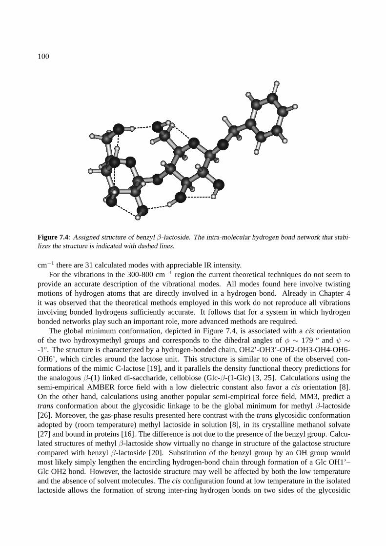
100
Figure 7.4: Assigned structure of benzyl β-lactoside. The intra-molecular hydrogen bond network that stabi-lizes the structure is indicated with dashed lines.
cm−1 there are 31 calculated modes with appreciable IR intensity.For the vibrations in the 300-800 cm−1 region the current theoretical techniques do not seem to
provide an accurate description of the vibrational modes. All modes found here involve twistingmotions of hydrogen atoms that are directly involved in a hydrogen bond. Already in Chapter 4it was observed that the theoretical methods employed in this work do not reproduce all vibrationsinvolving bonded hydrogens sufficiently accurate. It follows that for a system in which hydrogenbonded networks play such an important role, more advanced methods are required.
The global minimum conformation, depicted in Figure 7.4, is associated with acis orientationof the two hydroxymethyl groups and corresponds to the dihedral angles ofφ ∼ 179 o andψ ∼-1o. The structure is characterized by a hydrogen-bonded chain, OH2’-OH3’-OH2-OH3-OH4-OH6-OH6’, which circles around the lactose unit. This structure is similar to one of the observed con-formations of the mimic C-lactose [19], and it parallels the density functional theory predictions forthe analogousβ-(1) linked di-saccharide, cellobiose (Glc-β-(1-Glc) [3, 25]. Calculations using thesemi-empirical AMBER force field with a low dielectric constant also favor acis orientation [8].On the other hand, calculations using another popular semi-empirical force field, MM3, predict atrans conformation about the glycosidic linkage to be the global minimum for methylβ-lactoside[26]. Moreover, the gas-phase results presented here contrast with thetransglycosidic conformationadopted by (room temperature) methyl lactoside in solution [8], in its crystalline methanol solvate[27] and bound in proteins [16]. The difference is not due to the presence of the benzyl group. Calcu-lated structures of methylβ-lactoside show virtually no change in structure of the galactose structurecompared with benzylβ-lactoside [20]. Substitution of the benzyl group by an OH group wouldmost likely simply lengthen the encircling hydrogen-bond chain through formation of a Glc OH1’–Glc OH2 bond. However, the lactoside structure may well be affected by both the low temperatureand the absence of solvent molecules. Thecis configuration found at low temperature in the isolatedlactoside allows the formation of strong inter-ring hydrogen bonds on two sides of the glycosidic
101
linkage to create a rigid di-saccharide structure and because of this, the enthalpic benefit of the stronghydrogen bonds is somewhat diminished by the loss in flexibility. There is an entropic cost, and it isthe transconfiguration that is favored entropically [18]. Gibbs free energy calculations (room tem-perature; harmonic frequencies) indeed significantly stabilize thetrans conformer over thecis andpredict thetransto be the most stable conformer at room temperature by∼ 2 kJ/mole [20]. However,the low-temperature favoredcis conformer should still be present, at about half the abundance of thetransconformer. Specific complexation of the di-saccharide with water molecules may also alter thepreferred conformation about the glycosidic linkage [4].
7.3 Conclusions
The IR absorption spectrum of benzylβ-lactoside is recorded in the 330–1650 cm−1 region. Theprevious assignment, based on spectroscopic information in the 3350–3700 cm−1 region, to a con-formation in which the two monosaccharide units are linked in acis-configuration is confirmed. Theobservation of benzylβ-lactoside in thecis-configuration is in contrast with the generally accepted liq-uid phase structure (atrans-configuration), but it is not unlikely that the stabilization due to a circlingnetwork of intra-molecular hydrogen bonds is favored by the cold, isolated environment experiencedin the free-jet expansion. The observed vibrational structure could well be used to identify the confor-mational structure, but the present theoretical methods are as yet insufficient to treat the low-energytorsion-like vibrations that are coupled through the hydrogen bonding network. The present data canthus form a good benchmark for future theoretical methods.
102
References
[1] R. A. Dwek, Chem. Rev.96, 683 (1996).
[2] H. Lis and N. Sharon, Chem. Rev.98, 637 (1998).
[3] J. Yu-Jen Chen and K. J. Naidoo, J. Phys. Chem. B107, 9558 (2003).
[4] K. J. Naidoo and J. Yu-Jen Chen, Mol. Phys.101, 2687 (2003).
[5] M. R. Wormald, A. J. Petrescu, Y.-L. Pao, A. Glithero, T. Elliott, and R. A. Dwek, Chem. Rev.102, 371(2002).
[6] A. Imberty and S. Perez, Chem. Rev.100, 4567 (2000).
[7] A. D. French, G. P. Johnson, A.-M. Kelterer, M. K. Dowd, and C. J. Cramer, Int. J. Quantum Chem.84,416 (2001).
[8] L. Asensio and J. Jimenez-Barbero, Biopolymers35, 55 (1995).
[9] S. B. Engelsen, J. Koca, I. Braccini, C. Herve du Penhoat, and P. Perez, Carbohydr. Res.276, 1 (1995).
[10] K. J. Naidoo and J. Brady, J. Am. Chem. Soc.121, 2244 (1999).
[11] F. O. Talbot and J. P. Simons, Phys. Chem. Chem. Phys.4, 3562 (2002).
[12] R. A. Jockusch, F. O. Talbot, and J. P. Simons, Phys. Chem. Chem. Phys.5, 1502 (2003).
[13] R. A. Jockusch, R. T. Kroemer, F. O. Talbot, and J. P. Simons, J. Phys. Chem. A (2525).
[14] M. L. Hayes, A. S. Serianni, and R. Barker, Carbohydr. Res.100, 87 (1982).
[15] N. W. Cheetham, P. Dasgupta, and G. E. Ball, Carbohydr. Res.338, 955 (2003).
[16] H. M. Berman, J. Westbrook, Z. Feng, G. Gilliland, T. Bhat, H. Weissig, I. N. Shindyalov, and P. E.Bourne, Nucl. Acids. Res.28, 235 (2000).
[17] F. Cloran, I. Carmichael, and A. S. Serianni, J. Am. Chem. Soc.121, 9843 (1999).
[18] S. B. Engelsen and K. Rasmussen, J. Carbohydr. Chem16, 773 (1997).
[19] J.-F. Espinosa, F. J. Canada, J. L. Asensio, M. Martın-Pastor, H. Dietrich, M. Martın-Lomas, R. R.Schmidt, and J. Jimenez-Barbero, J. Am. Chem. Soc.118, 10862 (1996).
[20] R. A. Jockush, R. T. Kroemer, F. O. Talbot, J. P. Simons, L. C. Snoek, P. Carcabal, M. Havenith, J. M.Bakker, I. Compagnon, G. Meijer, et al., J. Am. Chem. Soc.126, 5709 (2004).
[21] A. Roen, J. I. Padron, and J. T. Vazquez, J. Org. Chem.68, 4615 (2003).
[22] Theoretical calculations for this work are performed by Romano Kroemer.
[23] M. Clark, R. Cramer, and N. Opdenbosch, J. Comput. Chem.10, 982 (1989).

103
[24] M. J. Frisch, G. W. Trucks, H. B. Schlegel, G. E. Scuseria, M. A. Robb, J. R. Cheeseman, V. G. Za-krzewski, J. J. A. Montgomery, R. E. Stratmann, J. C. Burant, et al.,Gaussian 98, Revision A.7, Pittsburgh,PA (1998).
[25] G. L. Strati, J. L. Willett, and F. A. Momany, Carbohydr. Res.337, 1833 (2002).
[26] A. Rivet, C. Sabin, K. Mazeau, A. Imberty, and S. Perez,http://www.cermav.cnrs.fr/(2003), accessedMay 2003.
[27] R. Stenutz, M. Y. Shang, and A. S. Serianni, Acta Cryst.C55, 1719 (1999).

104
SUMMARY AND OUTLOOK
Weak intra- and intermolecular interactions as well as subtle electronic effects can have a large in-fluence on molecular structure. Infrared (IR) spectroscopy can be a useful tool to investigate theseeffects. In this thesis, a Free-Electron Laser is used to study several molecular model systems usingUV-IR double-resonance techniques. The aim is to compare the experimental spectra with resultsfrom quantum chemical methods to obtain information on the species studied. Whether this will beof success depends on the answer to two simple questions: a) Is the experiment good enough and b)Is theory good enough.
In the experiments on the gas-phase biomolecules, a key question is whether conformational dif-ferences in flexible molecules can actually be observed in the mid-IR wavelength range that is coveredby FELIX. Based on the work presented here, this question can be answered positively. In all sys-tems studied, well-discernible IR spectral structure is observed that is sharp enough to be able todifferentiate between various conformers. Specifically, in Chapter 5, where three different conform-ers of a single amino acid are investigated, it is convincingly demonstrated that the spectroscopictechniques are adequate for this purpose. In all other cases, the investigation of single conformersof biomolecules has led to spectroscopic data that are sufficiently characteristic that they could eitherlead to unambiguous assignments or, at least, to strong evidence for specific classes of conformations.
The second question is whether the currently used (standard) theoretical methods are sufficientlyaccurate to allow for the spectroscopic data to be evaluated meaningfully. It must be concluded thatpresent theory can be very accurate for certain types of vibrations, but less so for others. For highfrequency stretching vibrations (as studied by others) and other strongly localized bending and defor-mation vibrations, the theoretical methods are adequate. For, mostly low-frequency, de-localized andcoupled vibrations, problems can arise. Particularly in the experimental study on benzyl-β-lactoside(Chapter 7), the theoretical treatment of the O–H twisting vibrations, which could be key spectral fea-ture for the derivation of structural conformation, is inadequate. This could potentially be improvedby the evaluation of anharmonicities in the calculations of vibrations, a gargantuan task.
For the future use of the here employed techniques, several restrictions can be foreseen: first ofall, these studies depend strongly on double-resonance techniques. It is not at all certain that (much)larger molecules facilitate such convenient conformer-specific detection schemes as the ones usedhere. A possible complication is then the simultaneous probing of several conformers at once.
Second, larger molecules will exhibit more congested IR spectra, as the number of resonanceswill increase linearly with the number of atoms that form the molecule. For theoretical treatment theincrease in number of atoms will be an even more serious problem, as the requirements on compu-
105

106
tational resources grow strongly non-linear with molecular size. A possible solution is to resort tocarefully parametrized force field molecular mechanics methods.
Thirdly, to bridge the gap between IR spectroscopy of gas-phase biomolecules and the well-established field of liquid-phase IR spectroscopy of solvated biomolecules, it is required to study mi-crosolvation effects,i.e., an extension of the study of gas-phase biomolecules to gas-phase biomoleculesthat are complexed with one or more solvent molecules. Although already quite some work on thissubject has been done in the near-IR, mid-IR spectral studies on such systems are imperative, giventhe before mentioned spectral congestion that is to be expected and the complications that arise withsolvent-induced broadening effects.
It is hoped that the work described in this thesis, together with the work that is being done in thenear-IR spectral range, can form a good starting point for future investigations.
Berlin, fall 2004
SAMENVATTING
Op het raakvlak van de biologie en scheikunde bevindt zich het vakgebied dat zich bezighoudt metvraagstukken aangaande de microscopische interacties tussen moleculen die noodzakelijk zijn voorlevende organismen, de biomoleculaire chemie. Deze tak van wetenschap stelt zich ten doel bi-ologisch macroscopische processen te vertalen naar intermoleculaire processen. Een van haar uit-gangspunten is het ontrafelen van de driedimensionale structuur van de betrokken moleculen.
In het algemeen zijn moleculen met een biologische functie (kortweg biomoleculen) tamelijkgroot. Een typisch eiwit bijvoorbeeld, bevat honderden individuele atomen, gerangschikt in een lang,ketenvormig molecuul. Het is vanzelfsprekend dat een dergelijk lang molecuul een enorme flexi-biliteit bezit die het in staat stelt zich te vervormen en als het ware op te rollen tot een compactekluwen. Er is gebleken dat veel eigenschappen van de intermoleculaire reacties die plaatsvindendirect gerelateerd zijn aan de precieze vorm van de moleculen in kwestie. Veel interacties tussen bi-ologische moleculen vinden plaats op basis van een donor-receptor proces waarbij de partners elkaardoor middel van moleculaire patroonherkenning vinden. Een bekend voorbeeld hiervan zijn deoxyri-bonucleınezuur (DNA) en ribonucleınezuur (RNA), de moleculen die verantwoordelijk zijn voor deoverdracht van de genetische code van levende organismen. Deze moleculen blijken maar opeenunieke wijze met elkaar te wisselwerken. Ook de functionele werking van eiwitten en suikers is vaakuitsluitend bepaald door de driedimensionale structuur van het biomolecuul. De unieke eigenschapvan lange ketenmoleculen in het algemeen en biomoleculen in het bijzonder is dat ze, ondanks hetenorme aantal vrijheidsgraden dat ze bezitten, tijdsgemiddeld eigenlijk altijd ineen zelfde opgeroldetoestand ofwel conformatie worden aangetroffen. Dit bepaalt voor een belangrijk deel de functionelewerking van de moleculen.
Om een beschrijving mogelijk te maken van de driedimensionale structuur van biomoleculenwordt de gehele structuur van biomoleculaire clusters in vier lagen aangeduid. Allereerst bestaat er deprimaire structuur, die beschrijft hoe het molecuul met covalente bindingen uit de individuele atomenis gevormd. Het is ook gebruikelijk met de primaire structuur aan te duiden hoe macromoleculen uitindividuele kleinere moleculen zijn opgebouwd. Men spreekt dan ook vaak van de ketenvolgorde ofsequentie. Een veelgebruikte onderzoeksmethode in de biochemie is het ontwarren van de primairestructuur door middel van hetsequencen, het bepalen van de volgorde van de bouwmoleculen.
Van secundaire structuur spreekt men om de (vaak locale) ruimtelijke orientatie van verschillendezijgroepen ten opzichte van elkaar te karakteriseren. Deze secundaire structuur wordt hoofdzakelijkbepaald door zwakkere interatomaire wisselwerkingen zoals elektrostatische (waterstofbruggen) endispersieve interacties. Typisch zijn deze wisselwerkingen een ordegrootte zwakker dan de covalente
107
108
bindingen waardoor de primaire structuur wordt bepaald. Voorbeelden van secundaire structuren zijndeα-helixstructuur, deβ-geplooide bladstructuur en de verschillende bochtstructuren.
Een verdere karakterisatie vindt plaats met de tertiaire structuur, die op grotere schaal de substruc-turen binnen het macromolecuul beschrijft. De quartaire structuur tenslotte, beschrijft de conformatievan meerdere in elkaar verwikkelde biomoleculen.
Voor een goed begrip van de eigenschappen van biomoleculen is het van belang dat de inherenteeigenschappen van de moleculen die de secundaire en hogere orde structuren bepalen in detail wor-den bestudeerd met een focus op de inter- en intramoleculaire krachten. De laatstgenoemde interactiesworden sterk beınvloed door de aanwezigheid van watermoleculen in de natuurlijke, waterige omgev-ing van biologische systemen. Deze zogenaamde oplossingseffecten versluieren de fundamenteleeigenschappen van de biomoleculen, hetgeen het onderscheid tussen de bijdragen van het molecuulzelf en die van de watermoleculen aan de stabilisering vaneen bepaalde conformatie bemoeilijkt.Om dit onderscheid wel te kunnen maken worden in dit proefschrift experimenten beschreven aanmoleculen die in de gasfase geısoleerd zijn van hun omgeving. Op deze wijze zijn de fundamenteleeigenschappen van de moleculen zelf ontdaan van oplossingsgerelateerde effecten. In eventuele ver-volgstudies kunnen dan clusters van biomoleculen en watermoleculen worden bestudeerd waarbij bijsequentiele toevoeging van watermoleculen de oplossingseffecten als het ware worden opgebouwd.
Voor de karakterisatie van de structuren van biomoleculen in de gasfase wordt in dit proefschriftgebruik gemaakt van infraroodspectroscopie. Het infraroodspectrum is zeer nauw verbonden metde structuur van een molecuul aangezien het door de moleculaire vibraties wordt bepaald, en dusook door de daaraan sterk gerelateerde moleculaire bindingen. Daarom wordt met behulp van ver-schillende spectroscopische technieken van gasfase moleculen het infraroodspectrum opgemeten terbepaling van hun driedimensionale structuur.
Beschrijving van de experimenten
Om de infraroodspectra van de moleculen in kwestie te meten worden de moleculen samen met eeninert draaggas door een gepulste klep in een vacuumkamer gelaten. Daar ondergaat de gaswolk eenadiabatische expansie. De interne energie van de gaswolk wordt omgezet in kinetische energie. Allemoleculen in het gas staan door middel van binaire botsingen met draaggasatomen hun interne energieaf totdat ze zich in hun elektronische en vibrationele grondtoestand bevinden. Een gedeelte van deuitzettende gaswolk wordt een tweede vacuumkamer ingelaten. Hier vindt interactie plaats met deverschillende laserbronnen.
Aangezien het geen triviale zaak is om alle typen moleculen intact in de gasfase te brengen,worden er verschillende technieken gebruikt om dit te bewerkstelligen. Van sommige moleculen heeftde vastestof of vloeistof een voldoende hoge dampdruk om een dichte wolk voor het experiment tecreeren. Bij andere moleculen moet de vastestof worden verhit om de gewenste dampdruk te bereiken.Helaas blijken biomoleculen in het algemeen slecht bestand tegen verhitting, omdat er fragmentatieplaatsvindt voordat sublimatie vanuit de vastestof kan optreden. Voor deze moleculen is een techniekontwikkeld, waarbij de moleculen op een goed gedefinieerd tijdstip vanuit een grafieten matrix meteen gepulste desorptielaser in de gasfase worden gebracht. Dit proces gebeurd op een dusdanige plaatsen tijd dat de biomoleculen onmiddellijk worden meegevoerd in de nog expanderende draaggaswolken vrijwel al hun interne energie kwijtraken door botsingen met de draaggasatomen.
De infraroodspectra van de moleculen worden gemeten met twee verschillende spectroscopische
109
technieken. De eerste techniek, infrarood geınduceerde fotodissociatie, wordt gebruikt voor geladensystemen. Hierbij wordt het spectrum van een heel precies geprepareerd molecuulion dat een com-plex vormt met een inert edelgasatoom gemeten. Aangenomen wordt dat de aanwezigheid van hetedelgasatoom een verwaarloosbaar kleine invloed op het spectrum heeft. Het resulterende infrarood-spectrum is dus in zeer goede benadering gelijk aan dat van het kale ion.
De tweede techniek, infraroodion-dip spectroscopie, is universeel toepasbaar om de infrarood-spectra van neutrale moleculen te meten. In deze techniek wordt de moleculaire populatie van degrondtoestand van het molecuul bemonsterd door in het molecuul een zeer goed gedefinieerde elektro-nische overgang te induceren met een ultraviolet foton. Een tweede foton ioniseert dan het molecuulwaarna het wordt gedetecteerd in een massaspectrometer. Kort voordat de elektronische overgangwordt geınduceerd vindt er een interactie plaats tussen de moleculen en de infraroodlaser. De laatstekan een vibrationele overgang in de moleculen induceren. Wanneer dit het geval is, dan wordt eendeel van de bevolking vanuit de grondtoestand naar een vibrationeel geexciteerde toestand overge-bracht. Dit heeft tot gevolg dat er minder moleculen in de grondtoestand achterblijven die bemonsterdkunnen worden door de ultravioletlaser en leidt tot een vermindering van het aantal geproduceerdeen gedetecteerde ionen, eenion-dip. Door nu het aantal ionen te meten als een functie van de in-fraroodgolflengte wordt het infraroodion-dip spectrum verkregen. Dit kan weer tot een infraroodabsorptiespectrum worden geconverteerd. Aangezien de elektronische overgangen die door de ultra-violetlaser worden geınduceerd voor elk conformeer van een molecuul anders zijn wordt voor eengekozen ultraviolette golflengte slechts het infraroodspectrum vaneen moleculair conformeer geme-ten.
Een uniek gegeven voor dit werk is de beschikbaarheid van de vrije-elektronenlaser FELIX alsbron voor afstembare infraroodstraling. FELIX is een bij uitstek geschikt instrument voor het uitvo-eren van spectroscopisch werk aan moleculen in het mid- tot verinfrarode golflengtegebied (ruwwegvan 5µm tot 250µm) aangezien zich in dit gebied het merendeel van de vibraties bevindt. Naast hetgrote bereik van de golflengte maken de continue golflengteafstembaarheid, de resulterende lichten-ergie die beschikbaar is voor de gasfase experimenten (typisch 100 mJ in 5µs) en de voor vibrationelespectroscopie zeer acceptabele bandbreedte dit instrument tot een ideaal hulpmiddel.
Voor de interpretatie van de gemeten spectra worden quantumchemische berekeningen uitgevoerd.Van een aantal verschillende conformaties van het molecuul wordt het infraroodspectrum uitgerekendwaarmee het gemeten spectrum vergeleken kan worden. Op basis van een gevonden gelijkenis kanhet gemeten spectrum dan aan een specifieke structuur worden toegekend.
Resultaten
Alhoewel alle experimenten die in dit proefschrift worden beschreven als modelsysteem kunnen wor-den beschouwd, kan het eerste toch als een klassieker worden beschouwd: in hoofdstuk 2 wordt hetinfraroodspectrum van het benzeenion gemeten door middel van infrarood fotodissociatie. Het magverbazing wekken dat van een zo belangrijk molecuul het infraroodspectrum van het ion aan het eindevan de 20e eeuw nog niet bekend is. De huidige meetmethoden nemen de barriere weg om het in-fraroodspectrum van het benzeenion te bestuderen. Daarnaast lenen de experimenten zich uitstekendvoor een verkenning van de mogelijkheden van de beschikbare methoden. Bovendien is het benzee-nion een klassiek modelsysteem voor bestudering van het Jahn-Tellereffect, een quantummechanischeffect waarin door middel van een koppeling tussen elektronische en bepaalde vibrationele toestanden

110
de hoogsymmetrische structuur van benzeen wordt vervormd.
De in dit proefschrift beschreven experimenten aan het benzeenion dragen bij tot een quantitatiefbegrip van de vibratie-eigenschappen van dit belangrijke molecuul-ion. Ook wordt aangetoond datde invloed die het edelgasatoom heeft op het infraroodspectrum miniem is in het geval van neonals gebonden edelgasatoom, maar dat er aantoonbare verschillen bestaan als argon wordt gebruikt.Tenslotte wordt beschreven dat het er sterk op lijkt dat in het systeem benzeen-Ar2
+ ook de tweeargonatomen, die zich aan weerszijden van het benzeenmolecuul bevinden, onderhevig zijn aan hetJahn-Tellereffect.
In hoofdstuk 3 wordt het neutrale benzeen dimeer bestudeerd. Van dit systeem, dat model staatvoor de interactie tussen twee moleculen door puur dispersieve krachten, bestaan twee voorspeldestructuren: een T-vormige, waarbijeen van de benzeenmoleculen de stam eneen de dwarsbalk van deT vormt, en een parallelle structuur waarin de benzeenmoleculen enigszins van elkaar afgeschovenzijn. Historisch zijn er zowel voorstanders van de T-vormige als van de parallel verplaatste structurengeweest. De hier beschreven experimenten wijzen in de richting van een T-vormige structuur. Erzijn zelfs tekenen die op een enigszins vervormde T-structuur wijzen. Tevens blijkt dat het infrarood-spectrum van het dimeersysteem nauwelijks afwijkt van dat van het monomeer, een bewijs van deslechts uiterst kleine invloed van de dispersieve krachten die het dimeer stabiliseren op de individuelemonomeren waaruit het dimeer bestaat.
Na de studie van de invloed van dispersieve krachten op het infraroodspectrum wordt in hoofdstuk4 een systeem onder de loep genomen dat door middel van twee waterstofbruggen verbonden is: hetbenzoeenzuur dimeer. De verschillen tussen de spectra van het monomeer en van het dimeer zijnaanzienlijk. Hieruit blijkt dat de aanwezigheid van een dubbele waterstofbinding het infraroodspec-trum sterk beınvloedt.
Omdat dit een berekeningstechnisch nog vrij klein systeem is geldt het als een proeve voor derekenmethodes die gebruikt kunnen worden voor grotere systemen. De gebruikte theoretische meth-ode slaagt goed in de voorspelling van de infraroodspectra van zowel monomeer als dimeer en kandus met vertrouwen gebruikt worden in meer gecompliceerde systemen. Een aspect van de – vaak ge-bruikte – theoretische methode blijkt niet toereikend in de beschrijving van dit moleculair systeem: deberekende frequentie van de vibratie waarbij de beide waterstofatomen in de C–O–H· · ·O=C bindin-gen door het vlak van de moleculen bewegen wijkt sterk af van de geobserveerde.
In hoofdstuk 5 wordt het zeer flexibele molecuul tryptophaan bestudeerd. Tryptophaan kan ineen groot aantal verschillende conformaties, die worden gekenmerkt door intramoleculaire waterstof-bindingen, voorkomen. Van drie van de in totaal zes ooit waargenomen conformeren is het infrarood-spectrum gemeten en er blijken tussen de spectra van verschillende conformeren grote verschillen tebestaan. De in hoofdstuk 4 geteste theoretische methode blijkt wederom in staat om op basis vande vergelijking tussen berekende en gemeten spectra de gemeten spectra uniek toe te kennen aanbepaalde berekende structuren. Nadat in eerder onderzoek in het midinfrarode golflengtegebied alwas gebleken dat de spectra gevoelig waren voor conformatieverschillen, bewijst dit eerste onderzoekvan een zo flexibel molecuul in het midinfrarode golflengtegebied dat ook hier structurele identificatiegoed mogelijk is.
Een zeer interessant biologisch systeem wordt onderzocht in hoofdstuk 6: het gasfasecluster vanguanine en cytosine, twee van de vier nucleobases in DNA. De meest interessante vraag is hier ofde nucleobases in afwezigheid van de suiker-fosfaat keten die de helixstructuur stabiliseert nog altijddezelfde drievoudige waterstofverbinding aangaan in de gasfase. Uit de vergelijking van het experi-
111
menteel waargenomen spectrum met berekende spectra voor verschillende configuraties van guanine-cytosine blijkt dat niet de klassieke “Watson-Crick” configuratie wordt waargenomen. Het hierwaargenomen guanine-cytosine complex bevat een tautomeer van guanine. Een definitieve toeken-ning van het geobserveerde spectrum aan een bepaalde configuratie blijkt in het hier bestudeerdegolflengtebereik niet mogelijk te zijn. Hoogstwaarschijnlijk komt de “Watson-Crick” configuratiewel voor in de bundel, maar is het de UV detectie die observatie niet mogelijk maakt.
Tenslotte worden in hoofdstuk 7 experimenten beschreven aan de gesynthetiseerde disaccharidebenzyl–β–lactoside. De experimenten tonen aan dat ook moleculen met een dergelijke omvang nogeen uniek infraroodspectrum bezitten dat aan de hand van theoretisch berekende spectra toegekendkan worden aan een unieke conformatie. Helaas blijkt hier wel dat met de huidige voorhanden zijndetheoretische modellen bepaalde typen vibraties niet adequaat berekend worden en dat het wenselijkis deze beschrijving te verbeteren.
In grote lijnen bewijst het werk dat is beschreven in dit proefschrift dat het toepassen van midin-frarode straling voor de identificatie van structurele conformaties van flexibele (bio)moleculen eensuccesvolle methode is waarmee de intra- en intermoleculaire interacties op gedetailleerde wijzebestudeerd kunnen worden. In alle beschreven experimenten blijkt unieke identificatie aan de handvan een midinfarood absorptiespectrum mogelijk. Een toekomstige belemmering zou wel eens kun-nen bestaan uit het falen van theoretische berekeningen aan gekoppelde en sterk gedelocaliseerdevibraties, alsmede uit de afwezigheid van een geschikt detectiemechanisme met ultraviolet laserlicht.Niettemin vormt de grootte van het molecuul in ieder geval nog geen belemmering voor identificatie.
De in dit proefschrift beschreven methoden bewijzen daarmee dat infraroodspectroscopie eenkrachtig instrument vormt in de identificatie van flexibele moleculaire structuur in de gasfase.

112
DANKWOORD
Vroeger ging ik promoveren. Dat zou gebeuren op een dag ver, ver in de toekomst. Tot die tijd zou ikwat rondlummelen in een laboratorium, af en toe wat lasers uitlijnen, spectra van moleculen opmetenen de ermee samenhangende, lastige problemen proberen te doorgronden. Wie weet zou ik er zelf eenbijdrage toe leveren dat zulke problemen beter begrepen zouden kunnen worden. Een ding was zeker:de weg naar de promotie zou lang, en vooral zwaar zijn. Loodzwaar.
Het bleek een vergissing. Ik heb rondgelummeld, uitgelijnd, opgemeten en doorgrond. Ik heb eenbijdrage kunnen leveren om natuurwetenschappelijke problemen van beter begrip te voorzien. Maarzwaar? Zwaar was het niet. Het was een feest.
Op mijn feest waren een heleboel gasten. Sommigen waren er de hele tijd, anderen kwamen evensnel gedag zeggen om naar een volgende afspraak te gaan. Van ieders aanwezigheid heb ik genoten.
Het feest is nu over, de lichten gaan aan. Het laatste biertje wordt getapt, wie weet volgt straksnog koffie. Het uur is gekomen om na te praten, om de uitnodigingen voor een volgende gelegenheidte sturen. Maar vooral om te bedanken voor de aanwezigheid op mijn feest.
Laat ik beginnen de feestzaal te complimenteren. Wat een mooi instituut is dat Rijnhuizen toch!Met zijn prachtige kasteel, zijn tuin en zijn rustige omgeving een ideale plek om een promotie tevolbrengen. Ik wil hier alle afdelingen van het instituut bedanken voor de prachtige tijd die ik hebmogen doorbrengen tussen jullie. Dankzij jullie hulpvaardigheid, inspiratie of gewoon koffie metkletspraat heb ik zo’n mooie tijd gehad.
Dan de ceremoniemeester, Gerard Meijer. Gerard, in de afgelopen vier jaar heb je je voor mijbewezen als een Weltklasse wetenschapper, motivator en manager. Al duurden je bezoeken aan mijnlab in de regel niet langer dan 5 minuten, in die tijd bleek je altijd in staat je enthousiasme voormoleculen over te brengen. Je enorme parate kennis, al dan niet doorspekt met een portie bluf dieje in het westen niet zou misstaan, wist je altijd wel aan te wenden om me in de goede richting teduwen als het weer eens ergens wat stroever liep. Ook toen je transfer naar Berlijn allang in kannenen kruiken was bleek je met een verbluffende efficientie in staat me bij te staan. Ik heb er dan ook eenvol vertrouwen in dat de komende periode minstens zo succesvol zal zijn. Dat je me ooit nog eenszult poorten, waag ik overigens te betwijfelen.
Een ambitieuze hoogleraar verpoost veel van zijn tijd onderweg om zijn ideeen te verkopen. InGerards afwezigheid ontpopte Gert von Helden zich als de drijvende kracht in de groep. Gert, ik vondhet een groot plezier met je te werken. Vooral in de overgang van het eeuwigdurende benzeen naar debiomoleculen heb ik je enthousiasme en je ijzersterke kwaliteiten op het vlak van quantum chemischeberekeningen leren kennen. Met je galgehumor en je eeuwige geklaag over de Nederlandse gebruiken
113
114
en eetgewoontes was je daarnaast ook een voortdurende inspiratiebron voor wetenschappelijk miss-chien minder verantwoorde maar des te vermakelijkere gesprekken.
Onze groepstechnicus Jan Pluygers heb ik eeuwigvaak lastiggevallen voor hulp op het praktischevlak. Jan, je werd nooit (te) moe van me en hebt me vele malen van mijn twee linkerhanden gered.Het is voor mij zonneklaar dat zonder jou dit boekje niet tot stand was gekomen.
Avec Isabelle Compagnon j’ai fait beaucoup de voyages nocturnes entre Utrecht et Amsterdamapres des shifts pleins de beaux resultats sur les biomolecules. Isabelle, j’ai apprecie les derniersshifts, et j’ai confiance que tu emporteras beaucoup de succes avec mon experiment.
Dat ik Isabelle heb kunnen inwijden in de fijne kneepjes van het werken met FELIX, is onderandere de verdienste van Hans Piest. Hans heeft me in een maand of vier alle nukken en eige-naardigheden van de opstelling geleerd. Hans, bedankt!
Middenin kreeg ik de kans mijn eigen pedagogische vaardigheden uit te oefenen op een Fransegaststudent, Luke Mac Aleese. Luke, c’a marche pas mal, eh? Merci pour cette periode et bonnechance avec tesetudes, toi-meme.
Toen ik begon in de groep Moleculaire Dynamica liep daar zo’n druk, opgewonden standje rond,Deniz van Heijnsbergen. In de 3 jaar sindsdien hebben we met groot plezier het instituut verlevendigd,varierend van ’hulp’ bij Gerts interne verhuizing en Gerards computerproblemen tot de verkeersher-structurering binnen het instituut. Dat we ook na onze promoties goede vrienden blijven is voor mijbuiten twijfel. Daar helpt zelfs verhuizen naar New York niet tegen.
Samen met Rob Satink deelden Deniz en ik lange tijd een kamer. Rob, dat je al het gekleuter hebtweten te overleven mag een klein wonder heten. Anderzijds heb we ook binnen de kamer enorme lolgehad. Dankzij jouw eerdere werk aan benzeen en het Jahn-Teller effect kon ik een vliegende startmaken.
Van mijn laatste kamergenoot, Dave Moore, heb ik veel plezier gehad. Nooit heb ik een Amerikaanzo snel na aankomst in Nederland zo goed Nederlands horen praten. Voor elk probleem had hijeenontstellend lang, dan wel meerdere antwoorden. Bij al zijn Amerika-bashing zou je bijna vergeten dathij zelf Amerikaan is.
Ook de mensen binnen de groep met wie ik minder direct samengewerkt heb, Nick Polfer, AndreFielicke, Karine Demyk, Britta Redlich en Jos Oomens bedank ik voor de uitstekende sfeer en be-hulpzaamheid.
Bij alle experimenten beschreven in dit proefschrift is gebruik gemaakt van de vrije-elektronenlaser FELIX. De hulp van de FELIX-staf, bestaande uit Lex van der Meer, Britta Redlich (daar isze weer), Giel Berden, Rene van Buuren, Wim Mastop, Peter Delmee en Theo Ram, is daarvooronontbeerlijk geweest. Ik dank jullie allen nogmaals voor de uitstekende omstandigheden waaronderik heb kunnen werken en voor de bereidheid om me tot vaak diep in de nacht al dan niet telefonischuit de brand te helpen.
Aan de andere kant van het gebouw was een tweede groep gespecialiseerd in molecuulfysica, deKoude Moleculen. In willekeurige volgorde Rick Bethlem, Floris Crompvoets, Jacqueline van Veld-hoven, Bas van de Meerakker, Rienk Jongma, Sophie Schlunk, Jochen Kupper, Dave Carty, AllardMosk, Wieland Schollkopf, Boris Sartakov, Paul Smeets en Andre van Roij bleken een uitstekendebron van kennis, materialen en plezier. Ik heb ondertussen wel gezien dat Bas, nu hij niet meer zohoeft te steunen om het Amsterdam-Rijnkanaal over te fietsen, een enorm succesvolle fysicus gaatworden.
Mochten er ondanks of dankzij voornoemde mensen toch nog enige frustraties hebben bestaan,
115
dan was er dinsdags en donderdags alle gelegenheid om die af te reageren op de bal. Menige poeierheb ik daar uitgedeeld, en het was fijn dat Rob, Deniz, Aad, Anton, Frank, Dick, Jan-Cornelis, Toon,Dave, David en Jochen daar ook waren om me nog wat verder geırriteerd te krijgen.
Halverwege mijn promotie heb ik een kleine tweeeneenhalve maand doorgebracht in de groep vanMattanjah de Vries aan de University of California in Santa Barbara. Van Mattanjah’s promovendusAli Abo-Riziq heb ik de zwarte magie van de laser-desorptie geleerd die onontbeerlijk is geblekenvoor het slagen van dit proefschrift. Later heeft Mattanjah een succesvol tegenbezoek gebracht waar-van onder andere Hoofdstuk 6 het resultaat is. Mattanjah and Ali: thanks very much for both yourhospitality and the generosity to let me have a look into your kitchen. Especially Ali’s mastery of themachine and his will power to force the experiments his way were a true inspiration.
Voor het laatstgenoemde hoofdstuk zijn de berekeningen uitgevoerd door Pavel Hobza en MartinKabelac van het Institute of Organic Chemistry and Biochemistry, van de Tsjechische Academie derWetenschappen in Praag. Martin and Pavel, it was a pleasure to be able to collaborate.
De zomer van 2003 bestond niet. Althans, niet dat ik weet. Het grootste deel van de maand augus-tus heb ik opgesloten in de FELIX kelder doorgebracht met Isabelle, Lavina Snoek en Pierre Carcabalvan het Physical and Theoretical Chemistry Laboratory van de Universiteit van Oxford. Succesvolleexperimenten aan tryptophaan-water clusters en aan benzyl-β-lactoside (zoals beschreven in Hoofd-stuk 7 behoorden tot onze bezigheden. With much pleasure I think back to this period, in which weshared the fears whether the airco would hold it and the continuous battle for signal. I especiallycherish the valuable lessons by Pierre on the proper treatment of liquid nitrogen. After much cor-respondence on the benzyl-β-lactoside experiments I finally got to meet with Rebeccah Jockusch,Romano Kroemer (who put all his efforts in calculating the structure of benzyl-β-lactoside), FrancisTalbot and John Simons at a lavish Christmas dinner in Oxford.
Ook de herfst van 2003 was druk: buiten begonnen de bladeren alweer te vallen, maar daar hadik geen erg in. Oktober 2003 zat ik weer in de kelder, deze keer met Christian Plutzer, Isabel Hunigen Karl Kleinermanns van de Heinrich-Heine Universiteit uit Dusseldorf. Weliswaar is werk uit dezeperiode niet opgenomen in dit proefschrift, een publicatie zal hiervan zeker verschijnen. Christian,Isabel und Carlo, es war mir ein wahres Vergnugen mit euch zu arbeiten.
Het lijkt wel alsof er buiten het lab geen leven was. Dat was bijna zo. ’s Ochtends was er gelukkigaltijd het welkome “mogguh” vanaf de spam. Bruce, Vins, Len, Alec, Tors, Frey, Peet, Tom: “mogguhtrug”. Voor verdere onzin en goede raad kon ik terecht bij Suus en Died. In Amsterdam viel er zekereens in de zoveel tijd van concertjes, lekker eten en drinken te genieten met Kirsten, Freya en Joris.
’s Zondags had ik altijd het team der teams om naar uit te kijken: VVA/De Spartaan Zondag 4.Ondanks de mislukte poging om de Knudde Cup te veroveren heb ik vertrouwen in de toekomst, enik weet jullie te vinden, bij welke club jullie dan ook weer rondzwerven.
Thuis op de Biesbosch moeten ze er ook van genoten hebben: vier jaar lang zo’n 5 tot 10 keer inde maand die fiets uit Utrecht weer op de overloop omdat Joost weer zo nodig moest meten. Alec,Tom, Marcel en Jo, ik zou nooit van mijn leven meer in dat kot willen wonen, maar denk er met enormveel plezier aan terug.
Pa, moe, Erin, Mirte en Theo, ik dank jullie voor het vertrouwen dat jullie altijd in me gehadhebben, al was ik er zelf nooit zo zeker van.
En Lou natuurlijk. Kom Lou, het is af. We gaan bierdrinken!

116
CURRICULUM VITAE
Ik ben geboren op 8 oktober 1975 in het Noord-Hollandse Uitgeest, waar ik mijn hele jeugd hebgewoond. Na de lagere school in Uitgeest heb ik mijn middelbare schooltijd doorgebracht aan hetMurmellius Gymnasium in Alkmaar. Zomer 1993 haalde ik mijn diploma en begon in Amsterdam destudie Natuurkunde aan de Universiteit van Amsterdam. Mijn afstudeerwerk heb ik uitgevoerd aanhet FOM instituut voor Atoom- en Molecuulfysica (AMOLF) in Amsterdam in de groep van BartNoordam. Onder begeleiding van Diederik Maas en Marcel Warntjes deed ik studies aan getsjilpteexcitatie van rubidium (Rb) en de dynamica van Rydbergtoestanden van stikstofmonooxide (NO).
Na deze studie heb ik een jaar (1998-99) in de FEL groep van Alan Schwettman aan StanfordUniversity in Palo Alto, Californie, Verenigde Staten gewerkt. Hier werkte ik onder andere aan deontwikkeling en karakterisatie van een nieuwe methode om staaroperaties te kunnen uitvoeren.
Na terugkomst begon ik op 1 januari 2000 aan het FOM instituut voor Plasmafysica “Rijnhuizen”aan het onderzoek wat tot deze promotie zal leiden. Hiertoe voerde ik in de groep “Moleculaire Dy-namica” van Gert von Helden en Gerard Meijer metingen uit aan de infrarood eigenschappen vankleine modelsystemen van moleculen met een biologische relevantie. De resultaten van dit werk zijnbeschreven in dit proefschrift.
Sinds 1 maart 2004 werk ik in de groep van Achim Peters van de Humboldt Universiteit in Berlijnaan de constructie van een magneetval voor ultrakoude moleculen die met de techniek van buffergaskoeling geladen kan worden. Dit project wordt uitgevoerd op het Fritz-Haber-Institut der Max-Planck-Gesellschaft en is een samenwerkingsverband tussen de groepen van Achim Peters en Gerard Meijer.
117

118

PUBLICATIELIJST
[1] Joost M. Bakker, Christian Plutzer, Isabel Hunig, Thomas Haber, Isabelle Compagnon, Gertvon Helden, Gerard Meijer, and Karl Kleinermanns,Folding structures of isolated peptides asrevealed by gas-phase mid-infrared spectroscopy, Chem. Phys. Chem. , in press.
[2] Isabelle Compagnon, Jos Oomens, Joost M. Bakker, Gerard Meijer, and Gert von Helden,Vi-brational spectroscopy of a non-aromatic amino acid-based peptide: identification of theγ-turnmotif of the peptide backbone, submitted for publication .
[3] Pierre Carcabal, Romano T. Kroemer, Lavina C. Snoek, John P. Simons, Joost M. Bakker,I. Compagnon, Gerard Meijer, and Gert von Helden,Hydrated complexes of tryptophan: ion dipinfrared spectroscopy in the molecular fingerprint region, 100–2000 cm−1, Phys. Chem. Chem.Phys.6, 4546 (2004).
[4] Joost M. Bakker, Isabelle Compagnon, Gerard Meijer, Gert von Helden, Martin Kabelac, PavelHobza, and Mattanjah S. de Vries,The mid-IR absorption spectrum of gas-phase clusters of thenucleobases guanine and cytosine, Phys. Chem. Chem. Phys.6, 2810 (2004).
[5] Rebecca A. Jockush, Romano T. Kroemer, Francis O. Talbot, John P. Simons, Lavina C. Snoek,Pierre Carcabal, Martina Havenith, Joost M. Bakker, Isabelle Compagnon, Gerard Meijer, andGert von Helden,Probing the glycosidic linkage: UV and IR ion-dip spectroscopy of a lactoside,J. Am. Chem. Soc.126, 5709 (2004).
[6] Joost M. Bakker, Luke Mac Aleese, Gerard Meijer, and Gert von Helden,Fingerprint IR spec-troscopy to probe amino acid conformations in the gas phase, Phys. Rev. Lett.91, 203003(2003).
[7] Joost M. Bakker, Luke Mac Aleese, Gert von Helden, and Gerard Meijer,The infrared absorp-tion spectrum of the gas phase neutral benzoic acid monomer and dimer, J. Chem. Phys.119,11180 (2003).
[8] Jos Oomens, Joost M. Bakker, Boris G. Sartakov, Gerard Meijer, and Gert von Helden,Theinfrared spectrum of the benzoyl cation, Chem. Phys. Lett.367, 576 (2003).
119

120
[9] Rob G. Satink, Joost M. Bakker, Gerard Meijer, and Gert von Helden,Vibrational lifetimes ofaniline-noble gas complexes, Chem. Phys. Lett.359, 163 (2002).
[10] Joost M. Bakker, Rob G. Satink, Gert von Helden, and Gerard Meijer,Infrared photodissocia-tion spectroscopy of benzene–Ne,Ar complex cations, Phys. Chem. Chem. Phys.4, 24 (2002).
[11] J.A. (Hans) Piest, Jos Oomens, Joost Bakker, Gert von Helden, and Gerard Meijer,Vibrationalspectroscopy of gas-phase neutral and cationic phenanthrene in their electronic groundstates,Spec. Acta A57, 717 (2001).
[12] A.H. Chin, J.M. Bakker, and J. Kono,Ultrafast electroabsorption at the transition betweenclassical and quantum response, Phys. Rev. Lett.85, 3293 (2000).
[13] J.B.M. Warntjes, F. Robicheaux, J.M. Bakker, and L.D. Noordam,Autoionizing Rydberg statesin NO in strong electric fields, J. Chem. Phys.111, 2556 (1999).