isolation and structural identification of compounds with ... · isolation and structural...
TRANSCRIPT
International Journal of Scientific & Engineering Research, Volume 6, Issue 11, November-2015 1023 ISSN 2229-5518
IJSER © 2015 http://www.ijser.org
Isolation and Structural Identification of Compounds with Antioxidant, Nematicidal
and fungicidal Activities from Punica granatum L. var. nana.
Emam, A.M1, Ahmed, M.A.M1. Tammam, M.A.A1, Hala, A.M. El-Dakar2 and Zawam,S. Hanaa2
1- Dept. of Biochemis., Fac. Agric., Fayoum Univ.. 2.Integ. Cont. Res. Dept., Plant Pathol. Res. Inst., A.R.C., Giza, Egypt 3-Dept. of Nematology., Plant Pathol. Res. Inst., A.R.C., Giza, Egypt
ABSTRACT-Pure compounds have been isolated from methanolic extract of Punica granatum L. var. nana. The crude extract and pure isolated compounds have been tested for their antioxidant, nematicidal and fungicidal activities against three nematode species, i.e. Meloidagyne incognita, Rotylenchulu reniformis and Pratylenchus penetrans as well as three phytopathogenic fungi, i.e Fusarium oxysorum f.sp. lycopersici ,Rhizoctonia solani and Sclerotium rolfsii. The methanolic extract exhibited free radical scavenging activity against DPPH radicals (IC50=25.97µg/ml-1). Activity guided separation resulted in isolation of three active compounds namely; Cyanidin 3,5-diglucoside (IC50=5.2 µg/ml-1), Galloyl glucoside (IC50=4.1 µg/ml-1) and 2-methyl-pyran-4-one-3-O-β-D-glucopyranoside (IC50=5.5 µg/ml-1). The structures of the isolated compounds were elucidated by UV, 1H and 13C – NMR spectroscopic techniques. The fungicidal and nematicidal activities of the isolated compounds were performed in vitro and in vivo. The bioactive components of Punica granatum L. var. nana (A, B and C) resulted in significant reduction to the linear growth and sclerotial viability of the three tested fungi. Also, they caused exhibited larvacidal and ovacidal to the tested nematode. Moreover, these compounds resulted in increasing plant weight and length of tomato plants and reducing number of nematodes / 250 cm3, galls , egg masses and developmental stage as well as the infection by any of the three tested fungi when tomato plants grown in soil infested with M. incognita and any of the tested fungi. This effect was increased by increasing the concentration of the tested bioactive components. Key words: Punica granatum L. var. nana, Lythraceae, secondary metabolites, phenolic compounds, Anthocyanins,
Fungicidal and nematicidal activity.
—————————— —————————— 1 INTRODUCTION People have used plants for food and medicinal purposes for thousand years and have acquired extensive knowledge of their properties (Brouwer et al., 2005). Many higher plants accumulate extractable organic substances in quantities sufficient to be economically used in management of diseases in Agricultural and Medicinal fields. Plants have been a rich source of pesticides and medicine drugs because they produce wide array of bioactive molecules, most of which probably evolved as chemical defense against predation or infection. The demand for nature –based biopesticides, predominantly those derived from plants is rising steady all over the world. This is because of increased public awareness of environment and the pollution potential and health hazards related to many conventional (synthetic) pesticides. Biopesticides are cost effective, safer, readily available, biodegradable and therefore more environment-friendly and will offer alternative to those conventional pesticides (Ranasing, 2007). Punica granatum L. var. nana is a dwarf variety of P. granatum popularly planted as an ornamental plant in gardens and larger containers and used as a bonsai specimen tree. It could well be a wild form with distinct origin. The variety has a very compact pomegranate from with showy double flowers. It does not usually produce edible fruit (Hellyer, 1978 and Gupta and Diskshit, 2010). Pomegranate is a symbol of life, longevity, health, feminity, fecundity, knowledge, morality, immortality and spirituality, if not Divinity (Mahdihassan, 1984). In Aurvedic medicine; The pomegranate is considered "a pharmacy unto itself". The pharmacological properties of various
extracts of different parts of this plant have been reported by many investigators including antifungal activity against plant pathogenic fungi (Tehranifar et al., 2011 and Al-Askar, 2012). The pharmacological properties of various different parts of this plant have been attributed to its high content of bioactive secondary metabolites, such as polyphenols glycosides, triterpenes, sterols, flavonoids, anthocyanins, triglycerides, tannins and alkaloids (Tantray et al., 2009). The tree could be divided into several anatomical compartments (1) bark (2) root (3) peel (4) flowers (5) juice (6) seed (7) leaves. Despite several previous studies on the phytochemistry and pharmacological actions of all Punica granatum L. components, to date there is no any research about the variety nana. The objective of this study was to evaluate in vitro both the nematicidal activity of this plant extract against three nematode species, Meloidegyne incognita Rotylenculus reniformes and Pratylencus penetrans, causing root knot, and canker in many economically important crops such as vegetables , cotton, ….etc ; and the fungicidal activity against three phytopathogenic fungi, Fusarium oxysporum, f. sp. Lycopersici, Sclerotium rolfsii and Rhizoctonia solani, causing root rot and wilt in many economic important crops such as tomato, beans ……etc; along with the isolation and structural elucidation of the active constituents responsible for these activities.
2 MATERIAL AND METHODS 1. Plant material: Leaves of Punica granatum L. var. nana (Family; Lythraceae) were collected at the flowering stage during May of 2009 from El-Tahreir garden, on the front of Bani-Suef general hospital, Bani-Suef
IJSER
International Journal of Scientific & Engineering Research, Volume 6, Issue 11, November-2015 1024 ISSN 2229-5518
IJSER © 2015 http://www.ijser.org
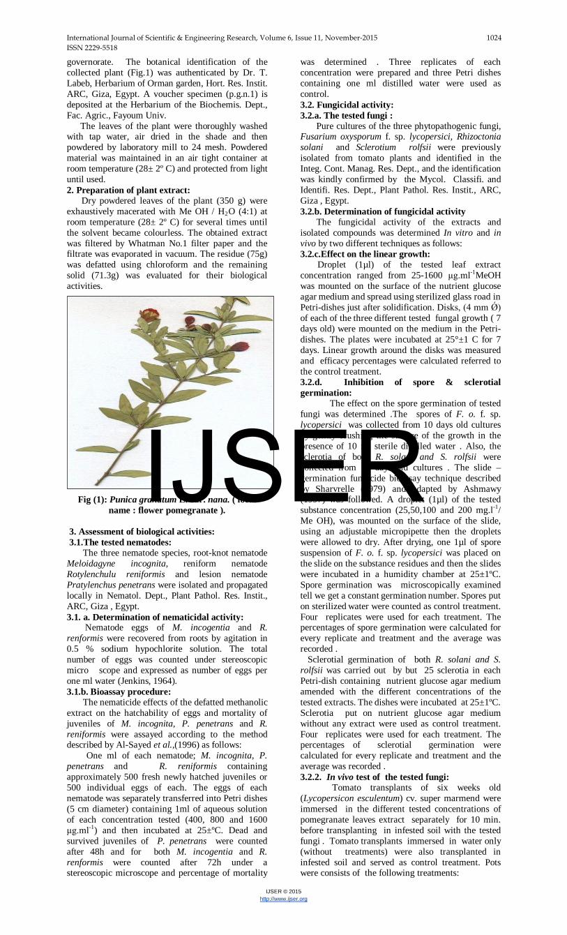
governorate. The botanical identification of the collected plant (Fig.1) was authenticated by Dr. T. Labeb, Herbarium of Orman garden, Hort. Res. Instit. ARC, Giza, Egypt. A voucher specimen (p.g.n.1) is deposited at the Herbarium of the Biochemis. Dept., Fac. Agric., Fayoum Univ. The leaves of the plant were thoroughly washed with tap water, air dried in the shade and then powdered by laboratory mill to 24 mesh. Powdered material was maintained in an air tight container at room temperature (28± 2º C) and protected from light until used. 2. Preparation of plant extract: Dry powdered leaves of the plant (350 g) were exhaustively macerated with Me OH / H2O (4:1) at room temperature (28± 2º C) for several times until the solvent became colourless. The obtained extract was filtered by Whatman No.1 filter paper and the filtrate was evaporated in vacuum. The residue (75g) was defatted using chloroform and the remaining solid (71.3g) was evaluated for their biological activities.
Fig (1): Punica granatum L. var. nana. ( local name : flower pomegranate ).
3. Assessment of biological activities: 3.1.The tested nematodes: The three nematode species, root-knot nematode Meloidagyne incognita, reniform nematode Rotylenchulu reniformis and lesion nematode Pratylenchus penetrans were isolated and propagated locally in Nematol. Dept., Plant Pathol. Res. Instit., ARC, Giza , Egypt. 3.1. a. Determination of nematicidal activity: Nematode eggs of M. incogentia and R. renformis were recovered from roots by agitation in 0.5 % sodium hypochlorite solution. The total number of eggs was counted under stereoscopic micro scope and expressed as number of eggs per one ml water (Jenkins, 1964). 3.1.b. Bioassay procedure: The nematicide effects of the defatted methanolic extract on the hatchability of eggs and mortality of juveniles of M. incognita, P. penetrans and R. reniformis were assayed according to the method described by Al-Sayed et al.,(1996) as follows: One ml of each nematode; M. incognita, P. penetrans and R. reniformis containing approximately 500 fresh newly hatched juveniles or 500 individual eggs of each. The eggs of each nematode was separately transferred into Petri dishes (5 cm diameter) containing 1ml of aqueous solution of each concentration tested (400, 800 and 1600 μg.ml P
-1P) and then incubated at 25±ºC. Dead and
survived juveniles of P. penetrans were counted after 48h and for both M. incogentia and R. renformis were counted after 72h under a stereoscopic microscope and percentage of mortality
was determined . Three replicates of each concentration were prepared and three Petri dishes containing one ml distilled water were used as control. 3.2. Fungicidal activity: 3.2.a. The tested fungi : Pure cultures of the three phytopathogenic fungi, Fusarium oxysporum f. sp. lycopersici, Rhizoctonia solani and Sclerotium rolfsii were previously isolated from tomato plants and identified in the Integ. Cont. Manag. Res. Dept., and the identification was kindly confirmed by the Mycol. Classifi. and Identifi. Res. Dept., Plant Pathol. Res. Instit., ARC, Giza , Egypt. 3.2.b. Determination of fungicidal activity The fungicidal activity of the extracts and isolated compounds was determined In vitro and in vivo by two different techniques as follows: 3.2.c.Effect on the linear growth: Droplet (1µl) of the tested leaf extract concentration ranged from 25-1600 μg.ml P
-1PMeOH
was mounted on the surface of the nutrient glucose agar medium and spread using sterilized glass road in Petri-dishes just after solidification. Disks, (4 mm Ǿ) of each of the three different tested fungal growth ( 7 days old) were mounted on the medium in the Petri-dishes. The plates were incubated at 25°±1 C for 7 days. Linear growth around the disks was measured and efficacy percentages were calculated referred to the control treatment. 3.2.d. Inhibition of spore & sclerotial germination: The effect on the spore germination of tested fungi was determined .The spores of F. o. f. sp. lycopersici was collected from 10 days old cultures by gently brushing the surface of the growth in the presence of 10 ml sterile distilled water . Also, the sclerotia of both R. solani and S. rolfsii were collected from 10 days old cultures . The slide – germination fungicide bioassay technique described by Sharvrelle (1979) and adapted by Ashmawy (1997) was followed. A droplet (1µl) of the tested substance concentration (25,50,100 and 200 mg.l P
-1P/
Me OH), was mounted on the surface of the slide, using an adjustable micropipette then the droplets were allowed to dry. After drying, one 1µl of spore suspension of F. o. f. sp. lycopersici was placed on the slide on the substance residues and then the slides were incubated in a humidity chamber at 25±1ºC. Spore germination was microscopically examined tell we get a constant germination number. Spores put on sterilized water were counted as control treatment. Four replicates were used for each treatment. The percentages of spore germination were calculated for every replicate and treatment and the average was recorded . Sclerotial germination of both R. solani and S. rolfsii was carried out by but 25 sclerotia in each Petri-dish containing nutrient glucose agar medium amended with the different concentrations of the tested extracts. The dishes were incubated at 25±1ºC. Sclerotia put on nutrient glucose agar medium without any extract were used as control treatment. Four replicates were used for each treatment. The percentages of sclerotial germination were calculated for every replicate and treatment and the average was recorded . 3.2.2. In vivo test of the tested fungi: Tomato transplants of six weeks old (Lycopersicon esculentum) cv. super marmend were immersed in the different tested concentrations of pomegranate leaves extract separately for 10 min. before transplanting in infested soil with the tested fungi . Tomato transplants immersed in water only (without treatments) were also transplanted in infested soil and served as control treatment. Pots were consists of the following treatments:
IJSER
International Journal of Scientific & Engineering Research, Volume 6, Issue 11, November-2015 1025 ISSN 2229-5518
IJSER © 2015 http://www.ijser.org
1-M. incognita+ compound A , P. penetrans+ compound A and R. reniformis + compound A. 2-F. o. f.sp. lycopersici + compound A , R. solani + compound A and S. rolfsii + compound A . 3- M . incognita+ F.o. f.sp. lycopersici+ compound A . 4- M. incognita+ R. solani+ compound A. 5- M. incognita+ S. rolfsii + compound A . 6- Tomato with all infestation individuals without compound A. The aforementioned treatments have been repeated with the other two isolated compounds, i.e. B and C. Each treatment was replicated three times. Pots were watered twice per week .Tomato seedlings were infested with nematodes by using 2000 freshly extracted juveniles that were poured around the roots of tomato transplants (7 days after transplanted). Forty five days after transplanting the percentages of disease incident were recorded. Also, tomato roots were carefully pulled off and then washed to get rid of the adhering soil particles to determine height and weight of plants also the number of nematode larvae in 250 cm3 of soil, number of galls, egg-masses, RF (Reproductive factor) and DS (Developmental stages) were determined according to Norton (1978). Reproductive factor (RF) = No. eggs + developmental stages + Free nematode in soil Initial nematode population Developmental stages (DS) = number of developed juveniles (second, third and fourth stages) embedded in the roots. All treatments of the experiments were statistically arranged in a complete randomize design according to Snedecor and Cochran (1989), where mean values were compared using L.S.D. at 5% level. 4. Preliminary phytochemical screening: The preliminary screening of the defatted methanol extract for the following classes of phytoconstituents was preformed according to the methods described by (Farnsworth, 1966) using the indicated detection tests: 1- Saponins were identified by Froth test. 2- Sterols and/or triterpenoids were identified by Liebermann-Burchard test. 3- Tannins were identified by Ferric Chloride 5%. 4- Flavonoids were identified by Aluminium Chloride 5%. 5- Alkaloids were identified by modified Dragendorff's reagent. 6- Glycosides and / or Carbohydrates were identified by Molisch test. 7- Anthocyanins were identified by NHR3R/ HCl. 5. Isolation and structure identification of the active compounds:
5.1. Analytical and preparative Thin Layer Chromatography (TLC): Analytical and preparative TLC was carried out on Merck pre-coated silica gel plates (F254 thickness = 0.25 mm and 2.0 mm respectively) using the following Solvent systems. 1- n-Butanol - Acetic acid – Water (4:1:5) upper layer. 2-Ethyl acetate – Acetic acid – Formic acid – Water (100:11:11:27). 3-Chloroform – Methanol – Water (75:25:2, 70:30:5 and 65:35:2). 4-Chloroform – Methanol (80:20). 5-Dichloromethane – Methanol – Water (50:25:5). 6-Chloroform – Acetone (50:6). Spots on TLC were detected under UV lights (254 and 365 nm) and / or by spraying with concentrated H2SO4 followed by heating at 105 ̊C
for 5 min. and / or by Spraying with; FeClR3R to detect tannins; AlClR3R 5% to detect flavonoids and NHR3R/HCl to detect anthocyanins. Sugars were detected by spraying with nphthoresorcinol phosphoric acid followed heating at 105 ̊C for 10 min. 5.2. Isolation of the bioactive compounds: The bioactive defatted methanol extract was subjected to the isolation of the antioxidant components as follows:
Twenty grams of the defatted methanol extract residue were loaded onto a chromatographic column (5 cm × 100 cm) packed with silica gel (800g, 230 – 400 mesh, Merck) and eluted with a gradient of chloroform : methanol : water (80:20:0, 75:25:2 and 70:30:5 V/V/V; 3L for each eluent). Thirty 100 ml fractions of each eluent were collected and analyzed by TLC. According to differences in composition monitored by TLC system (CHClR3R: MeOH: HR2RO, 70:30:5), 12 fractions were obtained and then tested for antioxidant activity. The three most bioactive fractions No. 4, 5 and 8 were further purified as shown in (Fig.2) to give three compound designated as A, B and C. The purity of these compounds was established by the resolution of each one as a single spot in four different TLC solvent systems. 5.2.1. Structure identification of the purified compounds: The purified compounds were characterized by detection tests (as mentioned previously in 3. Preliminary Phytochemical Screening), acid hydrolysis and spectroscopic methods.
5.2.1.1. Acid hydrolysis: The purified compound to be hydrolyzed (2mg) was heated with aqueous 10% HCl (2ml) in a sealed tube at 100 ˚C water bath for 4 hours. The aglycone was extracted with diethyl ether and analyzed by TLC with chloroform – acetone (50:6). The aqueous layer was neutralized with N, Ndioctylamine (10% in CHClR3R). After evaporation to dryness, the sugars were identified by TLC with dichloromethane – methanol – water (50:25:5) by comparison with Rf of authentic samples. 5.2.1.2. Spectroscopic Methods: 5.2.1.3. Nuclear Magnetic Resonance (NMR) Spectroscopy: P
1PH and P
13PC NMR spectra were recorded in
deuteron methanol (CDR3ROD) on a varion Mercury VXR, 300 and 500 spectrometers (500 MHz for P
1PH
and 75 MHz P
13PC) the chemical shifts (ppm) were
related to the solvent. The spectroscopic NMR experiments were performed at the Nuclear Magnetic Resonance Laboratory, National Research Center, El-Dokki, Cairo and the Central Laboratory, Faculty of Science, Cairo University. 5.2.1.4. Mass Spectroscopy (MS):
Mass spectra were recorded on a GCMS. QP 1000 EX Shimadzu Mass spectrometer at 70e.v. The MS experiments were carried out at Biochemical Genetics Laboratory, National Research Center, El-Dokki, Cairo. 5.2.1.5. Ultraviolet Spectroscopy (UV): The UV spectra were recorded on Cecil 3000 series spectrophotometer according to (Mabry et al., 1970). 6. Statistical analysis: Data were statistically analyzed using the standard procedures for complete randomize block and split designs as mentioned by Snedecor and Cochran (1989). The averages were compared at 5% level using least significant differences (L.S.D) according to Fisher (1948).
IJSER

International Journal of Scientific & Engineering Research, Volume 6, Issue 11, November-2015 1026 ISSN 2229-5518
IJSER © 2015 http://www.ijser.org
Fig (2). Follow Diagram for the isolation of the active compound A-C.
Fig. (2) Follow diagram for the isolation of the active compounds A-C. 3 RESULTS AND DISCUSSION 1- Fungicidal activity : The antifungal efficacy of the defatted methanolic extract on the linear growth of the three well- known phytopathogenic fungi, i.e. Fusarium oxysporum f.sp. lycopersici , Rhizoctonia solani and Sclerotium rolfsii was studied in vitro and the results are shown in Table (1). The obtained results show that the defatted methanol extract exhibited fungicidal activity against the three tested fungi at concentration ranging from 25 to 1600 ppm. It is , also, clear that the efficacy of the defatted methanol extract on the linear growth of the three tested fungi ,i.e. F. o. f.sp. lycopersici, R. solani and S. rolfsii was increased with increasing the concentration from 25 to 1600 μg.ml-1 . The percentage of inhibition of the tested fungi increased
from 23.8, 28.8 and 16.3 to 83.8, 85.3 and 73.8%, respectively. The data obtained revealed that the defatted methanolic extract was more effective against the fungus R. solani than the other tested fungi. Recently several plant extracts have been reported as botanical fungicides against the three phytopathogenic fungi F. oxysporum. f.sp. lycopersici, R. solani and S. rolfsii (Mamdouh and Mohamed, 2007; Belal et al., 2007; Jasso de Rodriguez et al., 2007; Sharma and Kumar, 2009; Satish et al., 2009; Emam et al., 2010; Castillo et al., 2010; Osorio et al., 2010; Mahlo et al., 2010; Bhardwaj, 2012; Hadian, 2012 and Plodpai et al., 2013).
Table (1): In vitro fungicidal activity of the defatted methanol extract on the linear growth (mm.) of the
tested fungi Concentration (μg.ml-1)
F. oxysporum f.sp. lycopersici R. solani S. rolfsii Mean Linear growth
% Efficacy
Linear growth
% Efficacy
Linear growth
% Efficacy
25 6.1 23.8 5.7 28.8 6.7 16.3 6.2 50 5.8 27.5 5.2 35.0 5.4 32.5 5.4 100 5.1 36.3 4.9 38.8 5.0 37.8 5.0
IJSER

International Journal of Scientific & Engineering Research, Volume 6, Issue 11, November-2015 1027 ISSN 2229-5518
IJSER © 2015 http://www.ijser.org
200 4.8 40.0 4.1 45.8 4.7 41.3 4.6 400 4.3 47.8 3.6 55.8 4.1 48.8 4.0 800 3.2 60.0 2.8 65.0 3.2 60.0 3.1 1600 1.3 83.8 1.6 85.3 2.1 73.8 1.7
Control 8.0 0.0 8.0 0.0 8.0 0.0 8.0 Mean 4.8 ------ 4.5 ------ 4.9 -----
L.S.D. at 5 % for: Concentration (C)=1.3 , Fungi (F)= n.s. and CxF= 3.4
2- Nematicidal activity : The nematicide effect of the defatted methanol extract on the hatchability of eggs and mortality of juveniles of the three nematods M. incognita, R. reniformis and P. penetrans were studied in vitro and the results are shown in Table (2). As shown in (Table 2) the methanolic extract exhibited larvacidal and ovacidal activities at concentration ranging from 400 to 1600 μg.ml-1. In general, the extract with higher concentration i.e. 1600 μg.ml-1 showed more activity when compared with lower concentration i.e. 400 μg.ml-1. This observation showed that, an increase in concentration represent a supplementary input of different active compounds (Wabo et al., 2011). The data obtained
also revealed that methanolic extract was more efficient against the juveniles than eggs as the % of inhibition at the same concentrations were the highest and more effective against the juveniles of P. penetrans than the other nematode species i.e. M. incognita, R. reniformis as the percentage of juvenile mortality was the highest at all tested concentration. During the last two decades various plant extracts have been reported as botanical nematicides against the three nematodes tested (Tiyagi and Alam, 1995; Begum et al., 2000; Oka et al., 2001; Tsay et al., 2004; Ahmad et al., 2004; Ibrahim et al., 2006; Belal et al., 2007; El-Badri et al., 2008; Abo- Elyousr et al., 2010; Hussain et al., 2011; Kayani et al., 2012 ).
Table (2): In vitro nematicidal activity of the defatted methanolic extract on the hatchability of eggs and juveniles
mortality of nematodes.
Tested nematodes Concentration (μg.ml-1)
%Inhibition of egg hatching % Mortality of juveniles
M. incognita
400 21 33
800 40 51
1600 56 83
R. reniformis
400 25 34
800 41 58
1600 55 74
P. penetrans
1600 N.t. 92
800 N.t. 66
400 N.t. 44
L.S.D. at 5 % 3.0 4.0
N.T. = Not tested. The methanolic extract of P. granatum L. var. nana shown marvelous inhibitory effect against egg hatching and juvenile of test nematodes, which might be due to the presence of various secondary metabolites such as Flavonoids, Terpenoids, phenols…..etc in the plant sample. Some phytochemical have also been reported in Literature and possess nematicidal activity (Begum et al., 2000; Oka et al., 2000; Calvet et al., 2001; Belal et al., 2007 and Shakil et al., 2008). Hence , this extract could be used for protection plant against the three nematodes tested which causes rot not and die back in various economically important crops such as vegetables, cotton, citrus..….etc. (Kayani et al., 2012). 4. 2. Preliminary phytochemical screening: Screening test for Saponins, Sterols, Triterpenoids, Tannins, Flavonoids, Anthocyanins and Alkaloids in the defatted methanolic extract of the dry powdered leaves of Punica granatum L. var. nana were made and results are given in Table (3), which reveals the presence of all the aforementioned phytoconstituents.
3 .Preliminary phytochemical screening: Screening test for Saponins, Sterols, Triterpenoids, Tannins, Flavonoids, Anthocyanins and Alkaloids in the defatted methanolic extract of the dry powdered leaves of Punica granatum L. var. nana were made and results are given in Table (3), which reveals the presence of all the aforementioned phytoconstituents. It is noteworthy that many plant derived compounds belong to the classes of secondary metabolites such as Saponins, Flavonoids, Tanins, Anthocyanins and Alkaloids have been used as botanical pesticides such as fungicides, insecticides and nematicides as well as natural antioxidant and hapatoprotective agents in the agricultural and medical fields. (Marston et al., 1988, Begum et al., 2000, Chatterjee, 2000; Mnaa et al., 2008; Sudjaroen et al., 2005; Feng et al., 2010; Bhardwaj, 2012 and Abdel-Salam et al., 2012). Therefore, the biological activities of this plant may be attributed to the presence of a compound or more belongs to one or more of these classes of plant secondary metabolites.
IJSER
International Journal of Scientific & Engineering Research, Volume 6, Issue 11, November-2015 1028 ISSN 2229-5518
IJSER © 2015 http://www.ijser.org
Table (3): Phytochemical screening tests for constituents of P. granatum L. var.nana methanoliv extract.
Constituent Detection test Result
Saponins Froth test (+)
Sterols or Triterpenoids Liebermann –Burchard test. (+)
Tannins Ferric Chloride 5%. (+)
Flavonoids Aluminium Chloride5%. (+)
Alkaloids Modified Dragendorff's reagent. (+)
Glycosids Molisch test (+)
Anthocyanins NH3 / HCl (+)
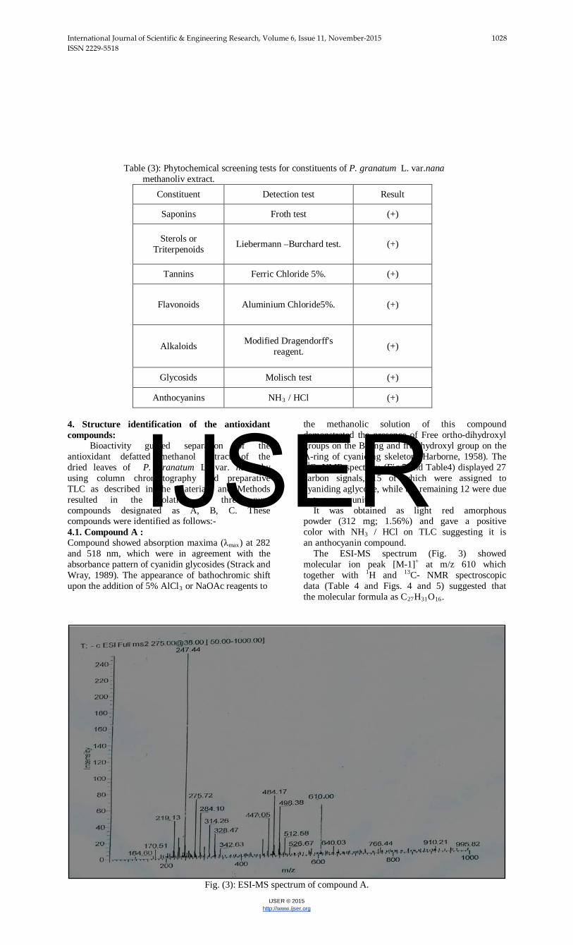
4. Structure identification of the antioxidant compounds: Bioactivity guided separation of the antioxidant defatted methanol extract of the dried leaves of P. granatum L. var. nana by using column chromatography and preparative TLC as described in the Materials and Methods resulted in the isolation of three pure compounds designated as A, B, C. These compounds were identified as follows:- 4.1. Compound A : Compound showed absorption maxima (λmax) at 282 and 518 nm, which were in agreement with the absorbance pattern of cyanidin glycosides (Strack and Wray, 1989). The appearance of bathochromic shift upon the addition of 5% AlCl3 or NaOAc reagents to
the methanolic solution of this compound demonstrated the presence of Free ortho-dihydroxyl groups on the B-ring and free hydroxyl group on the A-ring of cyaniding skeleton (Harborne, 1958). The 13C- NMR spectrum (Fig.5 and Table4) displayed 27 carbon signals, 15 of which were assigned to cyaniding aglycone, while the remaining 12 were due to two sugar units. It was obtained as light red amorphous powder (312 mg; 1.56%) and gave a positive color with NH3 / HCl on TLC suggesting it is an anthocyanin compound. The ESI-MS spectrum (Fig. 3) showed molecular ion peak [M-1]+ at m/z 610 which together with 1H and 13C- NMR spectroscopic data (Table 4 and Figs. 4 and 5) suggested that the molecular formula as C27H31O16.
Fig. (3): ESI-MS spectrum of compound A.
IJSER
International Journal of Scientific & Engineering Research, Volume 6, Issue 11, November-2015 1029 ISSN 2229-5518
IJSER © 2015 http://www.ijser.org
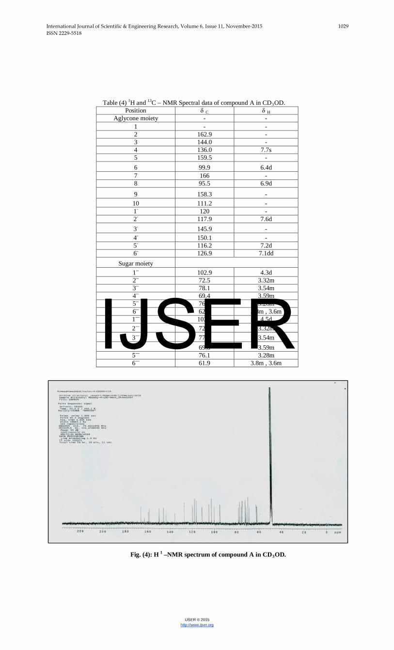
Table (4) 1H and 13C – NMR Spectral data of compound A in CD3OD.
Position δ C δ H Aglycone moiety - -
1 - - 2 162.9 - 3 144.0 - 4 136.0 7.7s 5 159.5 - 6 99.9 6.4d 7 166 - 8 95.5 6.9d
9 158.3 - 10 111.2 - 1- 120 - 2- 117.9 7.6d 3- 145.9 - 4- 150.1 - 5- 116.2 7.2d 6- 126.9 7.1dd
Sugar moiety 1-- 102.9 4.3d
2-- 72.5 3.32m 3-- 78.1 3.54m 4-- 69.4 3.59m 5-- 76.4 3.28m 6-- 62.1 3.8m , 3.6m 1--- 103.8 4.5d 2--- 72.8 3.32m 3--- 77.9 3.54m 4--- 69.8 3.59m 5--- 76.1 3.28m 6--- 61.9 3.8m , 3.6m
Fig. (4): H 1 –NMR spectrum of compound A in CD3OD.
IJSER
International Journal of Scientific & Engineering Research, Volume 6, Issue 11, November-2015 1030 ISSN 2229-5518
IJSER © 2015 http://www.ijser.org
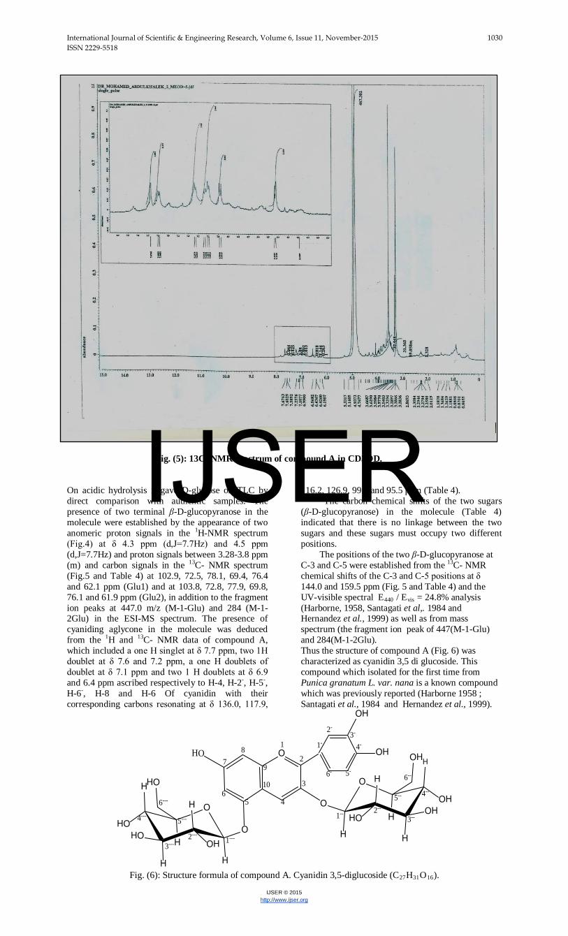
Fig. (5): 13C- NMR spectrum of compound A in CD3OD.
On acidic hydrolysis it gave D-glucose on TLC by direct comparison with authentic samples. The presence of two terminal β-D-glucopyranose in the molecule were established by the appearance of two anomeric proton signals in the 1H-NMR spectrum (Fig.4) at δ 4.3 ppm (d,J=7.7Hz) and 4.5 ppm (d,J=7.7Hz) and proton signals between 3.28-3.8 ppm (m) and carbon signals in the 13C- NMR spectrum (Fig.5 and Table 4) at 102.9, 72.5, 78.1, 69.4, 76.4 and 62.1 ppm (Glu1) and at 103.8, 72.8, 77.9, 69.8, 76.1 and 61.9 ppm (Glu2), in addition to the fragment ion peaks at 447.0 m/z (M-1-Glu) and 284 (M-1-2Glu) in the ESI-MS spectrum. The presence of cyaniding aglycone in the molecule was deduced from the 1H and 13C- NMR data of compound A, which included a one H singlet at δ 7.7 ppm, two 1H doublet at δ 7.6 and 7.2 ppm, a one H doublets of doublet at δ 7.1 ppm and two 1 H doublets at δ 6.9 and 6.4 ppm ascribed respectively to H-4, H-2-, H-5-, H-6-, H-8 and H-6 Of cyanidin with their corresponding carbons resonating at δ 136.0, 117.9,
116.2, 126.9, 99.9 and 95.5 ppm (Table 4). The carbon chemical shifts of the two sugars (β-D-glucopyranose) in the molecule (Table 4) indicated that there is no linkage between the two sugars and these sugars must occupy two different positions. The positions of the two β-D-glucopyranose at C-3 and C-5 were established from the 13C- NMR chemical shifts of the C-3 and C-5 positions at δ 144.0 and 159.5 ppm (Fig. 5 and Table 4) and the UV-visible spectral E440 / Evis = 24.8% analysis (Harborne, 1958, Santagati et al,. 1984 and Hernandez et al., 1999) as well as from mass spectrum (the fragment ion peak of 447(M-1-Glu) and 284(M-1-2Glu). Thus the structure of compound A (Fig. 6) was characterized as cyanidin 3,5 di glucoside. This compound which isolated for the first time from Punica granatum L. var. nana is a known compound which was previously reported (Harborne 1958 ; Santagati et al., 1984 and Hernandez et al., 1999).
OHO
OH
OH
O
H
HO
H
HO
H
H
OHHO
HO O
H
OH
H
OH
H
H
HO HO
OH1
2
3
456
78
9
10
1-
2-3-
4-
5-6-
1-- 2--
3--
4--5--
6--
1---2---
3---
4---5---
6---
Fig. (6): Structure formula of compound A. Cyanidin 3,5-diglucoside (C27H31O16).
IJSER

International Journal of Scientific & Engineering Research, Volume 6, Issue 11, November-2015 1031 ISSN 2229-5518
IJSER © 2015 http://www.ijser.org
4.2. Compound B: It was obtained as a white amorphous powder (294 mg; 1.47 %) that gave positive color reaction with 5% FeCl3 test on TLC suggesting it is a phenolic compound. The electron spray ionization mass spectrometric (ESI-MS) spectrum of this compound (Fig. 7) showed a molecular ion peak (M+1)+ at m/z
of 333, which together with 1H and 13C- NMR spectroscopic data (Table 5 and Figs. 8 and 9) suggested that the molecular formula as C13H16O10.
Fig. (7): ESI-MS spectrum of compound B.
Fig. (8): 1H- NMR spectrum of compound B in CD3OD.
IJSER
International Journal of Scientific & Engineering Research, Volume 6, Issue 11, November-2015 1032 ISSN 2229-5518
IJSER © 2015 http://www.ijser.org
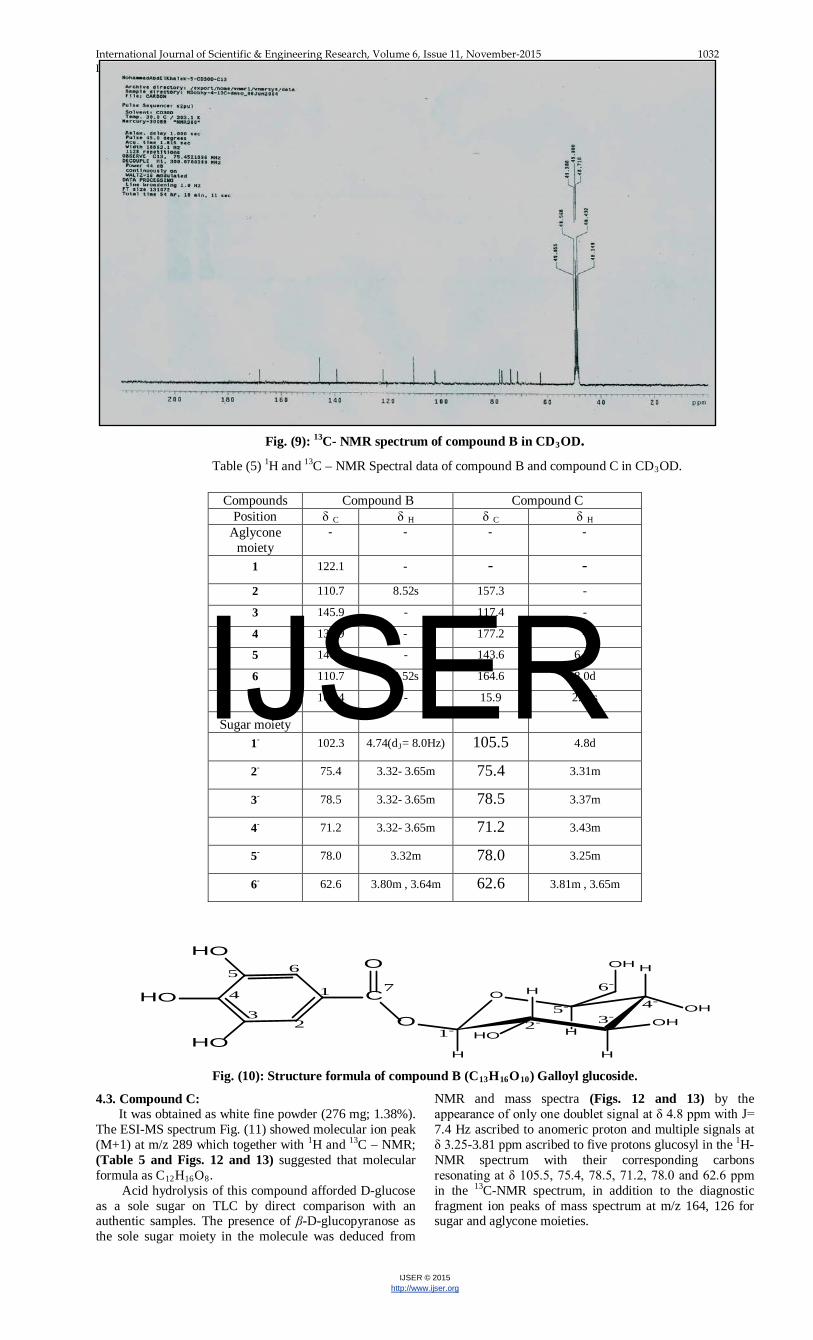
Fig. (9): 13C- NMR spectrum of compound B in CD3OD. Table (5) 1H and 13C – NMR Spectral data of compound B and compound C in CD3OD.
HO
HO
HO
C
O
O
H
OH
H
OH
H
H
HO HO
OH65
4
32
1 7
1- 2- 3-4-
5-
6-
Fig. (10): Structure formula of compound B (C13H16O10) Galloyl glucoside.
4.3. Compound C: It was obtained as white fine powder (276 mg; 1.38%). The ESI-MS spectrum Fig. (11) showed molecular ion peak (M+1) at m/z 289 which together with 1H and 13C – NMR; (Table 5 and Figs. 12 and 13) suggested that molecular formula as C12H16O8. Acid hydrolysis of this compound afforded D-glucose as a sole sugar on TLC by direct comparison with an authentic samples. The presence of β-D-glucopyranose as the sole sugar moiety in the molecule was deduced from
NMR and mass spectra (Figs. 12 and 13) by the appearance of only one doublet signal at δ 4.8 ppm with J= 7.4 Hz ascribed to anomeric proton and multiple signals at δ 3.25-3.81 ppm ascribed to five protons glucosyl in the 1H-NMR spectrum with their corresponding carbons resonating at δ 105.5, 75.4, 78.5, 71.2, 78.0 and 62.6 ppm in the 13C-NMR spectrum, in addition to the diagnostic fragment ion peaks of mass spectrum at m/z 164, 126 for sugar and aglycone moieties.
Compounds Compound B Compound C Position δ C δ H δ C δ H
Aglycone moiety
- - - -
1 122.1 - - - 2 110.7 8.52s 157.3 -
3 145.9 - 117.4 -
4 138.9 - 177.2 -
5 146.9 - 143.6 6.5d
6 110.7 8.52s 164.6 8.0d
7 168.4 - 15.9 2.44s
Sugar moiety
1P
- 102.3 4.74(dRJR= 8.0Hz) 105.5 4.8d
2P
- 75.4 3.32- 3.65m 75.4 3.31m
3P
- 78.5 3.32- 3.65m 78.5 3.37m
4P
- 71.2 3.32- 3.65m 71.2 3.43m
5P
- 78.0 3.32m 78.0 3.25m
6P
- 62.6 3.80m , 3.64m 62.6 3.81m , 3.65m
IJSER

International Journal of Scientific & Engineering Research, Volume 6, Issue 11, November-2015 1033 ISSN 2229-5518
IJSER © 2015 http://www.ijser.org
Fig. (11): Mass spectrum of compound C.
Fig.(12): 1H- NMR spectrum of compound C in CD3OD .
Fig. (13): 13C- NMR spectrum of compound C in CD3OD.
The 13C-NMR spectrum (Fig. 13) displayed 12 carbon signals. Out of which 6 carbons accounted for the sugar moiety (Glu). The remaining 6 carbons were due to the aglycone moiety. In the aglycone part of the molecule, the 13C-NMR spectrum revealed the presence of methyl group (C7; δ 15.9 ppm), three quaternary carbon including carbonyl group (C4; δ 177.2 ppm; CO) and two methine group including oxymethine (C6; δ 164.6 ppm) characteristic for 2,3 disubstitud pyran-4-one ring. This observation was further supported by the 1H-NMR spectrum which exhibited signals for two doublets (each for 1H) centered at δ8.0 ppm (H6; J= 5.3Hz) and δ6.5 ppm (H5; J= 5.3Hz) and one singlet at δ2.44 ppm (H7; 3Hs) for methyl group. The glycosylation site at C-3 and the methyl
group at C-2 were deduced from the chemical shifts of the C-2 and C-3 positions (δ117.4 and 157.3 ppm). On the basis of the above finding compound C (Fig.14) was identified as 2-methyl-pyran-4-one-3-O-β-D-glucopyranoside. This compound which isolated here for the first time from P. granatum L. var. nana is a known compound recently isolated only from the leaves of Punica granatum (Balwani et al., 2011). It is interesting to note that this is the first report about the antioxidant activity of this compound (IC50 = 5.8 μg.ml-1).
IJSER
International Journal of Scientific & Engineering Research, Volume 6, Issue 11, November-2015 1034 ISSN 2229-5518
IJSER © 2015 http://www.ijser.org
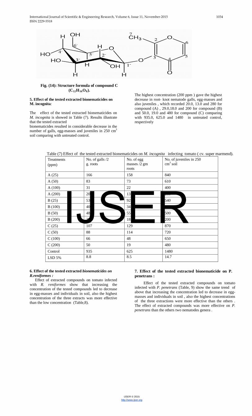
Fig. (14): Structure formula of compound C
(C12H16O8). 5. Effect of the tested extracted bionematicides on M. incognita: The effect of the tested extracted bionematicides on M. incognita is showed in Table (7). Results illustrate that the tested extracted bionematicides resulted in considerable decrease in the number of galls, egg-masses and juveniles in 250 cm3
soil comparing with untreated control.
The highest concentration (200 ppm ) gave the highest decrease in root- knot nematode galls, egg-masses and also juveniles , which recorded 20.0, 13.0 and 280 for compound (A) , 29.0,18.0 and 200 for compound (B) and 50.0, 19.0 and 480 for compound (C) comparing with 935.0, 625.0 and 1480 in untreated control, respectively
Table (7) Effect of the tested extracted bionematicides on M. incognita infecting tomato ( cv. super marmend).
Treatments (ppm)
No. of galls /2 g. roots
No. of egg masses /2 gm roots
No. of juveniles in 250 cm3 soil
A (25) 166 158 840 A (50) 83 73 610 A (100) 31 22 400 A (200) 20 13 280 B (25) 53 92 540 B (100) 40 34 380 B (50) 48 55 500 B (200) 29 18 200 C (25) 107 129 870 C (50) 88 114 720 C (100) 66 48 650 C (200) 50 19 480 Control 935 625 1480 LSD 5% 8.8 8.5 14.7
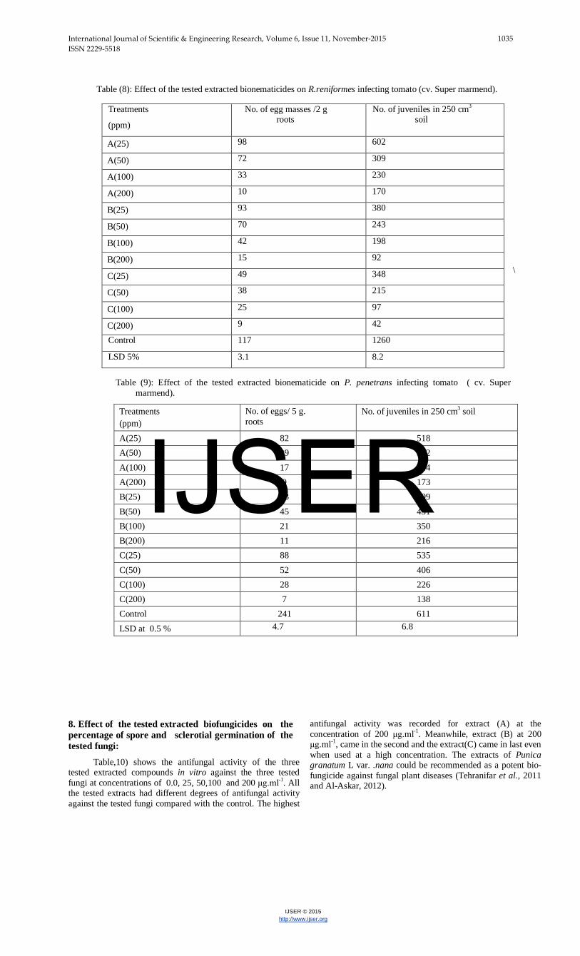
6. Effect of the tested extracted bionematicides on R.reniformes : Effect of extracted compounds on tomato infected with R. reniformes show that increasing the concentration of the tested compounds led to decrease in egg-masses and individuals in soil, also the highest concentration of the three extracts was more effective than the low concentration (Table,8).
7. Effect of the tested extracted bionematicide on P. penetrans :
Effect of the tested extracted compounds on tomato infected with P. penetrans (Table, 9) show the same trend of above that increasing the concentration led to decrease in egg-masses and individuals in soil , also the highest concentrations of the three extractions were more effective than the others . The effect of extracted compounds was more effective on P. penetrans than the others two nematodes genera .
IJSER
International Journal of Scientific & Engineering Research, Volume 6, Issue 11, November-2015 1035 ISSN 2229-5518
IJSER © 2015 http://www.ijser.org
Table (8): Effect of the tested extracted bionematicides on R.reniformes infecting tomato (cv. Super marmend).
\
Table (9): Effect of the tested extracted bionematicide on P. penetrans infecting tomato ( cv. Super marmend).
No. of juveniles in 250 cm3 soil No. of eggs/ 5 g. roots
Treatments (ppm)
518 82 A(25) 342 39 A(50) 214 17 A(100) 173 9 A(200) 609 93 B(25) 431 45 B(50) 350 21 B(100) 216 11 B(200) 535 88 C(25) 406 52 C(50) 226 28 C(100) 138 7 C(200) 611 241 Control
6.8 4.7 LSD at 0.5 %
8. Effect of the tested extracted biofungicides on the percentage of spore and sclerotial germination of the tested fungi:
Table,10) shows the antifungal activity of the three tested extracted compounds in vitro against the three tested fungi at concentrations of 0.0, 25, 50,100 and 200 μg.mlP
-1P. All
the tested extracts had different degrees of antifungal activity against the tested fungi compared with the control. The highest
antifungal activity was recorded for extract (A) at the concentration of 200 μg.mlP
-1P. Meanwhile, extract (B) at 200
μg.mlP
-1P, came in the second and the extract(C) came in last even
when used at a high concentration. The extracts of Punica granatum L var. .nana could be recommended as a potent bio-fungicide against fungal plant diseases (Tehranifar et al., 2011 and Al-Askar, 2012).
Treatments
(ppm)
No. of egg masses /2 g roots
No. of juveniles in 250 cmP
3P
soil
A(25) 98 602
A(50) 72 309
A(100) 33 230
A(200) 10 170
B(25) 93 380
B(50) 70 243
B(100) 42 198
B(200) 15 92
C(25) 49 348
C(50) 38 215
C(100) 25 97
C(200) 9 42
Control 117 1260
LSD 5% 3.1 8.2
IJSER

International Journal of Scientific & Engineering Research, Volume 6, Issue 11, November-2015 1036 ISSN 2229-5518
IJSER © 2015 http://www.ijser.org
Table (10): Effect of the tested extracted biofungicides on the percentage of spore and sclerotial germination of
the tested fungi .
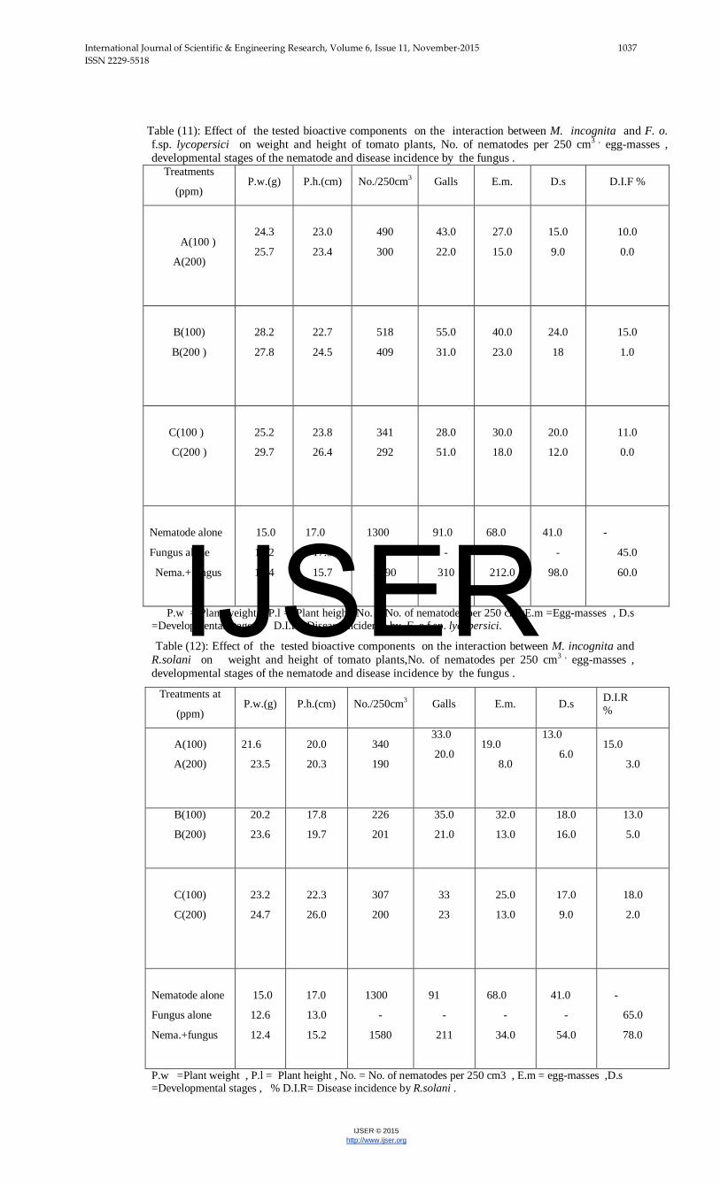
9. Effect of the tested bioactive components on the interaction between M. incognita and F. o. f.sp. lycopersici :
Data presented in Table (11) reveal that the tested bioactive components ( A, B and C) of Punica granatum L. var. nana resulted in increasing plant weight and length and reducing number of nematodes / 250 cmP
3P, galls , egg mases and
developmental stage as well as the infection by F. o. f.sp. lycopersici when tomato plants grown in soil infested with both M. incognita and F. o. f.sp. lycopersici . This effect was increased by increasing the concentration of the bioactive components of pomegrate from 100 to 200 ppm. On the other hand, tomato plants grown in soil unamended with the bioactive components of pomegrate and infested with any of M. incognita and F. o. f.sp. lycopersici and their combination showed low value of plant weight and length and great increase in of the estimated critiria of number of nematodes / 250 cm3, galls , egg mases and developmental stage as well as the infection by F. o. f.sp. lycopersici.
The highest effect refer to the treatment with compounds A and C, which gave no disease incidence with Fusarium interaction with M. incognita . Whereas, the percentage of disease incidence increased to 60% without treatments and 45% in case of the infection with Fusarium alone . Moreover, the effect on nematode infection was decreased clearly in galls , egg-masses and developmental stages at the highest concentration .Meanwhile, the effect was differed among the three extracted compound .
10. Effect of the tested bioactive components on the interaction between M. incognita and R.solani:
Data presented in Table (12) show that the tested bioactive components ( A,B and C) resulted in increasing plant weight and length and reducing number of nematodes / 250 cmP
3P, galls , egg mases and
developmental stage as well as the infection by R.solani when tomato plants grown in soil infested with both M. incognita and R.solani . This effect was increased by increasing the concentration of the bioactive components of pomegrate from 100 to 200 ppm. On the other hand, tomato plants grown in soil unamended with the bioactive components of pomegrate and infested with any of M. incognita and R.solani and their combination showed low value of plant weight and length and great increase in the estimated critiria of number of nematodes / 250 cmP
3P,
galls , egg mases and developmental stage as well as the infection by R.solani.
The highest effect refer to the treatment with compounds C, A and B, which gave 2.0 , 3.0 and 5.0% disease incidence with Rhizoctonia interaction with M.incognita , respectively . The infection with the two pathogens ( nematode + fungus) gave 78% disease incidence and 45% in case of the infection with Fusarium alone. The effect on nematode infection was decreased clearly in galls , egg-masses and developmental stages at highest concentration(200 ppm), which recorded 20.0 ,8.0 and 6.0 , respectively in treatment of compound (A). Whereas, it recorded 91.0,68.0 and 41.0 in case of the infection with the nematode alone.
% Inhibition of germination of Treatments
(ppm) S. rolfsii R. solani F. oxysporum
Efficacy% Sclerotia Efficacy% Sclerotia Efficacy% Spores 23.83 68.4 67.29 31.2 22.72 61.2 A(25) 36.19 57.3 73.79 25 29.04 56.2 A(50) 79.39 18.5 86.89 12.5 52.56 37.5 A(100) 91.98 7.2 93.50 6.2 84.21 12.5 A(200) 22.71 69.4 47.58 50 13.26 68.7 B(25) 35.19 58.2 60.69 37.5 29.04 56.2 B(50) 56.90 38.7 86.89 12.5 44.82 43.7 B(100) 86.08 12.5 93.50 6.2 76.38 18.7 B(200) 30.40 62.5 18.02 78.2 7.32 73.4 C(25) 51.34 43.7 86.89 12.5 21.08 62.5 C(50) 79.17 18.7 93.50 6.2 52.56 37.5 C(100) 86.08 12.5 100 0.0 68.43 25 C(200)
--------- 89.8 --------- 95.4 --------- 79.2 Control --------- 3.5 --------- 3.9 --------- 3.1 LSD at 5%
IJSER
International Journal of Scientific & Engineering Research, Volume 6, Issue 11, November-2015 1037 ISSN 2229-5518
IJSER © 2015 http://www.ijser.org
Table (11): Effect of the tested bioactive components on the interaction between M. incognita and F. o. f.sp. lycopersici on weight and height of tomato plants, No. of nematodes per 250 cm3 , egg-masses , developmental stages of the nematode and disease incidence by the fungus .
Treatments
(ppm) P.w.(g) P.h.(cm) No./250cm3 Galls E.m. D.s D.I.F %
A(100 )
A(200)
24.3
25.7
23.0
23.4
490
300
43.0
22.0
27.0
15.0
15.0
9.0
10.0
0.0
B(100)
B(200 )
28.2
27.8
22.7
24.5
518
409
55.0
31.0
40.0
23.0
24.0
18
15.0
1.0
C(100 )
C(200 )
25.2
29.7
23.8
26.4
341
292
28.0
51.0
30.0
18.0
20.0
12.0
11.0
0.0
Nematode alone
Fungus alone
Nema.+fungus
15.0
16.2
12.4
17.0
17.5
15.7
1300
-
2090
91.0
-
310
68.0
-
212.0
41.0
-
98.0
-
45.0
60.0
P.w = Plant weight . P.l = Plant height, No. = No. of nematodes per 250 cm3 ,E.m =Egg-masses , D.s =Developmental stages, D.I.F =Disease incidence by F. o.f.sp. lycopersici.
Table (12): Effect of the tested bioactive components on the interaction between M. incognita and R.solani on weight and height of tomato plants,No. of nematodes per 250 cm3 , egg-masses , developmental stages of the nematode and disease incidence by the fungus .
Treatments at
(ppm) P.w.(g) P.h.(cm) No./250cm3 Galls E.m. D.s D.I.R
%
A(100)
A(200)
21.6
23.5
20.0
20.3
340
190
33.0
20.0
19.0
8.0
13.0
6.0
15.0
3.0
B(100)
B(200)
20.2
23.6
17.8
19.7
226
201
35.0
21.0
32.0
13.0
18.0
16.0
13.0
5.0
C(100)
C(200)
23.2
24.7
22.3
26.0
307
200
33
23
25.0
13.0
17.0
9.0
18.0
2.0
Nematode alone
Fungus alone
Nema.+fungus
15.0
12.6
12.4
17.0
13.0
15.2
1300
-
1580
91
-
211
68.0
-
34.0
41.0
-
54.0
-
65.0
78.0
P.w =Plant weight , P.l = Plant height , No. = No. of nematodes per 250 cm3 , E.m = egg-masses ,D.s =Developmental stages , % D.I.R= Disease incidence by R.solani .
IJSER

International Journal of Scientific & Engineering Research, Volume 6, Issue 11, November-2015 1038 ISSN 2229-5518
IJSER © 2015 http://www.ijser.org
11. Effect of the tested bioactive components on the interaction between M. incognita and S. rolfsii:
Data presented in Table (13) reveal that the bioactive components of Punica granatum L. var. nana resulted in increasing plant weight and length and reducing number of nematodes / 250 cm3, galls , egg mases and developmental stage as well as the infection by S.rolfsii when tomato plants grown in soil infested with both M. incognita and S. rolfsii . This effect was increased by increasing the concentration of bioactive components of pomegrate from 100 to 200 ppm. On the other hand, tomato plants grown in soil unamended with the bioactive components of pomegrate and infested with each of M. incognita and S. Rolfsii and their combination showed low value of plant
weight and length and great increase in the estimated critiria of number of nematodes / 250 cm3, galls , egg mases and developmental stage as well as the infection by S.rolfsii.
The highest effect refer to the treatment with compounds C, A and B at 200 ppm, which reduced disease incidence in the interaction between Sclerotium and M. incognita to 7.0 , 8.0 and 9.0%, respectively . The percentage of disease incidence increased to 78% without any treatments and 65% in case of infection with S.rolfsii alone . Moreover, the effect on nematode infection was decreased clearly in galls , egg-masses and developmental stages at the highest concentration.
Table (13): Effect of the tested bioactive components on the interaction between M. incognita and S. rolfsii on weight and height of tomato plants, No. of nematodes per 250 cm3 , egg-masses , developmental stages of the nematode and disease incidence by the fungus.
Treatments at
(ppm) P.w.(g) P.h.(cm) No. /250
cm3 Galls E.m. Ds. D.I.S%
A (100 )
A (200 )
25.5
28.2
24.1
21.4
460
240
51.0
30.0
31.0
18.0
22.0
15.0
13.0
9.0
B (100 )
B (200 )
24.2
26.3
29.4
27.3
392
321
65.0
31.0
35.0
23.0
41.0
23 .0
13.0
8.0
C (100 )
C (200 )
22.6
25.1
21.4
28.5
407
337
43.0
28.0
29.0
18.0
24.0
12.0
18.0
7.0
Nematode alone
Fungus alone
Nema.+fungus
15.0
12.6
12.4
17.0
13.0
15.2
1300
-
1580
91.0
-
211.0
68.0
-
34.0
41.0
-
54.0
-
65.0
78.0
P.w= Plant weight , P.h = Plant height , No. = No. of nematodes per 250 cm3, E.m =egg-masses , D.s =Developmental stages , D.I.S.= Disease incidence by S. rolfsii
It has been found the bioactive components of Punica granatum L. var. nana resulted in increasing plant weight and length and reducing number of nematodes / 250 cm3, galls , egg masses and developmental stage as well as the infection any of the three tested fungi when tomato plants grown in soil infested with M. incognita and any of the tested fungi. This effect was increased by increasing the concentration of the tested bioactive components from 100 to 200 ppm. On the other hand, tomato plants grown in soil unamended with the bioactive components of pomegrate and infested with each of M. incognita and the three tested fungi and their combination showed low value of plant weight and height and great increase in the estimated criteria of number of nematodes / 250 cm3, galls , egg masses and
developmental stage as well as the infection by any of these fungi. The mode of action of the tested bioactive components of Punica granatum L. var. nana may be due to their direct toxic effect , activation of enzymes responsible for disease resistance and / or induced systemic acquired resistance to both nematode and fungi (Sudheesh et al. ,2005; Ibrahim et al.,2006; Tehranifar et al.,2011 and Abd El- Salam et al.(2012). Some phytochemical have also been reported in Literature and possess nematicidal activity (Begum et al., 2000; Oka et al., 2000; Calvet et al., 2001; Belal et al., 2007 and Shakil et al., 2008). The obtained data are of great interest , where the bioactive components of Punica granatum L. var. nana could be used as an alternative safe trial for managing the
IJSER
International Journal of Scientific & Engineering Research, Volume 6, Issue 11, November-2015 24 ISSN 2229-5518
IJSER © 2015 http://www.ijser.org
infection by nematode and soil borne fungi . The obtained results are in accordance with the obtained data by
different plant extracts by Hernandez et al. (1999); Osorio et al. (2010) and Abd El- Salam et al.(2012).
4 REFERENCES
[1] Abd El- Salam, O.M.; Sleem, A.A. and Omara, E. (2012). Micronized purified flavonoid fraction alleviates the carbon tetrachloride-induced hepatic injury. Comp. Clin. Pathol. Dot. 10. 1007/s00580-012-1542-2.
[2] Abo-Elyousr, K.; Khan, Z.; El- Morsi, A. and Abedol- Moneim, M. (2010). Evaluation of plant extracts and Pseudomonas spp. for control of root-knot nematode, Meloidagyne incognita on tomato. Nematropica, 40: 289 – 299.
[3] Ahmad, S.M.; Mukhtar, T. and Ahmad, R. (2004). Some studies on the control of citrus nematode (Tylenchulus semipenetrans) by leaf extracts of three plants and their effects on plant growth variables. Asian J. Plant. Sci., 3(5): 544-548.
[4] Al-Askar, A.A.A. (2012). In vitro antifungal activity of three Saudi plant extracts aganist some phytopathogenic fungi. J. of Plant Protec. Res., 52(4): 458-462.
[5] Al-Sayed, A.A.; Abadir, S.K.; Hashim, E.F. and Kheir, A.M. (1996). Effect of certain medicinal plant extracts on the mortility of Meloidogyne incognita. Annals. Agric. Sci. Moshtohor, 34(2): 727-732.
[6] Ashmawy, E.M.A. (1997). Studies on acquired resistance of Alternaria solani on tomato. M.Sc.Thesis, Economic of Entomology and Pesticides Dept., Fac. of Agric., Cairo Univ..
[7] Balwani, S.; Nandi, D.; Jaisankar, P. and Ghosh, B. (2011).2-Methyl-pyran-4-one-3-O-β-D-glucopyrano-side isolated from leaves of Punica granatum inhibits the TNFα- induced cell adhesion molecules expression by blocking nuclear transcription factor KB (NF- KB). Biochimie, 93: 921-930.
[8] Begum, S.; Wahab, A.; Siddiqui, B. and Qamar, F. (2000). Nematicidal constituents of the Aerial parts of Lcintana camaro. J. Nat. Prod., 63: 765-767.
[9] Belal, M.; Emam, A.; Megally, N.; ELDaker, H. and Hammad, E. (2007). Isolation and structural identification of components with both nematicidal and fungicidal activities from Acacia saligna leaves. J. Biol. Chem. Environ. Sci. 2(1): 27-49.
[10] Bhardwaj, S (2012). Evaluation of plant extracts as antifungal agents against Fusarium solani. World J. of Agric. Sci., 8(4): 385-388.
[11] Brouwer, N.; Liu, Q.; Harrington, D.; Kohen, J.;
Vemylped, S.; Jamie, J.; Randall, M. and Randall, D. (2005). An ethno pharmacological study of medicinal plants in New South Wales. Molecules, 10(10), 1252-1262.
[12] Calvet, C.; Pinochet, J.; Camprubi, A.; Estam, V. and Rodrignez – Kabana, R. (2001). Evaluation of natural chemical compounds against root - lesion and root knot nematodes and side effects on the infectivity of arbuscular mycorrhizal fungi. Eur. J. Plant Pathol.; 107: 601:605.
[13] Castillo, F.; Hernandez, D.; Gallegos, G.; Mendez, M.; Rodriguez, R; Reyes, A. and Aguilar, C. (2010). In vitro antifungal activity of plant extract obtained with alternative organic solvent against Rhizoctonia solani kühn. Industrial Crops and Products, 32: 324-328.
[14] El-Badri, G. A.; Lee, W. D.; Park, C. J.; Yu, B.H. and Choo, Y.H. (2008). Evaluation of various plant extracts for their nematicidal efficacies against juveniles of Meloidogyne incognita. Journal of Asia-Pacific Entomology, 11: 99-102.
[15] El-Dahshan, O. A. (2011). Isolation and structure elucidation of phenolic compounds of Garob leaves grown in Egypt. Current Research Journal of Biological Sciences, 3(1): 52-55.
[16] Emam, A. M.; Mohamed, M. A.; Diab, Y. M. and Megally, N. Y. (2010). Isolation and structure elucidation of antioxidant compounds from leaves of Laurus nobilis and Emex spinosus. Drug Discoveries &
Therapeutics, 4 (3): 202 – 207. [17] Farnsworth, N. R. (1966). Biological and phyto-
chemical screening of plants. J. Pharm. Sci., 55: 225-276.
[18] Feng, Y.; Siu, K.; Ye, X.; Wang, N.; Yuen, M.; Leung, C.; Tong, Y. and Kobayashi, S. (2010). Hepatoprotective effects of berberine on carbon tetrachloride-induced acute hepatotoxicity in rats. Chinese Medicine, 5(33): 1-6.
[19] Fisher R.A. (1948). Statistical Methods 6th ed. Iowa State Univ. Press, Ames, Iowa, USA.
[20] Gupta, S. and Diskshit, A. (2010). Biopesticides: an eco-friendly approach for pest control. Journal of Biopesticides, 3:186-188.
[21] Hadian, S. (2012). Antifungal activity of some plant extracts against some plant pathogenic fungi in Iran. Asian J. Exp. Biol. Sci., 3(4): 714-718.
[22] Harborne, J.B. (1958). Spectral methods of characteri-zing anthocyanins. Biochemes. J., 70: 22-28.
[23] Hellyer, A.G.L. (1978). Sanders' encyclopaedia of gardening. The Hamlyn Publishing Group LTD. London, ISBN 0 600 44110 5, pp 406.
[24] Hernandez, F.; Melgrejo, P; Tomas-Barberan, F. and Artes, F. (1999). Evaluation of juice anthocyanins during of new selected pomegranate (Punica granatum) clones. Europ. Food Res. and Technol., 210: 39-42.
[25] Huang, T.H.; HPeng, G.; Kota, B.P.; Li, G.Q.; Yamahara, J.; Roufogalis, B.D. and Li, Y. (2005a). Anti-diabetic action of Punica granatum flower extract: activation of PPAR-gamma and identification of an active component. Toxicol. and Appl. Pharmacol., 207: 160–169.
[26] Hussain, M.; Mukhtar, T. and Kayani, M. (2011). Efficacy evaluation of Azadirachta indica, Calotropis procera, Datura stramonium and Tagetes erecta against root – knot nematodes Meloidagyne incognita. Pak. J. Bot., 41: 197 – 204.
[27] Ibrahim, S.; Traboulsi, A. and EL- Haj, S. (2006). Effect of essential oils and plant extracts on hatching, migration and mortality of Meloidogyne incognita. Phytopathol. Medit., 45: 238-246.
[28] Jasso de Rodriguez, D.; Hernandez- Castillo, D.; Angulo-Sanchez, L.; Roderiguez-Garcia, R.; Villarreal Quintanilla, J.A. and Lira-Saldivar, R. (2007). Antifungal activity in vitro of Flourensia spp. Extracts on Alternaria sp. , Rhizoctonia solani and Fusarium oxysporum. Industrial Crops and Products, 25: 111-116.
[29] Jenkins, A. (1964). Centrifugation technique for separating nematodes from soil. Plant Dis. Report.; 48:692.
[30] Kayani, M.; Mukhatar, T. and Hussani, A. (2012). Evaluation of nematicidal effects of Cannabis sativa L. and Zanthoxylum alatum Roxb. against root-knot nematodes, Meloidogyne incognita. Crop protect., 31(5): 352-358.
[31] Mabry, T. J.; Markham, K. R. and Thomas, M. B.
(1970). The systematic identification of flavonoids.
Springer Verlag, Berlin, Heidlberg, New York. [32] Mahdihassan, S. (1984). Outline of the beginnings of
alchemy and its antecedents. American Journal of Chinese Medicine, 12: 32–42.
[33] Mahlo, S.; Mcgaw, J. and Eloff, J. (2010). Antifungal activity of leaf extracts from south African trees against plant pathogens. Crop Protec., 29(12): 1529-1533.
[34] Mamdouh, A. and Mohamed, E. (2007). Isolation and identification of antifungal acridone alkaloid from Ruta chalepensis L. leaves. J. Biol. Chem. Environ. Sci., 2(1): 263-278.
[35] Marston, A.; Gafner, F.; Dossaji, S. F. and Hostettmann, K. (1988). Fungicidal and molluscicidal
IJSER

International Journal of Scientific & Engineering Research, Volume 6, Issue 11, November-2015 1040 ISSN 2229-5518
IJSER © 2015 http://www.ijser.org
saponins from Dolichos kilimandscharicus. Phytochemistry, 27: 1325- 1326.
[36] Mnaa, S.; Mahmoud, H. and Shaker, E. (2008). Using silimaryin, as antioxidant component and Silybum marianum extract in treatment of liver damage. African J. Biol. Sci., 4(2): 111-119.
[37] Norton, D.C. (1978): Ecology of plant parasitic nematode. John Willey and Sons, New York, USA. 238 pp.
[38] Oka, Y.; Daniel, B.H.B. and Cohen, Y. (2001). Nematicidal activity of powder and extracts of Inula viscosa. Nematology, 3: 735-742.
[39] Osorio, E.; Flores, M.; Hernandes, D.; Ventura, J.; Rodriguez, R. and Aguilar, C. (2010). Biological efficiency of polyphenolic extracts from pecan nuts shell (Carya illinoensis), Pomegranata husk (Punica granatum) and creosote bush leaves (Larrea tridentate Cov.) against plant pathogenic fungi. Industrial Crops and Products, 31:153-157.
[40] Plodpai, P.; Chuenchitt, S.; Petcharat, V.; Chakthong, S. and Voravuthikunchai, S. (2013). Anti-Rhizoctonia solani activity by Desmos chinensis extract and its mechanism of action. Crop Protec., 32(9): 65-71.
[41] Plodpaia, P.; Petcharatb, V.; Chuenchitb, S.; Chakthongc, S.; Joycharatd, N. and Voravuthikunchai, P. (2013). Desmos chinensis: A new candidate as natural antifungicide to control rice diseases. Industrial Crops and Products, 42: 324-331.
[42] Ranasing, N.(2007). Biopesticides: an economic approach for pest management. Orrisa Review 77-79.
{43} Satish, S.; Raghaveendra, M.P. and Raveesha, K. (2009). Antifungal potentiality of some plant extracts against Fusarium sp. Archi. of phytopathol. and Plant Protec., 42(7):618-625.
[44] Shakil, A.N.; Pankaj; Kumar, J.; Pandey, R.K. and Saxena, D.B. (2008). Nematicidal prenylated flavanones from Phyllanthus niruri. Phytochemistry, 69: 759-764.
[45] Sharma , B. and Kumar, P. (2009). In vitro antifungal potency of some plant extracts against Fusarium oxysporum .International Journal of Green Pharmacy, 3(1): 63-65.
[46] Santagati, N.; Duro, R. and Duro, F. (1984). Study on pigments present in pomegranate seeds. Journal of Commodity Science, 23: 247-254. Sharvrella, E.G.
(1979). Plant Disease Control. AVI Publishing Company, I. N. C. Westport. pp, 331.
[47] Snedecor, G.W. and W.G. Cochran (1989). Statistical Methods. Eighth Ed. Iowa: Blackwell Publishing.
[48] Strack, Di. and Wray, V. (1989). Methods in Plant Biochemistry (Ed.J.B. Harborne). Academic Press, London (1), pp. 325-356.
[49] Sudheesh, S. and Vijayalakshmi, N.R. (2005). Flavonoids from Punica granatum ,potential antiperoxidative agents. Fitoterapia, 76: 181–186.
[50] Sun, J.; Shi, J.; Jiang, Y.; Xue, S.; and Wei, X. (2007). Identification of two polyphenolic compounds with antioxidant activities in Longan pericarp Tissues. J. Agric. Food chem., 55: 5864-5868.
[51] Tantray, M.; Akbar, S.; Khan, R.; Tariq, K. and Shawi, A. (2009). Humarain: A new dimeric gallic acid glycoside from Punica granatum L. bark. Fitoerapia, 80: 223-225.
[52] Tehranifar, A.; Selahvarzi, Y.; Kharrazi, M. and Bakhsh, V.J. (2011). High potential of agro-industrial by-products of pomegranate (Punica granatum L.) as the powerful antifungal and antioxidant substances. Industrial Crops and Products, 34: 1523-1527.
[53] Tiyagi, S. and Alam, M. (1995). Efficacy of oil seed cakes against plant – parasitic nematodes and soil inhabiting fungi on Mung bean and Chickpea. Bioresource Technology, 51: 233 – 239.
[54] Tsay, T.; Wn, S. and Lin, Y. (2004). Evaluation of Asteraceae plants for control of Meloidagyne incognita. J. Nematol. 39: 36 – 41.
[55] Wabo, P.; Nagankam, N.; Bilong, B. and Mpoame, M. (2011). Comparative study of the ovacidal and Larvacidal activities of aqueous and ethanolic extract of pawpaw seed Carica papaya (Caricaceae) on Heligmosamaides bakeri. Asian Pac: J. Trop. Med., 4(6): 447-450.
IJSER