structures of mono-unsaturated triacylglycerols. iv. the … · research papers acta cryst. (2008)....
TRANSCRIPT

UvA-DARE is a service provided by the library of the University of Amsterdam (http://dare.uva.nl)
UvA-DARE (Digital Academic Repository)
Structures of mono-unsaturated triacylglycerols. IV. The highest melting '-2polymorphs of trans-mono-unsaturated triacylglycerols and related saturated TAGsand their polymorphic stabilityvan Mechelen, J.B.; Peschar, R.; Schenk, H.
Published in:Acta Crystallographica. Section B-Structural Science
DOI:10.1107/S0108768108004825
Link to publication
Citation for published version (APA):van Mechelen, J. B., Peschar, R., & Schenk, H. (2008). Structures of mono-unsaturated triacylglycerols. IV. Thehighest melting '-2 polymorphs of trans-mono-unsaturated triacylglycerols and related saturated TAGs and theirpolymorphic stability. Acta Crystallographica. Section B-Structural Science, B64(2), 249-259. DOI:10.1107/S0108768108004825
General rightsIt is not permitted to download or to forward/distribute the text or part of it without the consent of the author(s) and/or copyright holder(s),other than for strictly personal, individual use, unless the work is under an open content license (like Creative Commons).
Disclaimer/Complaints regulationsIf you believe that digital publication of certain material infringes any of your rights or (privacy) interests, please let the Library know, statingyour reasons. In case of a legitimate complaint, the Library will make the material inaccessible and/or remove it from the website. Please Askthe Library: http://uba.uva.nl/en/contact, or a letter to: Library of the University of Amsterdam, Secretariat, Singel 425, 1012 WP Amsterdam,The Netherlands. You will be contacted as soon as possible.
Download date: 11 Jul 2018

research papers
Acta Cryst. (2008). B64, 249–259 doi:10.1107/S0108768108004825 249
Acta Crystallographica Section B
StructuralScience
ISSN 0108-7681
Structures of mono-unsaturated triacylglycerols. IV.The highest melting b000-2 polymorphs of trans-mono-unsaturated triacylglycerols and related saturatedTAGs and their polymorphic stability
Jan B. van Mechelen,* Rene
Peschar and Henk Schenk
University of Amsterdam, HIMS/FNWI/Kristallo-
graphie, Valckenierstraat 65, 1018 XE
Amsterdam, The Netherlands
Correspondence e-mail: [email protected]
# 2008 International Union of Crystallography
Printed in Singapore – all rights reserved
The �01-2 crystal structures of a series of mixed-chain saturated
and trans-mono-unsaturated triacylglycerols containing palmi-
toyl, stearoyl and elaidoyl acyl chains have been solved from
high-resolution powder diffraction data, from synchrotron as
well as laboratory X-ray sources. The structures crystallized in
the space group I2 with two independent molecules forming a
dimer in the asymmetric unit, and packed in double-chain
length layers. Unlike the corresponding �-2 structures the
solved �01-2 structures have different molecular conformations
for the symmetric and the asymmetric mixed triacylglycerols,
both with the sn-2 chain in a leg position of the chair-shaped
conformation. A transformation to the �-2 structure with the
sn-2 chain in the back position is complicated and unlikely to
take place in the solid state. A novel �0-2 polymorph of PSS
has been crystallized and its structure has been solved. The
melting point (239 K) of this so-called �00-2 polymorph is 2 K
above that of the �01-2 polymorph and almost equal to that of
the �-2 polymorph of PSS. The difference in packing of the �00-
2 versus �01-2 structure explains the slow �01-2 to �00-2 phase
transition. The transition is strikingly similar to the �2-3 to �1-3
transition in cis-mono-unsaturated triacylglycerols.
Received 10 January 2008
Accepted 19 February 2008
1. Introduction
Fat blends are used as shortenings in bakery products and in
table spreads to provide these products with the appropriate
stability, firmness, and melting and crystallization properties.
For most applications the mixture of triacylglycerols (TAGs)
in the fat blends should be in the �0 polymorph. The �0 phase
crystallites are small, have a needle-like crystal habit and tend
to aggregate into a network, in which air and liquid compo-
nents are better stabilized than by the larger � phase crys-
tallites. As a result, the �0 phase provides a smooth feel in the
mouth (Wiedermann, 1978; Ghotra et al., 2002). Stability of
the �0 phase is important and phase segregation or a transition
to the � phase should be avoided because of deterioration of
the quality of the product. For example, the problem of grai-
niness in margarines due to the occurrence of �-phase crys-
tallites is well known (Watanabe et al., 1992).
Most vegetable oils are too soft to be applied as such in the
above-mentioned consumer products. They need modification,
e.g. via fractionation of the natural product (Timms, 2005;
Kellens et al., 2007) or interesterification with other, harder
types of fat (Sreenivasan, 1978). A third option is (partial)
hydrogenation of the cis double bonds that are present in
mono-unsaturated oleic acid chains (cis-9-octadecenoic acid:
O; one of the most abundant unsaturated fatty-acid chains in
natural fats and oils), and other polyunsaturated fatty-acid
groups. For example, palm oil and low-melting palm-oil frac-
tions such as palm olein contain large amounts of PPO
(palmitic acid, hexadecanoic acid: P), POO and POP. These oil
components may be a starting source for the production of �0-stable PSP (stearic acid, octadecanoic acid: S), PPS and PSS.1
Although partial hydrogenation reduces the degree of
unsaturation, increases the melting point and provides an
improved stability against oxidation, it also allows the
isomerization of the cis double bond into the trans isomer, e.g.
the oleic fatty-acid chain becomes its trans isomer, elaidic acid
(trans-9-octadecenoic acid; E). An overwhelming amount of
research has been carried out to assess the potential health
hazards of these trans fatty acids because of their suspected
competition with essential fatty acids (Valenzuela & Morgado,
1999). Generally this research has led to the opinion that trans
fatty acids hold certain health risks, resulting in a strong
tendency to reduce their amount in food products.
Elaidoyl containing TAGs such as PEP and PPE are
commonly believed to be structurally similar to their fully
saturated analogues, PSP and PPS, respectively, although very
little is known about their actual packing. Fully saturated and
trans-mono-unsaturated TAGs can crystallize in various
polymorphs, with melting points that usually increase in the
order �, �02, �01 and �, but the occurrence and stability of the
polymorphs depend on the precise fatty-acid composition of
the TAGs. The existence of both �02-2 and �01-2 has been
reported for PSP (Gibon et al., 1985) as well as for the trans-
mono-unsaturated TAGs PEP and PPE (Elisabettini et al.,
1998), while for PSS and PPS only the higher melting �01-2 has
been reported (Lutton et al., 1948; Lutton, 1950).
Conformational and packing differences influence the
polymorphic stability and phase transition behavior of the
various TAGs. To obtain a better understanding of these
processes, we used DSC and time-resolved X-ray powder
diffraction (XRPD), together with crystal structure determi-
nation from the XRPD data. We found evidence for the
existence of the lower melting �02-2 for PSP and PSS from the
diffraction data and discovered a novel �0 polymorph of PSS,
coined �00-2, that melts at a higher temperature than the �01-2
polymorph. In addition to the crystal structure of this novel
�00-2 polymorph of PSS, we also present the crystal structures
of the �01-2 polymorphs of PEP, PPE, PSP and PPS.
The structures will be analysed in terms of their methyl end-
plane packing, and compared with the �-2 phase structures of
these and similar TAGs that have been reported in paper III of
this series (van Mechelen et al., 2008) and a few �0-type crystal
structures of TAGs that were solved earlier: CLC (single
crystal), MPM (powder; van Langevelde et al., 2000) and PPM
(single-crystal; Sato et al., 2001).
2. Experimental
2.1. Samples, sample preparation and data collection
Samples of PEP and PPE have been obtained from Larodan
Fine Chemicals AB (Malmo, Sweden). Molten sample mate-
rial was placed into a capillary and then crystallized. To
increase the crystallite size samples have been annealed at a
temperature just below their highest melting point (�01 poly-
morph) for a few minutes. The samples of PSP, PPS and PSS
have been obtained from Unilever Research Laboratories
(Vlaardingen, The Netherlands). The PSP sample was deliv-
ered as a �01 phase powder and measured as such in a glass
capillary. Samples of PPS as well as PSS have been crystallized
from the melt in a glass capillary. The formation of the �01phase in PPS was stimulated by heating the sample just above
the �02 to �01 conversion temperature. In the case of PSS this
procedure delivered initially a �01 polymorph that transformed
into a novel �0-type polymorph after storage in the laboratory
for several weeks.
For PEP, PPE, PSS and PPS XRPD data for structure
solution were collected at room temperature (298 K) at an
X’pert Pro MPD diffractometer, equipped with a sealed Cu X-
ray tube, a hybrid monochromator, primary and secondary
soller slits with 0.01 rad divergence and an X’celerator strip
detector (PANalytical, Almelo, The Netherlands). The
continuous scans were binned with a step size of 0.008� 2�.
High-resolution synchrotron powder (HR-SPD) data of PSP
were collected at the synchrotron beamline BM01b at the
ESRF (Grenoble, France) at 250 K. The continuous scans
were binned with a step size of 0.005� 2�. During the data
collection of PSP, the temperature at the capillary was
controlled with an Oxford Instruments Cryostream
(Abingdon, England), mounted with the temperature-
controlled N2 gas stream perpendicular to the capillary. This
limited the temperature-controlled length of the capillary to
4 mm.
To determine the melting points (Tm) of the polymorphs
and the phase-transition points, time- and temperature-
resolved XRPD experiments were carried out using an X’pert
Pro MPD instrument with an elliptical mirror, primary and
secondary soller slits with 0.02 rad divergence, an X’celerator
strip detector and an Oxford Instruments Cryostream for
temperature control. The latter was mounted with the
temperature-controlled N2 gas stream being parallel to the
capillary and with an X-ray transparent cylindrical polymer
film guiding the stream along the capillary. The capillary
samples were heated at 0.5 K min�1 and the diffraction
pattern was monitored continuously in 1 min scans from 0.5–
30� 2� with a step size of 0.016� 2�. Capillaries were spun
continuously in all experiments. Melting and phase-transition
temperatures were determined by quenching (�30 K min�1) a
seed-free melt from � 10 K above the melting point of the
most stable (�0) polymorph to 253 K so that the � phase
crystallizes. Subsequently, the sample was heated slowly at
0.5 K min�1 and simultaneously the diffraction pattern was
recorded with a time resolution of one minute. This protocol
shows the development of the polymorphs: � transforms into
�02, �02 transforms into �01 and, finally, heating of the �01 ends in
a melt. It should be noted that a higher melting (�-2) poly-
morph of these TAGs exists, but this polymorph is difficult or
even impossible to obtain from the melt (van Mechelen et al.,
2008).
research papers
250 Jan B. van Mechelen et al. � Structures of mono-unsaturated triacylglycerols. IV Acta Cryst. (2008). B64, 249–259
1 Carbon chain lengths are represented by the acronyms S: C18, P: C16 and E:C18:1 (trans), respectively.

Phase transitions were analysed with DSC using a Linkam
DSC600 instrument (Linkam Scientific Instruments Ltd,
Tadworth, England). Samples were heated at 0.5 K min�1 and
quenched at �30 K min�1. Although the �02 to �01 transition
process is clearly visible with our time-resolved XRPD
equipment, it is not easy to trace with DSC using the same
temperature profile.
Phase-transition points and melting points are listed in
Table 1. The Tm of �00-2 of PSS has been determined in a
separate XRPD run with �00 as the starting phase.
2.2. Indexing, model building, structure determination andrefinement
As already explained in other publications (van Mechelen et
al., 2006a,b), indexing of TAG powder diffraction patterns is
not straightforward because of the dominant low-angle and
higher-angle zones. The program McMaille (Le Bail, 2004) can
generate a list of cell suggestions, provided the search space is
limited by applying restrictions to the allowed cell dimensions.
With the help of the program Chekcell (Laugier & Bochu,
2001) a further selection and cell refinement could be carried
out. Eventually this led to the monoclinic and orthorhombic
unit cells that enabled the solution of the �0-2 structures.
The TAGs discussed in this paper all crystallize in a chair-
shaped conformation (Fig. 1). The positions of the three sn-
acyl chains, numbered 1, 2, 3 from left to right in the three-
character TAG acronyms, may differ from TAG to TAG and
therefore a numerical identifier [x–y] is used to discriminate
the conformations. In the notation [x–y] the ‘x’ is the sn chain
(number) that is in the chair’s back-leg position and the ‘y’ the
sn chain that forms the seat plus the front-leg position. For
symmetric molecules, such as PEP and PSP a [1–2] confor-
mation is equivalent to a [3–2] conformation, but for asym-
metric molecules, such as PPE and PPS, the presence of the
longer chain at either the back or at the seat/front-leg position,
e.g. [2–1] PPE versus [2–3] PPE, respectively, imply different
structural models with potentially different types of packings.
Fig. 2 shows the diffraction patterns of the �0 structures. The
pattern of �0-PSP has been rescaled to Cu K�1 radiation to
simplify the comparison with the other patterns. The lowest-
angle observed reflection in all the patterns (d spacing � 43–
44 A) is indicative of a double-chain length packing and is
denoted by the addition of ‘-2’ to the polymorph name.
For the structure solution of the �0-2 structures with FOX
(Favre-Nicolin & Cerny, 2002) a starting model with a [1–2]
conformation was built in a Z-matrix description. At the start-
up of the structure solution process (parallel tempering) rigid
molecular models were used with translational and rotational
freedom only. In the course of the structure solution process,
only torsion angles around the central glycerol moiety were
released, one by one.
Structure refinement was carried out with the program
GSAS (Larson & Von Dreele, 1987). The background was
modelled by a Chebyshev polynomial. By using profile func-
tion 4, (hkl)-dependent peak broadening as well as low-angle
peak asymmetry was taken into account. Soft restraints were
applied to all the distances and angles. Soft planar restraints
were applied to the saturated acyl chains (S, P) in the legs and
the back of the chair-shaped molecules. Planar restraints were
also applied to the carbon acyl chains at either side of the trans
double bond in the E-chain. Atomic displacement parameters
have not been refined. Correction for preferred orientation
([001] as direction) with the March–Dollase function (March,
1932; Dollase, 1986) in the final state of the refinement
affected the R values only slightly. A summary of the results of
the Rietveld refinements is listed in Table 2.2
3. Results and discussion
3.1. Structure determination process
3.1.1. b01-2 PEP, b01-2 PSP, b01-2 PPE and b01-2 PPS. The
patterns of �01-2 PEP, �01-2 PSP, �01-2 PPE and �01-2 PPS could
be indexed as monoclinic with eight molecules in the unit cell
in view of the expected density of � 1.0 g cm�3. The lower-
angle part of the fingerprint area of these patterns is domi-
nated by the (31‘) (‘ = even) reflections, while the (31‘) (‘ =
odd) reflections are absent (Fig. 3). Also the (00‘) (‘ = odd)
reflections are absent, but no other systematic absences are
detectable, implying I1–1 or the non-standard (and related)
A1–1 as possible extinction symbols. Structure determination
runs with mirror-plane-containing space groups were not
successful, leaving I2 (and A2) as the possible space group
option(s), with two independent molecules in the asymmetric
unit.
research papers
Acta Cryst. (2008). B64, 249–259 Jan B. van Mechelen et al. � Structures of mono-unsaturated triacylglycerols. IV 251
Table 1Phase transition (T in K) and melting points (Tm in K) of polymorphs ofPEP, PSP, PPE, PPS and PSS.
TAG T � to �02-2 T �02-2 to �01-2 Tm �01-2 Tm �
00-2 Tm �-2
PEP 303 312 329 – 327PSP 317 319 343 – 339.5PPE 304 311 320 – 320PPS 321 323 332 – 338PSS 323 330 337 339 339
Figure 1Schematic drawing of a TAG molecule in the [1–3] conformation. Thedouble bond has been drawn in the sn-2 chain but, if present, may also belocated in one of the other chains. The structural subscripts l, n and mlabel the number of C atoms in the acyl chain.
2 Supplementary data for this paper are available from the IUCr electronicarchives (Reference: DR5019). Services for accessing these data are describedat the back of the journal.

Fig. 3 shows that the (31‘) reflections of �01-2 PEP and �01-2
PSP are grouped in pairs with opposite signs for the ‘ values.
This pairing also occurs in �01-2 PPE and �01-2 PPS, but with
unequal ‘ values. With the program Chekcell an alternative
indexing in space group A2 was found for �01-2 PPE and �01-2
PPS with pairs of equal ‘ values of opposite sign while keeping
the (600) at its position. In this alternative A2 cell for �01-2 PPE
as well as �01-2 PPS [2–3] models could be refined to quite
acceptable Rp values, just above (� 1%) those of the final I2
models. Although the major observed intensities were covered
well, discrepancies at minor features, especially at lower angle,
led to the conclusion that these alternative A2 models are
incorrect.
Eventually, in the space group I2 structural models could be
refined for �01-2 PEP, �01-2 PSP, �01-2 PPE and �01-2 PPS. The [1–
2] conformation worked out well for PEP and PSP. In PPE,
however, this conformation led to unacceptable bumping
problems (i.e. opposing molecules having contact distances
which are too short) at the methyl end-plane interface. A [3–2]
conformation did not solve this problem and even had an
empty space between the aligned sn-2 chains. Analogous to
the single-crystal structure of PPM (Sato et al., 2001), combi-
nations of two PPE molecules with different conformations,
[2–1] and [2–3], were tested, but these models were also
improbable owing to bumping problems at the methyl end-
research papers
252 Jan B. van Mechelen et al. � Structures of mono-unsaturated triacylglycerols. IV Acta Cryst. (2008). B64, 249–259
Table 2Summary of the results of Rietveld refinement.
[1–2]�01-2 PEP [1–2]�01-2 PSP [2–3]�01-2 PPE [2–3]�01-2 PPS [1–2]�00-2 PSS
Chemical form C53H100O6 C53H102O6 C53H100O6 C53H102O6 C55H106O6
Mr 833.38 835.39 833.38 835.39 863.45Cell setting, space
groupMonoclinic, I2 Monoclinic, I2 Monoclinic, I2 Monoclinic, I2 Monoclinic, C2/c
Temperature (K) 293 250 298 297 297a, b, c (A) 22.715 (5), 5.656 (2),
85.110 (4)22.253 (3), 5.634 (1),
85.263 (4)22.988 (14), 5.641 (5),
86.265 (7)22.751 (10), 5.650 (5),
86.746 (5)22.651 (6), 5.653 (3),
89.462 (4)� (�) 90.20 (1) 90.80 (3) 93.52 (12) 93.968 (11) 90.01 (6)V (A3) 10934.0 (6) 10688.6 (6) 11164.5 (11) 11123.2 (12) 11455.5 (8)Z (Z0) 4 (2) 4 (2) 4 (2) 4 (2) 8 (1)Dx (Mg m�3) 1.01 1.04 0.99 1.00 1.00Radiation type Cu K�1 Synchrotron Cu K�1 Cu K�1 Cu K�1
Specimen form,colour
Solid fat, white Solid fat, white Solid fat, white Solid fat, white Solid fat, white
Specimen size (mm) 12 � 1 � 1 4 � 1.5 � 1.5 12 � 0.7 � 0.7 12 � 0.7 � 0.7 12 � 0.7 � 0.7Diffractometer Xpert-Pro BM01b ESRF Xpert-Pro Xpert-Pro Xpert-Pro2� (�) range 0.8–40 0.14–35.5 0.8–45 0.8–40 0.8–45R factors and
goodness-of-fitRp = 0.061, Rwp = 0.086,
Rexp = 0.019, S = 4.89Rp = 0.059, Rwp = 0.070,
Rexp = 0.022, S = 3.35Rp = 0.064, Rwp = 0.095,
Rexp = 0.041, S = 3.40Rp = 0.060, Rwp = 0.090,
Rexp = 0.034, S = 2.78Rp = 0.069, Rwp = 0.106,
Rexp = 0.037, S = 3.09Wavelength (A) 1.54059 0.79948 1.54059 1.54059 1.54059No. of parameters 986 1005 985 996 540
Figure 3Fingerprint area of the �0-2 diffraction patterns of PEP, PSP, PPE, PPSand PSS with PSP rescaled to Cu K�1. Peaks are marked with Millerindices. From the (31‘) reflections (between 20 and 23 �2�) only ‘ is given.
Figure 2�0-2 diffraction patterns of PEP, PSP, PPE, PPS and PSS with PSPrescaled to Cu K�1.
plane. Only with conformations [2–1] and [2–3] for �01-2 PPE
were plausible structural models found. The slightly lower Rp
value of the final [2–3] PPE model suggests this to be the more
probable solution. The saturated analogue PPS showed the
same conformational preference: the [2–1] model had a
bumping problem and a void at the methyl end-plane.
Therefore, the choice for the [2–3] PPS model was obvious and
in line with the findings for PPE.
3.1.2. b00-2 PSS. Unlike the �0 patterns of PEP, PSP, PPE and
PPS discussed above, in the case of �00-2 PSS the reflections
(31‘) for ‘ = odd were observed, thus excluding the space
group I2 as a potential solution. Although, eventually, in the
space group C2/c a structural model was obtained, its
correctness was questioned because of the monoclinic � angle
that is close to 90�. After testing orthorhombic space groups
with an eightfold general position, possible models were
obtained only in C2221 and Pbna. The latter was dismissed
because of a 270 A void at the methyl end-plane. The C2221
model refined to a final R value that is 1% higher than that of
the C2/c model. Therefore, the
latter is taken as the more probable
structure solution.
3.2. The role of temperature ininterpretation of XRPD patterns
In Table 3 the long spacings and
strong fingerprint lines of the
currently known polymorphs of
PEP, PSP, PPE, PPS and PSS are
listed together with the tempera-
ture Tdatacoll (in K) at which the data
have been collected. Anisotropic
thermal expansion properties
predominantly influence the posi-
tion of the strong fingerprint line
with the smallest d value (x3.1, van
Mechelen et al., 2006b, 2008) and
this should be taken into account
when comparing the data from
Table 3 with literature data that
have been collected at other
temperatures. It should also be kept
in mind that the positions of the
diffraction maxima can be shifted
because of axial divergence and, in
the case of Bragg–Brentano reflec-
tion geometry, small sample displa-
cement errors.
Although the characteristic d
values for long spacings and
fingerprint lines listed in Table 3
agree rather well with the limited
literature data available (Elisa-
bettini et al., 1998; Lutton et al.,
1948; Lutton, 1950; Lutton & Fehl,
1970), some discrepancies can be
discerned. The long spacings of PEP and PPE of Elisabettini et
al. (given hereafter in parentheses) are systematically longer
than ours: differences of 1–2 A are found for �01-2 PEP (44 A),
�01-2 PPE (44 A) and � PPE (48 A), but even up to 3–4 A for �PEP (48 A), �02-2 PEP (47 A) and �02-2 PPE (46 A). The larger
long spacings of Elisabettini and co-workers may be explained
by the larger axial divergence, by sample displacement error
(because in the reflection geometry they used the positioning
of the sample is very critical for accurate low-angle positions),
and a lower resolution that may have hidden the presence of a
residue of the � polymorph. Presumably, the resolution of the
data used by Elisabettini et al. was too low to observe
(resolved) long spacings of the � and �02-2 polymorphs. The
resolution of our time-resolved XRPD transmission geometry
data was just high enough to establish the presence of both the
� and the �02 long spacings, with the former being a clear
shoulder of the latter. In the fingerprint area it is difficult to
detect a broad � peak in the presence of �02-2 peaks that are
also broad.
research papers
Acta Cryst. (2008). B64, 249–259 Jan B. van Mechelen et al. � Structures of mono-unsaturated triacylglycerols. IV 253
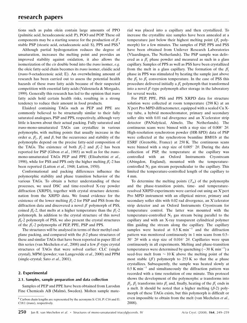
Table 3d values of long spacings and strong fingerprint lines (both in A) of polymorphs of PEP, PSP, PPE, PPSand PSS.
X-ray data collected at Texp (K).
TAG � Texp �02-2 Texp �01-2 Texp �00-2 Texp �-2 Texp
PEP 43.3 (vs) 296 43.1 (vs) 296 42.4 (vs) 324 – 43.3 (vs) 2964.14 (m) 4.32 (m) 4.34 (m) 4.59 (s)
4.17 (s) 4.19 (s) 4.55 (ms)4.00 (m) 4.02 (w) 4.45 (w)n.d. (w) 3.84 (m) 4.0 (w)
3.83 (ms)3.74 (ms)
PSP 44.7 (vs) 295 43.4 (vs) 316 42.8 (vs) 298 – 41.4 (vs) 2964.12 (m) 4.30 (ms) 4.32 (ms) 4.59 (s)
4.17 (s) 4.17 (s) 4.55 (s)4.04 (m) 4.00 (m) 4.45 (m)3.84 (w) 3.77 (m) 3.97 (w)
3.80 (ms)3.72 (s)
PPE 47.1 (vs) 296 43.5 (vs) 308 43.1 (vs) 296 – 41.9 (vs) 2964.14 (m) – 4.40 (vw) 4.59 (s)
overlapping 4.37 (w) 4.55 (ms)broad 4.28 (ms) 4.46 (vw)fingerprint 4.23 (ms) 3.87 (ms)peaks 4.13 (m) 3.74 (m)– 4.08 (m) 3.63 (w)3.9 (m) 3.82 (s)
PPS 47.7 (vs) 295 43.8 (versus) 324 43.5 (vs) 298 – 42.3 (vs) 2964.11 (m) – 4.37 (w) 4.59 (s)
overlapping 4.27 (s) 4.56 (s)broad 4.24 (s) 3.83 (ms)fingerprint 4.12 (ms) 3.69 (ms)lines 4.08 (ms)3.84 (m) 3.78 (s)
PSS 48.3 (vs) 295 45.1 (vs) 328 44.7 (vs) 333 44.7 (vs) 295 45.7 (vs) 2934.11 (m) 4.31 (ms) 4.35 (m) 4.51 (vw) 4.62 (s)
4.21 (s) 4.22 (s) 4.47 (vw) 4.55 (s)4.07 (ms) 4.06 (m) 4.38 (w) 4.44 (vw)3.86 (m) 3.85 (ms) 4.32 (w) 3.96 (w)
4.19 (ms) 3.84 (ms)4.11 (ms) 3.73 (w)4.03 (w) 3.66 (ms)3.77 (s)
The �-2 long spacings of PSP, PPE and PPS are smaller than
those of the �01-2 polymorphs, while for PEP and PSS they are
longer. It might be that this explains the relatively high Tdatacoll
of �01-2 PEP and �01-2 PSS.
The (600) is the highest-angle strong-intensity reflection in
most of the �01 patterns, but its position has been shifted
remarkably in �01-2 PSP (Fig. 3). This shift is attributed to the
considerably lower Tdatacoll (250 K) of PSP, compared with the
297 K of the other samples, and this led to an anisotropic
shrinkage of the unit-cell parameters that mainly affected the
middle-sized unit-cell axis.
3.3. Phase transitions and stability of polymorphs
The Tm and phase-transition temperatures obtained with
the constant heating-rate experiments (Table 1) show that the
symmetric PEP and PSP are �0 stable. The asymmetric PPE,
PPS and PSS are � stable, although the difference in Tm
between the highest melting �0 form and the � form is very
small for PSS and PPE. The similar melting points explain why
a �01-2 to �-2 conversion was not observed for PSS and PPE
within a week of annealing �01-2 2 K below its melting point.
For PPS the conversion did occur and was completed in 1 d.
With respect to the reproducibility of the �0 melting points
of mono-acid TAGs determined with DTA, Lutton & Fehl
(1970) reported that ‘under the best conditions’ an error of
�1 K can be achieved, although stabilization and sample
preparation also affect the melting points. Variations in Tm’s
up to 3 K have been attributed to these phenomena (Lutton et
al., 1948). The melting and phase-transition points listed in
Table 1 are expected to have an uncertainty of the same order.
An optimal stabilization was not feasible for many of the
metastable polymorphs because of potential phase transitions.
The heating rate used must be regarded as a parameter that
influences the observed temperatures. For example, Elisa-
bettini et al. (1998) obtained for PEP with DSC (heating
5 K min�1) a much higher �02-2 to �01-2 transition temperature
(320 K) than the 303 K obtained with our XRPD at
0.5 K min�1. The notion that even a modest heating rate may
lead to a significant overshoot of the phase-transition
temperature implies that one should be careful with conclu-
sions about melting or phase-transition temperatures that
have not been measured under the same experimental
conditions.
3.3.1. The a! b02 phase transition. The �! �02 transition is
difficult to analyze with time-resolved XRPD because of the
overlap at low angles and in the fingerprint area. With DSC
(0.5 K min�1) the symmetric TAGs (PSP and PEP) show no
significant melting peak before the � ! �02 transition.
However, in the case of the asymmetric TAGs (PPE, PPS and
PSS) the formation of �02 is clearly preceded by the melting of
the � phase (DSC data not shown). This is in line with the
results of Elisabettini et al. (1998) who concluded from
5 K min�1 DSC traces that in PEP the �! �02 transition is a
solid-state transition (no melting peak), whereas in PPE a melt
is involved. Apparently, for asymmetric TAGs the � ! �02transition is more complicated than for symmetric TAGs,
suggesting larger conformational changes in the former.
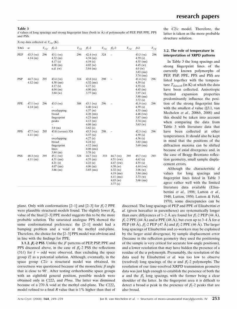
3.3.2. The b02 ! b01 phase transition. In Fig. 4 a selection of
diffraction patterns shows the melt and crystallization
experiment of PSS. The determined melt and phase-transition
temperatures, including the �02-2! �01-2 transition point, are
marked at the right-hand side of the patterns. The virtually
equal positions of the �01-2 and �02-2 fingerprint maxima, within
the accuracy and resolution of the data, and the growth of
sharper �01-2 peaks at the centres of the broad(er) �02-2
diffraction maxima suggests that the �01-2 is just a higher-
crystalline form of the �02-2 polymorph. The close structural
relation between the �02-2 and �01-2 polymorphs, also suggested
by Kellens et al. (1990), and the relatively small amount of
energy involved in such a transition may explain the difficulty
in locating it in the DSC trace. The systematically lower
intensity of the (600) reflection in the �02-2 patterns compared
research papers
254 Jan B. van Mechelen et al. � Structures of mono-unsaturated triacylglycerols. IV Acta Cryst. (2008). B64, 249–259
Figure 4Melting and recrystallization of PSS polymorphs: from bottom to top: thestarting polymorph �00 melts, and after quenching (�30 K min�1) andsubsequent heating (0.5 K min�1) �, �02 and �01 appear and melt. Relevanttemperatures are listed to the right of the graph.
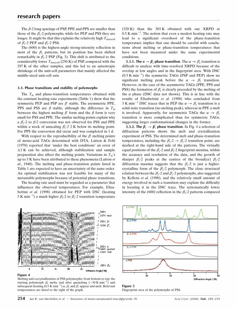
Figure 5Fingerprint area of the polymorphs of PSS.
with �01-2 (Fig. 5) may be an indication of a type of disorder in
the direction of the a axis in �02-2 polymorphs.
The stability of �02-2 differs drastically between TAG groups
and also depends on the thermal treatment. When slowly
heating (0.5 K min�1), the transition of the least stable �02-2 to
the �01-2 starts 2 K (4 min) after the former’s appearance
(Table 1, PSP and PPS). For PSS and PPE this interval is 7 K
and for PEP 11 K. This suggests that an exchange of S by E
considerably delays the appearance of �01-2 and thus stabilizes
the �02-2 polymorph. While the existence of the �02-2 poly-
morph is difficult to prove for a mono-acid trisaturated TAG
such as SSS because of instability (Simpson & Hageman,
1982), substitution of one (n) chain by a longer (nþ 2) one
stabilizes the �02-2 polymorph and substitution by a shorter
(n� 2) chain stabilizes it even more.
3.3.3. Stability of b01-2. The gap between the �02-2 ! �01-2
transition point and the melting point of the �01 polymorph is
different for symmetric versus asymmetric TAGs. For the
asymmetric samples the �01 melts within 9 K (18 min) after its
formation, while the symmetric PEP and PSP melt 17 K
(34 min) and 24 K (48 min), respectively, above their appear-
ance temperature. From this considerable difference in ‘life-
time’ it can be concluded that asymmetry destabilizes the �02-2
polymorph, presumably as a result of the different confor-
mation. In going from PSP to PEP the �01-2 lifetime drops by
7 K (14 min), but in the asymmetric TAGs the exchange of S
by E does not seem to have a significant lifetime influence on
the �01-2 phase. Only the absolute values of the �01-2 melting
points show the influence of an exchange of S by E since it
causes a considerable drop of the melting point, just as for the
� melting points.
3.3.4. b00-2 PSS. In older literature PSS has been reported to
be �0 stable, just like PSP (Lutton et al., 1948), but also to have
equally stable �0 and � polymorphs (Lutton, 1950). With our
crystallization procedure and heating the sample just above
the �02 to �01 conversion temperature, a �01 polymorph was
initially obtained. Surprisingly, after storage in the laboratory
for several weeks at room temperature (T ’ 294 K), all the
prepared capillaries appeared to contain a novel �0-type
polymorph (diffraction pattern at the bottom of Fig. 2) that
differed from �01-2. The novel �0-type PSS polymorph will be
denoted as �00-2, because its melting point (339 K) is higher
than that of the �01-2 polymorph (336 K). A melt and recrys-
tallization experiment carried out with this novel polymorph
(Fig. 4) delivered a pattern that resembles those of the �01-2
polymorphs shown in Fig. 2.
Fig. 5 gives an overview of the fingerprint areas of all known
polymorphs of PSS. Although the �00-2 melting point almost
equals that of the �-2 polymorph, the �00-2 polymorph cannot
be mistaken as a �-2 polymorph because the �00-2 XRPD
pattern clearly lacks the characteristic �-2 reflections between
19 and 19.5 �2� (upper trace Fig. 5). Also, the characteristic �0
bend conformation (see below) and the �0-typical (600)
reflection at 23.6 �2� classifies it as a �0 family member.
The Tm values of the �00-2 and �-2 polymorphs (Table 1),
determined using two different samples from the same PSS
batch, are equal within the accuracy of the temperature
measurement and suggest an equal stability. A time- and
temperature-resolved diffraction experiment has been carried
out with a third PSS capillary sample, taken from the same
batch and prepared in the same way as the other two. Unlike
the other two samples, this third sample contained �00, but also
a small amount of �. Upon heating this sample at 0.5 K min�1
the diffraction patterns show that the �00 melts 1.5–2 K before
�, so it is concluded that PSS is not �00-2 but �-2 stable.
Questions concerning the precise conditions under which
the �00-2 and the �-2 crystallize, and whether a tempering
process may stimulate this, remain as yet unanswered. Repe-
ated partial melting of �01-2 PSS at 336 K and followed by
cooling to 335 K sharpened the �01-2 peaks in the XRPD
pattern, but did not induce a conversion to �00-2, or to �-2.
After 3 months storage of �01-2 at 330 K a small amount of �00-2
was detectable. These experiments demonstrate the influence
of sample history on Tm values and show that one should take
care in drawing conclusions from experimentally obtained
thermal data, even if the samples originate from the same
batch.
research papers
Acta Cryst. (2008). B64, 249–259 Jan B. van Mechelen et al. � Structures of mono-unsaturated triacylglycerols. IV 255
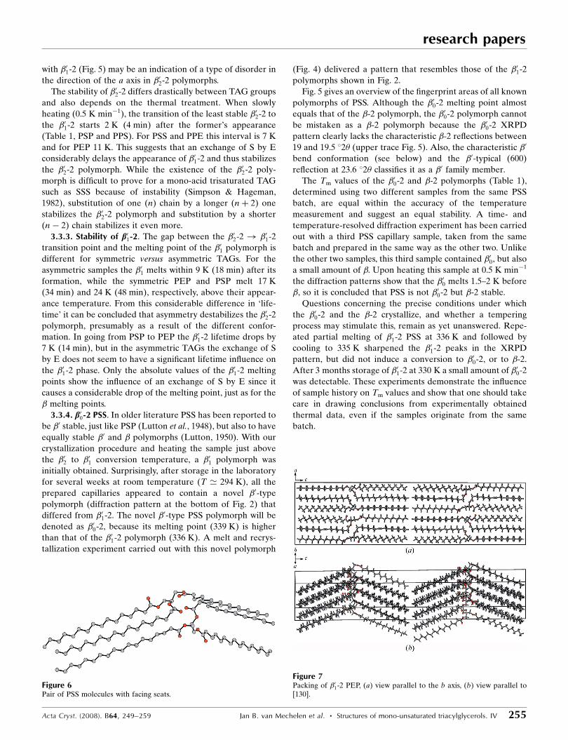
Figure 6Pair of PSS molecules with facing seats.
Figure 7Packing of �01-2 PEP, (a) view parallel to the b axis, (b) view parallel to[130].

3.4. Packing and methyl end-plane
3.4.1. b01-2 PEP, b01-2 PSP, b01-2 PPE and b01-2 PPS. In all the
�01-2 structures discussed in this publication the conformation
of the chair-shaped molecules shows the typical �0 bend of
� 130� between the back and back leg of the chair (the same
as in van Langevelde et al., 2000; Sato et al., 2001). Also, in all
the �0 structures the molecules are packed with seats facing
each other, but being slightly tilted, while the back of one
molecule is adjacent to the front leg of the other one (Fig. 6).
The legs of one (upper) molecule (Fig. 6, left-hand side) are
packed in the same layer as the back of the other (lower)
molecule, but the legs of the lower molecule and the back leg
of the lower molecule (Fig. 6, right-hand side) are packed in
another, different layer than its front leg and the back of the
upper molecule. The pairs of molecules form ‘two-pack’ layers
with a double-chain length thickness. The unit cell of the �01-2
structure contains two such ‘two-packs’ that are related to
each other by a (12,
12,
12) translation. The bends in the molecules
point in the same direction, as a result of which the ‘two-packs’
approach each other at the methyl end-plane with the same
angle as the bend in the molecules (Fig. 7). The methyl end-
groups at one side of the interface between the ‘two-packs’
point in between two methyl end-groups of the adjacent ‘two-
pack’ (Fig. 7b). The view along the b axis shows a difference
between the packing of the symmetric and asymmetric �01-2
structures: In the symmetric structures the chains at the
methyl end-plane are aligned projected along the ac plane
(Fig. 7a), whereas in the asymmetric structures the chain ends
of one ‘two-pack’ point between two other chains of the
neighbouring ‘two-pack’ (Fig. 8).
The precise location of the seat of the chair-shaped mole-
cules between the dominating columns of electron density of
the zigzag chains is a point that deserves extra attention. If
opposing chair seats at the front-leg side bump, the seat
position is likely to be incorrect. The solution to this bumping
problem is similar to that described for �-2 structures: rotation
of the seat plus front leg along the back-leg axis until the front
leg coincides with a neighbouring column of electron density
(van Mechelen et al., 2006b).
However, at the given resolution of the data two serious
ambiguities exist in the packing of the �01 structures of PPS,
PPE, PSP and PEP. The first is the position of the methyl end-
plane. When the molecules are shifted by c/4 and rotated by
180� along the direction of the c axis, a packing can be realised
that fills the columns of electron density of the parallel parts of
the acyl chains, but with the methyl end-plane and the glycerol
zone interchanged and having the seat at a different position.
The R values for both packings are virtually the same so from
the XRPD data no choice can be made for the methyl end-
plane position. This ambiguity may be solved by comparing
with the single-crystal data of �01 CLC. At room temperature
this crystal structure is orthorhombic, but at lower tempera-
tures �01 CLC becomes monoclinic. Since this orthorhombic to
monoclinic transition is reversible, it is likely that no changes
are involved other than small shifts and rotations. The simi-
larity between the powder pattern of the monoclinic �01 CLC
and the �01 patterns of the present study supports a structural
equivalence and for this reason the methyl end-plane position
has been taken as conforming to that in �01 CLC (van
Langevelde et al., 1999).
The second ambiguity is the orientation of the second
molecule in the asymmetric unit of the I2 structure. Each
molecule forms a seat-facing pair with a symmetry copy of
itself. When the lower-left quarter of the unit cell (Fig. 7a) is
filled by a molecule pair of molecule (1), the upper-left quarter
is filled by molecule (2). When molecule (1) has approximately
the proper configuration, molecule (2) may be obtained in two
ways: by shifting a copy of molecule (1) by a/2 relative to
molecule (1) (option 1), or by shifting a copy of molecule (1)
by a/2 plus an additional 180� rotation with respect to an axis
at (3a/4, c/4) perpendicular to the ac plane (option 2). After
optimization by FOX, both molecule combinations lead to
structure solutions that differ mainly in the position of the seat
of the second molecule. Just as with the previous ambiguity,
the single-crystal data of �01 CLC has been used to select the
second option. The seat-position ambiguity is not unique for
structure solution of TAGs from powder diffraction data.
Even in the single-crystal structure solution of �01 CLC, the
seat position was a problem (private communication) and only
from a 2mF0 � DFcalc electron-density map could it be
established unambiguously (van Langevelde et al., 2000).
3.4.2. b00-2 PSS. The unit cell of the �00-2 structure of PSS
also contains two ‘two packs’, but these are related by an
inversion centre. This makes the bends in the two ‘two-packs’
point in opposite directions, whereas the chains of the
approaching ‘two-packs’ at the methyl end-plane are not
inclined but parallel and aligned (Fig. 9). The methyl end-
plane of the ‘two-packs’ is
stepped (Fig. 9a) and can be
denoted as a h2–2i interface as the
sn-2 chains of neighbouring ‘two-
packs’ are in line, analogous to
the �-2 polymorphs of these
TAGs (van Mechelen et al., 2008).
3.5. Comparison of b000 structures
The fingerprint area of the
diffraction patterns of all the �0-2structures is dominated by the
research papers
256 Jan B. van Mechelen et al. � Structures of mono-unsaturated triacylglycerols. IV Acta Cryst. (2008). B64, 249–259
Figure 8Packing of [2–3]�01-2 PPS: view along the b axis.

(600) and (31‘) reflections (Fig. 3). The lattice planes (600),
(318) and (31�88) are related to the most intense fingerprint
diffraction maxima because of their orientation relative to the
chain packing (Fig. 10). Based on FT-IR measurements on
microcrystals, Yano et al. (1997) suggested an O? subcell for
the �01-2 structure. This subcell is found in the single-crystal
structure of the �01-2 CLC structure (van Langevelde et al.,
2000) and does not conflict with the data of the �01-2 structures
of this paper. However, at the resolution to which the �01 TAGs
diffract (not beyond 3 A), the orientation of the zigzag planes
of the acyl chains, and thus the subcell, cannot be established
unambiguously. Since the same problem applies to �00-2, it is
obvious that the usefulness of the subcell is quite limited.
The ‘two-packs’ in the �0 structures can be described as two
stacked dimers that each consist of two symmetry-related
molecules. The dimeric symmetry (21 axis parallel to the b
axis) is the same in all cases, but the stacking of the dimers can
be different. In the �01-2 structures of this work the dimers in
the ‘two-packs’ are not symmetry related. In the orthorhombic
�01-2 (room-temperature) crystal structure of CLC (van
Langevelde et al., 2000) they are symmetry-related by a b glide
perpendicular to the a axis and in the �00-2 of PSS the dimers
are related by a (12,
12,0) translation.
The symmetric and asymmetric �01-2 TAGs appear to have
different conformations, the former a [1–2] and the latter [2–
3]. This difference in conformation suggests a relation to their
different behaviour at the � ! �02 transition. For the
symmetrical group that does not exhibit a clear � melting it
seems likely that the [1–2] �0 conformation is maintained from
the � form. However, the asymmetric TAGs do show �melting
and this may be related to the change from the [1–2] confor-
mation to the [2–3] conformation.
Judging from the �01-2 models and the �00-2 PSS model, the
transition of �01-2 PSS to �00-2 PSS has to involve an inversion
of the orientation of every other ‘two-pack’. This symmetry
relation between the ‘two-packs’ in �01-2 versus �00-2 is similar
to that found for the ‘three-packs’ in the �2-3 versus �1-3
polymorphs of cis-mono-unsaturated TAGs: in the lower-
melting polymorph the ‘three-packs’ are related via the
translation (12,
12,
12), while in the higher-melting one they are
related by a centre of symmetry (van Mechelen et al., 2006b).
It should be noted that apart from the �02-2, �01-2 and �00-2
discussed in the present paper, other types of �0 structures do
exist. For example, the crystal structure of a �0-2 polymorph of
PPM has been solved from single-crystal data (Sato et al.,
2001). This asymmetric �0-2 structure has two molecules in the
asymmetric unit, just like the I2 structures of the present
paper, but the two molecules have different conformations, [2–
1] and [2–3], that together form a seat-facing pair. A (calcu-
lated) powder diffraction pattern clearly shows that this
compound belongs to a different class of �0-2 structures
because the reflection (600) is missing. It seems relevant to
note that the �0-2 PPM single crystal was crystallized from n-
hexane, whilst the �0 polycrystalline material used for the
present work was obtained without solvent.
3.6. Comparison of b000 and the b structures
The �0-2 polymorphs presented in this paper are either in a
[1–2] conformation (PEP, PSP, PSS) or in a [2–3] conformation
(PPE, PPS), and have a bend between the back and the back
leg. It was noted that in all cases the seat of the chair is a C18
chain (E or S), apparently a conformation that is energetically
favourable, and this may explain why the [2–3] conformation
of PPE and PPS both have a shorter sn-2 chain.
research papers
Acta Cryst. (2008). B64, 249–259 Jan B. van Mechelen et al. � Structures of mono-unsaturated triacylglycerols. IV 257
Figure 10View parallel to the chain direction of half a �01-2 PSP ‘two-pack’. Dottedlines mark the orientation of the lattice planes corresponding to the mainfingerprint lines. Solid lines: the O? subcell in line with that present in �01-2 CLC.
Figure 9Packing of �00-2 PSS: (a) view parallel to the b axis; (b) view parallel to[310].
In contrast, the �-2 polymorphs of all these materials (see
paper III of this series; van Mechelen et al., 2008) are all in a
[1–3] conformation. In this conformation the back and the
back-leg of the seat, formed by the sn-1 and sn-2 chains, are
lined up and the parallel planes of the zigzag chains form a
triclinic subcell. The different molecular conformations in �0-2versus �-2 imply that rather complicated changes are required
in the transition from �0-2 to �-2 and this may explain the
difficulty in obtaining a � phase. The symmetric PEP and PSP
are �0 stable and since the �-2 of PSP and PEP melt at lower
temperatures, a �0 to � conversion cannot be observed. It
seems likely that this � phase can be obtained only if the melt-
mediated �0-2 can be avoided, e.g. by crystallization from a
solvent (Lutton & Hugenberg, 1960). The latter authors
reported that �-2 PSP transforms into �01-2 at 338 K via the
melt. The melting indicates a change in conformation and/or
packing, in line with the structural differences determined.
The asymmetric PPS and PSS, and probably also PPE, are �-2
stable, as shown from the melting points. The �01 to �conversion rate in these cases is very slow, if conversion occurs
at all, so measuring the conversion with DSC is impossible.
With temperature-resolved XRPD the precise conditions to
stimulate the transition in a controlled and reproducible way
have not been established yet.
4. Conclusions
The �01-2 structures of PSP, PEP, PPS and PPE have been
solved from high-resolution powder diffraction data in the
space group I2. The packing is in line with the orthorhombic
single-crystal structure of �01-2 CLC (van Langevelde et al.,
2000). The presence of two molecules in the asymmetric unit
complicated the structure solution and, together with domi-
nant zone problems and peak overlap in the diffraction
pattern, it hindered the determination of the orientation of the
zigzag planes of the acyl chains. Unlike the �-2 structures of
these compounds (van Mechelen et al., 2008) the symmetric
and asymmetric �01-2 structures have different molecular
conformations. The structure of a novel �0 polymorph of PSS,
named �00-2, was solved in the space group C2/c. The molecule
has a characteristic �0 bend conformation. Time- and
temperature-dependent XRPD experiments showed that both
�01-2 PSS and �00-2 PSS melt at lower temperatures than the �polymorph, so justify the conclusion that PSS is � stable. The
difference in molecular conformation between the �0-2 poly-
morphs and the �-2 polymorph makes a �0-2 to �-2 solid-state
transition unlikely. For the �01-2 structures as well as �00-2, the
dominant zones in the fingerprint area of the diffraction
pattern can be correlated with the layered packing of the acyl
chains.
In the case of the structure determination of cis-mono-
unsaturated �-3 type TAGs it was shown that the diffraction
data are not very sensitive to rotational freedom of zigzag
chains around their long axis (van Mechelen et al., 2006a). This
also holds for the �0-2 structure of PSS in C2/c. This structure
has a single molecule in the asymmetric unit just like the �-3
structures. The rotational freedom is an even more serious
problem in the �01-2 structures because they have two mole-
cules in the asymmetric unit, while their powder patterns do
not have more independent reflections. Thus, no firm conclu-
sions are possible about the orientation of the planes of the
zigzag chains on the basis of non-atomic resolution XRPD
data alone.
The authors thank Unilever Research for the PSP sample.
The authors acknowledge the ESRF (Grenoble, France) for
providing the facilities to perform the synchrotron diffraction
experiments and they thank W. van Beek of the Swiss–
Norwegian CRG beamline BM01b for his valuable help
during the experimental sessions. The authors also thank E.
Sonneveld for his help in data collection during the experi-
mental session at BM01b. The Shell Research and Technology
Centre in Amsterdam is acknowledged for making the DSC
cell available. The investigations have been supported by the
Netherlands Foundation for Chemical Research (NWO/CW)
with financial aid from the Netherlands Technology Founda-
tion (STW; project 790.35.405). The members of the User
Committee of this project are thanked for stimulating
discussions and continuous interest.
References
Dollase, W. A. (1986). J. Appl. Cryst. 19, 267–272.Elisabettini, P., Lognay, G., Desmedt, A., Culot, C., Istasse, N.,
Deffense, E. & Durant, F. (1998). J. Am. Oil Chem. Soc. 75, 285–291.
Favre-Nicolin, V. & Cerny, R. (2002). J. Appl. Cryst. 35, 734–743.Ghotra, B. S., Dyal, S. D. & Narine, S. S. (2002). Food Res. Int. 35,
1015–1048.Gibon, V., Blanpain, P., Durant, F. & Deroanne, Cl. (1985). Belg. J.
Food Chem. Biotechnol. 40, 119–134.Kellens, M., Gibon, V., Hendrix, M. & De Greyt, W. (2007). Eur. J.
Lipid Sci. Technol. 109, 336–349.Kellens, M., Meeussen, W. & Reynaers, H. (1990). Chem. Phys.
Lipids, 55, 163–178.Langevelde, A. van, van Malssen, K., Driessen, R., Goubitz, K.,
Hollander, F., Peschar, R., Zwart, P. & Schenk, H. (2000). ActaCryst. B56, 1103–1111.
Langevelde, A. van, van Malssen, K., Sonneveld, E., Peschar, R. &Schenk, H. (1999). J. Am. Oil Chem. Soc. 76, 603–609.
Larson, A. C. & Von Dreele, R. (1987). GSAS. Report No. LAUR-86-748. Los Alamos National Laboratory, New Mexico, USA.
Laugier, J. & Bochu, B. (2001). Chekcell; http://www.inpg.Fr/LMPG.Le Bail, A. (2004). Powder Diffr. 19, 249–254.Lutton, E. S. (1950). J. Am. Oil Chem. Soc. 27, 276–281.Lutton, E. S. & Fehl, A. J. (1970). Lipids, 5, 90–99.Lutton, E. S. & Hugenberg, F. R. (1960). J. Chem. Eng. Data, 5, 489–
490.Lutton, E. S., Jackson, F. L. & Quimby, O. T. (1948). J. Am. Chem.
Soc. 70, 2441–2445.March, A. (1932). Z. Kristallogr. 81, 285–297.Mechelen, J. B. van, Peschar, R. & Schenk, H. (2006a). Acta Cryst.
B62, 1121–1130.Mechelen, J. B. van, Peschar, R. & Schenk, H. (2006b). Acta Cryst.
B62, 1131–1138.Mechelen, J. B. van, Peschar, R. & Schenk, H. (2008). Acta Cryst. B64,
240–248.Sato, K., Goto, M., Yano, J., Honda, K., Kodali, D. R. & Small, D. M.
(2001). J. Lipid Res. 42, 338–345.
research papers
258 Jan B. van Mechelen et al. � Structures of mono-unsaturated triacylglycerols. IV Acta Cryst. (2008). B64, 249–259

Simpson, T. D. & Hageman, J. W. (1982). J. Am. Oil Chem. Soc. 59,169–171.
Sreenivasan, B. (1978). J. Am. Oil Chem. Soc. 55, 796–805.Timms, R. E. (2005). Eur. J. Lipid Sci. Technol. 107, 48–57.Valenzuela, A. & Morgado, N. (1999). Biol. Res. 32, 273–287.
Watanabe, A., Tashima, I., Matsuzaki, N., Kurashige, J. & Sato, K.(1992). J. Am. Oil Chem. Soc. 69, 1077–1080.
Wiedermann, L. H. (1978). J. Am. Oil Chem. Soc. 55, 823–829.Yano, J., Kaneko, F., Kobayashi, M., Kodali, D. R., Small, D. M. &
Sato, K. (1997). J. Phys. Chem. B, 101, 8120–8128.
research papers
Acta Cryst. (2008). B64, 249–259 Jan B. van Mechelen et al. � Structures of mono-unsaturated triacylglycerols. IV 259